The Effect of Low Temperature on Physiological, Biochemical and Flowering Functions of Olive Tree in Relation to Genotype

 , and
, and
Abstract
:
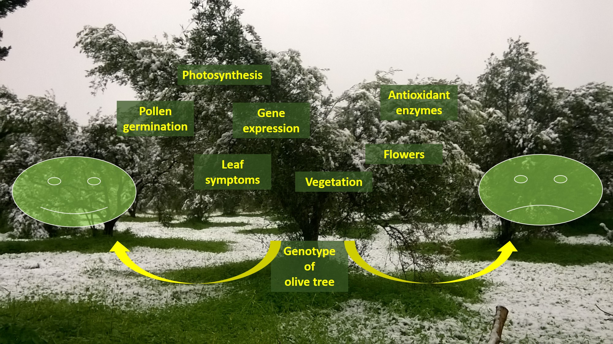
1. Introduction
2. Materials and Methods
2.1. Plant Material and Treatments
2.2. Gas Exchange Measurements
2.3. Determination of Antioxidant Enzymes Activity
2.4. Determination of H2O2 and Thiobarbituric Acid Reactive Substances (TBARS)
2.5. Phenological Measurements
2.6. Pollen Germination
2.7. RNA Isolation and Gene Expression Analysis
2.8. Statistical Analysis
3. Results
3.1. Photosynthesis Parameters
3.2. Antioxidant Enzymes Activities, H2O2 and TBARS
3.3. Phenology
3.4. Pollen Germination
3.5. Gene Expression Analysis
4. Discussion
Supplementary Materials
Author Contributions
Funding
Conflicts of Interest
References
- Langgut, D.; Cheddadi, R.; Carrión, J.S.; Cavanagh, M.; Colombaroli, D.; Eastwood, W.J.; Greenberg, R.; Litt, T.; Mercuri, A.M.; Miebach, A.; et al. The origin and spread of olive cultivation in the Mediterranean Basin: The fossil pollen evidence. Holocene 2019, 29, 902–922. [Google Scholar] [CrossRef]
- Bartolozzi, F.; Fontanazza, G. Assessment of frost tolerance in olive (Olea europaea L.). Sci. Hortic. (Amsterdam) 1999, 81, 309–319. [Google Scholar] [CrossRef]
- Campoy, J.A.; Ruiz, D.; Egea, J. Dormancy in temperate fruit trees in a global warming context: A review. Sci. Hortic. (Amsterdam) 2011, 130, 357–372. [Google Scholar] [CrossRef]
- Haberman, A.; Bakhshian, O.; Cerezo-Medina, S.; Paltiel, J.; Adler, C.; Ben-Ari, G.; Mercado, J.A.; Pliego-Alfaro, F.; Lavee, S.; Samach, A. A possible role for flowering locus T-encoding genes in interpreting environmental and internal cues affecting olive (Olea europaea L.) flower induction. Plant Cell Environ. 2017, 40, 1263–1280. [Google Scholar] [CrossRef]
- Koubouris, G.C.; Metzidakis, I.T.; Vasilakakis, M.D. Influence of cross pollination on the development of parthenocarpic -shotberries- olive (Olea europaea L.) fruits. Exp. Agric. 2010, 46, 67–76. [Google Scholar] [CrossRef]
- Woodward, F.I. Climate and Plant Distribution (Cambridge Studies in Ecology); Cambridge University Press: Cambridge, UK, 1987; ISBN 0521282144. [Google Scholar]
- Pearce, R. Plant Freezing and Damage. Ann. Bot. 2001, 87, 417–424. [Google Scholar] [CrossRef]
- Larcher, W. Temperature stress and survival ability of mediterranean sclerophyllous plants. Plant Biosyst. 2000, 134, 279–295. [Google Scholar] [CrossRef]
- Gururani, M.A.; Venkatesh, J.; Tran, L.S.P. Regulation of photosynthesis during abiotic stress-induced photoinhibition. Mol. Plant 2015, 8, 1304–1320. [Google Scholar] [CrossRef] [Green Version]
- Koubouris, G.C.; Kavroulakis, N.; Metzidakis, I.T.; Vasilakakis, M.D.; Sofo, A. Ultraviolet-B radiation or heat cause changes in photosynthesis, antioxidant enzyme activities and pollen performance in olive tree. Photosynthetica 2015, 53, 279–287. [Google Scholar] [CrossRef]
- Liakoura, V.; Stavrianakou, S.; Liakopoulos, G.; Karabourniotis, G.; Manetas, Y. Effects of UV-B Radiation on Olea Europaea: Comparisons Between a Greenhouse and a Field Experiment. Tree Physiol. 1999, 19, 905–908. [Google Scholar] [CrossRef]
- Sofo, A.; Manfreda, S.; Fiorentino, M.; Dichio, B.; Xiloyannis, C. The olive tree: A paradigm for drought tolerance in Mediterranean climates. Hydrol. Earth Syst. Sci. 2008, 12, 293–301. [Google Scholar] [CrossRef] [Green Version]
- Mittler, R.; Vanderauwera, S.; Gollery, M.; Van Breusegem, F. Reactive oxygen gene network of plants. Trends Plant Sci. 2004, 9, 490–498. [Google Scholar] [CrossRef]
- Bacelar, E.A.; Santos, D.L.; Moutinho-Pereira, J.M.; Lopes, J.I.; Gonçalves, B.C.; Ferreira, T.C.; Correia, C.M. Physiological behaviour, oxidative damage and antioxidative protection of olive trees grown under different irrigation regimes. Plant Soil 2007, 292, 1–12. [Google Scholar] [CrossRef]
- Bowler, C.; Montagu, M.V.; Inze, D. Superoxide Dismutase and Stress Tolerance. Annu. Rev. Plant Physiol. Plant Mol. Biol. 1992, 43, 83–116. [Google Scholar] [CrossRef]
- León, A.M.; Palma, J.M.; Corpas, F.J.; Gómez, M.; Romero-Puertas, M.C.; Chatterjee, D.; Mateos, R.M.; Del Río, L.A.; Sandalio, L.M. Antioxidative enzymes in cultivars of pepper plants with different sensitivity to cadmium. Plant Physiol. Biochem. 2002, 40, 813–820. [Google Scholar] [CrossRef]
- Masoumi, H.; Masoumi, M.; Darvish, F.; Daneshian, J.; Nourmohammadi, G.; Habibi, D. Change in several antioxidant enzymes activity and seed yield by water deficit stress in soybean (Glycine max L.) cultivars. Not. Bot. Horti Agrobot. Cluj-Napoca 2010, 38, 86–94. [Google Scholar] [CrossRef]
- Ortega-García, F.; Peragón, J. The response of phenylalanine ammonia-lyase, polyphenol oxidase and phenols to cold stress in the olive tree (Olea europaea L. cv. Picual). J. Sci. Food Agric. 2009, 89, 1565–1573. [Google Scholar] [CrossRef]
- Mayer, A.M. Polyphenol oxidases in plants and fungi: Going places? A review. Phytochemistry 2006, 67, 2318–2331. [Google Scholar] [CrossRef]
- Thipyapong, P.; Hunt, M.D.; Steffens, J.C. Antisense downregulation of polyphenol oxidase results in enhanced disease susceptibility. Planta 2004, 220, 105–117. [Google Scholar] [CrossRef]
- Thipyapong, P.; Hunt, M.D.; Steffens, J.C. Systemic wound induction of potato (Solanum tuberosum) polyphenol oxidase. Phytochemistry 1995, 40, 673–676. [Google Scholar] [CrossRef]
- Boeckx, T.; Winters, A.L.; Webb, K.J.; Kingston-Smith, A.H. Polyphenol oxidase in leaves: Is there any significance to the chloroplastic localization? J. Exp. Bot. 2015, 66, 3571–3579. [Google Scholar] [CrossRef] [Green Version]
- Raimbault, A.K.; Marie-Alphonsine, P.A.; Horry, J.P.; Francois-Haugrin, M.; Romuald, K.; Soler, A. Polyphenol oxidase and peroxidase expression in four pineapple varieties (Ananas comosus L.) after a chilling injury. J. Agric. Food Chem. 2011, 59, 342–348. [Google Scholar] [CrossRef]
- Hashempour, A.; Ghasemnezhad, M.; Ghazvini, F.R.; Sohani, M.M. Olive (Olea europaea L.) freezing tolerance related to antioxidant enzymes activity during cold acclimation and non acclimation. Acta Physiol. Plant. 2014, 36, 3231–3241. [Google Scholar] [CrossRef]
- Satoh, Y.; Tajima, K.; Munekata, M.; Keasling, J.D.; Lee, T.S. Engineering of L-tyrosine oxidation in Escherichia coli and microbial production of hydroxytyrosol. Metab. Eng. 2012, 14, 603–610. [Google Scholar] [CrossRef]
- Saimaru, H.; Orihara, Y. Biosynthesis of acteoside in cultured cells of Olea europaea. J. Nat. Med. 2010, 64, 139–145. [Google Scholar] [CrossRef]
- Trantas, E.; Navakoudis, E.; Pavlidis, T.; Nikou, T.; Halabalaki, M.; Skaltsounis, L.; Ververidis, F. Dual pathway for metabolic engineering of Escherichia coli to produce the highly valuable hydroxytyrosol. PLoS ONE 2019, 14, e0212243. [Google Scholar] [CrossRef] [Green Version]
- Mougiou, N.; Trikka, F.; Trantas, E.; Ververidis, F.; Makris, A.; Argiriou, A.; Vlachonasios, K.E. Expression of hydroxytyrosol and oleuropein biosynthetic genes are correlated with metabolite accumulation during fruit development in olive, Olea europaea, cv. Koroneiki. Plant Physiol. Biochem. 2018, 128, 41–49. [Google Scholar] [CrossRef]
- Geßler, A.; Keitel, C.; Kreuzwieser, J.; Matyssek, R.; Seiler, W.; Rennenberg, H. Potential risks for European beech (Fagus sylvatica L.) in a changing climate. Trees Struct. Funct. 2007, 21, 1–11. [Google Scholar] [CrossRef]
- Koubouris, G.; Limperaki, I.; Darioti, M.; Sergentani, C. Effects of various winter chilling regimes on flowering quality indicators of Greek olive cultivars. Biol. Plant. 2019, 63, 504–510. [Google Scholar] [CrossRef]
- Koubouris, G.C.; Metzidakis, I.T.; Vasilakakis, M.D. Impact of temperature on olive (Olea europaea L.) pollen performance in relation to relative humidity and genotype. Environ. Exp. Bot. 2009, 67, 209–214. [Google Scholar] [CrossRef]
- Malik, N.S.A.; Bradford, J.M. Inhibition of flowering in “Arbequina” olives from chilling at lower temperatures. J. Food Agric. Environ. 2009, 7, 429–431. [Google Scholar]
- Connor, D.J.; Fereres, E. The Physiology of Adaptation and Yield Expression in Olive. In Horticultural Reviews; John Wiley & Sons, Inc.: Oxford, UK, 2010; Volume 31, pp. 155–229. [Google Scholar]
- Martin, G.C.; Sibbett, S.G. Botany of the olive. In Olive Production Manual; Sibbett, S.G., Ferguson, L., Coviello, J.L., Lindstrand, M., Eds.; University of California, Agriculture and Natural Resources: Oakland, CA, USA, 2005; pp. 15–19. [Google Scholar]
- Sofo, A.; Dichio, B.; Xiloyannis, C.; Masia, A. Antioxidant defences in olive trees during drought stress: Changes in activity of some antioxidant enzymes. Funct. Plant Biol. 2005, 32, 45–53. [Google Scholar] [CrossRef]
- Sanz-Cortés, F.; Martinez-Calvo, J.; Badenes, M.L.; Bleiholder, H.; Hack, H.; Llacer, G.; Meier, U. Phenological growth stages of olive trees (Olea europaea). Ann. Appl. Biol. 2002, 140, 151–157. [Google Scholar] [CrossRef]
- Koubouris, G.C.; Tzortzakis, N.; Kourgialas, N.N.; Darioti, M.; Metzidakis, I. Growth, photosynthesis and pollen performance in saline water treated olive plants under high temperature. Int. J. Plant Biol. 2015, 6, 28–32. [Google Scholar] [CrossRef] [Green Version]
- Allen, D.J.; Ort, D.R. Impacts of chilling temperatures on photosynthesis in warm-climate plants. Trend Plant Sci. 2001, 6, 36–42. [Google Scholar] [CrossRef]
- Pavel, E.W.; Fereres, E. Low soil temperatures induce water deficits in olive (Olea europaea) trees. Physiol. Plant. 1998, 104, 525–532. [Google Scholar] [CrossRef]
- Pérez-López, D.; Gijón, M.C.; Mariño, J.; Moriana, A. Water relation response to soil chilling of six olive (Olea europaea L.) cultivars with different frost resistance. Spanish J. Agric. Res. 2010, 8, 780. [Google Scholar] [CrossRef] [Green Version]
- López-Bernal, Á.; García-Tejera, O.; Testi, L.; Orgaz, F.; Villalobos, F.J. Low winter temperatures induce a disturbance of water relations in field olive trees. Trees 2015, 29, 1247–1257. [Google Scholar] [CrossRef]
- Araújo, M.; De Oliveira, J.M.P.F.; Santos, C.V.; Moutinho-Pereira, J.M.; Correia, C.; Dias, M.C. Responses of olive plants exposed to different irrigation treatments in combination with heat shock: Physiological and molecular mechanisms during exposure and recovery. Planta 2019, 249, 1583–1598. [Google Scholar] [CrossRef]
- Trabelsi, L.; Gargouri, K.; Hassena, B.A.; Mbadra, C.; Ghrab, M.; Ncube, B.; Amoo, S.O.; Gargouri, R. Impact of drought and salinity on olive water status and physiological performance in an arid climate. Agric. Water Manag. 2019, 213, 749–759. [Google Scholar] [CrossRef]
- Monk, L.S.; Fagerstedt, K.V.; Crawford, R.M.M. Oxygen toxicity and superoxide dismutase as an antioxidant in physiological stress. Physiol. Plant. 1989, 76, 456–459. [Google Scholar] [CrossRef]
- Gulen, H.; Cansev, A.; Eris, A. Cold hardiness of olive (Olea europaea L.) cultivars in cold-acclimated and non-acclimated stages: Seasonal alteration of soluble sugars and phospholipids. J. Agric. Sci. 2009, 147, 459–467. [Google Scholar] [CrossRef]
- Saadati, S.; Baninasab, B.; Mobli, M.; Gholami, M. Measurements of freezing tolerance and their relationship with some biochemical and physiological parameters in seven olive cultivars. Acta Physiol. Plant. 2019, 41, 51. [Google Scholar] [CrossRef]
- Cansev, A.; Gulen, H.; Eris, A. The activities of catalase and ascorbate peroxidase in olive (Olea europaea L. cv. Gemlik) under low temperature stress. Hortic. Environ. Biotechnol. 2011, 52, 113–120. [Google Scholar] [CrossRef]
- Koubouris, G.C.; Metzidakis, I.T.; Vasilakakis, M.D. Phenological, morphological and functional indicators of genetic variability and their implication in the sexual reproductive system of Olea europaea L. (Oleaceae). Sci. Hortic. 2010, 123, 547–550. [Google Scholar] [CrossRef]
- Chakrabarti, B.; Singh, S.D.; Nagarajan, S.; Aggarwal, P.K. Impact of temperature on phenology and pollen sterility of wheat varieties. Aust. J. Crop Sci. 2011, 5, 1039–1043. [Google Scholar]
- Kumar, S.; Nayyar, H.; Bhanwara, R.; Upadhyaya, H. Chilling stress effects on reproductive biology of chickpea. J. SAT Agric. Res. 2010, 8, 1–14. [Google Scholar]
- Thomashow, M.F. PLANT COLD ACCLIMATION: Freezing Tolerance Genes and Regulatory Mechanisms. Annu. Rev. Plant Biol. 1999, 50, 571–599. [Google Scholar] [CrossRef] [Green Version]
- Bruno, L.; Chiappetta, A.; Muzzalupo, I.; Gagliardi, C.; Iaria, D.; Bruno, A.; Greco, M.; Giannino, D.; Perri, E.; Bitonti, M.B. Role of geranylgeranyl reductase gene in organ development and stress response in olive (Olea europaea) plants. Funct. Plant Biol. 2009, 36, 370–381. [Google Scholar] [CrossRef]


| Primer Code | Gene Name | Gene Accession Number | Oligonucleotide Sequence (5′–3′) | Strand |
|---|---|---|---|---|
| KVI106 | OeCuAO | CCTTACCTCCAGCTGATCCAT | Forward | |
| KVI107 | KP968841 | GATCATTGGGATCTCCATAGG | Reverse | |
| KVI108 | OeTDC | GGCTACTGCATTCGGGTTAACA | Forward | |
| KVI109 | KP968842 | GTGTGCATTGAAACTGAATGAATC | Reverse | |
| KVI110 | OePPO | CTATGAAAGAATATTGGGCAAACTG | Forward | |
| KVI111 | KP968843 | ACGCTGCGAATCATTTCACTATAT | Reverse | |
| KVI140 | OeALDH | TGGTACATGGTCTGAATATGTCG | Forward | |
| KVI141 | KP968844 | CCTCCAGGCAGATCCAA | Reverse | |
| KVI164 | OeTIP41 | CAACGGTGTCTCTCTTTTGACAGT | Forward | |
| KVI165 | XM_023020543 | TCATAAGCACTCCATCCACTCTCA | Reverse |
Publisher’s Note: MDPI stays neutral with regard to jurisdictional claims in published maps and institutional affiliations. |
© 2020 by the authors. Licensee MDPI, Basel, Switzerland. This article is an open access article distributed under the terms and conditions of the Creative Commons Attribution (CC BY) license (http://creativecommons.org/licenses/by/4.0/).
Share and Cite
Mougiou, N.; Baalbaki, B.; Doupis, G.; Kavroulakis, N.; Poulios, S.; Vlachonasios, K.E.; Koubouris, G.C. The Effect of Low Temperature on Physiological, Biochemical and Flowering Functions of Olive Tree in Relation to Genotype. Sustainability 2020, 12, 10065. https://doi.org/10.3390/su122310065
Mougiou N, Baalbaki B, Doupis G, Kavroulakis N, Poulios S, Vlachonasios KE, Koubouris GC. The Effect of Low Temperature on Physiological, Biochemical and Flowering Functions of Olive Tree in Relation to Genotype. Sustainability. 2020; 12(23):10065. https://doi.org/10.3390/su122310065
Chicago/Turabian StyleMougiou, Niki, Boushra Baalbaki, Georgios Doupis, Nektarios Kavroulakis, Stylianos Poulios, Konstantinos E. Vlachonasios, and Georgios C. Koubouris. 2020. "The Effect of Low Temperature on Physiological, Biochemical and Flowering Functions of Olive Tree in Relation to Genotype" Sustainability 12, no. 23: 10065. https://doi.org/10.3390/su122310065