
Episodic Vertigo: A Narrative Review Based on a Single-Center Clinical Experience

Abstract
:1. Introduction
2. Materials and Methods
- (a)
- Patients with first ever attack of acute vertigo.
- (b)
- Patients who reported recurring vertigo and dizziness (present or not at the time of observation) for at least three episodes, both spontaneous and provoked (for example, by movements of the head, change in body positions, physical efforts, etc.).
- (c)
- Patients with chronic imbalance.
3. Results
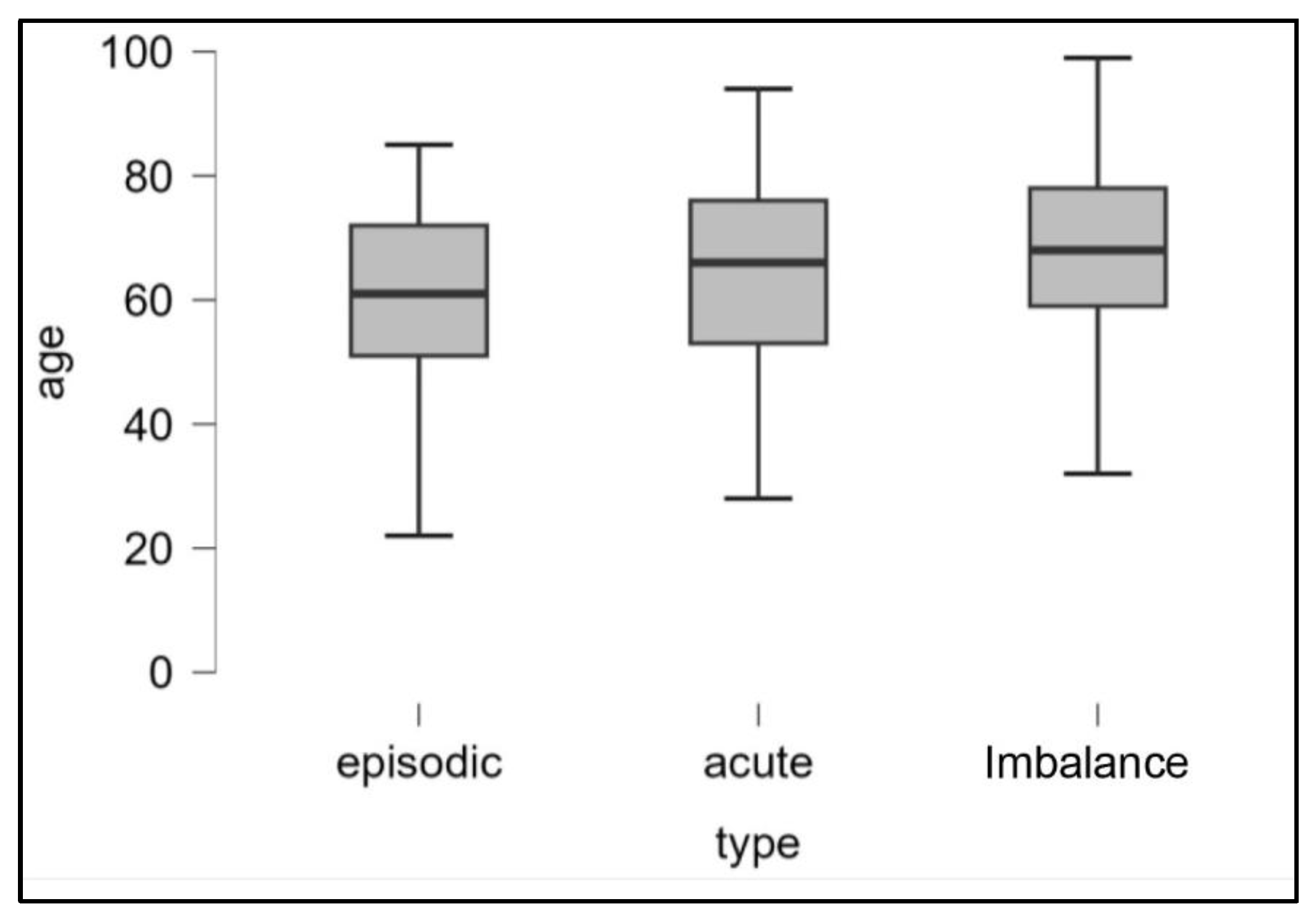
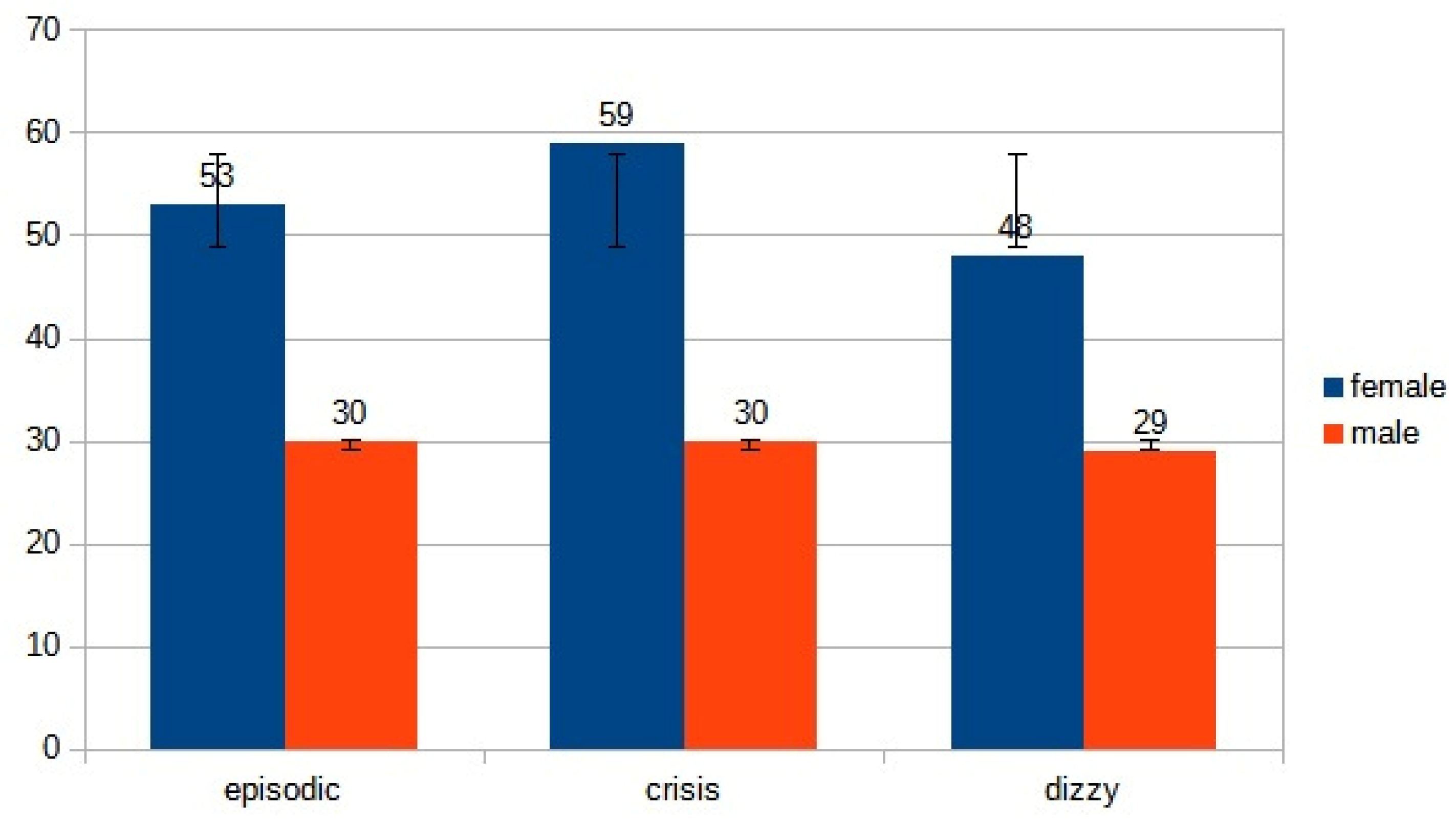
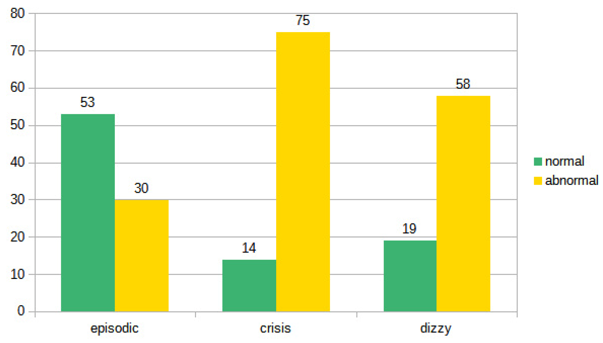
- 83 (53 females, 30 males, mean age ranging from 22 years to 85 years, mean 53.5 years) patients suffering from episodic vertigo and dizziness attacks.
- 89 (59 females, 30 males, mean age ranging from 29 years to 92 years, mean 60.5 years) with a first-ever attack of acute vertigo.
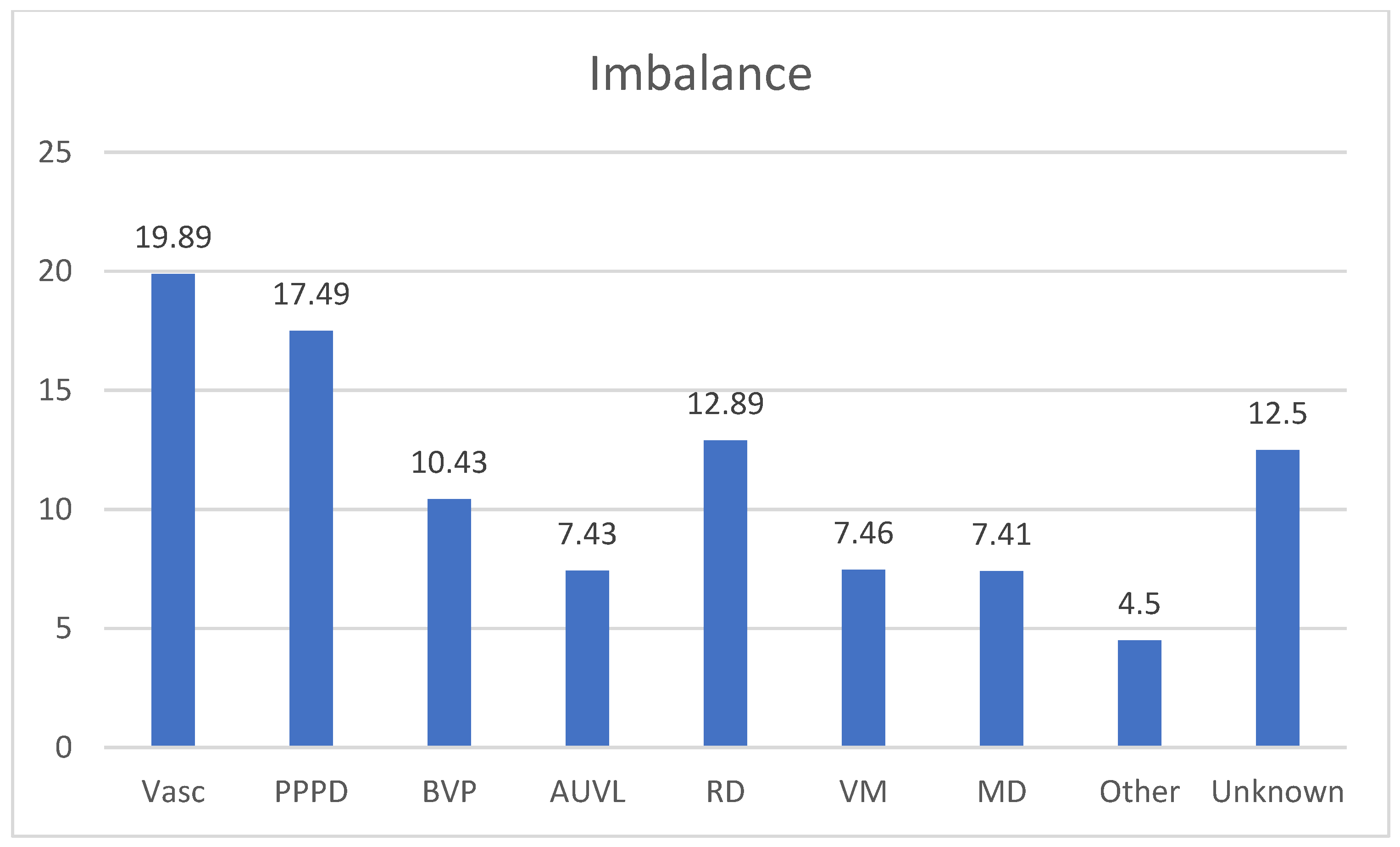
- 77 (48 females, 29 males, mean age ranging from 30 years to 99 years, mean 64.5 years) with imbalance.
4. Discussion
- (1)
- Benign paroxysmal positional vertigo.
- (2)
- Vestibular Migraine.
- (3)
- Menière’s disease.
- (4)
- Vertebrobasilar TIAs.
- (5)
- Orthostatic hypotension.
- (1)
- BPPV from posterior and lateral canalithiasis. It is a vertigo typically evoked by head movements, which the patient generally remembers well and is able to report. Furthermore, the average duration of the symptoms is in the order of weeks [4], so the patient can be seen again in the acute phase: at that point we can also make the diagnosis of the previous episode. Horizontal canalolithiasis could have a shorter course [19,20] with more possibility to not be diagnosed, but the frequency of the recurrences and the characteristics of the vertigo are generally significant. It is practically impossible to find objectivity in the intercritical phase: only in some cases, ‘Ocular tilt reaction’ can be seen in the overcompensation stage but this does not allow a diagnosis of certainty [21,22]. A diagnosis in the event of a negative Dix–Hallpike maneuver is not acceptable: at most, a diagnostic doubt can be granted to be confirmed as soon as possible.
- (2)
- According to the international guidelines, the diagnosis of VM is based exclusively on the clinical history and on the exclusion of other causes of vertigo [9]; therefore, it is always possible even in a non-critical phase (observing the migraine patient in the acute stage is rare and signs are not specific). In our series, VM is, as expected, a common cause of episodic vertigo; however, we found a large number of patients who presented with a first episode of acute vertigo who then developed a VM, confirming that VM pathology is, however, an increasingly recognized cause of acute vertigo [15]. We must keep in mind that following a first-ever presentation of vertigo (without a history of multiple episodes), it is often challenging to differentiate acute VM from other causes of acute vertigo, especially when acute VM is associated with positional vertigo and nystagmus thus mimicking BPPV [23]. Recently, the evaluation of a sign associated with migraine-associated vertigo has been proposed [24]: these patients, even outside the crisis, would show a pupillary nystagmus (“hippus”) characterized by alternating dilation and narrowing of the diameter of the pupils. This sign seems to be more easily (if not exclusively) detectable in patients suffering from migraine vertigo and would be linked to the alteration of the pupillary cycle of migraine as previously reported [25]. Therefore, it may be worthwhile to assess the presence or absence of pupillary nystagmus and further the medical history in case of inconsistency. Patients with VM frequently report a special sensitivity to head motion and complex visual surroundings [9]. Vestibular symptoms are generally triggered or worsened by self-motion and visual motion. These patients are prone to develop the so-called «Visual Dependence» (VD); this condition is defined as a reduced ability to disregard visual clues in complex or conflicting environments (supermarket, traffic) [26]. In brief, VD may correspond to the reduced ability to use complex visual input in a correct way in combination with an incorrect interaction with vestibular input. Since the migraine patient is very often a “visual dependent”, a negative or inconclusive clinical and instrumental examination associated with abnormal results of fHIT only under an optokinetic background could support the diagnosis of vestibular migraine [27]. These instrumental data could be helpful in correctly identifying patients with VM, particularly when the international diagnostic criteria are not completely met. Finally, in our experience, the effectiveness of a prophylactic therapy for VM (we reserved for cases with very frequent crises or in any case perceived as disabling) [11,28] could be useful as a diagnostic confirmation although some confounding factors (placebo effect, and spontaneous improvement) may interfere with the outcome of drug treatment [29].
- (3)
- MD is a purely clinical diagnosis (except for audiogram): electrocochleography, VEMPs (cervical and ocular), video-HIT and even MRI with intratympanic gadolinium do not enter the diagnostic criteria for this disease [30]. However, in the early stages of the disorder, vestibular or cochlear symptoms may occur in isolation. Vertigo attacks are the most frequent symptom in the initial phase [31]. Less than 30% of MD patients have the three symptoms in the initial phase [32]. After 1 or 2 years, however, the complete set of symptoms is usually established. Patients with a longstanding history of recurrent isolated vertigo (e.g., no definitive auditory features) are therefore unlikely to have MD. The dissociation of thermal tests and video-HIT may be helpful [33,34], but the fundamental finding is the detection and documentation of fluctuating hearing loss.
- (4)
- Vestibular TIAs in the posterior cranial fossa. Vertebrobasilar TIA is a common cause of isolated recurrent vertigo. The Vertebro-Basilar Insufficiency (VBI) can cause damage both at a central level, with involvement of the brainstem and cerebellum, and at a peripheral level, with involvement of the cochlea and vestibule, thus giving rise to an extremely polymorphic symptomatologic and objective picture. For this reason, circulatory insufficiency in the vertebro-basilar district is a common cause of vertigo, especially in subjects over 50, especially in the presence of vascular risk factors (smoking, diabetes, arterial hypertension and hyperlipemia); the high incidence of this symptom in the initial symptoms of VBI confirms the importance of posterior circulation disorders in the genesis of many vertigo syndromes [35]. The typical patient with vertigo caused by vertebro-basilar TIA is older than 60 years and has vascular risk factors such as history of smoking, hypertension, diabetes, or hyperlipidemia. Attacks may occasionally present with isolated vertigo but more commonly include associated symptoms from the posterior circulation territory. The duration of TIAs has been arbitrarily limited to 24 h; most, however, last minutes or 1 to 2 h. In this case, there are no residual signs in the intercritical phase and should be suspected in case of short-term, recurring vertigo [36]. They are often associated with other signs, always transient (visual disturbances, hearing loss, tinnitus, paresthesia, headache, etc.) but vertigo can be the only symptom. HINTs examination [37] is a valuable screening tool for distinguishing a central cause of acute vertigo (mainly cerebellar stroke) [38,39] from AUVL, such as vestibular neuritis, [40] but its value in case of VB-TIAs seems to be not so useful, due to the fact that after a transient ischemic attack no objective or instrumental signs are evident at the time of observation. On the contrary, the ABCD criterion (Table 1) remains valid, whose items can be reconstructed anamnestically, giving an idea of the circulatory defect in the posterior cranial fossa [41].
- (5)
- Orthostatic Hypotension (OH) is characterized by brief episodes of dizziness lasting seconds to minutes after standing up and can be diagnosed when a drop of systolic blood pressure of >20 mm Hg occurs after standing up [47]. Dizziness after standing up due to OH is a common type of dizziness with a lifetime prevalence of 12.5% and it may have severe consequences, causing syncope in 19% and traumatic injury in 5% of affected individuals [48]. Orthostatic hypotension can be associated with a “positional vertigo” (in reality it is a fainting sensation, but the patient often has difficulty describing it), in which the malaise does not manifest itself when taking the supine position but when assuming orthostatism or even prolonged standing. This symptom is typical of patients undergoing treatment with drugs that can cause hypotension or reduced alertness. In these cases, it is worthwhile to check the pressure and heart rate in orthostasis and in supine position.
- (6)
- Perilymphatic fistula is an abnormal communication between the perilymphatic space and the outside, resulting in perilymph leakage [49]. Vertigo may be episodic, with attacks lasting seconds to days, or chronic, with superimposed fluctuations. Tinnitus and aural fullness may also occur. It can be a consequence of trauma or surgery, but the diagnostic problem arises in cases of idiopathic (spontaneous) fistula and may originate from straining or minor trauma on the background of a preexisting subclinical abnormality, such as local thinning of the labyrinthine capsule [49,50]. It may be enough to blow your nose, sneeze, laugh or even bend over to cause a fistula [51]. Audiological (sudden hearing loss, tinnitus, fullness in the ears) or vestibular (imbalance or vertigo) symptoms may be present, or some combination of them. It can enter into the differential diagnosis with vestibular migraine, endolymphatic hydrops, and functional vertigo. The problem is that there are no precise diagnostic criteria: the majority of the methods (audiovestibular examination, imaging, or laboratory testing for perilymph biomarkers) used to identify PF lacked the sensitivity and specificity to provide consistent diagnosis [49,51]. The diagnosis of PF is very challenging: a careful history (with the aim of evaluate a preceding event), associated with a complete battery of vestibular, auditory and high-resolution imaging studies (and, when possible, biomarker testing) is necessary before planning a surgical exploration of the middle ear [49,51]. In our experience, we encountered only one case of perilymphatic fistulae. We encountered a similar combination of auditory and vestibular symptoms in two patients suffering from Superior Canal Dehiscence Syndrome (SCDS) [52]. Perlymphatic fistula is an opening of the inner ear with a communication to the middle ear allowing fluid to move and stimulate the vestibular end organ in response to sound or pressure changes [53]. On the contrary, the symptomatology of SCDS is the consequence of a thinning or discontinuity of the bone that separates the superior canal from the middle cranial fossa [52]. The presence of this “third window” can disrupt the harmony of hearing and balance residing in the same otic capsule, leading to physiologic stimuli causing excitatory or ampullofugal deflection of the cupula of the superior semicircular canal. Patients with SCDS may experience symptoms of pressure- or sound-induced vertigo, hyperacusis to bone conducted sounds, and pulsatile tinnitus. Chronic disequilibrium is common. The diagnosis is based on the presence of one of the previous described symptoms in association with a high resolution CT scan reformatted in the plane of the superior SC demonstrating a dehiscence and at least one of the following diagnostic test: negative bone conduction on pure tone audiometry, low cervical VEMPs threshold or high ocular VEMP amplitude, and elevated summating potential to action potential ratio on electrocochleography in the absence of a sensorineural hearing loss [54,55].
5. Conclusions
Author Contributions
Funding
Institutional Review Board Statement
Informed Consent Statement
Data Availability Statement
Acknowledgments
Conflicts of Interest
References
- Welgampola, M.S.; Young, A.S.; Pogson, J.M.; Bradshaw, A.P.; Halmagyi, G.M. Dizziness demystified. Pract. Neurol. 2019, 19, 492–501. [Google Scholar] [CrossRef]
- Cutfield, N.J.; Seemungal, B.M.; Millington, H.; Bronstein, A.M. Diagnosis of acute vertigo in the emergency department. J. Emerg. Med. 2011, 28, 9. [Google Scholar] [CrossRef]
- Agarwal, K.; Harnett, J.; Metha, N.; Humphries, F.; Kaski, D. Acute vertigo: Getting the diagnosis right. BMJ 2022, 378, e069850. [Google Scholar] [CrossRef]
- Nuti, D.; Zee, D.S.; Mandalà, M. Benign paroxysmal positional vertigo: What we do and do not know. Semin. Neurol. 2020, 40, 49–58. [Google Scholar] [CrossRef]
- Peña Navarro, P.; Pacheco López, S.; Almeida Ayerve, C.N.; Marcos Alonso, S.; Serradilla López, J.M.; Santa Cruz Ruiz, S.; Gómez Sánchez, J.C.; Kaski, D.; Batuecas Caletrío, Á. Early Diagnosis of Central Disorders Mimicking Horizontal Canal Cupulolithiasis. Brain Sci. 2023, 13, 562. [Google Scholar] [CrossRef]
- Lempert, T. Recurrent spontaneous attacks of dizziness. CONTINUUM Lifelong Learn. Neurol. 2012, 18, 1086–1101. [Google Scholar] [CrossRef] [PubMed]
- Staab, J.P.; Eckhardt-Henn, A.; Horii, A.; Jacob, R.; Strupp, M.; Brandt, T.; Bronstein, A. Diagnostic criteria for persistent postural-perceptual dizziness (PPPD): Consensus document of the committee for the Classification of Vestibular Disorders of the Bárány Society. J. Vestib. Res. 2017, 27, 191–208. [Google Scholar] [CrossRef] [PubMed]
- Lopez-Escamez, J.A.; Carey, J.; Chung, W.-H.; Goebel, J.A.; Magnusson, M.; Mandalà, M.; Newman-Toker, D.E.; Strupp, M.; Suzuki, M.; Trabalzini, F.; et al. Diagnostic criteria for Menière’s disease. J. Vestib. Res. 2015, 25, 1–7. [Google Scholar] [CrossRef] [PubMed]
- Lempert, T.; Olesen, J.; Furman, J.; Waterston, J.; Seemungal, B.; Carey, J.; Bisdorff, A.; Versino, M.; Evers, S.; Newman-Toker, D. Vestibular migraine: Diagnostic criteria. J. Vestib. Res. 2012, 22, 167–172. [Google Scholar] [CrossRef] [PubMed]
- Von Brevern, M.; Bertholon, P.; Brandt, T.; Fife, T.; Imai, T.; Nuti, D.; Newman-Toker, D. Benign paroxysmal positional vertigo: Diagnostic criteria. Consensus document of the Committee for the Classification of Vestibular Disorders of the Barany Society. J. Vestib. Res. 2015, 25, 105–117. [Google Scholar] [CrossRef] [PubMed]
- Bronstein, A.; Lempert, T. Dizziness. A practical approach to diagnosis and management. Cambridge University Press. J. Neurol. Neurosurg. Psychiatry 2007, 78, 779. [Google Scholar]
- Agrawal, Y.; Ward, B.K.; Minor, L.B. Vestibular dysfunction: Prevalence, impact and need for targeted treatment. J. Vestib. Res. 2013, 23, 113–117. [Google Scholar] [CrossRef] [PubMed]
- Jeong, S.S.; Simpson, K.N.; Johnson, J.M.; Rizk, H.G. Assessment of the Cost Burden of Episodic Recurrent Vestibular Vertigo in the US. JAMA Otol. Head Neck Surg. 2022, 148, 1103–1110. [Google Scholar] [CrossRef] [PubMed]
- Espinosa Sanchez, J.M.; Lopez-Escamez, J.A. Menière’s disease. In Handbook of Clinical Neurology; Furman, J., Lempert, T., Eds.; Elsevier: Amsterdam, The Netherlands, 2016; Volume 137, pp. 257–277. [Google Scholar]
- Seemungal, B.; Kaski, D.; Lopez-Escamez, J.A. Early diagnosis and management of acute vertigo from vestibular migraine and Ménière’s disease. Neurol. Clin. 2015, 33, 619–628. [Google Scholar] [CrossRef] [PubMed]
- Casani, A.P.; Sellari-Franceschini, S.; Napolitano, A.; Muscatello, L.; Dallan, I. Otoneurologic dysfunctions in migraine patients with or without vertigo. Otol. Neurotol. 2009, 30, 961–967. [Google Scholar] [CrossRef]
- Young, A.S.; Nham, B.; Bradshaw, A.P.; Calic, Z.; Pogson, J.M.; D’Souza, M.; Halmagyi, G.M.; Welgampola, M.S. Clinical, oculographic, and vestibular test characteristics of vestibular migraine. Cephalalgia 2021, 41, 1039–1052. [Google Scholar] [CrossRef]
- Fu, W.; Wang, Y.; He, F.; Wei, D.; Bai, Y.; Han, J.; Wang, X. Vestibular and oculomotor function in patients with vestibular migraine. Am. J. Otolaryngol. 2021, 42, 103152. [Google Scholar] [CrossRef]
- Shim, D.B.; Ko, K.M.; Lee, J.H.; Park, H.J.; Song, M.H. Natural history of horizontal canal benign paroxysmal positional vertigo is truly short. J. Neurol. 2015, 262, 74–80. [Google Scholar] [CrossRef]
- Gufoni, M.; Ducci, N.; Casani, A.P. Update on the treatment of benign positional paroxysmal vertigo of the horizontal semi-circular canal. J. Otolaryngol. 2022, 72, 101–106. [Google Scholar] [CrossRef]
- Halmagyi, G.M.; Curthoys, I.S.; Brandt, T.; Dieterich, M. Ocular tilt reaction: Clinical sign of vestibular lesion. Acta Otolaryngol. Suppl. 1991, 481, 47–50. [Google Scholar] [CrossRef]
- Gufoni, M.; Vianini, M.; Casani, A.P. Analysis of the Skew Deviation to Evaluate the Period of Onset of a Canalolithiasis after Macular Damage. Front. Neurol. 2020, 11, 572531. [Google Scholar] [CrossRef] [PubMed]
- Rocha, M.F.; Sacks, B.; Al-Lamki, A.; Koohi, N.; Kaski, D. Acute vestibular migraine: A ghost diagnosis in patients with acute vertigo. J. Neurol. 2023, 270, 1–4. [Google Scholar]
- Gufoni, M.; Casani, A.P. The Pupillary (Hippus) Nystagmus: A Possible Clinical Hallmark to Support the Diagnosis of Vestibular Migraine. J. Clin. Med. 2023, 12, 1957. [Google Scholar] [CrossRef]
- Cortez, M.M.; Rae, N.; Millsap, L.; McKean, N.; Brennan, K.C. Pupil cycle time distinguishes migraineurs from subjects without headache. Front. Neurol. 2019, 10, 478. [Google Scholar] [CrossRef]
- Bronstein, A.M. Visual vertigo syndrome: Clinical and posturography findings. J. Neurol. Neurosurg. Psychiatry 1995, 59, 472–476. [Google Scholar] [CrossRef]
- Casani, A.P.; Lazzerini, F.; Marconi, O.; Vernassa, N. The role of the functional head impulse test with and without optokinetic stimuli in vestibular migraine and acute unilateral vestibulopathy: Discovering a dynamic visual dependence. J. Clin. Med. 2021, 10, 3787. [Google Scholar] [CrossRef] [PubMed]
- Von Brevern, M.; Lempert, T. Vestibular migraine: Treatment and prognosis. Semin. Neurol. 2020, 40, 83–86. [Google Scholar] [CrossRef]
- Lempert, T.; Olesen, J.; Furman, J.; Waterston, J.; Seemungal, B.; Carey, J.; Bisdorff, A.; Versino, M.; Evers, S.; Kheradmand, A.; et al. Vestibular migraine: Diagnostic criteria. An update. J. Vestib. Res. 2022, 32, 1–6. [Google Scholar] [CrossRef]
- Basura, G.J.; Adams, M.E.; Monfared, A.; Schwartz, S.R.; Antonelli, P.J.; Burkard, R.; Bush, M.L.; Bykowski, J.; Colandrea, M.; Derebery, J.; et al. Clinical Practice Guideline: Ménière’s Disease. Otolaryngol.-Head Neck Surg. 2020, 162, S1–S55. [Google Scholar]
- Pyykkö, I.; Nakashima, T.; Yoshida, T.; Zou, J.; Naganawa, S. Ménière’s disease: A reappraisal supported by a variable latency of symptoms and the MRI visualization of endolymphatic hydrops. BMJ Open 2013, 3, e001555. [Google Scholar] [CrossRef]
- Perez-Carpena, P.; Lopez-Escamez, J.A. Current understanding and clinical management of meniere’s disease: A systematic review. Semin. Neurol. 2020, 40, 138–150. [Google Scholar] [PubMed]
- Cerchiai, N.; Navari, E.; Dallan, J.; Sellari-Franceschini, S.; Casani, A.P. Assessment of Vestibulo-oculomotor Reflex in Ménière’s Disease: Defining an Instrumental Profile. Otol. Neurotol. 2016, 37, 380–384. [Google Scholar] [CrossRef] [PubMed]
- Hannigan, I.P.; Welgampola, M.S.; Watson, S.R.D. Dissociation of Caloric and Head Impulse Tests: A Marker of Meniere’s Disease. J. Neurol. 2021, 268, 431–439. [Google Scholar] [PubMed]
- Hotson, J.R.; Baloh, R.W. Acute vestibular syndrome. N. Engl. J. Med. 1998, 339, 680–685. [Google Scholar]
- Yao, K.; Zu, H.B. Isolated transient vertigo due to TIA: Challenge for diagnosis and therapy. J. Neurol. 2023, 270, 769–779. [Google Scholar] [CrossRef] [PubMed]
- Kattah, J.C.; Talkad, A.V.; Wang, D.Z.; Hsieh, Y.H.; Newman-Toker, D.E. HINTS to diagnose stroke in the acute vestibular syndrome: Three-step bedside oculomotor examination more sensitive than early MRI diffusion-weighted imaging. Stroke 2009, 40, 3504–3510. [Google Scholar] [CrossRef]
- Lee, H.; Sohn, S.I.; Cho, Y.W.; Lee, S.R.; Ahn, B.H.; Park, B.R.; Baloh, R.W. Cerebellar infarction presenting isolated vertigo: Frequency and vascular topographical patterns. Neurology 2006, 67, 1178–1183. [Google Scholar]
- Casani, A.P.; Dallan, I.; Cerchiai, N.; Lenzi, R.; Cosottini, M.; Sellari Franceschini, S. Cerebellar infarctions mimicking acute peripheral vertigo: How to avoid misdiagnosis? Otolaryngol. Head Neck Surg. 2013, 148, 475–481. [Google Scholar]
- Gerlier, C.; Hoarau, M.; Fels, A.; Vitaux, H.; Mousset, C.; Farhat, W.; Firmin, M.; Pouyet, V.; Paoli, A.; Chatellier, G.; et al. Differentiating central from peripheral causes of acute vertigo in an emergency setting with the HINTS, STANDING, and ABCD2 tests: A diagnostic cohort study. Acad. Emerg. Med. Off. J. Soc. Acad. Emerg. Med. 2021, 28, 1368–1378. [Google Scholar]
- Fonseca, A.C.; Merwick, Á.; Dennis, M.; Ferrari, J.; Ferro, J.M.; Kelly, P.; Lal, A.; Ois, A.; Olivot, J.M.; Purroy, F. European Stroke Organization (ESO) guidelines on management of transient ischaemic attack. J. Eur. Stroke 2021, 6, 163–186. [Google Scholar] [CrossRef]
- Ahmad, H.; Cerchiai, N.; Mancuso, M.; Casani, A.P.; Bronstein, A.M. Are white matter abnormalities associated with “unexplained dizziness”? J. Neurol. Sci. 2015, 358, 428–431. [Google Scholar] [CrossRef] [PubMed]
- Cerchiai, N.; Mancuso, M.; Navari, E.; Giannini, N.; Casani, A.P. Aging with Cerebral Small Vessel Disease and Dizziness: The Importance of Undiagnosed Peripheral Vestibular Disorders. Front. Neurol. 2017, 8, 241. [Google Scholar] [CrossRef] [PubMed]
- Ibitoye, R.T.; Castro, P.; Cooke, J.; Allum, J.; Arshad, Q.; Murdin, L.; Wardlaw, J.; Kaski, D.; Sharp, D.J.; Bronstein, A.M. A link between frontal white matter integrity and dizziness in cerebral small vessel disease. NeuroImage Clin. 2022, 35, 103098. [Google Scholar]
- Kim, J.; Lee, H. Vertigo Due to Posterior Circulation Stroke. Semin. Neurol. 2013, 33, 179–184. [Google Scholar] [CrossRef] [PubMed]
- Kim, H.A.; Oh, E.H.; Choi, S.Y.; Choi, J.H.; Park, J.Y.; Lee, H.; Choi, K.D. Transient Vestibular Symptoms Preceding Posterior Circulation Stroke: A Prospective Multicenter Study. Stroke 2021, 52, 224–228. [Google Scholar]
- Kim, H.A.; Bisdorff, A.; Bronstein, A.M.; Lempert, T.; Rossi-Izquierdo, M.; Staab, J.P.; Strupp, M.; Kim, J.S. Hemodynamic orthostatic dizziness/vertigo: Diagnostic criteria. J. Vestib. Res. 2019, 29, 45–56. [Google Scholar] [CrossRef]
- Radtke, A.; Lempert, T.; Von Brevern, M.; Feldamnn, M.; Lezius, F.; Neuhauser, H. Prevalence and complications of orthostatic dizziness in the general population. Clin. Auton. Res. 2011, 21, 161–168. [Google Scholar] [CrossRef]
- Sarna, B.; Abouzari, M.; Merna, C.; Jamshidi, S.; Saber, T.; Djalilian, H.R. Perilymphatic Fistula: A Review of Classification, Etiology, Diagnosis, and Treatment. Front. Neurol. 2020, 11, 1046. [Google Scholar] [CrossRef]
- Rizer, F.M.; House, J.W. Perilymph fistulas: The house ear clinic experience. Otolaryngol. Head Neck Surg. 1991, 104, 239–243. [Google Scholar] [CrossRef]
- Comacchio, F.; Mion, M. Sneezing and Perilymphatic Fistula of the Round Window: Case Report and Systematic Review of the Literature. J. Int. Adv. Otol. 2018, 14, 106–111. [Google Scholar] [CrossRef]
- Minor, L.B.; Solomon, D.; Zinreich, J.S.; Zee, D.S. Sound- and/or pressure-induced vertigo due to bone dehiscence of the superior semicircular canal. Otolaryngol.-Head Neck Surg. 1998, 124, 249–258. [Google Scholar] [CrossRef] [PubMed]
- Weinreich, H.M.; Carey, J.P. Perilymphatic Fistulas and Superior Semi-Circular Canal Dehiscence Syndrome. Adv. Otorhinolaryngol. 2019, 82, 93–100. [Google Scholar] [PubMed]
- Ward, B.K.; Carey, J.P.; Minor, L.B. Superior canal dehiscence syndrome: Lessons from the first 20 years. Front. Neurol. 2017, 8, 177. [Google Scholar] [CrossRef] [PubMed]
- Ward, B.K.; Van De Berg, R.; Van Rompaey, V.; Bisdorff, A.; Hullar, T.E.; Welgampola, M.S.; Carey, J.P. Superior semicircular canal dehiscence syndrome: Diagnostic criteria consensus document of the committee for the classification of vestibular disorders of the Bárány Society. J. Vestib. Res. 2021, 31, 131–141. [Google Scholar] [CrossRef]
- Dlugaiczyk, J.; Lempert, T.; Lopez-Escamez, J.A.; Teggi, R.; Von Brevern, M.; Bisdorff, A. Recurrent Vestibular Symptoms Not Otherwise Specified: Clinical Characteristics Compared with Vestibular Migraine and Menière’s Disease. Front. Neurol. 2021, 12, 674092. [Google Scholar] [CrossRef]
- Van Leeuwen, R.B.; Colijn, C.; Van Esch, B.F.; Schermer, T.R. Benign Recurrent Vertigo: The Course of Vertigo Attacks Compared to Patients with Menière’s Disease and Vestibular Migraine. Front. Neurol. 2022, 13, 817812. [Google Scholar] [CrossRef]
- Teggi, R.; Colombo, B.; Cangiano, I.; Gatti, O.; Bussi, M.; Filippi, M. Similarities and Differences between Vestibular Migraine and Recurrent Vestibular Symptoms—Not Otherwise Specified (RVS-NOS). Audiol. Res. 2023, 13, 466–472. [Google Scholar] [CrossRef]
- Teggi, R.; Gatti, O.; Familiari, M.; Cangiano, I.; Bussi, M. Skull Vibration-Induced Nystagmus Test (SVINT) in Vestibular Migraine and Menière’s Disease. Audiol. Res. 2021, 11, 603–608. [Google Scholar] [CrossRef]
- Ruthberg, J.S.; Rasendran, C.; Kocharyan, A.; Mowry, S.E.; Otteson, T.D. The economic burden of vertigo and dizziness in the United States. J. Vestib. Res. 2021, 31, 81–90. [Google Scholar] [CrossRef]


| Clinical Factor | Score |
|---|---|
| Age 60 years or older | 1 |
| BP ≥ 140/90 mm Hg | 1 |
| Clinical: speech disturbance | 1 |
| Clinical: unilateral weakness | 2 |
| Duration < 10 min | 0 |
| Duration 10–59 min | 1 |
| Duration 10–59 min | 1 |
Disclaimer/Publisher’s Note: The statements, opinions and data contained in all publications are solely those of the individual author(s) and contributor(s) and not of MDPI and/or the editor(s). MDPI and/or the editor(s) disclaim responsibility for any injury to people or property resulting from any ideas, methods, instructions or products referred to in the content. |
© 2023 by the authors. Licensee MDPI, Basel, Switzerland. This article is an open access article distributed under the terms and conditions of the Creative Commons Attribution (CC BY) license (https://creativecommons.org/licenses/by/4.0/).
Share and Cite
Casani, A.P.; Gufoni, M.; Ducci, N. Episodic Vertigo: A Narrative Review Based on a Single-Center Clinical Experience. Audiol. Res. 2023, 13, 845-858. https://doi.org/10.3390/audiolres13060074
Casani AP, Gufoni M, Ducci N. Episodic Vertigo: A Narrative Review Based on a Single-Center Clinical Experience. Audiology Research. 2023; 13(6):845-858. https://doi.org/10.3390/audiolres13060074
Chicago/Turabian StyleCasani, Augusto Pietro, Mauro Gufoni, and Nicola Ducci. 2023. "Episodic Vertigo: A Narrative Review Based on a Single-Center Clinical Experience" Audiology Research 13, no. 6: 845-858. https://doi.org/10.3390/audiolres13060074