Article

An Evaluation of the Caudal End Deviation of the Nasal Septum Using the Quantitative Analysis of Computed Tomography

1
Department of Otorhinolaryngology, Head and Neck Surgery, Hiroshima Prefectural Hospital, Hiroshima 7348530, Japan
2
Department of Otorhinolaryngology, Head and Neck Surgery, Graduate School of Biomedical Sciences, Hiroshima University, Hiroshima 7348551, Japan
*
Author to whom correspondence should be addressed.
Surg. Tech. Dev. 2023, 12(3), 145-155; https://doi.org/10.3390/std12030014
Submission received: 31 May 2023 / Revised: 9 August 2023 / Accepted: 21 August 2023 / Published: 24 August 2023

Abstract

:
Objectives: This study was designed to determine objective surgical indications of correcting caudal end deviation of the nasal septum. Methods: We employed quantitative computed tomographic (CT) analysis and assessed the validity by comparing this with anterior rhinoscopic findings (AR findings). The study population consisted of 300 patients. The archived CT data were transferred to a workstation, and 3D CT volume-rendered images were generated using computer graphics tools. In the plane of the nostril entrance, we calculated ratios of the cross-sectional area of the convex side (narrower side) and the concave side (wider side), which is abbreviated as the N/W ratio. We also examined the presence of laterality between the right and the left cross-sectional area of the nasal valve based on the AR findings. Surgical procedures for whether to expose the caudal end were planned based on the AR findings and the N/W ratio. Results: A significant correlation was found between the AR findings and the N/W ratio. After surgery, the average N/W ratio improved from 0.53 ± 0.15 to 0.81 ± 0.15, and the average values of VAS scaling for nasal obstruction improved from 8.1 ± 0.2 to 1.0 ± 0.1. Conclusions: The quantitative CT analysis proposed in the study is a useful modality to objectively determine the surgical indications of managing the caudal end of the nasal septum.

1. Introduction

Nasal obstruction is a common presenting symptom to both primary care physicians and otolaryngologists and may be caused by a wide range of anatomic, physiologic, and pathophysiologic factors [1]. Airflow resistance is essential during breathing for good pulmonary function. The anterior portion of the nasal cavities is the place of highest nasal resistance to airflow [2]. Previous observers have suggested that the main site of respiratory airflow resistance is localized in the vestibular region of the nose [3].
The inter nasal valve is a structure that regulates air or liquid flow within the human body [4]. The inter nasal valve is considered the narrowest part of nasal airway, and hence, has the greatest resistance flow. Physiological studies demonstrate that this complex region significantly regulates both nasal airflow and nasal resistance [5]. Collapse or obstruction of the inter nasal valve is mostly the cause of nasal airway obstruction [2]. This perplexing problem greatly inhibits normal nasal breathing through the narrowing of the external valve area and the nasal valve angle [6].
A caudal end deviation of the nasal septum is the most important cause of internal nasal valve obstruction [7]. It is therefore likely to contribute to symptomatic obstruction. Nasal septum deviation does not only affect adults but also children. It causes ailments that reduce the quality of life [8]. Therefore, it is very important to correct a caudal end deviation using septoplasty. A caudal end deviation is a common diagnosis made by otolaryngologists but is one that is not usually based on objective measurements. As a result, there can be a significant interobserver variability in terms of diagnosing the condition, verifying its precise location, quantifying the degree of deviation, and assessing its clinical impact on patients. This subjectivity can lead to unnecessary surgical treatments, patient complications, and low patient satisfaction rates [9]. Some surgeons have reported on the operating methods for correcting caudal end deviation [6,10,11]. Meanwhile, there are few reports on objective surgical indications. When an appropriate evaluation is not carried out on the presence or absence of caudal end deviation, nasal obstruction might not be improved. For the same reason, we think it very important to evaluate the presence or absence of caudal end deviation objectively and correct it as necessary.
The internal nasal valve exam needs to be conducted carefully, since the very introduction of the nasal speculum in the nasal vestibule, as we normally visualize the nasal cavity itself, deforms the ostium internum. With the tip of the nasal speculum the nostril is opened, thus providing visualization of the vestibule and internal nasal valve. The endoscopic exam is also useful [4].
In this study, we used the method of quantitative analysis of computed tomography (CT), and the validity of this method was examined by comparing with anterior rhinoscopic findings (AR findings) and intraoperative findings. These adaptations include increased nasal projection, anterior nasal convexity, exaggerated nasal angles, anterior nasal spine prominence, and an intricate nasal cartilaginous tip [1]. The aim of this study was to determine the objective surgical indications for correcting a caudal septal deviation.

2. Material and Methods
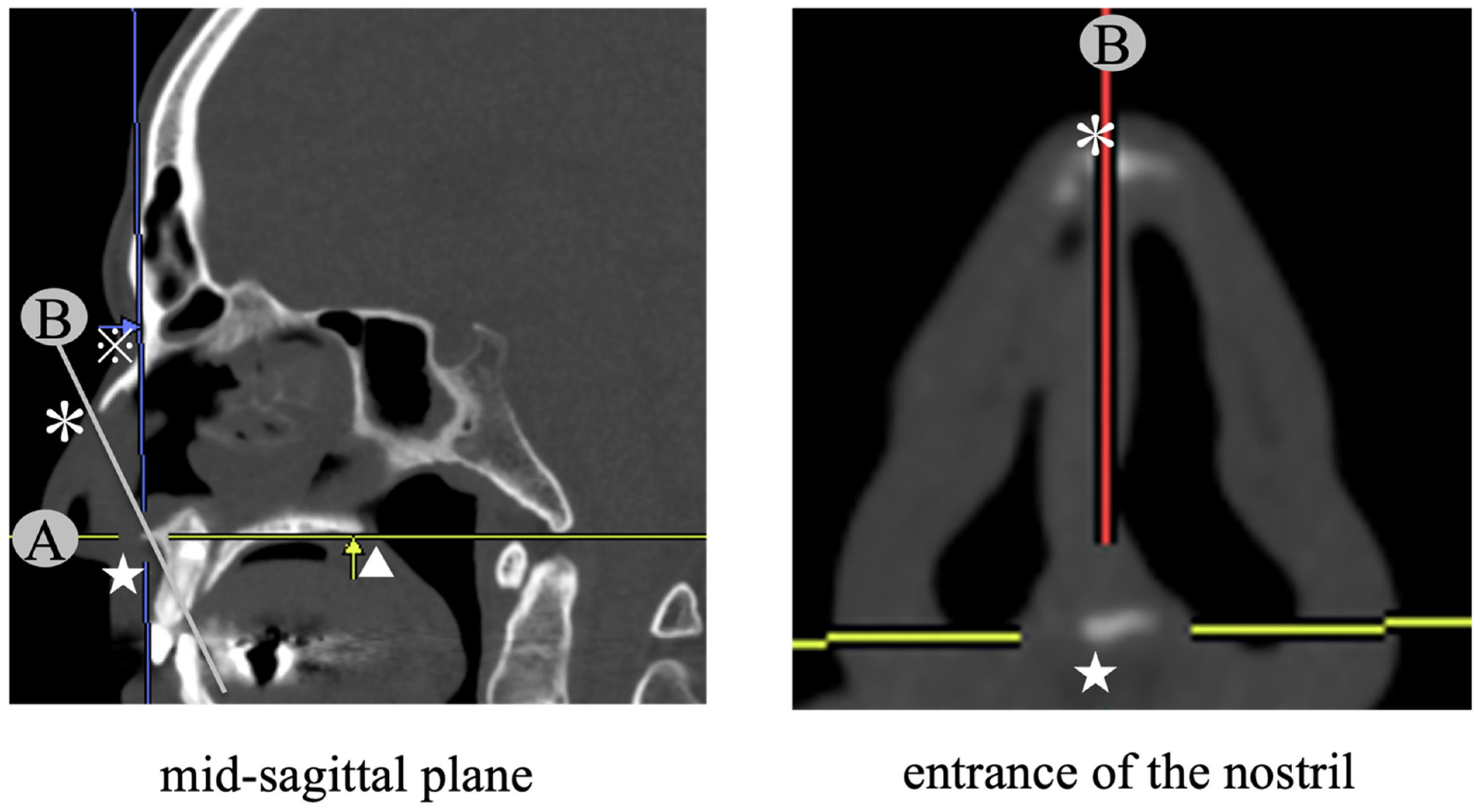
2.1. CT Analysis

The degree of caudal end deviation in all participants was evaluated by analyzing CT data using soft SYNAPSE VINCENT® (FUJIFILM Corporation, Tokyo, Japan).As the degree of the caudal end deviation becomes severe, it is thought that the differences between the cross-sectional area of the convex side and concave side at the entrance of the nostril become increased. Therefore, we calculated ratios of the cross-sectional area of the convex side and the concave side. In deciding the entrance of the nostril, it was desirable to render images under equivalent conditions. In this study, to standardize conditions, we utilized four anatomical landmarks (anterior nasal spine, posterior nasal spine, nasion, and anterior tip of nasal bone) (Figure 1). In the beginning, a line connecting two anatomical landmarks (anterior nasal spine and posterior nasal spine) was determined; Line A. Next, a sagittal plane containing Line A and the nasion was rendered; mid-sagittal plane [12]. Subsequently, we designed a coronal plane that crosses the mid-sagittal plane at 90 degrees and includes two landmarks (anterior nasal spine and anterior tip of nasal bone: Line B connected both landmarks). As a result, we defined the cross-sectional image rendered on this plane as the plane of entrance of the nostril.
The contours of the left and right nasal passages were plotted in the plane of the entrance to the nostrils, and the area of each was measured. The ratio of the cross-sectional area of the convex side (narrower side)/the cross-sectional area of the concave side (wider side) was calculated, and this was abbreviated as the N/W ratio [13]. Figure 2 shows a concrete example of a case in which the N/W ratio improved after caudal septum correction. The pre-operative N/W ratio was 0.36 (76/210) and the post-operative N/W ratio was 0.98 (190/193).
The contours of the left and right nasal passages were plotted in the plane of the entrance to the nostrils, and the area of each was measured. The ratio of the cross-sectional area of the convex side (narrower side)/the cross-sectional area of the concave side (wider side) was calculated, and this was abbreviated as the N/W ratio. In the above case, the pre-operative N/W ratio was 0.36 (76/210) and the post-operative N/W ratio was 0.98 (190/193).

2.2. Patients

In total, 300 patients who underwent septoplasty for the purpose of improvement in nasal obstruction at our department between April 2020 and November 2022 were enrolled in this study. Of the 300 patients, 113 patients underwent septoplasty alone, without concomitant endoscopic sinus surgery. On the other hand, 187 patients underwent septoplasty with concomitant endoscopic sinus surgery.
The group consisted of 222 males and 78 females, aged 18–84 (mean age 42 years). Post-operative follow-up was performed about once every 1 to 4 weeks up to 3 months after surgery. Cases with severe external nasal deformity or insufficient anterior nasal air inclusion due to the presence of polyps or tumor were excluded.
The presence of caudal end deviation was evaluated using two methods: anterior rhinoscopy and CT analysis. Anterior rhinoscopies were performed by two independent surgeons. They examined the presence of laterality between the right and the left cross-sectional area of the nasal valve (Figure 3) caused by a deviation of the anterior portion of the nasal septum based on the anterior rhinoscopic findings (AR findings), and they were classified as “Laterality (+)” or “Laterality (−)”, which is abbreviated as “L (+)” or “L (−)”. When at least one doctor judged that there was a laterality, we classified it as “L (+)”, and when both the doctors judged that there was not a laterality, we classified it as “L (−)”. A representative case of “L (+)” is shown in Figure 4. This case had a laterality between the cross-sectional area of the right and left nasal valves caused by a deviation of the anterior portion of the nasal septum. When performing septoplasty, for all cases of “L (−)”, we did not expose the caudal end and preserved it. On the other hand, in cases of “L (+)”, we determined whether we exposed the caudal end intraoperatively by calculating the N/W ratio. For the cases with an N/W ratio ≥ 0.8, we did not expose the caudal end and preserved it. Meanwhile, for the cases with an N/W ratio < 0.8, we exposed the caudal end and corrected it as necessary.
The nasal valve region is located between the ostium internum and the istmus nasi [4]. The ostium internum is located 1–1.5 cm from the nostril. It has the inferior border of the superior lateral cartilage as a lateral border, the caudal end of the nasal septum as the medial one, and the nasal cavity floor inferiorly, which is not composed of erectile tissue. The inside of the isthmus nasi is composed of the nasal septum including the nasal septum cavernous body and the lamina perpendiculars, and the lateral side is composed of the anterior part of the inferior nasal concha [14].
This case had a laterality between the cross-sectional area of the right and left nasal valves caused by a deviation of the anterior portion of the nasal septum.
As for correcting the deviation of the caudal end, we used two methods depending on the caudal end (Figure 5).
In cases of a curved caudal end with its inferior part not being dislocated from anterior nasal spine, we adopted cutting and suturing techniques [6]. The caudal strut was cut using scissors at the convex-most region in the caudocephalic direction. The excess portions of the upper and lower caudal strut were then overlapped, and the overlapping cartilages were sutured together. They were reinforced with a batten graft.
In cases when the inferior part of the caudal end was dislocated from the anterior nasal spine, it was necessary to prevent the development of saddle nose due to the instability of the caudal end. For the above reason, we resected excess cartilage, and the lower edge of the cartilage was fixed to the anterior nasal spine properly using a “Figure 8” suture [8], and then they were reinforced with a batten graft.
Specifically, we adopted cutting and suturing techniques in cases of a curved caudal end with its inferior part not being dislocated from the anterior nasal spine [7]. In cases when the inferior part of the caudal end was dislocated from the anterior nasal spine, we resected the overlapping portion of the cartilage, and the septal cartilage was then repositioned and fixed properly using a “Figure 8” suture [8].
Of the 117 patients who underwent correcting caudal end deviation, 56 patients performed septoplasty without endoscopic sinus surgery. Of these 56 patients, for subjective evaluation, nasal obstruction status using a visual analogue scale (VAS) from 0 (no nasal obstruction) to 10 (complete nasal obstruction) [7] was compared pre-operatively and 3 months post-operatively. For objective evaluation, the N/W ratio was compared pre-operatively and 3 months post-operatively in 64 cases (those who agreed to undergo a sinus CT scan post-operatively). We further investigated the relationship between the AR findings and the N/W ratio in all cases. The relationship between the AR findings and the N/W ratio was obtained using Fisher’s exact test. Statistical significance was set at p < 0.05.
The study protocol was approved by the Review Board of the Hiroshima Prefectural Hospital Ethics Committee (approval number: R4-22-3, 2022). All participants provided informed consent for participation in this study.
A.
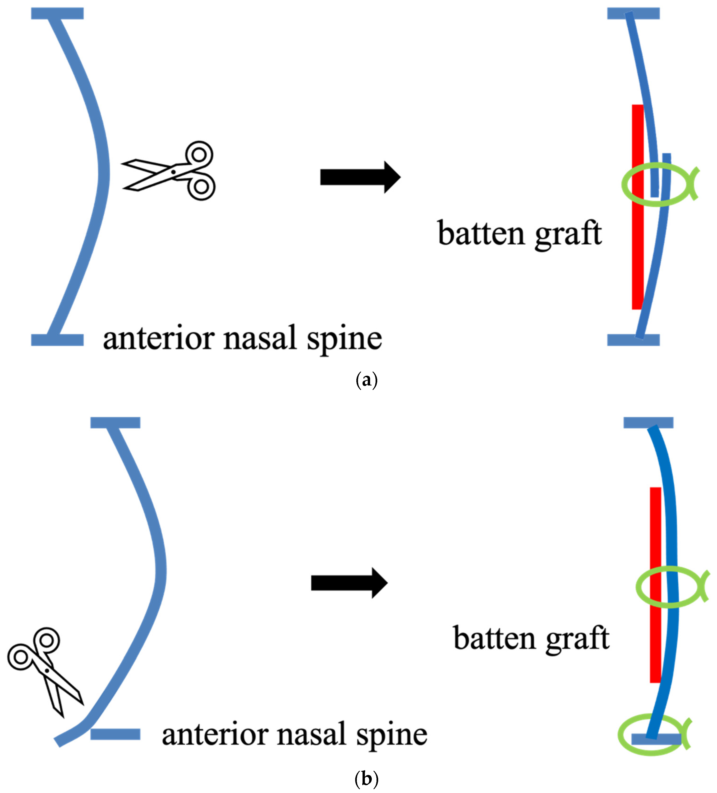
For the cases where the inferior part of the curved caudal end was not dislocated from the anterior nasal spine, cutting and suturing techniques [6] were performed. The caudal strut was cut using scissors at the convex-most region in the caudocephalic direction. The excess portions of the upper and lower caudal strut were then overlapped, and the overlapping cartilages were sutured together. They were reinforced with a batten graft.
B.
For the cases where the inferior part of the caudal end was dislocated from anterior the nasal spine, the excess cartilage was resected and the lower edge of the cartilage was fixed to the anterior nasal spine. They were reinforced with a batten graft.

3. Results

3.1. Participant Division

The participant selection flow chart is depicted in Figure 6. Initially, they were divided into two groups based on the AR findings: “L (−)” (n = 152) and “L (+)” (n = 148). As for the latter 148 cases, the number of cases that both doctors judged as “L (+)” was 119, and the number of cases that only a single doctor judged whether there was laterality was 29. The cases of “L (+)” were successively divided into two groups based on CT analysis: N/W ratio ≥ 0.8 (n = 18) and N/W ratio < 0.8 (n = 130). We exposed the caudal end for the latter group (n = 130) and the number of cases that recognized the caudal end deviation (including those cases in which the inferior part of caudal end was dislocated from anterior nasal spine) intraoperatively was 117 (90.0%), and the number of cases without the caudal end deviation was 13 (10.0%). In the former group (n = 117), we adopted cutting and suturing techniques in 76 cases, and we resected the excess cartilage and rebuild the caudal end with a batten graft in 41 cases. There were 13 cases in which the caudal end deviation was not recognized. In those cases, deformities of the premaxillary wing were considered the main cause of a smaller rate of the N/W ratio.

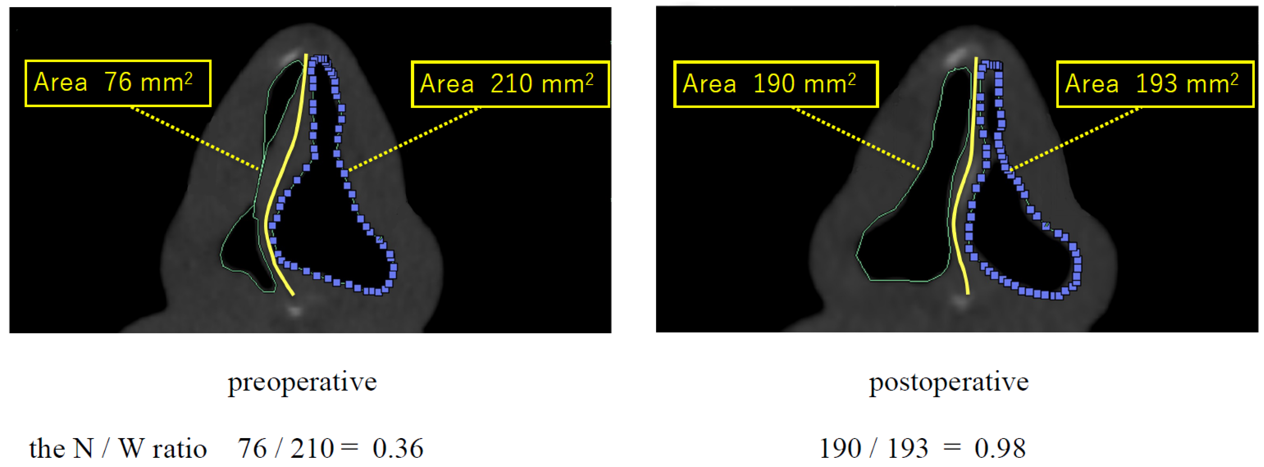
3.2. The Relationship between the AR Findings and the N/W Ratio

Table 1 shows the relationship between the AR findings and the N/W ratio. Regarding the cases of “L (−)” (n = 152), the number of the N/W ratio ≥ 0.8 was 135 (88.8%), and the number of the N/W ratio < 0.8 was 17 (11.2%). And, in the cases of “L (+)” (n = 148), the number of the N/W ratio ≥ 0.8 was 18 (12.2%), and the number of the N/W ratio < 0.8 was 130 (87.8%). A significant correlation was found between the two groups with Fisher’s exact test (p > 0.05).

3.3. Correcting Effect

As a correcting effect, the average of the ratio of N/W was improved from 0.53 ± 0.15 (pre-operatively) to 0.81 ± 0.15 (3 months post-operatively). Moreover, the average of the VAS scale improved from 8.8 ± 0.3 (pre-operatively) to 0.6 ± 0.1 (3 months post-operatively).

4. Discussion

Nasal septum deviation causes various symptoms other than nasal obstruction. A rhinogenic contact point headache is characterized by contact between different anatomical structures such as the nasal septum and the middle, superior turbinate, or the anteromedial wall of the ethmoid sinus associated with frontal–orbital pain radiating to the root of the nose [15]. Patients with persistent rhinogenic cephalea may require more extensive sinus surgery to address associated chronic sinonasal disease. Turbinate surgery is a procedure aimed at reducing the size of the nasal turbinates, bony structures lined with soft tissue that can cause airway obstruction if enlarged. Headaches, or cephalgia, can be associated with nasal issues such as a deviated nasal septum and enlarged turbinates, which can affect the ventilation and drainage of the paranasal sinuses [16]. Septoplasty is a surgical procedure that corrects the deviation of the nasal septum, improving respiratory function and potentially reducing the frequency of headaches associated with nasal issues, and increasing nasal volume to facilitate airflow.
The anterior portion of the nasal cavity, from the nostril to the nasal valve, is the place of highest nasal resistance to airflow, paramount to nasal physiology [4]. Cole et al. [17] studied simulated septal deviations and their effects on nasal resistance to respiratory airflow and concluded that small septal deviations in the anterior nasal part can cause significant obstruction. Mink [18] suggested that the nasal valve was the narrow opening formed between the caudal portion of the superior lateral cartilage and the nasal septum at an angle of 10–15°. In cases with caudal end deviation, the angle between the caudal portion of the superior lateral cartilage and the nasal septum of the convex side might become smaller and cause nasal obstruction. For the same reason, we think it very important to evaluate the presence or absence of caudal end deviation objectively, and to correct it as needed.
Mladina [19] divides nasal septal deformities into seven categories based on the examination of the nose (anterior rhinoscopy) without the use of nasal decongestion, superficial mucosal anesthesia, or an endoscope. Mladina’s classification is a gold standard whenever clinical research on nasal septum is concerned. More than 40 clinical studies based on this classification have been performed to date [20]. The classification is as follows: Type 1; a unilateral vertical septal ridge in the valve region that does not reach the valve itself; it does not change the physiologic valve angle. Type 2; a unilateral vertical septal ridge in the valve region that touches the nasal valve, thus diminishing the physiologic valve angle. Type 3; a unilateral vertical ridge that is located more deeply in the nasal cavity, opposite the head of the middle turbinate. Type 4; a bilateral deformity consisting of Type 2 on one side and Type 3 on the other. Type 5; an almost horizontal septal spur that sticks laterally and deeply into the nasal cavity. The opposite side of the nasal septum is always flat. Type 6; a massive unilateral intermaxillary bone wing with a gutter between it and the rest of the septum on this septal side. On the other septal side, there is an anteriorly positioned basal septal crest. Type 7; a very variable combination of the previous types. Examinations via anterior rhinoscopy showed that almost 90% of the ear, nose, and throat patients in the various geographic regions in the world had 1 of the 7 types of septal deformities [21]. Patel [22] reported a method of analyzing the static component of the internal nasal valve (INV) by measuring the degree of middle turbinate visualization, which can serve as a marker of internal nasal valve obstruction. The internal nasal valve is graded according to the degree of middle turbinate visualization. On endoscopic imaging, this is assessed in each nostril at rest, at the level of the head of the inferior turbinate. On anterior rhinoscopy, INV is graded based on a horizontal line at the level of the head of the inferior turbinate. Grade 0 signifies that the head of the middle turbinate is easily visible. Grade 1 signifies that the middle turbinate is partially obscured. Grade 2 signifies that the middle turbinate is not visible. Sunnychan [23] endoscopically measured the inter nasal valve in various nasal deformities and derived its association with the inter nasal valve for 75 cases. In association with the Mladina classification, Grade 0 INV was most common in type 1 (62.5%) while Grade 2 INV was most common in Type 7. Type 2–5 Grade 1 INV on anterior rhinoscopy examination was more common. Thus, a linear relationship, i.e., Grade 0 INV in Type 1, Grade 1 INV in Types 2, 3, 4, and 5, and Grade 2 in Type 7 were observed in our study. Another important observation in the study was that all the patients with Type 7 had INV angle < 9 degrees in the endoscopic assessment. In both the tools of assessment of INV, i.e., anterior rhinoscopic examination and endoscopic assessment, we observed that Type 7 was most common in Grade 2 (anterior rhinoscopic examination) and less than 9 degrees (endoscopic assessment), thus establishing a positive and complimentary role of both these assessment tools. In this study, the cases judged as “L (+)” include the cases of Type 1, Type 2, Type 4, Type 6, and Type 7.
To evaluate the degree of caudal end deviation other than anterior rhinoscopy, various methods have been reported. Acoustic rhinometry [24], rhinomanometry [25], and nasal spectral sound analysis [26] have been documented in the literature to assess septal deviation. However, these index tests lack sensitivity and specificity in identifying the presence, location, and severity of nasal septum deviation [9]. Some researchers [10,11,27] have utilized a method of quantitative CT analysis, but in those studies, the measurement spots of the area were chosen on an arbitrary slice; hence, they lacked in repeatability. In this study, we rendered the measuring planes definitively and repeatably by utilizing four anatomical landmarks (anterior nasal spine, posterior nasal spine, nasion, and anterior tip of nasal bone). Moreover, those landmarks were not affected by surgical procedure, so we could precisely compare the change of the data pre- and post-operatively.
The reason why we defined the cross-sectional area of the plane, which meets the mid-sagittal plane, as the plane of entrance of the nostril is as follows. The nasal valve is a tridimensional structure made of the ostium internum anteriorly, and by the isthmus nasi posteriorly [4]. The plane of entrance of the nostril is approximately same as the ostium internum (Figure 3). The ostium internum is located 1–1.5 cm from the nostril and regulates airflow resistance [4]. The ostium internum is the anterior segment and the isthmus nasi is the posterior segment of the nasal valve region [1]. Huizing [14] proposed that the ostium internum is the nasal cavity entrance by itself. Furthermore, the ostium internum has the inferior border of the superior lateral cartilage as the lateral border, and the caudal end of the nasal septum and the nasal cavity floor inferiorly, and is not composed of erectile tissue. Hence, the airflow of the ostium internum reflects the caudal end deviation sensitively. Moreover, the inside of the isthmus nasi is composed of the nasal septum including the nasal septum cavernous body and the lamina perpendiculars, and the lateral side is composed of the anterior part of the inferior nasal concha. Therefore, the airflow of the isthmus nasi is easily influenced by the nasal cycle. For these reasons, we decided it was appropriate to calculate the N/W ratio on the plane of entrance of the nostril to evaluate the degree of caudal end deviation.
As for the relationship between the AR findings and the N/W ratio, there was a significant correlation between them. It was thought that the larger the difference between the cross-sectional area of the convex side and that of the concave side, the smaller the N/W ratio became. When setting the cutoff value of the N/W ratio, we set it to 0.9, 0.8, 0.7, and 0.6, respectively, and the N/W ratio of less than 0.8 showed the highest correlation in Fisher’s exact test, so 0.8 was selected. As a result of exposing the caudal end for the cases with “L (+)” and the N/W ratio < 0.8 (n = 130), we confirmed caudal end deviation in 117 cases (90.0%). In those cases, the desirable results were obtained by correcting the caudal end deformities. When the existence of the caudal end deviation was in doubt based on the AR findings (“L (+)”), we often hesitated to expose the caudal end intraoperatively because it might become an excessive invasion. From the results of this study, if the N/W ratio is <0.8, the probability of the presence of a caudal end deviation is approximately 90%. Therefore, we think that it is valid to decide whether we expose the caudal end intraoperatively by combining the AR findings with the N/W ratio. Furthermore, in cases of “L (+)” with a N/W ratio ≥ 0.8, even if the caudal end deviation potentially exits, we think it is not necessary to expose the caudal end because the degree of the caudal end deviation is mild.

5. Conclusions

We evaluated the degree of caudal end deviation using AR findings and the quantitative analysis of CT data. Surgical procedures that involved the exposure of the caudal end were planned based on the AR findings and the N/W ratio. A significant correlation was found between the AR findings and the N/W ratio. After surgery, both the average N/W ratio and the average values of VAS scaling for nasal obstruction improved. We concluded that quantitative CT analysis is a useful modality to objectively determine the surgical indications of managing the caudal end of the nasal septum. It was desirable to correct the caudal end deviation in those cases with “L (+)” and a N/W ratio < 0.8.

Author Contributions

Conceptualization, T.H. and S.T.; methodology, T.H.; validation, T.I.; formal analysis, T.H.; data curation, T.H.; writing—original draft preparation, T.H.; writing—review and editing, T.H.; visualization, T.U.; supervision, S.T. All authors have read and agreed to the published version of the manuscript.

Funding

This research was funded partly by a grant from the Japan Society for the Promotion of Science KAKENHI, a Health Labor Sciences Research grant, and the Society for promotion of In-ternational Oto-Rhino-Laryngology (SPIO) Japan, SODA TOYOJI SPIO Research Grant 2022 (No. SR22001).

Institutional Review Board Statement

Ethical review and approval were waived for this study due to the nature of the study. In the study, no patient data are reported.

Informed Consent Statement

Written informed consent was obtained from the patient(s) to publish this paper.

Data Availability Statement

The original dataset generated during the current study is available from the corresponding author on reasonable request.

Conflicts of Interest

The authors declare no conflict of interest.

References

  1. Hsu, D.W.; Suh, J.D. Anatomy and Physiology of Nasal Obstruction. Otolaryngol. Clin. N. Am. 2018, 51, 853–865. [Google Scholar] [CrossRef] [PubMed]
  2. Shafik, A.G.; Alkady, H.A.; Tawfik, G.M.; Mohamed, A.M.; Rabie, T.M.; Huy, N.T. Computed tomography evaluation of internal nasal valve angle and area and its correlation with NOSE scale for symptomatic improvement in rhinoplasty. Braz. J. Otorhinolaryngol. 2020, 86, 343–350. [Google Scholar] [CrossRef]
  3. Haight, J.S.J.; Cole, P. The site and function of the nasal valve. Laryngoscope 1983, 93, 49–55. [Google Scholar] [CrossRef] [PubMed]
  4. Nigro, C.E.N.; Nigro, J.F.d.A.; Mion, O.; Mello, J.F. Nasal Valve: Anatomy and physiology. Braz. J. Otorhinolaryngol. 2009, 75, 305–310. [Google Scholar] [CrossRef] [PubMed]
  5. Tasca, I.; Compadretti, C.; Sorace, F. Nasal valve surgery. Acta Otorhinolaryngol. Ital. 2013, 33, 196–201. [Google Scholar] [PubMed]
  6. Jang, Y.J.; Yeo, N.-K.; Wang, J.H. Cutting and Suture Technique of the Caudal Septal Cartilage for the Management of Caudal Septal Deviation. Arch. Otolaryngol. Head Neck Surg. 2009, 135, 1256–1260. [Google Scholar] [CrossRef]
  7. Slavit, D.H.; Bansberg, S.F.; Facer, G.W.; Kern, E.B. Reconstruction of Caudal End of Septum: A Case for Transplantation. Arch. Otolaryngol. Neck Surg. 1995, 121, 1091–1098. [Google Scholar] [CrossRef]
  8. Chmielik, L.P.; Mielnik-Niedzielska, G.; Kasprzyk, A.; Stankiewicz, T.; Niedzielski, A. An Evaluation of Health-Related Quality of Life in Children with Nasal Septum Deviation. Children 2022, 9, 1714. [Google Scholar] [CrossRef]
  9. Aziz, T.; Biron, V.L.; Ansari, K.; Flores-Mir, C. Measurement tools for the diagnosis of nasal septal deviation: A systematic review. J. Otolaryngol. Head Neck Surg. 2014, 43, 11. [Google Scholar] [CrossRef]
  10. Nakayama, T.; Okushi, T.; Yamakawa, S.; Kuboki, A.; Haruna, S.-I. Endoscopic single-handed septoplasty with batten graft for caudal septum deviation. Auris Nasus Larynx 2014, 41, 441–445. [Google Scholar] [CrossRef]
  11. Iimura, J.; Miyawaki, T.; Kikuchi, S.; Tsumiyama, S.; Mori, E.; Nakajima, T.; Kojima, H.; Otori, N. A new “J septoplasty” technique for correction of mild caudal septal deviation. Auris Nasus Larynx 2020, 47, 79–83. [Google Scholar] [CrossRef] [PubMed]
  12. Elkenawy, I.; Fijany, L.; Colak, O.; Paredes, N.A.; Gargoum, A.; Abedini, S.; Cantarella, D.; Dominguez-Mompell, R.; Sfogliano, L.; Moon, W. An assessment of the magnitude, parallelism, and asymmetry of micro-implant-assisted rapid maxillary expansion in non-growing patients. Prog. Orthod. 2020, 21, 42. [Google Scholar] [CrossRef]
  13. Hirai, T.; Fukushima, N.; Go, K.; Takahashi, S.; Nishida, M.; Masuda, S.; Nagamine, H. Clinical Analysis of Septoplasty Performed via the Hemitransfixion Approach for Caudal End Deviation of the Nasal Septum. Nippon. Jibiinkoka Gakkai Kaiho 2018, 121, 664–672. [Google Scholar] [CrossRef]
  14. Huizing, E.H. Incorrect terminology in nasal anatomy and surgery, suggestions for improvement. Rhinol. J. 2003, 41, 129–133. [Google Scholar]
  15. Maniaci, A.; Merlino, F.; Cocuzza, S.; Iannella, G.; Vicini, C.; Cammaroto, G.; Lechien, J.R.; Calvo-Henriquez, C.; La Mantia, I. Endoscopic surgical treatment for rhinogenic contact point headache: Systematic review and meta-analysis. Eur. Arch. Oto Rhino Laryngol. 2021, 278, 1743–1753. [Google Scholar] [CrossRef] [PubMed]
  16. Cocuzza, S.; Maniaci, A.; Di Luca, M.; La Mantia, I.; Grillo, C.; Spinato, G.; Motta, G.; Testa, D.; Ferlito, S. Long-term results of nasal surgery: Comparison of mini-invasive turbinoplasty. J. Biol. Regul. Homeost. Agents 2020, 34, 1203–1208. [Google Scholar] [CrossRef]
  17. Cole, P.; Chaban, R.; Naito, K.; Oprysk, D. The Obstructive Nasal Septum: Effect of Simulated Deviations on Nasal Airflow Resistance. Arch. Otolaryngol. Neck Surg. 1988, 114, 410–412. [Google Scholar] [CrossRef]
  18. Mink, P.J. The nasal valves: Changes in anatomy and physiology in normal subjects. Rhinology 2000, 38, 7–12. [Google Scholar]
  19. Mladina, R. The role of maxillar morphology in the development of pathological septal deformities. Rhinol. J. 1987, 25, 199–205. [Google Scholar]
  20. Mladina, R.; Skitarelić, N.; Poje, G.; Šubarić, M. Clinical implications of nasal septal deformities. Balk. Med. J. 2015, 32, 137–146. [Google Scholar] [CrossRef]
  21. Mladina, R.; Čujić, E.; Šubarić, M.; Vuković, K. Nasal septal deformities in ear, nose, and throat patients: An international study. Am. J. Otolaryngol. 2008, 29, 75–82. [Google Scholar] [CrossRef] [PubMed]
  22. Patel, B.; Virk, J.S.; Randhawa, P.S.; Andrews, P.J. The internal nasal valve: A validated grading system and operative guide. Eur. Arch. Oto Rhino Laryngol. 2018, 275, 2739–2744. [Google Scholar] [CrossRef] [PubMed]
  23. Sunnychan, S.; Deshmukh, P. Association Between Anterior Rhinoscopic/Endoscopic Assessment of Internal Nasal Valve and Various Nasal Deformities in the Rural Population of Vidarbha Region of India: A Cross-Sectional Study. Cureus 2023, 15, e35682. [Google Scholar] [CrossRef] [PubMed]
  24. Jackson, A.C.; Swift, D.L.; Fujimoto, Y.; Huang, J.; Fukunaga, T.; Kato, R.; Higashino, M.; Shinomiya, S.; Kitadate, S.; Takahara, Y.; et al. Acoustic rhinometry: Evaluation of nasal cavity geometry by acoustic reflection. J. Appl. Physiol. 1989, 66, 295–303. [Google Scholar] [CrossRef]
  25. Stoksted, P.; Nielsen, J.Z. XV Rhinomanometric Measurements of the Nasal Passage. Ann. Otol. Rhinol. Laryngol. 1957, 66, 187–197. [Google Scholar] [CrossRef]
  26. Choi, H.; Park, I.-H.; Geun, H.; Lee, H.-M. Diagnostic Accuracy Evaluation of Nasal Sound Spectral Analysis Compared with Peak Nasal Inspiratory Flow in Nasal Septal Deviation. Am. J. Rhinol. Allergy 2011, 25, e86–e89. [Google Scholar] [CrossRef]
  27. Kimura, S.; Suzuki, M.; Honma, A.; Nakazono, A.; Osawa, M.; Nakamaru, Y.; Homma, A. Objective evaluation of caudal deviation of the nasal septum and selection of the appropriate septoplasty technique. Auris Nasus Larynx 2022, 49, 67–76. [Google Scholar] [CrossRef]
Figure 1. Decision making of the entrance of the nostril. ★: anterior nasal spine, ▲: posterior nasal spine, ※: nasion, ✻: anterior tip of the nasal bone. Line A: connecting anterior nasal spine and posterior nasal spine, mid-sagittal plane: containing Line A and nasion, Line B: connecting anterior nasal spine and anterior tip of the nasal bone, entrance of the nostril: a coronal plane that crosses the mid-sagittal plane at 90 degrees and includes anterior nasal spine and anterior tip of nasal bone (Line B).
Std 12 00014 g001
Figure 2. The change of the N/W ratio pre- and post-operatively.
Std 12 00014 g002
Figure 3. Nasal valve drawing.
Std 12 00014 g003
Figure 4. A representative case of “L (+)”. ✻: the blades of an anterior rhinoscopy.
Std 12 00014 g004
Figure 5. Surgical procedure of correcting caudal end deviation. (a) Cutting and suturing techniques. (b) Resecting excess cartilage and fixing the lower edge of the cartilage to the anterior nasal spine.
Std 12 00014 g005
Figure 6. Participant flow diagram. L (+); the cases with laterality between the right and the left area of the nasal valve. L (−); the cases without laterality between them.
Std 12 00014 g006
Table 1. The relationship between the AR findings and the N/W ratio.
The N/W Ratio
≥0.8<0.8
AR findingsLaterality (−)13517
Laterality (+)18130
Disclaimer/Publisher’s Note: The statements, opinions and data contained in all publications are solely those of the individual author(s) and contributor(s) and not of MDPI and/or the editor(s). MDPI and/or the editor(s) disclaim responsibility for any injury to people or property resulting from any ideas, methods, instructions or products referred to in the content.

