

Ophthalmic Manifestations in Patients with Blood Malignancies

 , , , , and
Abstract
:1. Introduction
2. Primary Disease Involvement of the Eye
2.1. Ocular Adnexal Lymphomas
2.2. Intraocular Lymphomas
3. Secondary Disease Involvement
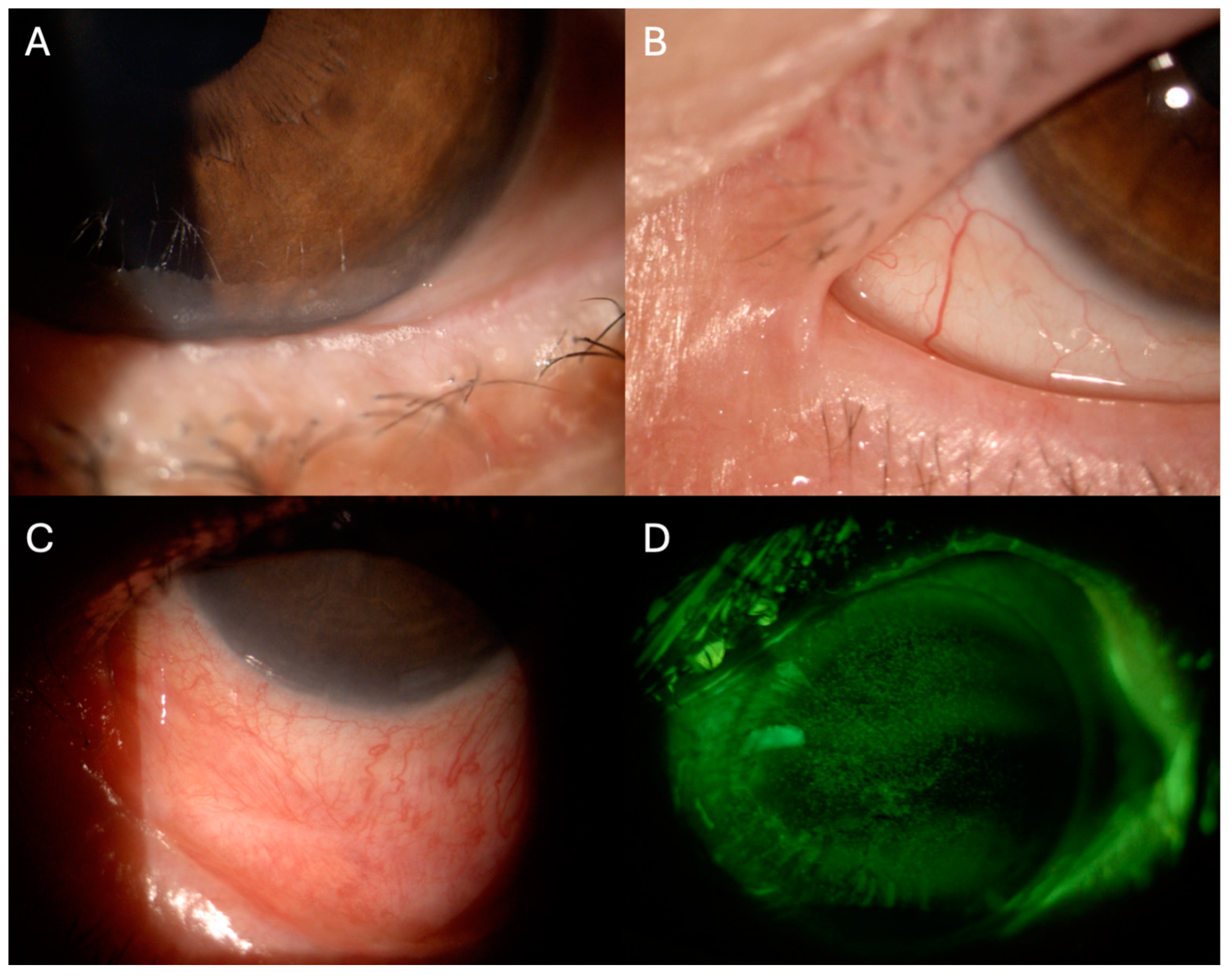
4. Ocular GVHD
5. Toxicity to Antineoplastic Therapies

| Agent | Sign/Symptom | Underlying Mechanism |
|---|---|---|
| Busulfan [98,99,100] | Posterior subcapsular cataracts | Inhibition of nucleic acid formation in lens epithelium |
| Vincristine [101] | Temporary or permanent loss of vision | Axonal damage due to microtubule disruption |
| Dexamethasone [102] | Increased intraocular pressure; cataract | Increased resistance to aqueous outflow; unclear, likely non-enzymatic formation of Schiff base intermediates |
| Fludarabine [103,104] | Rapid progressive loss of vision | Direct damage to retinal bipolar and ganglion cells; gray and white matter |
| Cytarabine [105,106] | Reversible corneal toxicity and conjunctivitis | Unknown, likely inhibition of DNA synthesis corneal and conjunctival epithelium |
| Imatinib [107] | Periorbital edema, conjunctival hemorrhage | Periocular soft tissue expression of molecular targets for Imatinib |
| Immune checkpoint inhibitors [108] | Uveitis | Activation of complement cascade, recruitment of innate immunity cells in cerebrospinal fluid, loss of immune-privilege |
6. Conclusions
Author Contributions
Funding
Institutional Review Board Statement
Data Availability Statement
Conflicts of Interest
References
- Talcott, K.E.; Garg, R.J.; Garg, S.J. Ophthalmic manifestations of leukemia. Curr. Opin. Ophthalmol. 2016, 27, 545–551. [Google Scholar] [CrossRef] [PubMed]
- Buchan, J.; McKibbin, M.; Burton, T. The prevalence of ocular disease in chronic lymphocytic leukaemia. Eye 2003, 17, 27–30. [Google Scholar] [CrossRef] [PubMed]
- Sharma, T.; Grewal, J.; Gupta, S.; Murray, P.I. Ophthalmic manifestations of acute leukaemias: The ophthalmologist’s role. Eye 2004, 18, 663–672. [Google Scholar] [CrossRef]
- Giannaccare, G.; Pellegrini, M.; Bernabei, F.; Scorcia, V.; Campos, E. Ocular surface system alterations in ocular graft-versus-host disease: All the pieces of the complex puzzle. Graefe’s Arch. Clin. Exp. Ophthalmol. 2019, 257, 1341–1351. [Google Scholar] [CrossRef] [PubMed]
- Olsen, T.G.; Heegaard, S. Orbital lymphoma. Surv. Ophthalmol. 2019, 64, 45–66. [Google Scholar] [CrossRef]
- Tanenbaum, R.E.; Galor, A.; Dubovy, S.R.; Karp, C.L. Classification, diagnosis, and management of conjunctival lymphoma. Eye Vis. 2019, 6, 22. [Google Scholar] [CrossRef] [PubMed]
- Witmer, M.T. Primary Vitreoretinal Lymphoma: Management of Isolated Ocular Disease. Cancer Control 2016, 23, 110–116. [Google Scholar] [CrossRef]
- Hassan, W.M.; Bakry, M.S.; Hassan, H.M.; Alfaar, A.S. Incidence of orbital, conjunctival and lacrimal gland malignant tumors in USA from Surveillance, Epidemiology and End Results, 1973–2009. Int. J. Ophthalmol. 2016, 9, 1808. [Google Scholar]
- Valenzuela, J.; Echegaray, J.J.; Dodds, E.; Kurup, S.K.; Lowder, C.; Ondrejka, S.L.; Singh, A.D. Ophthalmic Manifestations of Hodgkin Lymphoma: A Review. Ocul. Oncol. Pathol. 2021, 7, 381–389. [Google Scholar] [CrossRef]
- Olsen, T.G.; Holm, F.; Mikkelsen, L.H.; Rasmussen, P.K.; Coupland, S.E.; Esmaeli, B.; Finger, P.T.; Graue, G.F.; Grossniklaus, H.E.; Honavar, S.G.; et al. Orbital Lymphoma—An International Multicenter Retrospective Study. Am. J. Ophthalmol. 2019, 199, 44–57. [Google Scholar] [CrossRef]
- WHO. WHO Classification of Tumours of Haematopoietic and Lymphoid Tissues; WHO: Geneva, Switzerland, 2009. [Google Scholar]
- Goda, J.S.; Le, L.W.; Lapperriere, N.J.; Millar, B.-A.; Payne, D.; Gospodarowicz, M.K.; Wells, W.; Hodgson, D.C.; Sun, A.; Simpson, R.; et al. Localized Orbital Mucosa-Associated Lymphoma Tissue Lymphoma Managed with Primary Radiation Therapy: Efficacy and Toxicity. Int. J. Radiat. Oncol. Biol. Phys. 2011, 81, e659–e666. [Google Scholar] [CrossRef]
- Kharod, S.M.; Herman, M.P.; Morwith, C.G.; Lightsey, J.; Mendenhall, W.M.; Mendenhall, N.P. Radiotherapy in the Management of Orbital Lymphoma. Am. J. Clin. Oncol. Cancer Clin. Trials 2015, 41, 100–106. [Google Scholar] [CrossRef] [PubMed]
- Qualls, D.; Imber, B.S.; Okwali, M.; Hamlin, P.A.; Kumar, A.; Lahoud, O.B.; Matasar, M.J.; Noy, A.; Owens, C.; Zelenetz, A.D.; et al. Long-term outcomes of patients with limited-stage ocular adnexal DLBCL treated with combined modality therapy in the rituximab era. Br. J. Haematol. 2023, 200, 524–527. [Google Scholar] [CrossRef]
- Salar, A.; Domingo-Domenech, E.; Panizo, C.; Nicolás, C.; Bargay, J.; Muntañola, A.; Canales, M.; Bello, J.L.; Sancho, J.M.; Tomás, J.F.; et al. Long-term results of a phase 2 study of rituximab and bendamustine for mucosa-associated lymphoid tissue lymphoma. Blood 2017, 130, 1772–1774. [Google Scholar] [CrossRef]
- Zucca, E.; Conconi, A.; Martinelli, G.; Bouabdallah, R.; Tucci, A.; Vitolo, U.; Martelli, M.; Pettengell, R.; Salles, G.; Sebban, C.; et al. Final results of the IELSG-19 randomized trial of mucosa-associated lymphoid tissue lymphoma: Improved event-free and progression-free survival with rituximab plus chlorambucil versus either chlorambucil or rituximab monotherapy. J. Clin. Oncol. 2017, 35, 1905–1912. [Google Scholar] [CrossRef]
- Soussain, C.; Malaise, D.; Cassoux, N. Primary vitreoretinal lymphoma: A diagnostic and management challenge. Blood 2021, 138, 1519–1534. [Google Scholar] [CrossRef] [PubMed]
- Soussain, C.; Houillier, C.; Ghesquieres, H.; Soubeyran, P.; Chinot, O.; Taillandier, L.; Houot, R.; Ahle, G.; Gyan, E.; Chabrot, C.; et al. The French LOC Network for Primary CNS Lymphoma (PCNSL) Patients: What Can We Learn from a Large National Database? Blood 2016, 128, 926. [Google Scholar] [CrossRef]
- Levasseur, S.D.; Wittenberg, L.A.; White, V.A. Vitreoretinal lymphoma: A 20-year review of incidence, clinical and cytologic features, treatment, and outcomes. JAMA Ophthalmol. 2013, 131, 50–55. [Google Scholar] [CrossRef]
- Reichstein, D. Primary vitreoretinal lymphoma: An update on pathogenesis, diagnosis and treatment. Curr. Opin. Ophthalmol. 2016, 27, 177–184. [Google Scholar] [CrossRef]
- Kalogeropoulos, D.; Vartholomatos, G.; Mitra, A.; Elaraoud, I.; Ch’ng, S.W.; Zikou, A.; Papoudou-Bai, A.; Moschos, M.M.; Kanavaros, P.; Kalogeropoulos, C. Primary vitreoretinal lymphoma. Saudi J. Ophthalmol. 2019, 33, 66–80. [Google Scholar] [CrossRef]
- Chan, C.C.; Rubenstein, J.L.; Coupland, S.E.; Davis, J.L.; Harbour, J.W.; Johnston, P.B.; Cassoux, N.; Touitou, V.; Smith, J.R.; Batchelor, T.T.; et al. Primary vitreoretinal lymphoma: A report from an International Primary Central Nervous System Lymphoma Collaborative Group symposium. Oncologist 2011, 16, 1589–1599. [Google Scholar] [CrossRef] [PubMed]
- Sagoo, M.S.; Mehta, H.; Swampillai, A.J. Primary intraocular lymphoma. Surv. Ophthalmol. 2014, 59, 503–516. [Google Scholar] [CrossRef] [PubMed]
- Coupland, S.E.; Damato, B. Understanding intraocular lymphomas. Clin. Exp. Ophthalmol. 2008, 36, 564–578. [Google Scholar] [CrossRef]
- Gill, M.K.; Jampol, L.M. Variations in the presentation of primary intraocular lymphoma: Case reports and a review. Surv. Ophthalmol. 2001, 45, 463–471. [Google Scholar] [CrossRef] [PubMed]
- Mohd Fauzi Yap, M.F.B.; Mohd Zain, A.; Tumian, N.R.; Palaniappan, S.; Nasaruddin, R.A.; Md Din, N. Optic Nerve Infiltration in Systemic Metastatic Retinal Lymphoma (SMRL): Multimodal Imaging and Challenges in Diagnosis. Ocul. Immunol. Inflamm. 2021, 29, 479–484. [Google Scholar] [CrossRef] [PubMed]
- Costopoulos, M.; Touitou, V.; Golmard, J.L.; Darugar, A.; Fisson, S.; Bonnemye, P.; Le Lez, M.L.; Soussain, C.; Cassoux, N.; Lamy, T.; et al. ISOLD: A New Highly Sensitive Interleukin Score for Intraocular Lymphoma Diagnosis. Ophthalmology 2016, 123, 1626–1628. [Google Scholar] [CrossRef]
- Gao, X.; Li, B.; You, Q.; Peng, X. Primary extranodal marginal zone B-cell lymphoma with diffuse uveal involvement and focal infiltration of the trabecular meshwork: A case report and review of literature. BMC Ophthalmol. 2015, 15, 48. [Google Scholar] [CrossRef] [PubMed]
- Kakkassery, V.; Coupland, S.E.; Heindl, L.M. Iris lymphoma—A systematic guide for diagnosis and treatment. Surv. Ophthalmol. 2021, 66, 41–53. [Google Scholar] [CrossRef] [PubMed]
- Mashayekhi, A.; Shukla, S.Y.; Shields, J.A.; Shields, C.L. Choroidal lymphoma: Clinical features and association with systemic lymphoma. Ophthalmology 2014, 121, 342–351. [Google Scholar] [CrossRef]
- Valenzuela, J.; Yeaney, G.A.; Hsi, E.D.; Azzato, E.M.; Peereboom, D.M.; Singh, A.D. Large B-cell lymphoma of the uvea: Histopathologic variants and clinicopathologic correlation. Surv. Ophthalmol. 2020, 65, 361–370. [Google Scholar] [CrossRef]
- Sen, H.N.; Chan, C.C.; Caruso, R.C.; Fariss, R.N.; Nussenblatt, R.B.; Buggage, R.R. Waldenström’s macroglobulinemia-associated retinopathy. Ophthalmology 2004, 111, 535–539. [Google Scholar] [CrossRef] [PubMed]
- Shoji, M.K.; Chen, Y.; Topilow, N.J.; Abou Khzam, R.; Dubovy, S.R.; Johnson, T.E. Orbital Involvement in Multiple Myeloma. Ophthalmic Plast. Reconstr. Surg. 2023, 39, 347–356. [Google Scholar] [CrossRef] [PubMed]
- Lin, Y.C.; Liang, T.H.; Chang, H.N.; Lin, J.S.; Lin, H.Y. Behçet disease associated with myelodysplastic syndrome. J. Clin. Rheumatol. 2008, 14, 169–174. [Google Scholar] [CrossRef] [PubMed]
- Ahn, B.Y.; Choi, K.D.; Choi, Y.J.; Jea, S.Y.; Lee, J.E. Isolated monocular visual loss as an initial manifestation of polycythemia vera. J. Neurol. Sci. 2007, 258, 151–153. [Google Scholar] [CrossRef] [PubMed]
- Lin, A.L.; Burnham, J.M.; Pang, V.; Idowu, O.; Iyer, S. Ocular Manifestations of Primary Myelofibrosis. Retin. Cases Brief. Rep. 2016, 10, 364–367. [Google Scholar] [CrossRef] [PubMed]
- El Salloukh, N.A.; Hage, D.G.; Bashshur, A.Z.; Kheir, W.J. Early Ophthalmological Manifestations of Acute Myeloid Leukemia: Current Perspectives. Clin. Ophthalmol. 2022, 16, 2119–2127. [Google Scholar] [CrossRef] [PubMed]
- De Queiroz Mendonca, C.; Freire, M.V.; Viana, S.S.; Silva Tavares, M.K.G.; Almeida Silva, W.M.; Cipolotti, R. Ocular manifestations in acute lymphoblastic leukemia: A five-year cohort study of pediatric patients. Leuk. Res. 2019, 76, 24–28. [Google Scholar] [CrossRef] [PubMed]
- Reddy, S.C.; Jackson, N.; Menon, B.S. Ocular involvement in leukemia—A study of 288 cases. Ophthalmologica 2003, 217, 441–445. [Google Scholar] [CrossRef] [PubMed]
- Green, W.; Rao, P.K.; Harocopos, G.J. Extramedullary Relapse of Acute Myelogenous Leukemia Presenting as a Large Serous Retinal Detachment. Ocul. Oncol. Pathol. 2017, 3, 95–100. [Google Scholar] [CrossRef]
- Russo, V.; Scott, I.U.; Querques, G.; Stella, A.; Barone, A.; Delle Noci, N. Orbital and ocular manifestations of acute childhood leukemia: Clinical and statistical analysis of 180 patients. Eur. J. Ophthalmol. 2008, 18, 619–623. [Google Scholar] [CrossRef]
- Vishnevskia-Dai, V.; Sella King, S.; Lekach, R.; Fabian, I.D.; Zloto, O. Ocular Manifestations of Leukemia and Results of Treatment with Intravitreal Methotrexate. Sci. Rep. 2020, 10, 1994. [Google Scholar] [CrossRef] [PubMed]
- Reddy, S.C.; Jackson, N. Retinopathy in acute leukaemia at initial diagnosis: Correlation of fundus lesions and haematological parameters. Acta Ophthalmol. Scand. 2004, 82, 81–85. [Google Scholar] [CrossRef] [PubMed]
- Chen, B.; Yan, X.; Zhang, X.; Yang, H. Leukostasis retinopathy: An uncommon visual threatening complication of chronic myeloid leukemia with severe hyperleukocytosis—A case report and review of the literature. Indian J. Ophthalmol. 2018, 66, 1871–1874. [Google Scholar] [PubMed]
- Mandava, N.; Costakos, D.; Bartlett, H.M. Chronic myelogenous leukemia manifested as bilateral proliferative retinopathy. Arch. Ophthalmol. 2005, 123, 576–577. [Google Scholar] [PubMed]
- Johnson, J.S.; Lopez, J.S.; Kavanaugh, A.S.; Liang, C.; Mata, D.A. A 25-Year-Old Man with Exudative Retinal Detachments and Infiltrates without Hematological or Neurological Findings Found to Have Relapsed Precursor T-Cell Acute Lymphoblastic Leukemia. Case Rep. Ophthalmol. 2015, 6, 321–327. [Google Scholar] [CrossRef] [PubMed]
- Kumar, V.; Kumawat, D.; Dhakal, S. Leukemic retinopathy and foveal infiltrates. Int. Ophthalmol. 2018, 38, 1301–1303. [Google Scholar] [CrossRef] [PubMed]
- Vicini, G.; Nicolosi, C.; Malandrino, D.; Tozzetti, C.; Rizzo, S.; Sodi, A. Leukostasis retinopathy with leukemic infiltrates as onset manifestation of chronic myeloid leukemia: A case report. Eur. J. Ophthalmol. 2021, 31, NP116–NP121. [Google Scholar] [CrossRef] [PubMed]
- Sayadi, J.; Gouider, D.; Allouche, Y.; Choura, R.; Cherni, I.; Sayadi, M.; Benneji, H.; Zghal, I.; Malek, I.; Nacef, L. Ophthalmic Manifestations of Newly Diagnosed Acute Leukemia Patients in a Tunisian Cohort. Clin. Ophthalmol. 2022, 16, 3425–3435. [Google Scholar] [CrossRef]
- Veerappan Pasricha, M.; Callaway, N.F.; Nguyen, Q.D.; Do, D.V. Serous retinal detachment as a presenting sign of acute lymphoblastic leukemia: A case report and literature review. Am. J. Ophthalmol. Case Rep. 2021, 23, 101142. [Google Scholar] [CrossRef]
- Patel, A.V.; Miller, J.B.; Nath, R.; Shih, H.A.; Yoon, M.K.; Freitag, S.K.; Papaliodis, G.; Chen, T.C.; Eliott, D.; Kim, I.K. Unilateral Eye Findings: A Rare Herald of Acute Leukemia. Ocul. Oncol. Pathol. 2016, 2, 166–170. [Google Scholar] [CrossRef]
- Lin, Y.C.; Wang, A.G.; Yen, M.Y.; Hsu, W.M. Leukaemic infiltration of the optic nerve as the initial manifestation of leukaemic relapse. Eye 2004, 18, 546–550. [Google Scholar] [CrossRef] [PubMed]
- Liu, L.; Hadyah, S.; Park, A.; Akhtari, M.; Scott, J.; Ran-Castillo, D.; Chong, E.; Koh, H.; Oregel, K.; Khandelwal, K.; et al. Leukemic infiltration of the optic nerve in chronic lymphocytic leukemia: A case report and review of literature. Leuk. Res. Rep. 2023, 20, 100391. [Google Scholar] [CrossRef] [PubMed]
- Dini, G.; Capolsini, I.; Cerri, C.; Massei, M.S.; Mastrodicasa, E.; Perruccio, K.; Gorello, P.; Caniglia, M.; Verrotti, A.; Arcioni, F. Acute lymphoblastic leukemia relapse presenting with optic nerve infiltration. SAGE Open Med. Case Rep. 2023, 11, 1–5. [Google Scholar] [CrossRef] [PubMed]
- Kuan, H.C.; Mustapha, M.; Oli Mohamed, S.; Abdul Aziz, R.A.; Loh, C.K.; Mohammed, F.; Naffi, A.A.; Othman, O.; Nasaruddin, R.A.; Alias, H. Isolated Infiltrative Optic Neuropathy in an Acute Lymphoblastic Leukemia Relapse. Cureus 2022, 14, e25625. [Google Scholar] [CrossRef] [PubMed]
- Lee, V.; Farooq, A.V.; Shah, H.A. Leukemic and Lymphomatous Optic Neuropathy: A Case Series. J. Neuroophthalmol. 2021, 41, e796–e802. [Google Scholar] [CrossRef] [PubMed]
- Gruenewald, R.L.; Perry, M.C.; Henry, P.H. Leukemic iritis with hypopyon. Cancer 1979, 44, 1511–1513. [Google Scholar] [CrossRef] [PubMed]
- Ploysangam, P.; Reynolds, A.L. Iris Infiltration as a Sign of Relapse in a Child with Chronic Myeloid Leukemia. Ophthalmology 2021, 128, 780. [Google Scholar] [CrossRef] [PubMed]
- Türkoğlu, E.B.; Öcal, O. Anterior segment optical coherence tomography findings in leukemic iris infiltration. Photodiagn. Photodyn. Ther. 2022, 37, 102578. [Google Scholar] [CrossRef] [PubMed]
- Hakim, F.; Farooq, A. Conjunctival chronic lymphocytic leukemia presenting as bilateral chronic conjunctivitis. Am. J. Ophthalmol. Case Rep. 2022, 27, 101670. [Google Scholar] [CrossRef]
- Wood, W.J.; Nicholson, D.H. Corneal ring ulcer as the presenting manifestation of acute monocytic leukemia. Am. J. Ophthalmol. 1973, 76, 69–72. [Google Scholar] [CrossRef]
- Hodgson, K.; Ferrer, G.; Montserrat, E.; Moreno, C. Chronic lymphocytic leukemia and autoimmunity: A systematic review. Haematologica 2011, 96, 752–761. [Google Scholar] [CrossRef] [PubMed]
- Cullis, C.M.; Hines, D.R.; Bullock, J.D. Anterior segment ischemia: Classification and description in chronic myelogenous leukemia. Ann. Ophthalmol. 1979, 11, 1739–1744. [Google Scholar] [PubMed]
- Chan, C.C.; Wallace, D.J. Intraocular lymphoma: Update on diagnosis and management. Cancer Control 2004, 11, 285–295. [Google Scholar] [CrossRef]
- Jahnke, K.; Korfel, A.; Komm, J.; Bechrakis, N.E.; Stein, H.; Thiel, E.; Coupland, S.E. Intraocular lymphoma 2000–2005: Results of a retrospective multicentre trial. Graefe’s Arch. Clin. Exp. Ophthalmol. 2006, 244, 663–669. [Google Scholar]
- Mashayekhi, A.; Shields, C.L.; Shields, J.A. Iris involvement by lymphoma: A review of 13 cases. Clin. Exp. Ophthalmol. 2013, 41, 19–26. [Google Scholar] [CrossRef] [PubMed]
- Pathak, M.; Diep, P.P.; Lai, X.; Brinch, L.; Ruud, E.; Drolsum, L. Ocular findings and ocular graft-versus-host disease after allogeneic stem cell transplantation without total body irradiation. Bone Marrow Transplant. 2018, 53, 863–872. [Google Scholar] [CrossRef] [PubMed]
- Dietrich-Ntoukas, T.; Cursiefen, C.; Westekemper, H.; Eberwein, P.; Reinhard, T.; Bertz, H.; Nepp, J.; Lawitschka, A.; Heiligenhaus, A.; Seitz, B.; et al. Diagnosis and treatment of ocular chronic graft-versus-host disease: Report from the German-Austrian-Swiss Consensus Conference on Clinical Practice in chronic GVHD. Cornea 2012, 31, 299–310. [Google Scholar] [CrossRef] [PubMed]
- Nassiri, N.; Eslani, M.; Panahi, N.; Mehravaran, S.; Ziaei, A.; Djalilian, A.R. Ocular graft versus host disease following allogeneic stem cell transplantation: A review of current knowledge and recommendations. J. Ophthalmic Vis. Res. 2013, 8, 351–358. [Google Scholar] [PubMed]
- Wolff, D.; Radojcic, V.; Lafyatis, R.; Cinar, R.; Rosenstein, R.K.; Cowen, E.W.; Cheng, G.S.; Sheshadri, A.; Bergeron, A.; Williams, K.M.; et al. National Institutes of Health Consensus Development Project on Criteria for Clinical Trials in Chronic Graft-versus-Host Disease: IV. The 2020 Highly morbid forms report. Transplant. Cell. Ther. 2021, 27, 817–835. [Google Scholar] [CrossRef] [PubMed]
- Ogawa, Y.; Kuwana, M.; Yamazaki, K.; Mashima, Y.; Yamada, M.; Mori, T.; Okamoto, S.; Oguchi, Y.; Kawakami, Y. Periductal area as the primary site for T-cell activation in lacrimal gland chronic graft-versus-host disease. Investig. Ophthalmol. Vis. Sci. 2003, 44, 1888–1896. [Google Scholar] [CrossRef]
- Ogawa, Y.; Yamazaki, K.; Kuwana, M.; Mashima, Y.; Nakamura, Y.; Ishida, S.; Toda, I.; Oguchi, Y.; Tsubota, K.; Okamoto, S.; et al. A significant role of stromal fibroblasts in rapidly progressive dry eye in patients with chronic GVHD. Investig. Ophthalmol. Vis. Sci. 2001, 42, 111–119. [Google Scholar]
- Ogawa, Y.; Kodama, H.; Kameyama, K.; Yamazaki, K.; Yasuoka, H.; Okamoto, S.; Inoko, H.; Kawakami, Y.; Kuwana, M. Donor fibroblast chimerism in the pathogenic fibrotic lesion of human chronic graft-versus-host disease. Investig. Ophthalmol. Vis. Sci. 2005, 46, 4519–4527. [Google Scholar] [CrossRef] [PubMed]
- Ogawa, Y.; Shimmura, S.; Kawakita, T.; Yoshida, S.; Kawakami, Y.; Tsubota, K. Epithelial mesenchymal transition in human ocular chronic graft-versus-host disease. Am. J. Pathol. 2009, 175, 2372–2381. [Google Scholar] [CrossRef] [PubMed]
- Cheng, X.; Huang, R.; Huang, S.; Fan, W.; Yuan, R.; Wang, X.; Zhang, X. Recent advances in ocular graft-versus-host disease. Front. Immunol. 2023, 14, 1092108. [Google Scholar] [CrossRef] [PubMed]
- Ogawa, Y.; Okamoto, S.; Wakui, M.; Watanabe, R.; Yamada, M.; Yoshino, M.; Ono, M.; Yang, H.Y.; Mashima, Y.; Oguchi, Y.; et al. Dry eye after haematopoietic stem cell transplantation. Br. J. Ophthalmol. 1999, 83, 1125–1130. [Google Scholar] [CrossRef] [PubMed]
- Giannaccare, G.; Bonifazi, F.; Sebastiani, S.; Sessa, M.; Pellegrini, M.; Arpinati, M.; Moscardelli, F.; Versura, P.; Campos, E. Meibomian Gland Dropout in Hematological Patients Before Hematopoietic Stem Cell Transplantation. Cornea 2018, 37, 1264–1269. [Google Scholar] [CrossRef] [PubMed]
- Giannaccare, G.; Bonifazi, F.; Sessa, M.; Fresina, M.; Arpinati, M.; Bandini, G.; Versura, P. Dry Eye Disease Is Already Present in Hematological Patients Before Hematopoietic Stem Cell Transplantation. Cornea 2016, 35, 638–643. [Google Scholar] [CrossRef] [PubMed]
- Appenteng Osae, E.; Steven, P. Meibomian Gland Dysfunction in Ocular Graft vs. Host Disease: A Need for Pre-Clinical Models and Deeper Insights. Int. J. Mol. Sci. 2021, 22, 3516. [Google Scholar]
- Perez, V.L.; Mousa, H.M.; Soifer, M.; Beatty, C.; Sarantopoulos, S.; Saban, D.R.; Levy, R.B. Meibomian Gland Dysfunction: A Route of Ocular Graft-Versus-Host Disease Progression That Drives a Vicious Cycle of Ocular Surface Inflammatory Damage. Am. J. Ophthalmol. 2023, 247, 42–60. [Google Scholar] [CrossRef]
- Jabs, D.A.; Wingard, J.; Green, W.R.; Farmer, E.R.; Vogelsang, G.; Saral, R. The eye in bone marrow transplantation. III. Conjunctival graft-vs-host disease. Arch. Ophthalmol. 1989, 107, 1343–1348. [Google Scholar]
- Eberwein, P.; Issleib, S.; Böhringer, D.; Mittelviefhaus, H.; Schwartzkopff, J.; Finke, J.; Reinhard, T. Conjunctival HLA-DR and CD8 expression detected by impression cytology in ocular graft versus host disease. Mol. Vis. 2013, 19, 1492–1501. [Google Scholar] [PubMed]
- Sonawane, S.; Khanolkar, V.; Namavari, A.; Chaudhary, S.; Gandhi, S.; Tibrewal, S.; Jassim, S.H.; Shaheen, B.; Hallak, J.; Horner, J.H.; et al. Ocular surface extracellular DNA and nuclease activity imbalance: A new paradigm for inflammation in dry eye disease. Investig. Ophthalmol. Vis. Sci. 2012, 53, 8253–8263. [Google Scholar] [CrossRef]
- An, S.; Raju, I.; Surenkhuu, B.; Kwon, J.E.; Gulati, S.; Karaman, M.; Pradeep, A.; Sinha, S.; Mun, C.; Jain, S. Neutrophil extracellular traps (NETs) contribute to pathological changes of ocular graft-vs.-host disease (oGVHD) dry eye: Implications for novel biomarkers and therapeutic strategies. Ocul. Surf. 2019, 17, 589–614. [Google Scholar] [CrossRef]
- Tatematsu, Y.; Ogawa, Y.; Shimmura, S.; Dogru, M.; Yaguchi, S.; Nagai, T.; Yamazaki, K.; Kameyama, K.; Okamoto, S.; Kawakami, Y.; et al. Mucosal microvilli in dry eye patients with chronic GVHD. Bone Marrow Transplant. 2012, 47, 416–425. [Google Scholar] [CrossRef] [PubMed]
- Tabbara, K.F.; Al-Ghamdi, A.; Al-Mohareb, F.; Ayas, M.; Chaudhri, N.; Al-Sharif, F.; Al-Zahrani, H.; Mohammed, S.Y.; Nassar, A.; Aljurf, M. Ocular findings after allogeneic hematopoietic stem cell transplantation. Ophthalmology 2009, 116, 1624–1629. [Google Scholar] [CrossRef] [PubMed]
- Kheirkhah, A.; Coco, G.; Satitpitakul, V.; Dana, R. Subtarsal Fibrosis Is Associated with Ocular Surface Epitheliopathy in Graft-Versus-Host Disease. Am. J. Ophthalmol. 2018, 189, 102–110. [Google Scholar] [CrossRef] [PubMed]
- Sivaraman, K.R.; Jivrajka, R.V.; Soin, K.; Bouchard, C.S.; Movahedan, A.; Shorter, E.; Jain, S.; Jacobs, D.S.; Djalilian, A.R. Superior Limbic Keratoconjunctivitis-like Inflammation in Patients with Chronic Graft-Versus-Host Disease. Ocul. Surf. 2016, 14, 393–400. [Google Scholar] [CrossRef] [PubMed]
- Kheirkhah, A.; Qazi, Y.; Arnoldner, M.A.; Suri, K.; Dana, R. In Vivo Confocal Microscopy in Dry Eye Disease Associated with Chronic Graft-Versus-Host Disease. Investig. Ophthalmol. Vis. Sci. 2016, 57, 4686–4691. [Google Scholar] [CrossRef] [PubMed]
- He, J.; Ogawa, Y.; Mukai, S.; Saijo-Ban, Y.; Kamoi, M.; Uchino, M.; Yamane, M.; Ozawa, N.; Fukui, M.; Mori, T.; et al. In Vivo Confocal Microscopy Evaluation of Ocular Surface with Graft-Versus-Host Disease-Related Dry Eye Disease. Sci. Rep. 2017, 7, 10720. [Google Scholar] [CrossRef]
- Zhang, C.Y.; Farooq, A.V.; Harocopos, G.J.; Sollenberger, E.L.; Hou, J.H.; Bouchard, C.S.; Shieh, C.; Tran, U.L.; Lubniewski, A.J.; Huang, A.J.W.; et al. Corneal perforation in ocular graft-versus-host disease. Am. J. Ophthalmol. Case Rep. 2021, 24, 101224. [Google Scholar] [CrossRef] [PubMed]
- Jagasia, M.H.; Greinix, H.T.; Arora, M.; Williams, K.M.; Wolff, D.; Cowen, E.W.; Palmer, J.; Weisdorf, D.; Treister, N.S.; Cheng, G.S.; et al. National Institutes of Health Consensus Development Project on Criteria for Clinical Trials in Chronic Graft-versus-Host Disease: I. The 2014 Diagnosis and Staging Working Group report. Biol. Blood Marrow Transplant. 2015, 21, 389–401.e1. [Google Scholar] [CrossRef]
- Ogawa, Y.; Kim, S.K.; Dana, R.; Clayton, J.; Jain, S.; Rosenblatt, M.I.; Perez, V.L.; Shikari, H.; Riemens, A.; Tsubota, K. International Chronic Ocular Graft-vs-Host-Disease (GVHD) Consensus Group: Proposed diagnostic criteria for chronic GVHD (Part I). Sci. Rep. 2013, 3, 3419. [Google Scholar] [CrossRef]
- Singh, R.B.; Yung, A.; Coco, G.; Sinha, S.; Dohlman, T.H.; Yin, J.; Dana, R. Efficacy and retention of silicone punctal plugs for treatment of dry eye in patients with and without ocular graft-versus-host-disease. Ocul. Surf. 2020, 18, 731–735. [Google Scholar] [CrossRef]
- Le, Q.T.; Eulau, S.M.; George, T.I.; Hildebrand, R.; Warnke, R.A.; Donaldson, S.S.; Hoppe, R.T. Primary radiotherapy for localized orbital MALT lymphoma. Int. J. Radiat. Oncol. Biol. Phys. 2002, 52, 657–663. [Google Scholar] [CrossRef]
- Stafford, S.L.; Kozelsky, T.F.; Garrity, J.A.; Kurtin, P.J.; Leavitt, J.A.; Martenson, J.A.; Habermann, T.M. Orbital lymphoma: Radiotherapy outcome and complications. Radiother. Oncol. 2001, 59, 139–144. [Google Scholar] [CrossRef]
- Finger, P.T. Radiation therapy for orbital tumors: Concepts, current use, and ophthalmic radiation side effects. Surv. Ophthalmol. 2009, 54, 545–568. [Google Scholar] [CrossRef] [PubMed]
- Li, J.; Tripathi, R.C.; Tripathi, B.J. Drug-induced ocular disorders. Drug Saf. 2008, 31, 127–141. [Google Scholar] [CrossRef] [PubMed]
- Kaida, T.; Ogawa, T.; Amemiya, T. Cataract induced by short-term administration of large doses of busulfan: A case report. Ophthalmologica 1999, 213, 397–399. [Google Scholar] [CrossRef] [PubMed]
- Saito, T.; Ando, R.; Ohira, T.; Hoshiya, T.; Tamura, K. Ocular lesions induced in infant rats by busulfan. Histol. Histopathol. 2015, 30, 321–330. [Google Scholar]
- Lee, W.H.; You, S.K.; Lee, Y.H. Bilateral optic neuropathy following vincristine chemotherapy: A case report with description of multimodal imaging findings. Medicine 2021, 100, e24706. [Google Scholar] [CrossRef] [PubMed]
- Sugiyama, M.; Terashita, Y.; Hara, K.; Cho, Y.; Iguchi, A.; Chin, S.; Manabe, A. Corticosteroid-induced glaucoma in pediatric patients with hematological malignancies. Pediatr. Blood Cancer 2019, 66, e27977. [Google Scholar] [CrossRef]
- Bishop, R.J.; Ding, X.; Heller, C.K., 3rd; Illei, G.; Caruso, R.; Cunningham, D.; Pavletic, S.; Chan, C.C. Rapid vision loss associated with fludarabine administration. Retina 2010, 30, 1272–1277. [Google Scholar] [CrossRef]
- Ding, X.; Herzlich, A.A.; Bishop, R.; Tuo, J.; Chan, C.C. Ocular toxicity of fludarabine: A purine analog. Expert. Rev. Ophthalmol. 2008, 3, 97–109. [Google Scholar] [CrossRef]
- Hollander, D.A.; Aldave, A.J. Drug-induced corneal complications. Curr. Opin. Ophthalmol. 2004, 15, 541–548. [Google Scholar] [CrossRef] [PubMed]
- Mori, T.; Kato, J.; Yamane, A.; Aisa, Y.; Kawata, Y.; Ichimura, M.; Tsuneyama, C.; Yashima, T.; Ogawa, Y.; Tsubota, K.; et al. Prevention of cytarabine-induced kerato-conjunctivitis by eye rinse in patients receiving high-dose cytarabine and total body irradiation as a conditioning for hematopoietic stem cell transplantation. Int. J. Hematol. 2011, 94, 261–265. [Google Scholar] [CrossRef] [PubMed]
- Ho, W.L.; Wong, H.; Yau, T. The ophthalmological complications of targeted agents in cancer therapy: What do we need to know as ophthalmologists? Acta Ophthalmol. 2013, 91, 604–609. [Google Scholar] [CrossRef]
- Zhang, H.; Houadj, L.; Wu, K.Y.; Tran, S.D. Diagnosing and Managing Uveitis Associated with Immune Checkpoint Inhibitors: A Review. Diagnostics 2024, 14, 336. [Google Scholar] [CrossRef] [PubMed]


| National Institutes of Health (NIH) Diagnostic Criteria | International Consensus Group of Ophthalmologists’ Criteria |
|---|---|
| Schirmer’s test mean value of ≤5 mm at 5 min | Pathological ocular surface disease index (OSDI) questionnaire (<13) |
| New onset of keratoconjunctivitis sicca as determined by a slit lamp examination, with a mean Schirmer’s test value of 6 to 10 mm | Pathological Schirmer’s test (<10 mm in 5 min) without anesthesia |
| Positive corneal fluorescein staining | |
| Presence of conjunctival injection |
Disclaimer/Publisher’s Note: The statements, opinions and data contained in all publications are solely those of the individual author(s) and contributor(s) and not of MDPI and/or the editor(s). MDPI and/or the editor(s) disclaim responsibility for any injury to people or property resulting from any ideas, methods, instructions or products referred to in the content. |
© 2024 by the authors. Licensee MDPI, Basel, Switzerland. This article is an open access article distributed under the terms and conditions of the Creative Commons Attribution (CC BY) license (https://creativecommons.org/licenses/by/4.0/).
Share and Cite
Rossi, C.; Buizza, A.; Alessio, G.; Borselli, M.; Taloni, A.; Carnevali, A.; Carnovale Scalzo, G.; Lucisano, A.; Scorcia, V.; Giannaccare, G. Ophthalmic Manifestations in Patients with Blood Malignancies. Hematol. Rep. 2024, 16, 193-203. https://doi.org/10.3390/hematolrep16020020
Rossi C, Buizza A, Alessio G, Borselli M, Taloni A, Carnevali A, Carnovale Scalzo G, Lucisano A, Scorcia V, Giannaccare G. Ophthalmic Manifestations in Patients with Blood Malignancies. Hematology Reports. 2024; 16(2):193-203. https://doi.org/10.3390/hematolrep16020020
Chicago/Turabian StyleRossi, Costanza, Alessandro Buizza, Giuseppe Alessio, Massimiliano Borselli, Andrea Taloni, Adriano Carnevali, Giovanna Carnovale Scalzo, Andrea Lucisano, Vincenzo Scorcia, and Giuseppe Giannaccare. 2024. "Ophthalmic Manifestations in Patients with Blood Malignancies" Hematology Reports 16, no. 2: 193-203. https://doi.org/10.3390/hematolrep16020020