Case Report

Mutation in the STXBP1 Gene Associated with Early Onset West Syndrome: A Case Report and Literature Review

1
Department of Pediatrics, Kawasaki Municipal Tama Hospital, 1-30-37 Shukugawara, Tama-ku, Kawasaki City 214-8525, Kanagawa, Japan
2
Department of Pediatrics, St. Marianna University School of Medicine, 2-16-1 Sugao, Miyamae-ku, Kawasaki City 216-8511, Kanagawa, Japan
3
Department of Child Neurology, National Center of Neurology and Psychiatry, 4-1-1 Ogawahigashi-machi, Kodaira City 187-8511, Tokyo, Japan
4
Department of Pediatrics, Fukuoka Sanno Hospital, 3-6-45 Momochihama Sawara-ku, Fukuoka-City 814-0001, Fukuoka, Japan
5
Department of Pediatrics, Fukuoka University School of Medicine Comprehensive Medical Research Center, 45-1 7-Chome Nanakuma Jonan-Ku, Fukuoka-City 814-0180, Fukuoka, Japan
*
Author to whom correspondence should be addressed.
Pediatr. Rep. 2022, 14(4), 386-395; https://doi.org/10.3390/pediatric14040046
Submission received: 5 August 2022 / Revised: 5 September 2022 / Accepted: 13 September 2022 / Published: 20 September 2022

Abstract

:
Syntaxin-binding protein1 (STXBP1) is a member of the Sec1/Munc18-1 protein family, which comprises important regulators of the secretory and synaptic vesicle fusion machinery underlying hormonal and neuronal transmission, respectively. STXBP1 pathogenic variants are associated with multiple neurological disorders. Herein, we present the case of a Japanese girl with a mutation in the STXBP1 gene, who was born at 40 weeks without neonatal asphyxia. At 15 days old, she developed epilepsy and generalized seizures. Around 88 days old, she presented with a series of nodding spasms, with the seizure frequency gradually increasing. Interictal EEG indicated hypsarrhythmia and she presented with developmental regression. At 1.5 years old, genetic testing was performed and mutational analysis revealed an STXBP1 gene mutation (c.875G > A: p.Arg292His). Accordingly, she was diagnosed with developmental and epileptic encephalopathy, presenting West syndrome’s clinical characteristics caused by the STXBP1 gene mutation. Although drug treatment has reduced the frequency of epileptic seizures, her development has remained regressive. The relationship between the location and type of genetic abnormality and the phenotype remains unclear. Future studies should investigate the genotype–phenotype correlation and the underlying pathophysiology to elucidate the causal relationships among the multiple phenotype-determining factors.

1. Introduction

When determining the etiology of early-onset infantile seizures following neonatal seizures, genetic, congenital metabolic, and structural brain abnormalities as well as a hypoxic–ischemic state should be mainly considered. The major neonatal-onset epilepsy syndromes are self-limited neonatal epilepsy and early infantile developmental and epileptic encephalopathy (previously early myoclonic epilepsy [EME] and early infantile epileptic encephalopathy [EIEE]) [1]. Some neonatal seizures may present with conditions that are atypical of the classic EIEE or EME, which, due to repeated seizures, affect interictal brain function, leading to developmental delay and cognitive–motor function. This condition was reworded as developmental epileptic encephalopathy (DEE) in the 2017 revision of the International League Against Epilepsy [2]. More than 60 causative genes for DEE have been identified to date, including the syntaxin-binding protein 1 (STXBP1) gene.
STXBP1, also known as Munc-18-1, is a member of the Sec1/Munc18-1 protein family, which comprises important regulators of the secretory and synaptic vesicle fusion machinery involved in the hormonal and neuronal transmission, respectively [3]. Pathogenic STXBP1 variants usually impair neurotransmitter release, specifically in GABAergic interneurons, which causes uncontrolled excitatory neuron firing [4]. Moreover, they are associated with developmental delay, cognitive dysfunction, intellectual disability, and epilepsy [3]. STXBP1 encephalopathy with epilepsy is one type of DEE and is qualified by early-onset encephalopathy with epilepsy. Epilepsy syndromes include infantile spasms (IS), EIEE, Lennox–Gaustaut syndrome, Dravet syndrome (not SCN1A-related), classic Rett syndrome (not MECP2-related), and atypical Rett syndrome (not CDKL5-related) [4].
Patients with IS typically exhibit epileptic spasms along with the EEG pattern known as hypsarrhythmia. West syndrome is classified as a subtype of IS and is the most commonly described subtype, appearing in approximately 90% of instances of IS. It includes the combination of epileptic spasm, hypsarrhythmia, and developmental arrest or regression [5]. Over 130 other genes, in addition to STXBP1, have also been linked to IS. Examples include ARX, CDKL5, PAFAH1B1/LIS1, DC, and KCNQ2 [6]. Here, we present a case of a patient with an STXBP1 mutation who was clinically diagnosed with West syndrome.

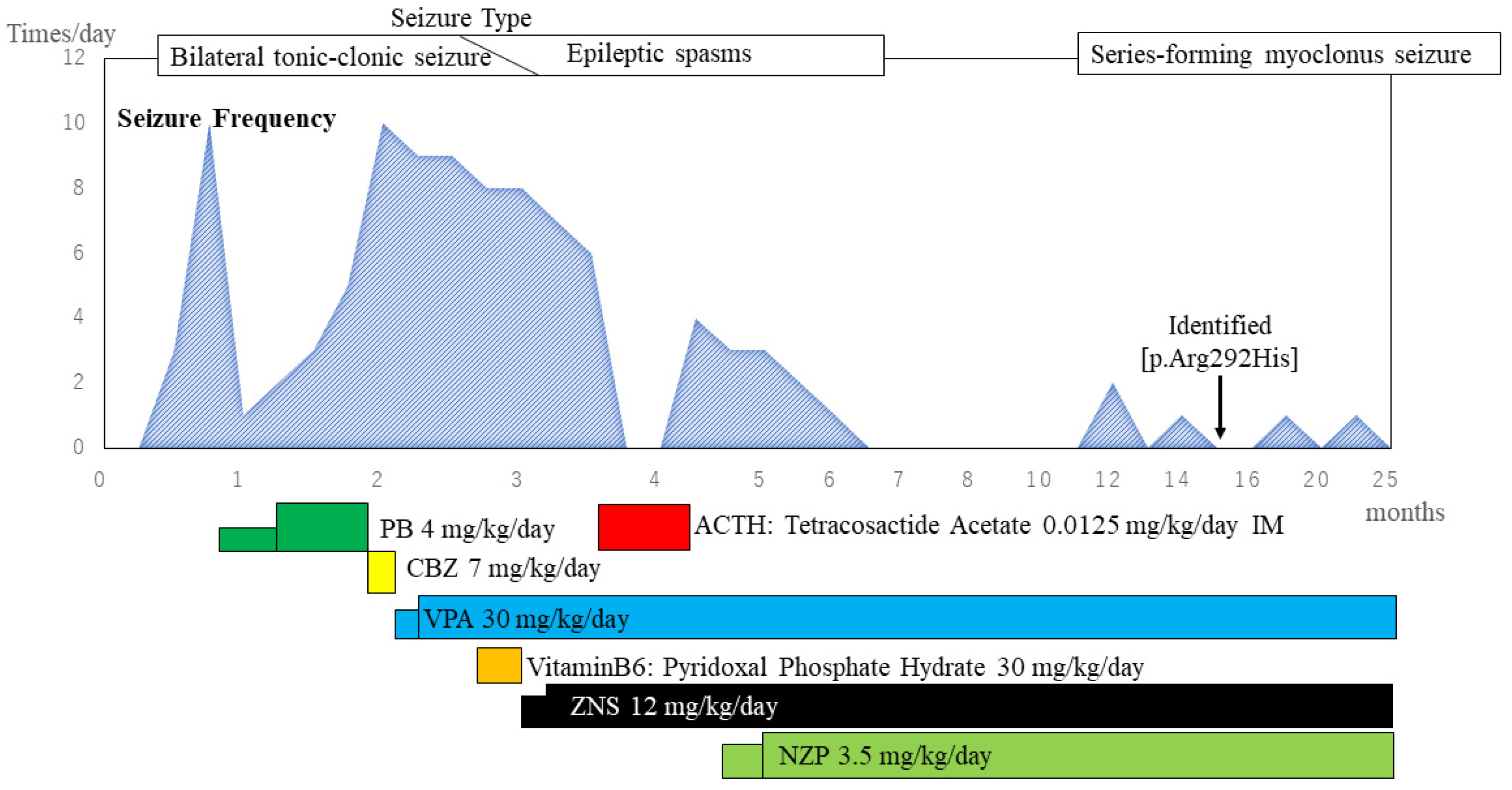
2. Case Presentation

Our patient was a girl who was 40 weeks in utero and born through natural vaginal delivery as the first child of a Japanese couple. Her birth weight, height, and head circumference were 3244 g (+0.67 standard deviation [SD]), 46.8 cm (−0.77 SD), and normal, respectively. Neonatal asphyxia was not observed at birth. Her family history lacked evidence regarding consanguineous marriage, epilepsy, or neuromuscular disease. The clinical course of the patient is shown in Figure 1. At 15 days old, she presented with bilateral tonic–clonic seizures that lasted for a few minutes, which started in the left or right upper extremity. The seizure frequency increased to approximately 10 per day; the patient was admitted to the hospital by her previous doctor. Interictal electroencephalography (EEG) revealed multifocal spontaneous spikes. The EEG showed isolated spike waves in the right frontal, central, and occipital regions that did not spread widely. It was not a suppression-burst or hypsarrhythmia pattern. General blood tests, plasma amino acid analysis, general spinal fluid tests, spinal fluid virus isolation, and urinary organic acid analysis revealed no abnormalities. Brain magnetic resonance imaging (MRI) revealed no obvious epilepsy-inducing structural abnormalities. She was started on phenobarbital for neonatal-onset epilepsy of unknown classification; however, she showed no improvement. She was referred to our hospital at the age of 75 days.
Physical examination and general neurological findings revealed no obvious facial abnormalities or deformities. Eye-tracking and smiling when someone touched or held her were appropriate for infant age. Interictal EEG revealed normal sleep waves; however, there were frequent localized spike waves and spike–slow wave complexes (Figure 2A). A tentative diagnosis of symptomatic localization-related epilepsy was made, and treatment was initiated. She continued to experience seizures approximately 10 times a day; therefore, phenobarbital was discontinued and carbamazepine was started at 78 days old. However, it was discontinued after a few days due to an increase in seizure frequency. Oral sodium valproate and zonisamide were initiated at 81 and 88 days old, respectively. Vitamin B6 was administered; however, it was discontinued after having no effect. After starting sodium valproate and zonisamide, there was a gradual decrease in the seizure frequency; however, even at the maximum dose, seizures were still observed 3–4 times a day. Initially, the seizures were generalized tonic convulsions starting from the left upper extremity. However, around the age of 88 days, she displayed a series of nodding spasms. After adjustment of medications, oral intake was decreased, and tube feeding was initiated. At 108 days old, the patient underwent examination for primary disease according to the Department of Child Neurology, National Center of Neurology and Psychiatry. Long-term EEG monitoring revealed frequent irregular high-amplitude spikes and slow waves without left-right synchrony during sleep and wakefulness, which indicated a hypsarrhythmia (Figure 2B). Ictal EEG revealed a fast-wave rhythm and positive slow waves preceding the spasm (Figure 2C). Eye tracking and chuckling during the early disease stage were no longer observed. Further, she was considered to be developmentally regressing and was diagnosed with West syndrome. Brain MRI revealed no structural abnormalities presenting as obvious epileptic foci, including focal cortical dysplasia (Figure 2D). The patient was started on adrenocorticotropic hormone (ACTH) therapy at 114 days old, which was administered daily for two weeks.
The seizures disappeared the day after starting ACTH therapy. Recurrent spasms were observed 8 days after initiating ACTH therapy. Subsequently, sodium valproate and zonisamide were continued; however, there was no change in seizure frequency. Nitrazepam was started at the age of 4 months, which decreased the seizure frequency to 1–2 per day. EEG revealed multifocal spikes; however, the typical hypsarrhythmia disappeared and there was improved background activity (Figure 2E). Cerebral cortical atrophy occurred as a side effect of ACTH therapy. Development remained regressive, without evidence of head control, eye tracking, or chuckling. Due to aspiration pneumonia, nutrition was maintained by tube feeding. The patient was discharged from the hospital at 6 months old. After 7 months old, the patient lacked obvious clinical seizures. At 10 months old, genetic testing for West syndrome was performed. Subsequently, she was hospitalized for a urinary tract infection and pneumonia at 10 months and 1.3 years old, respectively.
At approximately 1.2 years old, she began experiencing a series of seizures involving the twitching of her right leg during sleep for 2–3 min at a time. Ictal EEG revealed series-forming myoclonus seizures. Interictal EEG showed multifocal spikes and slow waves at sleep onset without hypsarrhythmia. Moreover, background activity during wakefulness revealed continuous slow waves. Brain MRI showed diffuse cerebral atrophy and corpus callosum thinning; however, there were no obvious abnormal structures (Figure 2F). Despite the observation of a new seizure type, three antiepileptic drugs were administered at extreme doses and no indication for active surgery was indicated.
At 1.5 years old, she underwent further genetic testing. Target capture sequencing was performed to screen 114 epilepsy-related genes in the proband of the Department of Pediatrics at Fukuoka. We identified a missense variant (RefSeq accession number NM_003165: c.875G > A [p.Arg292His] available at https://www.ncbi.nlm.nih.gov/refseq/ (accessed on 27 July 2022)) in the STXBP1 gene. Sanger sequencing confirmed the variant in the proband (Figure 3). A search for this variant in her parents through Sanger sequencing revealed that the variant was de novo. Finally, she was diagnosed with developmental and epileptic encephalopathies exhibiting clinical characteristics of West syndrome due to STXBP1 gene mutation (c.875G > A [p.Arg292His], previously reported in ClinVar [rs796053361] as a pathogenic variant). Other unidentified variants in addition to STXBP1-p.Arg292His may have contributed to her clinical phenotype; since she did not undergo whole genome or whole exome sequencing, we continue to investigate this possibility.
The patient is currently 2.1 years old. Although the frequency of epileptic seizures has decreased, her development remains regressive, and she has a severe intellectual disability. At the time of writing, apart from the neurological phenotype, the patient had no other symptoms, such as metabolic disease, primary immunodeficiency, or hyperinflammatory condition. She continues to receive medical treatment. Should the seizures worsen in the future, we will review the choice of drug according to the seizure type and consider palliative epilepsy surgical treatment to reduce seizure frequency.

3. Discussion

This report describes a patient with West syndrome associated with genetic etiology related to an STXBP1 gene mutation. West syndrome mostly occurs in the first year of life and presents as spasms with characteristic EEG changes known as hypsarrhythmia; moreover, it is strongly associated with developmental delay or regression [6,7]. STXBP1 encephalopathy is a type of West syndrome that causes encephalopathy. It is characterized by early-onset encephalopathy, with about half of cases presenting seizures in the neonatal period. Frequent seizure types include epileptic spasms, focal seizures, and tonic seizures. Epileptic spasms frequently occur at some stage of the disease course (65.3%) [8]. In addition to epilepsy, a pathogenic de novo STXBP1 gene mutation causes various neurological deficits, including severe early-onset forms of intellectual disability, autism, and movement disorders [3].
The patient experienced bilateral tonic–clonic seizures and had multifocal abnormalities on the EEG from the beginning of her epilepsy in her neonatal period, but not a suppression-burst pattern. Consequently, a diagnosis of neither conventional EIEE nor EME could be established, and the patient was first considered to be in a condition of DEE with no specific syndrome diagnosis and was followed up. Subsequently, the seizure type and EEG findings changed, developmental regression was observed, and the condition was diagnosed as West syndrome within the concept of DEE. Although many reports describe that the clinical spectrum of STXBP1 mutations may be broader and there are differences between reports on the early features of onset, the presence of EEG suppression-burst patterns and epileptic spasms may better characterize STXBP1 encephalopathy. According to a study by Saitsu et al., STXBP1 encephalopathy first manifests as a range of seizure forms; however, 22% of patients also experienced epileptic spasms [9]. In a report of 24 cases of epileptic patients with STXBP1 mutations, epileptic spasms were discovered in 78% of cases as the initial seizure. A hypsarrhythmic pattern was already evident from the initial EEG in one child at 2 months old, and multifocal asynchronous spikes and slow waves without discontinuity were seen in 78% and 27% of patients, respectively [10]. Even when there is not a noticeable early EEG suppression burst pattern, as there is in the current example, the likelihood of STXBP1 mutation should be considered. Although EIEE is the most common epileptic syndrome phenotype in STXBP1 encephalopathy (53%), a transition from EIEE to West syndrome was reported in 78% of patients (7 of 9 patients with EIEE) after a few to several months [9]. One of the features of this disease is that the type of seizure changes, as does the diagnosis. A previous study reported that the seizure types included bilateral tonic asymmetric, clonic, epileptic spasms, and bilateral axial myoclonic seizures, and that 60% of affected individuals had more than one seizure type during their lifetime [10,11]. In our case, three seizure types were identified during the patient’s clinical course, which is regarded as a manifestation of these symptoms.
In terms of neurodevelopment, the patient had seizures from the neonatal period; however, at the time of referral to our hospital at 2 months old, she was developing eye tracking and smiling when someone touched or held her. However, over the next three weeks, the seizure type changed to epileptic spasm, and the eye tracking and smiling disappeared, indicating a regression in neurodevelopment. Thereafter, she was unable to hold her head upright and was in the most severe state of psychomotor retardation. Previous reports indicate that 90% of patients with STXBP1 gene mutations have severe intellectual disability, and autism or autistic features have been reported in 31% of these patients [8]. Existing case series suggest that there is considerable variability in the types and severity of neurodevelopmental problems associated with STXBP1 variants, and this study revealed that, compared to the other intellectual disability group with pathogenic mutations in other intellectual disability-associated genes, the STXBP1 group had more severe global adaptive impairments, fine motor difficulties, and hyperactivity [12]. In our case, West syndrome and the most severe psychomotor retardation were present; therefore, the severe degree of developmental delay may represent a feature of STXBP1 encephalopathy.
With regard to the treatment of epilepsy induced by STXBP1 encephalopathy, about 25% of patients with STXBP1 encephalopathy were resistant to antiseizure medication (ASM) and more than 20% of patients with the condition required the use of two or more ASMs together [13]. The ketogenic diet was attempted to treat seizures in about 1% of patients with STXBP1 encephalopathy, and response to the diet was either minimal or nonexistent [14,15]. The chosen course of treatment for epilepsy was surgery, such as corpus callosotomy [16] and resection of the patient’s affected cortex if they had focal cortical dysplasia [14].
In our case, there was no apparent effect of ACTH treatment. Liu et al. reviewed 15 patients with epilepsy associated with STXBP1 gene abnormalities who were treated with ACTH and the seizures disappeared in 9 patients, although all these patients had severe psychomotor retardation [17]. Xian et al. conducted a comparative effectiveness assessment of ASM and treatment responses in STXBP1-related disorders in 534 individuals and ACTH therapy was more likely to reduce seizure frequency than to exacerbate symptoms or have no effect [18]. However, only partial and transient responses were reported in other studies [9,16]. The efficacy of ACTH therapy requires additional exploration; it may lessen seizures but has not been proven to enhance developmental prognosis. In the case series presented by Mihl et al., seizures disappeared in five patients between 6 and 18 months; however, severe psychomotor retardation persisted in all cases [11]. Moreover, STXBP1 encephalopathy may be characterized by a lack of improvement in developmental outcomes after seizures have subsided, and it is necessary to explain to patients and their families that anti-epileptic drug therapy, such as ACTH therapy has significant side effects and cannot be expected to improve developmental outcomes. Thus, patients and/or their families should carefully consider whether it should be implemented.
The clinical course of STXBP1-related encephalopathy has not yet been established, although several case reports and case series have been published. The clinical features of STXBP1-related encephalopathy may be clarified by accumulating more cases for further studies.
Our patient carries the c.875G > A (p.Arg292His) missense mutation in the STXBP1 gene, which has been previously reported in six patients (Table 1) [6,8,10,11,12,13,14,19,20,21,22,23], all of whom presented with epilepsy. Regarding the age at onset, four and two patients showed neonatal and childhood-onset, respectively. Six of the seven patients presented with developmental and epileptic encephalopathy; however, there were varying seizure types and EEG findings. The most unique case is Pt. 42, which was reported by Stamberger et al. [8]. Among these cases, there were clear differences in the disease course and seizure types. Although all cases showed global developmental delay, there were various phenotypic differences. Studies have reported a heterozygous loss of STXBP1 function, followed by reduced expression, which causes impaired synaptic transmission. Although there is no obvious domain selectivity in missense mutations, most STXBP1 mutations destabilize the original folding protein state, which increases the protein’s susceptibility to misfolding, aggregation, and degradation [3,24,25]. Moreover, recent reports indicate that STXBP1 regulates the self-renewing aggregation of α-synuclein, which is a presynaptic protein involved in various neurodegenerative diseases; further, missense mutations co-aggregate with both wild-type STXBP1 and α-synuclein [26]. STXBP1 gene mutations or haploinsufficiency can cause pathogenic gain-of-function phenotypes by disrupting the native STXBP1 function and reducing chaperone activity of α-synuclein, which causes delayed brain development and aggregation-induced neurodegeneration [3]. Given the phenotype diversity, various factors, such as other genetic and environmental factors, may be involved in determining the final phenotype. It is conceivable that these causes may have phenotypic differences.

4. Conclusions

Further research is required to investigate the genotype–phenotype correlation and the underlying pathophysiology, which may elucidate the causal relationships among the multiple phenotype-determining factors.

Author Contributions

K.T., Y.M., H.Y. and T.I. managed the patient’s care. K.T. wrote the original manuscript. N.S. (Naoki Shimizu) performed clinical supervision. E.T. and N.S. (Noriko Sumitomo) performed thorough examinations of the patient. A.I. and S.H. analyzed and interpreted the genetic data. All authors have read and agreed to the published version of the manuscript.

Funding

This work was supported by the Japan Society for the Promotion of Science (JSPS) Grants-in-Aid for Scientific Research (KAKENHI) [Grant Numbers JP12K34567 and 20H03651]; the Ministry of Health, Labour, and Welfare Research for Intractable Disease Program [Grant Numbers 20FC1039 and 21FC1005 to A.I.]; and the Japan Agency for Medical Research and Development [Grant Number JP21ek0109505 to S.H.]. This work was also supported, in part, by the following: JSPS Grants-in-Aid for Scientific Research (KAKENHI) [Grant Numbers 20H03443, 20H03651, and 20H04506 to S.H.] and Fukuoka University for The Research Institute for the Molecular Pathomechanisms of Epilepsy.

Institutional Review Board Statement

This study obtained ethical approval from the Fukuoka University Graduate School of Medicine Committee on the Ethics of Human Clinical Research (No. 06-51).

Informed Consent Statement

Written informed consent has been obtained from the patients’ parents to publish this paper.

Data Availability Statement

Not applicable.

Conflicts of Interest

The authors declare no conflict of interest.

References

  1. Pressler, R.M.; Cilio, M.R.; Mizrahi, E.M.; Moshé, S.L.; Nunes, M.L.; Plouin, P.; Vanhatalo, S.; Yozawitz, E.; de Vries, L.S.; Puthenveettil Vinayan, K.; et al. The ILAE Classification of Seizures and the Epilepsies: Modification for Seizures in the Neonate. Position Paper by the ILAE Task Force on Neonatal Seizures. Epilepsia 2021, 62, 615–628. [Google Scholar] [CrossRef]
  2. Scheffer, I.E.; Berkovic, S.; Capovilla, G.; Connolly, M.B.; French, J.; Guilhoto, L.; Hirsch, E.; Jain, S.; Mathern, G.W.; Moshé, S.L.; et al. ILAE Classification of the Epilepsies: Position Paper of the ILAE Commission for Classification and Terminology. Epilepsia 2017, 58, 512–521. [Google Scholar] [CrossRef]
  3. Lanoue, V.; Chai, Y.J.; Brouillet, J.Z.; Weckhuysen, S.; Palmer, E.E.; Collins, B.M.; Meunier, F.A. STXBP1 Encephalopathy: Connecting Neurodevelopmental Disorders with α-Synucleinopathies? Neurology 2019, 93, 114–123. [Google Scholar] [CrossRef]
  4. Hussain, S. Developing a PPI Inhibitor-Based Therapy for STXBP1 Haploinsufficiency-Associated Epileptic Disorders. Front. Mol. Neurosci. 2014, 7, 4–7. [Google Scholar] [CrossRef]
  5. Morrison-Levy, N.; Borlot, F.; Jain, P.; Whitney, R. Early-Onset Developmental and Epileptic Encephalopathies of Infancy: An Overview of the Genetic Basis and Clinical Features. Pediatric Neurol. 2021, 116, 85–94. [Google Scholar] [CrossRef]
  6. Pavone, P.; Polizzi, A.; Marino, S.D.; Corsello, G.; Falsaperla, R.; Marino, S.; Ruggieri, M. West Syndrome: A Comprehensive Review. Neurol. Sci. 2020, 41, 3547–3562. [Google Scholar] [CrossRef]
  7. Wheless, J.W.; Gibson, P.A.; Rosbeck, K.L.; Hardin, M.; O’Dell, C.; Whittemore, V.; Pellock, J.M. Infantile Spasms (West Syndrome): Update and Resources for Pediatricians and Providers to Share with Parents. BMC Pediatrics 2012, 12, 108. [Google Scholar] [CrossRef]
  8. Stamberger, H.; Nikanorova, M.; Accorsi, P.; Angriman, M.; Benkel-herrenbrueck, I.; Capovilla, G.; Erasmus, C.E.; Fannemel, M.; Giordano, L.; Helbig, K.L.; et al. A Neurodevelopmental Disorder Including Epilepsy. Neurology 2016, 86, 954–962. [Google Scholar] [CrossRef]
  9. Saitsu, H.; Kato, M.; Okada, I.; Orii, K.E.; Higuchi, T.; Hoshino, H.; Kubota, M.; Arai, H.; Tagawa, T.; Kimura, S.; et al. STXBP1 Mutations in Early Infantile Epileptic Encephalopathy with Suppression-Burst Pattern. Epilepsia 2010, 51, 2397–2405. [Google Scholar] [CrossRef]
  10. di Meglio, C.; Lesca, G.; Villeneuve, N.; Lacoste, C.; Abidi, A.; Cacciagli, P.; Altuzarra, C.; Roubertie, A.; Afenjar, A.; Renaldo-Robin, F.; et al. Epileptic Patients with de Novo STXBP1 Mutations: Key Clinical Features Based on 24 Cases. Epilepsia 2015, 56, 1931–1940. [Google Scholar] [CrossRef]
  11. Milh, M.; Villeneuve, N.; Chouchane, M.; Kaminska, A.; Laroche, C.; Barthez, M.A.; Gitiaux, C.; Bartoli, C.; Borges-Correia, A.; Cacciagli, P.; et al. Epileptic and Nonepileptic Features in Patients with Early Onset Epileptic Encephalopathy and STXBP1 Mutations. Epilepsia 2011, 52, 1828–1834. [Google Scholar] [CrossRef]
  12. O’Brien, S.; Ng-Cordell, E.; Astle, D.E.; Scerif, G.; Baker, K. STXBP1-Associated Neurodevelopmental Disorder: A Comparative Study of Behavioural Characteristics. J. Neurodev. Disord. 2019, 11, 17. [Google Scholar] [CrossRef]
  13. Deprez, L.; Weckhuysen, S.; Holmgren, P.; Suls, A.; van Dyck, T.; Goossens, D.; Del-Favero, J.; Jansen, A.; Verhaert, K.; Lagae, L.; et al. Clinical Spectrum of Early-Onset Epileptic Encephalopathies Associated with STXBP1 Mutations. Neurology 2010, 75, 1159–1165. [Google Scholar] [CrossRef]
  14. Weckhuysen, S.; Holmgren, P.; Hendrickx, R.; Jansen, A.C.; Hasaerts, D.; Dielman, C.; de Bellescize, J.; Boutry-Kryza, N.; Lesca, G.; von Spiczak, S.; et al. Reduction of Seizure Frequency after Epilepsy Surgery in a Patient with STXBP1 Encephalopathy and Clinical Description of Six Novel Mutation Carriers. Epilepsia 2013, 54, 74–80. [Google Scholar] [CrossRef]
  15. Saitsu, H.; Hoshino, H.; Kato, M.; Nishiyama, K.; Okada, I.; Yoneda, Y.; Tsurusaki, Y.; Doi, H.; Miyake, N.; Kubota, M.; et al. Paternal Mosaicism of an STXBP1 Mutation in OS. Clin. Genet. 2011, 80, 484–488. [Google Scholar] [CrossRef]
  16. Otsuka, M.; Oguni, H.; Liang, J.S.; Ikeda, H.; Imai, K.; Hirasawa, K.; Imai, K.; Tachikawa, E.; Shimojima, K.; Osawa, M.; et al. STXBP1 Mutations Cause Not Only Ohtahara Syndrome but Also West Syndrome-Result of Japanese Cohort Study. Epilepsia 2010, 51, 2449–2452. [Google Scholar] [CrossRef]
  17. Liu, S.; Wang, L.; Cai, X.T.; Zhou, H.; Yu, D.; Wang, Z. Therapeutic Benefits of ACTH and Levetiracetam in STXBP1 Encephalopathy with a de Novo Mutation. Medicine 2018, 97, e0663. [Google Scholar] [CrossRef]
  18. Xian, J.; Parthasarathy, S.; Ruggiero, S.M.; Balagura, G.; Fitch, E.; Helbig, K.; Gan, J.; Ganesan, S.; Kaufman, M.C.; Ellis, C.A.; et al. Assessing the Landscape of STXBP1 -Related Disorders in 534 Individuals. Brain 2022, 145, 1668–1683. [Google Scholar] [CrossRef]
  19. Li, T.; Cheng, M.; Wang, J.; Hong, S.; Li, M.; Liao, S.; Xie, L.; Jiang, L. De Novo Mutations of STXBP1 in Chinese Children with Early Onset Epileptic Encephalopathy. Genes Brain Behav. 2018, 17, e12492. [Google Scholar] [CrossRef]
  20. Zhang, Q.; Li, J.; Zhao, Y.; Bao, X.; Wei, L.; Wang, J. Gene Mutation Analysis of 175 Chinese Patients with Early-Onset Epileptic Encephalopathy. Clin. Genet. 2017, 91, 717–724. [Google Scholar] [CrossRef] [Green Version]
  21. Helbig, K.L.; Farwell Hagman, K.D.; Shinde, D.N.; Mroske, C.; Powis, Z.; Li, S.; Tang, S.; Helbig, I. Diagnostic Exome Sequencing Provides a Molecular Diagnosis for a Significant Proportion of Patients with Epilepsy. Genet. Med. 2016, 18, 898–905. [Google Scholar] [CrossRef]
  22. Trump, N.; McTague, A.; Brittain, H.; Papandreou, A.; Meyer, E.; Ngoh, A.; Palmer, R.; Morrogh, D.; Boustred, C.; Hurst, J.A.; et al. Improving Diagnosis and Broadening the Phenotypes in Early-Onset Seizure and Severe Developmental Delay Disorders through Gene Panel Analysis. J. Med. Genet. 2016, 53, 310–317. [Google Scholar] [CrossRef]
  23. Michaud, J.L.; Lachance, M.; Hamdan, F.F.; Carmant, L.; Lortie, A.; Diadori, P.; Major, P.; Meijer, I.A.; Lemyre, E.; Cossette, P.; et al. The Genetic Landscape of Infantile Spasms. Hum. Mol. Genet. 2014, 23, 4846–4858. [Google Scholar] [CrossRef]
  24. Saitsu, H.; Kato, M.; Mizuguchi, T.; Hamada, K.; Osaka, H.; Tohyama, J.; Uruno, K.; Kumada, S.; Nishiyama, K.; Nishimura, A.; et al. De Novo Mutations in the Gene Encoding STXBP1 (MUNC18-1) Cause Early Infantile Epileptic Encephalopathy. Nat. Genet. 2008, 40, 782–788. [Google Scholar] [CrossRef]
  25. Guiberson, N.G.L.; Pineda, A.; Abramov, D.; Kharel, P.; Carnazza, K.E.; Wragg, R.T.; Dittman, J.S.; Burré, J. Mechanism-Based Rescue of Munc18-1 Dysfunction in Varied Encephalopathies by Chemical Chaperones. Nat. Commun. 2018, 9, 3986. [Google Scholar] [CrossRef]
  26. Chai, Y.J.; Sierecki, E.; Tomatis, V.M.; Gormal, R.S.; Giles, N.; Morrow, I.C.; Xia, D.; Götz, J.; Parton, R.G.; Collins, B.M.; et al. Munc18-1 Is a Molecular Chaperone for α-Synuclein, Controlling Its Self-Replicating Aggregation. J. Cell Biol. 2016, 214, 705–718. [Google Scholar] [CrossRef] [Green Version]
Figure 1. Clinical course of the treatment. Seizure frequency and ASM are shown in this figure. All ASMs except ATCH were administered orally. The type of seizure was initially bilateral tonic–clonic seizure, but changed to epileptic spasm from around 88 days old. Blood levels of the main ASM were VPA: 86.1 µg/mL, ZNS 18.1 ng/mL, and NZP 0.13 µg/mL at the mean trough values in maximum dosage. Abbreviations: ASM, antiseizure medication; PB, phenobarbital; CBZ, carbamazepine; VPA, valproic acid; ZNS, zonisamide; NZP, nitrazepam; ACTH, adrenocorticotropic hormone; PO, by mouth (orally); IM, intramuscular.
Pediatrrep 14 00046 g001
Figure 2. EEGs and brain MRIs of the patient. (A) Interictal EEG at 2 months of age showing localized spike waves and spike–slow wave complexes in the right frontal, central, and occipital regions, which further broadened. Additionally, a left central spike wave was observed; (B) Interictal EEG at 3 months of age showing frequent irregular high-amplitude spikes and slow waves without left–right synchrony during sleep and wakefulness, indicative of hypsarrhythmia; (C) Ictal EEG at 3 months of age revealing spasms coinciding with irregular poly-spike waves followed by high-amplitude slow-wave complexes (arrowed area) and desynchronization; (D) Brain MRI at 3 months of age shows no obvious structural abnormalities that could cause seizures; (E) Interictal EEG at 11 months of age revealing slow waves at 3–4 Hz that were continuously observed on both sides as background waves without hypsarrhythmia; (F) Brain MRI at 1.4 years of age showing diffuse cerebral atrophy and corpus callosum thinning; however, there were no obvious abnormal structures.
Pediatrrep 14 00046 g002
Figure 3. Monoallelic gene mutation c.875G > A [p.Arg292His] is revealed both in the forward and reverse sequences.
Pediatrrep 14 00046 g003
Table 1. Information on patients with a previously reported STXBP1 gene c.875G > A [p.Arg292His] missense mutation.
Stamberger H [6]Li.T [10]Zhang Q [11]Helbig KL [12]Trump N [13]Michaud JL [14]Our patient
(Pt. 42)(Pt. 8)(Pt. 123)(Pt. 77)(Pt. 64)(Pt. 9)
Sex/age at studyFM/2 mo 3 dF/0.8 moNRMMF/2 Y
Age at onset8 Y15 d3 dChildhoodNR7 d15 d
Seizure semiology:initialFocal behavior arrestEye blinking, lip smackingNRFocal SeizuresNRFocal Seizures Epileptic spasmsFBTCS, FMS, Epileptic spasms, Myoclonic
Development prior to Seizure onsetGlobal developmental delay, severe ID
EEG at onsetSharp waves and poly spikes waves R central temporal regions (8 Y)Burst-suppression during sleep, multifocal epileptic activity during awake status (2 mo)NRNRBurst-suppressionMultifocal spikesMultifocal spikes
Response to initial treatmentOxcarbazepine: seizure freeNo effect to PBRefractory to AEDsNRNRRefractory to AEDs (tried 7 drugs)No effect to PB
MRI findings (age)Normal (5.3 Y)Normal (2 mo)NRNRNRAtrophy, Basal ganglia hyperintensitiesNormal (3 mo)
Evolution of AEDs and other regimensNot changeTPM, LEV, NZP, and Dex, MP, PRDNRNRNRNRCBZ, Vitamin B6, VPA, ZNS, NZP
Clinical diagnosisNon-convulsive status epilepticusOhtahara syndrome with evolution to West syndromeOhtahara syndromeEarly infantile epileptic encephalopathyEarly infantile epileptic encephalopathyWest syndromeWest syndrome
Current treatment OxcarbazepineLEV, NZPNRNRNRNRVPA, ZNS, NZP
Current statusSeizure freeSeizure freeSeizure not freeNRNRNRSeizure not free
Clinical examination at the end of follow up9 Y. Non ambulatory, No speech, Truncal and limb ataxia, Hyperreflexia hypotonia, Pubertas praecox1 y 3 mo. Poor head control, unable to sit independently, poor social contactGlobal developmental delayGlobal developmental delaySevere Global developmental delayGlobal developmental delay, moderate ID from infancy2 y 1 mo. Global developmental delay
Abbreviations: MP, methylprednisolone; DEX, dexamethasone; NZP, nitrazepam; PB, phenobarbital; PRD, prednisone; TPM, topiramate; VGB, vigabatrin; VPA, valproic acid; ZNS, zonisamide; AED, antiepileptic drug; NR, not reported; ID, intellectual disability; FBTCS, focal to bilateral tonic–clonic seizure; FMS, focal motor seizure.
Publisher’s Note: MDPI stays neutral with regard to jurisdictional claims in published maps and institutional affiliations.

