Article

Escherichia coli Isolated from Vegans, Vegetarians and Omnivores: Antibiotic Resistance, Virulence Factors, Pathogenicity Islands and Phylogenetic Classification

by
Ariane Tiemy Tizura
1,*,
João Gabriel Material Soncini
1,
Vanessa Lumi Koga
2,
Renata Katsuko Takayama Kobayashi
2,
Zuleica Naomi Tano
3 and
Eliana Carolina Vespero
1
1
Department of Pathology, Clinical and Toxicological Analysis, Center for Health Sciences, State University of Londrina, Londrina, Paraná 86038-350, Brazil
2
Department of Microbiology, Center for Biological Sciences, State University of Londrina, Londrina, Paraná 86057-970, Brazil
3
Department of Medical Clinic, Center for Health Sciences, State University of Londrina, Londrina, Paraná 86038-350, Brazil
*
Author to whom correspondence should be addressed.
Microbiol. Res. 2022, 13(4), 825-835; https://doi.org/10.3390/microbiolres13040058
Submission received: 1 September 2022 / Revised: 7 October 2022 / Accepted: 11 October 2022 / Published: 14 October 2022
(This article belongs to the Special Issue Zoonotic Bacteria: Infection, Pathogenesis and Drugs)

Abstract

:
Pathogenic strains of Escherichia coli have acquired virulence factors, which confer an increased ability to cause a broad spectrum of enteric diseases and extraintestinal infections. The aim of this study was to analyze the antimicrobial resistance profile of and the presence of virulence-associated genes (VAGs) in E. coli fecal isolates from omnivores, vegetarians and vegans. A control group of 60 isolates from omnivores, as well as a study group with 41 isolates from vegetarians and 17 from vegans, were analyzed. Isolates from both groups showed a high rate of resistance to ampicillin, amoxicillin-clavulanic acid and nalidixic acid, and some of them were positive for the ESBL test (12% of isolates from vegetarians/vegans and 5% of isolates from omnivores). The most predominant VAGs detected in isolates from omnivores were fimH (70%), iutA (32%), fyuA (32%) and traT (32%), while among isolates from vegetarians or vegans, the most predominant were traT (62%), kpsMT k1 (28%) and iutA (22%). Most isolates from omnivores (55%) were positive for PAI I536, while most of those from vegetarians/vegans (59%) were positive for PAI IV536. Phylogenetic group A, composed of commensal non-pathogenic isolates that survive in the intestinal tract, was the most prevalent in both control and study groups. Some VAGs were found in only one of the groups, such as the pathogenicity island PAI III536, found in 12% of the isolates from omnivores, while the kpsMT III gene (15%) was detected only among isolates from vegetarians/vegans. Interestingly, this gene codes for a polysaccharide capsule found mainly in E. coli isolates causing intestinal infections, including EPEC, ETEC and EHEC. Finally, our results show that there were no advantages in vegetarian or vegan diets compared to the omnivorous diet, as in both groups we detected isolates harboring VAGs and displaying resistance to antibiotics, especially those most commonly used to treat urinary tract infections.

1. Introduction

Escherichia coli is a member of the normal gut microbiota of healthy humans, but can also be an important human pathogen, capable of causing a variety of infections [1]. The pathogenic E. coli strains are subdivided into two main groups: diarrheagenic E. coli (DEC) and extraintestinal pathogenic E. coli (ExPEC). DEC cause diarrheal syndromes that vary in clinical presentation and pathogenesis, being distinguished in variants such as Enterotoxigenic E. coli (ETEC), Enteropathogenic E. coli (EPEC), Enterohemorrhagic E. coli (EHEC), Enteroinvasive E. coli (EIEC) and Enteroaggregative E. coli (EAEC) [2,3].
Molecular studies demonstrate the distinction of ExPEC from other E. coli, being pathogenically versatile and of great concern for human health. The ExPEC group is involved in severe infections at multiple anatomical sites, such as urinary tract infections (UTIs), pneumonia, meningitis and sepsis [4,5,6]. ExPEC is generally transmitted via the fecal–oral route and its entry into the human host from animal reservoirs can be straightforward, such as direct contact or consumption of foods made from animals, or more complicatedly, involving not only the original animal reservoir, but also other animals and environmental factors, being a pathogen of concern in relation to One Health issues [7]. The latter is defined as an integrative effort of multiple disciplines working locally, nationally and globally to achieve optimal outcomes through integrative management of human, animal and environmental health [8].
Bacterial resistance, as well as virulence factors, have become urgent topics among health professionals and institutions around the world. This is because the genes encoding resistance and virulence can be present in plasmids, easily transferred from one microorganism to another. This is even more so in the case of ExPEC, which can be present in the intestinal microbiota and dynamically interact with the external environment in a bidirectional manner, that is, bacteria can circulate among ecosystems: from animals to humans, through manure, feces, water and soil, returning to humans and animals by food and feed [8,9].
There is scant information in the literature on a possible role of animal-source food as a cause of developed resistance in bacteria, but there is growing concern about this contribution on the observed resistance to antibiotics in humans [10], since antimicrobials are prescribed in the pig, cattle and chicken industries, and also in the farmed seafood industry [11].
Considering that one of the main community infections caused by ExPEC is UTIs, epidemiological studies have suggested that there is an association between high pork and chicken meat intake and resistance to the main antibiotics used for UTI treatment, such as ciprofloxacin, ampicillin and third-generation cephalosporin [12]. In addition, several studies have shown similarities between E. coli extended-spectrum β-lactamase genes in broilers, retail chicken and human clinical isolates [13]. In addition, the human gut microbiota change according to different eating habits, and health depends on microbial metabolism within this community, although the influence of different lifestyles and diets on the microbiota composition is still not fully understood [14].
To investigate the possible influence of different diets on in the antimicrobial resistance profile of E. coli, people from a city in southern Brazil, Londrina, in the north of the State of Paraná, who eat different diets (omnivores, vegetarians and vegans) were selected. The aims of this study were to determine the antimicrobial resistance profile of E. coli isolated from stool samples of omnivores, vegetarians and vegans, to determine the phylogenetic relationship among these isolates and to detect their virulence-associated genes (VAG).

2. Materials and Methods

For this study, stool samples from omnivorous, vegetarian and vegan people were used. Volunteers were recruited through the dissemination of the project through social media, pamphlets and through people close to the researchers involved in the project.
To select the participants, a questionnaire was applied. Participants who followed their diet for at least six months were included in the study. Those who were hospitalized or used antibiotics in the last six months, as well as vegetarians and vegans who ate meat within the same period, were not included in the study. The control group included 60 omnivore isolates and the study group included 58 samples, 41 from vegetarians and 17 from vegans. Each member of the groups signed an informed consent form (Supplementary S1) and answered a sociodemographic questionnaire (Supplementary S2). The study was approved by the Ethics and Research Committee of the State University of Londrina CAAE 56869816.0.0000.5231.

2.1. Bacterial Isolates

Approximately 1 g of stool sample from each patient was inoculated into three separate tubes: one containing only 5 mL of E. coli (EC) broth (Merck, Darmstadt, Germany), another with 5 mL of EC broth and norfloxacin 4 µg/mL, and a third tube with 5 mL of EC broth and ceftriaxone 4 µg/mL, in order to select isolates with greater potential for antimicrobial resistance. The tubes were incubated at 44 °C in a water bath for 24 h; the temperature chosen was due to the ability of the fecal coliform group, present in warm-blooded animals such as humans, to differentiate from total coliforms by its ability to grow at 44.5 ± 0.5 °C [15]. After incubation, with the aid of 10 µL disposable loops, an aliquot from each tube was streaked, separately, in chromogenic agar (Becton Dickinson GmbH, Franklin Lakes, NJ, USA) and MacConkey agar (Merck, Darmstadt, Germany) and kept at 37 °C for 24 h. With the growth of colonies suggestive of E. coli (that grew on MacConkey and chromogenic agar, of medium to large size and with a pink to red color), about five colonies were chosen to be identified by the VITEK®2 system (bioMérieux, Paris, France), using VITEK®2 ID card. Bacterial isolates were stored both in nutrient agar (room temperature) and in Tryptic soy broth supplemented with 15% glycerol (−20 °C).

2.2. Antimicrobial Susceptibility

Antibiograms were performed according to the CLSI 2019 (Clinical and Laboratory Standards institute, Wayne, PA, USA, 2019), using the diffusion disc method on Mueller–Hinton agar, and interpreted also following CLSI 2019 criteria. The bacterial susceptibility was tested for 14 antibiotics: ampicillin, amoxicillin/clavulanate, ceftriaxone, cefepime, ertapenem, meropenem, nalidixic acid, ciprofloxacin, norfloxacin, gentamicin, amikacin, nitrofurantoin, trimethoprim-sulfamethoxazole and piperacillin-tazobactam.
Moreover, the disc approximation method was carried out to detect ESBL, also on Mueller–Hinton agar, using amoxicillin/clavulanic acid, ceftazidime, aztreonam, ceftriaxone and cefepime [16].

2.3. Detection of β-Lactamases

The detection of blaCTX-M1, blaCTX-M2, blaCTX-M8/25 and blaCTX-M9 genes was performed by conventional PCR, as described by Dallene et al. (2010) [17], and blaCTX-M15 according to Leflon-Guibolt et al. (2004) [18]. PCR was enhanced by using the TopTaq® Master Mix Kit (QIAGEN).
Genes encoding CTX-M ESBL enzymes and their respective primers and amplicon sizes (bp) are shown in Table 1.

2.4. Detection of Virulence-Associated Genes

Genes commonly associated with virulence factors in ExPEC were investigated by PCR assay [19]. Isolates were investigated for the presence of genes encoding haemolysins (hlyA and hlyF), cytotoxic necrotizing factors (cnf1 and cnf2), colicin V (cvaC), aerobactin (iutA), yersiniabactin (fyuA), salmochelin (iroN), type 1 fimbrialadhesin (fimH), P-fimbriae (papC and papG), S-fimbrialadhesins (sfaA and sfaS), afimbrialadhesin (afa), serum resistance (iss and traT), brain microvascular endothelium invasion (ibeA), capsules (kpsMT K1, kpsMT K5, kpsMT II, and kpsMT III) and an outer membrane protein (ompT). Genes encoding these virulence factors and their respective primers and amplicon sizes (bp) are shown in Table 2 [19,20].

2.5. Detection of Pathogenicity Island (PAI) Markers

Different PAIs (PAI I536, II536, III536, IV536, ICFT073, IICFT073, IJ96 and IIJ96), previously characterized in uropathogenic E. coli, were investigated according to Sabaté et al. (2006) [21] and Koga et al. (2015) [19] and are described in Table 3.

2.6. Phylogenetic Classification

Seven phylogenetic groups (A, B1, B2, C, D, E and F) were considered to classify E. coli isolates, based on the presence of the genes chuA, yjaA, arpA and trpA, as well as a DNA fragment (TSPE4.C2), all detected by PCR according to Clermont et al. (2013) [22].
Genes researched for phylogenetic classification and their respective primer sequences are described in Table 4.

2.7. Statistical Analysis

Categorical data were classified by frequencies and percentages and performed by Fisher’s exact test or chi-square test, as appropriate, at an alpha significance level of 0.05.
Data analysis was performed using Statistical Package for Social Sciences (SPSS–IBM Corp., New York, NY, USA), version 20.0 for windows.

3. Results

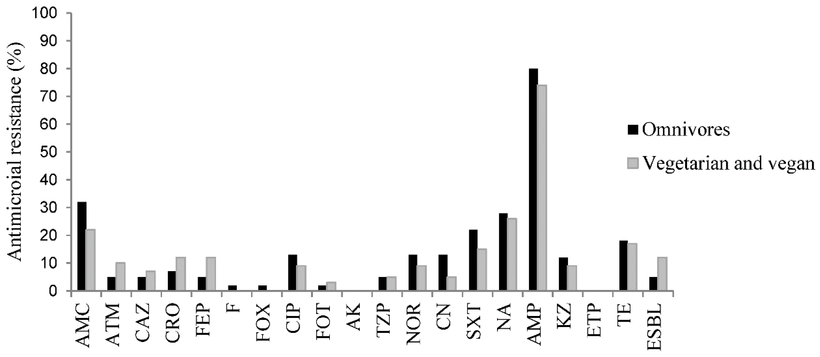
3.1. Antibiotic Resistance

Antimicrobial resistance in E. coli has been reported all over the world. Studies show that the occurrence and susceptibility profile have significant differences that vary according to population, environment and geographic location [23]. In our study, there was no statistically significant difference concerning the antimicrobial resistance profile between groups. However, vegetarians/vegans had a great number of isolates resistant to beta-lactams when compared to omnivores. Isolates from both groups exhibited a high rate of resistance to ampicillin (omnivores 80% and vegetarians/vegans 69%), amoxicillin-clavulanic acid (omnivores 32% and vegetarians/vegans 22%) and nalidixic acid (omnivores 28% and vegetarians/vegans 26%), and some of them were ESBL-positive (7–12% of isolates from vegetarians/vegans and 3–5% of isolates from omnivores).
The antimicrobial resistance profile is shown in Figure 1.
Among the resistance mechanisms of ESBL-producing bacteria, the CTX-M enzyme is one of the most common and important β-lactamases described. Herein, we found that 70% of the analyzed isolates were CTX-M-9-positive, being the most prevalent in both groups (67% omnivores and 71% vegetarians/vegans). All results of CTX-M ESBLs are shown in Table 5.

3.2. Virulence-Associated Genes

The most predominant VAGs among isolates from omnivores were fimH (70%), iutA (32%), fyuA (32%) and traT (32%), while among isolates from vegetarians/vegans, the most predominant were traT (62%), kpsMT k1 (28%) and iutA (22%).
Moreover, there was a statistically significant difference between control and study groups in relation to isolates that were kpsMT III-positive. This gene was detected in nine isolates from vegetarians/vegans and it was not detected in isolates from omnivores. In addition, 42 isolates from omnivores were fimH-positive, while only seven isolates from vegetarians/vegans presented this gene. Regarding cnf1, which codes for a necrotizing cytotoxic factor, it was detected in four isolates from vegetarians/vegans and it was not detected in isolates from omnivores. Finally, 36 isolates from vegetarians/vegans were traT-positive, while this gene was detected in 19 isolates from omnivores.
The results for the VAGS are described in Table 6.

3.3. PAIs

Most isolates from omnivores (55%) were PAI I536-positive(α-hemolysin, CS12 fimbriae and F17-like fimbriae adhesin) [21,24], while most isolates from vegetarians/vegans (59%) were PAI IV536-positive (yersiniabactin siderophore system). Moreover, seven (12%) isolates from omnivores were positive for PAI III536 (S-fimbriae, salmochelin, HmuR-like heme receptor, Sat toxin, Tsh-like hemoglobin protease, antigen 43), which was not detected in isolates from vegetarians/vegans.
Data related to the prevalence of PAIs of isolates are shown in Table 7.

3.4. Phylogeny

Phylogenetic group A, which contains most of the commensal non-pathogenic isolates that survive in the gastrointestinal system, was the most prevalent in both groups, and no statistically significant difference was found between them.
Data related to the phylogenetic classification of isolates are shown in Table 8.

4. Discussion and Conclusions

This study evaluated the virulence and antibiotic resistance profile in E. coli from feces of individuals following vegan, vegetarian and omnivorous diets. Isolates from both groups, vegetarians/vegans and omnivores, harbored genes related to ESBLs and exhibited a high rate of resistance to first-choice antimicrobials used in the treatment of UTIs.
In both groups, most of ESBL-positive isolates were positive for CTX-M-9 (two isolates among omnivores and five among vegetarians/vegans). Presently, CTX-M ESBLs include more than 220 different enzymes clustered into five subfamilies (CTX-M-1, CTX-M-2, CTX-M-8, CTX-M-9 and CTX-M-25) based on their amino acid identities, and enzymes that originated from subfamilies, such as CTXM-1 and CTX-M-9, which are widely distributed and commonly reported [25]. In general, CTX-M enzymes are more active against cefotaxime and ceftriaxone than ceftazidime, but point mutations involving the active site of some enzymes, especially those belonging to the CTX-M-1 and CTX-M-9 subfamilies, have significantly increased their ability to hydrolyze ceftazidime [26].
As we undergo antibiotic treatments many times throughout our lives, bacterial microbiota are exposed to selective pressure by antibiotics. Consequently, the gastrointestinal tract is highly exposed, especially during oral therapy, and this results in a natural selection of resistant isolates carrying an important genetic pool that might be capable of transferring their antibiotic resistance genes to other isolates present in the human intestine. Moreover, resistant food contaminants, such as organic fertilizers, originated from animals and consumed by humans, can also act as a gene pool (donors) of antibiotic resistance genes [8].
In agriculture, organic fertilizers, such as manure, are conventionally used to improve crop yield, since they are believed to be more effective than inorganic fertilizers. However, because of the indiscriminate use of antibiotics and the high spread of antibiotic resistance genes (ARGs), manure is regarded as an important food contaminant, since it may contain microorganisms harboring these ARGs, which pose a high risk to human health [27]. Animal manure is an important reservoir of antibiotic residues and ARGs, in addition to human pathogenic bacteria [28]. Perhaps for this reason, a greater number of ESBL-positive isolates was found among vegetarians and vegans (10) when compared to omnivores (3).
Furthermore, after the report of the first cases of antibiotic-resistant bacterial diseases in humans, recommendations were made for banning the use of antibiotics as growth promoters if the same drugs are also prescribed in human medicine [29]. However, recently, it was estimated that approximately 3,345,022 kg of antimicrobials were sold and used in the U.S. poultry industry in 2016, with 1,265,420 kg being “medically important” for human medical therapy [29,30]. Due to this, there is growing interest in sustainable food production, and research is currently being conducted to identify antibiotic alternatives that could support healthy growth and provide defense against pathogenic microbes [30].
Most E. coli isolates in both groups belong to phylogenetic group A, shared by E. coli isolates commensally inhabiting the mucosa of the gastrointestinal tract [31]. The next phylogenetic groups most found among isolates from vegetarians/vegans and omnivores were B2 and D, respectively. According to Sarowska et al. (2019) [32], B2 and D are often related to extra-intestinal infections. These results are in accordance with previous studies, which show that, despite being pathogenic, phylogenetic group B2 has a high prevalence (≥25%) in fecal E. coli [6,33] and is also related to ExPEC in the human intestinal microbiota.
Interestingly, the groups showed significant differences in relation to the presence of VAGs. Among omnivores, 70% (42) of the isolates harbored fimH, which codes for an adhesin responsible for supporting the binding of bacteria to specific host cells in structural or functional molecules associated with the cell membrane [34]. On the other hand, 62% (36) of isolates from vegetarians/vegans harbored traT, carried by conjugative plasmids, which favors its transmission. In addition, traT is associated with an increased serum resistance, observed mainly in ExPEC present in the bloodstream, and among commensal isolates, traT is a good predictor of urinary tract and bloodstream infections. The fimbriae play a significant role in the process of adhesion of the microorganism to the cells, increasing the virulence of pathogenic E. coli due to this close contact of the bacteria with the host cell wall. Most genes that determine the presence of fimbriae on the surface of bacterial cells are chromosomally encoded or, less frequently, within plasmid DNA, which facilitates their horizontal transfer between different isolates [32,35].
The pathogenicity island most frequent (55%) among isolates from omnivores was PAI I536, while only 12% (7) of isolates from vegetarians and vegans presented it. Remarkably, PAI IV536 was detected in 59% (36) and 38% (23) of isolates from vegetarians/vegans and omnivores, respectively. This PAI, which codes for the fyuA gene (yersiniabactin iron absorption system), is present in up to 57% of E. coli commensal isolates and is also the most frequent in enterobacteria.
Some VAGs were found in only one of the groups, such as PAI III536, found in 12% (7) of the isolates from omnivores, while the kpsMT III gene was detected only among 15% (9) of isolates from vegetarians/vegans. Interestingly, this gene codes for a polysaccharide capsule found mainly in E. coli isolates causing intestinal infections, including EPEC, ETEC and EHEC [36].
Regarding cnf1, it was detected in 7% (4) of isolates from omnivores. This gene codes for a necrotizing cytotoxic factor capable of facilitating bacterial internalization in host cells and contributing to the potential for invasion of pathogenic E. coli by manipulating epithelial and endothelial barriers [37]. It is often associated with extraintestinal conditions, especially urinary tract infections, present in around 40% of UPEC isolates, and less frequently, in bacteremia and neonatal meningitis [32].
A study by Chen et al. (2020) [38] shows that vegetarian diets (compared to a non-vegetarian diet) are associated with a lower risk of UTIs, especially in women, non-smokers and for the uncomplicated UTI subtype. In addition, other previous studies have suggested that meat-related food-based ExPECs may be the main pathogens in uncomplicated UTIs. However, despite the differences found between omnivores and vegans/vegetarians, it is not possible to conclude that one diet is better than the other, since the composition of these diets in terms of nutrients does not seem to be sufficient for them to shape the intestinal microbiota.
The present study also shows that even commensal E. coli isolated from the feces of healthy individuals have great potential to cause extraintestinal infections. This is due not only to the high prevalence of VAGs among these isolates, but also to the presence of mobile genetic elements capable of directly influencing the genomes of pathogenic bacteria and the horizontal transfer of resistance genes, which can spread rapidly among non-pathogenic isolates, transforming them into potential pathogens that can cause UTIs and several other extraintestinal infections.

Supplementary Materials

The following supporting information can be downloaded at: https://www.mdpi.com/article/10.3390/microbiolres13040058/s1.

Author Contributions

E.C.V., Z.N.T. and R.K.T.K. conceived and designed the experiments. A.T.T. and J.G.M.S. performed the experiments. E.C.V., J.G.M.S., V.L.K. and E.C.V. analyzed the data. E.C.V., Z.N.T. and R.K.T.K. contributed reagents and materials. E.C.V. and Z.N.T. reviewed the study. V.L.K. performed confirmation experiments. All authors have read and agreed to the published version of the manuscript.

Funding

The support was provided by the Bill and Melinda Gates Foundation’s Grand Challenges Explorations Brazil—New Approaches to Characterize the Global Burden of Antimicrobial Resistance (number OPP1193112). No funding agencies had any role in this study’s design, collection, analysis, data interpretation and manuscript writing.

Data Availability Statement

Not applicable.

Acknowledgments

We are grateful to the graduate program in clinical and laboratory pathophysiology, to the National Council for Scientific and Technological Development for the master’s scholarship awarded to Ariane Tiemy Tizura, and to Danilo Figueiredo for his help in publicizing the project.

Conflicts of Interest

The authors declare that the research was conducted in the absence of any commercial or financial relationships that could be construed as a potential conflict of interest.

References

  1. Micenková, L.; Beňová, A.; Frankovičová, L.; Bosák, J.; Vrba, M.; Ševčíková, A.; Kmeťová, M.; Šmajs, D. Human Escherichia coli isolates from hemocultures: Septicemia linked to urogenital tract infections is caused by isolates harboring more virulence genes than bacteremia linked to other conditions. Int. J. Med. Microbiol. 2017, 307, 182–189. [Google Scholar] [CrossRef]
  2. Pitout, J.D.D. Extraintestinal pathogenic Escherichia coli: A combination of virulence with antibiotic resistance. Front. Microbiol. 2012, 3, 9. [Google Scholar] [CrossRef] [PubMed] [Green Version]
  3. Köhler, C.-D.; Dobrindt, U. What defines extraintestinal pathogenic Escherichia coli. Int. J. Med. Microbiol. 2011, 301, 642–647. [Google Scholar] [CrossRef] [PubMed]
  4. Raeispour, M.; Ranjbar, R. Antibiotic resistance, virulence factors and genotyping of Uropathogenic Escherichia coli strains. Antimicrob. Resist. Infect. Control 2018, 7, 118. [Google Scholar] [CrossRef] [PubMed]
  5. Daga, A.P.; Koga, V.L.; Soncini, J.G.M.; De Matos, C.M.; Perugini, M.R.E.; Pelisson, M.; Kobayashi, R.; Vespero, E.C. Escherichia coli Bloodstream Infections in Patients at a University Hospital: Virulence Factors and Clinical Characteristics. Front. Cell Infect. Microbiol. 2019, 9, 191. [Google Scholar] [CrossRef]
  6. Starčič Erjavec, M.; Žgur-Bertok, D. Virulence potential for extraintestinal infections among commensal Escherichia coli isolated from healthy humans—The Trojan horse within our gut. FEMS Microbiol. Lett. 2015, 362, fnu061. [Google Scholar] [CrossRef] [PubMed] [Green Version]
  7. Fegan, N.; Gobius, K.S. Pathogenic Escherichia coli and One Health Implications. Curr. Top. Microbiol. Immunol. 2013, 366, 49–62. [Google Scholar] [PubMed]
  8. Schjørring, S.; Krogfelt, K.A. Assessment of bacterial antibiotic resistance transfer in the gut. Int. J. Microbiol. 2011, 2011, 1–10. [Google Scholar] [CrossRef] [Green Version]
  9. Soncini, J.G.M.; Cerdeira, L.; Sano, E.; Koga, V.L.; Tizura, A.T.; Tano, Z.N.; Nakazato, G.; Kobayashi, R.K.T.; Aires, C.A.M.; Lincopan, N.; et al. Genomic insights of high-risk clones of ESBL-producing Escherichia coli isolated from community infections and commercial meat in southern Brazil. Sci. Rep. 2022, 12, 9354. [Google Scholar] [CrossRef]
  10. Lawson, B.; De Pinna, E.; Horton, R.A.; MacGregor, S.K.; John, S.K.; Chantrey, J.; Duff, J.P.; Kirkwood, J.K.; Simpson, V.R.; Robinson, R.A.; et al. Epidemiological Evidence That Garden Birds Are a Source of Human Salmonellosis in England and Wales. PLoS ONE 2014, 9, e88968. [Google Scholar] [CrossRef]
  11. Done, H.Y.; Venkatesan, A.K.; Halden, R.U. Does the Recent Growth of Aquaculture Create Antibiotic Resistance Threats Different from those Associated with Land Animal Production in Agriculture? AAPS J. 2015, 17, 513–524. [Google Scholar] [CrossRef] [Green Version]
  12. Gozi, K.S.; Froes, J.R.; Ajude, L.P.T.D.; Da Silva, C.R.; Baptista, R.S.; Peiró, J.R.; Marinho, M.; Mendes, L.C.N.; Nogueira, M.C.L.; Casella, T. Dissemination of Multidrug-Resistant Commensal Escherichia coli in Feedlot Lambs in Southeastern Brazil. Front. Microbiol. 2019, 10, 1394. [Google Scholar] [CrossRef]
  13. Mulder, M.; Jong, J.C.K.; Goessens, W.H.F.; de Visser, H.; Ikram, M.A.; Verbon, A.; Stricker, B.H. Diet as a risk factor for antimicrobial resistance in community-acquired urinary tract infections in a middle-aged and elderly population: A case–control study. Clin. Microbiol. Infect. 2019, 25, 613–619. [Google Scholar] [CrossRef]
  14. De Filippo, C.; Cavalieri, D.; di Paola, M.; Ramazzotti, M.; Poullet, J.B.; Massart, S.; Collini, S.; Pieraccini, G.; Lionetti, P. Impact of diet in shaping gut microbiota revealed by a comparative study in children from Europe and rural Africa. Proc. Natl. Acad. Sci. USA 2010, 107, 14691–14696. [Google Scholar] [CrossRef] [Green Version]
  15. Marano, R.B.; Fernandes, T.; Manaia, C.M.; Nunes, O.; Morrison, D.; Berendonk, T.U.; Kreuzinger, N.; Tenson, T.; Corno, G.; Fatta-Kassinos, D.; et al. A global multinational survey of cefotaxime-resistant coliforms in urban wastewater treatment plants. Environ. Int. 2020, 144, 106035. [Google Scholar] [CrossRef]
  16. CLSI. Suggested Grouping of US-FDA Approved Antimicrobial Agents That Should Be Considered for Routine Testing and Reporting on Nonfastidious Organisms by Clinical Laboratories, 29th ed.; CLSI Guideline M100-S29; Clinical and Laboratory Institute: Wayne, PA, USA, 2019. [Google Scholar]
  17. Dallenne, C.; Da Costa, A.; Decré, D.; Favier, C.; Arlet, G. Development of a set of multiplex PCR assays for the detection of genes encoding important beta-lactamases in Enterobacteriaceae. J. Antimicrob. Chemother. 2010, 65, 490–495. [Google Scholar] [CrossRef] [Green Version]
  18. Leflon-Guibout, V.; Jurand, C.; Bonacorsi, S.; Espinasse, F.; Guelfi, M.C.; Duportail, F.; Heym, B.; Bingen, E.; Nicolas-Chanoine, M.H. Emergence and spread, of three clonally related virulent isolates of CTX-M-15-producing Escherichia coli with variable resistance to aminoglycosides and tetracycline in a French geriatric hospital. Antimicrob. Agents Chemother. 2004, 48, 3736–3742. [Google Scholar] [CrossRef] [Green Version]
  19. Koga, V.L.; Scandorieiro, S.; Vespero, E.C.; Oba, A.; de Brito, B.G.; de Brito, K.C.T.; Nakazato, G.; Kobayashi, R.K.T. Comparison of antibiotic resistance and virulence factors among Escherichia coli isolated from conventional and free-range poultry. BioMed. Res. Int. 2015, 2015, 618752. [Google Scholar] [CrossRef] [Green Version]
  20. Johnson, J.R.; Stell, A.L. Extended virulence genotypes of Escherichia coli strains from patients with urosepsis in relation to phylogeny and host compromise. J. Infect. Dis. 2000, 181, 261–272. [Google Scholar] [CrossRef] [Green Version]
  21. Sabaté, M.; Moreno, E.; Perez-Berezo, T.; Andreu, A.; Prats, G. Pathogenicity island markers in commensal and Uropathogenic Escherichia coli isolates. Clin. Microbiol. Infect. 2006, 12, 880–886. [Google Scholar] [CrossRef]
  22. Clermont, O.; Christenson, J.K.; Denamur, E.; Gordon, D.M. The Clermont Escherichia coli phylo-typing method revisited: Improvement of specificity and detection of new phylo-groups. Environ. Microbiol. Rep. 2013, 5, 58–65. [Google Scholar] [CrossRef]
  23. Von Baum, H.; Marre, R. Antimicrobial resistance of Escherichia coli and therapeutic implications. Int. J. Med. Microbiol. 2005, 295, 503–511. [Google Scholar] [CrossRef]
  24. Samei, A.; Haghi, F.; Zeighami, H. Distribution of pathogenicity island markers in commensal and uropathogenic Escherichia coli isolates. Folia. Microbiol. 2016, 61, 261–268. [Google Scholar] [CrossRef]
  25. Shurina, B.A.; Page, R.C. Structural Comparisons of Cefotaximase (CTX-M-ase) Sub Family 1. Front. Microbiol. 2021, 12, 688509. [Google Scholar] [CrossRef]
  26. Poirel, L.; Gniadkowski, M.; Nordmann, P. Biochemical analysis of the ceftazidime-hydrolysing extended-spectrum β-lactamase CTX-M-15 and of its structurally related β-lactamase CTX-M-3. J. Antimicrob. Chemother. 2002, 50, 1031–1034. [Google Scholar] [CrossRef] [Green Version]
  27. Naas, T.; Oueslati, S.; Bonnin, R.A.; Dabos, M.L.; Zavala, A.; Dortet, L.; Retailleau, P.; Iorga, B.I. Beta-lactamase database (BLDB) – structure and function. J. Enzym. Inhib. Med. Chem. 2017, 32, 917–919. [Google Scholar] [CrossRef] [Green Version]
  28. Yang, Q.; Zhang, H.; Guo, Y.; Tian, T. Influence of chicken manure fertilization on antibiotic-resistant bacteria in soil and the endophytic bacteria of pakchoi. Int. J. Environ. Res. Public Health 2016, 13, 662. [Google Scholar] [CrossRef]
  29. Yang, Y.; Ashworth, A.; Willett, C.; Cook, K.; Upadhyay, A.; Owens, P.R.; Ricke, S.C.; DeBruyn, J.; Moore, P.A., Jr. Review of Antibiotic Resistance, Ecology, Dissemination, and Mitigation in U. S. Broiler Poult. Syst. 2019, 10, 2639. [Google Scholar]
  30. Sneeringer, S.; MacDonald, J.M.; Key, N.; McBride, W.D.; Mathews, K. Economics of Antibiotic Use in U.S. livestock production. USDA Econ. Res. Rep. 2015. Available online: https://www.ers.usda.gov/webdocs/publications/err200/55529_err200.pdf (accessed on 10 October 2022).
  31. Aslam, N.; Khan, S.-U.; Usman, T.; Ali, T. Phylogenetic genotyping, virulence genes and antimicrobial susceptibility of Escherichia coli isolates from cases of bovine mastitis. J. Dairy Res. 2021, 88, 78–79. [Google Scholar] [CrossRef]
  32. Sarowska, J.; Futoma-Koloch, B.; Jama-Kmiecik, A.; Frej-Madrzak, M.; Ksiazczyk, M.; Bugla-Ploskonska, G.; Choroszy-Krol, I. Virulence factors, prevalence and potential transmission of extraintestinal pathogenic Escherichia coli isolated from different sources: Recent reports. Gut Pathog. 2019, 11, 10. [Google Scholar] [CrossRef] [Green Version]
  33. Nowrouzian, F.; Hesselmar, B.; Saalman, R.; Strannegård, I.; Åberg, N.; Wold, A.E.; Adlerberth, I. Escherichia coli in infants’ intestinal microflora: Colonization rate, strain turnover, and virulence gene carriage. Pediatr. Res. 2003, 54, 8–14. [Google Scholar] [CrossRef]
  34. Kline, K.A.; Fälker, S.; Dahlberg, S.; Normark, S.; Henriques-Normark, B. Bacterial Adhesins in Host-Microbe Interactions. Cell Host Microbe 2009, 5, 580–592. [Google Scholar] [CrossRef] [PubMed] [Green Version]
  35. Mjailovic, H.; Smith, S.G. Bacterial self-defence: How Escherichia coli evades serum killing. FEMS Microbiol. Lett. 2014, 354, 1–9. [Google Scholar] [CrossRef] [Green Version]
  36. Whitfield, C. Structure and Assembly of Escherichia coli Capsules. EcoSal Plus 2009, 354, 1–9. [Google Scholar] [CrossRef]
  37. Knust, Z.; Schmidt, G. Cytotoxic Necrotizing Factors (CNFs)−A Growing Toxin Family. Toxins 2010, 2, 116–127. [Google Scholar] [CrossRef] [Green Version]
  38. Chen, Y.-C.; Chang, C.-C.; Chiu, T.H.T.; Lin, M.-N.; Lin, C.-L. The risk of urinary tract infection in vegetarians and non-vegetarians: A prospective study. Sci. Rep. 2020, 10, 906. [Google Scholar] [CrossRef]
Figure 1. Percentage of antimicrobial resistance profile of E. coli isolated from omnivores and vegetarians/vegans. Amoxicillin-clavulanic acid (AMC), Aztreonam (ATM), Ceftazidime (CAZ), Ceftriaxone (CRO), Cefepime (FEP), Nitrofurantoin (F), Cefoxitin (FOX), Ciprofloxacin (CIP), Cefotaxime (FOT), Amikacin (AK), Piperacilin-tazobactam (TZP), Norfloxacin (NOR), Gentamicin (CN), Trimethoprim-sulfamethoxazole (SXT), Nalidixic acid (NA), Ampicilin (AMP), Cefazolin (KZ), Ertapenem (ETP), Tetracycline (TE) and Extended-spectrum β-Lactamase (ESBL).
Microbiolres 13 00058 g001
Table 1. Genes encoding CTX-M ESBL enzymes.
EnzymesGenesPrimer Sequences (5′–3′)Amplion Size (bp)References
CTX-M-1blaCTX-M1TTAGGAARTGTGCCGCTGYA
CGATATCGTTGGTGGTRCCAT
688Dallene et al., 2010 [17]
CTX-M-2blaCTX-M2CGTTAACGGCACGATGAC
CGATATCGTTGGTGGTRCCAT
404Dallene et al., 2010 [17]
CTX-M-8/25blaCTX-M8AACRCRCAGACGCTCTAC
TCGAGCCGGAASGTGTYAT
326Dallene et al., 2010 [17]
CTX-M-9blaCTX-M9TCAAGCCTGCCGATCTGGT
TGATTCTCGCCGCTGAAG
561Dallene et al., 2010 [17]
CTX-M-15blaCTX-M15ATA AAA CCG GCA GCG GTGGAA TTT TGA CGA TCG GGG483Leflon-Guibout et al., 2004 [18]
Table 2. Virulence-associated genes.
GenesPrimer Sequences (5′–3′)VAGsAmplion Size (bp)References
kpsMTIIGCG CAT TTG CTG ATA CTG TTGGroup 2 of capsular antigens 272Johnson and Stell, 2000 [20]
CAT CCA GAC GAT AAG CAT GAC CA
KpsMTIIITCC TCTT GCT ACT ATT CCC CCTGroup 3 of capsular antigens 392Johnson and Stell, 2000 [20]
AGG CGT ATC CAT CCC TCC TAA C
KpsMTk1TAG CAA ACG TTC TAT TGG TGCK1 capsule153Johnson and Stell, 2000 [20]
CAT CCA GAC GAT AAG CAT GAC CA
kpsMTk5CAG TAT CAG CAA TCG TTC TGT AK5 capsule159Johnson and Stell, 2000 [20]
CAT CCA GAC GAT AAG CAT GAC CA
cvaCCAC ACA CAA ACG GGA GCT GTTColicin V680Johnson and Stell, 2000 [20]
CTT CCC GCA GCA TAG TTC CAT
iutAGGC TGG ACA TCA TGG GAA CTG GAerobactin
siderophore receptor
300Johnson and Stell, 2000 [20]
CGT CGG GAA CGG GTA GAA TCG
fimHTGC AGA ACG GAT AAG CCG TGGFimbriae type 1508Johnson and Stell, 2000 [20]
GCA GTC ACC TGC CC TCC GGT A
fyuATGA TTA ACC CCG CGA CGG AAYersiniobactinsiderophore receptor880Johnson and Stell, 2000 [20]
CGC AGT AGG CAC GAT CTT GTA
papCGAC GGC TGT ACT GCA GGG TGT GGC GP Fimbriae 328Johnson and Stell, 2000 [20]
ATA TCC TTT CTG CAG GCA GGG TGT GGC
papGCTG TAA TTA CGG AAG TGA TTT CTGP Fimbriae1070Johnson and Stell, 2000 [20]
CTG TAA TTA CGG AAG TGA TTT CTG
sfaACTC CGG AGA ACT GGG TGC ATC TTA CSfa fimbriae410Johnson and Stell, 2000 [20]
CGG AGG AGT AAT TAC AAA CCT GGC A
sfaSGTG GAT ACG ACG ATT ACT GTGSfa fimbriae240Johnson and Stell, 2000 [20]
CCG CCA GCA TTC CCT GTA TTC
afaGGC AGA GGG CCG GCA ACA GGCAfa fimbriae750Johnson and Stell, 2000 [20]
CCC GTA ACG CGA CAG CAT CTC
ibeAAGG CAG GTG TGC GCC GCG TACInvasion of brain endothelium170Johnson and Stell, 2000 [20]
TGG TGC TCC GGC AAA CCA TGC
HlyAAC AAG GAT AAG CAC TGT TCT GGCHemolysin1177Johnson and Stell, 2000 [20]
ACC ATA TAA GCG GTC ATT CCC GTC
cnf1AGG AAG TTA TAT TTC CGT AGGCytotoxic necrotizing factor 1498Johnson and Stell, 2000 [20]
GTA TTT GCC TGA ACC GTA A
cnf2AAT CTA ATT AAA GAG AACCytotoxic necrotizing factor 2543Johnson and Stell, 2000 [20]
CAT GCT TTG TAT ATC TA
traTGGT GTG GTG CGA TGA GCA CAGSerum resistance 290Johnson and Stell, 2000 [20]
GGT GTG GTG CGA TGA GAC CAG
iroNAAT CCG GCA AAG AGA CGA ACC GCC TSalmochelinsiderophore receptor553Johnson and Stell, 2000 [20]
GTT CGG GCA ACC CCT GCT TTG ACT TT
ompTTCA TCC CGG AAG CCT CCC TCA CTA CTA TEpisomal outer membrane protease496Johnson and Stell, 2000 [20]
TAG CGT TTG CTG CAC TGG CTT CTG ATA C
hlyFGGC CAC AGT CGT TTA GGG TGC TTA CCPutative avian hemolysin450Johnson and Stell, 2000 [20]
GGC GGT TTA GGC ATT CCG ATA CTC AG
IssCAG CAA CCC GAA CCA CTT GAT GEpisomal increased serum survival323Johnson and Stell, 2000 [20]
AGC ATT GCC AGA GCG GCA GAA
Table 3. Genes encoding PAI markers.
PAIPrimersSequences (5′–3′)Amplicon Size (bp)References
PAI I536I.9TAA TGC CGG AGA TTC ATT GTC1800Koga et al., 2014 [19]
I.10AGG ATT TGT CTC AGG GCT TT
PAI II536orf1upCAT GTC CAA AGC TCG AGC C1000Sabaté et al., 2006 [21]
orf1downCTA CGT CAG GCT GGC TTT G
PAI III536sfaAI.1CGG GCA TGC ATC AAT TAT CTT TG161Sabaté et al., 2006 [21]
sfaAI.2TGT GTA GAT GCA GTC ACT CCG
PAI IV536IRP2 FPAAG GAT TCG CTG TTA CCG GAC300Sabaté et al., 2006 [21]
IRP2 RPTCG GGC AGC GTT TCT TCT
PAI ICFT073RPAiGGA CAT CCT GTT ACA GCG CGC A930Sabaté et al., 2006 [21]
RPAfTCG CCA ATC ACA GC GAA C
PAI IICFT073cft073.2Ent1ATG GAT GTT GTA TCG CGC400Sabaté et al., 2006 [21]
cft073.2Ent2ACG AGC ATG TGG ATC TGC
PAI IJ96papGIfTCG TGC TCA GGT CCG GAA TTT400Sabaté et al., 2006 [21]
papGIrTGG CAT CCC ACA TTA TCG
PAI IIJ96hlydGGA TCC ATG AAA ACA TGG TTA ATG GG2300Sabaté et al., 2006 [21]
cnfGAT ATT TTT GTT GCC ATT GGT TAC C
Table 4. Genes researched for phylogenetic classification.
PCR GenesPrimer Sequences (5′–3′)Amplicon Size (bp)References
MultiplexchuAGAC GAA CCA ACG GTC AGG AT279Clermont et al., 2013 [22]
TGC CGC CAG TAC CAA AGA CA
yjaATGA AGT GTC AGG AGA CGC TG211
ATG GAG AAT GCG TTC CTC AAC
TSPE4.C2GAG TAA TGT CGG GGC ATT CA152
CGC GCC AAC AAA GTA TTA CG
E grouparpAGAT TCC ATC TTG TCA AAA TAT GCC219Clermont et al., 2013 [22]
GAA AAG AAA AAG AAT TCC CAA GAG
C grouptrpAAGT TTT ATG CCC AGT GCG AG489Clermont et al., 2013 [22]
TCT GCG CCG GTC ACG CCC
Table 5. Prevalence of CTX-M ESBLs among E. coli isolated from omnivores and vegetarians/vegans.
GenesOmnivores
n (%)
Vegetarians/Vegans
n (%)
CTX-M-1-4 (57)
CTX-M-21 (33)3 (43)
CTX-M-8/25--
CTX-M-92 (67)5 (71)
CTX-M-15-2 (29)
Total of isolates310
Table 6. Prevalence of VAGs among E. coli isolated from omnivores and vegetarians/vegans.
Virulence-Associated GenesCoding ForOmnivores
n = 60 (%)
Vegetarians
Vegans
n = 58 (%)
p-Value
kpsMT IIGroup 2 of capsular antigens15 (25)10 (17)0.349
kpsMT IIIGroup 3 of capsular antigens09 (15)0.001 *
kpsMT k1K1 capsule9 (15)16 (28)0.078
kpsMT k5K5 capsule14 (23)8 (14)0.214
cvaCColicin V3 (5)7 (12)0.152
iutAAerobactinsiderophore receptor19 (32)13 (22)0.308
fimHFimbriae type 142 (70)7 (12)<0.001 *
fyuAYersiniobactinsiderophore receptor19 (32)11 (19)0.206
papCP Fimbriae7 (12)7 (12)0.894
papGP Fimbriae2 (3)2 (3)0.946
sfaASfa fimbriae6 (10)4 (7)0.582
sfaSSfa fimbriae4 (7)2 (3)0.451
afaAfa fimbriae2 (3)2 (3)0.946
ibeAInvasion of brain endothelium5 (8)3 (5)0.526
hlyHemolysin4 (7)8 (14)0.180
cnf1Cytotoxic necrotizing factor 104 (7)0.035 *
cnf2Cytotoxic necrotizing factor 23 (5)4 (7)0.631
traTSerum resistance19 (32)36 (62)0.001 *
iroNSalmochelinsiderophore receptor8 (13)9 (15)0.682
ompTEpisomal outer membrane protease4 (7)9 (15)0.110
hlyFPutative avian hemolysin2 (3)6 (10)0.118
IssEpisomal increased serum survival12 (203 (5)0.019 *
* indicates significant difference.
Table 7. Prevalence of PAIs among E. coli isolated from omnivores and vegetarians/vegans.
GenesOmnivores
n = 60 (%)
Vegetarians/Vegans
n = 58 (%)
p-Value
PAI
PAI I53633 (55)7 (12)<0.001 *
PAI II53600
PAI III5367 (12)00.008 *
PAI IV53623 (38)34 (59)0.018 *
PAI ICFT0736 (10)8 (14)0.483
PAI IICFT07311 (18)9 (15)0.744
PAI IJ9603 (5)0.070
PAI IIJ963 (5)00.090
* indicates significant difference.
Table 8. Phylogenetic classification among E. coli isolated from omnivores and vegetarians/vegans.
GenesOmnivores
n = 60 (%)
Vegetarians/Vegans
n = 58 (%)
p-Value
Phylogenetic Classification
A15 (25)13 (22)0.818
B18 (13)10 (17)0.506
B24 (7)11 (19)0.079
C2 (3)2 (3)0.703
D12 (20)8 (14)0.414
E9 (15)10 (17)0.506
F10 (17)4 (7)0.073
Publisher’s Note: MDPI stays neutral with regard to jurisdictional claims in published maps and institutional affiliations.

