Article

Survey of Fungal Foliar and Panicle Diseases in Smallholder Sorghum Cropping Systems in Different Agro-Ecologies of Lower Eastern Kenya

by
Irene Njeri Koima
1,2,*,
Dora Chao Kilalo
1,
Charles O. Orek
2,
John Maina Wagacha
3 and
Evans N. Nyaboga
4
1
Department of Plant Science and Crop Protection, University Nairobi, P.O. Box 29053, Nairobi 00625, Kenya
2
Department of Agricultural Sciences, South Eastern Kenya University, P.O. Box 170, Kitui 90200, Kenya
3
Department of Biology, University Nairobi, P.O. Box 30197, Nairobi 00100, Kenya
4
Department of Biochemistry, University Nairobi, P.O. Box 30197, Nairobi 00100, Kenya
*
Author to whom correspondence should be addressed.
Microbiol. Res. 2022, 13(4), 765-787; https://doi.org/10.3390/microbiolres13040055
Submission received: 20 August 2022 / Revised: 12 September 2022 / Accepted: 19 September 2022 / Published: 7 October 2022

Abstract

:
Sorghum is a staple food crop and plays a critical role in subsistence farming in Kenya due to its adaptability to marginal agro-ecological zones. However, fungal diseases are among the major biotic constraints of sorghum production, causing over 70% yield loss in susceptible cultivars. Information on the distribution and severity of fungal diseases is important to establish efficient and improved strategies for integrated disease management of sorghum fungal diseases. The aim of this study was to determine the prevalence, incidence, severity and spatial distribution of fungal diseases on sorghum across agro-ecological zones of lower eastern Kenya. A total of 384 smallholder farmers’ fields were surveyed, and in each field, 30 plants were assessed for prevalence and incidence of fungal diseases using a W-shaped pattern to cover the whole field. Sorghum anthracnose was the most prevalent disease (71%), followed by leaf blight (70.18%), rust (68.41%), smut (63.02%), sorghum mildew (55.33%), Alternaria leaf spot (48.39%) and rough leaf spot (46.02%). Disease prevalence, incidence and severity varied among the investigated agro-ecological zones. There was a significant difference (p ≤ 0.05) in fungal disease severity across the investigated agro-ecological zones. Spatially interpolated disease maps showed a high variation in the distribution of various sorghum fungal diseases across the investigated agro-ecological zones of lower eastern Kenya. Morpho-cultural identification revealed the association of Colletotrichum sublineola with anthracnose, Curvularia lunata and Bipolaris cynodontis with leaf blight, Puccinia purpurea with rust, Peronosclerospora sorghi with downy mildew, Alternaria alternata with Alternaria leaf spot, Ascochyta sorghi with rough leaf spot and Sporisorium sorghi with covered kernel smut symptoms. Information obtained in this study will be useful to update knowledge on sorghum fungal diseases and provide a basis for the development of strategies for management and control of the investigated diseases.

1. Introduction

Sorghum (Sorghum bicolor L.) is considered the most valuable cereal food crop in semi-arid zones of sub-Saharan Africa [1]. Typically, it is referred to as a crop of hot, semi-arid environments, potentially ensuring food security in East Africa [2]. Globally, it is ranked fifth among the primary staple cereals after maize, wheat, rice and barley, and in East Africa it is second after maize [3]. Sorghum is cultivated for feed, food and forage and has considerable potential in the biofuelindustry [4]. Despite its significant contribution to food security, a global decline in sorghum production and productivity has been reported [5]. In Kenya, a reduction in sorghum production and productivity has been observed, falling from 189,000 MT/ha in 2015 to 150,000MT/ha in 2019, with a stagnant growth rate of 0.8 MT/ha [5]. The decline in sorghum production is due to perennial biotic challenges, including pests, such as nematodes, and diseases caused by fungi, bacteria and viruses [6].
Fungal pathogens cause important sorghum diseases that cause considerable grain losses [6]. Diseases that exhibit cosmopolitan traits include leaf spots (Gloecerospora sorghi and Ramulispora sorghi) [7] and leaf blight (Exserohilum turcicum and Bipolaris maydis). Other diseases, such as anthracnose (Colletotrichum sublineola), ergot (Claviceps africana and Claviceps sorghi) and downy mildew (Peronosclerospora sorghi), are considered acute and result in severe grain losses [8]. Other fungal diseases, such as charcoal rot (Macrophomina phaseolina), grain mold and Fusarium stalk rot, cause yearly drag loss of both sorghum grain and forage yield [6]. The use of susceptible landrace cultivars has been reported as an important factor responsible for the observed fungal disease incidence in East Africa [9]. In Kenya, anthracnose, smuts, leaf blight, ladder leaf spot, zonate leaf spot, grey leaf spot, oval leaf spot, among others, are considered the most destructive diseases affecting sorghum in western and upper eastern Kenya [10,11,12]. However, information on the occurrence, severity and distribution of sorghum fungal diseases in other regions of Kenya is not available.
Disease surveys play an important role in the development of integrated disease management strategies. An effective disease management strategy depends on adequate information on the prevalence, distribution and severity of the disease in various cropping systems and agro-ecologies. The only survey on the occurrence of sorghum fungal diseases was reported two decades ago [10] and confirmed the presence of fourteen foliar and six panicle fungal diseases infecting sorghum in western Kenya. However, recent reports on the occurrence on individual diseases focused on turcicum leaf blight [11] and covered kernel smut [12], probably due to their economic importance with respect to sorghum production. However, there is a knowledge gap with respect to the occurrence and severity of sorghum fungal diseases in the dry and low humid agro-ecological zones of lower eastern Kenya. In addition, the incidence of sorghum fungal diseases may vary considerably depending on the geographical location. Therefore, in the present study, we aimed to assess the occurrence, severity and spatial distribution of sorghum fungal diseases in different agro-ecological zones of lower eastern Kenya. Information obtained in this study will be useful to update knowledge on sorghum fungal diseases and provide an important basis for effective integrated management and control of the investigated diseases. This study also will contribute to the collection of phytopathogenic fungi infecting sorghum for further analysis.

2. Materials and Methods

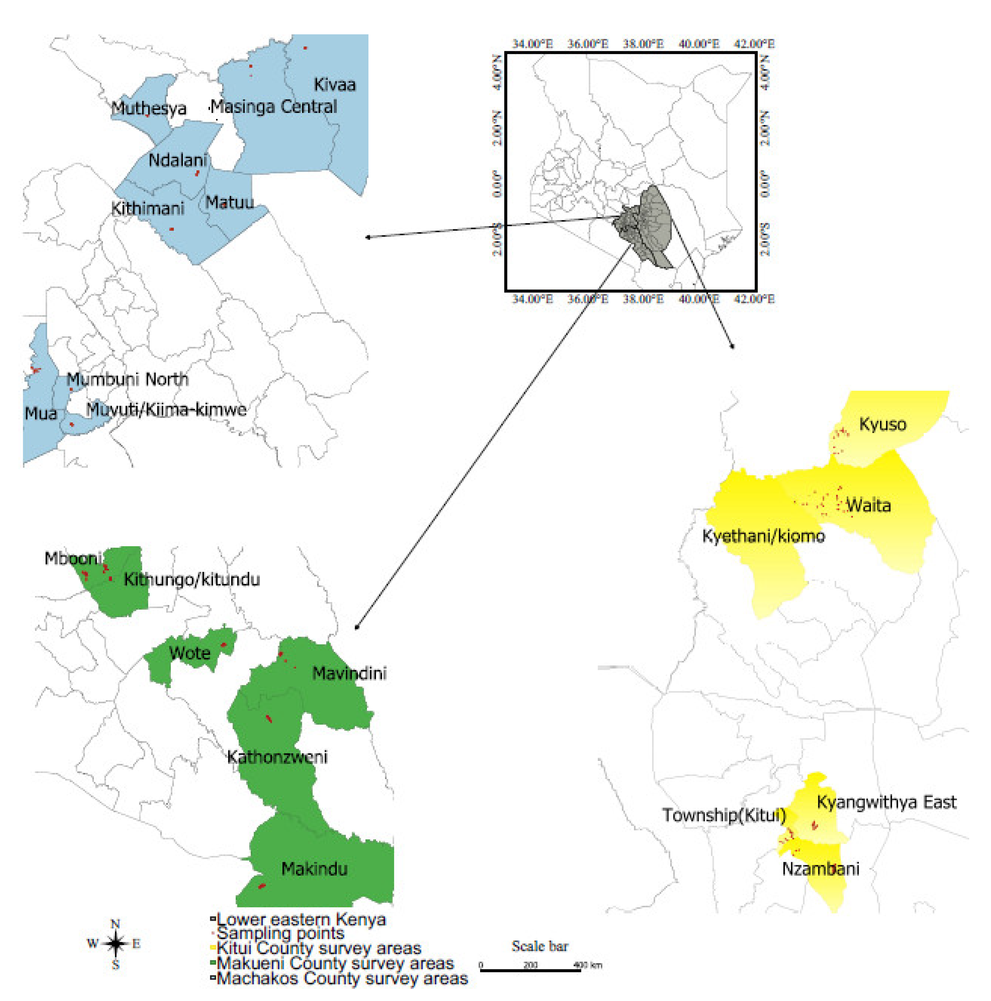
2.1. Description of the Study Areas and Selection of Sites

Disease surveys were conducted between May and September 2019 in smallholder sorghum famers’ fields in three counties, namely Kitui, Machakos and Makueni in lower eastern Kenya (Figure 1). Lower eastern Kenya lies between 37° and 38° longitude and between 1° and 3° latitude. The surveyed sites were located in Mwingi, central Kitui and southern Kitui in Kitui County; Machakos town, Masinga and Yatta in Machakos County; and Makueni town, Mbooni and western Kibwezi in Makueni County. The disease surveys were scheduled to coincide with crop maturity, as described by Ngugi et al. [10]. The surveyed sites were selected among sorghum growing locations across five (5) agro-ecological zones (Table 1), as described by Jactzold and Schmidt [13]. GPS coordinates for each surveyed field were recorded and plotted on a map of Kenya using Quantum GIS software.

2.2. Assessment of Prevalence, Incidence and Severity of Sorghum Fungal Diseases

Purposive sampling was implemented to select sorghum fields to assess the prevalence, incidence and severity of fungal diseases. Based on sample size calculation using the formula developed by Cochran [14], a total of 384 smallholder farmers’ fields were surveyed in the three counties.
Approximately 10 to 15 smallholder sorghum farms were sampled in each agro-ecological zone. The sampling procedure involved following main roads and the accessible routes of selected sorghum growing areas, and stops were made in intervals of approximately 0.5 to 1 km [10]. Sorghum fields of approximately 0.5 ha were sampled only if the sorghum plant was in milk stage or in later, when many fungal diseases are fully developed and symptomatic [6]. In each field, 30 plants were assessed for disease prevalence and incidence using a W pattern to cover the whole field/site.

2.3. Field Disease Assessment

Sorghum plants were assessed and rated for anthracnose by adopting a scale described by Prom et al. [15]. Disease was characterized by small red spots on both surfaces of the leaf blade. The centers of the spots were white and encircled by purple, red or brown margin. Numerous small black dots, such asacervuli, were present on the white surface of the necrotic lesions. There were circular cankers in the inflorescence, which showed characteristics of red rot.
Plants were assessed and rated for leaf blight disease using the scale developed by Beshir [16]. Plants showing symptoms were characterized by ovalwater-soaked spots on the leaves that grew into elongated, spindle-shaped necrotic lesions, leading to a complete blighting of foliage.
Plants were assessed and rated for Alternaria leaf spot disease using a disease rating scale developed by Prom et al. [17]. Plants with characteristic symptoms of Alternaria leaf spot showed circular to irregular, brown to dark brown spots of 1–2 mm in diameter, with concentric rings of alternate light and dark bands and a center denoted by a light-colored spot [18].
The disease symptoms associated with downy mildew in sorghum were assessed and rated using a scale described by Prom et al.[19]. Downy mildew symptoms involves white fluffy lesion on the underside of the leaves, shredding of infected leaves, greenish or yellow stripes along the leaf blade and severe chlorosis of the leaves [19].
Plants showing rust symptoms were assessed and rated using the scale developed by Prom et al. [15]. Rust symptoms were characterized by small red or tan flecks on lower leaves and the presence of pustules on both sides of the leaf surface. The pustules ruptured to produce a reddish, powdery mass of uredospores and teliospores [6].
Plants were assessed and rated for rough leaf spot disease using a disease rating scale described by Prom et al. [17]. Plants with characteristic symptoms of rough leaf spot had small red lesions on the upper leaf surface, in addition to the presence of long elliptical necrotic lesion with hard, black specks at the center of the lesion [20].
Covered smut disease was rated using the panicle disease rating scale developed by Prom et al. [21].The assessed sorghum plants showed smut symptoms that included a large, thick white–grey smut gall enclosed membrane on the young head. When the enclosed smut gall was ruptured, a mass of dark brown to black powdery teliospores intermingled with a network of long, thin, black broom-like filaments of vascular tissues was exposed [22].

2.4. Assessment of Prevalence, Disease Incidence and Severity

Disease prevalence and incidence were calculated using a method described by Cooke [23].
Disease   prevalence = ( No .   of   fields   with   the   disease / Total   number   of   fields   sur - veyed )     × 100 Disease   incidence = ( No .   of   plants   showing   symptoms / Total   number   of   plants   as - sessed )   ×   100
Foliar disease severity was estimated as the proportion of the area of the leaf infection on 30 sorghum plants selected randomly within each field. Each sorghum plant was divided to three portions—upper, middle and lower sections—and the average scores of the three portions represented the severity of foliar disease per plant [10]. A severity scale of 1 to 5 was used [15], where 1 = 0%, i.e., healthy leaves; 2 = 1–25%, i.e., no acervulus formation, no spread to other leaves and the presence of red spots on infected leaves; 3 = 26–50%, i.e., lesions present with the formation of small acervuli at the center of leaves located up to a third of the height of plant from the bottom; 4 = 51–75%, i.e., necrotic lesions consisting of acervuli on all leaves, except the flag leaf; and 5 = 76–100%, i.e., severe necrotic lesions comprising abundant acervuli covering the entire sorghum plant.
Symptomatic leaf samples were collected from 3–4 randomly selected plants. The collected leaf samples were placed in Ziplock bags, transferred to the laboratory and immediately processed.
Data on sorghum varieties grown by smallholder farmers in lower eastern Kenya, the source of seeds for planting and the preferred traits required by farmers were also collected. Furthermore, information on the cropping system and measures used to manage and control sorghum fungal diseases was collected.

2.5. Collection of Diseased Sorghum Samples and Isolation of Associated Fungi

A total of 108 symptomatic leaf samples were collected from different villages and used for isolation and identification of the causal agents of sorghum fungal diseases in lower eastern Kenya.
Fungal pathogens were isolated following the procedure developed by Photita et al. [24]. Briefly, symptomatic leaf samples were cut into pieces of about 1 cm2 with a sterile surgical blade and surfaced-sterilized in 1.3% sodium hypochlorite for three minutes, followed by rinsing three times with sterile water. Then, samples were allowed to dry under a laminar flow hood. After drying, the samples were placed onto Petri plates containing potato dextrose agar (PDA) amended with the antibiotic chloramphenicol (323.1 g/mol) and incubated at 25 ± 2 °C for 7 days. Pure cultures were prepared by transferring mycelia of about 10 mm from 7-day-old grown fungal culture to fresh PDA media in Petri plates and incubated for 7 days at 25 ± 2 °C.

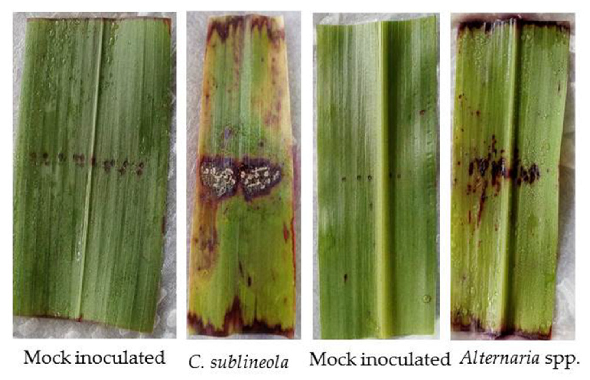
2.6. Pathogenicity of the Fungal Isolates

A Koch’s postulates test for pathogenicity of the fungal isolates was conducted on detached leaves of susceptible sorghum cultivar Serena. The leaves were washed under running tap water to remove any debris and surface-sterilized with 1.3% sodium hypochlorite for three minutes, followed by rinsing three times with sterile water. The leaves were cut and placed in a 90 mm diameter Petri dishes lined with two layers of sterilized and moist blotting papers. The leaves in the Petri plates were inoculated with 1 × 10⁵ mL concentration of spore suspension prepared from each isolate and left at 25 ± 2 °C for 7 days [25]. After 10 days, the fungal pathogens were re-isolated from the parts of the leaves showing symptoms, and the characteristics of the isolates were compared to those of the original cultures [25].

2.7. Morpho-Cultural Identification of Pathogenic Fungal Isolates

Fungal isolates were identified by microscopic examination following the taxonomic keys described by Sutton [26]. Morpho-cultural characteristics were recorded, including upper and reverse surface appearance of colony, shape, color, texture, acervulus and sclerotia.
Mycelia discs of approximately 5 mm were aseptically placed into Petri plates containing PDA and stored at 25 ± 2 °C for 7 days. The 7-day-old fungal cultures were flooded with sterile water and scraped to suspend the spores. Suspended spores were filtered through a cheese cloth to remove mycelia, and the spore suspension was diluted to a 10−6concentration [27]. A drop of 10−6suspension was deposited on a microscope slide and covered with a cover slip [27]. Spores were stained with lactophenol cotton blue and observed under a compound microscope. The shapes of the spores were recorded and observed for the presence or absence of septa in order to determine the taxonomic class of the isolate [26].
The procedure described by Hasanuddin [28] was used to confirm the causal agents of rough leaf spot and covered kernel smut diseases. Briefly, using a dissecting microscope, the surface of the leaves with lesions was examined for the presence of mycelia or spores. Spores were carefully removed using a scalpel and placed on a slide containing a drop of water, then covered with a cover slip and observed under a compound microscope [28]. Shapes of the spores were recorded and observed for the presence or absence of septa in order to determine the taxonomic class of the isolates [26].

2.8. Data Analysis

The data collected on fungal disease incidence, prevalence and severity were analyzed. Data analyses on mean disease incidence and severity were performed using Statistical Analysis System software version 9.2 [29]. Disease severity frequency curves were constructed to determine the relationship between each fungal disease and agro-ecological zones. Means were separated by least significant difference (LSD) at p ≤ 0.05. Arch GIS software version 3.2a was used to generate fungal disease spatial distribution maps of lower eastern Kenya. The deterministic spatial interpolation method (inverse distance weighting) was used in the analysis, and the surface was interpolated using GPS coordinate points and the associated field disease incidence data. A power of 2.00 coefficient interval was used to control for the influence of surrounding points [30].

3. Results

3.1. Occurrence of Fungal Diseases in the Field

Fungal disease symptoms were observed in all surveyed fields (Figure 2). The survey indicated that sorghum plants in farmers’ fields in lower eastern Kenya were infected with six foliar fungal diseases: anthracnose, leaf blight, rust, downy mildew, Alternaria leaf spot and rough leaf spot (Figure 2). The prevalence of the observed foliar fungal diseases, in decreasing order, were: sorghum anthracnose, with 71% prevalence, followed by sorghum leaf blight (70%), rust (68%), downy mildew (55%), Alternaria leaf spot (48%) and rough leaf spot (46%). Covered kernel smut was the major panicle disease observed, with 63% prevalence (Table 2).
A significant difference (p ≤ 0.05) in diseases prevalence was observed between the agro-ecological zones of lower eastern Kenya (Table 2). For instance, sorghum anthracnose prevalence in UM3/UM4 was 79%, which was significantly higher than the 59% prevalence recorded in LM5. The highest leaf blight prevalence (76%) was reported in UM3/UM4, which was significantly higher (p ≤ 0.05) than the 62% prevalence recorded in LM5. Rust disease was most common in LM3/LM4, with 72% prevalence, which was significantly higher (p ≤ 0.05) than that in UM3/UM4 (65%) (Table 2). Alternaria leaf spot (54%) and rough leaf spot (57%) were also prevalent in LM4 and LM5, significantly (p ≤ 0.05) higher than in UM4 (39%). The prevalence of downy mildew differed significantly (p ≤ 0.05) among the agro-ecological zones, and the disease was significantly moreprevalent in LM3/LM4 (58%) than in UM4 (53%) (Table 2). The prevalence of covered kernel smut was significantly higher in LM3/LM4 (68%) than in LM5 (59%) (p ≤ 0.05) (Table 2).
In terms of disease incidence, significant differences (p ≤ 0.05) were observed among the agro-ecological zones of lower eastern Kenya (Table 3). Sorghum anthracnose disease incidence was significantly higher in LM3/LM4 (77%) than in LM5 (37%). UM4 recorded the highest leaf blight prevalence (75%) which was significantly higher (p ≤ 0.05) than that in LM5 (43%). Rust disease was most common in LM3/LM4 and UM4, with 57% incidence, which was significantly higher (p< 0.05) than that in LM5 (43%) (Table 3).The incidences of Alternaria leaf spot (54%) and rough leaf spot (48%) were high in LM4 and LM5, differing significantly (p ≤ 0.05) from those in UM3/UM4 (33 and 30%, respectively). The incidence of downy mildew differed significantly (p ≤ 0.05) among the investigated agro-ecological zones. LM3/LM4 had the highest downy mildew disease incidence (63%), which was significantly higher than that in LM5 (37%) (Table 3). The incidence of covered kernel smut was significantly higher (p ≤ 0.05) in LM3/LM4 and UM4, with a disease incidence of 53% compared to UM3/UM4, with an incidence of 45% (Table 3).

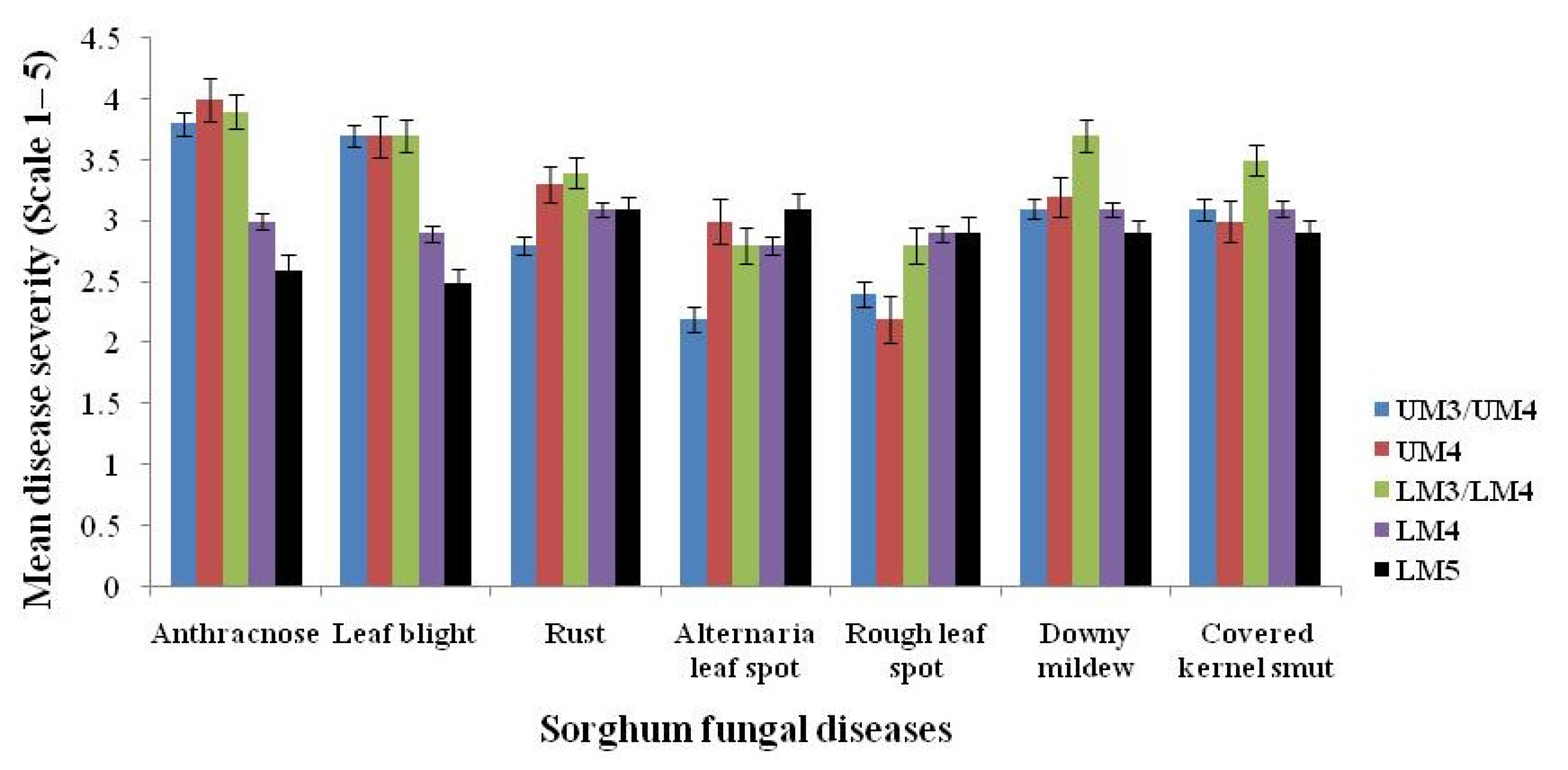
3.2. Fungal Disease Severity across Agro-Ecological Zones

We observed significant differences (p ≤ 0.05) in major fungal disease severity across the surveyed agro-ecological zones (Figure 3). Anthracnose and leaf blight were the most common in diseases in UM3/UM4, UM4 and LM3/LM4, with severity scores of more than 3.5 (70%) and significantly higher (p ≤ 0.001) than in LM5, with a severity score of less than 3 (60%) (Figure 3 and Figure 4)
Both rough leaf spot and Alternaria leaf spot showed a significantly higher (p ≤ 0.001) severity in LM5, with the lowest (p ≤ 0.017) severity in UM3/UM4 (Figure 3). The rust disease severity curve shows that the disease severity is high from average humid zones (UM4, LM3/LM4) to hot lower midland zones (LM4 and LM5) (Figure 4).The rust disease severity was significantly lower (p ≤ 0.002) in more humid zones (UM3/UM4) (Figure 3). The downy mildew disease severity curve was skewed to the left, with a peak at LM3/LM4, indicating that high severity is most common in more humid areas, with lower severity in lowland zones (Figure 4). The downy mildew disease severity of 3.7 recorded in LM3/LM4 was significantly higher (p ≤ 0.0001) compared to the 2.9 recorded in LM5 (Figure 3). The smut disease severity curve shows a similar distribution to that of downy mildew disease, with a peak at LM3/LM4 (Figure 4); however, its severity distribution did not show any significant differences in the other agro-ecological zones (Figure 3).

3.3. Spatial Distribution of Sorghum Fungal Diseases in the Agro-Ecological Zones of Lower Eastern Kenya

Spatially interpolated disease maps illustrated considerable variation in the distribution of different sorghum fungal diseases observed across the agro-ecological zones of lower eastern Kenya (Figure 5). High incidence rates of sorghum anthracnose (>61%) and leaf blight (>60%) were observed in high-humidity areas UM2, UM3, UM4, UM5, LH2, LM3 and LM4 (Figure 5) whereas relatively lower rates (<41%) of distribution were noted in the hot and low humid areas of LM4, LM5, LM6, IL5 and IL6 (Figure 5).
High rust disease incidence rates (>51%) were recorded in all agro-ecological zones of Makueni County, and rust was prevalent in the average humidity areas of UM4, LM4, LM3 and LM5, as well as parts of LM6 (Figure 5). Relatively lower rust incidence rates ranging from 31.8% to 49%were recorded in both high-humidity areas (UM3, UM5, UM6 and UM2) and hot, dry lowland areas (LM5, LM6, IL5 and IL6, as well as parts of LM4) (Figure 5).
Additionally, high incidence rates (>44.46%) of rough leaf spot were recorded in northern and southern parts of Kitui lying in LM4 and LM5, as well as parts of LM6 (Figure 5). The disease was also prevalent in parts of UM4 and UM2 of lower eastern Kenya. Lower rates (<41.16%) of rough leaf spot were observed in UM5, UM6, UM3 and LH2, as well as parts of LM4 in lower eastern Kenya (Figure 5). Alternaria leaf spot had a distribution pattern that is closely related to rough leaf spot and was common (>47%) in LM4, LM5 and LM6 and least common (<44.60%) in the high-humidity areas (UM3, UM4, UM5, UM6 and LH2) (Figure 5).
A high downy mildew disease incidence was recorded in central and southeastern parts of lower eastern Kenya and was common in UM4, UM3 and LM4, as well as parts of IL5, with incidence rates >48% (Figure 5). Low incidence rates (<46.22%) were present in Machakos central (UM5 and UM6) and in the hot, dry areas of IL5 and IL6 in lower Eastern Kenya (Figure 5). High incidence rates of covered kernel smut of more than 52% were observed across all agro-ecological zones of Kitui County, high-humidity areas of UM4 and UM3 in Makueni County and parts of UM4 and LM4 in Machakos County (Figure 5). Low smut incidence rates of less than 50% were exhibited in most agro-ecological zones of Machakos (Figure 5).

3.4. Sorghum Varieties, Source of Seeds and Cropping Systems in the Field

During the survey, we observed that both improved and landraces were cultivated by farmers in lower eastern Kenya. Varieties Gadam, Serena, Seredo and KARI Mtama-1 were the common types grown in the study area (Table 4). More than 50% of farmers were growing landrace varieties, and the rest were growing either singly or mixed sorghum varieties Serena, Seredo, KARI Mtama-1 and Gadam (Supplementary Figure S1). About 70% of farmers planted seeds from previously harvested sorghum crops, whereas few bought seeds from market centers or formal market systems (Supplementary Figure S2). Approximately 10% of the farmers sourced planting materials from a seed company or agro-vets (Supplementary Figure S2). Mixed cropping was the main cropping system observed in the survey. Sorghum was intercropped with maize, cowpea, green grams and pigeon peas (Supplementary Table S1). These crops were sparsely distributed within sorghum fields.
The main cultural practices for management of fungal diseases were burning of tobacco leaves and dusting the infected plants with ash. Approximately 23% of farmers used traditional methods of burning of tobacco leaves and dusting the affected plants with ash to control fungal diseases in the sorghum fields (Supplementary Table S2); 13% sprayed fungicide; 15% rotated sorghum crops with legumes, such as cowpea and pigeon peas; and 16% carried out rouging off the diseased plants. Approximately 33% of farmers did not manage the disease at all in any growing seasons (Supplementary Table S2).We observed a significant (p ≤ 0.05) relationship between disease management practices and their effectiveness. For instance, fungicides were reported by 67% respondents to effectively control fungal pathogens compared to crop rotation, which was reported by 33% of farmers, followed by traditional methods (27%), whereas rouging off was the least effective method and reported by 16% farmer respondents (Supplementary Table S2).

3.5. Isolation, Pathogenicity and Microscopic Identification of the Causal Agents of Sorghum Fungal Diseases

A total of 200 fungal isolates were obtained from all the collected symptomatic leaf samples. Of the 200 fungal isolates used for pathogenicity testing on sorghum, 150 isolates were pathogenic. After 7 days of inoculation, all inoculated leaves developed symptoms unique to the six fungal isolates, whereas controls had no symptoms (Figure 6). Six kinds of symptoms were observed in the pathogenicity test: (i) both small and elongated elliptical necrotic lesions on the leaf blade, as well as red lesions on the midrib and leaf sheath; (ii) small, circular, light-colored to reddish lesions, as well as hard, black specks on necrotic lesions; (iii) long, spindle-shaped blight spot on leaf surfaces, as well as long, reddish necrotic lesions; (iv) light yellow to whitish stripes running lengthwise along the leaf blade; (v) circular spots with dark brown fruiting bodies and many irregular red lesions on the leaf surface; and (vi) small, oval, water-soaked spots and irregular brown spots with yellow halos on the leaf surface. Diseased leaves were collected 10 days after inoculation, and the recovered fungi were consistent with the inoculation species. The 150 pathogenic fungal isolates were classified according to the color and shape of their colonies, as well as the morphology of their conidia and conidiophores (Table 5, Figure 7).
The following pathogenic fungal isolates were morphologically identified: Colletotrichum sp. (causing anthracnose) (Figure 7), Curvulariasp. and Bipolarissp. (causing leaf blight) (Figure 7) and Alternaria sp. (causal agent of Alternaria leaf spot) (Figure 7) (Table 5). Other fungal disease-causing agents included Ascochyta sp. (for rough leaf spot), Peronosclerospora sp. (for sorghum downy mildew), Puccinia sp. (for rust) and Sporisoriumsorghi (for covered kernel smut) (Table 5).

4. Discussion

This is the first report of a regional survey on the prevalence, incidence, severity and spatial distribution of fungal diseases infecting sorghum in lower eastern Kenya. This information is required to develop effective strategies to manage the economic losses associated with fungal diseases on the crop within different cropping agro-ecological zones. A previous study in the agro-ecological zones of lower eastern Kenya listed fungal diseases as one of the biotic stresses affecting sorghum production in the region [31]; however, the study did not provide information on the prevalence, incidence, severity or spatial distribution of the fungal diseases. Another previous study demonstrated that a complex of fourteen foliar and six panicle diseases infect sorghum in western Kenya [10]. In addition, fourteen fungal diseases were reported in Uganda and Tanzania in a report by Njoroge et al. [32]. This study showed the widespread occurrence of six foliar and one panicle diseases caused by various pathogens on sorghum. All the fungal diseases, namely, anthracnose, leaf blight, rust, rough leaf spot, Alternaria leaf spot, downy mildew and covered kernel smut, were consistently present in all the surveyed agro-ecological zones of lower eastern Kenya with variable incidence and severity levels, suggesting their endemic nature. The findings presented here are partly in agreement with those reported in other regional studies conducted across the sorghum-growing regions of Kenya [10] and other East African countries [32].
We observed a widespread distribution of anthracnose, leaf blight, rust, downy mildew, Alternaria leaf spot, rough leaf spot and covered kernel smut diseases in all four agro-ecological zones of lower eastern Kenya. This may be attributed to several factors, including favorable climatic conditions for the development and spread of these diseases, as well as the presence of susceptible sorghum varieties, such as Gadam, Serena, Seredo, mixed landraces and weeds, which serve as alternative hosts to the causal agents. Gadam, Seredo and Serena sorghum varieties were determined to be susceptible to fungal diseases because they exhibited fungal symptoms and these varieties were common in all the surveyed sites. In addition, a majority of the farmers used farm-saved seeds, which have high chances of carrying diseases to the next crop. Other sorghum fungal diseases reported in other regions, such as zonate leaf spot, ladder leaf spot, tar spot, ergot, head smut, loose smut and long smut, infecting sorghum in Kenya [10,32] were not encountered in any of the surveyed fields. It is possible that these diseases were not symptomatic during the sampling period due to either unfavorable environmental conditions for the symptoms to manifest and intercropping practices by farmers or the landraces planted here are tolerant to these diseases. Partial resistance to multiple sorghum diseases has been reported among local landraces [33], and sorghum varieties and species mixtures have been reported to reduce severity levels of some sorghum diseases under experimental conditions [31].
We observed differences in the prevalence and incidence of sorghum fungal diseases among the surveyed agro-ecological zones. One possible reason for these variations could be the disparity in agro-climatic conditions among the surveyed agro-ecological zones, differences in sorghum varieties and pathogen types, production systems and agronomic practices adopted by farmers, as well as other biotic and abiotic factors. The prevalence of the observed foliar and panicle fungal diseases ranged from 46% to 71% across the surveyed agro-ecological zones, indicating the seriousness of the damage caused by the fungal pathogens in areas where sorghum is cultivated. Anthracnose, leaf blight and rust were the most prevalent diseases found in farmers’ fields in lower eastern Kenya. In western Kenya, Anthracnose, leaf blight, grey leaf spot and zonate leaf spot were found to be the most frequently observed diseases in farmers’ fields [10]. A survey conducted by Njoroge et al. [32] on sorghum cultivated across different agro-ecological zones in Uganda and Tanzania revealed that leaf blight was the most prevalent disease. The differences in the disease patterns across agro-ecological zones could also be attributed to other factors that may have contributed to the high disease incidence in the study areas include mixed cropping and exchange of diseased seeds among farmers.
The severity of the various sorghum fungal diseases was not uniformly distributed among the fields in the surveyed agro-ecological zones. This variation in disease severity may indicate the existence of isolates differing in pathogenicity. High severity scores of anthracnose and leaf blight diseases were recorded in more humid agro-ecological zones with higher moisture levels, such as UM3, UM4 and LM3. Furthermore, based on inverse distance weighting (IDW), spatial interpolated disease maps show high severity rates of anthracnose and leaf blight in all cool and moist areas of Machakos, Kitui and Makueni, with the exception of Mwingi north, which is a dry, low-moisture zone mapped as a hot spot of anthracnose and leaf blight diseases. Reports show that humidity and rainy weather are requirements for complete manifestation of anthracnose and leaf blight diseases [34]. In addition, other studies have suggested that climatic conditions, such as high humidity alternating with dry weather periods, are perfect for leaf blight and anthracnose establishment, resulting to increased severity levels [8].
The severity of rough leaf spot and Alternaria leaf spot disease appeared to be favored by low moisture and humidity. Similar patterns of disease severity were reported by Ngugi et al. [10] in western Kenya. Additionally, interpolated disease maps (IDW) also show the dryer regions of lower eastern Kenya as a hot spot of rough leaf spot and Alternaria leaf spot diseases. The severity of sorghum rust appears to be lower in the high-humidity areas of UM3, increasing in UM4 and LM3 and then dropping to moderate severity in LM4 and LM5. This indicates that warm, humid conditions and moderate temperatures are favorable for full rust manifestation and that very cold temperatures and extremely dry weather could reduce the development of rust and therefore reduce severity levels. Severe rust has been reported more in humid tropical regions than in temperate zones [35]. White et al. [36] also suggested that in semi-arid areas, more frequent drought stress seems to interact with rust, resulting in increased yield losses.
High severity of downy mildew was observed in high-humidity zones of UM3 and UM4m with peak severity in LM3. Low severity was recorded in drier zones of LM4 and LM5. This result is in agreement with reports by Tesso et al. [8], who found that high humidity and warm weather conditions are favorable for severe downy mildew infections and that formation of dew is critical for the development of sporulating lesions on susceptible sorghum cultivars. The severity curve of covered kernel smut seems to show a distribution that is close to normal, with peak severity observed in the transitional zone of LM3 and LM4. Similar observations were reported by Ngugi et al. [10]. Further studies are required to investigate and elucidate the factors associated with the high severity of covered kernel smut.
In this study, variations in fungal symptoms on sorghum were observed in lower eastern Kenya, suggesting the likely establishment of various fungal pathogens causing sorghum fungal diseases. Eight fungal pathogens (Colletotrichum sublineolia, Curvularialunata, Bipolaris cynodontis, Alternaria alternata, Ascochyta sorghi, Peronosclerospora sorghi, Puccinia purpurea and Sporisorium sorghi) were identified as causal agents of fungal diseases infecting sorghum in lower eastern Kenya. These pathogens have been reported to cause sorghum fungal diseases of economic importance in most parts of sorghum production zones in the world [22]. The pathogens reported here were occasionally able to cause mixed infection in the field but were distinguished by pure culture isolations. These findings may contribute to the understanding of fungal species diversity in sorghum production zones of lower eastern Kenya and aid in the diagnosis of fungal pathogens and their management. Molecular characterization of fungal pathogens needs to be performed to determine the genetic variability, if any, related to the various agro-ecological zones of lower eastern Kenya. In addition, regular monitoring in sorghum fields for the possible presence of new or emerging pathogens is necessary to document various fungi-causing fungal diseases infecting sorghum in Kenya.
Cool and humid zones experienced a high severity of anthracnose, leaf blight, downy mildew and rust, whereas dry zones had a high severity of rough and Alternaria leaf spots. Covered kernel smut had spread in all surveyed regions of lower eastern Kenya, thereby limiting crop use in human and animal feed. Because sorghum is a potential food security crop and is ranked second behind maize as a major staple source of grain starch in Kenya, the development of effective management strategies for the identified fungal diseases is critical. To date, sorghum varieties that are tolerant to fungal diseases have not been screened and released to farmers in the region; therefore, it is important to develop genetic improvements of the current varieties to incorporate disease resistance within the current susceptible farmer-preferred sorghum varieties.

5. Conclusions

In this study, we explored the fungal pathogens causing fungal diseases in sorghum production zones of lower eastern Kenya based on culture and morphology. We identified anthracnose, leaf blight, rust, rough leaf spot, Alternaria leaf spot, downy mildew and covered kernel smut as the main fungal diseases infecting sorghum in the agro-ecological zones of lower eastern Kenya. These diseases were prevalent and widely distributed in all the surveyed sites of lower eastern Kenya, with varying incidence and severity levels. The inverse distance weighting (IDW) interpolation maps presented in this study demonstrate the detailed spatial distribution of anthracnose, leaf blight, rust, rough leaf spot, Alternaria leaf spot, downy mildew and smut diseases in the agro-ecological zones of lower eastern Kenya. Sorghum varieties being bred for resistance could be evaluated in disease hotspot areas revealed by interpolated maps before they are released to farmers. There is a need for successive assessments of the diseases spread among sorghum-growing areas over time to further identify sources of resistance to these diseases in Kenya.

Supplementary Materials

The following are available online at https://www.mdpi.com/article/10.3390/microbiolres13040055/s1, Table S1: Soil characteristics and sorghum cropping systems in different agro-ecological zones of lower eastern Kenya, Table S2: Management practices used by farmers to manage fungal diseases in lower eastern Kenya. Figure S1: Sorghum varieties grown by farmers in different agro-ecological zones of lower eastern Kenya; Figure S2: Sources of sorghum seeds for planting in different agro-ecological zones of lower eastern Kenya.

Author Contributions

Conceptualization, I.N.K., D.C.K., C.O.O., J.M.W. and E.N.N.; Methodology, I.N.K., D.C.K., C.O.O., J.M.W. and E.N.N.; Investigation, I.N.K.; Data Analysis, I.N.K., and E.N.N.; Supervision, D.C.K., C.O.O., J.M.W. and E.N.N.; Writing—Initial Draft Preparation, I.N.K.; Writing—Review and Editing, D.C.K., C.O.O., J.M.W. and E.N.N. All authors have read and agreed to the published version of the manuscript.

Funding

This research received no external funding.

Institutional Review Board Statement

This manuscriptdoes not contain any studies with human participants or vertebrate performed by any of the authors. Sorghum samples were collected from farmers’ fields upon obtaining their permission.

Informed Consent Statement

Not applicable.

Data Availability Statement

The datasets generated and/or analyzed during the current study are available from the corresponding author upon reasonable request.

Acknowledgments

The authors wish to acknowledge the Department of Biochemistry, University of Nairobi, for providing facilities for research. We also wish to thank Allan Wekesa Simyu, a research assistant at Kenya Agricultural and Livestock Research Organization (KALRO, Katumani) for his help during the survey. Special thanks go to farmers of lower eastern Kenya for allowing data collection in their sorghum fields.

Conflicts of Interest

The authors declare no conflict of interest.

References

  1. Tariq, S.; Akram, Z.; Shabdir, G.; Gulfraz, M.; Khan, K.; Iqbal, M.; Mahmood, T. Character association and inheritance studies of different sorghum genotypes for fodder yield and quality under irrigated and rain fed conditions. Afri. J. Biotechnol. 2012, 11, 9189–9195. [Google Scholar]
  2. Dalla, M.A.; Mancini, M.; Orlando, F.; Natali, L.; Capecchi, L.; Orlandini, S. Sweet sorghum for bioethanol production: Crop responses to different water stress levels. Biomass Bioenerg. 2020, 64, 211–219. [Google Scholar] [CrossRef]
  3. FAO (Food and Agriculture Organization of the United Nations). Rome, Italy. 2017. Available online: http://www.fao.org/ (accessed on 18 August 2022).
  4. Mocoeur, A.; Zhang, Y.; Liu, Z.; Shen, X.; Zhang, L.; Rasmussen, S.; Jing, H. Stability and genetic control of morphological and biomass and biofuel traits under temperate maritime and continental conditions in sweet sorghum (Sorghumbicolor). Theor. Appl. Genet. 2015, 128, 1685–1701. [Google Scholar] [CrossRef]
  5. FAS/USDA (Foreign Agriculture Services, United States Department of Agriculture). World Agricultural Production; USDA: Washington, DC, USA, 2020. [Google Scholar]
  6. Little, R.; Perumal, R.; Tesso, T.; Kofoid, K.; Prasad, V.; Aiken, R.; Bean, S.; Wilson, J.; Herald, T. Registration of nine grain sorghum seed parent lines. J. Plant Regist. 2015, 9, 244–248. [Google Scholar]
  7. Brady, R.; Noll, R.; Little, R. Disease severity and microsclerotium properties of the sorghum sooty stripe pathogen, Ramulisporasorghi. Plant Dis. 2011, 95, 853–859. [Google Scholar] [CrossRef] [PubMed] [Green Version]
  8. Tesso, T.; Perumal, R.; Little, C.R.; Adeyanju, A.; Radwan, G.L.; Prom, L.K.; Magill, C.W. Sorghum pathology and biotechnology-a fungal disease perspective: Part II. Anthracnose, stalk rot, and downy mildew. Eur. J. Plant Sci. Biotechnol. 2012, 6, 31–44. [Google Scholar]
  9. Reddy, T.; Prasad, V. Turcicum leaf blight—A review. Int. J. Recent Sci. Res. 2013, 976–3031. [Google Scholar]
  10. Ngugi, K.; King, B.; Abayo, O.; Reddy, R. Prevalence, incidence and severity of sorghum diseases in Western Kenya. J. Plant Dis. 2002, 86, 65–70. [Google Scholar] [CrossRef] [Green Version]
  11. Ogolla, F.O.; Muraya, M.M.; Onyango, B.O. Incidence and severity of turcicum leaf blight caused by Exserohilum turcicum (pass.) Leonard and Suggs) on sorghum populations in different regions of Tharaka Nithi County, Kenya. J. Sci. Eng. Res. 2018, 6, 104–111. [Google Scholar]
  12. Okong’o, C.; Ouma, E.; Gudu, S. A survey of sorghum covered kernel smut disease infection in Western Kenya. Int. J. Sci. Res. 2019, 8, 1349–1353. [Google Scholar]
  13. Jactzold, R.; Schmidt, H. Farm Management Handbook of Kenya; University of Trier: Trier, Germany, 2010. [Google Scholar]
  14. Cochran, W. Organizational research: Determining appropriate sample size in survey Research. Inf. Technol. Learn. Perform. J. 1997, 19, 43–50. [Google Scholar]
  15. Prom, L.K.; Perumal, R.; Erpelding, J.E.; Isakeit, T.; Montes-Garcia, N.; Magill, C.W. A pictorial technique for mass screening of sorghum germplasm for anthracnose (Colletotrichum sublineolum) resistance. Open Agric. J. 2009, 3, 20–25. [Google Scholar] [CrossRef] [Green Version]
  16. Beshir, M.; Ahmed, E.; Ali, M.; Babiker, H.; Rubaihayo, P.; Okori, P. Prevalence and severity of sorghum leaf blight in the sorghum growing areas of central Sudan. Wudpecker J. Agric. Res. 2015, 4, 54–60. [Google Scholar]
  17. Prom, K.; Isakeit, T.; Cuevas, H.; Rooney, L.; Perumal, R.; Magill, W. Reaction of sorghum lines of zonate leaf spot and rough leaf spot. Plant Health Prog. 2015, 10, 15–40. [Google Scholar] [CrossRef] [Green Version]
  18. Zhao, Y.; Shi, K.; Zhang, D.; Yu, X.; Dong, Y.; Bi, W.; Yu, H. First report of Alternaria alternata causing leaf spot on Sorghum in China. Am. Phytopathol. Soc. J. 2016, 10, 1215–1522. [Google Scholar] [CrossRef]
  19. Prom, L.K.; Perumal, R.; Cuevas, H.E.; Radwan, G.; Katile, S.; Isakeit, T.; Magill, C. Assessing the vulnerability of sorghum converted lines to anthracnose and downy mildew infection. J. Agric. Crops 2016, 2, 101–106. [Google Scholar]
  20. Xu, J.; Jiang, Y.; Hu, L.; Liu, K.; Xu, X.; Qin, W.; Kong, F.; Xin, Z. First report of rough leaf spot of sorghum caused by Ascochyta sorghi in China. Am. Phytopathol. Soc. J. 2018, 10, 1117–1850. [Google Scholar] [CrossRef]
  21. Prom, L.K.; Perumal, R.; Erattaimuthu, S.R.; Erpelding, J.E.; Montes, N.; Odvody, G.N.; Greenwald, C.; Jin, Z.; Frederiksen, R.; Magill, C.W. Virulence and molecular genotyping studies of Sporisorium reilianum isolates in sorghum. Plant Dis. 2011, 95, 523–529. [Google Scholar] [CrossRef] [Green Version]
  22. Little, C.R.; Ramasamy, P.; Tesfaye, T.; Louis, K.; Gary, N.; Clint, W. Sorghum pathology and biotechnology. A fungal disease perspective: Part I. Grain mold, head smut and ergot. Eur. J. Plant Sci. Biotechnol. 2012, 6, 10–30. [Google Scholar]
  23. Cooke, B.M. Disease assessment and yield loss. In The Epidemiology of Plant Diseases; Cooke, B.M., Gareth Jones, D., Kaye, B., Eds.; Springer: Dordrecht, The Netherlands, 2006; pp. 48–80. [Google Scholar]
  24. Photita, W.; Taylor, P.; Ford, R.; Lumyong, P.; McKenzie, H.; Hyde, K. Morphological and molecular characterization of Colletotrichum species from herbaceous plants in Thailand. J. Fungal Divers. 2005, 18, 117–133. [Google Scholar]
  25. Binyam, T.; Girma, A.; Fikre, L. Distribution and importance of sorghum anthracnose (Colletotrichum sublineolum) in south western Ethiopia. Plant Pathol. J. 2016, 15, 75–85. [Google Scholar]
  26. Sutton, B. The genus Glomerella and its anamorphic Colletotrichum. In Colletotrichum: Biology, Pathology and Control; Bailey, J.A., Jeger, M.J., Eds.; CAB International: Wallingford, UK, 1980; pp. 1–26. [Google Scholar]
  27. Kumar, K.; Bhagat, S.; Madhuri, K.; Amaresan, N.; Srivastava, R. Morphological and molecular characterization of Colletotrichum species causing anthracnose disease in Bay Islands, India. J. Mycol. Plant Pathol. 2010, 40, 322–330. [Google Scholar]
  28. Hasanuddin. Spore dimensional characteristics of Peronosclerospora sp. from corn leaves informer of sugar cane plantation. Earth Environ. Sci. 2020, 454, 012031. [Google Scholar]
  29. SAS. SAS Guide for Personal Computers, Version 9.2; SAS Institute Inc.: Cary, NC, USA, 2008. [Google Scholar]
  30. Chang, K.-T. Introduction to Geographic Information Systems, 5th ed.; McGraw Hill: New York, NY, USA, 2010; pp. 327–340. [Google Scholar]
  31. Anyamba, A.; Small, J.; Britch, S.; Tucker, J.; Pak, W.; Reynolds, A. Recent weather extremes and impacts on agricultural production and vector-borne disease outbreak patterns. PLoS ONE 2014, 9, e92538. [Google Scholar] [CrossRef]
  32. Njoroge, S.M.; Takan, J.P.; Letayo, E.A.; Okoth, P.S.; Ajaku, D.O.; Kumar, A.; Rathore, A.; Ojulong, H.; Manyasa, E. Survey of fungal foliar and panicle diseases of sorghum in important agro-ecological zones of Tanzania and Uganda. Plant Health Prog. 2018, 19, 265–271. [Google Scholar] [CrossRef]
  33. ICRISAT (International Centre for Research in Semi- Arid Tropics). Technical Advisory Committee: Consultative Group on International Agricultural Research Priorities and Strategies for Resource Allocation during 1998–2000 at Patancheru, India; ICRISAT: Patancheruvu, India, 1996. [Google Scholar]
  34. Prom, K.; Perumal, R.; Isakeit, T.; Radwan, G.; Rooney, W.; Magill, C. The impact of weather conditions on response of sorghum genotypes to anthracnose (Colletotrichum sublineolum) infection. Am. J. Exp. Agric. 2015, 6, 242–250. [Google Scholar]
  35. Bandyopadhyay, R.; Frederiksen, R. Contemporary global movement of emerging plant diseases. Ann. N. Y. Acad. Sci. 2006, 894, 28–36. [Google Scholar] [CrossRef] [Green Version]
  36. White, J.; Ryley, M.; George, D.; Kong, G. Optimal environmental conditions for infection and development of Puccinia purpurea on sorghum. Australas. Plant Path. 2014, 43, 447–457. [Google Scholar] [CrossRef]
Figure 1. Map showing regions and locations of sorghum fields sampled during the survey between May and September, 2019.
Microbiolres 13 00055 g001
Figure 2. Fungal diseases observed in sorghum fields in different agro-ecological zones of lower eastern Kenya during the survey period. (a) Anthracnose disease symptoms on the leaf sheath and panicle parts; (b) leaf blight symptoms: long, elliptical, yellowish lesions on the leaf blade; (c,d) Alternaria leaf spot disease symptoms: oval, brown spots with whitish to yellowish center on the leaf blade; (e)sorghum rust: rust pustules with red mass of spores on the leaf blade; (f) sorghum mildew (Peronosclerospora spp.): yellow stripes running along the leaf blade; and (g,h) covered kernel smut (Sporisorium sorghi).
Microbiolres 13 00055 g002
Figure 3. Severity of sorghum fungal diseases identified in the investigatedagro-ecological zones of lower eastern Kenya. LM = lower midland zone; UM = upper midland zone.
Microbiolres 13 00055 g003
Figure 4. Mean severity scores observed across five agro-ecological zones surveyed in lower eastern Kenya. UM3/UM4 = transitional zone between sub-humid upper midland zones 3 and 4; UM4 = sub-humid upper midland zone 4; LM3/LM4 = transitional zone between lower midland zone 3 and lower midland zone 4; LM4 = warm lower midland zone 4; LM5 = warm lower midland zone 5.
Microbiolres 13 00055 g004aMicrobiolres 13 00055 g004b
Figure 5. Inverse distance weighting (IDW) interpolated maps indicating the spatial distribution of major sorghum fungal disease incidence in lower eastern Kenya. UM = upper midland zone; LM = lower midland zone; LH = lower highland zone; IL = inner lands.
Microbiolres 13 00055 g005aMicrobiolres 13 00055 g005b
Figure 6. Sorghum leaf blades of cultivar Serena 10 days after inoculation with Colletotrichum sublineola and Alternaria spp.
Microbiolres 13 00055 g006
Figure 7. Representative colonies formed on PDA and conidial morphological characteristicsof the various sorghum fungal pathogens.
Microbiolres 13 00055 g007
Table 1. Summary description of the agro-ecological zones in lower eastern Kenya.
Agro-Ecological ZonesCounties/LocationsGeneral DescriptionAltitude (m)Mean Temperature (°C)Mean Annual Rainfall (mm)R/PET (%)
UM3/UM4 Machakos:Kithimani, Mua and Mumbuni north;
Makueni:Mbooni;
Kitui: Kitui township
Temperate-sub humid1340–183020.2–18.6900–105050–80
UM4 Machakos:Muvuti/Kiima-Kimwe;
Makueni:Wote and Kathonzweni
Warm-humid1180–155020.9–19.0850–1000>80
LM3/LM4 Makueni:Kithungo/Kitundu and Kiima Kiu/Kalanzoni;
Makueni:Waita;
Kitui:Nzambani and Kyanwithya East
Warm-sub humid1160–135022.0–20.9800–100050–65
LM4 Machakos:Muthesya and Ndalani;
Makueni:Makindu;
Kitui:Kyethani/Kiomo, Matuu and Masinga central
Warm-transitional760–128022.0–21.3700–100040–50
LM5 Machakos:Kivaa;
Makueni:Mavindini;
Kitui:Mutomo, Ikanga/Kyatune and Ikutha
Semi-arid760–122024.0–21.6550–80025–40
Adapted from classification by Jactzold and Schmidt [13]. R/PET represents rainfall as a proportion of potential evapotranspiration, a measure of moisture availability. Machakos, Makueni and Kitui are the three counties of lower eastern Kenya, and the locations representing each of the agro-ecological zones are indicated for each county.
Table 2. Foliar and panicle fungal disease prevalence observed in agro-ecological zones of lower eastern Kenya.
Agro-Ecological ZoneMean Disease Prevalence (%)
AnthracnoseLeaf BlightRustAlternaria Leaf SpotRough Leaf SpotDowny MildewSmut
UM3/UM4 79.31 a75.64 a65.11 bc42.53 b42.11 c55.84 b61.86 ab
UM4 77.24 a74.00 a69.40 b39.00 b38.40 c53.40 c61.00 ab
LM3/LM4 71.38 b72.26 a71.55 a51.67 a43.45 c57.86 a68.33 a
LM4 67.31 bc66.53 b67.23 ab54.28 a48.84 b54.91 c64.77 b
LM5 59.38 c62.45 ab68.75 c54.46 a57.32 a54.64 c59.11 ab
Total mean71.0070.1868.4148.3946.0255.3363.02
Std. Error 0.6070.6870.6170.7690.7410.8750.728
LSD (0.05)7.295.161.747.147.861.843.98
Data are presented as mean ± SE. Disease prevalence was determined as the total number of fields infected, expressed as a percentage of the total number of fields assessed per agro-ecological zone. LSD = Fisher’s protected significance difference test at p ≤ 0.05. Means with similar superscripts in the same column indicate no significant difference between the means.
Table 3. Foliar and panicle fungal disease incidence observed in agro-ecological zones of lower eastern Kenya.
Agro-Ecological
Zone
Mean Disease Incidence (%)
Anthracnose Leaf BlightRustAlternaria Leaf SpotRough Leaf SpotDowny MildewSmut
UM3/UM4 73.42 a67.11 b47.11 b33.42 bc30.00 b46.58 c45.26 b
UM4 74.00 a75.00 a57.00 a37.00 bc31.00 b48.00 b53.00 a
LM3/LM4 77.38 a69.64 b57.14 a54.76 a44.64 a63.10 a53.57 a
LM4 55.15 ab58.13 b55.03 a48.08 b44.68 a46.60 c51.92 a
LM5 37.95 c43.30 c43.75 c47.77 b47.77 a37.05 c47.32 b
Total mean63.5862.6452.0144.2139.6248.2750.22
Std. Error 1.2811.4721.3571.5091.3961.3391.301
LSD (0.05)17.7613.646.217.147.2810.462.76
Data are presented as mean ± SE. Disease incidence was determined as the number of plants infected, expressed as a percentage of the total number of units assessed per field. LSD = Fisher’s protected significance difference test at p ≤ 0.05. Means with similar superscripts in same column denote no significant differences between the means.
Table 4. Varieties of sorghum identified in the surveyed fields in lower eastern Kenya.
Agro-Ecological ZoneSorghum Varieties Grown by FarmersVarieties with Symptoms in the Surveyed FieldsVarieties without Symptoms in the Surveyed Fields
UM4/UM3Improved: Serena, Seredo andlandraces (unknown landraces)Serena, Seredo, KARI Mtama-1 and unknown landracesNone
UM4Improved: Gadam, Serena and Seredo
Local: Kateng’u
Landraces: Unknown landraces
Unknown landraces, Kateng’u, Serena, Seredo, KARI Mtama-1 and GadamNone
LM3/LM4Improved: Gadam, KARI Mtama-1, Serena and Seredo
Local: Kateng’u
Landraces: Unknown landraces
Kateng’u, Serena, Seredo, unknown landraces and GadamKARI Mtama-1
LM4Improved: Gadam and KARI Mtama-1
Local: Kateng’u
Landraces: Unknown landraces
Kateng’u, Gadam and
unknown landraces
KARI Mtama-1
LM5Improved: Gadam and KARI Mtama-1
Local: Kateng’u
Landraces: Unknown landraces
Kateng’u, Gadam and unknown landracesKARI Mtama-1
KARI, Kenya Agricultural and Research Institute.
Table 5. Morphological characteristics of pathogenic fungal isolates observed on PDA after 7 days at approximately 25 °C under microscopic examination.
Agro-Ecological ZoneSample IDSymptoms Observed in the FieldFungal DiseaseMorpho-Cultural Characteristics of the Fungal IsolateIdentity of the Fungal Isolate
UM3/UM4KT001, MKS078, KK038, KY049Both small and elongated elliptical necrotic lesions on the leaf blade
Red lesions on the midrib and leaf sheath
AnthracnoseColony: top view—white; reverse—yellowish
Conidia: falcate and non-septate
Colletotrichum sublineola
KK037, KY046, KT002, M074Small, circular, light-colored to reddish lesions
Hard, black specks on necrotic lesions
Rough leaf spotPycnidia: darkbrown, gregarious, globose, depressed, papillate
Pycnidiospores: hyaline, oblong elliptic; some were one-septate and constricted at the septum, whereas others were non-septate
Ascochyta sorghi
MB041, KY045, MB043, KT003Long, spindle-shaped blight spot on the leaf surface
Long, reddish necrotic lesions
Leaf blightColony: top view—grey; reverse—greyish with dark center
Conidia: straight with seven septa
Bipolaris cynodontis
M073, M075, MB044 KK039Light yellow to whitish stripes running lengthwise along the leaf bladeSorghum mildewConidia: single-celled, globose, hyaline and thin-walled; some spores werechained from un-branched germ tube hypha
Oospores: spherical, thick-walled and deep brown
Peronosclerospora sorghi
MKS076, KT004, KK040, MB042Circular spots with dark brown fruiting bodies
Many irregular red lesions on the leaf surface
Sorghum rustTeliospores: brown in color, slightly constricted at the septum, two-celled and rounded at the apex with one germ pore in each cellPuccinia purpurea
KY047, KY048, MKS077 Small, oval, water-soaked spots
Irregular brown spots with yellow halos on the leaf surface
Alternaria leaf spotColony: top—greyish black; reverse—brown
Conidia: septate with both transverse and longitudinal; obclavate and dark pigmentation
Alternaria alternata
UM4W058, KAM030 KAT054Long, spindle-shaped blight spot on the leaf surface
Long, reddish to purplish necrotic lesions
Leaf blightColony: top view—grey; reverse—greyish with dark center
Conidia: straight with sevensepta
Bipolaris cynodontis
KAT050, KAT055
KI080, KAM035
Small and elongated elliptical necrotic lesions on the leaf blade
Necrotic sunken lesions on the leaf blade
AnthracnoseColony: top view—cotton; reverse—white with slightly dark center
Conidia: allantoid and non-septate
Colletotrichum sublineola.
W056, KAM034. KAM033, KAM036Oval and circular brown spots with light yellow halos on the leaf surfaceAlternaria leaf spotColony: top—whitish grey; reverse—white with olive grey at the center
Conidia: obclavate with six to seven transverse septa and two longitudinal septa with subcylindric secondary conidiophores
Alternaria alternata
W059, W60, KAT052,Yellow stripes along the leaf blade
Deposits on a white downy growth on the underside of the leaf
Sorghum mildewConidia: single-celled, globose, hyaline and thin-walled; some spores were chained from un-branched germ tube hypha
Oospores: spherical, thick-walled and deep brown
Peronosclerospora sorghi
KI079, KAT051, W057, KAM031Reddish powdery mass of urediosporesSorghum rustTeliospores: brown in color, slightly constricted at the septum, two-celled and rounded at the apex with one germ pore in each cellPuccinia purpurea
KI081, KAT053, KAM032Chlorotic halos on the lesions
Elongated, light-colored lesions with defined margin near the end of the leaf
Rough leaf spotPycnidia: darkbrown, gregarious, globose, depressed, papillate
Pycnidiospores: hyaline, oblong elliptic; some were one-septate and constricted at the septum, whereas others were non-septate
Ascochyta sorghi
LM3/LM4MT083, KY007, MY087Rust pustules on both leaf surfacesSorghum rustTeliospores: brown in color, septum, slightly constricted at the end, two-celled and rounded at the apex with one germ pore in each cellPuccinia purpurea
NZ009, WA024, MY086 MV090, WA025Circular to irregular brown spots with light yellow halos on the leaf surfaceAlternaria leaf spotColony: top—grey; reverse—brown
Conidia: septate with both transverse and longitudinal; obclavate and dark pigmentation
Alternaria alternata
MT082, MT084, NZ010Presence of small, hard, black specks on necrotic lesionsRough leaf spotPycnidia: darkbrown, gregarious, globose, depressed, papillate
Pycnidiospores: hyaline, oblong elliptic; some were one-septate and constricted at the septum, whereas others were non-septate
Ascochyta sorghi
MY085, WA023, KY005Long, elliptical yellowish lesionsLeaf blightConidia: slightly curved, pale brown, slightly dark basal protuberant hila;three-septate and four cells, dark central spectrum and enlarged central sectionCurvularia lunata
MV089, WA022,
KY006
Small, elliptical red lesions with tan centers
Presence of fruiting bodies on dark lesions
AnthracnoseColony: top view–cotton; reverse–white with slightly dark center
Conidia: allantoid and non-septate
Colletotrichum sublineola
WA020, NZ008, MV088Whitish-yellow stripes on the leaf surfaceSorghum mildewConidia: single-celled, globose, hyaline and thin-walled; some spores were chained from un-branched germ tube hypha
Oospores: spherical, thick-walled and deep brown
Peronosclerospora sorghi
LM4KAY064, MTH093, IKU029, MAT097Whitish-yellow stripes on the leaf surface
White downy growths on the underside of the leaf
Sorghum mildewConidia: single-celled, globose, hyaline and thin-walled; some spores were chained from un-branched germ tube hypha
Oospores: spherical, thick-walled and deep brown
Peronosclerospora sorghi
MTH092, IKU027, MAT094, KAY062, ND098Small, elliptical red lesions with tan centers
Reddish discoloration on the midrib
AnthracnoseColony: top view—white; reverse—light pink
Conidia: cylindrical, slightly curved and non-septate
Colletotrichum sublineola
ND099, KAY063, MAK066Long, elliptical yellowish lesionsLeaf blightColony: top—grey; reverse—dark
Conidia: slightly curved, pale brown, three-septate and four cells, dark central spectrum and enlarged central section
Curvularia lunata
MAT095, MAK067, IKU025 Rust pustules on both surfaces of the leafSorghum rustTeliospores: brown in color, slightly constricted at the septum, two-celled and rounded at the apex with one germ pore in each cellPuccinia purpurea
MAK069, KAY065, MAT096Small, circular, reddish lesion with hard, black specks Rough leaf spotPycnidia: darkbrown, gregarious, globose, depressed, papillate
Pycnidiospores: hyaline, oblong elliptic; some were one-septate and constricted at the septum, whereas others were non-septate
Ascochyta sorghi
IKU026, MTH091, MAK068, ND100, KAY061Many oval brown spots with whitish to yellowish centersAlternaria leaf spotColony: top—whitish grey; reverse—white with olive grey at the center
Conidia: obclavate with six to seven transverse septa and two longitudinal septa with subcylindric secondary conidiophores
Alternaria alternata
LM5MAS103, IKT011, MAV071, MAS101Dark brown circular spots
Necrotic sunken lesions on the leaf surface
AnthracnoseColony: top view—white; reverse—light pink
Conidia: cylindrical, slightly curved and non-septate
Colletotrichum sublineola
IKA014, MAV070, MAS104Elliptical pustules parallel to the leaf veins
Small, red flecks on the leaf surface
Sorghum rustTeliospores: brown in color, two-celled and rounded at the apex with one germ pore in each cellPuccinia purpurea
IKA012,
MUT019, KIV106
Red elliptical necrotic lesions on the leaf surface and midribRough leaf spotPycnidia: darkbrown, gregarious, globose, depressed, papillate
Pycnidiospores: hyaline, oblong elliptic; some were one-septate and constricted at the septum, whereas others were non-septate
Ascochyta sorghi
IKA015, KIV108, IKU028 MAV072, MUT017Numerous small, brown spots with yellow halos on the leaf surfaceAlternaria leaf spotColony: top—whitish grey; reverse—white with olive grey at the center
Conidia: obclavate with six to seven transverse septa, two longitudinal septa with subcylindric secondary conidiophores
Alternaria alternata
MAS102, MUT018, IKA013, KIV105Light yellow stripes running along the leaf surfaceSorghum mildewConidia: single-celled, globose, hyaline and thin-walled; some spores were chained from unbranched germ tube hypha
Oospores: spherical, thick-walled and deep brown
Peronosclerospora sorghi
KIV107, IKA016Long, cigar-shaped, water-soaked necrotic spotsLeaf blightColony: top view—grey; reverse—greyish with dark center
Conidia: straight with seven septa
Bipolaris cynodontis
Publisher’s Note: MDPI stays neutral with regard to jurisdictional claims in published maps and institutional affiliations.

