Article

Tolerance and Cadmium (Cd) Immobilization by Native Bacteria Isolated in Cocoa Soils with Increased Metal Content

by
Pedro F. Feria-Cáceres
1,2,*,
Lucas Penagos-Velez
2 and
Claudia X. Moreno-Herrera
1
1
Grupo de Microbiodiversidad y Bioprospección, Laboratorio de Biología Celular y Molecular, Universidad Nacional de Colombia sede Medellín, Street 59A #63-20, Medellín 05003, Colombia
2
Center for Research, Development and Quality—CIDCA, Compañía Nacional de Chocolates, Km.2 vía Belén, Rionegro 54048, Colombia
*
Author to whom correspondence should be addressed.
Microbiol. Res. 2022, 13(3), 556-573; https://doi.org/10.3390/microbiolres13030039
Submission received: 20 May 2022 / Revised: 7 July 2022 / Accepted: 19 July 2022 / Published: 14 August 2022

Abstract

:
Twelve cadmium native bacteria previously isolated in soils of cocoa farms located in the western Colombian Andes (Santander), and tolerant to 2500 µM CdCl2 (120 mg Cd/L), were chosen in order to test their tolerance and Cd immobilization using liquid culture medium (Nutritive broth) at different concentrations of heavy metals. Furthermore, in the greenhouse experiments, the strains Exiguobacterium sp. (11-4A), Klebsiella variicola sp. (18-4B), and Enterobacter sp. (29-4B) were applied in combined treatments using CCN51 cacao genotype seeds grown in soil with different concentrations of Cd. All bacterial strains’ cell morphologies were deformed in TEM pictures, which also identified six strain interactions with biosorption and four strain capacities for bioaccumulation; FT-IR suggested that the amide, carbonyl, hydroxyl, ethyl, and phosphate groups on the bacteria biomass were the main Cd binding sites. In the pot experiments, the concentration of Cd was distributed throughout the cacao plant, but certain degrees of immobilization of Cd can occur in soil to prevent an increase in this level in roots with the presence of Klebsiella sp.
Keywords:
bacteria; native; soil; cacao; Cd; TEM; FT-IR

1. Introduction

One of the heavy metals in the earth’s crust is cadmium (Cd). It can be found in salt compounds, which have been shown to be chronically harmful to both humans and the environment. The World Health Organization has set a maximum daily intake of 2.5 mg/Kg for these compounds [1]. Because bacteria in particular have a defense mechanism to counteract Cd’s toxicity, this heavy metal could be biostabilized in soil by using microorganisms. There is information regarding microbial communities that have been isolated from the soil in different Asian and European countries, with a focus on their behavior when it comes to bioaccumulation and biosorption of Cd [2,3,4].
Active plants and microorganisms accumulate metals as a result of typical metabolic processes via exchange in the cell wall, complexation reactions in the cell wall, or intracellular precipitation [5]. Bioaccumulation entails the absorption of metals from media contaminated by living or dead organisms, inactive biomass, and active plants. The first method for metal absorption by inactive biomass is through ionic groups on the cell surface, and metal accumulation in biomass is more efficient than some ion exchange resins at eliminating metals from water [6]. A number of variables, including pH, temperature, biomass concentration and type, the presence of different metal ions in solution, and contact time, have an impact on the biosorption process [3].
Numerous reports discuss the use of local bacteria in paddy and soybean crops as a means of preventing both geogenic and anthropogenic Cd transfer to the plant. Cupriavidus sp. WS2 can survive in high Cd conditions and immobilize Cd by biosynthesizing intracellular Cd-containing nanoparticle inclusions, which decreases the accumulation of Cd in rice seedlings [7]. Burkholderia sp. Y4 inoculation may indirectly alter the availability of micronutrients and inhibit Cd accumulation in rice by preferential Cd-biosorption [8]. The effects of the acidophilic strain Pseudomonas putida 62BN and alkalophilic strain Pseudomonas monteilli 97AN on the remediation of Cd and the subsequent effects on soybean to improve plant growth in the presence of Cd, and reduce the concentration in plant and soil in acidic and alkaline soils, respectively, have been reported in soybean crops [9].
The presence of Cd has a negative impact on cocoa crops, and research is being carried out to determine what causes the high concentration of Cd in cocoa seeds from Latin America [10]. The manufacturing and processing of cocoa derivatives, as well as local and international sales that adhere to standards for Cd concentration in food, are all impacted by this circumstance, which also limits the places suitable for cultivation (soils with lower Cd concentrations) [11]. Meanwhile, different reports on isolated bacteria have been linked to geogenic Cd soil in Colombia’s cocoa crops [12,13]. The processes of natural strains against Cd concentration and in vitro studies using cocoa pots to determine the mechanisms of native strains against concentrations of Cd, as well as cocoa pot experiments to know the possibility of counteracting the Cd transfer to the cocoa plants, are not mentioned though.
The goal of this study is to assess the mechanisms underlying the response of cell morphology and analyze the cadmium tolerance of 12 native bacteria strains in in vitro assays when they are inoculated with CCN51 cacao genotype seeds under various concentrations of Cd.

2. Materials and Methods

2.1. Bacteria Strains and Growth Conditions

Native bacteria strains used in this study were previously isolated from non-rhizospheric soil cocoa farms in lands with cocoa crops between 15–20 years old; the farms located in the municipality of San Vicente de Chucurrí, department of Santander, Colombia [13]. Cadmium-resistant bacterium was isolated by adding to the media a cadmium concentration of 2500 μM CdCl2 (120 mg Cd/L), the growing strains were purified to obtain a pure culture, and then stored in cryovials with 10% (v/v) of glycerol at −20 °C for further study [13]. The native bacteria strains were selected whose taxonomic identification is summarized in Table 1.

2.2. Effect of Cadmium on Bacterial Growth

An inoculum (50 mL) was grown for all the strains at 32 °C for 24 h until reaching an optical density of 1.0 at 600 nm (2 × 10⁷ CFU/mL). They were inoculated at 1% v/v in nutrient broth at 32 °C in a shaker (150 rpm) at different Cd concentrations (0, 10, and 15 mg Cd/L), previously prepared the stock solution (500 mg Cd/L) dissolving CdCl2 in sterile deionized water by diluting to desired concentrations. The bacteria strains growth (each curve by triplicate) was determined every 3 h by the optical density at 600 nm. Cupriavidus metallidurans DSM 2839 was used as a positive control strain [3]. We calculated the µ, g, and K growth variables for each native bacteria in exponential stage; the µ is the specific growth rate constant (h−1) (log N − log N0 = +/2303 (t − t0)); g (generation time) is the time it takes for a population of bacteria to double in number (g = 0.693/µ); the inverse of the generation time is called growth rate (K), and it is expressed as generations /h (K = 1/g) [14].

2.3. Cadmium Uptake Ability of Bacteria Strains

Growing cell suspensions (2.5 mL) was introduced to culture broth (250 mL), modifying the concentration of Cd present in the medium, depending on the tolerance level of the strain, and incubated at 32 °C for 24 h without adjusting broth pH. All cells were precipitated by centrifugation at 4 °C for 10 min at 4000× g and the pellet was washed 3 times with sterile deionized water; then, the samples were washed with sterile 10 mM EDTA to remove Cd excess that could bind to the cell surface. The supernatant and cell pellets after centrifugation were used to measure total Cd by atomic flame absorption. All experiments were performed in triplicate and the mean values with standard deviation were reported [15]; furthermore, the ability of each strain to Cd bioaccumulate was evaluated in the calculation of bioaccumulation factor (BF), which is defined as the relationship between the concentration of the element in cellular biomass compared to the concentration of the element in the culture broth (BF = (Metal) biomass cellular/(Metal) culture broth) [16].

2.4. Transmission Electron Microscopy and Analyzer Energy Dispersive X-ray (TEM/EDX)

Bacteria cells were obtained by centrifugation at 4 °C for 5 min at 6000× g; afterward, the cells were washed gently 3 times with sterile deionized water, and fixed either with 2.5% glutaraldehyde in phosphate buffer (pH 7.2) at 4 °C for 4 h, washed with the same buffer, then immerse into 1% Osmium tetraoxide in phosphate buffer (pH 7.2) for 2 h. Cells were dehydrated with an ethanol gradient and deposited in ERL resin. The section was stained with 2% uranyl acetate for 1 h and then fixed with lead citrate for 15 min. The samples were them sectioned (50 nm) with a Du Point diamond knife in an LKB ultra microtome EM UC7 LEICA (Leica, Wetzlar, Germany) stained with uranyl acetate and lead citrate (2% w/v) and examiner under Tecnai G2 F20 Super Twin transmission electron microscope (TEM, Waltham, MA, USA) with GATAN camera US 1000XP-P (Pleasanton, CA, USA) to observe and photographed cell structures also simultaneous EDX analysis with an energy dispersive X-ray detector Oxford (XMAX Instruments, Oxford, UK) [17].

2.5. Fourier-Transform Infrared (FT-IR) Spectrum Analysis

Strains were grown at 32 °C in the presence and absence of cadmium concentrations (10 or 15 mg/L depend on uptake tolerance in numeral 2.3) for 24 h. The bacteria cells were collected by centrifugation at 4 °C for 10 min at 4000× g. Cells were washed with sterile deionized water 3 times and resuspended. The samples were freeze-dried, and cells deposited onto KBr plate and were scanned by IR spectrometer Perkin Elmer 100 (Waltham, MA, USA) with the default analysis settings as recommended by the manufacturer. The resolution for each spectrum FT-IR was 4 cm−1 and wavelength from 4000 to 450 cm−1 [18].

2.6. Greenhouse Experiment

2.6.1. Location and Experimental Design

The greenhouse location (area 160 m2) was in Yariguíes farm of Compañía Nacional de Chocolates in Barrancabermeja and San Vicente de Chucurí, department of Santander, (6°54′30″ N, 73°44′08″ W), 620 m above sea level, average temperature of 28 °C, annual rainfall of 2984 mm and daily solar radiation of 5500 w/m2. The study had experimental completely randomized design blocks with 4 × 2 × 2 factorial design. Four treatments corresponded to bacteria (Bacteria 1 (B1-Klebsiella sp. (18-4B)), Bacteria 2 (B2-Exiguobacterium sp. (11-4A)), Bacteria 3 (B3-Enterobacter sp. (29-4B)) and negative control (without bacteria). The treatments were compared between the two different Cd concentrations (0.332 and 12.95 mg Cd/Kg) two times (60 and 120 days) with 3 repetitions.

2.6.2. Pot Assays

Cacao seeds of CCN-51 were harvested in trees between 7 to 8 years old, which were phytosanitary and healthy belonging to the area planted only with the self-pollination variety of Yariguies farm [19]. After the harvesting pods were disinfected with sodium hypochlorite (100 mg/L), the beans with mucilage were covered, extracted, and cleaned to avoid seed rot [20]. Subsequently, the beans were submerged in sterile deionized water and left 2 days to verify the seed viability and achieve a greater germinating probability in the pots. Once the seeds were selected, we evaluated Cd’s total concentration (0.14 mg/Kg). The soil was extracted from the same farm area (depth 0–20 cm), dried at the sun, and sieve through mesh N° 20, to remove the stones and root plants, after being sterilized with humidity steam to destroy insects, larvae, and pathogens present in soil. In treatments for high Cd, it was necessary to increase the basal Cd concentration used, CdCl2.5/2H2O (Meyer®, Blue Springs, MO, USA), which was prepared as a standard solution with deionized sterile H2O (1000 mg Cd/L), applied in soil by the manual atomizer, shaking vigorously to homogenize and showed the soil Cd concentration between 10 to 15 mg Cd/Kg. The black plastic pods used (15 cm × 8 cm; 800 to 900 soil grams capacity) were sterilized with sodium hypochlorite (100 mg/L) and dried with paper towel. Bacteria inoculation in the soil was applied in a manual atomizer (spray) at 108 cell/gram soil count. Each treatment sample soil was left to rest for a week in a dark, dry, and cool place [7]. The seeds were sown after placing the soil according to treatment and randomized design in the arranged assembly. Each pod was irrigated and controlled with potable water every day to keep soil moisture (60–70%) at field capacity (Cd in water was not detected). After cacao seeds were germinated, fertilization, fumigation, control, and a monitoring plan was implemented according to commercial greenhouse practices, all the reagents used were evaluated for Cd concentration. We collected, according to the treatment, the leachates for each pot and checked the Cd level for each replication at different harvested times (60 and 120 days).

2.6.3. Physical and Chemical Analysis of Soil Properties

The soil properties (exchangeable elements) were measured following the protocols described in the national standard reference norms of the ISO representative in the country (ICONTEC): pH, electrical conductivity (CE), and cationic interchange effective capacity (C.E.C.I) by NTC 5167; texture (bouyoucos method); OM and C (NTC 5403); P (NTC 5530); K, Ca, Mg and Na according to NTC 5349; Cu, Fe, Mn, Al, and Zn by NTC 5526; S and B following to NTC 5404 and total Cd (NTC 3934).

2.6.4. Determination of Cd in Cacao Plant Parts

After harvesting, the plants were cleaned and roots, stems, and aerial parts were separated, and their fresh weight was registered. After the cacao shoots (roots, stems, and leaves), were used for total dry biomass determination in the stove to 80 °C to obtain constant weight. Soil and vegetal material were Cd analysis ICP-OES before digested in HNO3:HClO4 1:6 solution and calculated bioconcentration (BCF) and translocation (TF) factors [21].

2.7. Statistical Analysis

Statistical analysis was performed using R Studio software version 3.6.1 (Vienna, Austria), an analysis of variance (ANOVA) and Tukey proof to compare means to biomass, Cd concentration in parts of the cocoa plant by different harvested time and negative control. Statistical significance was determined at p < 0.05.

3. Results

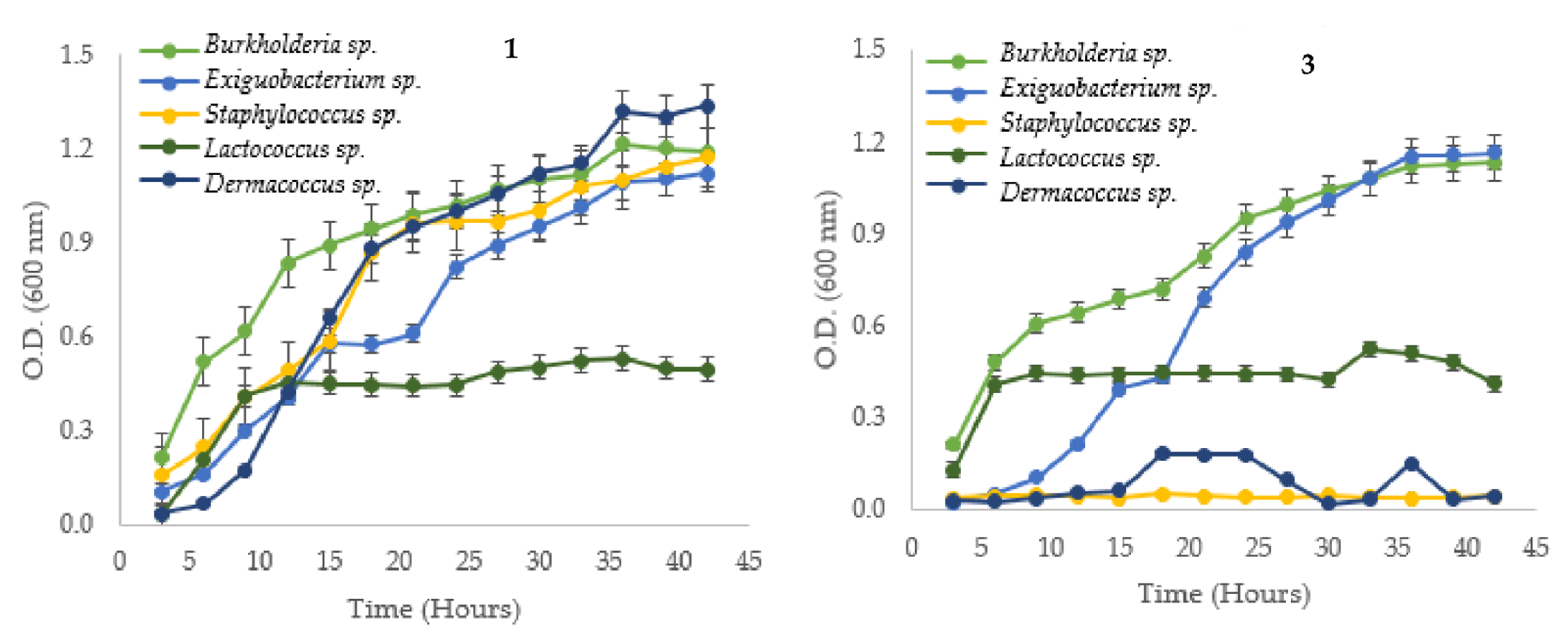
3.1. Bacterial Strain and Growth Curves Measurements

To study the behavior of 12 Cd tolerant strains in NB, their growth curves at different Cd concentrations (0, 10, and 15 mg Cd/L) were researched according to Cd levels in cocoa crop soils as previously reported (Figure 1). The results showed that Staphylococcus sp. (2-3) and Dermacoccus sp. (4-3) strains could not grow in both Cd concentrations, the other 10 strains were Cd tolerant at 10 and 15 mg Cd/L. In the presence of Cd, the growth curves were lower than without Cd; it was even observed that some strains clearly distinguish the growth phases (exponential/stationary).
The lag phase in most strains was shorter, suggesting that these strains have the adequate cellular machinery to hold the Cd level and quickly reach the exponential phase. Table 2 shows the µ, g, and K growth variables.

3.2. Cd Bioaccumulation in Native Bacteria Strains

A summary of the behavior of the strains after 24 h of incubation in nutrient broth with Cd can be found in Table 3. The FB (Cd accumulative capacity) for all bacteria strains is high, except for the Serratia sp. (6-2).

3.3. TEM, EDX, and FT-IR Analysis and Mechanisms of Cd Interaction Using Native Bacteria
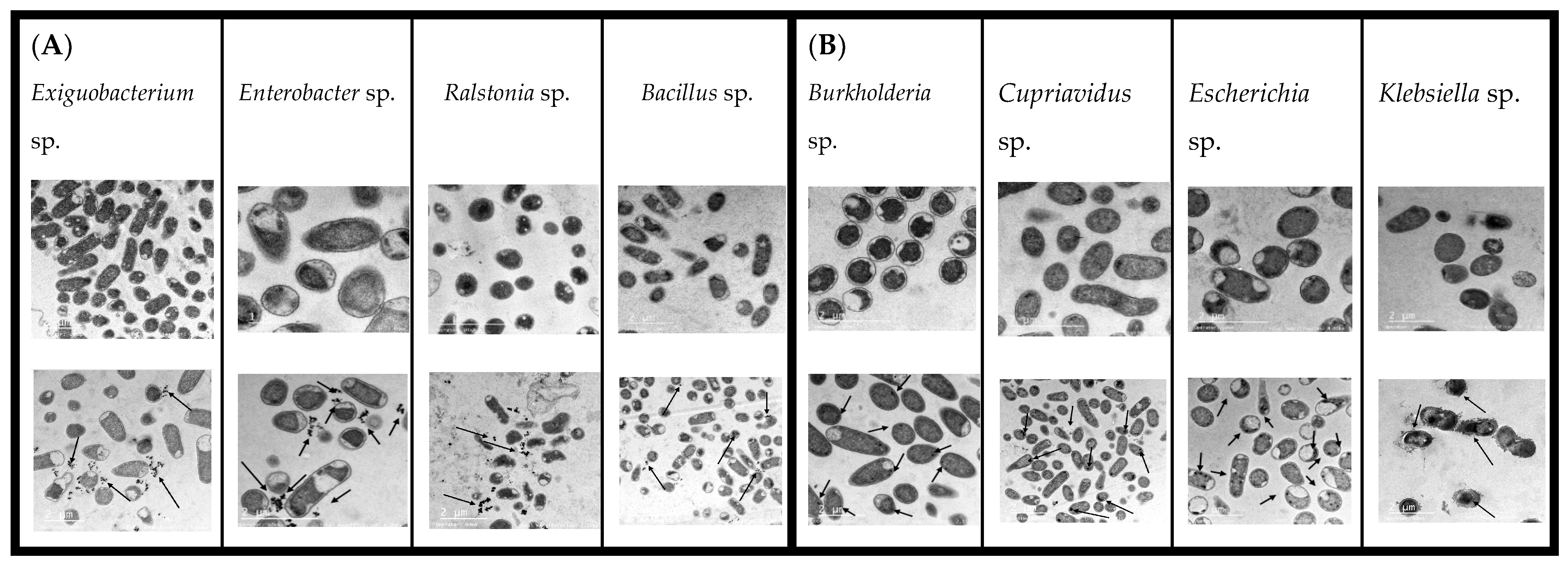
3.3.1. TEM Results

TEM was used to determine intra or extracellular Cd accumulation in native bacteria strains. In Serratia sp. (6-2), Exiguobacterium sp. (11-4A), Ralstonia sp. (16-1), Enterobacter sp. (29-4B), Lactococcus sp. (22-4), and Bacillus sp. (10-2), Cd dense granules were observed in external parts and cells bacteria showed morphological deformation (Figure 2A). For Burkholderia sp. (17-1), Cupriavidus sp. (15-1), Escherichia sp. (4-2), Klebsiella sp. (18-4B), and reference bacteria Cupriavidus metallidurans DSM 2839, Cd dense granules showed the inside of the cytoplasm with a cell morphological deformation (Figure 2B). Native Staphylococcus sp. (2-3) produced a sticky substance (exopolysaccharide (EPS)); it interacted with Cd and protected the cell lysis or cellular destruction to Cd concentration, even after 72 h in resuspended nutrient broth at a level of 15 mg Cd/L; the other native strain without Cd growth capacity Dermacoccus sp. (4-3) has a yellow–orange coloration intensity, the pellet cellular was intact and suspended even after 48 h in nutrient broth with 15 mg Cd/L concentration (Supplementary Figure S1).

3.3.2. EDX Spectra

EDX scan spectra images showed the Cd identification peaks from 3.2 to 3.4 KeV, the normal range for this heavy metal, and changed the counts in different bioaccumulative abilities. Other present metals in the spectra were C, O, Cl, Pb, Si, Os, and Cu peaks. Cl came from Cd analytic grade reactive (CdCl2); Cl, C, O, and Si were common elements of cell wall constituents and Os and Cu have come from fixation treatment and contrast staining to samples. In addition, in all the strains, an increase in oxygen after Cd biosorption was observed (Supplementary Figure S2).

3.3.3. FT-IR Spectra

FT-IR analysis revealed, before and after Cd binding, that many functional groups were involved in the biosorption process (Supplementary Figure S3). The main functional groups in the cell surfaces of all the spectral were observed changes around 3300 cm−1 –OH glucose and N–H proteins were in the range of 1100–1050 cm−1 assigned to vibration C–O and C–N stretch, respectively, and also in some samples, identified 1637–1529 cm−1 peaks corresponding to the C=O stretch. Table 4 resumed the functional groups and vibration types. The FT-IR results showed that more functional groups were involved in the Cd biosorption process in Bacillus sp. (10-2), Lactococcus sp. (22-4), Cupriavidus sp. (15-1), and Burkholderia sp. (17-1).

3.4. Responses of Inoculation to Native Tolerant Cd Bacteria in Young Cacao Plants at Differents Cd Levels in the Soil

3.4.1. Soil Analysis and Cd Concentration Chemicals Used in the Study

Soil properties (pH 4.88, electric conductivity 0.27 dS/m, organic matter 1.53%, total cadmium 0.332 mg/Kg, total nitrogen 0.074%, phosphorous 11.6 mg/g, clay 2%, sand 88%, and silt 10%) were recollected in cacao farm (Yariguies); the textural class of the soil was sandy silt loam according to Bouyoucos method. With respect to Cd content in chemicals employed in the study, only Ridomil Gold N60WP (fungicide) 2.67 mg/Kg and Basacote 12M (fertilizer NPK) 0.534 mg/Kg were detected. For Nilo 350SC (insecticide), Bélico 500SC, Imidogen 350SC (insecticide), Nutremin 200EC (insecticide), and Carrier (encapsulating aid), the Cd content was not detected.

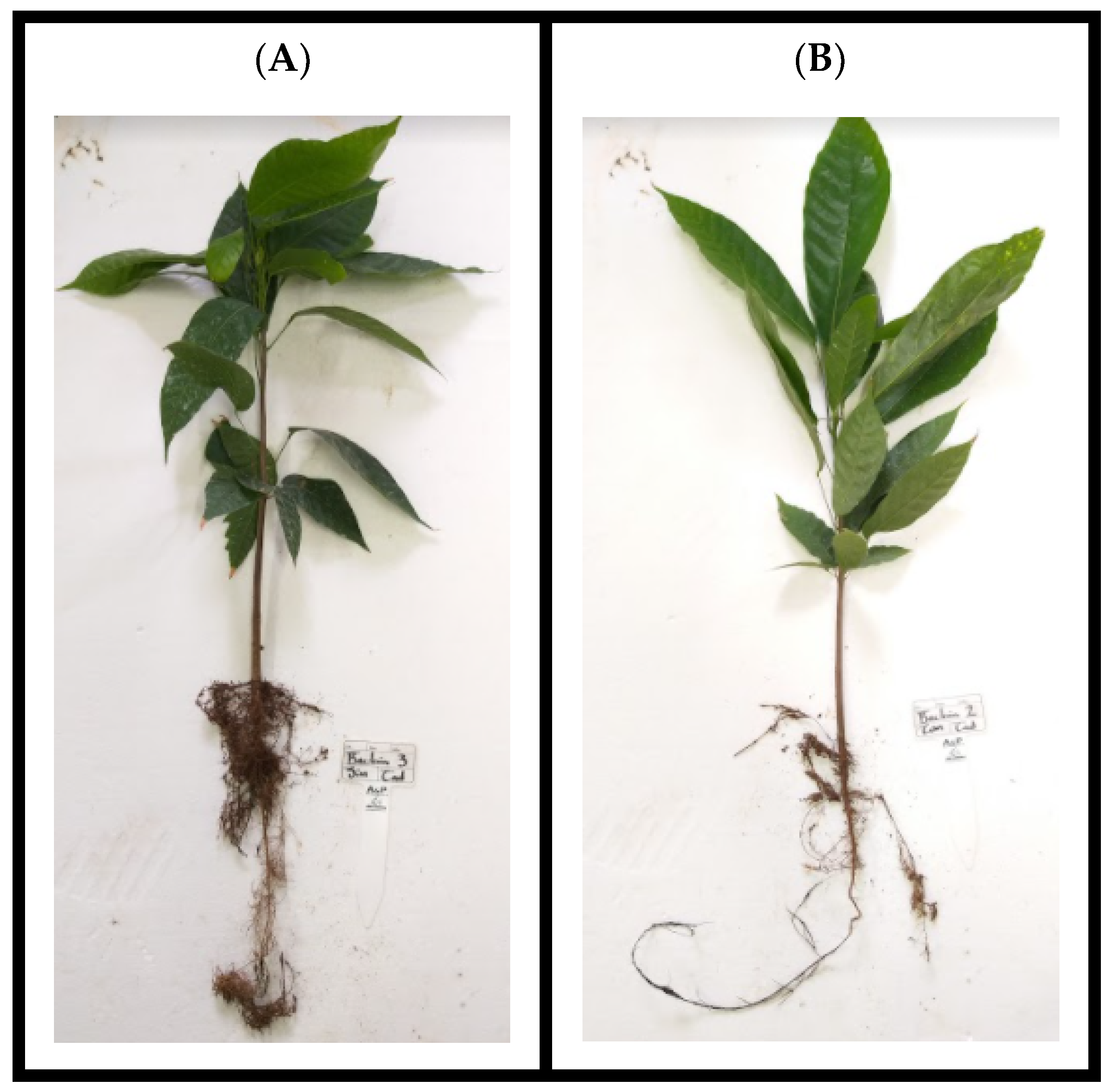
3.4.2. Roots and Aerial Parts Cacao Biomass

Roots and aerial parts of the cacao biomass in each plant part for each harvested time were resumed in Table 5. ANOVA analysis indicated that roots and aerial part the factors to influence biomass were time and Cd concentration in soil. The paired Tukey test refers to the biomass levels with no significant statistical differences between the control (without bacteria) and inoculated samples.

3.4.3. Cd Accumulation in Cacao Plants, Bioconcentration, and Translocation Factors

Effects of Klebsiella sp. (18-4B) (Bacteria 1), Exiguobacterium sp. (11-4A) (Bacteria 2), and Enterobacter sp. (29-4B) (Bacteria 3) on Cd accumulation in roots, sheet, and aerial parts each time (T1-T2). ANOVA analysis in Cd accumulation found no statistical differences in all treatments. Paired Tukey test indicated each time, no significant difference between the control and bacteria inoculated samples. Nevertheless, a decrease in the mean Cd accumulate concentration in roots for T1 (60 days) in Klebsiella sp. (18-4B) was 23.38% and 25.12% for T2 (120 days) in the Cd high concentration in soil. In the stems, a decrease in the mean Cd bioaccumulation at T1 for Klebsiella sp. (18-4B) was 19.34% and Enterobacter sp. (29-4B) was 10.93% at T2. The bacteria effects on Cd bioconcentration in roots showed no statistical differences in Cd soil concentrations and harvested time. However, the bioconcentration factor obtained for all treatments was equal or even less than the control, and especially reported for Klebsiella sp. (18-4B) in a different time and Cd level. The translocation factor indicated that the cacao plant was the Cd phytoextractor capability and increased directly proportional to the Cd soil concentration (Table 6). Figure 3 shows the physiological state of cacao plants at 120 days harvest without Cd concentrations.

4. Discussion

The isolation and characterization of native bacteria in complicated ecosystems or high pollution areas has lately become extremely relevant at the scientific level in order to find bacterial genera that display tolerance and advance treatments to address the substance that affects soil or water. There is a great deal of interest in the use of microorganisms in bioremediation since it has advantages, including high efficiency, low cost, and eco-friendliness. In addition to the fact that microorganisms can tolerate environmental stress through fast mutation and evolution, research of natural bacterial tolerance mechanisms can aid in the development of bioremediation solutions [22].
The present study established how 10 native bacterial strains behaved in nutrient broth at different Cd concentrations (0, 10, and 15 mg Cd/L), which corresponded to Cd levels in soils used to grow cocoa in the Santander Region in Colombia. It also demonstrated how Cd acts on the cell wall side chains and identified interactions between them. The growth curves showed a decrease in optical density conserving times to different phases after Cd was added to the broth; this delayed growth pattern suggested that bacteria strains must reduce growth and likely alter their physiological mechanisms in response to Cd toxicity [3]. Ten strains were grown at Cd concentrations regardless of Cd tolerance capacity, and two strains did not grow to full development, thus showing that Cd concentrations have an impact on growth conditions. The behavior of the positive control, Cupriavidus metallidurans DSM 2839, was similar to that which has been previously reported [3,23].
According to µ, the growth factors g and K demonstrated the behavior of each strain against Cd, even though the strains showed lower values with Cd, which gave them a survival advantage in the presence of a growth restrictor such as heavy metals. Regarding the two strains of Dermacoccus sp. (4-3) that did not develop in the presence of Cd concentrations, we discovered the yellow pigment generated by this strain in culture media. These microbial pigments, created by in vitro bacteria, have the ability to stop or chelate metal ions and have antioxidant qualities that can be used to prevent their toxic effects. By killing the cell pellet at high Cd levels, this mechanism can be prevented [24].
The other strain, Staphylococcus sp. (2-3), produced EPS, which may have anionic properties that promote interaction with cationic heavy metals (including Cd) and result in the formation of EPS-metal complexes [25]. After 24 h in nutrient broth, the native bacteria in this study, decreased the pH between 5.14 and 6.88, and also decreased the Cd absorption capacity from 8% to 79%. The removal rates of the ten strains were specific to the 10 mg/L group. The Lactococcus sp. (22-4) biosorption capture rate was larger than Exiguobacterium sp. (11-4A), while in the 15 mg/L groups, Klebsiella sp. (18-4B) removal was higher than Ralstonia sp. (16-1) and Escherichia sp. (4-2). These bacterial gene differences have been attributed to mechanisms for Cd interaction with the cell wall, Van der Waals forces, covalent bonds, redox reactions, extracellular precipitation, or combinations of these processes [26].
Another relevant aspect is the characteristics of the bacterial wall cells, which are more complex in Gram-negative bacteria than in Gram-positive ones, and have distinct biosorption capacities [27]. There are various ways that bacteria interact with heavy metals, including mechanism-independent (physicochemical interactions without energy intake) and process-dependent (transporting metal across the cell membrane with energy expenditure), with energy expenditure separating both [16].
Contaminant concentration and speciation have influenced the cumulative capacity, along with pH, which is key for heavy metal biosorption. The solubility, the ionic states of the functional groups on the biosorbent surface, and the competition with other ions are all affected by the pH of the solution [28,29]. According to TEM images, all the strains presented morphological changes, wrinkles, and deformed shapes after Cd exposition, which may be toxic effects conferred by heavy metals. These phenomena were related to the description of colony plates. Some strains exhibit swelling, irregular shapes, and liquid texture, others have a crusty surface, and the halo or colony is enlarged, covering a large plate area. According to Figure 2A, in the six native strains of Serratia sp. (6-2), Exiguobacterium sp. (11-4A), Ralstonia sp. (16-1), Enterobacter sp. (29-4B), Lactococcus sp. (22-4), and Bacillus sp. (10-2), dense Cd granules were observed in external parts.
The ability to accumulate Cd extracellularly has been reported in bacterial genera such as Serratia sp. [16], Bacillus sp. [30,31,32,33], Ralstonia sp. [34,35], Enterobacter sp. [36,37], Exiguobacterium sp. [38], and Lactococcus sp. [39]. This mechanism depends on Cd concentration and interaction time. Cd through efllux pumps is used by several bacterial species to maintain metal ions outside the cell to avoid their toxicity [3]. Another aspect of extracellular biosorption mechanism that also depends on growth conditions to produce Cd detoxification was CdS precipitated by lipoprotein biosurfactant. It conferred heavy metal tolerance and could express metallothioneins attached to membrane cells, also known as Lam B lipoprotein attached to peptidoglucane. Even metallothioneins capture heavy metals and mobilize them at specific sites in the cellular structure [40].
In Figure 2B, the reference bacteria Cupriavidus metallidurans DSM 2839 and four native strains, Burkholderia sp. (17-1), Cupriavidus sp. (15-1), Escherichia sp. (4-2), and Klebsiella sp. (18-4B) showed dense Cd granules inside the cytoplasm. Members of a number of bacterial species, such as Cupriavidus sp. [16], Klebsiella sp. [41,42,43], Escherichia sp. [44], and Burkholderia sp. [45] have been noted to possess this capacity to intracellularly collect Cd and other metals. This might occur because of the ability of living cells to produce energy. Compared to external bioabsorption, it is a slow absorption mechanism, but intercellular absorption results in higher capacity. Additionally, based on the inherent surface characteristics of cells and the active transport of a tiny quantity of Cd to interior cell structures, the Cd allocations of microbial cells change with contact time [46].
Additional details on elemental quantification in the biomass strains, as well as C, N, and oxygen peaks that are likely present in polysacharides and proteins in the cell envelop the bacterial biomass, are provided by EDX scanning. Ca has a significant impact on cell metabolism, especially when bound to a variety of cell envelope proteins. Due to the fact that Cd is a divalent metal such as Ca, as seen in TEM images [2], it can substitute for Ca and affect the structure of the cell. Thus, an oxide precipitate containing Cd is formed on the surface, as indicated by the increase in oxygen [47].
Through the interaction of metals with native bacterial strains, FT-IR is a method to identify functional groups. Cd was found to be involved in the stretching of functional groups in the cell after 24 h of incubation. There was a slight difference between the control groups and those treated with Cd. Different peaks at 3275, 1634, and 1531 cm−1 (amide groups) were identified for the native bacterial strains, which suggests that nitrogen atoms may be the main adsorption sites for Cd binding on the bacteria strains. The peaks at 914, 1057, and 1636 cm−1 also showed a shift, suggesting that the band has been broadened and stretched due to the presence of C=O groups. The peaks at 2958, 2923, 2873, 2852, 1467, 1455, and 860 cm−1 all show an intriguing alteration. It is possible that the oxygen atoms in the hydroxyl groups in the biomass strain are involved in this absorption process because this region corresponds to both C–H and O–H stretchings [48]. The peaks in the region of 966, 1230, and 1060 cm−1 indicate that the phosphate bond is involved in Cd biosorption and that cadmium phosphate can be produced on the biomass surface [2].
The extracellular interaction associated with each group was the amine (N–H) absorption peak at 3276 cm−1, at 2854 cm−1 with stretched alkane C–H leaps, and at 1637 cm−1 amide stretch signal I. Serratia sp. (6-2) had a low percentage of Cd absorption for each one, with a value of BF = 0.07. N–H is mainly stretched at 1529 cm−1 in the protein amide II band. The stretched C–H or C–OO groups emit a 1389 cm−1 signal, and the P=O bond is connected with a 1227 cm−1 band. C–OH is 1054 cm−1 and P=O is 1073 cm−1, respectively.
According to Chen and Xu [49], it is believed that the amine group, amide I and II, and the phosphoric acid group are the main groups involved in the Cd adsorption process. Cupriavidus sp. (15-1) exhibited intracellular bioaccumulation and had a 24% Cd absorption with a BF = 0.21. However, the FT-IR analysis detected interactions with C–H stretching around the C–OH bond at 2851 cm−1, 1452 cm−1, 1388 cm−1, and 1060 cm−1. It may be associated with metal ion transport mechanisms. The Klebsiella sp. (18-4B) strain showed intracellular bioaccumulation and 78% Cd absorption with a value of BF = 0.69.
Since CdS is created outside the cell and enters the cell through the Mg or Mn transporter system, Shamin and Rehman [41] suggested that Klebsiella pneumoniae may have an intracellular absorption due to sulfur formation and an increase in organic phosphorus. Furthermore, signals at 3275 cm−1, 1528 cm−1, 1226 cm−1, and 1061 cm−1, which are associated with N–H and P=O groups, and transport systems, also showed alterations in the FT-IR. Bacillus sp. (10-2) exhibited extracellular biosorption, BF = 0.23, and Cd capture at a rate of 28%. In addition, the FT-IR spectra changed to the following wavelengths: 3276 cm−1 and 1529 cm−1 for the N-H bond, 1455 cm−1 for the C-H bond, 1226 cm−1 and 1061 cm−1 for the stretch P=O bond. These signals were inferred to be phospholipids, carboxyl, amide I, amino, sulfates, and phosphate groups on the surface of strain 10-2, according to Huang and Jia [47].
Moreover, Ralstonia sp. (16-1) exhibits a biosorption mechanism with a 60.5% Cd absorption rate and BF = 0.58. According to Park, and Ko [34], Cd sulfite precipitation and an extracellular mechanism are present in Ralstonia sp. HM-1. The FT-IR showed 3277 cm−1, 2872 cm−1; 1636 cm−1; 1388 cm−1; 1170 cm−1; 1061 cm−1; 968 cm−1 which are mainly linked to cell wall functional groups of C=O, P=O, and N–H in saturated fatty acids, phosphate, and amine groups in proteins, or lipids, thus indicating that the cell wall components generated under Cd stress are involved in enhancing stress adaptation and increasing Cd adsorption capacity. These results were similar to Huang and Liu [50].
Escherichia sp. (4-2) showed intracellular bioaccumulation, BF = 0.48, and 54% Cd capture. Only three signals changed in the FT-IR, showing the C–H stretching and N–H bending vibrations of peptide bonds to membrane proteins or transport systems: 3274 cm−1, 2851 cm−1, and 1635 cm−1. These findings support those of Huang and Jia [47]. Additionally, mainly with amide group II, which represents C–H stretching and N–H bending vibration of peptide bond to membrane protein or transport system. These results agree with Huang and Jia [47]. In addition, 47% of Cd capture, BF = 0.37 with extracellular bioaccumulation potential following the Cd biosorption by Enterobacter sp. (29-4B).
Meanwhile, the FT-IR spectra showed changes at 3277 cm−1 (stretch N–H proteins), 2957 cm−1 (stretched C–H), 1529 cm−1 (asymmetric lipids/carbohydrates/proteins), and 1455 cm−1 (flexion N–H proteins); 1387 cm−1 (flexion C–H proteins/lipids), 1229 cm−1 (stretched C=O asymmetric fatty acids/amino acids), and 1169 cm−1 (stretched P=O asymmetric phospholipids/nucleic acids). For Enterobacter sp. DNB-S2, Sun and Wang [51] identified similar interaction groups with Cd. Meanwhile, Exiguobacterium sp. (11-4A) registered a 28% Cd capture rate with a biosorption capacity value of BF = 0.24. Bands 3276 cm−1, which correspond to stretch N-H proteins, 2958 cm−1, 2875 cm−1 and 2851 cm−1 which are assigned to stretch C-H asymmetric lipids/carbohydrates/and proteins, 1638 cm−1 which is assigned to stretch C=O proteins, 1220 cm−1, 1036 cm−1, and 965 cm−1 which are assigned to stretch P=O asymmetric phospholipids, and nucleic acids, were all found to change in FT-IR.
The behavior of this native Exiguobacterium sp. matched that of Park and Chon strain [38]. Meanwhile, Burkholderia sp. (17-1) demonstrated a 21% Cd capture rate, BF = 0.18, and intracellular Cd bioaccumulation, whereas FT-IR espectra showed changes in signals. Stretching N-H proteins at 3274 cm−1, C-H asymmetric lipids/carbohydrates/proteins at 2958 cm−1, 2873 cm−1, 2852 cm−1 flexioning C-H proteins/lipids at 1467 and 1455 cm−1, and stretching N-P proteins at 1390 cm−1. Stretch P=O symmetric phospholipids, nucleic acids, 1227 cm−1, 1075 cm−1, 1054 cm−1, and 965 cm−1 corresponding to asymmetric fatty acids and amino acids. This behavior resembles capture by biosorption; however, the results showed intracellular accumulation for Burkholderia sp. (17-1), maybe both mechanisms of action occur for this strain. According to Zhang and Li [45], Burkholderia cepacia GYP1 may have gone through various stages of fast immobilization of Cd(II) on the cell surface coordinated with functional groups, after transport of Cd(II) to cells and accumulation in cytoplasm, efflux of intracellular Cd(II) depended on energy, and entrapment or adsorbed of extracellular Cd(II) by EPS. Lactococcus sp. (22-4) had a BF = 0.40 and accumulated biosorption with a 46% Cd capture rate. OH and NH2 groups were, contrary to Sheng and Wang [39], responsible for Cd biosorption in Lactococcus lactis subsp. Lactis, according to our findings. Other proteins, with similar length to ours, include 1637 cm−1 stretch C=O proteins, 1526 cm−1 flexion N–H proteins, 1468 cm−1 and 1452 cm−1 with flexion C-H proteins/lipids, and 1219 cm−1, 1054 cm−1, and 964 cm−1.
Subsequently, for the selection of native strains to be studied in the greenhouse, it was carried out according to the highest growth variables, µ, g, and K. Exiguobacterium sp. (11-4A), Enterobacter sp. (29-4B), Lactococcus sp. (22-4), Cupriavidus sp. (15-1), and Klebsiella sp. (18-4B) strains grow faster in the presence of Cd. Another aspect of strain selection was the higher Cd concentration, an action mechanism found in this study and reported in the literature, finally selecting Exiguobacterium sp. (11-4A), Enterobacter sp. (29-4B) and Klebsiella sp. (18-4B).
In contrast, the cocoa plants in the greenhouse study were monitored under different treatments and harvest times in order to evaluate the behavior of the three native bacteria strains. The CCN51 cocoa variety was included in the greenhouse design because it was more prevalent in South American crops, had a high level of disease resistance, had an aromatic profile, contained more polyphenols, and had cocoa butter in the beans [52]. Concentration, exposure time, texture, and chemical characteristics of the soil, such as pH and organic matter, among others, all have a direct impact on Cd toxicity in cocoa [53]. Regarding soil texture, it offers benefits including ease of tilling, low compaction, quick water absorption, and high drainage; however, these conditions require permanent irrigation and fertilization. The pH of the soil is acidic due to the leaching of nutrients, which is an important factor in the Cd absorption process.
While the soil has a low percentage of organic matter, Qi and Lamb [54] suggested that Cd can interact with oxides of iron, manganese, and carbonates, which are generally immobile, allowing free mobility, phyto-availability, and Cd exchange. This study has a certain advantage because, according to Shahid and Dumat [55], Cd is directly influenced by humus, the substance responsible for maintaining an elevated pH level and highly reactive fulvic acid content, thereby promoting Cd bioavailability.
In order to contrast the effects of heavy metals on the plants and demand some sort of activity on the inoculating bacteria, we applied cadmium chloride, one of the most stable and water-soluble Cd sources, to enhance the basal Cd in the soil [56]. The fertilizer chosen for use in the greenhouse is crucial because, according to Zug and Huamaní Yupanqui [57], fertilizers regardless of Cd concentration can result in high Cd levels in crops, contaminate them directly, or generate Cd mobility in acid soils, thus allowing plants to grow faster or absorb more Cd through soil micelles. The fungicides and insecticides were added in a rotational manner established on the farm. They had low Cd concentrations, and the doses applied contributed to increasing the Cd total concentration by approximately 0.01 mg/L for each treatment without determining another influence in the experiment. This was carried out to fertilize the soil with NPK, to achieve seed germination, as well as to control possible pests or fungi that could affect the plants. Having innate mechanisms to bind free Cd in the cell wall, complex in the cytoplasm, or excrete chelating organic acids, some bacteria develop potential Cd resistance, dominating rhizospheric conditions, and supporting plant growth in Cd stress situations [58].
The native bacteria strains used in greenhouse experiments, Klebsiella sp. (18-4B) (Bacteria 1), and Enterobacter sp. (29-4B) (Bacteria 3) belong to the Proteobacteria phyla, they were Gram-negative bacillus and Exiguobacterium sp. (11-4A) (Bacteria 2), Gram-positive bacillus which belong to the Firmicutes phyla; these bacteria genera were characterized by fast growth, and high biomass production at 24 h without a supplemental medium. In terms of biomass, the plants were observed to increase in all treatments in T2, with no statistical differences; however, the biomass of roots and aerial parts of plants inoculated with bacteria 3 was slightly higher compared to control. This finding corresponded to Li and Liu [59], who report that Enterobacter sp. FM-1 cadmium-resistant bacteria promote the biomass production of plants cultivated in Cd polluted soil.
The root and aerial sections differ significantly in terms of exposure time and Cd concentration, according to variance analysis. Plants were found to have long, thin roots, little secondary root growth, and few aerial parts, which indicated lower dry weight in treatments with high Cd concentrations, symptoms of Cd toxicity from the soil [60]. The results showed a significant translocation rate for Cd, which was absorbed by the root system and deposited in all cacao plant organs. Aerial part > stem > root was the order of accumulation in plant parts at T1, but stem > aerial part > root was the order at T2. According to Verbruggen and Hermans [61], greater metal accumulation in the roots is a characteristic of intolerant plants, while plants that can translocate metals to the aerial parts are regarded as tolerant factors. The above is referred to as the bioconcentration factor (BCF) and translocation factor (TF), which indicate the relationship between the Cd concentration in the soil and roots (BCF), and the Cd concentration in the stem and roots (TF); BCF < 1 and TF < 1 indicated that the plant excluded the Cd; BCF > 1 and TF > 1 indicate that the plant accumulated the Cd; and BCF > 10 indicates hyperaccumulation [21].
In our study, all T1 and T2 treatments had high Cd concentrations, and the BCF indicated that cocoa plants were capable of accumulating and phytoextracting Cd; this BCF depended on the cacao variety and the soil properties, not just for Cd, but also for trace elements [62,63]. Several publications have cited the existence of proteins that conferred natural resistance to Cd translocation, which may have been mediated by metal ion transporters that increased Cd content in roots and mobilized it into xylem and phloem, according to Arevalo-Hernandez and Arevalo-Gardini [64]. According to final Cd levels in soil and leachates for treatments with low Cd, they were 0.254 and 0.005 mg kg−1; high Cd 12.94 and 0.63 mg kg−1 for T1; 0.22 and 0.045 mg kg−1, high Cd 9.78 and 1.17 mg kg−1, respectively, for T2. In contrast, at initial concentrations, Cd translocated in plants came exclusively from the soil.
The findings of this study demonstrated that different bacterial strains recovered from soils on Cd-polluted cocoa farms interact with Cd in distinct ways. The bacterial growth kinetics were studied using the proposed methodology, and the key to understanding the adaptation mechanism was discovered to be an inverse relationship between growth rate and heavy metal concentrations, but in native bacteria tolerant strains, growth is a response to the external restriction [65]. Some strains had morphological changes, which may have been a signal to infer maximum Cd tolerance level independently of genera.
Additional research needs to be carried out to identify metabolic pathways and enhance the ability of this type of bacteria to absorb heavy metals. To reduce the impact of cadmium in agricultural soils, the same methodology should be used to test different criollo cocoa varieties to find which variety has the lowest capacity to translocate Cd. The correct combination of native Cd-tolerant bacterial strains must also be used with different types of amendments, fertilizers, and agrochemicals.

5. Conclusions

Twelve native bacteria strains that had previously been molecularly described and were isolated from soil with Cd concentrations participated in the current study. These bacteria revealed two distinct ways of Cd capture, such as extracellular capacity (biosorption) and extracellular mechanism (Cd cytoplasm precipitation) in different concentrations, that depended on time and incubation conditions. All the genera have been reported in the literature with different Cd capability properties. Furthermore, in the pot assay under controlled conditions, the native strain Klebsiella sp. (18-4B) produced a certain degree of Cd immobilization in the soil to prevent the accumulation of Cd in the roots of cacao plants, thus demonstrating that native bacteria is one of the strategies for bioremediation to apply in soil cocoa crops with Cd geogenic presence. We identified the major functional groups on cell surfaces and reported the chemical changes before and after Cd interaction. The most significant issue to be tackled in recent years is environmental contamination, particularly the presence of heavy metals in soil used for crops. The native bacteria strains with action and tolerance to Cd were inoculated with a certain tendency to counteract Cd bioaccumulation in roots, which can reduce the negative effects of Cd on growth and productivity in cocoa crops in soils with cobalt concentrations, which is one of the limitations for the chocolate industry, which is booming due to the quality of Colombian beans.

Supplementary Materials

The following supporting information can be downloaded at: https://www.mdpi.com/article/10.3390/microbiolres13030039/s1, Figure S1: Strains without Cd growth; Figure S2: EDX spectra in some strains; Figure S3: FT-IR spectra.

Author Contributions

Conceptualization, P.F.F.-C. and C.X.M.-H.; methodology, P.F.F.-C., L.P.-V. and C.X.M.-H.; validation, P.F.F.-C., L.P.-V. and C.X.M.-H.; formal analysis, P.F.F.-C.; investigation, P.F.F.-C.; resources, L.P.-V. and C.X.M.-H.; writing—original draft preparation, P.F.F.-C.; writing—review and editing, P.F.F.-C., L.P.-V. and C.X.M.-H.; visualization, P.F.F.-C.; supervision, C.X.M.-H.; project administration, L.P.-V. All authors have read and agreed to the published version of the manuscript.

Funding

This work was supported by the Compañía Nacional de Chocolates.

Institutional Review Board Statement

Not applicable.

Informed Consent Statement

Not applicable.

Data Availability Statement

Not applicable.

Acknowledgments

We acknowledge the personal in Yariguies Farm, in special Juan Fernando Valenzuela by agricultural development director and Juan Camilo Mazo Rivas R&D manager by Compañía Nacional de Chocolates.

Conflicts of Interest

The authors declare that the research was conducted in the absence of any commercial or financial relationships that could be construed as a potential conflict of interest.

References

  1. Zao, M.; Zhang, C.; Zeng, G.; Huang, D.; Xu, P.; Cheng, M. Growth. Growth, metabolism of Phanerochaete chrysosporium and route of lignin degradation in response to cadmium stress in solid-state fermentation. Chemosphere 2015, 138, 560–567. [Google Scholar] [CrossRef] [PubMed]
  2. Chakravarty, R.; Banerjee, P. Mechanism of cadmium binding on the cell wall of an acidophilic bacterium. Bioresour. Technol. 2012, 108, 176–183. [Google Scholar] [CrossRef] [PubMed]
  3. Siripornadulsil, S.; Siripornadulsil, W. Cadmium-tolerant bacteria reduce the uptake of cadmium in rice: Potential for microbial bioremediation. Ecotoxicol. Environ. Saf. 2013, 94, 94–103. [Google Scholar] [CrossRef] [PubMed]
  4. Tossapol, L.; Sooksawat, N. Bioaccumulation and biosorption of Cd2+ and Zn2+ by bacteria isolated from a zinc mine in Thailand. Ecotoxicol. Environ. Saf. 2015, 122, 322–330. [Google Scholar] [CrossRef]
  5. Al-Saraj, M.; Abdel-Latif, M. Bioaccumulation of some hazardous metals by sol gel entrapped microorganisms. J. Non-Cryst. Solids 1999, 248, 137–140. [Google Scholar] [CrossRef]
  6. Wong, C.; Cobbett, C. HMA P-type ATPases are the major mechanism for root to shoot Cd translocation in Arabidopsis thaliana. New Phytol. 2009, 181, 71–78. [Google Scholar] [CrossRef]
  7. Wang, C.; Huang, Y. Burkholderia sp. Y4 inhibits cadmium accumulation in rice by increasing essential nutrient uptake and preferentially absorbing cadmium. Chemosphere 2020, 252, 126603. [Google Scholar] [CrossRef]
  8. Shi, Z.; Zhang, Z. Characterization of a high cadmium accumulating soil bacterium, Cupriavidus sp. WS2. Chemosphere 2020, 247, 125834. [Google Scholar] [CrossRef]
  9. Rani, A.; Souche, Y. Comparative assessment of in situ bioremediation potential of cadmium resistant acidophilic Pseudomonas putida 62BN and alkalophilic Pseudomonas monteilli 97AN strains on soybean. Int. Biodeterior. Biodegrad. 2009, 63, 62–66. [Google Scholar] [CrossRef]
  10. Engbersen, N.; Gramlich, A. Cadmium accumulation and allocation in different cacao cultivars. Sci. Total Environ. 2019, 678, 660–670. [Google Scholar] [CrossRef]
  11. Chavez, E.; He, Z. Concentration of cadmium in cacao beans and its relationship with soil cadmium in southern Ecuador. Sci. Total Environ. 2015, 533, 205–214. [Google Scholar] [CrossRef] [PubMed]
  12. Bravo, D.; Pardo-Díaz, S. Cadmium and Cadmium-tolerant soil bacteria in cacao crops from northeastern Colombia. J. Appl. Microbiol. 2018, 124, 1175–1194. [Google Scholar] [CrossRef] [PubMed]
  13. Feria-Cáceres, P.; Penagos-Vélez, L. Theobroma cacao L. agricultural soils with natural low and high cadmium (Cd) in Santander (Colombia), contain a persistent shared bacterial composition shaped by multiple soil variables and bacterial isolates highly resistant to Cd concentrations. Curr. Res. Microb. Sci. 2021, 2, 100086. [Google Scholar] [CrossRef]
  14. Madigan, M.; Martinko, D. Brock Biology of Microorganisms, 13th ed.; Benjamin Cummings Editorial©: San Francisco, CA, USA, 2012. [Google Scholar]
  15. Huang, F.; Guo, C.-L.; Lu, G.-N.; Yi, X.-Y.; Zhu, L.-D.; Dang, Z. Bioaccumulation characterization of cadmium by growing Bacillus cereus RC-1 and its mechanism. Chemosphere 2014, 109, 134–142. [Google Scholar] [CrossRef]
  16. Ferreira, P.; Bomfeti, C. Cupriavidus necator strains: Zinc and cadmium tolerance and bioaccumulation. Sci. Agric. 2018, 75, 452–460. [Google Scholar] [CrossRef]
  17. Chen, Y.; Zhu, Q. How Serratia marcescens HB-4 absorbs Cadmium and its implication on phytoremediation. Ecol. Environ. Saf. 2019, 185, 109723. [Google Scholar] [CrossRef]
  18. Suarez, C.; Restrepo, J.; Quinchia, A. Colombian vegetal fibers as a reinforcement in polymeric matrix composites. Tecnura 2017, 21, 51. [Google Scholar] [CrossRef]
  19. Pereira de Araújo, R.; Furtado de Almeida, A. Photosynthetic, antioxidative, molecular and ultrastructural responses of young cacao plants to Cd toxicity in the soil. Ecotoxicol. Environ. Saf. 2017, 144, 148–157. [Google Scholar] [CrossRef]
  20. Kongor, J.; Hinneh, M. Factors influencing quality variation in cocoa (Theobroma cacao) bean flavour profile: A review. Food Reseach Int. 2016, 82, 44–52. [Google Scholar] [CrossRef]
  21. Padmavathiamma, P.; Loretta, L. Phytoremediation Technology: Hyper-accumulation Metals in Plants. Water Air Soil Pollut. 2007, 184, 105–126. [Google Scholar] [CrossRef]
  22. Yin, K.; Wang, Q. Microorganism remediation strategies towards heavy metals. Review. Chem. Eng. J. 2019, 360, 1553–1563. [Google Scholar] [CrossRef]
  23. Zoropogui, A.; Gambarelli, S. CzcE from Cupriavidus metallidurans CH34 is a copper-binding protein. Biochem. Biophys. Res. Commun. 2008, 365, 735–739. [Google Scholar] [CrossRef] [PubMed]
  24. Banik, A.; Pandya, P. Characterization of halotolerant, pigmented, plant growth promoting bacteria of groundnut rhizosphere and its in-vitro evaluation of plant-microbe protocooperation to withstand salinity and metal stress. Sci. Total Environ. 2018, 630, 231–242. [Google Scholar] [CrossRef] [PubMed]
  25. Shatrohan, L.; Sheel, R.; Ben, S.; Rajesh, K. Biosurfactant and exopolysaccharide-assisted rhizobacterial technique for the remediation of heavy metal contaminated soil: An advancement in metal phytoremediation technology. Environ. Technol. Innov. 2018, 10, 243–263. [Google Scholar] [CrossRef]
  26. Rajendran, P.; Muthukrishnan, J. Microbes in heavy metal remediation. Indian J. Exp. Biol. 2003, 41, 935–944. [Google Scholar]
  27. Xie, Y.; Li, H. Kinetic simulating of Cr (VI) removal by the waste Chlorella vulgaris biomass. J. Taiwan Inst. Chem. Eng. 2014, 45, 1773–1782. [Google Scholar] [CrossRef]
  28. Ozdemir, S.; Kilinc, E. Biosorption of Cd, Cu, Ni, Mn and Zn from aqueous solutions by thermophilic bacteria, Geobacillus toebii sub.sp. decanicus and Geobacillus thermoleovorans sub.sp. stromboliensis: Equilibrium, kinetic and thermodynamic studies. Chem. Eng. J. 2009, 152, 195–206. [Google Scholar] [CrossRef]
  29. Long, J.; Yu, M. Characterization of cadmium biosorption by inactive biomass of two cadmium-tolerant endophytic bacteria Microbacterium sp. D2-2 and Bacillus sp. C9-3. Ecotoxicology 2021, 30, 1419–1428. [Google Scholar] [CrossRef]
  30. Huang, F.; Dang, Z. Biosorption of Cd(II) by live and dead cells of Bacillus cereus RC-1 isolated from cadmium-contaminated soil. Colloids Surf. Biointerfaces 2013, 107, 11–18. [Google Scholar] [CrossRef]
  31. Mathew, B.; Biju, V. Accumulation of lead (Pb II) metal ions by Bacillus toyonensis SCE1 species, innate to industrial-area ground water and nanoparticle synthesis. Appl. Nanosci. 2019, 9, 49–66. [Google Scholar] [CrossRef]
  32. Kim, S.; Jin, M. Biosortion of cationic basic dye and cadmium by the novel biosorbent Bacillus catenulatus JB-022 strain. J. Biosci. Bioeng. 2015, 119, 433–439. [Google Scholar] [CrossRef] [PubMed]
  33. Chi, Y.; Huang, Y. Two plant growth promoting bacterial Bacillus strains possess different mechanisms in adsortion and resistence to cadmium. Sci. Total Environ. 2020, 741, 140422. [Google Scholar] [CrossRef] [PubMed]
  34. Pugazhendhi, A.; Boovaragamoorthy, G. New insight into effective biosorption of lead from aqueos solution using Ralstonia solanacearum: Characterization and mechanism studies. J. Clean. Prod. 2018, 174, 1234–1239. [Google Scholar] [CrossRef]
  35. Park, Y.; Ko, J. Enhancement of bioremediation by Ralstonia sp. HM-1 in sediment polluted by Cd and Zn. Bioresour. Technol. 2008, 99, 7458–7463. [Google Scholar] [CrossRef] [PubMed]
  36. Zhou, J.; Li, P. Isolation, characterization and inoculation of Cd tolerant rice endophytes and their impacts on rice under contamined environment. Environ. Pollut. 2020, 260, 113990. [Google Scholar] [CrossRef] [PubMed]
  37. Wang, Q.; Li, Q. Biochemical and genetic basis of cadmium biosorption by Enterobacter ludwigii LY6, isolated from industrial contaminated soil. Environ. Pollut. 2020, 264, 114637. [Google Scholar] [CrossRef]
  38. Park, J.; Chon, H. Characterization of cadmium biosorption by Exiguobacterium sp. isolated from farmland soil near Cu-Pb-Zn mine. Environ. Sci. Pollut. Res. 2016, 23, 11814–11822. [Google Scholar] [CrossRef]
  39. Sheng, Y.; Wang, Y. Cadmium tolerant characteristic of a newly isolated Lactococcus lactis subsp. lactis. Environ. Toxicol. Pharmacol. 2016, 48, 183–190. [Google Scholar] [CrossRef]
  40. Valls, M.; Gonzalez-Duarte, R. Bioaccumulation of heavy metals with protein fusions of metallothionein to bacterial OMPs. Biochimie 1998, 80, 855–861. [Google Scholar] [CrossRef]
  41. Afzal, A.; Rasool, M. Assesment of heavy metal tolerance and biosorptive potencial of Klebsiella variicola isolated from industrial effluents. AMB Express 2017, 7, 184. [Google Scholar] [CrossRef]
  42. Shamin, S.; Rehman, A. Cadmium resistance and acumulation potencial of Klebsiella pneumoniae strain CBL-1 isolated from industrial wastewater. Pak. J. Zool. 2012, 44, 203–208. [Google Scholar]
  43. Holmes, J.; Richardson, D. Cadmium-specific formation of metal sulfide ‘Q-particles’ by Klebsiella pneumoniae. Microbiology 1997, 143, 2521–2530. [Google Scholar] [CrossRef] [PubMed]
  44. Sriram, M.; Gayathiri, S. Novel lipopeptide biosurfactant produced by hydrocarbon degrading and heavy metal tolerant bacterium Escherichia fergusonii KLU01 as a potential tool for bioremediation. Bioresour. Technol. 2011, 102, 9291–9295. [Google Scholar] [CrossRef] [PubMed]
  45. Zhang, J.; Li, Q. Bioaccumulation and distribution of cadmium by Burkholderia cepacia GYP1 under oligotrophic condition and mechanism analysis at proteome level. Ecotoxicol. Environ. Saf. 2019, 176, 162–169. [Google Scholar] [CrossRef]
  46. Li, J.; Liu, Y. Sorption mechanism and distribution of cadmium by different microbial species. J. Environ. Manag. 2019, 237, 552–559. [Google Scholar] [CrossRef]
  47. Huang, H.; Jia, Q. Screening strains for microbial technology of cadmium. Chemosphere 2020, 251, 126428. [Google Scholar] [CrossRef]
  48. BeĆ, K.; Grabska, J. Biomolecular and bioanalytical applications of infrared spectroscopy—A review. Anal. Chim. Acta 2020, 1133, 150–177. [Google Scholar] [CrossRef]
  49. Chen, Y.; Xu, C. Cadmium adsorption mechanism of Serratia marcescens HB-4. CIE J. 2017, 68, 1574–1581. [Google Scholar] [CrossRef]
  50. Huang, J.; Liu, C. Identification of a novel heavy metal resistant Ralstonia strain and its growth response to cadmium exposure. J. Hazard. Mater. 2021, 416, 125942. [Google Scholar] [CrossRef]
  51. Sun, R.; Wang, L. Cadmium resistance mechanisms of a functional strain Enterobacter sp. DNB-S2, isolated from black soil in Northeast China. Environ. Pollut. 2020, 263, 114612. [Google Scholar] [CrossRef]
  52. Boza, E.J.; Motamayor, J.C.; Amores, F.M.; Cedeno-Amador, S.; Tondo, C.L.; Livingstone, D.S.; Gutiérrez, O.A. Genetic Characterization of the cacao (Theobroma cacao L.) clone ‘CCN 51′ and its impact and significance on global cacao improvement and production. J. Am. Soc. Hortic. Sci. 2014, 139, 219–229. [Google Scholar] [CrossRef]
  53. He, S.; He, Z. Soil biogeochemistry, plant physiology and phytoremediation of cadmium contaminated soils. Adv. Agron. 2015, 134, 135–225. [Google Scholar] [CrossRef]
  54. Qi, F.; Lamb, D. Cadmium solubility and bioavailability in soils amended with acidic and neutral biochar. Sci. Total Environ. 2018, 610, 1457–1466. [Google Scholar] [CrossRef] [PubMed]
  55. Shahid, M.; Dumat, C. Cadmium bioavailability, uptake, toxicity and detoxifcation in soilplant system. Rev. Environ. Contam. Toxicol. 2016, 241, 73–137. [Google Scholar] [CrossRef]
  56. Sauvé, S.; Norvell, W. Speciation and complexation of cadmium in extracted soil solutions. Environ. Sci. Technol. 2000, 34, 291–296. [Google Scholar] [CrossRef]
  57. Zug, K.; Huamaní Yupanqui, H. Cadmium Accumulation in Peruvian Cacao (Theobroma cacao L.) and Opportunities for Mitigation. Water Air Soil Pollut. 2019, 230, 72. [Google Scholar] [CrossRef]
  58. Liu, H.; Xie, Y. Efect of Serratia sp. K3 combined with organic materials on cadmium migration in soil-Vetiveria zizanioides L. system and bacterial community in contaminated soil. Chemosphere 2020, 242, 125164. [Google Scholar] [CrossRef]
  59. Li, Y.; Liu, K. Improvement of cadmium phytoremediation by Centella asiatica L. after soil inoculation with cadmium-resistant Enterobacter sp. FM-1. Chemosphere 2018, 202, 280–288. [Google Scholar] [CrossRef]
  60. Ehsan, M.; Santamaría-Delgado, K. Phytostabilization of cadmium contaminated soils by Lupinus uncinatus Schdl. Span. J. Agric. Res. 2009, 7, 390–397. [Google Scholar] [CrossRef]
  61. Verbruggen, N.; Hermans, C. Mechanisms to cope with arsenic or cadmium excess in plants. Curr. Opin. Plant Biol. 2009, 12, 364–372. [Google Scholar] [CrossRef]
  62. Antoniadis, V.; Levizou, E. Trace elements in the soil-plant interface: Phytoavailability, translocation, and phytoremediation–A review. Earth Sci. Rev. 2017, 171, 621–645. [Google Scholar] [CrossRef]
  63. Barraza, F.; Schreck, E. Beyond cadmium accumulation: Distribution of other trace elements in soils and cacao beans in Ecuador. Environ. Res. 2021, 192, 110241. [Google Scholar] [CrossRef] [PubMed]
  64. Arevalo-Hernandez, C.; Arevalo-Gardini, E. Growth and nutritional responses of wild and domesticated cacao genotypes to soil Cd stress. Sci. Total Environ. 2021, 763, 144021. [Google Scholar] [CrossRef] [PubMed]
  65. Oladipo, O.; Ezeokoli, O. Tolerance and growth kinetics of bacteria isolated from gold and gemstone mining sites in response to heavy metal concentrations. J. Environ. Manag. 2018, 212, 357–366. [Google Scholar] [CrossRef] [PubMed]
Figure 1. Growth curve native bacteria strains; 1: 0 mg Cd/L, 2: 15 mg Cd/L, 3: 10 mg Cd/L. * Strains grow in both Cd concentrations with the growth curve at the highest concentration.
Microbiolres 13 00039 g001aMicrobiolres 13 00039 g001b
Figure 2. Some TEM pictures for cells without Cd (up), cell tolerance to 15 mg Cd/L (below). (A) Biosorption ability; the black arrows indicated external deposits of Cd. (B) Intracellular ability; the black arrows indicated intracellular deposits of Cd.
Microbiolres 13 00039 g002
Figure 3. (A). Cacao plant without Cd: high biomass aerial, stem large, thick, and various branches; principal root large and straight and abundant secondary roots. (B) Cacao plant with Cd concentration: low biomass aerial, stem short, thin; branches cover the top of the stem; principal root was large, thin, curved, and fragile; a few secondary roots.
Microbiolres 13 00039 g003
Table 1. Homology of the 16S rDNA sequences of the native cadmium-tolerant soil bacteria.
Phylogenetic Affiliation (Related Reference Sequence)Gram StainGenBank Accesión Number
Bacillus toyonensis+MN587894
Burkholderia arborisMN587896
Cupriavidus necátorMN587892
Escherichia fergusoniiMN587901
Exiguobacterium acetylicum+MN587893
Ralstonia solanacearumMN587895
Serratia marcescensMN587899
Dermacoccus barathriMN587890
Enterobacter tabacoMN587891
Klebsiella variicolaMN587897
Lactococcus lactis+MN587898
Staphylococcus capitis+MN587900
Table 2. Mean values of growth parameters with and without Cd in nutrient broth.
Native Strainµ (h−1)g (h)K (Generation/h)
Without CdWith CdWithout CdWith CdWithout CdWith Cd
Cupriavidus metallidurans *0.0930.0587.49211.9070.1330.084
Serratia sp. (6-2)0.0760.0899.1427.3880.1090.128
Cupriavidus sp. (15-1)0.0890.1067.7606.5190.1290.153
Klebsiella sp. (18-4B)0.0990.1267.0075.4910.1430.182
Bacillus sp. (10-2)0.1000.0946.9447.3880.1440.135
Ralstonia sp. (16-1)0.1230.0655.63910.7280.1770.093
Escherichia sp. (4-2)0.0660.07410.5489.3650.0950.107
Enterobacter sp. (29-4B)0.1110.1046.2496.6570.1600.150
Exiguobacterium sp. (11-4A)0.1150.1446.0054.8060.1670.208
Burkholderia sp. (17-1)0.0620.08211.1248.4410.0900.118
Lactococcus sp. (22-4)0.2400.3922.8851.7690.3470.565
* Commercial reference strain.
Table 3. Ability to tolerance and bioaccumulation for the native bacteria strains.
BacteriaInitial (Cd) (mg/L)Final pH Nutrient Broth(Cd) Supernatant (mg/L)(Cd) Captured (mg/L)% Cd CaptureBioaccumulation Factor (BF)
Cupriavidus metallidurans * 6.36 ± 0.411.70 ± 0.111.61 ± 0.0748.63 ± 0.490.43 ± 0.02
Serratia sp. (6-2)3.73 ± 0.31 6.25 ± 0.17 3.14 ± 0.090.28 ± 0.108.02 ± 2.730.07 ± 0.03
Cupriavidus sp. (15-1)6.88 ± 0.302.38 ± 0.060.78 ± 0.1424.61 ± 3.350.21 ± 0.04
Klebsiella sp. (18-4B)6.60 ± 0.380.69 ± 0.082.59 ± 0.4078.86 ± 2.970.69 ± 0.11
Bacillus sp. (10-2)6.34 ± 0.232.20 ± 0.090.87 ± 0.1928.21 ± 5.370.23 ± 0.05
Ralstonia sp. (16-1)6.62 ± 0.251.41 ± 0.192.17 ± 0.2160.59 ± 5.590.58 ± 0.06
Escherichia sp. (4-2)6.22 ± 0.421.50 ± 0.211.78 ± 0.3953.93 ± 9.090.48 ± 0.11
Enterobacter sp. (29-4B)6.73 ± 0.441.56 ± 0.051.39 ± 0.0447.16 ± 1.400.37 ± 0.01
Exiguobacterium sp. (11-4A)3.22 ± 0.27 5.14 ± 0.382.92 ± 0.030.90 ± 0.0928.24 ± 2.00.24 ± 0.02
Burkholderia sp. (17-1)6.06 ± 0.202.50 ± 0.050.68 ± 0.0221.33 ± 2.210.18 ± 0.01
Lactococcus sp. (22-4)5.16 ± 0.191.73 ± 0.041.51 ± 0.1846.38 ± 3.450.40 ± 0.05
* Commercial reference strain.
Table 4. Functional group identification of Cd-tolerant bacteria.
Wavenumber (cm−1)AssignmentVibration Types
~3275Amide AStretch N-H proteins
2958/2873Methyl CH3Stretch C-H asymmetric lipids/carbohydrates/proteins
2923/2852Methyl CH2Stretch C-H asymmetric lipids/carbohydrates/proteins
~1636Amide IStretch C=O proteins
1529Amide IIFlexion N-H proteins
1467Methyl CH2Flexion C-H proteins/lipids
1455Methyl CH3Flexion C-H proteins/lipids
1389CarbonylStretch C=O asymmetric fatty acids and amino acids
~1230/~1060Phosphate PO2−Stretch P=O asymmetric phospholipids, nucleic acids
~966Phosphate PO2−Stretch P=O symmetric phospholipids, nucleic acids
~914Ketones C-O ring vibrations of nucleic acids “sugars”
~860C-H groupTri-substituted bending C-H
Table 5. Effects of the bacterial inoculation (control, not inoculated; bacteria, inoculated seedlings) on morphological and physiological parameters of cocoa plants.
Cd LevelTreatmentFresh Weight (g)Dry Weight (g)
RootsStemsAerial PartsRootsStemsAerial Parts
T1 aT2 bT1 aT2 bT1 aT2 bT1 aT2 bT1 aT2 bT1 aT2 b
LowControl5.55 ± 1.916.58 ± 2.107.4 ± 4.28.0 ± 1.28.3 ± 1.810.5 ± 0.20.74 ± 0.471.17 ± 0.151.37 ± 0.552.31 ± 0.291.89 ± 0.583.52 ± 0.49
Bacteria 13.56 ± 0.096.13 ± 2.564.6 ± 0.18.5 ± 2.76.9 ± 0.912.0 ± 4.90.42 ± 0.061.14 ± 0.131.29 ± 0.632.29 ± 0.541.57 ± 0.823.49 ± 1.52
Bacteria 23.39 ± 0.085.70 ± 1.735.4 ± 0.98.4 ± 0.87.7 ± 0.512.7 ± 1.90.58 ± 0.191.13 ± 0.231.58 ± 0.472.18 ± 0.271.85 ± 0.743.95 ± 0.96
Bacteria 33.43 ± 0.527.34 ± 2.495.1 ± 1.59.5 ± 2.17.7 ± 0.614.0 ± 2.30.44 ± 0.111.35 ± 0.101.31 ± 0.432.50 ± 0.881.78 ± 0.604.17 ± 0.84
HighControl3.83 ± 1.224.81 ± 2.034.9 ± 1.78.6 ± 1.16.6 ± 0.510.4 ± 0.50.44 ± 0.051.01 ± 0.361.19 ± 0.342.47 ± 0.291.57 ± 0.653.25 ± 0.51
Bacteria 12.53 ± 1.173.97 ± 0.704.2 ± 0.87.0 ± 1.35.4 ± 0.79.5 ± 1.80.40 ± 0.020.86 ± 0.181.14 ± 0.412.04 ± 0.291.33 ± 0.783.25 ± 0.58
Bacteria 23.18 ± 0.494.28 ± 2.365.0 ± 0.56.2 ± 2.85.9 ± 1.08.3 ± 3.60.48 ± 0.260.87 ± 0.141.05 ± 0.251.81 ± 0.521.58 ± 0.952.81 ± 1.07
Bacteria 33.13 ± 1.363.42 ± 1.284.2 ± 1.96.1 ± 0.55.2 ± 1.47.8 ± 1.90.50 ± 0.070.79 ± 0.411.04 ± 0.301.92 ± 0.581.22 ± 0.762.77 ± 0.87
ANOVA (Type II)
Treatment8.821.916.465.811.530.090.220.16
Time12.5664.0918.3118.7710.912.81 ***10.7 ***38.81 ***
Cd level3.1032.25*19.5921.1459.92*0.51 **0.903.75 *
Treatment: Cd1.665.554.680.8529.350.020.381.09
Values were mean ± standard deviation. Different letters were statistical significance p < 0.05 (paired Tukey test). Significance level: *** p < 0.001; ** p < 0.01; * p < 0.05. T1 (60 days); T2 (120 days).
Table 6. Bioconcentration and translocation factors (mean, SD, min.–max.) for all treatments.
TimeFactors
Treatment
Bioconcentration (BCF)Translocatión (TF)
LowHighLowHigh
T1Control4.92 ± 4.17
(0.39–11.72)
2.54 ± 0.07
(0.97–4.70)
1.13 ± 1.54
(0.25–13.72)
4.30 ± 2.54
(0.97–4.70)
Klebsiella sp. (18-4B)3.12 ± 0.70
(0.79–6.27)
1.51 ± 0.07
(0.77–2.16)
0.76 ± 0.68
(0.36–3.29)
8.24 ± 6.61
(0.77–2.16)
Exiguobacterium sp. (11-4A)2.75 ± 1.91
(0.93–7.28)
2.39 ± 1.06
(1.40–3.57)
0.24 ± 0.17
(1.65–7.44)
5.18 ± 3.03
(1.40–3.57)
Enterobacter sp. (29-4B)3.56 ± 1.64
(0.82–6.75)
2.51 ± 1.36
(0.82–5.16)
0.95 ± 1.20
(0.31–4.62)
3.77 ± 1.31
(0.82–5.16)
T2Control4.60 ± 1.49
(0.18–18.57)
3.36 ± 0.90
(0.82–10.39)
5.82 ± 10.83
(0.19–18.57)
7.69 ± 10.47
(0.50–13.75)
Klebsiella sp. (18-4B)2.28 ± 0.59
(0.15–7.88)
2.27 ± 0.56
(0.51–5.54)
5.32 ± 7.80
(0.43–7.88)
7.76 ± 9.47
(0.084–15.12)
Exiguobacterium sp. (11-4A)4.64 ± 3.19
(0.45–8.01)
2.53 ± 0.28
(0.74–4.94)
5.82 ± 10.83
(0.45–33.84)
6.39 ± 6.70
(0.69–13.49)
Enterobacter sp. (29-4B)3.53 ± 3.99
(0.31–17.73)
2.48 ± 0.77
(0.71–3.66)
6.64 ± 11.87
(0.31–17.73)
4.37 ± 1,62
(0.73–7.22)
Publisher’s Note: MDPI stays neutral with regard to jurisdictional claims in published maps and institutional affiliations.

