
Autochthonous North American Leprosy: A Second Case in Canada


Abstract
:1. Introduction
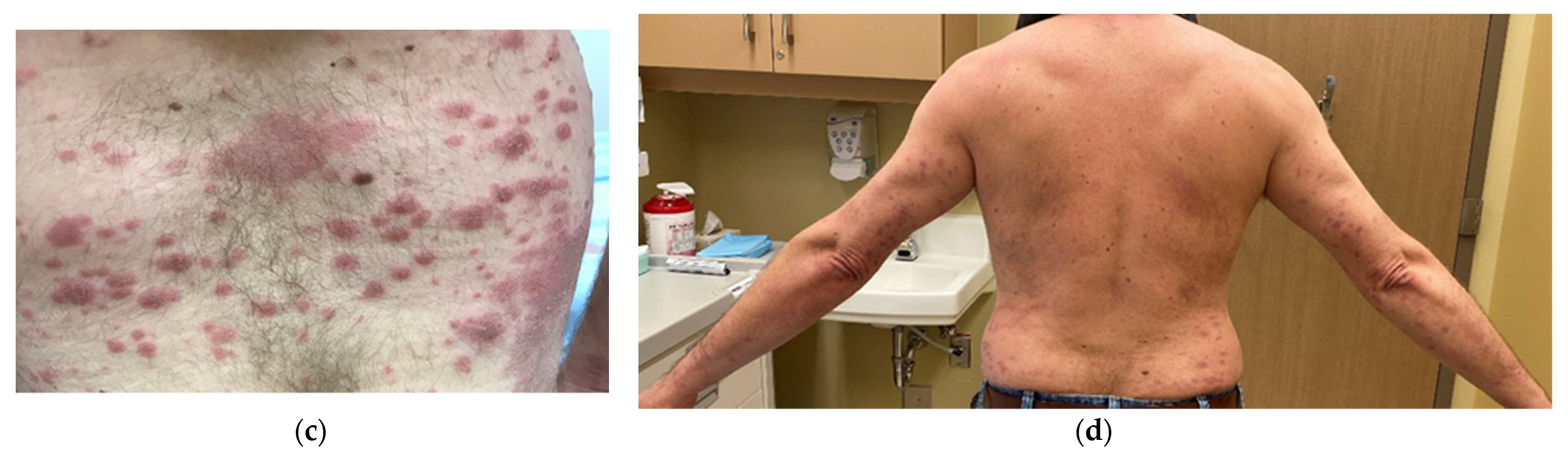
2. Case
3. Laboratory Results
4. Case Management and Progress
5. Discussion
6. Conclusions
Author Contributions
Funding
Institutional Review Board Statement
Informed Consent Statement
Data Availability Statement
Acknowledgments
Conflicts of Interest
References
- Government of Canada. Reported Cases from 1924 to 2019 in Canada Notifiable Diseases On-Line. Available online: https://diseases.canada.ca/notifiable/charts?c=pl (accessed on 24 March 2020).
- World Health Organization. Leprosy Number of New Leprosy Cases: Data by Country. Available online: https://apps.who.int/gho/data/node.main.A1639 (accessed on 24 March 2021).
- Bonnar, P.E.; Cunningham, N.P.; Boggild, A.K.; Walsh, N.M.; Sharma, R.; Davis, I.R.C. Leprosy in nonimmigrant canadian man without travel outside North America, 2014. Emerg. Infect. Dis. 2018, 24, 165–166. [Google Scholar] [CrossRef] [Green Version]
- US Department of Health and Human Services: Health Resources and Services Administration. Leprosy in U.S. May be Transmitted by Armadillos, Study Finds. Available online: https://www.hrsa.gov/about/news/press-releases/2011-04-27-hansens.html (accessed on 24 March 2021).
- Rendini, T.; Levis, W. Autochthonous leprosy without armadillo exposure, Eastern United States. Emerg. Infect. Dis. 2017, 23, 1928. [Google Scholar] [CrossRef]
- Sharma, R.; Singh, P.; Loughry, W.J.; Lockhart, J.M.; Inman, W.B.; Duthie, M.S.; Pena, M.T.; Marcos, L.A.; Scollard, D.M.; Cole, S.T.; et al. Zoonotic leprosy in the Southeastern United States. Emerg. Infect. Dis. 2015, 21, 2127–2134. [Google Scholar] [CrossRef]
- Barth-Jaeggi, T.; Steinmann, P.; Mieras, L.; van Brakel, W.; Richardus, J.H.; Tiwari, A.; Bratschi, M.; Cavaliero, A.; Vander Plaetse, B.; Mirza, F.; et al. Leprosy Post-Exposure Prophylaxis (LPEP) programme: Study protocol for evaluating the feasibility and impact on case detection rates of contact tracing and single dose rifampicin. BMJ Open 2016, 6, e013633. [Google Scholar] [CrossRef] [Green Version]
- Moet, F.J.; Pahan, D.; Oskam, L.; Richardus, J.H. Effectiveness of single dose rifampicin in preventing leprosy in close contacts of patients with newly diagnosed leprosy: Cluster randomised controlled trial. BMJ 2008, 336, 761–764. [Google Scholar] [CrossRef] [PubMed] [Green Version]
- Government of Alberta. Public Health Disease Management Guidelines: Leprosy. Available online: https://open.alberta.ca/publications/leprosy (accessed on 24 March 2021).
- Walsh, G.P.; Meyers, W.M.; Binford, C.H.; Gormus, B.J.; Baskin, G.B.; Wolf, R.H.; Gerone, P.J. Leprosy as a zoonosis: An update. Acta Leprol. 1988, 6, 51–60. [Google Scholar] [PubMed]
- Avanzi, C.; Del-Pozo, J.; Benjak, A.; Stevenson, K.; Simpson, V.R.; Busso, P.; McLuckie, J.; Loiseau, C.; Lawton, C.; Schoening, J.; et al. Red squirrels in the British Isles are infected with leprosy bacilli. Science 2016, 354, 744–747. [Google Scholar] [CrossRef] [Green Version]
- Fulton, N.; Anderson, L.F.; Watson, J.M.; Abubakar, I. Leprosy in England and Wales 1953–2012: Surveillance and challenges in low incidence countries. BMJ Open 2016, 6, e010608. [Google Scholar] [CrossRef] [PubMed] [Green Version]
- Truman, R.W.; Singh, P.; Sharma, R.; Busso, P.; Rougemont, J.; Paniz-Mondolfi, A.; Kapopoulou, A.; Brisse, S.; Scollard, D.M.; Gillis, T.P.; et al. Probable zoonotic leprosy in the southern United States. N. Engl. J. Med. 2011, 364, 1626–1633. [Google Scholar] [CrossRef] [Green Version]
- Domozych, R.; Kim, E.; Hart, S.; Greenwald, J. Increasing incidence of leprosy and transmission from armadillos in Central Florida: A case series. JAAD Case Rep. 2016, 2, 189–192. [Google Scholar] [CrossRef] [Green Version]
- Levis, W.R.; Vides, E.A.; Cabrera, A. Leprosy in the Eastern United States. JAMA 2000, 283, 1004–1005. [Google Scholar] [CrossRef] [PubMed]
- Keo, T.; Martiniuk, F.; Latkowski, J.; Cabrera, A.; Rom, W.; Levis, W.R. Molecular origin of endemic leprosy in New York City. Clin. Infect. Dis 2008, 46, 899–901. [Google Scholar] [CrossRef] [PubMed] [Green Version]
- Levis, W.R.; Paraskevas, L.R.; Jacobson, M.; Spencer, J.; Spencer, T.; Martiniuk, F. Endemic leprosy in New York City. Arch. Dermatol. 2011, 147, 624–626. [Google Scholar] [CrossRef] [Green Version]
- Lane, J.E.; Walsh, D.S.; Meyers, W.M.; Klassen-Fischer, M.K.; Kent, D.E.; Cohen, D.J. Borderline tuberculoid leprosy in a woman from the state of Georgia with armadillo exposure. J. Am. Acad. Dermatol. 2006, 55, 714–716. [Google Scholar] [CrossRef]
- Ramos, J.M.; Romero, D.; Belinchón, I. Epidemiology of Leprosy in Spain: The role of the international migration. PLoS Negl. Trop. Dis. 2016, 10, e0004321. [Google Scholar] [CrossRef] [PubMed]
- Britton, W.J. 108—Leprosy. In Infectious Diseases, 4th ed.; Cohen, J., Powderly, W.G., Opal, S.M., Eds.; Elsevier: Amsterdam, The Netherlands, 2017; pp. 954–960.e951. [Google Scholar]
- Sharma, R.; Singh, P.; Pena, M.; Subramanian, R.; Chouljenko, V.; Kim, J.; Kim, N.; Caskey, J.; Baudena, M.A.; Adams, L.B.; et al. Differential growth of Mycobacterium leprae strains (SNP genotypes) in armadillos. Infect. Genet. Evol. 2018, 62, 20–26. [Google Scholar] [CrossRef] [PubMed]
- World Health Organization. Guidelines for the Diagnosis, Treatment and Prevention of Leprosy. Available online: https://apps.who.int/iris/bitstream/handle/10665/274127/9789290226383-eng.pdf (accessed on 24 March 2021).
- Program, N.H.s.D. NHDP Guide to the Management of Hansen’s Disease. Available online: https://www.hrsa.gov/sites/default/files/hrsa/hansens-disease/pdfs/hd-guide-management.pdf (accessed on 24 March 2021).
- Malathi, M.; Thappa, D.M. Fixed-duration therapy in leprosy: Limitations and opportunities. Indian J. Dermatol. 2013, 58, 93–100. [Google Scholar] [CrossRef]
- Nery, J.A.C.; Sales, A.M.; Hacker, M.; Moraes, M.O.; Maia, R.C.; Sarno, E.N.; Illarramendi, X. Low rate of relapse after twelve-dose multidrug therapy for hansen’s disease: A 20-year cohort study in a brazilian reference center. PLoS Negl. Trop. Dis. 2021, 15, e0009382. [Google Scholar] [CrossRef] [PubMed]
- Kumar, A.; Girdhar, A.; Girdhar, B.K. Twelve months fixed duration WHO multidrug therapy for multibacillary leprosy: Incidence of relapses in Agra field based cohort study. Indian J. Med. Res. 2013, 138, 536–540. [Google Scholar] [PubMed]
- Fitness, J.; Tosh, K.; Hill, A.V.S. Genetics of susceptibility to Leprosy. Genes Immun. 2002, 3, 441–453. [Google Scholar] [CrossRef] [PubMed] [Green Version]
- Sartori, P.V.U.; Penna, G.O.; Bührer-Sékula, S.; Pontes, M.A.A.; Gonçalves, H.S.; Cruz, R.; Virmond, M.C.L.; Dias-Baptista, I.M.F.; Rosa, P.S.; Penna, M.L.F.; et al. Human genetic susceptibility of Leprosy recurrence. Sci. Rep. 2020, 10, 1284. [Google Scholar] [CrossRef] [PubMed]
- World Health Organization. Global Leprosy Strategy (2016–2020). Available online: https://apps.who.int/iris/rest/bitstreams/1091054/retrieve (accessed on 11 October 2021).
- World Health Organization (Regional Office for South-East Asia). Global Strategy for Further Reducing the Leprosy Burden and Sustaining Leprosy Control Activities (2006–2010): Operational Guidelines. Available online: https://www.who.int/lep/resources/SEAGLP20062.pdf (accessed on 10 October 2021).


Publisher’s Note: MDPI stays neutral with regard to jurisdictional claims in published maps and institutional affiliations. |
© 2021 by the authors. Licensee MDPI, Basel, Switzerland. This article is an open access article distributed under the terms and conditions of the Creative Commons Attribution (CC BY) license (https://creativecommons.org/licenses/by/4.0/).
Share and Cite
Naidu, P.; Sharma, R.; Kanji, J.N.; Marks, V.; King, A. Autochthonous North American Leprosy: A Second Case in Canada. Infect. Dis. Rep. 2021, 13, 917-923. https://doi.org/10.3390/idr13040083
Naidu P, Sharma R, Kanji JN, Marks V, King A. Autochthonous North American Leprosy: A Second Case in Canada. Infectious Disease Reports. 2021; 13(4):917-923. https://doi.org/10.3390/idr13040083
Chicago/Turabian StyleNaidu, Prenilla, Rahul Sharma, Jamil N. Kanji, Vilma Marks, and Arienne King. 2021. "Autochthonous North American Leprosy: A Second Case in Canada" Infectious Disease Reports 13, no. 4: 917-923. https://doi.org/10.3390/idr13040083