Systematic Review

Altered Cardiac Autonomic Regulation in Individuals with Myasthenia Gravis—A Systematic Review and Meta-Analysis

by
Monika Zawadka-Kunikowska
1,*,
Łukasz Rzepiński
2,3,
Małgorzata Tafil-Klawe
1,
Nicola Veronese
4,
Mario Barbagallo
4,
Mario Habek
5,6 and
Nils E. Gilhus
7,8
1
Department of Human Physiology, Nicolaus Copernicus University Ludwik Rydygier Collegium Medicum in Bydgoszcz, Karłowicza 24, 85-092 Bydgoszcz, Poland
2
Sanitas—Neurology Outpatient Clinic, Dworcowa 110, 85-010 Bydgoszcz, Poland
3
Department of Neurology, 10th Military Research Hospital and Polyclinic, 85-681 Bydgoszcz, Poland
4
Geriatrics Section, Department of Internal Medicine, University of Palermo, 90133 Palermo, Italy
5
Department of Neurology, Referral Center for Autonomic Nervous System Disorders, University Hospital Center Zagreb, 10000 Zagreb, Croatia
6
School of Medicine, University of Zagreb, 10000 Zagreb, Croatia
7
Department of Neurology, Haukeland University Hospital, 5021 Bergen, Norway
8
Department of Clinical Medicine, University of Bergen, 5020 Bergen, Norway
*
Author to whom correspondence should be addressed.
Neurol. Int. 2023, 15(3), 1140-1154; https://doi.org/10.3390/neurolint15030071
Submission received: 18 July 2023 / Revised: 25 August 2023 / Accepted: 5 September 2023 / Published: 8 September 2023
(This article belongs to the Special Issue Advances in Multiple Sclerosis, Volume II)

Abstract

:
The aim of this systematic review with meta-analysis was to determine differences in cardiovascular autonomic parameters between patients with myasthenia gravis (MG) and healthy controls (HCs). Two reviewers searched four electronic databases, namely PubMed, Web of Science, EMBASE, and SCOPUS, from database inception to 7 July 2023 for studies investigating cardiovascular autonomic parameters in MG vs. HCs. A random-effects meta-analysis was performed to compute Hedges’ g ± 95% confidence intervals (CI). Out of a total of 2200 records, 8 observational studies with a sample size of 301 patients with MG and 454 HCs were included in the systematic review. Meta-analysis revealed lower values of expiration/inspiration ratio (g = −0.45, I2 = 74.7), baroreflex sensitivity (g = −0.56, 95%CI −0.80, −0.33; I2 = 0.3), percentage of adjacent NN intervals differing by more than 50 ms (g = −1.2, I2 = 82.8), square root of the mean of squared differences between successive beat intervals (g = −1.94, I2 = 95.1), mean of the standard deviations of all NN intervals (g = −0.83, 95%CI −1.37, −0.28; I2 = 55.5), and high frequency of HRV during tilt (g = −0.75, 95%CI −0.11, −0.39; I2 = 0). MG patients vs. HCs had higher systolic blood pressure (g = 0.39; I2 = 56.1), sympathovagal balance at rest/during tilt (LF/HF-RRIsupine, g = 0.44; I2 = 0; LF/HF-RRItilt, g = 0.86; I2 = 0; LF/HFtilt, g = 0.40; I2 = 0). As a group, MG patients have altered cardiac autonomic function, including decreased parasympathetic function, lower baroreflex sensitivity, and higher sympathovagal balance at rest and during orthostatic challenges.

1. Introduction

Myasthenia gravis (MG) is a rare, chronic autoimmune disease affecting more than 700,000 people worldwide [1]. MG is the largest group of neuromuscular disorders caused by antibodies that target the neuromuscular junction (NMJ), mainly the acetylcholine receptor (AChR), muscle-specific kinase (MuSK), and lipoprotein-associated protein (LRP4). Approximately 80% of patients with ocular symptoms will develop generalized myasthenia within the first 1–2 years after symptom onset. Muscle weakness and fatigability are predominant manifestations of MG [2,3]. However, the disease is also characterized by autonomic dysfunction (AD), which affects the cardiovascular system under the sympathetic and parasympathetic control of the autonomic nervous system [4].
The concept of cardiac involvement in MG was presented for the first time in 1901 when Bramwell described a 30-year-old male myasthenic patient who had tachycardia and heart failure, and defined it as cardiomyasthenia [5]. Since then, research has described myositis and/or myocarditis, conduction and diastolic left ventricle abnormalities, and cardiac autonomic dysfunction (CAD) in MG [6,7,8,9,10,11,12,13,14,15,16,17]. CAD has remained poorly characterized, probably due to subclinical forms and the lack of routine cardiac autonomic evaluation in MG patients.
Several forms of CAD have been documented among MG patients, comprising altered heart rate variability (HRV), abnormal baroreflex sensitivity (BRS), cardiac arrhythmias, and orthostatic intolerance symptoms [8,11,18,19]. The evidence has not favored one particular type of autonomic dysfunction. Studies into CAD in MG patients have shown both normal and abnormal sympathetic and parasympathetic function, either at rest or in response to hemodynamic challenges [8,9,10,11,12,13,14,15,16]. Some studies suggest that MG patients exhibit sympathetic hyperresponsiveness, with an increased heart rate and elevated blood pressure [10,11,12]. Inconsistency can be explained by a wide spectrum of MG patients with varying disease severity, presence of thymoma, antibody status (anti-AChR antibodies or MuSK antibodies), and variation in autonomic assessment methods.
A recent international expert consensus document recommended that assessment of CAD should include tests of cardiac sympathetic (adrenergic) and cardiovagal function, and quantify its severity. The cardiovagal function is typically assessed through heart rate variation to deep breathing (DBT) and to the Valsalva maneuver (VM), whilst the sympathetic function is evaluated by blood pressure measurements [20]. Other methods such as analysis of HRV in both the time and frequency domain, blood pressure variability (BPV), and BRS measurement have also been used in clinical trials. Furthermore, a combination of autonomic tests should be considered to achieve an accurate measure of total autonomic function [20,21].
The aim of this systematic review with meta-analysis was to determine if there are any differences in cardiovascular autonomic parameters between patients with MG and healthy controls (HCs).

2. Methods

This systematic review and meta-analysis was conducted in accordance with Preferred Reporting Items for Systematic Reviews and Meta-Analyses (PRISMA) statement [22] and Meta-analysis of Observational Studies in Epidemiology (MOOSE) guidelines [23] for observational studies. An a priori established but unpublished protocol was followed (available upon request; https://doi.org/10.17605/OSF.IO/ZXCNJ accessed on 3 September 2023).

2.1. Information Sources and Search Strategies

Two reviewers (MZK and ŁR) systematically and independently performed a literature screening using PubMed, Web of Science, EMBASE, and SCOPUS, from database inception to 7 July 2023 for studies investigating cardiovascular autonomic parameters in MG. Any disagreements that occurred during full-text screening were resolved via consensus with a third reviewer (NV). There were no restrictions on publication date, age, or setting. Only articles published in English were considered.
The research question was formulated in the PICO (S) format as follows:
(Participants) People affected by MG diagnosed using standardized criteria; (Intervention) none; (Comparison) healthy controls; (Outcome) cardiovascular autonomic parameters using validated diagnostic tools for autonomic assessment/testing; (S) Observational (case–control, cross-sectional, cohort studies).
We built a comprehensive strategy integrating search terms derived from the PICO elements. The search strategy developed for PubMed was as follows:
(“Myasthenia gravis” OR MuSK MG ) AND (“Autonomic Nervous System Diseases” [mh] OR ANS Disease OR Autonomic Disease OR Autonomic Nervous System OR Autonomic Dysfunction OR parasympathethic OR sympathethic OR Hyperreflexia OR Dysreflexia OR Autonomic OR Dysautonomia OR Orthostatic Intolerance OR Orthostatic Hypotension OR Postural Orthostatic Tachycardia Syndrome OR Postural Tachycardia Syndrome OR Tilt-Table Test OR Valsalva Maneuver OR Baroreflex OR Baroreceptor Reflex OR Blood Pressure OR Breath* Test OR Heart Rate OR Heartbeat OR Cardiac Rate OR Pulse OR Rate Control OR handgrip strength OR Grip Strength OR Hand Strength). The search was then adapted to other databases.

2.2. Eligibility Criteria

The inclusion criteria included the following: (1) Adult (≥18 years) patients diagnosed with MG according to standardized criteria (e.g., diagnosis confirmed by fluctuating ocular and/or extraocular muscle weakness, and fulfillment of at least one of the subsequent criteria: presence of positive AChR or MUSK autoantibodies, electrophysiological results such as repetitive nerve stimulation and/or single-fiber electromyography, observed clinical responsiveness to cholinesterase inhibitors; (2) using validated tools for the detection of CAD, measured by at least one cardiovascular autonomic parameter (e.g., battery of autonomic functions tests: (i) tilt test (blood pressure and/or heart rate variation, (ii) Valsalva maneuver, (iii) handgrip test, (iv) heart rate response to deep breathing, and other non-invasive approaches currently available to evaluate cardiovascular autonomic function such HRV, BRS, and blood pressure variability (BPV)); and (3) [20] observational studies (case–control, cross-sectional, and prospective).
Studies were excluded if (1) they included pediatric participants; (2) they did not include humans; (3) a control group of healthy controls was not included; or (4) they did not clearly report data regarding autonomic function test in MG patients and/or controls.

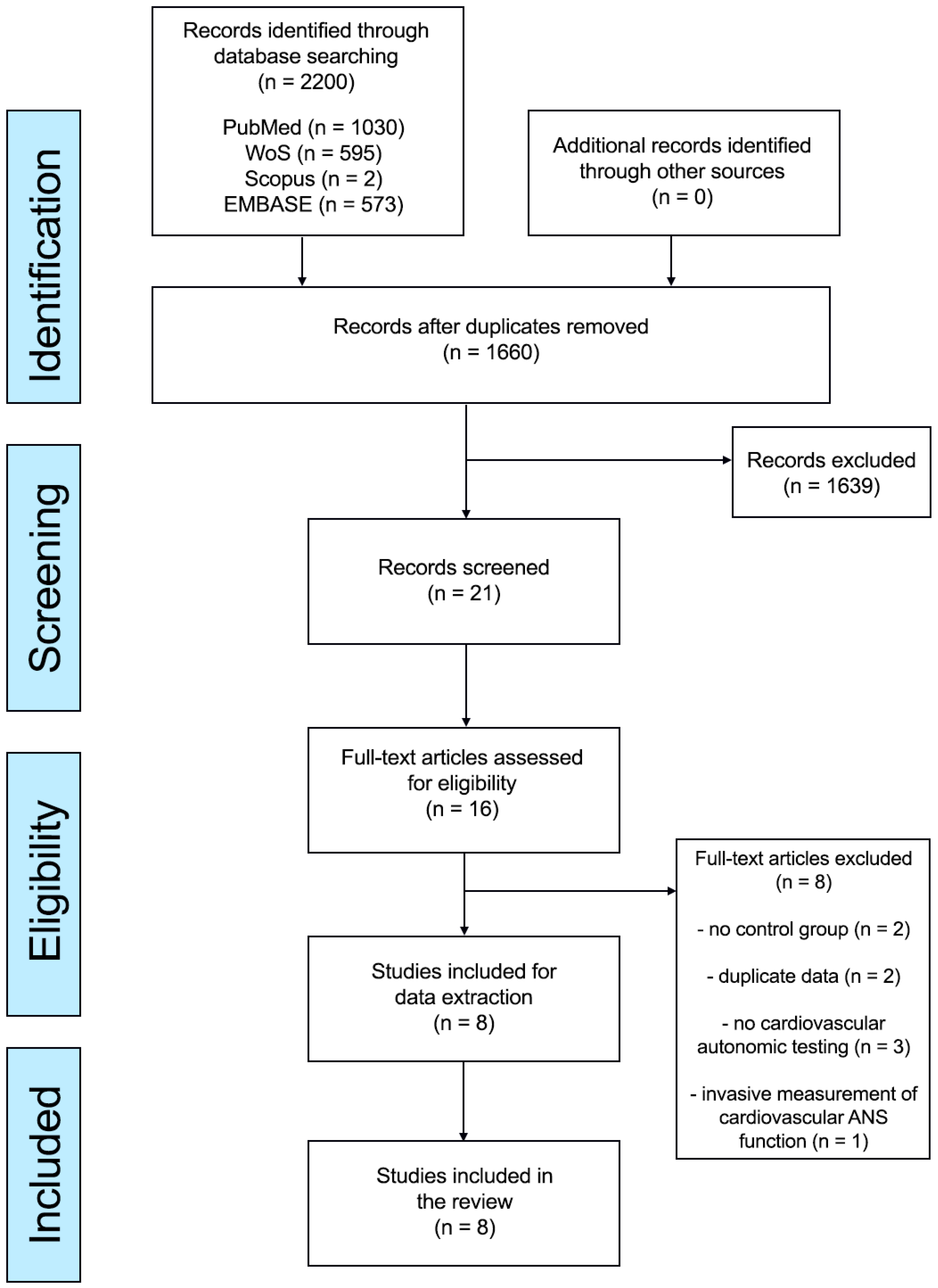
2.3. Study Selection

We adhered to the guidelines outlined in the Cochrane Handbook for Systematic Reviews to select studies for inclusion. During the selection process, through abstract screening, two reviewers (MZK, ŁR) independently screened studies identified through the electronic search engines based on titles and/or abstracts. A third independent reviewer (NV) checked the extracted data.
The process of study selection involved an initial screening based on titles and/or abstracts, followed by a secondary selection retrieved from this preliminary stage using full-text manuscripts. Relevant items were extracted from the retrieved full-text articles.
Data were stored for identification of the manuscript (e.g., first author’s last name, year of publication, country) and characteristics of participants (e.g., sample size, age, and gender distribution), a diagnostic tool for autonomic assessment (e.g., tilt test, Valsalva maneuver, etc.), and cardiovascular autonomic parameters, as well as data on baseline clinical MG characteristics (disease severity, disease duration, antibody status, presence of thymoma).

2.4. Outcomes

The primary outcomes were expressed as the mean values and the corresponding standard deviations (SDs) of the verified tools of cardiovascular autonomic testing, comparing the values of MG patients with HCs. If the data were presented as medians (interquartile ranges), they were transformed into mean and SDs. Selected parameters are described in Supplementary File S1. We studied baroreflex sensitivity (BRS), Valsalva ratio, E/I ratio, rMSSD, pNN50, and HF indices denoting the parasympathetic ANS, and PSD, SDNN, SDNN index, SDANN, and mean RRI representing the overall HRV modulation. LF indexes mainly reflect the sympathetic cardiac modulation but also reflect parasympathetic (vagal) nervous system activity. Valsalva ratio and E/I ratio represent parasympathetic (cardiovagal) function. BRS is an index of both arms of the ANS, given its involvement in both sympathetic input and vagal output.

2.5. Assessment of Risk of Bias

Studies underwent a methodological quality evaluation for risk of bias using the Newcastle Ottawa Scale (NOS). The NOS scale determinates a maximum of nine points, considering three aspects (subject selection, comparability, and outcome), distributed as follows: 1–3 for low, 4–6 for moderate, and 7–9 for high quality [24].

2.6. Data Synthesis

Analyses were performed using PS IMAGO ver. 9.0. Outcomes evaluated by at least two studies (n ≥ 2) were included in the meta-analysis. The main analysis compared cardiovascular autonomic parameters in MG patients and HCs. We calculated the difference between the means of the MG and HC groups using standardized mean differences (SMDs) with 95% confidence intervals (CIs), employing a random-effects model [25].
Heterogeneity between the studies was conducted using both the I2 metric and χ2 statistics. If significant heterogeneity (I2 value ≥ 50%, p < 0.05) was observed and when there were at least ten studies (N ≥ 10), for a given outcome, we performed a meta-regression analysis using moderators.
Publication bias was evaluated by visual inspection funnel plots, as well as using the objective tests (Begg–Mazumdar Kendall tau, and Egger bias test) [26,27]. To address any potential bias, the trim-and-fill method was employed. If asymmetries were detected, adjustment was made for potential effects of unpublished (trimmed) studies [28].
Significance for all analyses was determined by a p-value of less than 0.05.

3. Results

Our search identified 2200 results from 4 electronic databases: PubMed: 1030; Web of Sciences: 595; Scopus: 2; and Embase: 573. After eliminating duplicates, 1660 abstracts were reviewed. From these, 16 full-text articles were considered for eligibility, and eight studies met the inclusion criteria for qualitative and quantitative synthesis. The PRISMA flowchart is shown in Figure 1.

3.1. Characteristics of Included Studies and Participant Details

Study and subject characteristics are summarized in Table 1. All the included studies were conducted retrospectively. Six studies were carried out in Europe and two in India. Most of the studies were published during the last ten years. We were able to identify studies reporting at least one abnormal test and seven studies reporting a minimum of two (Table 2). The eight meta-analyzed studies included a total of 755 participants, including 301 subjects with MG and 454 HCs. The participants with MG had a mean age of 45.2 (±13.9) years. A total of 61.9% of the MG patients were women, 64.4% had serum antibodies to the acetylcholine receptor (Abs AchR+), and 23.9% had a thymoma.
All studies included medicated MG subjects. In the HC group, the mean age was 43.9 (±13.2) years, with a share of 60.7% females across eight studies. Seven studies eliminated individuals with additional cardiovascular conditions or pathologies that could potentially involve dysautonomia, such as diabetes. Table 1 summarizes the details of the included studies and participants.
The two domains of analysis (time and frequency) were used in three of the eight studies [8,9,13]. An additional study reported only time domain analyses.
With respect to cardiovascular reflex tests, six studies [8,9,10,12,13,14] reported sympathetic autonomic function by assessing blood pressure response to a sustained handgrip test, while seven assessed it during an orthostatic challenge. Studies expressed their findings either as a percentage of abnormal results or as absolute values. For the handgrip test, one study used both heart rate (HR) and diastolic blood pressure (dBP), one used mean arterial pressure, and one reported only dBP. Five studies assessed parasympathetic function expressed as HRV in response to standing up and DBT, while six studies reported it using the Valsalva maneuver. Overall, two studies used mean RRI, two studies used RRIV, and one study did not present detailed data. Four studies used BRS at rest, and one during tilt.
The mean NOS score among the studies was 6 (ranging from 5 to 7). The summarized scores are displayed in Table 1.

3.2. Meta-Analysis Results

Comprehensive details of all meta-analysis results, including heterogeneity and publication bias, are provided in Table 3.

3.2.1. Time Domain HRV Parameters: SDNN; SDANN, rMSSD, pNN50, SDNN Index

In total, five parameters of HRV were analyzed, with the rMSSD and SDNN being the most common.
Data pooled from two studies [8,9] including 96 subjects with MG and 76 HCs demonstrated that patients with MG had a lower pNN50 compared to HCs (g = −1.50, 95% CI −2.52–−0.483 p = 0.004). Significant heterogeneity was observed (I2 = 82.8, p = 0.02).
Data from three studies [8,9,13] including 126 subjects with MG and 106 HCs revealed that MG patients had a lower rMSSD compared to HCs (g = −1.94, 95% CI −3.57, −0.32, p = 0.019). There was significant heterogeneity (I2 = 95.1, p = 0.02) but no evidence of publication bias (Egger = 1.35, p = 0.531). (Supplementary File S2).
Data pooled from two studies [8,9] including 96 subjects with MG and 76 HCs demonstrated that MG patients had a lower SDNN index compared to HCs (g = −0.83, 95% CI −1.37–−0.28 p = 0.003), with non-significant heterogeneity (I2 = 55.5, p = 0.13).
There were no differences between MG patients and HCs when comparing SDNN (g = −1.20, 95% CI −3.13–0.73, p = 0.22) and SDANN (g = 0.19, 95% CI −0.26–0.64, p = 0.51) (Table 3).

3.2.2. Frequency Domain HRV Parameters at Rest: LFnu, HFnu, LF, HF, LF/HF, LF/HF of HRV, PSD

The frequency domain HRV measures were used in five studies [8,9,10,11,12,13]. In total, seven parameters were analyzed, with LFnusupine and HFnusupine being the most common.
Four studies [8,9,10,11] conducted LF/HF-RRIsupine assessment in 164 subjects with MG and 136 HCs. The pooled data demonstrated that MG patients had a higher LF/HF-RRIsupine at rest compared to HCs (g = 0.44, 95% CI 0.21–0.68, p < 0.001). There was no heterogeneity (I2 = 0; p = 0.78) and no evidence of publication bias (Egger = 0.42, p = 0.53).
There were no differences between MG patients and HCs at rest when comparing LFnu-RRIsupine (g = 1.34, 95% CI −0.88–3.55, p = 0.24), LF-RRIsupine (g = −0.39, 95% CI −0.39–1.17, p = 0.33), HFnu-RRIsupine (g = −2.81, 95% CI −7.72–2.10, p = 0.26), HF-RRIsupine (g = −0.40, 95% CI −1.16–0.36, p = 0.30), LF/HFsupine (g = 1.80, 95% CI −1.04–4.65, p = 0.21), and PSD-RRIsupine (g = −0.11, 95% CI −0.46–0.24, p = 0.55) (Table 3).

3.2.3. Frequency Domain HRV Parameters in Response to Tilt: LFtilt, HFtilt, LF/HFtilt, LF/HFtilt of HRV, PSDtilt

Data pooled from two studies [10,11] including 68 subjects with MG and 60 HCs demonstrated that patients with MG had a lower HF-RRI during tilt (g = −0.75, 95% CI −0.11–−0.39, p < 0.001). There was no heterogeneity (I2 = 0; p = 0.98).
Data pooled from two studies [10,11] including 68 subjects with MG and 60 HCs demonstrated that patients with MG had a higher LF/HF-RRItilt (g = 0.86, 95% CI 0.50–1.25 p < 0.001) and LF/HFtilt compared to HCs (g = 0.81, 95% CI 0.45–1.18 p < 0.001), with no heterogeneity (I2 = 0, p = 0.52, I2 = 0, p = 0.80, respectively).
There were no differences between MG patients and HCs at rest when comparing LF-RRItilt (g = −0.13, 95% CI −1.05–1.32, p = 0.83) and PSD-RRItilt (g = −0.24, 95% CI −0.65–0.17, p = 0.526) (Table 3).

3.3. Baroreflex Sensitivity (BRS)

Data from four studies [8,9,10,11] including 136 subjects with MG and 164 HCs measured BRS. There was a decreased BRS response among patients with MG compared to HCs (g = −0.56, 95% CI −0.89–−0.33, p < 0.00) with no heterogeneity (I2 = 0.3, p = 0.49)

3.4. Valsalva Ratio

Data from three studies [12,13,15] including 120 people with MG and 301 HCs measured heart rate response to the Valsalva maneuver (ratio). The Valsalva ratio was used to assess cardiovascular parasympathetic function. There was no decreased Valsalva ratio among people with MG compared to HCs (g= −0.22, 95% CI −0.69–0.25, p = 0.36).

3.5. Mean R-R Interval

Data from two studies [8,9] including 96 subjects with MG and 76 HCs measured RRI. There was no increased RRI among patients with MG compared to HCs (g = 0.16, 95% CI −0.14–0.47, p = 0.29) and no heterogeneity (I2 = 81.1, p = 0.49)

3.6. E/I Ratio

Data from two studies [11,13] including 68 people with MG and 60 HCs measured heart rate response to DBT (E/I ratio). E/I ratio was also used to assess cardiovascular parasympathetic function. There was a decreased heart rate response among people with MG compared to HCs (g = −0.45, 95% CI −1.18–0.24, p = 0.01), with significant heterogeneity (I2 = 74.7, p = 0.047).

3.7. Heart Rate and Blood Pressure Parameters at Rest and in Response to Tilt

Five studies [8,10,11,12,14] (patients n = 221, controls n = 373) reported HR at rest (HRrest) and four reported HR in response to HUTT (HRtilt) (patients n = 146, controls n = 318). There were no differences between MG patients and HCs at rest when comparing HRsupine (g = 0.40, 95% CI −0.47–1.26, p = 0.37) and HRtilt (g = 0.19, 95% CI −0.57–0.95, p = 0.63) (Table 3). Both sBPsupine and dBPsupine at rest were reported by four studies, [8,10,11,12] and four studies [10,11,12,14], respectively, during HUTT. Similarly, two studies [11,14] reported mBP at rest (mBPrest) and in response to HUTT (mBPtilt).
Data pooled from four studies [8,10,11,12] including 204 subjects with MG and 356 HCs demonstrated that MG patients had a higher sBPsupine at rest compared to HCs (g = 0.39, 95% CI 0.09–0.68, p = 0.01). There was heterogeneity (I2 = 84.6; p = 0.78) and no evidence of publication bias (Egger = 1.24 p = 0.15).
There were no differences between MG patients and HCs at rest when comparing dBPsupine (g = −0.08, 95% CI −0.42–0.27, p = 0.66), mBPsupine (g = −0.25, 95% CI −0.14–0.66, p = 0.20), and during tilt sBPtilt (g = 0.34, 95% CI −0.27–0.94, p = 0.28), dBPtilt (g = 0.04, 95% CI −0.61–0.69, p = 0.90), mBPtilt (g = −0.04, 95% CI −0.92–0.83, p = 0.92). (Supplementary File S2).

4. Discussion

Across the eight included studies (including 301 MG patients and 454 HCs), we found evidence that MG patients, compared to HCs, have altered cardiac autonomic functions, including decreased parasympathetic activity, lower baroreflex sensitivity, and higher sympathovagal balance at rest and during orthostatic challenge. Patients with MG had significantly lower E/I ratio, as well as lower pNN50, rMSSD, SDNN index, and HF-RRItilt, and higher systolic blood pressure, LF/HF-RRIsupine at rest, and during tilt (LF/HF-RRItilt, LF/HFtilt), as measured by HRV, compared to controls.
The pathophysiological mechanisms through which decreased HRV increases the risk of cardiovascular events and development of CAD are multifactorial. Autoimmunity with immune activation and chronic inflammation is probably a main inducer [29,30]. Previous studies have demonstrated that systemic inflammation markers expressed abnormally in MG patients. In this context, interplay between chronic systemic inflammation and sympathetic nervous system (SNS) overactivation may play a significant role in the autoimmune dysfunction [29]. Inflammation may lead to heightened excitability of premotor sympathetic neurons and suppression of cardiac vagal preganglionic neurons within the medulla [31]. This could lead to an increase in sympathetic outflow to the heart and vasculature, as well as a decrease in parasympathetic outflow to the heart [31]. The result could be elevated blood pressure and a heightened cardiovascular risk [32], as illustrated in our meta-analysis. Another possible mechanism behind AD in MG is cross-reactivity for the skeletal nAChR antibodies, with some idiotypes also binding to ganglionic AChR. Such cross-reactivity has been reported in patients with MG and autoimmune autonomic ganglionopathy (AAG) [33]. In the peripheral nervous system, nicotinic acetylcholine receptors (nAChRs) mediate fast synaptic transmission at autonomic ganglionic (sympathetic, parasympathetic, enteric) and neuromuscular synapses. Furthermore, autoantibodies targeting nAChR in autonomic ganglia have been implicated in AD [34]. Another mechanism of cardiac impairment involving autoantibodies against heart muscle, such as the Ryanodine receptor, adrenergic receptors (beta1- and beta2), titin, and Kv1.4, may also be present. Myositis and/or myocarditis are the most serious manifestations of MG and are related to poor outcomes [16].
There is evidence of autonomic involvement in individuals with MG [6,15] before and after pharmacological treatment. Cholinergic stimulation induced by AChE inhibitor treatment influences autonomic function, including cardiac function, and improves BRS and HRV in both humans and animal disease models [35,36]. AChE inhibitors also increase the HF while decreasing the LF and sympathovagal balance. Moreover, immunosuppressive agents could potentially affect autonomic function [37], thereby increasing the potential risk of arrhythmia, cardiac hypertrophy, and abnormal vascular remodeling [38]. Corticosteroids hold the potential to impact cardiovascular autonomic function, resulting in improved responses in terms of sympathovagal balance (Prednisone/Prednisolone), as observed in patients with Duchenne Muscular Dystrophy [39]. One study, using echocardiography techniques including Tissue Doppler imaging, found that before pyridostigmine intake, compared to controls, MG patients exhibited lower early diastolic AV-plane velocity and diminished peak systolic strain.
However, differences between the groups were equalized following the administration of the drug, suggesting that pyridostigmine restores diastolic function [7]. Nalbantoglu et al. [15], in a study of 22 MG patients, found no significant difference in parasympathetic tests (RR interval variability and Valsalva ratio) before and one hour after the administration of the drug pyridostigmine. Additionally, lower sympathetic skin response (SSR) amplitudes were observed after drug intake, suggesting that pyridostigmine exerts a noncumulative peripheral sympathetic cholinergic effect. However, the utilization of SSR amplitudes and latencies as a quantitative measure is regarded as debatable [40,41]. Evaluating sudomotor function through the qualitative analysis of SSR based on its presence/absence could potentially be a more reliable method [42].
Activation of the SNS and autonomic imbalance are the first reactions to alterations in cardiac loading or myocardial injury and a common pathway to increased morbidity and mortality [43]. AD may be detected before the onset of hypertension [44], as well as diabetes [45], which are non-autoimmune concomitant comorbidities associated with MG [46,47]. Emerging data suggest that reduced HRV and BRS, representing lower vagal activity, are independently associated with a poor cardiovascular prognosis [48]. In addition, increased sympathetic activity has been considered a plausible cause for both atrial and ventricular arrhythmias, hypertension, and, later, heart failure, all of which are conditions observed in MG patients [46].
In support, a large German study with 1660 participants showed that three-quarters of MG patients reported at least one concurrent disease with cardiovascular diseases prevailing (37%) [47]. The burden of MG, including cardiovascular disease, might be exacerbated by the adverse effects of prolonged corticosteroid use and other immunosuppressive therapies [49,50]. A case-control study with a Dutch population (198 patients) demonstrated a higher prevalence of hypertension (35%) and heart diseases (18%), including heart failure and arrhythmia. They reported a higher prevalence of hypertension and type 2 diabetes than in the general population [50]. Similarly, Harris et al. [46] studied the clinical burden in 1149 MG patients and found that patients with refractory MG had a higher occurrence of hypertension, diabetes, and congestive heart failure in comparison to healthy controls. This finding was also related to prolonged corticosteroid use. Results of a retrospective study in Taiwan revealed that individuals with MG exhibited higher prevalence of hypertension, coronary artery disease, diabetes mellitus, dyslipidemia, and thyroid and cerebrovascular diseases. These comorbidities could potentially be linked to the prolonged use of immunosuppressive treatments for MG [51]. High-quality studies designed to examine the relationship between immunosuppressive/glucocorticoid treatments (such as azathioprine) and ischemic stroke are lacking [52].
Despite the advancements in therapeutic management, MG is still associated with an increased mortality rate, with respiratory causes being the most common [6,16]. A study of 1121 Swedish MG patients found no significant difference in cardiovascular diseases, including heart and cerebrovascular diseases, as causes of death compared to the general population [53].
The results of our meta-analysis suggest that sympathetic function, measured as HRV indices, appears to be relatively normal or less compromised in patients with MG. However, vagal impairment can lead to a relative predominance of sympathetic activity at rest and during cardiovascular challenges.
Previous studies evaluating cardiovascular adrenergic function in patients with MG using different methodological approaches have reported mixed findings, including physiologically preserved, depressed [12], or increased sympathetic reactivity [10,11]. Shukla et al. indicated that MG patients, compared to HCs, showed sympathetic hyperreactivity in terms of HR and BP during HUTT, while showing a lesser increase in cardiovascular parameters during the handgrip test.
The authors state that the smaller increase in HR and BP observed during the handgrip test in MG patients may be attributed to MG-related weakness [12]. In contrast, others found similar cardiovascular responses during the handgrip test or orthostatic challenge [9,10]. Nikolic et al. found a higher percentage of sympathetic dysfunction in thymoma-associated MG patients (80% vs. 34.8%), but similar percentages of abnormal results (handgrip test and orthostatic hypotension test) in AChR-positive MG patients without thymoma and MuSK-positive MG patients compared to controls, respectively [8].
Autonomic measures representing parasympathetic activity, including pNN50, rMSSD, and E/I ratio, appear to be lower for patients with MG than for controls. However, these measures were affected by significant heterogeneity, likely due to the limited number of included studies., differences in in other variables such as the percentage of patients with thymoma, and disease severity. Four studies evaluated BRS, which is widely used to quantify the vagal part of the reflex. These studies had low heterogeneity, and the reliability of the results is high.
All studies included were of medium quality (NOS score, mean 6) and had relatively consistent diagnostic criteria, which allowed for better control of bias. However, one study included patients with concomitant comorbidities (23%), which might interfere with the results [12].

5. Limitations

The limitations of the MG studies include their relatively small sample sizes, retrospective design, and the absence of therapy-naïve patients. Furthermore, the quality of the included studies was moderate, and there was a risk of patient selection bias.
Nevertheless, our findings establish a solid foundation for further investigations, also including measures other than cardiac autonomic assessments when examining dysautonomia in MG. Future studies should compare the differences in autonomic function between MG patients with different antibody statuses.

6. Conclusions

As a group, MG patients have altered cardiac autonomic function, including decreased parasympathetic function, lower baroreflex sensitivity, and higher sympathovagal balance at rest and during orthostatic challenges.

Supplementary Materials

The following supporting information can be downloaded at: https://www.mdpi.com/article/10.3390/neurolint15030071/s1. Supplementary File S1 (Table S1: Selected parameters), Supplementary File S2 (Figure S1: Baroreflex sensitivity and Figure S2: Low frequency to high frequency ratio of HRV).

Author Contributions

Conceptualization, M.Z.-K.; Formal analysis, M.Z.-K. and N.V.; methodology, M.Z.-K., Ł.R. and N.V.; project administration, M.Z.-K.; software, M.Z.-K.; supervision, M.T.-K., N.V. and M.B.; writing—original draft preparation, M.Z.-K.; writing—review and editing, M.Z.-K., M.T.-K., Ł.R., M.B., M.H. and N.E.G. All authors have read and agreed to the published version of the manuscript.

Funding

This research received no external funding.

Data Availability Statement

The datasets generated during and/or analyzed during the current study are available from the corresponding author on reasonable request.

Conflicts of Interest

The authors declare no conflict of interest.

References

  1. Sanders, D.B.; Wolfe, G.I.; Benatar, M.; Evoli, A.; Gilhus, N.E.; Illa, I.; Kuntz, N.; Massey, J.M.; Melms, A.; Murai, H.; et al. International consensus guidance for management of myasthenia gravis: Executive summary. Neurology 2016, 87, 419–425. [Google Scholar] [CrossRef] [PubMed]
  2. Gilhus, N.E.; Tzartos, S.; Evoli, A.; Palace, J.; Burns, T.M.; Verschuuren, J.J.G.M. Myasthenia gravis. Nat. Rev. Dis. Primers 2019, 5, 30. [Google Scholar] [CrossRef]
  3. Binks, S.; Vincent, A.; Palace, J. Myasthenia gravis: A clinical-immunological update. J. Neurol. 2016, 263, 826–834. [Google Scholar] [CrossRef] [PubMed]
  4. Khemani, P.; Mehdirad, A.A. Cardiovascular Disorders Mediated by Autonomic Nervous System Dysfunction. Cardiol. Rev. 2020, 28, 65–72. [Google Scholar] [CrossRef] [PubMed]
  5. Gibson, T.C. The heart in myasthenia gravis. Am. Heart J. 1975, 90, 389–396. [Google Scholar] [CrossRef] [PubMed]
  6. Owe, J.F.; Davidsen, E.S.; Eide, G.E.; Gerdts, E.; Gilhus, N.E. Left ventricular long-axis function in myasthenia gravis. J. Neurol. 2008, 255, 1777–1784. [Google Scholar] [CrossRef]
  7. Johannessen, K.-A.; Mygland, A.; Gilhus, N.E.; Aarli, J.; Vik-Mo, H. Left ventricular function in myasthenia gravis. Am. J. Cardiol. 1992, 69, 129–132. [Google Scholar] [CrossRef]
  8. Nikolić, A.; Perić, S.; Nišić, T.; Popović, S.; Ilić, M.; Stojanović, V.R.; Lavrnić, D. The presence of dysautonomia in different subgroups of myasthenia gravis patients. J. Neurol. 2014, 261, 2119–2127. [Google Scholar] [CrossRef]
  9. Peric, S.; Rakocevic-Stojanovic, V.; Nisic, T.; Pavlovic, S.; Basta, I.; Popovic, S.; Damjanovic, S.; Lavrnic, D. Cardiac autonomic control in patients with myasthenia gravis and thymoma. J. Neurol. Sci. 2011, 307, 30–33. [Google Scholar] [CrossRef]
  10. Kocabas, Z.U.; Kizilay, F.; Basarici, I.; Uysal, H. Evaluation of cardiac autonomic functions in myasthenia gravis. Neurol. Res. 2018, 40, 405–412. [Google Scholar] [CrossRef]
  11. Zawadka-Kunikowska, M.; Rzepiński, Ł.; Tafil-Klawe, M.; Klawe, J.J.; Zalewski, P.; Słomko, J. Association of Cardiac Autonomic Responses with Clinical Outcomes of Myasthenia Gravis: Short-Term Analysis of the Heart-Rate and Blood Pressure Variability. J. Clin. Med. 2022, 11, 3697. [Google Scholar] [CrossRef]
  12. Shukla, G.; Gupta, S.; Goyal, V.; Singh, S.; Srivastava, A.; Behari, M. Abnormal sympathetic hyper-reactivity in patients with myasthenia gravis: A prospective study. Clin. Neurol. Neurosurg. 2013, 115, 179–186. [Google Scholar] [CrossRef]
  13. Puneeth, C.S.; Chandra, S.R.; Yadav, R. Heart rate and blood pressure variability in patients with myasthenia gravis. Ann. Indian Acad. Neurol. 2013, 16, 329–332. [Google Scholar]
  14. Elsais, A.; Kerty, E.; Russell, K.; Toska, K. Does cardiovascular autonomic dysfunction contribute to fatigue in myasthenia gravis? Physiol. Res. 2022, 71, 79–91. [Google Scholar] [CrossRef] [PubMed]
  15. Nalbantoglu, M.; Akalin, M.A.; Gunduz, A.; Kiziltan, M. Electrophysiological investigation for autonomic dysfunction in patients with myasthenia gravis: A prospective study. Clin. Neurosci. 2021, 74, 33–40. [Google Scholar] [CrossRef] [PubMed]
  16. Cheng, C.-Y.; Cheng, G.-Y.; Shan, Z.-G.; Baritussio, A.; Lorenzoni, G.; Tyminska, A.; Ozieranski, K.; Iliceto, S.; Marcolongo, R.; Gregori, D.; et al. Efficacy of immunosuppressive therapy in myocarditis: A 30-year systematic review and meta analysis. Autoimmun. Rev. 2021, 20, 102710. [Google Scholar] [CrossRef] [PubMed]
  17. Benjamin, R.N.; Aaron, S.; Sivadasan, A.; Devasahayam, S.; Sebastin, A.; Alexander, M. The Spectrum of Autonomic Dysfunction in Myasthenic Crisis. Ann. Indian Acad. Neurol. 2018, 21, 42–48. [Google Scholar] [CrossRef] [PubMed]
  18. Rzepiński, Ł.; Zawadka-Kunikowska, M.; Newton, J.L.; Zalewski, P. Cardiac Autonomic Dysfunction in Myasthenia Gravis and Relapsing-Remitting Multiple Sclerosis—A Pilot Study. J. Clin. Med. 2021, 10, 2173. [Google Scholar] [CrossRef] [PubMed]
  19. Zawadka-Kunikowska, M.; Rzepiński, Ł.; Cieślicka, M.; Klawe, J.J.; Tafil-Klawe, M. Alterations in short-term blood pressure variability related to disease severity and autonomic symptoms in myasthenia gravis patients. Neurol. Sci. 2023, 28. [Google Scholar] [CrossRef]
  20. Cheshire, W.P.; Freeman, R.; Gibbons, C.H.; Cortelli, P.; Wenning, G.K.; Hilz, M.J.; Spies, J.M.; Lipp, A.; Sandroni, P.; Wada, N.; et al. Electrodiagnostic assessment of the autonomic nervous system: A consensus statement endorsed by the American Autonomic Society, American Academy of Neurology, and the International Federation of Clinical Neurophysiology. Clin. Neurophysiol. 2021, 132, 666–682. [Google Scholar] [CrossRef]
  21. Low, P.A. Composite autonomic scoring scale for laboratory quantification of generalized autonomic failure. Mayo Clin. Proc. 1993, 68, 748–752. [Google Scholar] [CrossRef] [PubMed]
  22. Sarkis-Onofre, R.; Catalá-López, F.; Aromataris, E.; Lockwood, C. How to properly use the PRISMA Statement. Syst. Rev. 2021, 10, 117. [Google Scholar] [CrossRef]
  23. Stroup, D.F.; Berlin, J.A.; Morton, S.C.; Olkin, I.; Williamson, G.D.; Rennie, D.; Moher, D.; Becker, B.J.; Sipe, T.A.; Thacker, S.B. Meta-analysis of observational studies in epidemiology: A proposal for reporting. Meta-analysis Of Observational Studies in Epidemiology (MOOSE) group. JAMA 2000, 283, 2008–2012. [Google Scholar] [CrossRef] [PubMed]
  24. Wells, G.; Shea, B.; O’Connell, D.; Peterson, J. The Newcastle-Ottawa Scale (NOS) for Assessing the Quality of Nonrandomised Studies in Meta-Analyses. Available online: http://www.ohri.ca/programs/clinical_epidemiology/oxford.asp (accessed on 15 June 2023).
  25. Higgins, J.P.; Altman, D.G.; Gøtzsche, P.C.; Jüni, P.; Moher, D.; Oxman, A.D.; Savović, J.; Schulz, K.F.; Weeks, L.; Sterne, J.A.C.; et al. The Cochrane Collaboration’s tool for assessing risk of bias in randomised trials. BMJ 2011, 343, d5928. [Google Scholar] [CrossRef] [PubMed]
  26. Begg, C.B.; Mazumdar, M. Operating characteristics of a rank correlation test for publication bias. Biometrics 1994, 50, 1088–1101. [Google Scholar] [CrossRef] [PubMed]
  27. Egger, M.; Davey Smith, G.; Schneider, M.; Minder, C. Bias in meta-analysis detected by a simple, graphical test. BMJ 1997, 315, 629–634. [Google Scholar] [CrossRef]
  28. Duval, S.; Tweedie, R. Trim and Fill: A simple funnel-plot-based method of testing and adjusting for publication bias in meta-analysis. Biometrics 2000, 56, 455–463. [Google Scholar] [CrossRef]
  29. Jammoul, M.; Naddour, J.; Madi, A.; Reslan, M.A.; Hatoum, F.; Zeineddine, J.; Abou-Kheir, W.; Lawand, N. Investigating the possible mechanisms of autonomic dysfunction post-COVID-19. Auton. Neurosci. 2023, 245, 103071. [Google Scholar] [CrossRef]
  30. Spudich, S.; Nath, A. Nervous system consequences of COVID-19. Science 2022, 375, 267–269. [Google Scholar] [CrossRef]
  31. Adlan, A.M.; Paton, J.F.; Lip, G.Y.; Kitas, G.D.; Fisher, J.P. Increased sympathetic nerve activity and reduced cardiac baroreflex sensitivity in rheumatoid arthritis. J. Physiol. 2017, 595, 967–981. [Google Scholar] [CrossRef]
  32. Pongratz, G.; Straub, R.H. The sympathetic nervous response in inflammation. Arthritis Res. Ther. 2014, 16, 504. [Google Scholar] [CrossRef] [PubMed]
  33. Vernino, S.; Adamski, J.; Kryzer, T.J.; Fealey, R.D.; Lennon, V.A. Neuronal nicotinic ACh receptor antibody in subacute autonomic neuropathy and cancer-related syndromes. Neurology 1998, 50, 1806–1813. [Google Scholar] [CrossRef] [PubMed]
  34. Roy, A.; Guatimosim, S.; Prado, V.F.; Gros, R.; Prado, M.A.M. Cholinergic activity as a new target in diseases of the heart ashbeel. Mol. Med. 2015, 20, 527–537. [Google Scholar] [CrossRef] [PubMed]
  35. Barboza, C.A.; Fukushima, A.R.; Carrozzi, N.; Machi, J.F.; Dourado, P.M.M.; Mostarda, C.T.; Irigoyen, M.C.; Nathanson, L.; Morris, M.; Caperuto, E.C.; et al. Cholinergic Stimulation by Pyridostigmine Bromide Before Myocardial Infarction Prevent Cardiac and Autonomic Dysfunction. Sci. Rep. 2019, 9, 2481. [Google Scholar] [CrossRef]
  36. Gardim, C.B.; Veiga, A.C.; Aguilar, B.A.; Philbois, S.V.; Souza, H.C.D. Effects of chronic cholinergic stimulation associated with aerobic physical training on cardiac morphofunctional and autonomic parameters in spontaneously hypertensive rats. Sci. Rep. 2021, 11, 17141. [Google Scholar] [CrossRef]
  37. Robinson, T.G.; Carr, S.J. Cardiovascular autonomic dysfunction in uremia. Kidney Int. 2002, 62, 1921–1932. [Google Scholar] [CrossRef]
  38. Elezaby, A.; Dexheimer, R.; Sallam, K. Cardiovascular effects of immunosuppression agents. Front. Cardiovasc. Med. 2022, 9, 981838. [Google Scholar] [CrossRef]
  39. Dias, R.M.; Hoshi, R.A.; Vanderlei, L.C.M.; Monteiro, C.B.d.M.; Alvarez, M.P.B.; Crocetta, T.B.; Grossklauss, L.F.; Fernani, D.C.G.L.; Dantas, M.T.A.P.; Martins, F.P.A.; et al. Influence of Different Types of Corticosteroids on Heart Rate Variability of Individuals with Duchenne Muscular Dystrophy—A Pilot Cross Sectional Study. Life 2021, 11, 752. [Google Scholar] [CrossRef]
  40. Kucera, P.; Goldenberg, Z.; Kurca, E. Sympathetic skin response: Review of the method and its clinical use. Bratisl Lek List. 2004, 105, 108–116. [Google Scholar]
  41. Emmer, A.; Mangalo, S.; Kornhuber, M.E. Augmentation of the sympathetic skin response after electrical train stimuli. Front. Neurol. 2012, 3, 152. [Google Scholar] [CrossRef]
  42. Hubli, M.; Krassioukov, A.V. How reliable are sympathetic skin responses in subjects with spinal cord injury? Clin. Auton. Res. 2015, 25, 117–124. [Google Scholar] [CrossRef] [PubMed]
  43. Gronda, E.; Dusi, V.; D’elia, E.; Iacoviello, M.; Benvenuto, E.; Vanoli, E. Sympathetic activation in heart failure. Eur. Heart J. Suppl. 2022, 24, E4–E11. [Google Scholar] [CrossRef] [PubMed]
  44. Jung, M.-H.; Ihm, S.-H.; Lee, D.-H.; Choi, Y.; Chung, W.-B.; Jung, H.O.; Hong, K.-S.; Youn, H.-J. Prehypertension is a comorbid state with autonomic and metabolic dysfunction. J. Clin. Hypertens. 2018, 20, 273–279. [Google Scholar] [CrossRef] [PubMed]
  45. Carnethon, M.R.; Jacobs, D.R.; Sidney, S.; Liu, K. Influence of autonomic nervous system dysfunction on the development of type 2 diabetes: The CARDIA study. Diabetes Care 2003, 26, 3035–3041. [Google Scholar] [CrossRef]
  46. Harris, L.; Graham, S.; MacLachlan, S.; Exuzides, A.; Jacob, S. A retrospective longitudinal cohort study of the clinical burden in myasthenia gravis. BMC Neurol. 2022, 22, 172. [Google Scholar] [CrossRef]
  47. Lehnerer, S.; Jacobi, J.; Schilling, R.; Grittner, U.; Marbin, D.; Gerischer, L.; Stascheit, F.; Krause, M.; Hoffmann, S.; Meisel, A. Burden of disease in myasthenia gravis: Taking the patient’s perspective. J. Neurol. 2022, 269, 3050–3063. [Google Scholar] [CrossRef]
  48. La Rovere, M.T.; Pinna, G.D.; Hohnloser, S.H.; Marcus, F.I.; Mortara, A.; Nohara, R.; Bigger, J.T., Jr.; Camm, A.J.; Schwartz, P.J. Autonomic Tone and Reflexes after Myocardial Infarcton. Baroreflex sensitivity and heart rate variability in the identification of patients at risk for life-threatening arrhythmias: Implications for clinical trials. Circulation 2001, 103, 2072–2077. [Google Scholar] [CrossRef]
  49. Rice, J.B.; White, A.G.; Scarpati, L.M.; Wan, G.; Nelson, W.W. Long-term systemic corticosteroid exposure: A systematic literature review. Clin. Ther. 2017, 39, 2216–2229. [Google Scholar] [CrossRef]
  50. Ruiter, A.M.; Strijbos, E.; de Meel, R.H.; Lipka, A.F.; Raadsheer, W.F.; Tannemaat, M.R.; Verschuuren, J.J. Accuracy of patient-reported data for an online patient registry of autoimmune myasthenia gravis and Lambert-Eaton myasthenic syndrome. Neuromuscul. Disord. 2021, 31, 622–632. [Google Scholar] [CrossRef]
  51. Chu, H.-T.; Tseng, C.-C.; Liang, C.-S.; Yeh, T.-C.; Hu, L.-Y.; Yang, A.C.; Tsai, S.-J.; Shen, C.-C. Risk of Depressive Disorders Following Myasthenia Gravis: A Nationwide Population-Based Retrospective Cohort Study. Front. Psychiatry 2019, 10, 481. [Google Scholar] [CrossRef]
  52. Marto, J.P.; Strambo, D.; Livio, F.; Michel, P. Drugs Associated with Ischemic Stroke: A Review for Clinicians. Stroke 2021, 52, e646–e659. [Google Scholar] [CrossRef] [PubMed]
  53. Westerberg, E.; Punga, A.R. Mortality rates and causes of death in Swedish Myasthenia Gravis patients. Neuromuscul. Disord. 2020, 30, 815–824. [Google Scholar] [CrossRef] [PubMed]
Figure 1. PRISMA flow diagram.
Neurolint 15 00071 g001
Table 1. Baseline characteristics of included studies.
StudyCountryTotal Population (MG, HC)Mean Age of Population (SD)Mean Age of MG (SD)Mean Age of HC (SD)Total %
Female (MG, HC)
Disease Duration (SD)Disease Severity, n% Abs AchR+% ThymomaNOS
Nalbantoglu et al., 2021 [15]Turkey59
(29, 30)
45.95 (14.85)47.86 (15.08)44.1 (14.64)57.58
(55.2; 60)
NR NR51.710.346
Kocabas et al., 2018 [10]Turkey60
(30, 30)
44.8
(11.92)
45.9 (12.08)43.77 (11.1)48.45
(50; 46.7)
8.6 (6.3)Remission: n = 12
MGFA I: n = 4
MGFA IIa: n = 9
MGFA IIb: n = 2
MGFA IIIb: n = 0
MGFA IIIa: 2
MGFA IVa: n = 1
73,926.667
Shukla et al., 2013 [12]India302
(61, 241)
NRNRNRNRNR NRNR65
Elsais, 2022 [14]Norway34
(17, 17)
45
(13.8)
45 (14)45 (14.0)58.8
(58.8; 58.8)
13 (11)Remission: n = 10
MGFA I: n = 7
MGFA IIa: n = 0
MGFA IIb: n = 0
MGFA IIIb: n = 0
MGFA IIIa: n = 0
MGFA IVa: n = 0
NR11.766
Puneeth et al., 2013 [13]India60
(30, 30)
36.05
(13.3)
36.2 (13.6)35.9 (13.3)60
(60; 60)
NR Remission: n = 4
Osserman I: n = 2
Osserman IIa, n = 24
66.76.666
Peric et al., 2011 [9]Serbia42
(21, 21)
52.9
(10.8)
53.2 (9.9)52.7 (11.9)47.6
(47.6; 47.6)
7,4 (6)Remission: n = 7
MGFA I: n =1
MGFA IIa: n = 2
MGFA IIb: n = 9
MGFA IIIa: n = 1
MGFA IIIb: n = 1
MGFA IVa: n = 0
NR 1006
Nicolic et al., 2014 [8]Serbia130
(75, 55)
46.80 (13.88)46.57
(14.42)
47.12 (13.25)75.1
(75.3; 74.9)
6.65 (6.32)Remission: n = 13
MGFA I: n = 24
MGFA IIa: n = 0
MGFA IIb: n = 26
MGFA IIIa: n = 7
MGFA IIIb: n = 5
MGFA IVa: n = 0
69.3276
Zawadka-Kunikowska et al., 2022 [11]Poland68
(38, 30)
41.1
(9.9)
42.8 (11.07)39 (8)81.75 (86.8; 76.7)4.4 (2.7)Remission: n = 0
MGFA I: n = 8
MGFA IIa: n = 19
MGFA IIb: n = 0
MGFA IIIa:11
MGFA IVa: n = 0
60.52.637
Myasthenia gravis, MG; HC, healthy control group; % positive AChR autoantibodies, %Abs AchR+; Newcastle Ottawa Scale, NOS.
Table 2. Summary of included studies.
StudyDiagnostic Tool for Cardiac Autonomic AssessmentCardiovascular Autonomic ParametersMain Results
Nalbantoglu et al., 2021 [15]RRIV
Valsalva maneuver
RRIV
Valsalva ratio
MG patients (both ocular/generalized) exhibit a subclinical parasympathetic abnormality, which is particularly prominent in the AchR antibody-negative group.
Kocabas et al., 2018 [10]BRS
Head-up tilt test
HRV; frequency domain
Handgrip test
DBT
Valsalva maneuver
Active standing: HR, sBP, dBP response
Record time: rest, tilt; sBP, dBP, HR; LF, HF, PSD, LF/HF, LF/HF of HRV
BRSrest, BRStilt,
E/I ratio
Valsalva ratio
30:15 ratio
HR and BP response to HG
Ewing’s battery: percentage of abnormal results
The balance between sympathetic and vagal activity has been disturbed in favor of sympathetic tone and parasympathetic insufficiency has become more prominent.
Shukla et al., 2013 [12]Head-up tilt: tilt 70°
Active standing: 3 min
Handgrip test: 3 min
Valsalva maneuver
R-R interval variation
Record time: rest; RRIV
Valsalva ratio
HR and sBP, dBP response to HG
HR and sBP, dBP response on Valsalva maneuver
HR and sBP, dBP response on HUTT
HR and sBP, dBP on active standing
Sympathetic hyperreactivity among individuals with MG.
Elsais, 2022 [14]Head-up tilt
Handgrip test
Record time: rest, during, after; HR, mBPMG patients experiencing fatigue show higher resting heart rates compared to suitably matched HCs. This distinction is more pronounced in patients who are not using acetylcholinesterase inhibitors.
Puneeth et al., 2013 [13]DBT
Handgrip test
Valsalva maneuver
HRV; frequency and time domains
Active standing
Record time: rest; SDNN, rMSSD; LFnu, HFnu, LF/HF
E-I difference
Valsalva ratio
BP response to HG
Orthostatic fall in systolic BP
Reduction in the values of HR-based tests, along with a BP-based test (isometric handgrip test), was observed in the study group in comparison to the controls. This reduction indicates a deficiency in parasympathetic activity and a minimal level of sympathetic deficiency.
Peric et al., 2011 [9]Handgrip test
DBT
Valsalva maneuver
Active standing
Orthostatic challenge
HRV; frequency and time domains
BRS
Record time: rest; LFnu, HFnu, LF/HF of HRV
SDNN, SDANN, SDNN index, rMSSD, pNN50
Mean R-R interval
BRSrest
Ewing’s battery: percentage of abnormal results
Predominantly, there is parasympathetic cardiac impairment in individuals with MG and thymoma.
Nicolic et al., 2014 [8]Handgrip test
Active standing
Orthostatic hypotension test
Valsalva maneuver
BRS
HRV; frequency and time domains
DBT
Record time: rest
HR, sBP, dBP
LFnu, HFnu, LF, HF, LF/HF ratio of HRV, PSD of HRV, mean R-R interval
SDANN, SDNN, SDNN index, rMSSD, pNN50
Ewing’s battery: percentage of abnormal results
The most prominent autonomic failure was noted among MG patients with thymoma association. Mild parasympathetic abnormalities were observed in AChR-positive thymoma-negative MG patients. MuSK-positive MG patients showed a mild degree of AD.
Zawadka-Kunikowska et al., 2022 [11]Head-up tilt
HRV; frequency domain
BPV
BRS
DBT
Record time: rest, tilt, delta
mBP, sBP, dBP, HR
LFnu, HFnu, LF, HF, LF/HF, LF/HF of HRV, PSD of HRV
LFnu, HFnu, LF, HF, LF/HF, LF/HF of HRV, PSD of sBPV
BRSrest, E/I ratio
CAD with predominant parasympathetic dysfunction.
MG, myasthenia gravis, C, control group; R-R interval variation, RRIV; R-R intervals; deep breathing test, DBT, baroreflex sensitivity (BRS); heart rate, HR; systolic blood pressure sBP; diastolic blood pressure dBP; mean blood pressure, mBP; heart rate variability, HRV; blood pressure variability, BPV; low frequency, LF; high frequency, HF; power spectral density, PSD, index of sympathovagal balance LF/HF, expiration/inspiration ratio the longest R-R interval during inspiration E/I ratio; handgrip test, HG; head-up tilt test, HUTT, expiration-inspiration difference, E-I difference; ratio between longest R-R interval at or around the 30th beat and shortest R-R interval at or around the 15th beat 30:15 ratio; SDNN, mean standard deviation of all normal RR intervals, standard deviation of the averages of NN intervals in all 5-min segments, SDANN; mean of the standard deviations of all NN intervals for all 5-min segments, SDNN index, square root of the mean of squared differences between successive beat intervals, rMSSD; number of pairs of adjacent NN intervals differing more by than 50 ms divided by the total number of all NN intervals, pNN50; nu, normalized units.
Table 3. Meta-analysis results of cardiac autonomic parameters versus healthy controls.
Number of Data SetsSample MGSample CEffect Size (95%CI)pI2p of I2Publication Bias
HRsupine52213730.40 (−0.47; −1.26)0.3794.8<0.001N/A
sBPsupine42043560.39 (0.09; −0.68)0.0156.10.09No
dBPsupine4204356−0.08 (−0.42; −0.27)0.6668.10.013No
mBPsupine255470.25 (−0.14, 0.66)0.200.00.412N/A
LFnusupine41641361.34 (−0.88, 3.55)0.2498.5<0.001No
HFnusupine4164134−2.81 (−7.72, 2.10)0.2699.7<0.001No
LFsupine268600.39 (−0.39, 1.17)0.3379.20.028N/A
HFsupine26860−0.40 (−1.16, 0.36)0.3077.90.033N/A
LF/HFsupine368601.80 (−1.04, 4.65)0.2198.4<0.001No
LF/HF-RRIsupine41641360.44 (0.21, 068)<0.00100.79No
RRIsupine296760.16( −0.14, –0.470.6661.70.11N/A
PSD-RRIsupine26860−0.11 (−0.46, 0.24)0.5500.47N/A
SDNN3126106−1.20 (−3.13, 0.73)0.2297.2<0.001No
SDANN296760.19 (−0.26, 064)0.4144.50.179N/A
rMSSD3126106−1.94 (−3.57, −0.32)0.0295.1<0.001No
pNN5029676−1.2 (−3.134, 0.735)0.00482.80.016N/A
SDNN index29676−0.83 (−1.37, −0.28)0.00355.50.13N/A
BRSsupine4136164−0.56 (−0.80, −0.33)<0.0010.30.49No
HRtilt41463180.19 (−0.57–0.95)0.6390.5<0.001No
sBPtilt31293010.34 (−0.27–0.94)0.2883.80.04No
dBPtilt31293010.04 (−0.61–0.69)0.9085.70.004No
mBPtilt25547−0.04 (−0.92–0.83)0.9277.30.036N/A
LFtilt268600.13 (−1.05, 1.32)0.8390.9<0.001N/A
HFtilt26860−0.75 (−1.11, −0.39)0.0000.98N/A
LF/HF-RRItilt268600.86 (0.50, 1.23)<0.0010.0.96N/A
LF/HFtilt268600.40 (0.05, 0.75)0.0200.57N/A
PSD-RRItilt26860−0.24 (−0.65, 0.17)0.2625.80.246N/A
Valsalva ratio3120301−0.22 (−0.69–0.25)0.3672.20.045No
E/I ratio26860−0.45 (−0.80, −0.09)0.0174.70.047N/A
MG, myasthenia gravis; C, control group; N/A = not applicable (<3 studies publication bias not applicable). Bold values represent significant results (p-value < 0.05).
Disclaimer/Publisher’s Note: The statements, opinions and data contained in all publications are solely those of the individual author(s) and contributor(s) and not of MDPI and/or the editor(s). MDPI and/or the editor(s) disclaim responsibility for any injury to people or property resulting from any ideas, methods, instructions or products referred to in the content.

