
Antimicrobial Activity of Selenium Nanoparticles (SeNPs) against Potentially Pathogenic Oral Microorganisms: A Scoping Review
 , , , and
Abstract
:1. Introduction
2. Materials and Methods
2.1. Protocol and Registration
2.2. Eligibility Criteria
2.3. Sources of Information and Search Strategy
2.4. Selection of Sources of Evidence
2.5. Data Charting Process
2.6. Critical Appraisal
2.7. Synthesis of Results
3. Results
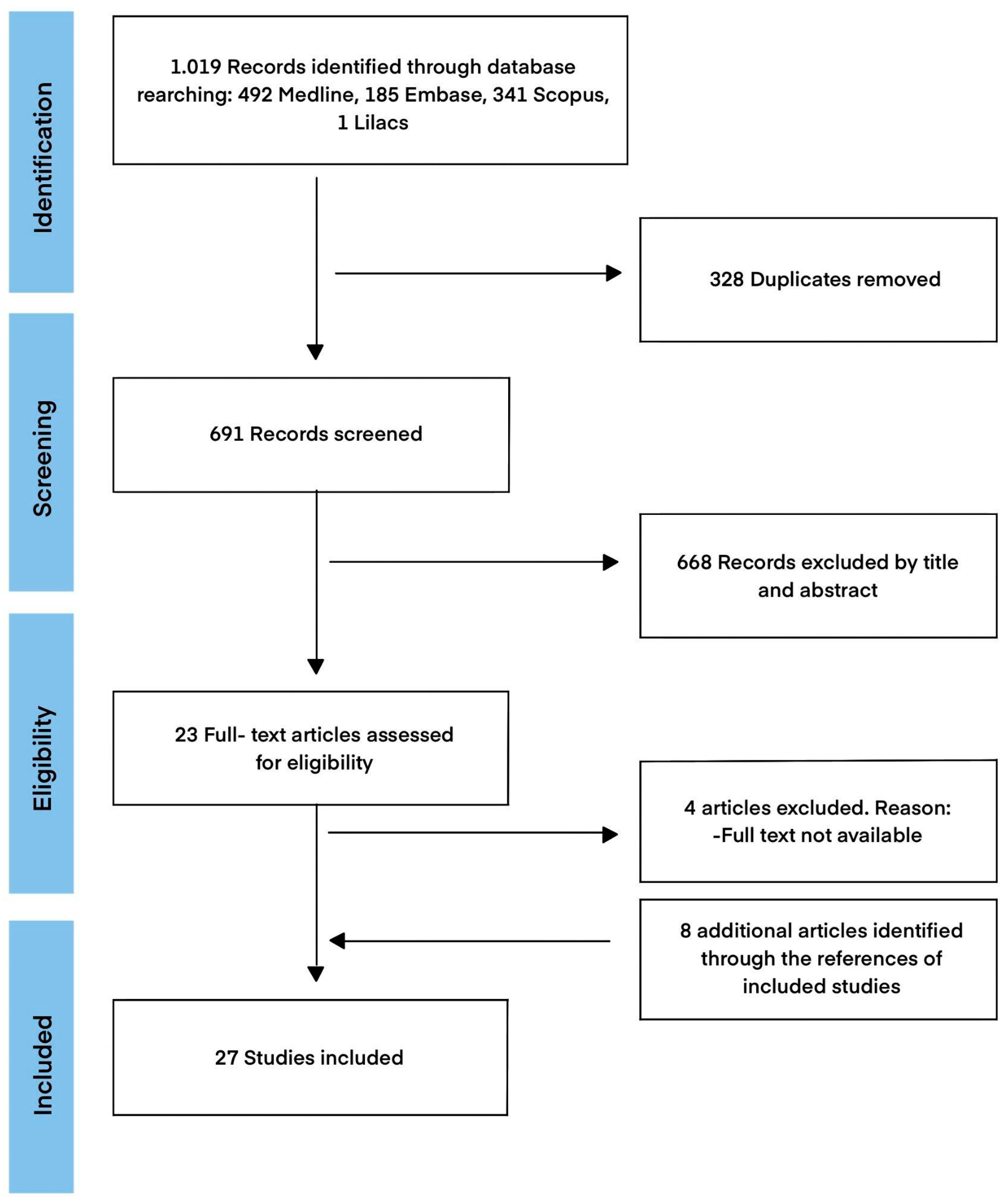
3.1. Selection of Sources of Evidence
3.1.1. Characteristics of Sources of Evidence
3.1.2. Critical Appraisal of Sources of Evidence
3.2. Synthesis of Results
3.2.1. Antimicrobial Efficacy on Planktonic Microorganisms
3.2.2. Antimicrobial Efficacy on Biofilm Microorganisms
3.2.3. Side Effects
- Cytotoxicity of SeNPs
- Antioxidant activity of SeNPs
4. Discussion
4.1. Antimicrobial Efficacy of SeNPs against Pathogenic Oral Microorganisms
4.2. In Vitro Side Effects of SeNPs
4.3. Limitations
4.4. Implications for Practice and Research
5. Conclusions
Supplementary Materials
Author Contributions
Funding
Institutional Review Board Statement
Informed Consent Statement
Data Availability Statement
Conflicts of Interest
References
- Dashper, S.G.; Nastri, A.; Abbott, P.V. Odontogenic Bacterial Infections. In Contemporary Oral Medicine; Farah, C., Balasubramaniam, R., McCullough, M., Eds.; Springer: Cham, Switzerland, 2017. [Google Scholar]
- Lemos, J.A.; Palmer, S.R.; Zeng, L.; Wen, Z.T.; Kajfasz, J.K.; Freires, I.A.; Abranches, J.; Brady, L.J. The Biology of Streptococcus mutans. Microbiol. Spectr. 2019, 7. [Google Scholar] [CrossRef] [PubMed]
- Xu, W.; Zhou, W.; Wang, H.; Liang, S. Roles of Porphyromonas gingivalis and its virulence factors in periodontitis. Adv. Protein. Chem. Struct. Biol. 2020, 120, 45–84. [Google Scholar] [PubMed]
- Dioguardi, M.; Di Gioia, G.; Illuzzi, G.; Arena, C.; Caponio, V.C.A.; Caloro, G.A.; Zhurakivska, K.; Adipietro, I.; Troiano, G.; Lo Muzio, L. Inspection of the Microbiota in Endodontic Lesions. Dent. J. 2019, 7, 47. [Google Scholar] [CrossRef]
- Vila, T.; Sultan, A.S.; Montelongo-Jauregui, D.; Jabra-Rizk, M.A. Oral Candidiasis: A Disease of Opportunity. J. Fungi. 2020, 6, 15. [Google Scholar] [CrossRef] [PubMed]
- Donlan, R.M.; Costerton, J.W. Biofilms: Survival Mechanisms of Clinically Relevant Microorganisms. Clin. Microbiol. Rev. 2002, 15, 167–193. [Google Scholar] [CrossRef] [PubMed]
- Berger, D.; Rakhamimova, A.; Pollack, A.; Loewy, Z. Oral Biofilms: Development, Control, and Analysis. High-Throughput 2018, 7, 24. [Google Scholar]
- Moser, C.; Trøstrup Pedersen, H.; Lerche, C.J.; Kolpen, M.; Line, L.; Thomsen, K.; Høiby, N.; Østrup Jensen, P. Biofilms and host response-helpful or harmful. APMIS 2017, 125, 320–338. [Google Scholar] [CrossRef]
- Pushalkar, S.; Li, X.; Kurago, Z.; Ramanathapuram, L.V.; Matsumura, S.; Fleisher, K.E.; Glickman, R.; Yan, W.; Li, Y.; Saxena, D. Oral microbiota and host innate immune response in bisphosphonate-related osteonecrosis of the jaw. Int. J. Oral. Sci. 2014, 6, 219–226. [Google Scholar] [CrossRef]
- Bessa, L.J.; Botelho, J.; Machado, V.; Alves, R.; Mendes, J.J. Managing Oral Health in the Context of Antimicrobial Resistance. Int. J. Environ. Res. Public Health 2022, 19, 16448. [Google Scholar] [CrossRef]
- Bowen, W.H.; Burne, R.A.; Wu, H.; Koo, H. Oral Biofilms: Pathogens, Matrix, and Polymicrobial Interactions in Microenvironments. Trends Microbiol. 2018, 26, 229–242. [Google Scholar] [CrossRef]
- Serrage, H.J.; Jepson, M.A.; Rostami, N.; Jakubovics, N.S.; Nobbs, A.H. Understanding the Matrix: The Role of Extracellular DNA in Oral Biofilms. Front. Oral Health 2021, 2, 640129. [Google Scholar] [CrossRef] [PubMed]
- Lebeaux, D.; Ghigo, J.M.; Beloin, C. Biofilm-Related Infections: Bridging the Gap between Clinical Management and Fundamental Aspects of Recalcitrance toward Antibiotics. Microbiol. Mol. Biol. Rev. 2014, 78, 510–543. [Google Scholar] [CrossRef] [PubMed]
- Cámara, M.; Green, W.; MacPhee, C.E.; Rakowska, P.D.; Raval, R.; Richardson, M.C.; Slater-Jefferies, J.; Steventon, K.; Webb, J.S. Economic significance of biofilms: A multidisciplinary and cross-sectoral challenge. NPJ Biofilms Microbiomes 2022, 8, 42. [Google Scholar] [CrossRef] [PubMed]
- Wang, L.; Hu, C.; Shao, L. The antimicrobial activity of nanoparticles: Present situation and prospects for the future. Int. J. Nanomed. 2017, 12, 1227–1249. [Google Scholar] [CrossRef] [PubMed]
- Vallet-Regí, M.; González, B.; Izquierdo-Barba, I. Nanomaterials as Promising Alternative in the Infection Treatment. Int. J. Mol. Sci. 2019, 20, 3806. [Google Scholar] [CrossRef]
- Mubeen, B.; Ansar, A.N.; Rasool, R.; Ullah, I.; Imam, S.S.; Alshehri, S.; Ghoneim, M.M.; Alzarea, S.I.; Nadeem, M.S.; Kazmi, I. Nanotechnology as a Novel Approach in Combating Microbes Providing an Alternative to Antibiotics. Antibiotics 2021, 10, 1473. [Google Scholar] [CrossRef]
- Khezerlou, A.; Alizadeh-Sani, M.; Azizi-Lalabadi, M.; Ehsani, A. Nanoparticles and their antimicrobial properties against pathogens including bacteria, fungi, parasites and viruses. Microb. Pathog. 2018, 123, 505–526. [Google Scholar] [CrossRef]
- Saraeva, I.; Tolordava, E.; Yushina, Y.; Sozaev, I.; Sokolova, V.; Khmelnitskiy, R.; Sheligyna, S.; Pallaeva, T.; Pokryshkin, N.; Khmelenin, D.; et al. Direct Bactericidal Comparison of Metal Nanoparticles and Their Salts against S. aureus Culture by TEM and FT-IR Spectroscopy. Nanomaterials 2022, 12, 3857. [Google Scholar] [CrossRef]
- Sans-Serramitjana, E.; Fusté, E.; Martínez-Garriga, B.; Merlos, A.; Pastor, M.; Pedraz, J.L.; Esquisabel, A.; Bachiller, D.; Vinuesa, T.; Viñas, M. Killing effect of nanoencapsulated colistin sulfate on Pseudomonas aeruginosa from cystic fibrosis patients. J. Cyst. Fibros. 2016, 15, 611–618. [Google Scholar] [CrossRef]
- Sans-Serramitjana, E.; Jorba, M.; Pedraz, J.L.; Vinuesa, T.; Viñas, M. Determination of the spatiotemporal dependence of Pseudomonas aeruginosa biofilm viability after treatment with NLC-colistin. Int. J. Nanomed. 2017, 12, 4409–4413. [Google Scholar] [CrossRef]
- Nair, A.; Mallya, R.; Suvarna, V.; Khan, T.A.; Momin, M.; Omri, A. Nanoparticles—Attractive Carriers of Antimicrobial Essential Oils. Antibiotics 2022, 11, 108. [Google Scholar] [CrossRef] [PubMed]
- Jamuna, B.A.; Ravishankar, R.V. Environmental Risk, Human Health, and Toxic Effects of Nanoparticles. In Nanomaterials for Environmental Protection; Kharisov, B.I., Kharissova, O.V., Dias, H.V.R., Eds.; Wiley: Hoboken, NJ, USA, 2014; pp. 523–535. [Google Scholar]
- Panáček, A.; Kvítek, L.; Smékalová, M.; Večeřová, R.; Kolář, M.; Röderová, M.; Dyčka, F.; Šebela, M.; Prucek, R.; Tomanec, O.; et al. Bacterial resistance to silver nanoparticles and how to overcome it. Nat. Nanotechnol. 2018, 13, 65–71. [Google Scholar] [CrossRef] [PubMed]
- Lu, J.; Wang, Y.; Jin, M.; Yuan, Z.; Bond, P.; Guo, J. Both silver ions and silver nanoparticles facilitate the horizontal transfer of plasmid-mediated antibiotic resistance genes. Water Res. 2020, 169, 115229. [Google Scholar] [CrossRef] [PubMed]
- Zhang, S.; Wang, Y.; Song, H.; Lu, J.; Yuan, Z.; Guo, J. Copper nanoparticles and copper ions promote horizontal transfer of plasmid-mediated multi-antibiotic resistance genes across bacterial genera. Environ. Int. 2019, 129, 478–487. [Google Scholar] [CrossRef]
- Zambonino, M.C.; Quizhpe, E.M.; Jaramillo, F.E.; Rahman, A.; Santiago Vispo, N.; Jeffryes, C.; Dahoumane, S.A. Green Synthesis of Selenium and Tellurium Nanoparticles: Current Trends, Biological Properties and Biomedical Applications. Int. J. Mol. Sci. 2021, 22, 989. [Google Scholar] [CrossRef]
- Kurokawa, S.; Berry, M.J. Selenium. Role of the Essential Metalloid in Health. Interrelations between Essential Metal Ions and Human Diseases. Met. Ions Life Sci. 2013, 13, 499–534. [Google Scholar]
- Marzo, T.; La Mendola, D. Strike a Balance: Between Metals and Non-Metals, Metalloids as a Source of Anti-Infective Agents. Inorganics 2021, 9, 46. [Google Scholar] [CrossRef]
- Zhang, J.; Wang, X.; Xu, T. Elemental selenium at nano size (Nano-Se) as a potential chemopreventive agent with reduced risk of selenium toxicity: Comparison with se-methylselenocysteine in mice. Toxicol. Sci. 2008, 101, 22–31. [Google Scholar] [CrossRef]
- Mal, J.; Veneman, W.J.; Nancharaiah, Y.V.; van Hullebusch, E.D.; Peijnenburg, W.J.; Vijver, M.G.; Lens, P.N. A comparison of fate and toxicity of selenite, biogenically, and chemically synthesized selenium nanoparticles to zebrafish (Danio rerio) embryogenesis. Nanotoxicology 2017, 11, 87–97. [Google Scholar] [CrossRef]
- Bai, K.; Hong, B.; He, J.; Hong, Z.; Tan, R. Preparation and antioxidant properties of selenium nanoparticles-loaded chitosan microspheres. Int. J. Nanomed. 2017, 12, 4527–4539. [Google Scholar] [CrossRef]
- Jin, Y.; He, Y.; Liu, L.; Tao, W.; Wang, G.; Sun, W.; Pei, X.; Xiao, Z.; Wang, H. Effects of Supranutritional Selenium Nanoparticles on Immune and Antioxidant Capacity in Sprague-Dawley Rats. Biol. Trace Elem. Res. 2021, 199, 4666–4674. [Google Scholar] [CrossRef] [PubMed]
- Varlamova, E.G.; Goltyaev, M.V.; Maltseva, V.N.; Turovsky, E.A.; Sarimov, R.M.; Simakin, A.V.; Gudkov, S.V. Mechanisms of the Cytotoxic Effect of Selenium Nanoparticles in Different Human Cancer Cell Lines. Int. J. Mol. Sci. 2021, 22, 7798. [Google Scholar] [CrossRef]
- Liao, G.; Tang, J.; Wang, D.; Zuo, H.; Zhang, Q.; Liu, Y.; Xiong, H. Selenium nanoparticles (SeNPs) have potent antitumor activity against prostate cancer cells through the upregulation of miR-16. World J. Surg. Oncol. 2021, 18, 81. [Google Scholar] [CrossRef] [PubMed]
- Truong, L.B.; Medina, D.; Mostafavi, E.; Rabiee, N. Selenium Nanomaterials to Combat Antimicrobial Resistance. Molecules 2021, 26, 3611. [Google Scholar] [CrossRef] [PubMed]
- Lin, W.; Zhang, J.; Xu, J.F.; Pi, J. The Advancing of Selenium Nanoparticles Against Infectious Diseases. Front. Pharmacol. 2021, 12, 682284. [Google Scholar] [CrossRef]
- Abbas, H.S.; Abou Baker, D.H.; Ahmed, E.A. Cytotoxicity and antimicrobial efficiency of selenium nanoparticles biosynthesized by Spirulina platensis. Arch. Microbiol. 2021, 203, 523–532. [Google Scholar] [CrossRef]
- Zhang, H.; Li, Z.; Dai, C.; Wang, P.; Fan, S.; Yu, B.; Qu, Y. Antibacterial properties and mechanism of selenium nanoparticles synthesized by Providencia sp. DCX. Environ. Res. 2021, 194, 110630. [Google Scholar] [CrossRef]
- Tricco, A.C.; Lillie, E.; Zarin, W.; O’Brien, K.K.; Colquhoun, H.; Levac, D.; Moher, D.; Peters, M.; Horsley, T.; Weeks, L.; et al. PRISMA extension for scoping reviews (PRISMA-ScR): Checklist and explanation. Ann. Intern. Med. 2018, 169, 467–473. [Google Scholar] [CrossRef]
- Schneider, K.; Schwarz, M.; Burkholder, I.; Kopp-Schneider, A.; Edler, L.; Kinsner-Ovaskainen, A.; Hartung, T.; Hoffmann, S. “ToxRTool”, a new tool to assess the reliability of toxicological data. Toxicol. Lett. 2009, 189, 138–144. [Google Scholar] [CrossRef]
- Green, B.N.; Johnson, C.D.; Adams, A. Writing narrative literature reviews for peer-reviewed journals: Secrets of the trade. J. Chiropr. Med. 2006, 5, 101–117. [Google Scholar] [CrossRef]
- Gunasekaran, G.; Ramamurthy, J.; Rajeshkumar, S. Evaluation of the Antimicrobial Activity of Solanum Nigrum Infused Selenium Nanoparticles. Int. J. Pharm. Res. 2020, 12. [Google Scholar] [CrossRef]
- Khalil, E.; Abdalla Elkhader, I.; Elsahn, N.A.; Sid Ahmed, A.; Raj, S.S. Antibacterial activity of novel selenium and zinc nanoparticles toward oral Candida albicans: An In vitro study. Drug Invent. Today 2020, 14, 36–38. [Google Scholar]
- Safaei, M.; Mozaffari, H.R.; Moradpoor, H.; Imani, M.M.; Sharifi, R.; Golshah, A. Optimization of Green Synthesis of Selenium Nanoparticles and Evaluation of Their Antifungal Activity against Oral Candida albicans Infection. Adv. Mater. Sci. Eng. 2022, 2022, 1376998. [Google Scholar] [CrossRef]
- Abdel-Moneim AM, E.; El-Saadony, M.T.; Shehata, A.M.; Saad, A.M.; Aldhumri, S.A.; Ouda, S.M.; Mesalam, N.M. Antioxidant and antimicrobial activities of Spirulina platensis extracts and biogenic selenium nanoparticles against selected pathogenic bacteria and fungi. Saudi J. Biol. Sci. 2022, 29, 1197–1209. [Google Scholar] [CrossRef] [PubMed]
- Anushya, P.; Preetha, S.; Prathap, L.; Jeevitha, M. Enhanced Antibacterial activity of Capparis decidua fruit mediated Selenium Nanoparticle against Enterococcus faecalis. Int. J. Res. Pharm. Sci. 2021, 12, 6–11. [Google Scholar] [CrossRef]
- Chellapa, L.R.; Rajeshkumar, S.; Arumugham, M.I.; Samuel, S.R. Biogenic Nanoselenium Synthesis and Evaluation of its antimicrobial, Antioxidant Activity and Toxicity. Bioinspired Biomim. Nanobiomaterials 2020, 9, 184–189. [Google Scholar] [CrossRef]
- Rao, C.K.; Rosaiah, G.; Mangamuri, U.K.; Sikharam, A.S.; Devaraj, K.; Kalagatur, N.K.; Kadirvelu, K. Biosynthesis of Selenium Nanoparticles from Annona muricata Fruit Aqueous Extract and Investigation of their Antioxidant and Antimicrobial potentials. Curr. Trends Biotechnol. Pharm. 2022, 16, 101–107. [Google Scholar]
- Darroudi, M.; Rangrazi, A.; Ghazvini, K.; Bagheri, H.; Boruziniat, A. Antimicrobial activity of colloidal selenium nanoparticles in chitosan solution against Streptococcus mutans, Lactobacillus acidophilus, and Candida albicans. Pesqui. Bras. Odontopediatria Clín. Integr. 2021, 21. [Google Scholar] [CrossRef]
- Hashem, A.H.; Khalil, A.M.A.; Reyad, A.M.; Salem, S.S. Biomedical applications of mycosynthesized selenium nanoparticles using Penicillium expansum ATTC 36200. Biol. Trace Elem. Res. 2021, 199, 3998–4008. [Google Scholar] [CrossRef]
- Rangrazi, A.; Bagheri, H.; Ghazvini, K.; Boruziniat, A.; Darroudi, M. Synthesis and antibacterial activity of colloidal selenium nanoparticles in chitosan solution: A new antibacterial agent. Mater. Res. Express 2019, 6, 1250h3. [Google Scholar] [CrossRef]
- Shakibaie, M.; Salari Mohazab, N.; Ayatollahi Mousavi, S.A. Antifungal Activity of Selenium Nanoparticles Synthesized by Bacillus species Msh-1 Against Aspergillus fumigatus and Candida albicans. Jundishapur J. Microbiol. 2015, 8, e26381. [Google Scholar] [CrossRef] [PubMed]
- Dhanraj, G.; Rajeshkumar, S. Anticariogenic Effect of Selenium Nanoparticles Synthesized Using Brassica oleracea. J. Nanomater. 2021, 2021, 8115585. [Google Scholar] [CrossRef]
- Cremonini, E.; Zonaro, E.; Donini, M.; Lampis, S.; Boaretti, M.; Dusi, S.; Melotti, P.; Lleo, M.M.; Vallini, G. Biogenic selenium nanoparticles: Characterization, antimicrobial activity and effects on human dendritic cells and fibroblasts. Microb. Biotechnol. 2016, 9, 758–771. [Google Scholar] [CrossRef] [PubMed]
- Filipovic, N.; Ušjak, D.; Milenkovic, M.T.; Zheng, K.; Liverani, L.; Boccaccini, A.R.; Stevanovic, M.M. Comparative Study of the Antimicrobial Activity of Selenium Nanoparticles with Different Surface Chemistry and Structure. Front. Bioeng. Biotechnol. 2021, 8, 624621. [Google Scholar] [CrossRef] [PubMed]
- Miglani, S.; Tani-Ishii, N. Biosynthesized selenium nanoparticles: Characterization, antimicrobial, and antibiofilm activity against Enterococcus faecalis. Peer J. 2021, 9, e11653. [Google Scholar] [CrossRef]
- Shahmoradi, S.; Shariati, A.; Amini, S.M.; Zargar, N.; Yadegari, Z.; Darban-Sarokhalil, D. The application of selenium nanoparticles for enhancing the efficacy of photodynamic inactivation of planktonic communities and the biofilm of Streptococcus mutans. BMC Res. Notes 2022, 15, 84. [Google Scholar] [CrossRef]
- Shahmoradi, S.; Shariati, A.; Zargar, N.; Yadegari, Z.; Asnaashari, M.; Amini, S.M.; Darban-Sarokhalil, D. Antimicrobial effects of selenium nanoparticles in combination with photodynamic therapy against Enterococcus faecalis biofilm. Photodiagnosis Photodyn. Ther. 2021, 35, 102398. [Google Scholar] [CrossRef]
- Kishen, A.; Rajeshkumar, S.; Preejitha, V.B. Cynodon dactylon Mediated Synthesis of Selenium Nanoparticles and Its Antimicrobial Activity Against Oral Pathogens. Int. J. Res. Pharm. Sci. 2020, 11, 4152–4156. [Google Scholar] [CrossRef]
- Hou, J.; Tamura, Y.; Lu, H.Y.; Takahashi, Y.; Kasugai, S.; Nakata, H.; Kuroda, S. An In Vitro Evaluation of Selenium Nanoparticles on Osteoblastic Differentiation and Antimicrobial Properties against Porphyromonas gingivalis. Nanomaterials 2022, 12, 1850. [Google Scholar] [CrossRef]
- Nile, S.H.; Thombre, D.; Shelar, A.; Gosavi, K.; Sangshetti, J.; Zhang, W.; Sieniawska, E.; Patil, R.; Kai, G. Antifungal Properties of Biogenic Selenium Nanoparticles Functionalized with Nystatin for the Inhibition of Candida albicans Biofilm Formation. Molecules 2023, 28, 1836. [Google Scholar] [CrossRef]
- Hashem, A.H.; Saied, E.; Ali, O.M.; Selim, S.; Al Jaouni, S.K.; Elkady, F.M.; El-Sayyad, G.S. Pomegranate Peel Extract Stabilized Selenium Nanoparticles Synthesis: Promising Antimicrobial Potential, Antioxidant Activity, Biocompatibility, and Hemocompatibility. Appl. Biochem. Biotechnol. 2023. [Google Scholar] [CrossRef] [PubMed]
- Hariharan, S.; Chauhan, S.; Velu, K.; Dharmaraj, S.; Kumar, V.; Ganesan, S. Biological Activities of Selenium Nanoparticles Synthesized from Camellia sinensis (L) Kuntze Leaves. Appl. Biochem. Biotechnol. 2023. [Google Scholar] [CrossRef] [PubMed]
- Nassar, A.R.; Eid, A.M.; Atta, H.M.; El Naghy, W.S.; Fouda, A. Exploring the antimicrobial, antioxidant, anticancer, biocompatibility, and larvicidal activities of selenium nanoparticles fabricated by endophytic fungal strain Penicillium verhagenii. Sci. Rep. 2023, 13, 9054. [Google Scholar] [CrossRef] [PubMed]
- Arthi, S.; Ramani, P.; Rajeshkumar, S. Green synthesis of Annona muricata mediated selenium nanoparticles and its antifungal activity against Candida albicans. J. Popul. Ther. Clin. Pharmacol. 2023, 30, 282–287. [Google Scholar]
- Guisbiers, G.; Lara, H.H.; Mendoza, R.; Naranjo, G.; Vincent, B.A.; Peralta, X.G.; Nash, K.L. Inhibition of Candida albicans biofilm by pure selenium nanoparticles synthesized by pulsed laser ablation in liquids. Nanomed. Nanotechnol. Biol. Med. 2016, 13, 1095–1103. [Google Scholar] [CrossRef] [PubMed]
- Lara, H.H.; Guisbiers, G.; Mendoza, J.; Mimun, L.C.; Vincent, B.A.; Lopez-Ribot, J.L.; Nash, K.L. Synergistic antifungal effect of chitosan-stabilized selenium nanoparticles synthesized by pulsed laser ablation in liquids against Candida albicans biofilms. Int. J. Nanomed. 2018, 13, 2697–2708. [Google Scholar] [CrossRef]
- Lyu, X.; Zhao, C.; Yan, Z.M.; Hua, H. Efficacy of nystatin for the treatment of oral candidiasis: A systematic review and meta-analysis. Drug Des. Dev. Ther. 2016, 10, 1161–1171. [Google Scholar] [CrossRef]
- Lee, N.Y.; Ko, W.C.; Hsueh, P.R. Nanoparticles in the Treatment of Infections Caused by Multidrug-Resistant Organisms. Front. Pharmacol. 2019, 10, 1153. [Google Scholar] [CrossRef]
- Lu, Z.; Rong, K.; Li, J.; Yang, H.; Chen, R. Size-dependent antibacterial activities of silver nanoparticles against oral anaerobic pathogenic bacteria. J. Mater. Sci. Mater. Med. 2013, 24, 1465–1471. [Google Scholar] [CrossRef]
- Ramalingam, B.; Parandhaman, T.; Das, S.K. Antibacterial Effects of Biosynthesized Silver Nanoparticles on Surface Ultrastructure and Nanomechanical Properties of Gram-Negative Bacteria viz. Escherichia coli and Pseudomonas aeruginosa. ACS Appl. Mater. Interfaces 2016, 8, 4963–4976. [Google Scholar]
- Fröber, K.; Bergs, C.; Pich, A.; Conrads, G. Biofunctionalized zinc peroxide nanoparticles inhibit peri-implantitis associated anaerobes and Aggregatibacter actinomycetemcomitans pH-dependent. Anaerobe 2020, 62, 102153. [Google Scholar] [CrossRef]
- Breijyeh, Z.; Jubeh, B.; Karaman, R. Resistance of Gram-Negative Bacteria to Current Antibacterial Agents and Approaches to Resolve It. Molecules 2020, 25, 1340. [Google Scholar] [CrossRef]
- Linklater, D.P.; Baulin, V.A.; Le Guével, X.; Fleury, J.; Hanssen, E.; Nguyen, T.H.P.; Juodkazis, S.; Bryant, G.; Crawford, R.J.; Stoodley, P.; et al. Antibacterial Action of Nanoparticles by Lethal Stretching of Bacterial Cell Membranes. Adv. Mater. 2020, 32, 2005679. [Google Scholar] [CrossRef] [PubMed]
- Behzadi, S.; Serpooshan, V.; Tao, W.; Hamaly, M.A.; Alkawareek, M.Y.; Dreaden, E.C.; Brown, D.; Alkilany, A.M.; Farokhzad, O.C.; Mahmoudi, M. Cellular uptake of nanoparticles: Journey inside the cell. Chem. Soc. Rev. 2017, 46, 4218–4244. [Google Scholar] [CrossRef] [PubMed]
- Nag, M.; Lahiri, D.; Sarkar, T.; Ghosh, S.; Dey, A.; Edinur, H.A.; Pati, S.; Ray, R.R. Microbial Fabrication of Nanomaterial and Its Role in Disintegration of Exopolymeric Matrices of Biofilm. Front. Chem. 2021, 9, 690590. [Google Scholar] [CrossRef] [PubMed]
- Piacenza, E.; Presentato, A.; Zonaro, E.; Lemire, J.A.; Demeter, M.; Vallini, G.; Turner, R.J.; Lampis, S. Antimicrobial activity of biogenically produced spherical Se-nanomaterials embedded in organic material against Pseudomonas aeruginosa and Staphylococcus aureus strains on hydroxyapatite-coated surfaces. Microb. Biotechnol. 2017, 10, 804–818. [Google Scholar] [CrossRef] [PubMed]
- Escobar-Ramírez, M.C.; Castañeda-Ovando, A.; Pérez-Escalante, E.; Rodríguez-Serrano, G.M.; Ramírez-Moreno, E.; Quintero-Lira, A.; Contreras-López, E.; Añorve-Morga, J.; Jaimez-Ordaz, J.; González-Olivares, L.G. Antimicrobial Activity of Se-Nanoparticles from Bacterial Biotransformation. Fermentation 2021, 7, 130. [Google Scholar] [CrossRef]
- Asadpoor, M.; Ithakisiou, G.N.; van Putten, J.P.M.; Pieters, R.J.; Folkerts, G.; Braber, S. Antimicrobial Activities of Alginate and Chitosan Oligosaccharides Against Staphylococcus aureus and Group B Streptococcus. Front. Microbiol. 2021, 12, 700605. [Google Scholar] [CrossRef] [PubMed]
- Ikono, R.; Vibriani, A.; Wibowo, I.; Saputro, K.E.; Muliawan, W.; Bachtiar, B.M.; Mardliyati, E.; Bachtiar, E.W.; Rochman, N.T.; Kagami, H.; et al. Nanochitosan antimicrobial activity against Streptococcus mutans and Candida albicans dual-species biofilms. BMC Res. Notes 2019, 12, 383. [Google Scholar] [CrossRef]
- Menichetti, A.; Mavridi-Printezi, A.; Mordini, D.; Montalti, M. Effect of Size, Shape and Surface Functionalization on the Antibacterial Activity of Silver Nanoparticles. J. Funct. Biomater. 2023, 14, 244. [Google Scholar] [CrossRef]
- Dumore, N.S.; Mukhopadhyay, M. Antioxidant properties of aqueous selenium nanoparticles (ASeNPs) and its catalysts activity for 1, 1-diphenyl-2-picrylhydrazyl (DPPH) reduction. J. Mol. Struct. 2019, 1205, 127637. [Google Scholar] [CrossRef]
- Dumore, N.S.; Mukhopadhyay, M. Sensitivity enhanced SeNPs-FTO electrochemical sensor for hydrogen peroxide detection. J. Electroanal. Chem. 2020, 878, 114544. [Google Scholar] [CrossRef]
- Ferrisse, T.M.; Dias, L.M.; de Oliveira, A.B.; Jordão, C.C.; Mima, E.G.O.; Pavarina, A.C. Efficacy of Antimicrobial Photodynamic Therapy Mediated by Photosensitizers Conjugated with Inorganic Nanoparticles: Systematic Review and Meta-Analysis. Pharmaceutics 2022, 14, 2050. [Google Scholar] [CrossRef] [PubMed]
- Misba, L.; Kulshrestha, S.; Khan, A.U. Antibiofilm action of a toluidine blue O-silver nanoparticle conjugate on Streptococcus mutans: A mechanism of type I photodynamic therapy. Biofouling 2016, 32, 313–328. [Google Scholar] [CrossRef] [PubMed]
- Darabpour, E.; Kashef, N.; Mashayekhan, S. Chitosan nanoparticles enhance the efficiency of methylene blue-mediated antimicrobial photodynamic inactivation of bacterial biofilms: An in vitro study. Photodiagn. Photodyn. Ther. 2016, 14, 211–217. [Google Scholar] [CrossRef]
- Shahabadi, N.; Zendehcheshm, S.; Khademi, F. Selenium nanoparticles: Synthesis, in-vitro cytotoxicity, antioxidant activity and interaction studies with ct-DNA and HSA, HHb and Cyt c serum proteins. Biotechnol. Rep. 2021, 30, e00615. [Google Scholar] [CrossRef]
- Chung, S.; Zhou, R.; Webster, T.J. Green Synthesized BSA-Coated Selenium Nanoparticles Inhibit Bacterial Growth While Promoting Mammalian Cell Growth. Int. J. Nanomed. 2020, 15, 115–124. [Google Scholar] [CrossRef]
- Gharbavi, M.; Johari, B.; Mousazadeh, N.; Rahimi, B.; Parvinzad Leilan, M.; Sadegh Eslami, S.; Sharafi, A. Hybrid of niosomes and bio-synthesized selenium nanoparticles as a novel approach in drug delivery for cancer treatment. Mol. Biol. Rep. 2020, 47, 6517–6529. [Google Scholar] [CrossRef]
- Varlamova, E.G.; Turovsky, E.A.; Blinova, E.V. Therapeutic Potential and Main Methods of Obtaining Selenium Nanoparticles. Int. J. Mol. Sci. 2021, 22, 10808. [Google Scholar] [CrossRef]
- Xu, C.; Qiao, L.; Guo, Y.; Ma, L.; Cheng, Y. Preparation, characteristics and antioxidant activity of polysaccharides and proteins-capped selenium nanoparticles synthesized by Lactobacillus casei ATCC 393. Carbohydr. Polym. 2018, 195, 576–585. [Google Scholar] [CrossRef]
- Sheiha, A.M.; Abdelnour, S.A.; El-Hack, A.; Mohamed, E.; Khafaga, A.F.; Metwally, K.A.; Ajarem, J.S.; Maodaa, S.N.; Allam, A.A.; El-Saadony, M.T. Effects of dietary biological or chemical-synthesized nano-selenium supplementation on growing rabbits exposed to thermal stress. Animals 2020, 10, 430. [Google Scholar] [CrossRef] [PubMed]
- Buchman, J.T.; Hudson-Smith, N.V.; Landy, K.M.; Haynes, C.L. Understanding Nanoparticle Toxicity Mechanisms to Inform Redesign Strategies to Reduce Environmental Impact. Acc. Chem. Res. 2019, 52, 1632–1642. [Google Scholar] [CrossRef] [PubMed]
- Ajdary, M.; Moosavi, M.A.; Rahmati, M.; Falahati, M.; Mahboubi, M.; Mandegary, A.; Jangjoo, S.; Mohammadinejad, R.; Varma, R.S. Health Concerns of Various Nanoparticles: A Review of Their in Vitro and in Vivo Toxicity. Nanomaterials 2018, 8, 634. [Google Scholar] [CrossRef] [PubMed]
- Koo, H.; Allan, R.; Howlin, R.; Stoodley, P.; Hall-Stoodley, L. Targeting microbial biofilms: Current and prospective therapeutic strategies. Nat. Rev. Microbiol. 2017, 15, 740–755. [Google Scholar] [CrossRef] [PubMed]


| Type of SeNPs | Microorganisms | Bacterial Incubation Time | Concentration of SeNPs (μg/mL) | Size of SeNPs (nm) | Efficacy | Main Conclusion | Reference |
|---|---|---|---|---|---|---|---|
| Biogenic | C. albicans | 24 h | 25–200 | 79.40 ± 44.26 | MIC (μg/mL) = 25 | SeNPs showed potent antifungal activity. | [38] |
| Biogenic | C. albicans | 5 days | 100–500 | 45–80 | ZOI (mm) = 13.1 (100 μg/mL); 15.5 (200 μg/mL); 17.2 (300 μg/mL); 19.5 (400 μg/mL); 20.9 (500 μg/mL); Diniconazole (20 mg/mL) = 27.9 MIC (μg/mL) = 75 | SeNPs exhibited antifungal activities, which increased in a concentration-dependent manner. | [45] |
| Biogenic | E. faecalis | ND | ND | 29–195 | ZOI (mm) = 32 ± 1 (50 μL); 35 ± 1 (100 μL); 36 ± 1 (150 μL) | SeNPs possessed antibacterial activity against E. faecalis. | [46] |
| Biogenic | S. mutans, E. faecalis and C. albicans | 24–48 h | >5000 | 16–132 | ZOI (mm) S. mutans = 10 (0.25 mg); 12 (0.5 mg); 15 (1 mg) E. faecalis = 8 (0.25 mg); 12 (0.5 mg); 20 (1 mg) C. albicans = 10 (0.25 mg); 24 (0.5 mg); 28 (1 mg) Ampicillin (5 mg) = 22 (S. mutans); 27 (E. faecalis) Cycloheximide (5 mg) = 34 (C. albicans) | SeNPs showed great potential as an oral antimicrobial agent. | [47] |
| Biogenic | E. faecalis | 24 h | 100–300 | 80–120 | ZOI (mm) = 12.20 ± 0.63 (100 μg); 16.73 ± 0.27 (200 μg); 23.41 ± 0.50 (300 μg); Ampicillin (100 μg) = 21.16 ± 0.88 MIC (μg/mL) = 23.12 ± 1.89; Ampicillin = 10.41 ±1.06 MBC (μg/mL) = 52.21± 2.80; Ampicillin =18.56 ± 0.72 | SeNPs exhibited antibacterial activity against E. faecalis. | [48] |
| Synthetic | S. mutans and C. albicans | 24 h | 5000 | 81.4 | MIC (μg/mL) = 68 (S. mutans); 274 (C. albicans) MBC (μg/mL) S. mutans 274 (after 1–2 h); 137 (after 6–24 h) C. albicans not bactericidal effect (after 1–2 h); 274 (after 6–24 h) | Chit-SeNPs had significant antimicrobial activity against both S. mutans and C. albicans. | [49] |
| Biogenic | C. albicans and S. mutans | 24–48 h | ND | ND | ZOI (mm) C. albicans = 30 (50 μL); 32 (100 μL); 35 (150 μL) S. mutans = 9 (50 μL); 11 (100 μL); 14 (150 μL) | SeNPs had antifungal and antibacterial activity. | [43] |
| Biogenic | C. albicans | 3–5 days | 15.62–2000 | 4–12.7 | ZOI (mm) = 8.7 ± 0.1 (1.25 μg); 25.6 ± 0.7 (20 μg) MIC (μg/mL) = 125 | SeNPs exhibited effective antimicrobial activity against unicellular fungi. | [50] |
| Biogenic | C. albicans | 24–48 h | ND | ND | ZOI (mm) = 10 ± 1.8 (25 μL); 15 ± 2.1 (50 μL); 31 ± 3.3 (100 μL) | SeNPs had antimicrobial efficacy against C. albicans. | [44] |
| Synthetic | E. faecalis | 24 h | 5000 | 50–105 | MIC (μg/mL) = 274 MBC (μg/mL) = not bactericidal effect (after 1–2 h); 274 (after 6–24 h) | Chit-SeNPs revealed good antimicrobial activity against Gram-positive bacteria. | [51] |
| Synthetic | C. albicans | 48 h | 10–200 | 80–220 | MIC (μg/mL) = 70 | SeNPs had good antifungal activity. | [52] |
| Biogenic | C. albicans | 48 h | ND | 5–25 | Fungal growth inhibition (%) = 70.86 | SeNPs had desirable antifungal properties. | [53] |
| Biogenic | S. mutans, E. faecalis and C. albicans | 24 h | ND | 10–25 | ZOI (mm) E. faecalis = 12 (25 μL); 12 (50 μL); 12 (100 μL) S. mutans = 8 (25 μL); 13 (50 μL); 15 (100 μL) C. albicans = 8 (25 μL); 8 (50 μL); 8 (100 μL) | SeNPs displayed antimicrobial activity against oral pathogens. The maximum inhibitory effect was observed against S. mutans at the volume of 100 μL. | [54] |
| Biogenic and Synthetic | C. albicans | ND | 50–500 | 160.6 ± 52.24 | MIC (μg/mL) Sm-SeNPs = 256 Bm-SeNPs = 512 Ch-SeNPs = >512 | Neither the biogenic nor the synthetic SeNPs did not inhibit the growth of planktonic C. albicans strains. | [55] |
| Synthetic | E. faecalis and C. albicans | 24 h | 400 | 100–200 | MIC (μg/mL) C. albicans SeNPs-BSA & SeNPs-Chit =25 E. faecalis SeNPs-Gluc =72 SeNPs-BSA & SeNPs-Chit =100 SeNPs-Gluc =290 | SeNPs-BSA and SeNPs-Chit showed higher antimicrobial activity than SeNPs-Gluc, for both E. faecalis and C. albicans. | [56] |
| Biogenic | E. faecalis | 48 h | 1000 | 40–150 | MIC (µg/mL) = 25; 17 (Gentamicin) ZOI (mm) = 1.33 (10 μg); 16.50 (20 μg); 21 (30 μg); 28.50 (40 μg). | SeNPs had potential as an effective antimicrobial agent for root canal disinfection. | [57] |
| Synthetic | S. mutans | 24 h | 64–128 | 77 ± 27 | VCC LED + MB + SeNPs 128 μg/mL = no bacterial reduction LED + MB + SeNPs 64 μg/mL = no bacterial reduction SeNPs 128 μg/mL = no bacterial reduction SeNPs 64 μg/mL = no bacterial reduction LED + MB = no bacterial reduction LED = no bacterial reduction | The combination of LED + MB + SeNPs and SeNPs alone did not significantly reduce the number of planktonic S. mutans compared to the control group. | [58] |
| Synthetic | E. faecalis | 24 h | 64–128 | 77 ± 27 | VCC LED + MB + SeNPs 128 μg/mL = Log 2 CFU/mL reduction LED + MB + SeNPs 64 μg/mL = Log 1 CFU/mL reduction LED + MB = Log 1 CFU/mL reduction LED = Log 0 CFU/mL reduction | LED + MB + SeNPs 128 μg/mL showed a slight reduction in planktonic E. faecalis. | [59] |
| Biogenic | S. mutans and C. albicans | 24 h | ND | 30–200 | ZOI (mm) S. mutans = 25 (50 μL); 30 (100 μL); 33 (150μL); 15 (Amoxicillin) C. albicans = 9 (50 μL); 10 (100 μL); 11 (150μL); 12 (Fluconazole) | SeNPs showed high antimicrobial activity against C. albicans and S. mutans. | [60] |
| Synthetic | P. gingivalis | 96 h | 2–2048 | 70 | VCC 2048 μg/mL = Log 3 CFU/mL reduction 1024 μg/mL = Log 1 CFU/mL reduction <1024 μg/mL = Log 0 CFU/mL reduction MBC (μg/mL): 2048 μg/mL = not bactericidal effect | SeNPs showed an inhibitory effect against P. gingivalis, which is concentration-dependent. The highest concentration of SeNPs was unable to kill P. gingivalis bacteria for a fixed period of time. | [61] |
| Biogenic | C. albicans | 48 h | 3.9–500 | 220–242 | Fungal growth inhibition (%) = SeNPs = 3 (3.9 μg/mL); 5 (15.62 μg/mL); 20 (62.50 μg/mL); 30 (125 μg/mL); 45 (500 μg/mL) SeNP@PVP-Nystatin = 60 (3.9 μg/mL); 70 (15.62 μg/mL); 80 (62.50 μg/mL); 85 (125 μg/mL); 100 (500 μg/mL) | The biogenic nanoconjugate SeNP@PVP-Nystatin inhibited the growth of C. albicans. | [62] |
| Biogenic | S. mutans and C. albicans | 3–5 days | 2000 | 14.5 | ZOI (mm) S. mutans = 54 ± 1.48 C. albicans = 41 ± 0.70 MIC (μg/mL) S. mutans = 3.9 C. albicans = 15.62 | SeNPs exhibited antimicrobial efficacy against C. albicans and S. mutans. | [63] |
| Biogenic | C. albicans | 24 h | 10–100 | 64–93 | ZOI (mm) SeNPs = 8 (10 μg/mL); 12 (50 μg/mL); 11 (100 μg/mL) SeNPs + plant extract (Camellia sinensis) = 12 (10 μg/mL); 14 (50 μg/mL); 10 (100 μg/mL) Ampicillin = 9 (10 μg/mL); 11 (50 μg/mL); 6 (100 μg/mL) | SeNPs possessed antimicrobial potential, being higher when used in combination with the plant extract. | [64] |
| Biogenic | C. albicans | 24 h | 50–400 | 25–75 | ZOI (mm) = 8 (50 μg/mL); 10 (100 μg/mL); 12(200 μg/mL); 15(300 μg/mL); 18 (400 μg/mL) | SeNPs exhibited high activity against C. albicans. | [65] |
| Biogenic | C. albicans | 5 h | 25–100 | ND | Growth inhibition (%) = 75 (100 μg/mL); Ciprofloxacin (100 μg/mL) = 65 | SeNPs showed antimicrobial activity against C. albicans. | [66] |
| Type of SeNPs | Microorganisms | Bacterial Incubation Time | Concentration of SeNPs (μg/mL) | Size of SeNPs (nm) | Efficacy | Main Conclusion | Reference |
|---|---|---|---|---|---|---|---|
| Synthetic | C. albicans | 24 h | 5–25 | 50–400 | BI (%) SeNPs (just after their synthesis) = 10 (5 μg/mL); 20 (15 μg/mL); 30 (25 μg/mL) SeNPs (with the smallest size) = 15 (5 μg/mL); 40 (15 μg/mL); 50 (25 μg/mL) SeNPs (with crystalline structure) = 10 (5 μg/mL); 30 (15 μg/mL); 50 (25 μg/mL) | SeNPs seemed to be a good candidate as antifungal agents. The size and crystallinity of the produced SeNPs are key parameters for inhibiting C. albicans biofilm. | [67] |
| Synthetic | C. albicans | 24 h | 0.05–2500 | 96 | BI (%) SeNPs 25 μg/mL = 59 ± 7 Chit 2500 μg/mL + SeNPs 25 μg/mL = 80 ± 2 | Chit-SeNPs showed the most potent inhibition against C. albicans biofilms. | [68] |
| Biogenic and Synthetic | C. albicans | ND | 50–500 | 160.6 ± 52.24 | BI (%) Sm-SeNPs = 61 ± 0.5 (50 μg/mL); 60 ± 1 (250 μg/mL; 94 ± 1 (500 μg/mL) Bm-SeNPs = 60 ± 6.5 (50 μg/mL); 74 ± 2.5 (250 μg/mL); 93 ± 0.5 (500 μg/mL) Ch-SeNPs = No biofilm inhibition (50 μg/mL); No biofilm inhibition (250 μg/mL); 9 ± 0.7 (500 μg/mL) | Biogenic SeNPs were potentially suitable as antimicrobial agents for C. albicans biofilm. | [55] |
| Synthetic | C. albicans | 24 | 400 | 100–200 | BI (%) = 96 ± 4.2 | SeNPs displayed significant inhibition on C. albicans biofilms. | [56] |
| Biogenic | E. faecalis | 48 h | 1000 | 40–150 | BI (%) = 65 VCC (%) = 21.38 (24 h); 12.13 (48 h) VCC (%) Ca(OH)2 =72.20–58.10; CHX = 30.03–19.15; NaOCl = 27.09–17 (24–48 h, respectively) | SeNPs demonstrated their potential to be a root canal disinfectant combating bacterial biofilm. | [57] |
| Synthetic | E. faecalis | 24 h | 64–128 | 77 ± 27 | VCC LED + MB + SeNPs 128 μg/mL = Log 2 CFU/mL reduction LED + MB + SeNPs 64 μg/mL = Log 2 CFU/mL reduction LED + MB = Log 1 CFU/mL reduction LED = Log 1 CFU/mL reduction | SeNPs could promote aPDT efficiency and provide appropriate antibacterial properties against E. faecalis biofilms. | [59] |
| Synthetic | S. mutans | 24 h | 64–128 | 77 ± 27 | VCC LED + MB + SeNPs 128 μg/mL = no bacterial reduction LED + MB + SeNPs 64 μg/mL = no bacterial reduction SeNPs 128 μg/mL = no bacterial reduction SeNPs 64 μg/mL = no bacterial reduction LED + MB = no bacterial reduction LED = no bacterial reduction | SeNPs did not enhance aPDT activity or provide a considerable antibiofilm effect against S. mutans. | [58] |
| Biogenic | C. albicans | 48 h | 3.9–500 | 220–242 nm | BI (%) SeNPs = 5 (3.9 μg/mL); 10 (15.62 μg/mL); 20 (62.50 μg/mL); 25 (125 μg/mL); 40 (500 μg/mL) SeNP@PVP-Nystatin = 50 (3.9 μg/mL); 70 (15.62 μg/mL); 80 (62.50 μg/mL); 90 (125 μg/mL); 100 (500 μg/mL) | The biogenic nanoconjugate SeNP@PVP-Nystatin showed inhibition against C. albicans biofilms as concentration increased. | [62] |
| Type of SeNPs | Cell Line | Time | Side Effects | Conclusion | Reference |
|---|---|---|---|---|---|
| Biogenic |
| 48 h | IC50 (μg/mL) Kidney cells = 233.08 Liver cells = 849.21 | Biogenic SeNPs were less toxic in normal liver cells and much safer than in normal kidney cells. | [38] |
| Biogenic | BSLA | 48 h | LC50 (μg/mL) = 20 | SeNPs were safe and exhibited limited toxicity. | [47] |
| Biogenic and Synthetic |
| 24 h | Cell viability (%) Dendritic cells: Sm-SeNPs = 98 (50 μg/mL); 98 (100 μg/mL); 90 (500 μg/mL) Bm-SeNPs = 98 (50 μg/mL); 95 (500 μg/mL) Ch-SeNPs = 100 (50 μg/mL); 95 (500 μg/mL) Fibroblasts cells: Sm-SeNPs = 100 (50 μg/mL); 100 (500 μg/mL) Bm-SeNPs = 100 (50 μg/mL); 100 (500 μg/mL) Ch-SeNPs = 100 (50 μg/mL); 100 (500 μg/mL) |
| [55] |
| Synthetic |
| 24 h | Cell viability (%) SeNPs-BSA = 70 (400 μg/mL); 100 (20 μg/mL); 90 (1 μg/mL); 100 (0.1 μg/mL) SeNPs-Chit = N/A (400 μg/mL); 90 (20 μg/mL); 90 (1 μg/mL); 90 (0.1 μg/mL) SeNPs-Gluc = 18 (400 μg/mL); 70 (20 μg/mL); 85 (1 μg/mL); 65 (0.1 μg/mL) |
| [56] |
| Biogenic |
| ND | IC50 (μg/mL) Vero cell line = 316.73 PC3 cells = 99.25 |
| [50] |
| Synthetic |
| 24 h | CC50 (μg/mL) = 26.3 |
| [68] |
| Synthetic |
| 24 h | Cell viability (%) = 85 (4 μg/mL); 78 (16 μg/mL); 55 (64 μg/mL); 50 (128 μg/mL) | Human fibroblast cells had increased survival at low SeNPs concentrations. | [59] |
| Synthetic |
| 3,5,7 days | Cell viability (Absorbance 450/620 nm) Day 3: Control (0 μg/mL) =1.8 2 μg/mL = 1.7 64 μg/mL = 1.6 2048 μg/mL = 1.8 Day 5: Control (0 μg/mL) =1.9 2 μg/mL =1.9 64 μg/mL = 1.7 2048 μg/mL = 1.8 Day 7: Control (0 μg/mL) =1.8 2 μg/mL =1.8 64 μg/mL = 1.4 2048 μg/mL = 1.3 | SeNPs did not show significant toxicity on days 3, 5, or 7 at any concentration tested. | [61] |
| Biogenic |
| 24 h | HEK Cell Growth (%) = 99 (3.9 μg/mL); 99 (31.25 μg/mL); 90 (62.50 μg/mL) | The biogenic nanoconjugate SeNP@PVP-Nystatin was not cytotoxic at concentrations lower than 125 μg/mL in HEK-293 cells. | [62] |
| Biogenic |
| ND | IC50 (μg/mL) Vero cell line = 113.73 MCF7 = 69.8 MG-63 = 47.9 Vero viability (%) = 90 (31.25 μg/mL); 45 (125 μg/mL); 10 (500 μg/mL); 5 (1000 μg/mL) Cell proliferation inhibition (%) MCF7 = 15 (31.25 μg/mL); 80 (125 μg/mL); 92 (500 μg/mL); 95 (1000 μg/mL) MG-63 = 30 (31.25 μg/mL); 88 (125 μg/mL); 95 (500 μg/mL); 98 (1000 μg/mL) |
| [63] |
| Biogenic |
| 24 h | IC50 (μg/mL) = 1.02 ± 0.8 Cell viability (%) = 20.79 (12.5 μg/mL) | SeNPs demonstrated a high cytotoxicity against Vero cells. | [64] |
| Biogenic |
| 24 h | Cell viability (%) MCF7 = 99.6 ± 3.2 (125 μg/mL); 26.3 ± 1.8 (500 μg/mL) PC3 = 89.7 ± 0.9 (125 μg/mL); 8.3 ± 0.9 (500 μg/mL) Vero = 98.4 ± 3.1 (125 μg/mL); 44.4 ± 0.7 (500 μg/mL) WI38 = 99.9 ± 1.2 (125 μg/mL); 43.1 ± 0.9 (500 μg/mL) IC50 (μg/mL) MCF7 = 283.8± 7.5 PC3 = 225.7 ± 3.6 Vero = 472.8 ± 5.8 WI38 = 454.8 ±29.9 |
| [65] |
| Type of SeNPs | Antioxidant Assay | Antioxidant Activity | Conclusion | Reference |
|---|---|---|---|---|
| Biogenic | ABTS assay DPPH assay | ABTS and DPPH radical inhibition (%) = 93 and 90, respectively (Control); 92 and 89, respectively (500 μg/mL SeNPs) | SeNPs significantly scavenged the ABTS and DPPH radicals. The antioxidant activity of SeNPs increased in a concentration-dependent manner. | [45] |
| Biogenic | DPPH assay | DPPH radical inhibition (%) = 93.15 (50 μg/mL of SeNPs) | SeNPs showed potent antioxidant activity. | [47] |
| Biogenic | ABTS assay DPPH assay | DPPH assay—IC50 (μg/mL) = 58.98 ± 0.70 ABTS assay—IC50 (μg/mL) = 66.10 ± 1.01 | SeNPs had a dose-dependent antioxidant effect. | [48] |
| Biogenic | DPPH assay | Antioxidant activity (%) = 50 (30 μg/mL of SeNPs) | SeNPs had strong antioxidant activity. | [50] |
| Biogenic | DPPH assay | SeNPs DPPH radical inhibition (%) = 55 (10 μg/mL), 60 (20 μg/mL), 65 (30 μg/mL), 68 (40 μg/mL), 70 (50 μg/mL) BHT (Control) DPPH radical inhibition (%) = 45 (10 μg/mL), 55 (20 μg/mL), 68 (30 μg/mL), 80 (40 μg/mL), 90 (50 μg/mL) | SeNPs displayed effective antioxidant activity. | [54] |
| Biogenic | DPPH assay | DPPH radical inhibition (%) = 50 (31.25 μg/mL); 75 (125 μg/mL); 95 (500 μg/mL); 100 (1000 μg/mL) DPPH assay—IC50 (μg/mL) = 27 | SeNPs exhibited strong antioxidant activity. | [63] |
| Biogenic | DPPH assay | DPPH radical inhibition (%) = 5 (100 μg/mL); 10 (300 μg/mL); 38.6 (500 μg/mL) | The antioxidant activity of SeNPs increased as concentration increased. | [64] |
| Biogenic | DPPH assay | DPPH radical inhibition (%) = 19.3 ± 4.5 (1.95 μg/mL); (86.6 ± 0.6 (1000 μg/mL) DPPH assay—EC50 (μg/mL) = 28.7 ± 1.6 | SeNPs possessed a high antioxidant activity. | [65] |
Disclaimer/Publisher’s Note: The statements, opinions and data contained in all publications are solely those of the individual author(s) and contributor(s) and not of MDPI and/or the editor(s). MDPI and/or the editor(s) disclaim responsibility for any injury to people or property resulting from any ideas, methods, instructions or products referred to in the content. |
© 2023 by the authors. Licensee MDPI, Basel, Switzerland. This article is an open access article distributed under the terms and conditions of the Creative Commons Attribution (CC BY) license (https://creativecommons.org/licenses/by/4.0/).
Share and Cite
Sans-Serramitjana, E.; Obreque, M.; Muñoz, F.; Zaror, C.; Mora, M.d.L.L.; Viñas, M.; Betancourt, P. Antimicrobial Activity of Selenium Nanoparticles (SeNPs) against Potentially Pathogenic Oral Microorganisms: A Scoping Review. Pharmaceutics 2023, 15, 2253. https://doi.org/10.3390/pharmaceutics15092253
Sans-Serramitjana E, Obreque M, Muñoz F, Zaror C, Mora MdLL, Viñas M, Betancourt P. Antimicrobial Activity of Selenium Nanoparticles (SeNPs) against Potentially Pathogenic Oral Microorganisms: A Scoping Review. Pharmaceutics. 2023; 15(9):2253. https://doi.org/10.3390/pharmaceutics15092253
Chicago/Turabian StyleSans-Serramitjana, Eulàlia, Macarena Obreque, Fernanda Muñoz, Carlos Zaror, María de La Luz Mora, Miguel Viñas, and Pablo Betancourt. 2023. "Antimicrobial Activity of Selenium Nanoparticles (SeNPs) against Potentially Pathogenic Oral Microorganisms: A Scoping Review" Pharmaceutics 15, no. 9: 2253. https://doi.org/10.3390/pharmaceutics15092253