
Smart Nanocarriers for the Targeted Delivery of Therapeutic Nucleic Acid for Cancer Immunotherapy
Abstract
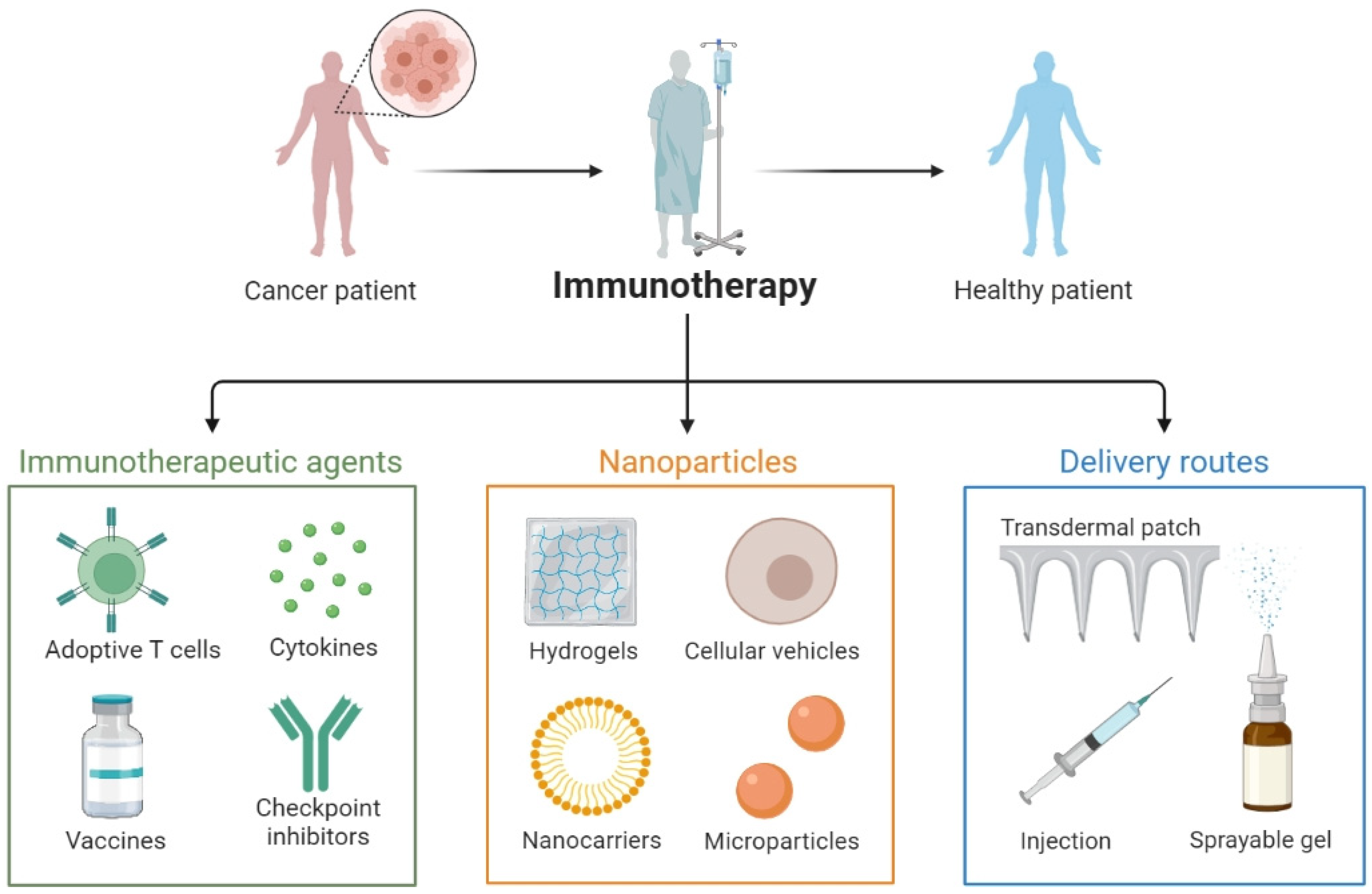
:1. Introduction

| Years | Milestones in Immunotherapy | Ref. |
|---|---|---|
| 1995 | Cytosine guanosine deoxynucleotide-mediated immune stimulation by bacterial DNA | [4] |
| 2001 | A clinical trial of mRNA with dendritic cells | [16] |
| 2003 | CTLA-4-specific aptamer used to manipulate the immune system | [17] |
| 2005 | miRNA cluster used to modulate tumor formation | [18] |
| 2008 |
| [1] [3] |
| 2016 | A clinical trial of saRNA therapeutic CEPBA-51 in lipid nanoparticles for liver cancer | [19] |
| 2018 | A clinical trial of poly-ICLC, Hiltonol© combined with anti-PD-1 for the treatment of advanced solid cancer | [20] |
2. Nucleic Acid Types and Structure
2.1. Circular RNA
2.2. mRNA
2.3. DNA
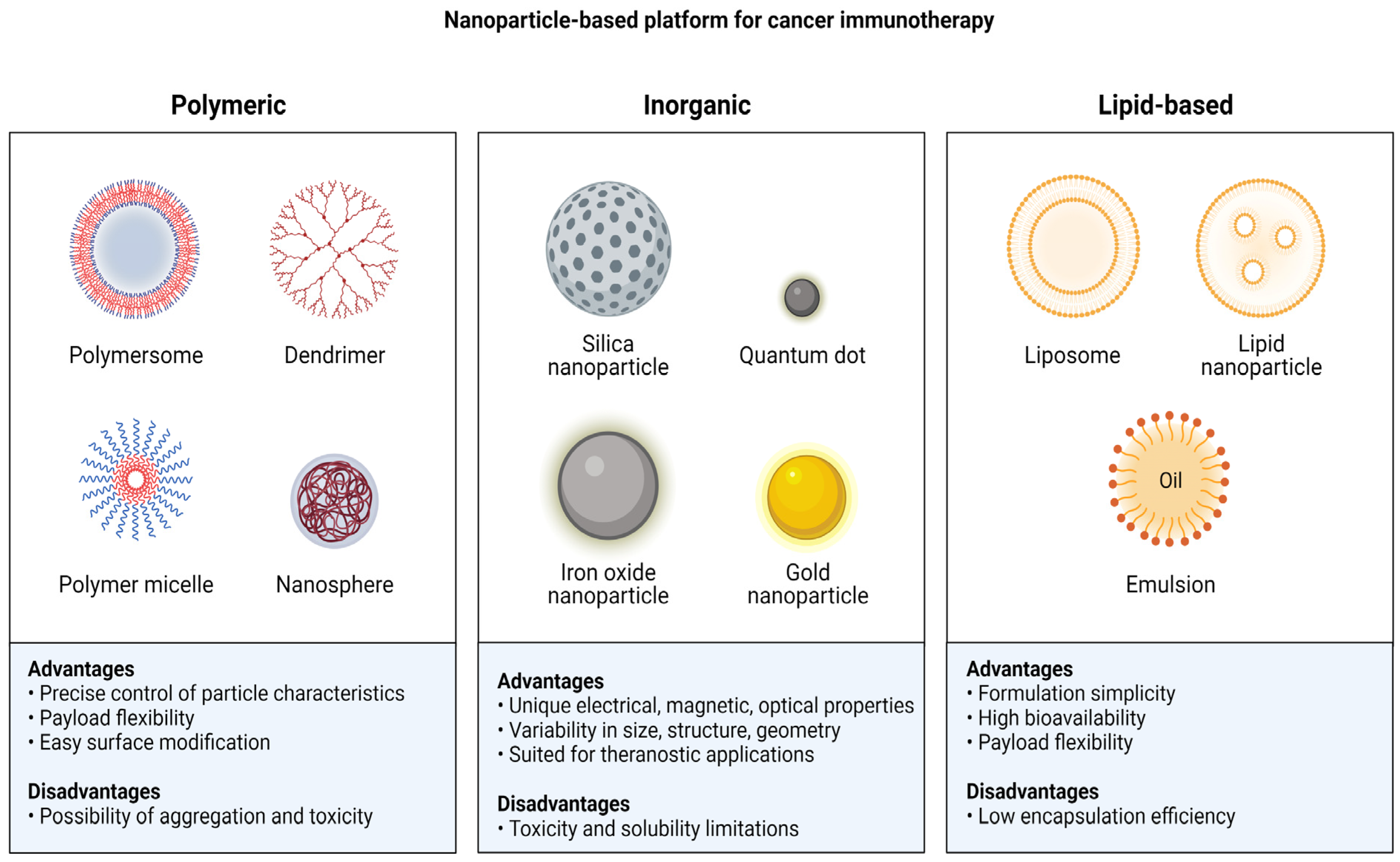
3. Nanoparticle-Mediated Nucleic Acid Delivery Systems for Immunotherapy
3.1. Lipid Nanoparticles
3.1.1. Lipid-Nanoparticle-Mediated circRNA Delivery
3.1.2. Lipid-Nanoparticle-Mediated mRNA Delivery
3.1.3. Lipid-Nanoparticle-Mediated DNA Delivery
3.2. Extracellular Vehicles
3.3. Polymeric Nanoparticles
3.4. Inorganic Nanoparticles
3.5. Hybrid Nanoparticles
3.6. Spherical Nucleic Acids (SNAs)
3.6.1. Selection of an Appropriate Core for SNAs
Inorganic Cores for SNA
Organic Cores for SNA
- Liposome
- Protein
4. Nucleic Acid Nanoparticles in the Cancer Immunity Cycle

5. Challenges for Nucleic-Acid-Mediated Nanotherapeutics
6. Conclusions and Future Outlook
Author Contributions
Funding
Institutional Review Board Statement
Informed Consent Statement
Data Availability Statement
Acknowledgments
Conflicts of Interest
References
- Zhou, S.; Chen, W.; Cole, J.; Zhu, G. Delivery of Nucleic Acid Therapeutics for Cancer Immunotherapy. Med. Drug Discov. 2020, 6, 100023. [Google Scholar] [CrossRef] [PubMed]
- Pastor, F.; Berraondo, P.; Etxeberria, I.; Frederick, J.; Sahin, U.; Gilboa, E.; Melero, I. An RNA Toolbox for Cancer Immunotherapy. Nat. Rev. Drug Discov. 2018, 17, 751–767. [Google Scholar] [CrossRef] [PubMed]
- Ishikawa, H.; Barber, G.N. STING Is an Endoplasmic Reticulum Adaptor That Facilitates Innate Immune Signalling. Nature 2008, 455, 674–678. [Google Scholar] [CrossRef] [PubMed] [Green Version]
- Sridharan, K.; Gogtay, N.J. Therapeutic Nucleic Acids: Current Clinical Status. Br. J. Clin. Pharmacol. 2016, 82, 659–672. [Google Scholar] [CrossRef] [PubMed] [Green Version]
- Mukalel, A.J.; Riley, R.S.; Zhang, R.; Mitchell, M.J. Nanoparticles for Nucleic Acid Delivery: Applications in Cancer Immunotherapy. Cancer Lett. 2019, 458, 102–112. [Google Scholar] [CrossRef]
- Moffett, H.F.; Coon, M.E.; Radtke, S.; Stephan, S.B.; McKnight, L.; Lambert, A.; Stoddard, B.L.; Kiem, H.P.; Stephan, M.T. Hit-and-Run Programming of Therapeutic Cytoreagents Using MRNA Nanocarriers. Nat. Commun. 2017, 8, 389. [Google Scholar] [CrossRef] [Green Version]
- Stewart, M.P.; Sharei, A.; Ding, X.; Sahay, G.; Langer, R.; Jensen, K.F. In Vitro and Ex Vivo Strategies for Intracellular Delivery. Nature 2016, 538, 183–192. [Google Scholar] [CrossRef] [Green Version]
- Ramasamy, T.; Munusamy, S.; Ruttala, H.B.; Kim, J.O. Smart Nanocarriers for the Delivery of Nucleic Acid-based Therapeutics: A Comprehensive Review. Biotechnol. J. 2021, 16, 1900408. [Google Scholar] [CrossRef]
- Kenchegowda, M.; Rahamathulla, M.; Hani, U.; Begum, M.Y.; Guruswamy, S.; Osmani, R.A.M.; Gowrav, M.P.; Alshehri, S.; Ghoneim, M.M.; Alshlowi, A. Smart Nanocarriers as an Emerging Platform for Cancer Therapy: A Review. Molecules 2021, 27, 146. [Google Scholar] [CrossRef]
- McNamara, M.A.; Nair, S.K.; Holl, E.K. RNA-Based Vaccines in Cancer Immunotherapy. J. Immunol. Res. 2015, 2015, 794528. [Google Scholar] [CrossRef] [Green Version]
- Wang, C.; Ye, Y.; Hu, Q.; Bellotti, A.; Gu, Z. Tailoring Biomaterials for Cancer Immunotherapy: Emerging Trends and Future Outlook. Adv. Mater. 2017, 29, 1606036. [Google Scholar] [CrossRef] [PubMed]
- Milling, L.; Zhang, Y.; Irvine, D.J. Delivering Safer Immunotherapies for Cancer. Adv. Drug Deliv. Rev. 2017, 114, 79–101. [Google Scholar] [CrossRef] [PubMed]
- Riley, R.S.; June, C.H.; Langer, R.; Mitchell, M.J. Delivery Technologies for Cancer Immunotherapy. Nat. Rev. Drug Discov. 2019, 18, 175–196. [Google Scholar] [CrossRef] [PubMed]
- Whitehead, K.A.; Dorkin, J.R.; Vegas, A.J.; Chang, P.H.; Veiseh, O.; Matthews, J.; Fenton, O.S.; Zhang, Y.; Olejnik, K.T.; Yesilyurt, V. Degradable Lipid Nanoparticles with Predictable in Vivo SiRNA Delivery Activity. Nat. Commun. 2014, 5, 4277. [Google Scholar] [CrossRef] [PubMed] [Green Version]
- Kamaly, N.; Yameen, B.; Wu, J.; Farokhzad, O.C. Degradable Controlled-Release Polymers and Polymeric Nanoparticles: Mechanisms of Controlling Drug Release. Chem. Rev. 2016, 116, 2602–2663. [Google Scholar] [CrossRef] [PubMed] [Green Version]
- Gilboa, E.; Vieweg, J. Cancer Immunotherapy with MRNA-transfected Dendritic Cells. Immunol. Rev. 2004, 199, 251–263. [Google Scholar] [CrossRef]
- Santulli-Marotto, S.; Nair, S.K.; Rusconi, C.; Sullenger, B.; Gilboa, E. Multivalent RNA Aptamers That Inhibit CTLA-4 and Enhance Tumor Immunity. Cancer Res. 2003, 63, 7483–7489. [Google Scholar]
- He, L.; Thomson, J.M.; Hemann, M.T.; Hernando-Monge, E.; Mu, D.; Goodson, S.; Powers, S.; Cordon-Cardo, C.; Lowe, S.W.; Hannon, G.J. A MicroRNA Polycistron as a Potential Human Oncogene. Nature 2005, 435, 828–833. [Google Scholar] [CrossRef] [Green Version]
- Reebye, V.; Huang, K.-W.; Lin, V.; Jarvis, S.; Cutilas, P.; Dorman, S.; Ciriello, S.; Andrikakou, P.; Voutila, J.; Saetrom, P. Gene Activation of CEBPA Using SaRNA: Preclinical Studies of the First in Human SaRNA Drug Candidate for Liver Cancer. Oncogene 2018, 37, 3216–3228. [Google Scholar] [CrossRef]
- Rodríguez-Ruiz, M.E.; Perez-Gracia, J.L.; Rodríguez, I.; Alfaro, C.; Oñate, C.; Pérez, G.; Gil-Bazo, I.; Benito, A.; Inogés, S.; de Cerio, A.L.-D. Combined Immunotherapy Encompassing Intratumoral Poly-ICLC, Dendritic-Cell Vaccination and Radiotherapy in Advanced Cancer Patients. Ann. Oncol. 2018, 29, 1312–1319. [Google Scholar] [CrossRef]
- Iurescia, S.; Fioretti, D.; Rinaldi, M. Nucleic Acid Sensing Machinery: Targeting Innate Immune System for Cancer Therapy. Recent Pat. Anti-Cancer Drug Discov. 2018, 13, 2–17. [Google Scholar] [CrossRef] [PubMed]
- Cerezo, M.; Robert, C.; Liu, L.; Shen, S. The Role of Mrna Translational Control in Tumor Immune Escape and Immunotherapy Resistance. Cancer Res. 2021, 81, 5596–5604. [Google Scholar] [CrossRef] [PubMed]
- Liu, M.A. A Comparison of Plasmid DNA and MRNA as Vaccine Technologies. Vaccines 2019, 7, 37. [Google Scholar] [CrossRef] [PubMed] [Green Version]
- Li, C.X.; Parker, A.; Menocal, E.; Xiang, S.; Borodyansky, L.; Fruehauf, J.H. Delivery of RNA Interference. Cell Cycle 2006, 5, 2103–2109. [Google Scholar] [CrossRef] [Green Version]
- Soldevilla, M.M.; Villanueva, H.; Pastor, F. Aptamers: A Feasible Technology in Cancer Immunotherapy. J. Immunol. Res. 2016, 2016, 1083738. [Google Scholar] [CrossRef] [Green Version]
- Levay, A.; Brenneman, R.; Hoinka, J.; Sant, D.; Cardone, M.; Trinchieri, G.; Przytycka, T.M.; Berezhnoy, A. Identifying High-Affinity Aptamer Ligands with Defined Cross-Reactivity Using High-Throughput Guided Systematic Evolution of Ligands by Exponential Enrichment. Nucleic Acids Res. 2015, 43, e82. [Google Scholar] [CrossRef] [Green Version]
- Afrasiabi, S.; Pourhajibagher, M.; Raoofian, R.; Tabarzad, M.; Bahador, A. Therapeutic Applications of Nucleic Acid Aptamers in Microbial Infections. J. Biomed. Sci. 2020, 27, 6. [Google Scholar] [CrossRef]
- Chen, L.-L.; Yang, L. Regulation of CircRNA Biogenesis. RNA Biol. 2015, 12, 381–388. [Google Scholar] [CrossRef]
- AbouHaidar, M.G.; Venkataraman, S.; Golshani, A.; Liu, B.; Ahmad, T. Novel Coding, Translation, and Gene Expression of a Replicating Covalently Closed Circular RNA of 220 Nt. Proc. Natl. Acad. Sci. USA 2014, 111, 14542–14547. [Google Scholar] [CrossRef] [Green Version]
- Pamudurti, N.R.; Bartok, O.; Jens, M.; Ashwal-Fluss, R.; Stottmeister, C.; Ruhe, L.; Hanan, M.; Wyler, E.; Perez-Hernandez, D.; Ramberger, E. Translation of CircRNAs. Mol. Cell 2017, 66, 9–21. [Google Scholar] [CrossRef] [Green Version]
- Legnini, I.; Di Timoteo, G.; Rossi, F.; Morlando, M.; Briganti, F.; Sthandier, O.; Fatica, A.; Santini, T.; Andronache, A.; Wade, M. Circ-ZNF609 Is a Circular RNA That Can Be Translated and Functions in Myogenesis. Mol. Cell 2017, 66, 22–37. [Google Scholar] [CrossRef] [Green Version]
- Zhang, Z.; Yang, T.; Xiao, J. Circular RNAs: Promising Biomarkers for Human Diseases. EBioMedicine 2018, 34, 267–274. [Google Scholar] [CrossRef] [Green Version]
- Enuka, Y.; Lauriola, M.; Feldman, M.E.; Sas-Chen, A.; Ulitsky, I.; Yarden, Y. Circular RNAs Are Long-Lived and Display Only Minimal Early Alterations in Response to a Growth Factor. Nucleic Acids Res. 2016, 44, 1370–1383. [Google Scholar] [CrossRef] [Green Version]
- Ji, P.; Wu, W.; Chen, S.; Zheng, Y.; Zhou, L.; Zhang, J.; Cheng, H.; Yan, J.; Zhang, S.; Yang, P. Expanded Expression Landscape and Prioritization of Circular RNAs in Mammals. Cell Rep. 2019, 26, 3444–3460. [Google Scholar] [CrossRef] [PubMed] [Green Version]
- Singh, S.; Narang, A.S.; Mahato, R.I. Subcellular Fate and Off-Target Effects of SiRNA, ShRNA, and MiRNA. Pharm. Res. 2011, 28, 2996–3015. [Google Scholar] [CrossRef] [PubMed]
- Kulkarni, J.A.; Witzigmann, D.; Chen, S.; Cullis, P.R.; van der Meel, R. Lipid Nanoparticle Technology for Clinical Translation of SiRNA Therapeutics. Acc. Chem. Res. 2019, 52, 2435–2444. [Google Scholar] [CrossRef]
- Guarnerio, J.; Zhang, Y.; Cheloni, G.; Panella, R.; Mae Katon, J.; Simpson, M.; Matsumoto, A.; Papa, A.; Loretelli, C.; Petri, A. Intragenic Antagonistic Roles of Protein and CircRNA in Tumorigenesis. Cell Res. 2019, 29, 628–640. [Google Scholar] [CrossRef] [PubMed]
- Jiang, Q.; Liu, C.; Li, C.-P.; Xu, S.-S.; Yao, M.-D.; Ge, H.-M.; Sun, Y.-N.; Li, X.-M.; Zhang, S.-J.; Shan, K. Circular RNA-ZNF532 Regulates Diabetes-Induced Retinal Pericyte Degeneration and Vascular Dysfunction. J. Clin. Investig. 2020, 130, 3833–3847. [Google Scholar] [CrossRef]
- He, A.T.; Liu, J.; Li, F.; Yang, B.B. Targeting Circular RNAs as a Therapeutic Approach: Current Strategies and Challenges. Signal Transduct. Target. Ther. 2021, 6, 185. [Google Scholar] [CrossRef] [PubMed]
- Weidle, U.H.; Birzele, F. Triple-Negative Breast Cancer: Identification of CircRNAs With Efficacy in Preclinical In Vivo Models. Cancer Genom. Proteom. 2023, 20, 117–131. [Google Scholar] [CrossRef]
- Zhang, C.; Huo, S.T.; Wu, Z.; Chen, L.; Wen, C.; Chen, H.; Du, W.W.; Wu, N.; Guan, D.; Lian, S. Rapid Development of Targeting CircRNAs in Cardiovascular Diseases. Mol. Ther. Acids 2020, 21, 568–576. [Google Scholar] [CrossRef] [PubMed]
- Zhang, M.; Xu, C.; Wang, H.; Peng, Y.; Li, H.; Zhou, Y.; Liu, S.; Wang, F.; Liu, L.; Chang, Y. Soft Fibrin Matrix Downregulates DAB2IP to Promote Nanog-Dependent Growth of Colon Tumor-Repopulating Cells. Cell Death Dis. 2019, 10, 151. [Google Scholar] [CrossRef] [PubMed] [Green Version]
- Sang, Y.; Chen, B.; Song, X.; Li, Y.; Liang, Y.; Han, D.; Zhang, N.; Zhang, H.; Liu, Y.; Chen, T. CircRNA_0025202 Regulates Tamoxifen Sensitivity and Tumor Progression via Regulating the MiR-182-5p/FOXO3a Axis in Breast Cancer. Mol. Ther. 2019, 27, 1638–1652. [Google Scholar] [CrossRef] [PubMed]
- Du, W.W.; Yang, W.; Li, X.; Awan, F.M.; Yang, Z.; Fang, L.; Lyu, J.; Li, F.; Peng, C.; Krylov, S.N. A Circular RNA Circ-DNMT1 Enhances Breast Cancer Progression by Activating Autophagy. Oncogene 2018, 37, 5829–5842. [Google Scholar] [CrossRef]
- Liang, W.-C.; Wong, C.-W.; Liang, P.-P.; Shi, M.; Cao, Y.; Rao, S.-T.; Tsui, S.K.-W.; Waye, M.M.-Y.; Zhang, Q.; Fu, W.-M. Translation of the Circular RNA Circβ-Catenin Promotes Liver Cancer Cell Growth through Activation of the Wnt Pathway. Genome Biol. 2019, 20, 84. [Google Scholar] [CrossRef]
- Yu, J.; Yang, M.; Zhou, B.; Luo, J.; Zhang, Z.; Zhang, W.; Yan, Z. CircRNA-104718 Acts as Competing Endogenous RNA and Promotes Hepatocellular Carcinoma Progression through MicroRNA-218-5p/TXNDC5 Signaling Pathway. Clin. Sci. 2019, 133, 1487–1503. [Google Scholar] [CrossRef]
- Dong, W.; Bi, J.; Liu, H.; Yan, D.; He, Q.; Zhou, Q.; Wang, Q.; Xie, R.; Su, Y.; Yang, M. Circular RNA ACVR2A Suppresses Bladder Cancer Cells Proliferation and Metastasis through MiR-626/EYA4 Axis. Mol. Cancer 2019, 18, 95. [Google Scholar] [CrossRef] [Green Version]
- Chen, L.; Nan, A.; Zhang, N.; Jia, Y.; Li, X.; Ling, Y.; Dai, J.; Zhang, S.; Yang, Q.; Yi, Y. Circular RNA 100146 Functions as an Oncogene through Direct Binding to MiR-361-3p and MiR-615-5p in Non-Small Cell Lung Cancer. Mol. Cancer 2019, 18, 13. [Google Scholar] [CrossRef]
- Wei, S.; Zheng, Y.; Jiang, Y.; Li, X.; Geng, J.; Shen, Y.; Li, Q.; Wang, X.; Zhao, C.; Chen, Y. The CircRNA CircPTPRA Suppresses Epithelial-Mesenchymal Transitioning and Metastasis of NSCLC Cells by Sponging MiR-96-5p. EBioMedicine 2019, 44, 182–193. [Google Scholar] [CrossRef] [Green Version]
- Zhang, L.; Song, X.; Chen, X.; Wang, Q.; Zheng, X.; Wu, C.; Jiang, J. Circular RNA CircCACTIN Promotes Gastric Cancer Progression by Sponging MiR-331-3p and Regulating TGFBR1 Expression. Int. J. Biol. Sci. 2019, 15, 1091–1103. [Google Scholar] [CrossRef] [Green Version]
- Rong, D.; Lu, C.; Zhang, B.; Fu, K.; Zhao, S.; Tang, W.; Cao, H. CircPSMC3 Suppresses the Proliferation and Metastasis of Gastric Cancer by Acting as a Competitive Endogenous RNA through Sponging MiR-296-5p. Mol. Cancer 2019, 18, 25. [Google Scholar] [CrossRef] [PubMed] [Green Version]
- Yang, F.; Hu, A.; Li, D.; Wang, J.; Guo, Y.; Liu, Y.; Li, H.; Chen, Y.; Wang, X.; Huang, K. Circ-HuR Suppresses HuR Expression and Gastric Cancer Progression by Inhibiting CNBP Transactivation. Mol. Cancer 2019, 18, 158. [Google Scholar] [CrossRef] [Green Version]
- Zhang, X.; Zhang, J.; Liu, Q.; Zhao, Y.; Zhang, W.; Yang, H. Circ-CUX1 Accelerates the Progression of Neuroblastoma via MiR-16-5p/DMRT2 Axis. Neurochem. Res. 2020, 45, 2840–2855. [Google Scholar] [CrossRef] [PubMed]
- Li, X.; Wang, X. The Emerging Roles and Therapeutic Potential of Exosomes in Epithelial Ovarian Cancer. Mol. Cancer 2017, 16, 92. [Google Scholar] [CrossRef] [PubMed] [Green Version]
- Hajj, K.A.; Whitehead, K.A. Tools for Translation: Non-Viral Materials for Therapeutic MRNA Delivery. Nat. Rev. Mater. 2017, 2, 17056. [Google Scholar] [CrossRef]
- Shen, T.; Zhang, Y.; Zhou, S.; Lin, S.; Zhang, X.-B.; Zhu, G. Nucleic Acid Immunotherapeutics for Cancer. ACS Appl. Bio Mater. 2020, 3, 2838–2849. [Google Scholar] [CrossRef]
- Van Nuffel, A.M.T.; Benteyn, D.; Wilgenhof, S.; Pierret, L.; Corthals, J.; Heirman, C.; Van Der Bruggen, P.; Coulie, P.G.; Neyns, B.; Thielemans, K. Dendritic Cells Loaded with MRNA Encoding Full-Length Tumor Antigens Prime CD4+ and CD8+ T Cells in Melanoma Patients. Mol. Ther. 2012, 20, 1063–1074. [Google Scholar] [CrossRef] [Green Version]
- Al Doghaither, H.; Gull, M. Plasmids as Genetic Tools and Their Applications in Ecology and Evolution. In Plasmid; IntechOpen: London, UK, 2019; ISBN 183880238X. [Google Scholar]
- Lowe, D.B.; Shearer, M.H.; Jumper, C.A.; Kennedy, R.C. Towards Progress on DNA Vaccines for Cancer. Cell. Mol. Life Sci. 2007, 64, 2391–2403. [Google Scholar] [CrossRef]
- Koyande, N.P.; Srivastava, R.; Padmakumar, A.; Rengan, A.K. Advances in Nanotechnology for Cancer Immunoprevention and Immunotherapy: A Review. Vaccines 2022, 10, 1727. [Google Scholar] [CrossRef]
- Kutzler, M.A.; Weiner, D.B. DNA Vaccines: Ready for Prime Time? Nat. Rev. Genet. 2008, 9, 776–788. [Google Scholar] [CrossRef]
- Yin, H.; Kanasty, R.L.; Eltoukhy, A.A.; Vegas, A.J.; Dorkin, J.R.; Anderson, D.G. Non-Viral Vectors for Gene-Based Therapy. Nat. Rev. Genet. 2014, 15, 541–555. [Google Scholar] [CrossRef] [PubMed]
- Vollmer, J.; Krieg, A.M. Immunotherapeutic Applications of CpG Oligodeoxynucleotide TLR9 Agonists. Adv. Drug Deliv. Rev. 2009, 61, 195–204. [Google Scholar] [CrossRef]
- Yu, G.; Dong, F.; Ge, W.; Sun, L.; Zhang, L.; Yuan, L.; Li, N.; Dai, H.; Shi, L.; Wang, Y. Self-Assembled Nanospheres Mediate Phototherapy and Deliver CpG Oligodeoxynucleotides to Enhance Cancer Immunotherapy of Breast Cancer and Melanoma. Nano Today 2022, 44, 101498. [Google Scholar] [CrossRef]
- Su, T.; Zhang, Y.; Valerie, K.; Wang, X.-Y.; Lin, S.; Zhu, G. STING Activation in Cancer Immunotherapy. Theranostics 2019, 9, 7759. [Google Scholar] [CrossRef] [PubMed]
- Pan, L.; Wang, Z.; Li, Y.; Xu, F.; Zhang, Q.; Zhang, C. Nicking Enzyme-Controlled Toehold Regulation for DNA Logic Circuits. Nanoscale 2017, 9, 18223–18228. [Google Scholar] [CrossRef] [PubMed]
- Harrison, E.B.; Azam, S.H.; Pecot, C.V. Targeting Accessories to the Crime: Nanoparticle Nucleic Acid Delivery to the Tumor Microenvironment. Front. Pharmacol. 2018, 9, 307. [Google Scholar] [CrossRef] [PubMed]
- Ibarguren, M.; López, D.J.; Escribá, P. V The Effect of Natural and Synthetic Fatty Acids on Membrane Structure, Microdomain Organization, Cellular Functions and Human Health. Biochim. Biophys. Acta (BBA)-Biomembr. 2014, 1838, 1518–1528. [Google Scholar] [CrossRef] [Green Version]
- Simberg, D.; Weisman, S.; Talmon, Y.; Barenholz, Y. DOTAP (and Other Cationic Lipids): Chemistry, Biophysics, and Transfection. Crit. Rev. Ther. Drug Carr. Syst. 2004, 21, 257–317. [Google Scholar] [CrossRef]
- Manolova, V.; Flace, A.; Bauer, M.; Schwarz, K.; Saudan, P.; Bachmann, M.F. Nanoparticles Target Distinct Dendritic Cell Populations According to Their Size. Eur. J. Immunol. 2008, 38, 1404–1413. [Google Scholar] [CrossRef]
- Haraguchi, N.; Ishii, H.; Mimori, K.; Tanaka, F.; Ohkuma, M.; Kim, H.M.; Akita, H.; Takiuchi, D.; Hatano, H.; Nagano, H. CD13 Is a Therapeutic Target in Human Liver Cancer Stem Cells. J. Clin. Investig. 2010, 120, 3326–3339. [Google Scholar] [CrossRef] [Green Version]
- Li, W.; Liu, J.; Chen, M.; Xu, J.; Zhu, D. Circular RNA in Cancer Development and Immune Regulation. J. Cell. Mol. Med. 2022, 26, 1785–1798. [Google Scholar] [CrossRef]
- Yu, L.-L.; Xiao, Q.; Yu, B.; Lv, Q.-L.; Liu, Z.-Q.; Yin, J.-Y. CircRNAs in Tumor Immunity and Immunotherapy: Perspectives from Innate and Adaptive Immunity. Cancer Lett. 2023, 564, 216219. [Google Scholar] [CrossRef]
- Li, H.; Peng, K.; Yang, K.; Ma, W.; Qi, S.; Yu, X.; He, J.; Lin, X.; Yu, G. Circular RNA Cancer Vaccines Drive Immunity in Hard-to-Treat Malignancies. Theranostics 2022, 12, 6422. [Google Scholar] [CrossRef]
- Yang, J.; Zhu, J.; Sun, J.; Chen, Y.; Du, Y.; Tan, Y.; Wu, L.; Zhai, M.; Wei, L.; Li, N. Intratumoral Delivered Novel Circular MRNA Encoding Cytokines for Immune Modulation and Cancer Therapy. Mol. Ther. Acids 2022, 30, 184–197. [Google Scholar] [CrossRef] [PubMed]
- Chen, Y.G.; Chen, R.; Ahmad, S.; Verma, R.; Kasturi, S.P.; Amaya, L.; Broughton, J.P.; Kim, J.; Cadena, C.; Pulendran, B. N6-Methyladenosine Modification Controls Circular RNA Immunity. Mol. Cell 2019, 76, 96–109. [Google Scholar] [CrossRef] [PubMed]
- Yang, J.; Wang, C.; Shi, S.; Dong, C. Nanotechnologies for Enhancing Cancer Immunotherapy. Nano Res. 2020, 13, 2595–2616. [Google Scholar] [CrossRef]
- Griffiths, G.; Gruenberg, J.; Marsh, M.; Wohlmann, J.; Jones, A.T.; Parton, R.G. Nanoparticle Entry into Cells; the Cell Biology Weak Link. Adv. Drug Deliv. Rev. 2022, 188, 114403. [Google Scholar] [CrossRef]
- Behzadi, S.; Serpooshan, V.; Tao, W.; Hamaly, M.A.; Alkawareek, M.Y.; Dreaden, E.C.; Brown, D.; Alkilany, A.M.; Farokhzad, O.C.; Mahmoudi, M. Cellular Uptake of Nanoparticles: Journey inside the Cell. Chem. Soc. Rev. 2017, 46, 4218–4244. [Google Scholar] [CrossRef] [PubMed]
- Xu, E.; Saltzman, W.M.; Piotrowski-Daspit, A.S. Escaping the Endosome: Assessing Cellular Trafficking Mechanisms of Non-Viral Vehicles. J. Control. Release 2021, 335, 465–480. [Google Scholar] [CrossRef] [PubMed]
- Donahue, N.D.; Acar, H.; Wilhelm, S. Concepts of Nanoparticle Cellular Uptake, Intracellular Trafficking, and Kinetics in Nanomedicine. Adv. Drug Deliv. Rev. 2019, 143, 68–96. [Google Scholar] [CrossRef]
- Kranz, L.M.; Diken, M.; Haas, H.; Kreiter, S.; Loquai, C.; Reuter, K.C.; Meng, M.; Fritz, D.; Vascotto, F.; Hefesha, H. Systemic RNA Delivery to Dendritic Cells Exploits Antiviral Defence for Cancer Immunotherapy. Nature 2016, 534, 396–401. [Google Scholar] [CrossRef] [PubMed]
- Sprangers, A.J.; Hao, L.; Banga, R.J.; Mirkin, C.A. Liposomal Spherical Nucleic Acids for Regulating Long Noncoding RNAs in the Nucleus. Small 2017, 13, 1602753. [Google Scholar] [CrossRef] [Green Version]
- Tenchov, R.; Bird, R.; Curtze, A.E.; Zhou, Q. Lipid Nanoparticles─ from Liposomes to MRNA Vaccine Delivery, a Landscape of Research Diversity and Advancement. ACS Nano 2021, 15, 16982–17015. [Google Scholar] [CrossRef] [PubMed]
- Beck, J.D.; Reidenbach, D.; Salomon, N.; Sahin, U.; Türeci, Ö.; Vormehr, M.; Kranz, L.M. MRNA Therapeutics in Cancer Immunotherapy. Mol. Cancer 2021, 20, 69. [Google Scholar] [CrossRef] [PubMed]
- Tzeng, S.Y.; Patel, K.K.; Wilson, D.R.; Meyer, R.A.; Rhodes, K.R.; Green, J.J. In Situ Genetic Engineering of Tumors for Long-Lasting and Systemic Immunotherapy. Proc. Natl. Acad. Sci. USA 2020, 117, 4043–4052. [Google Scholar] [CrossRef] [PubMed] [Green Version]
- Ho, W.; Gao, M.; Li, F.; Li, Z.; Zhang, X.; Xu, X. Next-generation Vaccines: Nanoparticle-mediated Dna and Mrna Delivery. Adv. Healthc. Mater. 2021, 10, 2001812. [Google Scholar] [CrossRef]
- Chen, W.; Jiang, M.; Yu, W.; Xu, Z.; Liu, X.; Jia, Q.; Guan, X.; Zhang, W. CpG-Based Nanovaccines for Cancer Immunotherapy. Int. J. Nanomed. 2021, 16, 5281. [Google Scholar] [CrossRef]
- Smith, T.T.; Stephan, S.B.; Moffett, H.F.; McKnight, L.E.; Ji, W.; Reiman, D.; Bonagofski, E.; Wohlfahrt, M.E.; Pillai, S.P.S.; Stephan, M.T. In Situ Programming of Leukaemia-Specific T Cells Using Synthetic DNA Nanocarriers. Nat. Nanotechnol. 2017, 12, 813–820. [Google Scholar] [CrossRef] [Green Version]
- Yang, B.; Jeang, J.; Yang, A.; Wu, T.C.; Hung, C.-F. DNA Vaccine for Cancer Immunotherapy. Hum. Vaccin. Immunother. 2014, 10, 3153–3164. [Google Scholar] [CrossRef] [Green Version]
- Rodolfo, C.; Eusébio, D.; Ventura, C.; Nunes, R.; Florindo, H.F.; Costa, D.; Sousa, Â. Design of Experiments to Achieve an Efficient Chitosan-Based DNA Vaccine Delivery System. Pharmaceutics 2021, 13, 1369. [Google Scholar] [CrossRef]
- Syn, N.L.; Wang, L.; Chow, E.K.-H.; Lim, C.T.; Goh, B.-C. Exosomes in Cancer Nanomedicine and Immunotherapy: Prospects and Challenges. Trends Biotechnol. 2017, 35, 665–676. [Google Scholar] [CrossRef] [PubMed]
- Becker, A.; Thakur, B.K.; Weiss, J.M.; Kim, H.S.; Peinado, H.; Lyden, D. Extracellular Vesicles in Cancer: Cell-to-Cell Mediators of Metastasis. Cancer Cell 2016, 30, 836–848. [Google Scholar] [CrossRef] [PubMed] [Green Version]
- Liu, Y.; Gu, Y.; Cao, X. The Exosomes in Tumor Immunity. Oncoimmunology 2015, 4, e1027472. [Google Scholar] [CrossRef] [PubMed] [Green Version]
- Théry, C.; Boussac, M.; Véron, P.; Ricciardi-Castagnoli, P.; Raposo, G.; Garin, J.; Amigorena, S. Proteomic Analysis of Dendritic Cell-Derived Exosomes: A Secreted Subcellular Compartment Distinct from Apoptotic Vesicles. J. Immunol. 2001, 166, 7309–7318. [Google Scholar] [CrossRef] [PubMed] [Green Version]
- Patel, A.; Sant, S. Hypoxic Tumor Microenvironment: Opportunities to Develop Targeted Therapies. Biotechnol. Adv. 2016, 34, 803–812. [Google Scholar] [CrossRef] [Green Version]
- Wolfers, J.; Lozier, A.; Raposo, G.; Regnault, A.; Théry, C.; Masurier, C.; Flament, C.; Pouzieux, S.; Faure, F.; Tursz, T. Tumor-Derived Exosomes Are a Source of Shared Tumor Rejection Antigens for CTL Cross-Priming. Nat. Med. 2001, 7, 297–303. [Google Scholar] [CrossRef]
- Elsabahy, M.; Wooley, K.L. Design of Polymeric Nanoparticles for Biomedical Delivery Applications. Chem. Soc. Rev. 2012, 41, 2545–2561. [Google Scholar] [CrossRef] [PubMed] [Green Version]
- Kumar, S.; Kesharwani, S.S.; Kuppast, B.; Bakkari, M.A.; Tummala, H. Pathogen-Mimicking Vaccine Delivery System Designed with a Bioactive Polymer (Inulin Acetate) for Robust Humoral and Cellular Immune Responses. J. Control. Release 2017, 261, 263–274. [Google Scholar] [CrossRef]
- Tabatabaei Mirakabad, F.S.; Nejati-Koshki, K.; Akbarzadeh, A.; Yamchi, M.R.; Milani, M.; Zarghami, N.; Zeighamian, V.; Rahimzadeh, A.; Alimohammadi, S.; Hanifehpour, Y. PLGA-Based Nanoparticles as Cancer Drug Delivery Systems. Asian Pac. J. Cancer Prev. 2014, 15, 517–535. [Google Scholar] [CrossRef] [Green Version]
- Jones, M.-C.; Leroux, J.-C. Polymeric Micelles–a New Generation of Colloidal Drug Carriers. Eur. J. Pharm. Biopharm. 1999, 48, 101–111. [Google Scholar] [CrossRef]
- Hoare, T.R.; Kohane, D.S. Hydrogels in Drug Delivery: Progress and Challenges. Polymer 2008, 49, 1993–2007. [Google Scholar] [CrossRef] [Green Version]
- Baker, A.; Khan, M.S.; Iqbal, M.Z.; Khan, M.S. Tumor-Targeted Drug Delivery by Nanocomposites. Curr. Drug Metab. 2020, 21, 599–613. [Google Scholar] [CrossRef] [PubMed]
- Baker, A.; Syed, A.; Iram, S.; Elgorban, A.M.; Al-Harthi, H.F.; Al-Rejaie, S.S.; Kim, J.; Khan, M.S. AR Independent Anticancer Potential of Enza against Prostate Cancer. Colloids Surf. A Physicochem. Eng. Asp. 2022, 642, 128598. [Google Scholar] [CrossRef]
- Baker, A.; Khan, M.S. Biogenic Nanomaterials Derived ROS for Cancer Therapy. In Handbook of Oxidative Stress in Cancer: Therapeutic Aspects; Springer: Berlin/Heidelberg, Germany, 2022; pp. 1–14. [Google Scholar]
- Iram, S.; Zahera, M.; Wahid, I.; Baker, A.; Raish, M.; Khan, A.; Ali, N.; Ahmad, S.; Khan, M.S. Cisplatin Bioconjugated Enzymatic GNPs Amplify the Effect of Cisplatin with Acquiescence. Sci. Rep. 2019, 9, 13826. [Google Scholar] [CrossRef] [PubMed] [Green Version]
- Almeida, J.P.M.; Lin, A.Y.; Figueroa, E.R.; Foster, A.E.; Drezek, R.A. In Vivo Gold Nanoparticle Delivery of Peptide Vaccine Induces Anti-tumor Immune Response in Prophylactic and Therapeutic Tumor Models. Small 2015, 11, 1453–1459. [Google Scholar] [CrossRef] [Green Version]
- Dykman, L.A.; Khlebtsov, N.G. Gold Nanoparticles in Biology and Medicine: Recent Advances and Prospects. Acta Nat. 2011, 3, 34–55. [Google Scholar] [CrossRef] [Green Version]
- Huang, X.; Jain, P.K.; El-Sayed, I.H.; El-Sayed, M.A. Gold Nanoparticles: Interesting Optical Properties and Recent Applications in Cancer Diagnostics and Therapy. Future Med. 2007, 2, 681–693. [Google Scholar] [CrossRef] [Green Version]
- Libutti, S.K.; Paciotti, G.F.; Byrnes, A.A.; Alexander, H.R.; Gannon, W.E.; Walker, M.; Seidel, G.D.; Yuldasheva, N.; Tamarkin, L. Phase I and Pharmacokinetic Studies of CYT-6091, a Novel PEGylated Colloidal Gold-RhTNF NanomedicinePEGylated Colloidal Gold-RhTNF Nanomedicine Phase I Trial. Clin. Cancer Res. 2010, 16, 6139–6149. [Google Scholar] [CrossRef] [Green Version]
- Elahi, N.; Kamali, M.; Baghersad, M.H. Recent Biomedical Applications of Gold Nanoparticles: A Review. Talanta 2018, 184, 537–556. [Google Scholar] [CrossRef]
- Maurer-Jones, M.A.; Lin, Y.-S.; Haynes, C.L. Functional Assessment of Metal Oxide Nanoparticle Toxicity in Immune Cells. ACS Nano 2010, 4, 3363–3373. [Google Scholar] [CrossRef]
- Sailor, M.J.; Park, J. Hybrid Nanoparticles for Detection and Treatment of Cancer. Adv. Mater. 2012, 24, 3779–3802. [Google Scholar] [CrossRef] [PubMed]
- Chen, G.; Qian, Y.; Zhang, H.; Ullah, A.; He, X.; Zhou, Z.; Fenniri, H.; Shen, J. Advances in Cancer Theranostics Using Organic-Inorganic Hybrid Nanotechnology. Appl. Mater. Today 2021, 23, 101003. [Google Scholar] [CrossRef]
- Luo, L.; Zhu, C.; Yin, H.; Jiang, M.; Zhang, J.; Qin, B.; Luo, Z.; Yuan, X.; Yang, J.; Li, W. Laser Immunotherapy in Combination with Perdurable PD-1 Blocking for the Treatment of Metastatic Tumors. ACS Nano 2018, 12, 7647–7662. [Google Scholar] [CrossRef] [PubMed]
- Xiong, H.; Liu, S.; Wei, T.; Cheng, Q.; Siegwart, D.J. Theranostic Dendrimer-Based Lipid Nanoparticles Containing PEGylated BODIPY Dyes for Tumor Imaging and Systemic MRNA Delivery in Vivo. J. Control. Release 2020, 325, 198–205. [Google Scholar] [CrossRef] [PubMed]
- Kim, M.; Jeong, M.; Hur, S.; Cho, Y.; Park, J.; Jung, H.; Seo, Y.; Woo, H.A.; Nam, K.T.; Lee, K. Engineered Ionizable Lipid Nanoparticles for Targeted Delivery of RNA Therapeutics into Different Types of Cells in the Liver. Sci. Adv. 2021, 7, eabf4398. [Google Scholar] [CrossRef]
- Wang, S.; Chen, Y.; Wang, S.; Li, P.; Mirkin, C.A.; Farha, O.K. DNA-Functionalized Metal–Organic Framework Nanoparticles for Intracellular Delivery of Proteins. J. Am. Chem. Soc. 2019, 141, 2215–2219. [Google Scholar] [CrossRef] [Green Version]
- Mirkin, C.A.; Letsinger, R.L.; Mucic, R.C.; Storhoff, J.J. A DNA-Based Method for Rationally Assembling Nanoparticles into Macroscopic Materials. Nature 1996, 382, 607–609. [Google Scholar] [CrossRef]
- Mitchell, G.P.; Mirkin, C.A.; Letsinger, R.L. Programmed Assembly of DNA Functionalized Quantum Dots. J. Am. Chem. Soc. 1999, 121, 8122–8123. [Google Scholar] [CrossRef]
- Azharuddin, M.; Zhu, G.H.; Das, D.; Ozgur, E.; Uzun, L.; Turner, A.P.F.; Patra, H.K. A Repertoire of Biomedical Applications of Noble Metal Nanoparticles. Chem. Commun. 2019, 55, 6964–6996. [Google Scholar] [CrossRef]
- Banga, R.J.; Chernyak, N.; Narayan, S.P.; Nguyen, S.T.; Mirkin, C.A. Liposomal Spherical Nucleic Acids. J. Am. Chem. Soc. 2014, 136, 9866–9869. [Google Scholar] [CrossRef]
- Brodin, J.D.; Sprangers, A.J.; McMillan, J.R.; Mirkin, C.A. DNA-Mediated Cellular Delivery of Functional Enzymes. J. Am. Chem. Soc. 2015, 137, 14838–14841. [Google Scholar] [CrossRef] [PubMed] [Green Version]
- Cutler, J.I.; Zhang, K.; Zheng, D.; Auyeung, E.; Prigodich, A.E.; Mirkin, C.A. Polyvalent Nucleic Acid Nanostructures. J. Am. Chem. Soc. 2011, 133, 9254–9257. [Google Scholar] [CrossRef] [PubMed] [Green Version]
- Buonerba, A.; Grassi, A. Trends in Sustainable Synthesis of Organics by Gold Nanoparticles Embedded in Polymer Matrices. Catalysts 2021, 11, 714. [Google Scholar] [CrossRef]
- Khan, S.; Rizvi, S.M.D.; Avaish, M.; Arshad, M.; Bagga, P.; Khan, M.S. A Novel Process for Size Controlled Biosynthesis of Gold Nanoparticles Using Bromelain. Mater. Lett. 2015, 159, 373–376. [Google Scholar] [CrossRef]
- Xia, J.; Wang, W.; Hai, X.; Shuang, E.; Shu, Y.; Wang, J. Improvement of Antibacterial Activity of Copper Nanoclusters for Selective Inhibition on the Growth of Gram-Positive Bacteria. Chin. Chem. Lett. 2019, 30, 421–424. [Google Scholar] [CrossRef]
- Macfarlane, R.J.; Jones, M.R.; Lee, B.; Auyeung, E.; Mirkin, C.A. Topotactic Interconversion of Nanoparticle Superlattices. Science 2013, 341, 1222–1225. [Google Scholar] [CrossRef] [PubMed]
- Baker, A.; Syed, A.; Alyousef, A.A.; Arshad, M.; Alqasim, A.; Khalid, M.; Khan, S. Sericin-Functionalized GNPs Potentiate the Synergistic Effect of Levofloxacin and Balofloxacin against MDR Bacteria. Microb. Pathog. 2020, 148, 104467. [Google Scholar] [CrossRef]
- Alkilany, A.M.; Murphy, C.J. Toxicity and Cellular Uptake of Gold Nanoparticles: What We Have Learned so Far? J. Nanoparticle Res. 2010, 12, 2313–2333. [Google Scholar] [CrossRef] [Green Version]
- Wang, S.; Qin, L.; Yamankurt, G.; Skakuj, K.; Huang, Z.; Chen, P.-C.; Dominguez, D.; Lee, A.; Zhang, B.; Mirkin, C.A. Rational Vaccinology with Spherical Nucleic Acids. Proc. Natl. Acad. Sci. USA 2019, 116, 10473–10481. [Google Scholar] [CrossRef] [Green Version]
- Guan, C.; Chernyak, N.; Dominguez, D.; Cole, L.; Zhang, B.; Mirkin, C.A. RNA-Based Immunostimulatory Liposomal Spherical Nucleic Acids as Potent TLR7/8 Modulators. Small 2018, 14, 1803284. [Google Scholar] [CrossRef]
- Teplensky, M.H.; Dittmar, J.W.; Qin, L.; Wang, S.; Evangelopoulos, M.; Zhang, B.; Mirkin, C.A. Spherical Nucleic Acid Vaccine Structure Markedly Influences Adaptive Immune Responses of Clinically Utilized Prostate Cancer Targets. Adv. Healthc. Mater. 2021, 10, 2101262. [Google Scholar] [CrossRef]
- Qin, L.; Wang, S.; Dominguez, D.; Long, A.; Chen, S.; Fan, J.; Ahn, J.; Skakuj, K.; Huang, Z.; Lee, A. Development of Spherical Nucleic Acids for Prostate Cancer Immunotherapy. Front. Immunol. 2020, 11, 1333. [Google Scholar] [CrossRef]
- Callmann, C.E.; Cole, L.E.; Kusmierz, C.D.; Huang, Z.; Horiuchi, D.; Mirkin, C.A. Tumor Cell Lysate-Loaded Immunostimulatory Spherical Nucleic Acids as Therapeutics for Triple-Negative Breast Cancer. Proc. Natl. Acad. Sci. USA 2020, 117, 17543–17550. [Google Scholar] [CrossRef]
- Skakuj, K.; Wang, S.; Qin, L.; Lee, A.; Zhang, B.; Mirkin, C.A. Conjugation Chemistry-Dependent T-Cell Activation with Spherical Nucleic Acids. J. Am. Chem. Soc. 2018, 140, 1227–1230. [Google Scholar] [CrossRef] [Green Version]
- Ferrer, J.R.; Sinegra, A.J.; Ivancic, D.; Yeap, X.Y.; Qiu, L.; Wang, J.-J.; Zhang, Z.J.; Wertheim, J.A.; Mirkin, C.A. Structure-Dependent Biodistribution of Liposomal Spherical Nucleic Acids. ACS Nano 2020, 14, 1682–1693. [Google Scholar] [CrossRef] [PubMed]
- Teplensky, M.H.; Evangelopoulos, M.; Dittmar, J.W.; Forsyth, C.M.; Sinegra, A.J.; Wang, S.; Mirkin, C.A. Multi-Antigen Spherical Nucleic Acid Cancer Vaccines. Nat. Biomed. Eng. 2023, 1–17. [Google Scholar] [CrossRef]
- Kusmierz, C.D.; Bujold, K.E.; Callmann, C.E.; Mirkin, C.A. Defining the Design Parameters for in Vivo Enzyme Delivery through Protein Spherical Nucleic Acids. ACS Cent. Sci. 2020, 6, 815–822. [Google Scholar] [CrossRef]
- Song, Y.; Song, W.; Lan, X.; Cai, W.; Jiang, D. Spherical Nucleic Acids: Organized Nucleotide Aggregates as Versatile Nanomedicine. Aggregate 2022, 3, e120. [Google Scholar]
- Yan, J.; Tan, Y.-L.; Lin, M.; Xing, H.; Jiang, J.-H. A DNA-Mediated Crosslinking Strategy to Enhance Cellular Delivery and Sensor Performance of Protein Spherical Nucleic Acids. Chem. Sci. 2021, 12, 1803–1809. [Google Scholar] [CrossRef] [PubMed]
- Mi, Y.; Hagan IV, C.T.; Vincent, B.G.; Wang, A.Z. Emerging Nano-/Microapproaches for Cancer Immunotherapy. Adv. Sci. 2019, 6, 1801847. [Google Scholar] [CrossRef] [PubMed] [Green Version]
- Veneziani, I.; Alicata, C.; Moretta, L.; Maggi, E. The Latest Approach of Immunotherapy with Endosomal TLR Agonists Improving NK Cell Function: An Overview. Biomedicines 2023, 11, 64. [Google Scholar] [CrossRef]
- Di Trani, C.A.; Fernandez-Sendin, M.; Cirella, A.; Segués, A.; Olivera, I.; Bolaños, E.; Melero, I.; Berraondo, P. Advances in MRNA-Based Drug Discovery in Cancer Immunotherapy. Expert Opin. Drug Discov. 2022, 17, 41–53. [Google Scholar] [CrossRef] [PubMed]
- Deng, K.; Yang, D.; Zhou, Y. Nanotechnology-Based SiRNA Delivery Systems to Overcome Tumor Immune Evasion in Cancer Immunotherapy. Pharmaceutics 2022, 14, 1344. [Google Scholar] [CrossRef] [PubMed]
- Lee, H.K.; Kim, C.-W.; Ahn, D.; Go, R.-E.; Choi, Y.; Choi, K.-C. Next-Generation Antisense Oligonucleotide of TGF-Β2 Enhances T Cell-Mediated Anticancer Efficacy of Anti-PD-1 Therapy in a Humanized Mouse Model of Immune-Excluded Melanoma. Cancers 2022, 14, 5220. [Google Scholar] [CrossRef] [PubMed]
- Zahedipour, F.; Majeed, M.; Kesharwani, P.; Sahebkar, A. Cancer Immunotherapy via Nucleic Acid Aptamers. In Aptamers Engineered Nanocarriers for Cancer Therapy; Elsevier: Amsterdam, The Netherlands, 2023; pp. 317–346. [Google Scholar]
- Chou, L.; Callmann, C.E.; Dominguez, D.; Zhang, B.; Mirkin, C.A. Disrupting the Interplay between Programmed Cell Death Protein 1 and Programmed Death Ligand 1 with Spherical Nucleic Acids in Treating Cancer. ACS Cent. Sci. 2022, 8, 1299–1305. [Google Scholar] [CrossRef]
- Zhang, D.; Wang, G.; Yu, X.; Wei, T.; Farbiak, L.; Johnson, L.T.; Taylor, A.M.; Xu, J.; Hong, Y.; Zhu, H. Enhancing CRISPR/Cas Gene Editing through Modulating Cellular Mechanical Properties for Cancer Therapy. Nat. Nanotechnol. 2022, 17, 777–787. [Google Scholar] [CrossRef]
- Haabeth, O.A.W.; Blake, T.R.; McKinlay, C.J.; Tveita, A.A.; Sallets, A.; Waymouth, R.M.; Wender, P.A.; Levy, R. Local Delivery of Ox40l, Cd80, and Cd86 MRNA Kindles Global Anticancer Immunity. Cancer Res. 2019, 79, 1624–1634. [Google Scholar] [CrossRef] [Green Version]
- Hewitt, S.L.; Bai, A.; Bailey, D.; Ichikawa, K.; Zielinski, J.; Karp, R.; Apte, A.; Arnold, K.; Zacharek, S.J.; Iliou, M.S. Durable Anticancer Immunity from Intratumoral Administration of IL-23, IL-36γ, and OX40L MRNAs. Sci. Transl. Med. 2019, 11, eaat9143. [Google Scholar] [CrossRef]
- Ma, Y.; Cai, L. Next Generation of Cancer Immunotherapy: Targeting the Cancer-Immunity Cycle with Nanotechnology. In Nanotechnology in Regenerative Medicine and Drug Delivery Therapy; Springer: Singapore, 2020; pp. 191–253. [Google Scholar]
- Song, W.; Musetti, S.N.; Huang, L. Nanomaterials for Cancer Immunotherapy. Biomaterials 2017, 148, 16–30. [Google Scholar] [CrossRef]
- Wu, J.; Chen, J.; Feng, Y.; Zhang, S.; Lin, L.; Guo, Z.; Sun, P.; Xu, C.; Tian, H.; Chen, X. An Immune Cocktail Therapy to Realize Multiple Boosting of the Cancer-Immunity Cycle by Combination of Drug/Gene Delivery Nanoparticles. Sci. Adv. 2020, 6, eabc7828. [Google Scholar] [CrossRef]
- Pecot, C.V.; Calin, G.A.; Coleman, R.L.; Lopez-Berestein, G.; Sood, A.K. RNA Interference in the Clinic: Challenges and Future Directions. Nat. Rev. Cancer 2011, 11, 59–67. [Google Scholar] [CrossRef] [PubMed] [Green Version]
- Zhang, Y.; Wang, Z.; Gemeinhart, R.A. Progress in MicroRNA Delivery. J. Control. Release 2013, 172, 962–974. [Google Scholar] [CrossRef] [PubMed] [Green Version]
- Mitchell, M.J.; Billingsley, M.M.; Haley, R.M.; Wechsler, M.E.; Peppas, N.A.; Langer, R. Engineering Precision Nanoparticles for Drug Delivery. Nat. Rev. Drug Discov. 2021, 20, 101–124. [Google Scholar] [CrossRef] [PubMed]


| CircRNA | Up Regulate | Down Regulate | Types of Cancer | Ref. |
|---|---|---|---|---|
| Circ-AGFG1 | ✓ | Triple-negative breast cancer (TNBC) | [40] | |
| Circ-HER2 | ✓ | [41] | ||
| Circ-TADA2A-E6 | ✓ | [42] | ||
| Has-circ-0025202 | ✓ | Breast cancer (BC) | [43] | |
| Circ-Dnmt1 | ✓ | [44] | ||
| Circβ-catenin | ✓ | Hepatocellular carcinoma (HCC) | [45] | |
| Circ-RNA-104718 | ✓ | [46] | ||
| Circ-TRIM33-12 | ✓ | [47] | ||
| Circ-RNA 100146 | ✓ | Non-small cell lung cancer (NSCLC) | [48] | |
| Circ-PTPRA | ✓ | [49] | ||
| Circ-CACTIN | ✓ | Gastric cancer (GC) | [50] | |
| Circ-PSMC3 | ✓ | [51] | ||
| Circ-HuR | ✓ | [52] | ||
| Circ-CUX1 | ✓ | Neuroblastoma (NB) | [53] | |
| Circ-LONP2 | ✓ | Colorectal cancer (CC) | [54] |
Disclaimer/Publisher’s Note: The statements, opinions and data contained in all publications are solely those of the individual author(s) and contributor(s) and not of MDPI and/or the editor(s). MDPI and/or the editor(s) disclaim responsibility for any injury to people or property resulting from any ideas, methods, instructions or products referred to in the content. |
© 2023 by the authors. Licensee MDPI, Basel, Switzerland. This article is an open access article distributed under the terms and conditions of the Creative Commons Attribution (CC BY) license (https://creativecommons.org/licenses/by/4.0/).
Share and Cite
Baker, A.; Lorch, J.; VanderWeele, D.; Zhang, B. Smart Nanocarriers for the Targeted Delivery of Therapeutic Nucleic Acid for Cancer Immunotherapy. Pharmaceutics 2023, 15, 1743. https://doi.org/10.3390/pharmaceutics15061743
Baker A, Lorch J, VanderWeele D, Zhang B. Smart Nanocarriers for the Targeted Delivery of Therapeutic Nucleic Acid for Cancer Immunotherapy. Pharmaceutics. 2023; 15(6):1743. https://doi.org/10.3390/pharmaceutics15061743
Chicago/Turabian StyleBaker, Abu, Jochen Lorch, David VanderWeele, and Bin Zhang. 2023. "Smart Nanocarriers for the Targeted Delivery of Therapeutic Nucleic Acid for Cancer Immunotherapy" Pharmaceutics 15, no. 6: 1743. https://doi.org/10.3390/pharmaceutics15061743