
Advancing Cancer Therapy with Copper/Disulfiram Nanomedicines and Drug Delivery Systems
 , , , and
Abstract
:1. Introduction
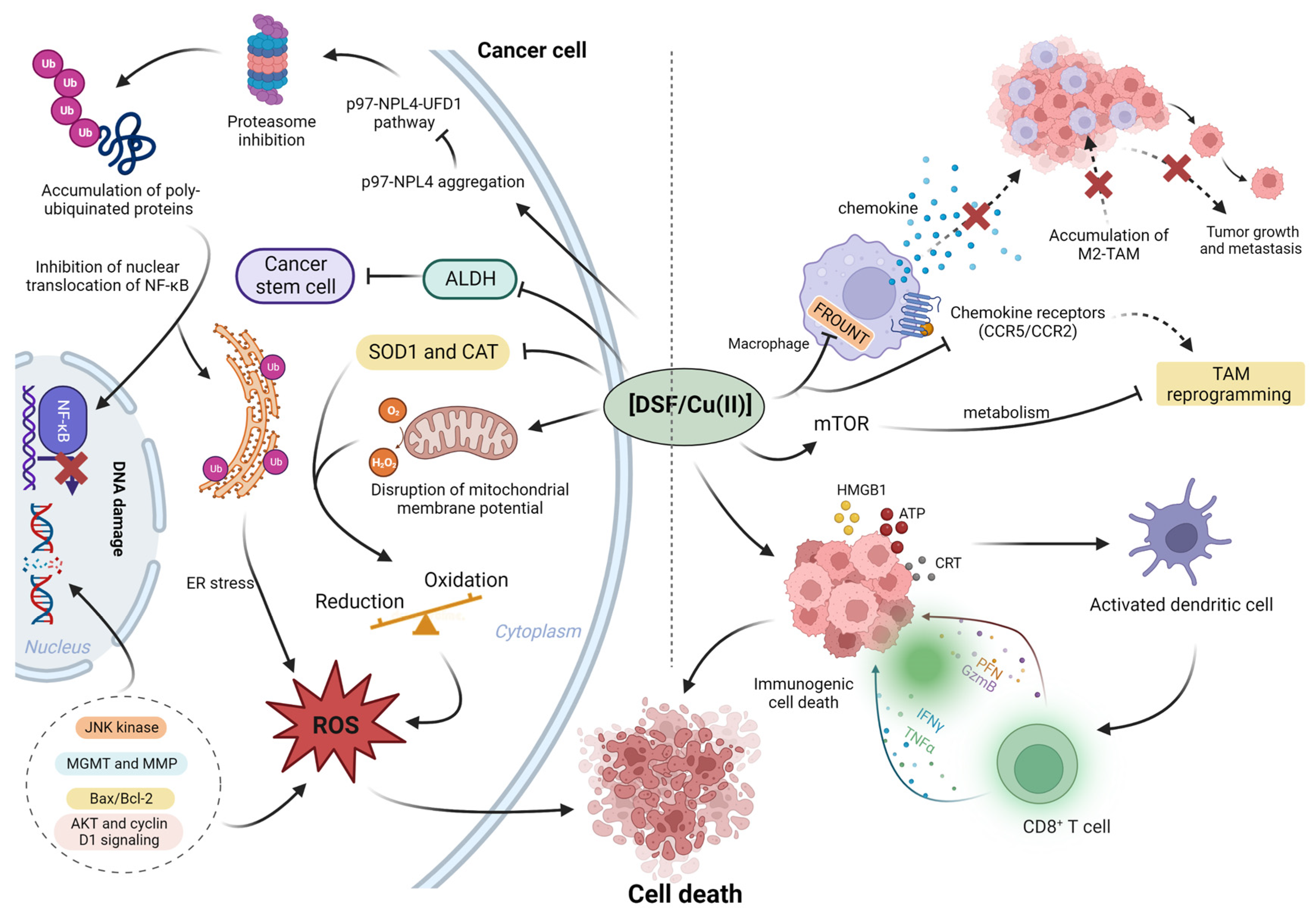
2. Anticancer Mechanisms of DSF/Cu (II)
2.1. Disulfiram/Cu with ROS
2.2. Enzyme Inhibition and DNA Damage
2.3. The Effect of DSF/Cu (II) on the Activity of the Proteasome System
2.4. The Effect on Transcription Factors
2.5. The Immunomodulatory Effects on the Tumor Microenvironment (TME)
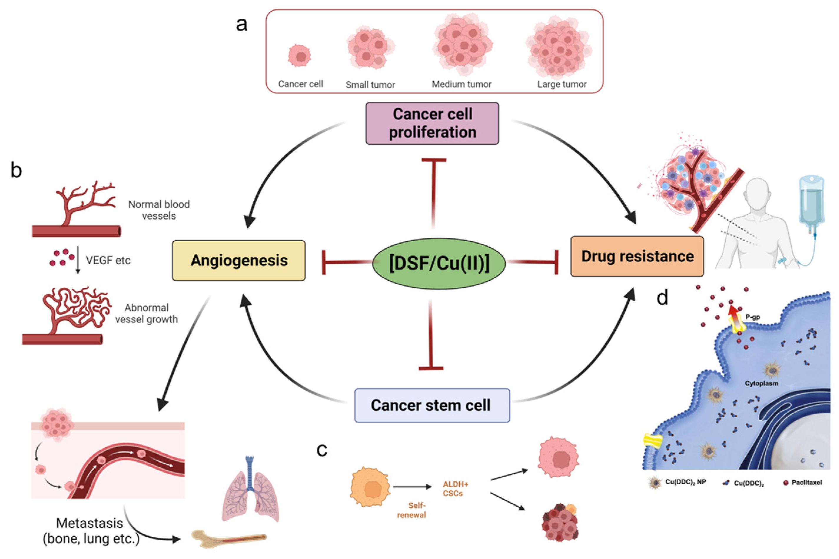
3. The Effect of DSF/Cu (II) on Cancer
3.1. DSF/Cu (II) on the Inhibition of Cancer Proliferation
3.2. DSF/Cu (II) Efficacy in Cancer Stem Cells (CSCs)
3.3. DSF/Cu (II) Effects on the Inhibition of Cancer Angiogenesis
3.4. DSF/Cu (II) Reverses Drug Resistance

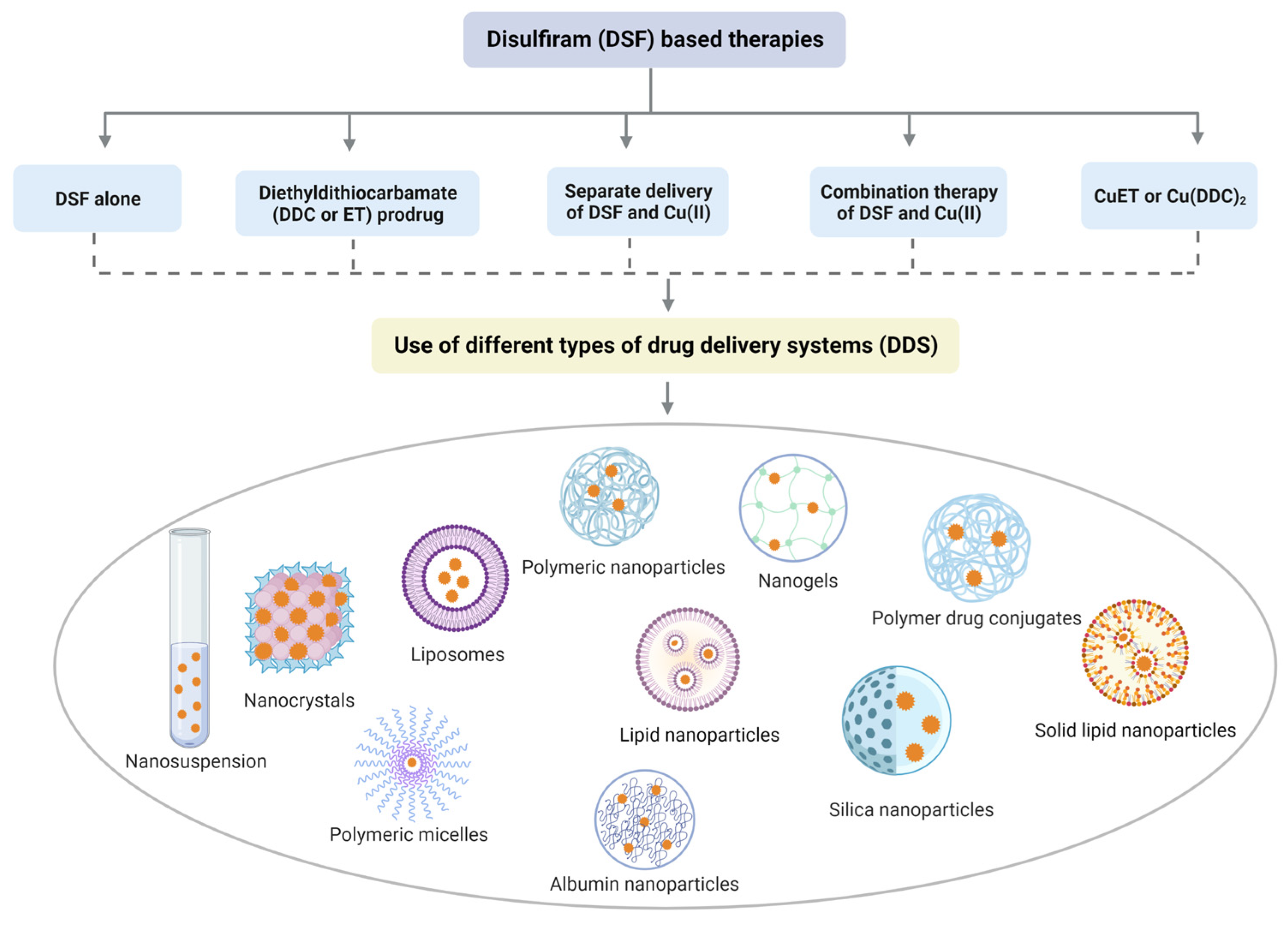
4. DSF-Based Therapies for the Treatment of Cancer
4.1. DSF Drug Delivery Systems and DDC Prodrug
4.2. Drug Delivery Systems for Cu (II) and DSF/Cu (II)
4.3. Drug Delivery Systems for CuET
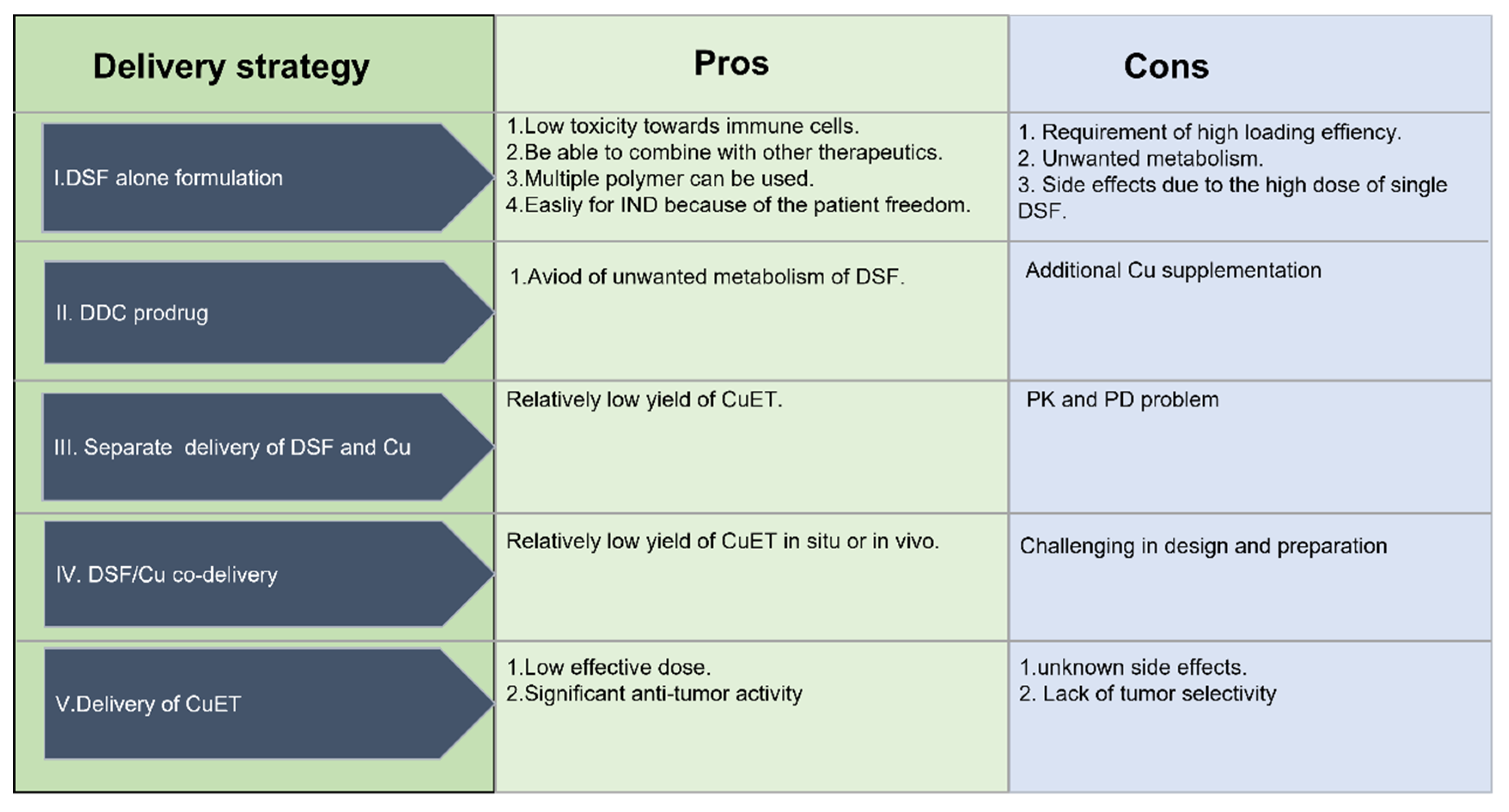
4.4. The Advantages and Disadvantages of Different Drug Delivery Systems
5. Summary and Future Directions
Author Contributions
Funding
Institutional Review Board Statement
Informed Consent Statement
Data Availability Statement
Conflicts of Interest
References
- Zhang, Z.; Zhou, L.; Xie, N.; Nice, E.C.; Zhang, T.; Cui, Y.; Huang, C. Overcoming cancer therapeutic bottleneck by drug repurposing. Signal Transduct. Target. Ther. 2020, 5, 113. [Google Scholar] [CrossRef] [PubMed]
- Chong, C.R.; Sullivan, D.J. New uses for old drugs. Nature 2007, 448, 645–646. [Google Scholar] [CrossRef] [PubMed]
- Corsello, S.M.; Nagari, R.T.; Spangler, R.D.; Rossen, J.; Kocak, M.; Bryan, J.G.; Humeidi, R.; Peck, D.; Wu, X.; Tang, A.A. Discovering the anticancer potential of non-oncology drugs by systematic viability profiling. Nat. Cancer 2020, 1, 235–248. [Google Scholar] [CrossRef]
- Ma, C.; Peng, Y.; Li, H.; Chen, W. Organ-on-a-chip: A new paradigm for drug development. Trends Pharmacol. Sci. 2021, 42, 119–133. [Google Scholar] [CrossRef]
- Lowndes, S.A.; Harris, A.L. The role of copper in tumour angiogenesis. J. Mammary Gland. Biol. Neoplasia 2005, 10, 299–310. [Google Scholar] [CrossRef] [PubMed]
- Antoniades, V.; Sioga, A.; Dietrich, E.M.; Meditskou, S.; Ekonomou, L.; Antoniades, K. Is copper chelation an effective anti-angiogenic strategy for cancer treatment? Med. Hypotheses 2013, 81, 1159–1163. [Google Scholar] [CrossRef] [PubMed]
- Wang, X.; Zhou, M.; Liu, Y.; Si, Z. Cope with copper: From copper linked mechanisms to copper-based clinical cancer therapies. Cancer Lett. 2023, 561, 216157. [Google Scholar] [CrossRef] [PubMed]
- da Silva, D.A.; De Luca, A.; Squitti, R.; Rongioletti, M.; Rossi, L.; Machado, C.M.L.; Cerchiaro, G. Copper in tumors and the use of copper-based compounds in cancer treatment. J. Inorg. Biochem. 2022, 226, 111634. [Google Scholar] [CrossRef]
- Tsvetkov, P.; Coy, S.; Petrova, B.; Dreishpoon, M.; Verma, A.; Abdusamad, M.; Rossen, J.; Joesch-Cohen, L.; Humeidi, R.; Spangler, R.D. Copper induces cell death by targeting lipoylated TCA cycle proteins. Science 2022, 375, 1254–1261. [Google Scholar] [CrossRef]
- Ghosh, P.; Vidal, C.; Dey, S.; Zhang, L. Mitochondria targeting as an effective strategy for cancer therapy. Int. J. Mol. Sci. 2020, 21, 3363. [Google Scholar] [CrossRef]
- Oliveri, V. Selective targeting of cancer cells by copper ionophores: An overview. Front. Mol. Biosci. 2022, 9, 841814. [Google Scholar] [CrossRef] [PubMed]
- Xie, J.; Yang, Y.; Gao, Y.; He, J. Cuproptosis: Mechanisms and links with cancers. Mol. Cancer 2023, 22, 46. [Google Scholar] [CrossRef] [PubMed]
- Johansson, B. A review of the pharmacokinetics and pharmacodynamics of disulfiram and its metabolites. Acta Psychiatr. Scand. 1992, 86, 15–26. [Google Scholar] [CrossRef] [PubMed]
- Mittal, M.; Bhagwati, S.; Siddiqi, M.I.; Chattopadhyay, N. A critical assessment of the potential of pharmacological modulation of aldehyde dehydrogenases to treat the diseases of bone loss. Eur. J. Pharmacol. 2020, 886, 173541. [Google Scholar] [CrossRef]
- Swift, R.; Davidson, D. Alcohol hangover: Mechanisms and mediators. Alcohol Health Res. World 1998, 22, 54–60. [Google Scholar]
- Yoshida, A.; Hsu, L.C.; Davé, V. Retinal oxidation activity and biological role of human cytosolic aldehyde dehydrogenase. Enzyme 1992, 46, 239–244. [Google Scholar] [CrossRef]
- Lewison, E.F. Spontaneous regression of breast cancer. Natl. Cancer Inst. Monogr. 1976, 44, 23–26. [Google Scholar]
- López-Lázaro, M. Dual role of hydrogen peroxide in cancer: Possible relevance to cancer chemoprevention and therapy. Cancer Lett. 2007, 252, 1–8. [Google Scholar] [CrossRef]
- Schmitt, S.M.; Frezza, M.; Dou, Q.P. New applications of old metal-binding drugs in the treatment of human cancer. Front. Biosci. 2012, 4, 375. [Google Scholar] [CrossRef]
- Wang, W.; McLeod, H.L.; Cassidy, J. Disulfiram-mediated inhibition of NF-κB activity enhances cytotoxicity of 5-fluorouracil in human colorectal cancer cell lines. Int. J. Cancer 2003, 104, 504–511. [Google Scholar] [CrossRef]
- Xu, B.; Shi, P.; Fombon, I.S.; Zhang, Y.; Huang, F.; Wang, W.; Zhou, S. Disulfiram/copper complex activated JNK/c-jun pathway and sensitized cytotoxicity of doxorubicin in doxorubicin resistant leukemia HL60 cells. Blood Cells Mol. Dis. 2011, 47, 264–269. [Google Scholar] [CrossRef] [PubMed]
- Yip, N.C.; Fombon, I.S.; Liu, P.; Brown, S.; Kannappan, V.; Armesilla, A.L.; Xu, B.; Cassidy, J.; Darling, J.L.; Wang, W. Disulfiram modulated ROS–MAPK and NFκB pathways and targeted breast cancer cells with cancer stem cell-like properties. Br. J. Cancer 2011, 104, 1564–1574. [Google Scholar] [CrossRef] [PubMed]
- Roudi, R.; Korourian, A.; Shariftabrizi, A.; Madjd, Z. Differential expression of cancer stem cell markers ALDH1 and CD133 in various lung cancer subtypes. Cancer Investig. 2015, 33, 294–302. [Google Scholar] [CrossRef]
- Koh, H.K.; Seo, S.Y.; Kim, J.H.; Kim, H.J.; Chie, E.K.; Kim, S.-K.; Kim, I.H. Disulfiram, a re-positioned aldehyde dehydrogenase inhibitor, enhances radiosensitivity of human glioblastoma cells in vitro. Cancer Res. Treat. Off. J. Korean Cancer Assoc. 2019, 51, 696–705. [Google Scholar] [CrossRef]
- Chen, W.; Yang, W.; Chen, P.; Huang, Y.; Li, F. Disulfiram copper nanoparticles prepared with a stabilized metal ion ligand complex method for treating drug-resistant prostate cancers. ACS Appl. Mater. Interfaces 2018, 10, 41118–41128. [Google Scholar] [CrossRef] [PubMed]
- Wang, N.-n.; Wang, L.-H.; Li, Y.; Fu, S.-Y.; Xue, X.; Jia, L.-N.; Yuan, X.-Z.; Wang, Y.-T.; Tang, X.; Yang, J.-Y. Targeting ALDH2 with disulfiram/copper reverses the resistance of cancer cells to microtubule inhibitors. Exp. Cell Res. 2018, 362, 72–82. [Google Scholar] [CrossRef] [PubMed]
- Liu, X.; Wang, L.; Cui, W.; Yuan, X.; Lin, L.; Cao, Q.; Wang, N.; Li, Y.; Guo, W.; Zhang, X. Targeting ALDH1A1 by disulfiram/copper complex inhibits non-small cell lung cancer recurrence driven by ALDH-positive cancer stem cells. Oncotarget 2016, 7, 58516–58530. [Google Scholar] [CrossRef] [PubMed]
- Denoyer, D.; Pearson, H.B.; Clatworthy, S.A.S.; Smith, Z.M.; Francis, P.S.; Llanos, R.M.; Volitakis, I.; Phillips, W.A.; Meggyesy, P.M.; Masaldan, S. Copper as a target for prostate cancer therapeutics: Copper-ionophore pharmacology and altering systemic copper distribution. Oncotarget 2016, 7, 37064–37080. [Google Scholar] [CrossRef]
- Orlov, A.P.; Orlova, M.A.; Trofimova, T.P.; Kalmykov, S.N.; Kuznetsov, D.A. The role of zinc and its compounds in leukemia. JBIC J. Biol. Inorg. Chem. 2018, 23, 347–362. [Google Scholar] [CrossRef]
- Gupte, A.; Mumper, R.J. Elevated copper and oxidative stress in cancer cells as a target for cancer treatment. Cancer Treat. Rev. 2009, 35, 32–46. [Google Scholar] [CrossRef]
- Chen, D.; Dou, Q.P. New uses for old copper-binding drugs: Converting the pro-angiogenic copper to a specific cancer cell death inducer. Expert Opin. Ther. Targets 2008, 12, 739–748. [Google Scholar] [CrossRef] [PubMed]
- Chang, Y.; Jiang, J.; Chen, W.; Yang, W.; Chen, L.; Chen, P.; Shen, J.; Qian, S.; Zhou, T.; Wu, L. Biomimetic metal-organic nanoparticles prepared with a 3D-printed microfluidic device as a novel formulation for disulfiram-based therapy against breast cancer. Appl. Mater. Today 2020, 18, 100492. [Google Scholar] [CrossRef] [PubMed]
- Lu, C.; Li, X.; Ren, Y.; Zhang, X. Disulfiram: A novel repurposed drug for cancer therapy. Cancer Chemother. Pharmacol. 2021, 87, 159–172. [Google Scholar] [CrossRef]
- Skrott, Z.; Mistrik, M.; Andersen, K.K.; Friis, S.; Majera, D.; Gursky, J.; Ozdian, T.; Bartkova, J.; Turi, Z.; Moudry, P. Alcohol-abuse drug disulfiram targets cancer via p97 segregase adaptor NPL4. Nature 2017, 552, 194–199. [Google Scholar] [CrossRef] [PubMed]
- Brar, S.S.; Grigg, C.; Wilson, K.S.; Holder, W.D., Jr.; Dreau, D.; Austin, C.; Foster, M.; Ghio, A.J.; Whorton, A.R.; Stowell, G.W. Disulfiram inhibits activating transcription factor/cyclic AMP-responsive element binding protein and human melanoma growth in a metal-dependent manner in vitro, in mice and in a patient with metastatic disease. Mol. Cancer Ther. 2004, 3, 1049–1060. [Google Scholar] [CrossRef] [PubMed]
- Wickström, M.; Danielsson, K.; Rickardson, L.; Gullbo, J.; Nygren, P.; Isaksson, A.; Larsson, R.; Lövborg, H. Pharmacological profiling of disulfiram using human tumor cell lines and human tumor cells from patients. Biochem. Pharmacol. 2007, 73, 25–33. [Google Scholar] [CrossRef]
- Li, H.; Wang, J.; Wu, C.; Wang, L.; Chen, Z.-S.; Cui, W. The combination of disulfiram and copper for cancer treatment. Drug Discov. Today 2020, 25, 1099–1108. [Google Scholar] [CrossRef]
- Ren, X.; Li, Y.; Zhou, Y.; Hu, W.; Yang, C.; Jing, Q.; Zhou, C.; Wang, X.; Hu, J.; Wang, L.; et al. Overcoming the compensatory elevation of NRF2 renders hepatocellular carcinoma cells more vulnerable to disulfiram/copper-induced ferroptosis. Redox Biol. 2021, 46, 102122. [Google Scholar] [CrossRef] [PubMed]
- Guo, F.; Yang, Z.; Kulbe, H.; Albers, A.E.; Sehouli, J.; Kaufmann, A.M. Inhibitory effect on ovarian cancer ALDH+ stem-like cells by Disulfiram and Copper treatment through ALDH and ROS modulation. Biomed. Pharmacother. 2019, 118, 109371. [Google Scholar] [CrossRef]
- Zhao, P.; Zhang, J.; Wu, A.; Zhang, M.; Zhao, Y.; Tang, Y.; Wang, B.; Chen, T.; Li, F.; Zhao, Q. Biomimetic codelivery overcomes osimertinib-resistant NSCLC and brain metastasis via macrophage-mediated innate immunity. J. Control. Release 2021, 329, 1249–1261. [Google Scholar] [CrossRef]
- Zheng, Z.; Zhang, J.; Jiang, J.; He, Y.; Zhang, W.; Mo, X.; Kang, X.; Xu, Q.; Wang, B.; Huang, Y. Remodeling tumor immune microenvironment (TIME) for glioma therapy using multi-targeting liposomal codelivery. J. Immunother. Cancer 2020, 8, e000207. [Google Scholar] [CrossRef] [PubMed]
- Morrison, B.W.; Doudican, N.A.; Patel, K.R.; Orlow, S.J. Disulfiram induces copper-dependent stimulation of reactive oxygen species and activation of the extrinsic apoptotic pathway in melanoma. Melanoma Res. 2010, 20, 11–20. [Google Scholar] [CrossRef] [PubMed]
- Liu, P.; Brown, S.; Goktug, T.; Channathodiyil, P.; Kannappan, V.; Hugnot, J.P.; Guichet, P.O.; Bian, X.; Armesilla, A.L.; Darling, J.L. Cytotoxic effect of disulfiram/copper on human glioblastoma cell lines and ALDH-positive cancer-stem-like cells. Br. J. Cancer 2012, 107, 1488–1497. [Google Scholar] [CrossRef] [PubMed]
- Nie, D.; Chen, C.; Li, Y.; Zeng, C. Disulfiram, an aldehyde dehydrogenase inhibitor, works as a potent drug against sepsis and cancer via NETosis, pyroptosis, apoptosis, ferroptosis, and cuproptosis. Blood Sci. 2022, 4, 152–154. [Google Scholar] [CrossRef] [PubMed]
- Huang, J.; Chaudhary, R.; Cohen, A.L.; Fink, K.; Goldlust, S.; Boockvar, J.; Chinnaiyan, P.; Wan, L.; Marcus, S.; Campian, J.L. A multicenter phase II study of temozolomide plus disulfiram and copper for recurrent temozolomide-resistant glioblastoma. J. Neuro-Oncol. 2019, 142, 537–544. [Google Scholar] [CrossRef] [PubMed]
- Huang, J.; Campian, J.L.; Gujar, A.D.; Tsien, C.; Ansstas, G.; Tran, D.D.; DeWees, T.A.; Lockhart, A.C.; Kim, A.H. Final results of a phase I dose-escalation, dose-expansion study of adding disulfiram with or without copper to adjuvant temozolomide for newly diagnosed glioblastoma. J. Neuro-Oncol. 2018, 138, 105–111. [Google Scholar] [CrossRef]
- Nechushtan, H.; Hamamreh, Y.; Nidal, S.; Gotfried, M.; Baron, A.; Shalev, Y.I.; Nisman, B.; Peretz, T.; Peylan-Ramu, N. A phase IIb trial assessing the addition of disulfiram to chemotherapy for the treatment of metastatic non-small cell lung cancer. Oncologist 2015, 20, 366–367. [Google Scholar] [CrossRef]
- Wang, L.; Yu, Y.; Zhou, C.; Wan, R.; Li, Y. Anticancer effects of disulfiram: A systematic review of in vitro, animal, and human studies. Syst. Rev. 2022, 11, 109. [Google Scholar] [CrossRef]
- Liou, G.-Y.; Storz, P. Reactive oxygen species in cancer. Free. Radic. Res. 2010, 44, 479–496. [Google Scholar] [CrossRef]
- Oberley, T.D. Oxidative damage and cancer. Am. J. Pathol. 2002, 160, 403–408. [Google Scholar] [CrossRef]
- Trachootham, D.; Alexandre, J.; Huang, P. Targeting cancer cells by ROS-mediated mechanisms: A radical therapeutic approach? Nat. Rev. Drug Discov. 2009, 8, 579–591. [Google Scholar] [CrossRef]
- Roy, D.; Sheng, G.Y.; Herve, S.; Carvalho, E.; Mahanty, A.; Yuan, S.; Sun, L. Interplay between cancer cell cycle and metabolism: Challenges, targets and therapeutic opportunities. Biomed. Pharmacother. 2017, 89, 288–296. [Google Scholar] [CrossRef] [PubMed]
- Wang, J.; Yi, J. Cancer cell killing via ROS: To increase or decrease, that is the question. Cancer Biol. Ther. 2008, 7, 1875–1884. [Google Scholar] [CrossRef] [PubMed]
- Yang, Z.; Guo, F.; Albers, A.E.; Sehouli, J.; Kaufmann, A.M. Disulfiram modulates ROS accumulation and overcomes synergistically cisplatin resistance in breast cancer cell lines. Biomed. Pharmacother. 2019, 113, 108727. [Google Scholar] [CrossRef] [PubMed]
- Li, Y.; Chen, F.; Chen, J.; Chan, S.; He, Y.; Liu, W.; Zhang, G. Disulfiram/copper induces antitumor activity against both nasopharyngeal cancer cells and cancer-associated fibroblasts through ROS/MAPK and ferroptosis pathways. Cancers 2020, 12, 138. [Google Scholar] [CrossRef] [PubMed]
- Allensworth, J.L.; Evans, M.K.; Bertucci, F.; Aldrich, A.J.; Festa, R.A.; Finetti, P.; Ueno, N.T.; Safi, R.; McDonnell, D.P.; Thiele, D.J. Disulfiram (DSF) acts as a copper ionophore to induce copper-dependent oxidative stress and mediate anti-tumor efficacy in inflammatory breast cancer. Mol. Oncol. 2015, 9, 1155–1168. [Google Scholar] [CrossRef]
- Calderon-Aparicio, A.; Strasberg-Rieber, M.; Rieber, M. Disulfiram anti-cancer efficacy without copper overload is enhanced by extracellular H2O2 generation: Antagonism by tetrathiomolybdate. Oncotarget 2015, 6, 29771–29781. [Google Scholar] [CrossRef]
- Cen, D.; Brayton, D.; Shahandeh, B.; Meyskens, F.L.; Farmer, P.J. Disulfiram facilitates intracellular Cu uptake and induces apoptosis in human melanoma cells. J. Med. Chem. 2004, 47, 6914–6920. [Google Scholar] [CrossRef]
- Fruehauf, J.P.; Trapp, V. Reactive oxygen species: An Achilles’ heel of melanoma? Expert Rev. Anticancer. Ther. 2008, 8, 1751–1757. [Google Scholar] [CrossRef]
- Cen, D.; Gonzalez, R.I.; Buckmeier, J.A.; Kahlon, R.S.; Tohidian, N.B.; Meyskens, F.L., Jr. Disulfiram induces apoptosis in human melanoma cells: A redox-related process. Mol. Cancer Ther. 2002, 1, 197–204. [Google Scholar]
- Leon, G.; MacDonagh, L.; Finn, S.P.; Cuffe, S.; Barr, M.P. Cancer stem cells in drug resistant lung cancer: Targeting cell surface markers and signaling pathways. Pharmacol. Ther. 2016, 158, 71–90. [Google Scholar] [CrossRef] [PubMed]
- Moreb, J.S.; Ucar, D.; Han, S.; Amory, J.K.; Goldstein, A.S.; Ostmark, B.; Chang, L.-J. The enzymatic activity of human aldehyde dehydrogenases 1A2 and 2 (ALDH1A2 and ALDH2) is detected by Aldefluor, inhibited by diethylaminobenzaldehyde and has significant effects on cell proliferation and drug resistance. Chem.-Biol. Interact. 2012, 195, 52–60. [Google Scholar] [CrossRef] [PubMed]
- Jin, N.; Zhu, X.; Cheng, F.; Zhang, L. Disulfiram/copper targets stem cell-like ALDH+ population of multiple myeloma by inhibition of ALDH1A1 and Hedgehog pathway. J. Cell. Biochem. 2018, 119, 6882–6893. [Google Scholar] [CrossRef] [PubMed]
- Yan, Z.; Xu, L.; Zhang, J.; Lu, Q.; Luo, S.; Xu, L. Aldehyde dehydrogenase 1A1 stabilizes transcription factor Gli2 and enhances the activity of Hedgehog signaling in hepatocellular cancer. Biochem. Biophys. Res. Commun. 2016, 471, 466–473. [Google Scholar] [CrossRef]
- Delude, C. Tumorigenesis: Testing ground for cancer stem cells. Nature 2011, 480, S43–S45. [Google Scholar] [CrossRef]
- Sharma, V.; Verma, V.; Lal, N.; Yadav, S.K.; Sarkar, S.; Mandalapu, D.; Porwal, K.; Rawat, T.; Maikhuri, J.P.; Rajender, S. Disulfiram and its novel derivative sensitize prostate cancer cells to the growth regulatory mechanisms of the cell by re-expressing the epigenetically repressed tumor suppressor—Estrogen receptor β. Mol. Carcinog. 2016, 55, 1843–1857. [Google Scholar] [CrossRef]
- Tesson, M.; Anselmi, G.; Bell, C.; Mairs, R. Cell cycle specific radiosensitisation by the disulfiram and copper complex. Oncotarget 2017, 8, 65900–65916. [Google Scholar] [CrossRef]
- Pegg, A.E. Repair of O6-alkylguanine by alkyltransferases. Mutat. Res./Rev. Mutat. Res. 2000, 462, 83–100. [Google Scholar] [CrossRef]
- Mishina, Y.; Duguid, E.M.; He, C. Direct reversal of DNA alkylation damage. Chem. Rev. 2006, 106, 215–232. [Google Scholar] [CrossRef]
- Gerson, S.L. Clinical relevance of MGMT in the treatment of cancer. J. Clin. Oncol. 2002, 20, 2388–2399. [Google Scholar] [CrossRef]
- Paranjpe, A.; Zhang, R.; Ali-Osman, F.; Bobustuc, G.C.; Srivenugopal, K.S. Disulfiram is a direct and potent inhibitor of human O 6-methylguanine-DNA methyltransferase (MGMT) in brain tumor cells and mouse brain and markedly increases the alkylating DNA damage. Carcinogenesis 2014, 35, 692–702. [Google Scholar] [CrossRef] [PubMed]
- Locasale, J.W.; Grassian, A.R.; Melman, T.; Lyssiotis, C.A.; Mattaini, K.R.; Bass, A.J.; Heffron, G.; Metallo, C.M.; Muranen, T.; Sharfi, H. Phosphoglycerate dehydrogenase diverts glycolytic flux and contributes to oncogenesis. Nat. Genet. 2011, 43, 869–874. [Google Scholar] [CrossRef]
- Spillier, Q.; Vertommen, D.; Ravez, S.; Marteau, R.; Thémans, Q.; Corbet, C.; Feron, O.; Wouters, J.; Frédérick, R. Anti-alcohol abuse drug disulfiram inhibits human PHGDH via disruption of its active tetrameric form through a specific cysteine oxidation. Sci. Rep. 2019, 9, 4737. [Google Scholar] [CrossRef] [PubMed]
- Turner, N.; Tutt, A.; Ashworth, A. Targeting the DNA repair defect of BRCA tumours. Curr. Opin. Pharmacol. 2005, 5, 388–393. [Google Scholar] [CrossRef]
- Tutt, A.; Ashworth, A. The relationship between the roles of BRCA genes in DNA repair and cancer predisposition. Trends Mol. Med. 2002, 8, 571–576. [Google Scholar] [CrossRef]
- Majera, D.; Skrott, Z.; Chroma, K.; Merchut-Maya, J.M.; Mistrik, M.; Bartek, J. Targeting the NPL4 Adaptor of p97/VCP Segregase by Disulfiram as an Emerging Cancer Vulnerability Evokes Replication Stress and DNA Damage while Silencing the ATR Pathway. Cells 2020, 9, 469. [Google Scholar] [CrossRef] [PubMed]
- Lecker, S.H.; Goldberg, A.L.; Mitch, W.E. Protein degradation by the ubiquitin–proteasome pathway in normal and disease states. J. Am. Soc. Nephrol. 2006, 17, 1807–1819. [Google Scholar] [CrossRef]
- Wang, F.; Zhai, S.; Liu, X.; Li, L.; Wu, S.; Dou, Q.P.; Yan, B. A novel dithiocarbamate analogue with potentially decreased ALDH inhibition has copper-dependent proteasome-inhibitory and apoptosis-inducing activity in human breast cancer cells. Cancer Lett. 2011, 300, 87–95. [Google Scholar] [CrossRef]
- Kleiger, G.; Mayor, T. Perilous journey: A tour of the ubiquitin–proteasome system. Trends Cell Biol. 2014, 24, 352–359. [Google Scholar] [CrossRef]
- Wang, H.; Yang, Q.; Dou, Q.P.; Yang, H. Discovery of natural proteasome inhibitors as novel anticancer therapeutics: Current status and perspectives. Curr. Protein Pept. Sci. 2018, 19, 358–367. [Google Scholar] [CrossRef]
- Chen, D.; Cui, Q.C.; Yang, H.; Dou, Q.P. Disulfiram, a clinically used anti-alcoholism drug and copper-binding agent, induces apoptotic cell death in breast cancer cultures and xenografts via inhibition of the proteasome activity. Cancer Res. 2006, 66, 10425–10433. [Google Scholar] [CrossRef]
- Chen, D.; Peng, F.; Cui, Q.C.; Daniel, K.G.; Orlu, S.; Liu, J.; Dou, Q.P. Inhibition of prostate cancer cellular proteasome activity by a pyrrolidine dithiocarbamate-copper complex is associated with suppression of proliferation and induction of apoptosis. Front. Biosci. 2005, 10, 2932–2939. [Google Scholar] [CrossRef] [PubMed]
- Meraz-Torres, F.; Plöger, S.; Garbe, C.; Niessner, H.; Sinnberg, T. Disulfiram as a therapeutic agent for metastatic malignant melanoma—Old myth or new logos? Cancers 2020, 12, 3538. [Google Scholar] [CrossRef] [PubMed]
- Lewis, D.J.; Deshmukh, P.; Tedstone, A.A.; Tuna, F.; O’Brien, P. On the interaction of copper (II) with disulfiram. Chem. Commun. 2014, 50, 13334–13337. [Google Scholar] [CrossRef] [PubMed]
- Li, L.; Yang, H.; Chen, D.; Cui, C.; Dou, Q.P. Disulfiram promotes the conversion of carcinogenic cadmium to a proteasome inhibitor with pro-apoptotic activity in human cancer cells. Toxicol. Appl. Pharmacol. 2008, 229, 206–214. [Google Scholar] [CrossRef]
- Pham, C.G.; Bubici, C.; Zazzeroni, F.; Knabb, J.R.; Papa, S.; Kuntzen, C.; Franzoso, G. Upregulation of Twist-1 by NF-κB blocks cytotoxicity induced by chemotherapeutic drugs. Mol. Cell. Biol. 2007, 27, 3920–3935. [Google Scholar] [CrossRef]
- Cvek, B.; Dvorak, Z. The value of proteasome inhibition in cancer: Can the old drug, disulfiram, have a bright new future as a novel proteasome inhibitor? Drug Discov. Today 2008, 13, 716–722. [Google Scholar] [CrossRef]
- Henkel, T.; Machleidt, T.; Alkalay, I.; Krönke, M.; Ben-Neriah, Y.; Baeuerle, P.A. Rapid proteolysis of I κ B-α is necessary for activation of transcription factor NF-κ B. Nature 1993, 365, 182–185. [Google Scholar] [CrossRef]
- Annunziata, C.M.; Davis, R.E.; Demchenko, Y.; Bellamy, W.; Gabrea, A.; Zhan, F.; Lenz, G.; Hanamura, I.; Wright, G.; Xiao, W. Frequent engagement of the classical and alternative NF-κB pathways by diverse genetic abnormalities in multiple myeloma. Cancer Cell 2007, 12, 115–130. [Google Scholar] [CrossRef]
- Hothi, P.; Martins, T.J.; Chen, L.; Deleyrolle, L.; Yoon, J.-G.; Reynolds, B.; Foltz, G. High-throughput chemical screens identify disulfiram as an inhibitor of human glioblastoma stem cells. Oncotarget 2012, 3, 1124–1136. [Google Scholar] [CrossRef]
- Brar, S.S.; Kennedy, T.P.; Whorton, A.R.; Sturrock, A.B.; Huecksteadt, T.P.; Ghio, A.J.; Hoidal, J.R. Reactive oxygen species from NAD (P) H: Quinone oxidoreductase constitutively activate NF-κB in malignant melanoma cells. Am. J. Physiol.-Cell Physiol. 2001, 280, C659–C676. [Google Scholar] [CrossRef] [PubMed]
- Dolcet, X.; Llobet, D.; Pallares, J.; Matias-Guiu, X. NF-kB in development and progression of human cancer. Virchows Arch. 2005, 446, 475–482. [Google Scholar] [CrossRef] [PubMed]
- Meyskens, F.L., Jr.; Buckmeier, J.A.; McNulty, S.E.; Tohidian, N.B. Activation of nuclear factor-κB in human metastatic melanoma cells and the effect of oxidative stress. Clin. Cancer Res. 1999, 5, 1197–1202. [Google Scholar]
- Askgaard, G.; Friis, S.; Hallas, J.; Thygesen, L.C.; Pottegård, A. Use of disulfiram and risk of cancer. Eur. J. Cancer Prev. 2014, 23, 225–232. [Google Scholar] [CrossRef] [PubMed]
- Pan, Q.; Kleer, C.G.; van Golen, K.L.; Irani, J.; Bottema, K.M.; Bias, C.; De Carvalho, M.; Mesri, E.A.; Robins, D.M.; Dick, R.D. Copper deficiency induced by tetrathiomolybdate suppresses tumor growth and angiogenesis. Cancer Res. 2002, 62, 4854–4859. [Google Scholar] [PubMed]
- Xu, B.; Wang, S.; Li, R.; Chen, K.; He, L.; Deng, M.; Kannappan, V.; Zha, J.; Dong, H.; Wang, W. Disulfiram/copper selectively eradicates AML leukemia stem cells in vitro and in vivo by simultaneous induction of ROS-JNK and inhibition of NF-κB and Nrf2. Cell Death Dis. 2017, 8, e2797. [Google Scholar] [CrossRef] [PubMed]
- Quail, D.F.; Joyce, J.A. Microenvironmental regulation of tumor progression and metastasis. Nat. Med. 2013, 19, 1423–1437. [Google Scholar] [CrossRef]
- Ostuni, R.; Kratochvill, F.; Murray, P.J.; Natoli, G. Macrophages and cancer: From mechanisms to therapeutic implications. Trends Immunol. 2015, 36, 229–239. [Google Scholar] [CrossRef]
- Yang, M.; Li, J.; Gu, P.; Fan, X. The application of nanoparticles in cancer immunotherapy: Targeting tumor microenvironment. Bioact. Mater. 2021, 6, 1973–1987. [Google Scholar] [CrossRef]
- Terashima, Y.; Toda, E.; Itakura, M.; Otsuji, M.; Yoshinaga, S.; Okumura, K.; Shand, F.H.W.; Komohara, Y.; Takeda, M.; Kokubo, K.; et al. Targeting FROUNT with disulfiram suppresses macrophage accumulation and its tumor-promoting properties. Nat. Commun. 2020, 11, 609. [Google Scholar] [CrossRef]
- Kang, X.; Cai, Y.; Wang, Q.; Wang, C.; Chen, W.; Yang, W.; Suryawanshi, A.; Zhou, G.; Chen, P.; Li, F. Near-infrared light triggered activation of pro-drug combination cancer therapy and induction of immunogenic cell death. Int. J. Pharm. 2021, 607, 120972. [Google Scholar] [CrossRef]
- You, S.-Y.; Rui, W.; Chen, S.-T.; Chen, H.-C.; Liu, X.-W.; Huang, J.; Chen, H.-Y. Process of immunogenic cell death caused by disulfiram as the anti-colorectal cancer candidate. Biochem. Biophys. Res. Commun. 2019, 513, 891–897. [Google Scholar] [CrossRef] [PubMed]
- Zhang, X.; Hu, P.; Ding, S.-Y.; Sun, T.; Liu, L.; Han, S.; DeLeo, A.B.; Sadagopan, A.; Guo, W.; Wang, X. Induction of autophagy-dependent apoptosis in cancer cells through activation of ER stress: An uncovered anti-cancer mechanism by anti-alcoholism drug disulfiram. Am. J. Cancer Res. 2019, 9, 1266–1281. [Google Scholar] [PubMed]
- Guo, Y.; Wang, S.-Z.; Zhang, X.; Jia, H.-R.; Zhu, Y.-X.; Zhang, X.; Gao, G.; Jiang, Y.-W.; Li, C.; Chen, X.; et al. In situ generation of micrometer-sized tumor cell-derived vesicles as autologous cancer vaccines for boosting systemic immune responses. Nat. Commun. 2022, 13, 6534. [Google Scholar] [CrossRef] [PubMed]
- Gao, X.; Huang, H.; Pan, C.; Mei, Z.; Yin, S.; Zhou, L.; Zheng, S. Disulfiram/Copper Induces Immunogenic Cell Death and Enhances CD47 Blockade in Hepatocellular Carcinoma. Cancers 2022, 14, 4715. [Google Scholar] [CrossRef]
- Wu, X.; Xue, X.; Wang, L.; Wang, W.; Han, J.; Sun, X.; Zhang, H.; Liu, Y.; Che, X.; Yang, J. Suppressing autophagy enhances disulfiram/copper-induced apoptosis in non-small cell lung cancer. Eur. J. Pharmacol. 2018, 827, 1–12. [Google Scholar] [CrossRef]
- Yang, Y.; Deng, Q.; Feng, X.; Sun, J. Use of the disulfiram/copper complex for breast cancer chemoprevention in MMTV-erbB2 transgenic mice. Mol. Med. Rep. 2015, 12, 746–752. [Google Scholar] [CrossRef]
- Cheriyan, V.T.; Wang, Y.; Muthu, M.; Jamal, S.; Chen, D.; Yang, H.; Polin, L.A.; Tarca, A.L.; Pass, H.I.; Dou, Q.P. Disulfiram suppresses growth of the malignant pleural mesothelioma cells in part by inducing apoptosis. PLoS ONE 2014, 9, e93711. [Google Scholar] [CrossRef]
- Lin, Y.; Bai, L.; Chen, W.; Xu, S. The NF-κB activation pathways, emerging molecular targets for cancer prevention and therapy. Expert Opin. Ther. Targets 2010, 14, 45–55. [Google Scholar] [CrossRef]
- Li, Y.; Wang, L.H.; Zhang, H.T.; Wang, Y.T.; Liu, S.; Zhou, W.L.; Yuan, X.Z.; Li, T.Y.; Wu, C.F.; Yang, J.Y. Disulfiram combined with copper inhibits metastasis and epithelial–mesenchymal transition in hepatocellular carcinoma through the NF-κB and TGF-β pathways. J. Cell. Mol. Med. 2018, 22, 439–451. [Google Scholar] [CrossRef]
- Bao, B.; Wang, Z.; Ali, S.; Ahmad, A.; Azmi, A.S.; Sarkar, S.H.; Banerjee, S.; Kong, D.; Li, Y.; Thakur, S. Metformin inhibits cell proliferation, migration and invasion by attenuating CSC function mediated by deregulating miRNAs in pancreatic cancer cells. Cancer Prev. Res. 2012, 5, 355–364. [Google Scholar] [CrossRef] [PubMed]
- Bleau, A.-M.; Hambardzumyan, D.; Ozawa, T.; Fomchenko, E.I.; Huse, J.T.; Brennan, C.W.; Holland, E.C. PTEN/PI3K/Akt Pathway Regulates the Side Population Phenotype and ABCG2 Activity in Glioma Tumor Stem-like Cells. Cell Stem Cell 2009, 4, 226–235. [Google Scholar] [CrossRef] [PubMed]
- Venere, M.; Hamerlik, P.; Wu, Q.; Rasmussen, R.D.; Song, L.A.; Vasanji, A.; Tenley, N.; Flavahan, W.A.; Hjelmeland, A.B.; Bartek, J.; et al. Therapeutic targeting of constitutive PARP activation compromises stem cell phenotype and survival of glioblastoma-initiating cells. Cell Death Differ. 2014, 21, 258–269. [Google Scholar] [CrossRef] [PubMed]
- Petersen, E.N. The pharmacology and toxicology of disulfiram and its metabolites. Acta Psychiatr. Scand. 1992, 86, 7–13. [Google Scholar] [CrossRef] [PubMed]
- Ginestier, C.; Hur, M.H.; Charafe-Jauffret, E.; Monville, F.; Dutcher, J.; Brown, M.; Jacquemier, J.; Viens, P.; Kleer, C.G.; Liu, S. ALDH1 is a marker of normal and malignant human mammary stem cells and a predictor of poor clinical outcome. Cell Stem Cell 2007, 1, 555–567. [Google Scholar] [CrossRef] [PubMed]
- Moreb, J.S. Aldehyde dehydrogenase as a marker for stem cells. Curr. Stem Cell Res. Ther. 2008, 3, 237–246. [Google Scholar] [CrossRef] [PubMed]
- Duan, X.; Xiao, J.; Yin, Q.; Zhang, Z.; Yu, H.; Mao, S.; Li, Y. Multi-targeted inhibition of tumor growth and lung metastasis by redox-sensitive shell crosslinked micelles loading disulfiram. Nanotechnology 2014, 25, 125102. [Google Scholar] [CrossRef]
- Chiba, T.; Suzuki, E.; Yuki, K.; Zen, Y.; Oshima, M.; Miyagi, S.; Saraya, A.; Koide, S.; Motoyama, T.; Ogasawara, S. Disulfiram eradicates tumor-initiating hepatocellular carcinoma cells in ROS-p38 MAPK pathway-dependent and-independent manners. PLoS ONE 2014, 9, e84807. [Google Scholar] [CrossRef]
- Triscott, J.; Lee, C.; Hu, K.; Fotovati, A.; Berns, R.; Pambid, M.; Luk, M.; Kast, R.E.; Kong, E.; Toyota, E. Disulfiram, a drug widely used to control alcoholism, suppresses self-renewal of glioblastoma and overrides resistance to temozolomide. Oncotarget 2012, 3, 1112–1123. [Google Scholar] [CrossRef]
- Mimeault, M.; Batra, S.K. Recent advances in the development of novel anti-cancer drugs targeting cancer stem/progenitor cells. Drug Dev. Res. 2008, 69, 415–430. [Google Scholar] [CrossRef]
- Manders, P.; Beex, L.; Tjan-Heijnen, V.C.G.; Geurts-Moespot, J.; Van Tienoven, T.; Foekens, J.A.; Sweep, C.G.J. The prognostic value of vascular endothelial growth factor in 574 node-negative breast cancer patients who did not receive adjuvant systemic therapy. Br. J. Cancer 2002, 87, 772–778. [Google Scholar] [CrossRef] [PubMed]
- Schreier, S.M.; Muellner, M.K.; Steinkellner, H.; Hermann, M.; Esterbauer, H.; Exner, M.; Gmeiner, B.M.K.; Kapiotis, S.; Laggner, H. Hydrogen sulfide scavenges the cytotoxic lipid oxidation product 4-HNE. Neurotox. Res. 2010, 17, 249–256. [Google Scholar] [CrossRef]
- Ayala, A.; Muñoz, M.F.; Argüelles, S. Lipid peroxidation: Production, metabolism, and signaling mechanisms of malondialdehyde and 4-hydroxy-2-nonenal. Oxidative Med. Cell. Longev. 2014, 2014, 360438. [Google Scholar] [CrossRef] [PubMed]
- Chapple, S.J.; Cheng, X.; Mann, G.E. Effects of 4-hydroxynonenal on vascular endothelial and smooth muscle cell redox signaling and function in health and disease. Redox Biol. 2013, 1, 319–331. [Google Scholar] [CrossRef] [PubMed]
- Camaré, C.; Vanucci-Bacqué, C.; Augé, N.; Pucelle, M.; Bernis, C.; Swiader, A.; Baltas, M.; Bedos-Belval, F.; Salvayre, R.; Nègre-Salvayre, A. 4-Hydroxynonenal contributes to angiogenesis through a redox-dependent sphingolipid pathway: Prevention by hydralazine derivatives. Oxidative Med. Cell. Longev. 2017, 2017, 9172741. [Google Scholar] [CrossRef]
- Liu, X.; Sun, X.; Liao, H.; Dong, Z.; Zhao, J.; Zhu, H.; Wang, P.; Shen, L.; Xu, L.; Ma, X. Mitochondrial aldehyde dehydrogenase 2 regulates revascularization in chronic ischemia: Potential impact on the development of coronary collateral circulation. Arterioscler. Thromb. Vasc. Biol. 2015, 35, 2196–2206. [Google Scholar] [CrossRef] [PubMed]
- Marikovsky, M.; Nevo, N.; Vadai, E.; Harris-Cerruti, C. Cu/Zn superoxide dismutase plays a role in angiogenesis. Int. J. Cancer 2002, 97, 34–41. [Google Scholar] [CrossRef]
- Shian, S.-G.; Kao, Y.-R.; Wu, F.Y.-H.; Wu, C.-W. Inhibition of invasion and angiogenesis by zinc-chelating agent disulfiram. Mol. Pharmacol. 2003, 64, 1076–1084. [Google Scholar] [CrossRef]
- Kast, R.E.; Halatsch, M.-E. Matrix Metalloproteinase-2 and-9 in glioblastoma: A trio of old drugs—Captopril, disulfiram and nelfinavir—Are inhibitors with potential as adjunctive treatments in glioblastoma. Arch. Med. Res. 2012, 43, 243–247. [Google Scholar] [CrossRef] [PubMed]
- Li, Y.; Fu, S.-Y.; Wang, L.-H.; Wang, F.-Y.; Wang, N.-N.; Cao, Q.; Wang, Y.-T.; Yang, J.-Y.; Wu, C.-F. Copper improves the anti-angiogenic activity of disulfiram through the EGFR/Src/VEGF pathway in gliomas. Cancer Lett. 2015, 369, 86–96. [Google Scholar] [CrossRef] [PubMed]
- Zhang, L.; Zhou, Y.; Sun, X.; Zhou, J.; Yang, P. CXCL12 overexpression promotes the angiogenesis potential of periodontal ligament stem cells. Sci. Rep. 2017, 7, 10286. [Google Scholar] [CrossRef] [PubMed]
- Wang, X.; Cao, Y.; Zhang, S.; Chen, Z.; Fan, L.; Shen, X.; Zhou, S.; Chen, D. Stem cell autocrine CXCL12/CXCR4 stimulates invasion and metastasis of esophageal cancer. Oncotarget 2017, 8, 36149–36160. [Google Scholar] [CrossRef]
- Ponti, D.; Costa, A.; Zaffaroni, N.; Pratesi, G.; Petrangolini, G.; Coradini, D.; Pilotti, S.; Pierotti, M.A.; Daidone, M.G. Isolation and in vitro propagation of tumorigenic breast cancer cells with stem/progenitor cell properties. Cancer Res. 2005, 65, 5506–5511. [Google Scholar] [CrossRef] [PubMed]
- Beckermann, B.M.; Kallifatidis, G.; Groth, A.; Frommhold, D.; Apel, A.; Mattern, J.; Salnikov, A.V.; Moldenhauer, G.; Wagner, W.; Diehlmann, A. VEGF expression by mesenchymal stem cells contributes to angiogenesis in pancreatic carcinoma. Br. J. Cancer 2008, 99, 622–631. [Google Scholar] [CrossRef]
- O’Brien, A.; Barber, J.E.B.; Reid, S.; Niknejad, N.; Dimitroulakos, J. Enhancement of cisplatin cytotoxicity by disulfiram involves activating transcription factor 3. Anticancer. Res. 2012, 32, 2679–2688. [Google Scholar] [PubMed]
- Olmo, F.; Urbanová, K.; Rosales, M.J.; Martín-Escolano, R.; Sánchez-Moreno, M.; Marín, C. An in vitro iron superoxide dismutase inhibitor decreases the parasitemia levels of Trypanosoma cruzi in BALB/c mouse model during acute phase. Int. J. Parasitol. Drugs Drug Resist. 2015, 5, 110–116. [Google Scholar] [CrossRef] [PubMed]
- Schmidtova, S.; Kalavska, K.; Gercakova, K.; Cierna, Z.; Miklikova, S.; Smolkova, B.; Buocikova, V.; Miskovska, V.; Durinikova, E.; Burikova, M. Disulfiram overcomes cisplatin resistance in human embryonal carcinoma cells. Cancers 2019, 11, 1224. [Google Scholar] [CrossRef]
- Lun, X.; Wells, J.C.; Grinshtein, N.; King, J.C.; Hao, X.; Dang, N.-H.; Wang, X.; Aman, A.; Uehling, D.; Datti, A. Disulfiram when Combined with Copper Enhances the Therapeutic Effects of Temozolomide for the Treatment of GlioblastomaDisulfiram/Copper Enhance Temozolomide Treatment for Glioblastoma. Clin. Cancer Res. 2016, 22, 3860–3875. [Google Scholar] [CrossRef]
- Thiery, J.P. Epithelial–mesenchymal transitions in development and pathologies. Curr. Opin. Cell Biol. 2003, 15, 740–746. [Google Scholar] [CrossRef]
- Thiery, J.P.; Acloque, H.; Huang, R.Y.J.; Nieto, M.A. Epithelial-mesenchymal transitions in development and disease. Cell 2009, 139, 871–890. [Google Scholar] [CrossRef]
- Kang, X.; Wang, J.; Huang, C.-H.; Wibowo, F.S.; Amin, R.; Chen, P.; Li, F. Diethyldithiocarbamate copper nanoparticle overcomes resistance in cancer therapy without inhibiting P-glycoprotein. Nanomed. Nanotechnol. Biol. Med. 2023, 47, 102620. [Google Scholar] [CrossRef] [PubMed]
- Han, D.; Wu, G.; Chang, C.; Zhu, F.; Xiao, Y.; Li, Q.; Zhang, T.; Zhang, L. Disulfiram inhibits TGF-β-induced epithelial-mesenchymal transition and stem-like features in breast cancer via ERK/NF-κB/Snail pathway. Oncotarget 2015, 6, 40907–40919. [Google Scholar] [CrossRef] [PubMed]
- Krasnovskaya, O.; Naumov, A.; Guk, D.; Gorelkin, P.; Erofeev, A.; Beloglazkina, E.; Majouga, A. Copper Coordination Compounds as Biologically Active Agents. Int. J. Mol. Sci. 2020, 21, 3965. [Google Scholar] [CrossRef] [PubMed]
- Kang, X.-j.; Wang, H.-y.; Peng, H.-g.; Chen, B.-f.; Zhang, W.-y.; Wu, A.-h.; Xu, Q.; Huang, Y.-z. Codelivery of dihydroartemisinin and doxorubicin in mannosylated liposomes for drug-resistant colon cancer therapy. Acta Pharmacol. Sin. 2017, 38, 885–896. [Google Scholar] [CrossRef]
- Alisi, A.; Cho, W.C.; Locatelli, F.; Fruci, D. Multidrug resistance and cancer stem cells in neuroblastoma and hepatoblastoma. Int. J. Mol. Sci. 2013, 14, 24706–24725. [Google Scholar] [CrossRef] [PubMed]
- Siddique, M.R. Improving Leukaemia Diagnosis and Management with Selected Ion Flow Tube Mass Spectrometry and Vibrational Spectroscopy Techniques. Ph.D. Thesis, Keele University, Newcastle, UK, 2017. [Google Scholar]
- Mafficini, A.; Scarpa, A. Genetics and Epigenetics of Gastroenteropancreatic Neuroendocrine Neoplasms. Endocr. Rev. 2019, 40, 506–536. [Google Scholar] [CrossRef]
- Farooq, M.A.; Aquib, M.; Khan, D.H.; Hussain, Z.; Ahsan, A.; Baig, M.M.F.A.; Wande, D.P.; Ahmad, M.M.; Ahsan, H.M.; Jiajie, J. Recent advances in the delivery of disulfiram: A critical analysis of promising approaches to improve its pharmacokinetic profile and anticancer efficacy. DARU J. Pharm. Sci. 2019, 27, 853–862. [Google Scholar] [CrossRef]
- Banerjee, P.; Geng, T.; Mahanty, A.; Li, T.; Zong, L.; Wang, B. Integrating the drug, disulfiram into the vitamin E-TPGS-modified PEGylated nanostructured lipid carriers to synergize its repurposing for anti-cancer therapy of solid tumors. Int. J. Pharm. 2019, 557, 374–389. [Google Scholar] [CrossRef]
- Zhong, Y.; Sun, R.; Geng, Y.; Zhou, Q.; Piao, Y.; Xie, T.; Zhou, R.; Shen, Y. N-Oxide polymer–cupric ion nanogels potentiate disulfiram for cancer therapy. Biomater. Sci. 2020, 8, 1726–1733. [Google Scholar] [CrossRef]
- Liu, X.; Chu, H.; Cui, N.; Wang, T.; Dong, S.; Cui, S.; Dai, Y.; Wang, D. In vitro and in vivo evaluation of biotin-mediated PEGylated nanostructured lipid as carrier of disulfiram coupled with copper ion. J. Drug Deliv. Sci. Technol. 2019, 51, 651–661. [Google Scholar] [CrossRef]
- Farooq, M.A.; Li, L.; Parveen, A.; Wang, B. Globular protein stabilized nanoparticles for delivery of disulfiram: Fabrication, characterization, in vitro toxicity, and cellular uptake. RSC Adv. 2020, 10, 133–144. [Google Scholar] [CrossRef] [PubMed]
- Tao, X.; Gou, J.; Zhang, Q.; Tan, X.; Ren, T.; Yao, Q.; Tian, B.; Kou, L.; Zhang, L.; Tang, X. Synergistic breast tumor cell killing achieved by intracellular co-delivery of doxorubicin and disulfiram via core–shell–corona nanoparticles. Biomater. Sci. 2018, 6, 1869–1881. [Google Scholar] [CrossRef] [PubMed]
- Song, W.; Tang, Z.; Lei, T.; Wen, X.; Wang, G.; Zhang, D.; Deng, M.; Tang, X.; Chen, X. Stable loading and delivery of disulfiram with mPEG-PLGA/PCL mixed nanoparticles for tumor therapy. Nanomed. Nanotechnol. Biol. Med. 2016, 12, 377–386. [Google Scholar] [CrossRef]
- Fasehee, H.; Zarrinrad, G.; Tavangar, S.M.; Ghaffari, S.H.; Faghihi, S. The inhibitory effect of disulfiram encapsulated PLGA NPs on tumor growth: Different administration routes. Mater. Sci. Eng. C 2016, 63, 587–595. [Google Scholar] [CrossRef] [PubMed]
- Fasehee, H.; Dinarvand, R.; Ghavamzadeh, A.; Esfandyari-Manesh, M.; Moradian, H.; Faghihi, S.; Ghaffari, S.H. Delivery of disulfiram into breast cancer cells using folate-receptor-targeted PLGA-PEG nanoparticles: In vitro and in vivo investigations. J. Nanobiotechnol. 2016, 14, 32. [Google Scholar] [CrossRef]
- Abu-Serie, M.M.; El-Fakharany, E.M. Efficiency of novel nanocombinations of bovine milk proteins (lactoperoxidase and lactoferrin) for combating different human cancer cell lines. Sci. Rep. 2017, 7, 16769. [Google Scholar] [CrossRef]
- Wang, Z.; Tan, J.; McConville, C.; Kannappan, V.; Tawari, P.E.; Brown, J.; Ding, J.; Armesilla, A.L.; Irache, J.M.; Mei, Q.-B. Poly lactic-co-glycolic acid controlled delivery of disulfiram to target liver cancer stem-like cells. Nanomed. Nanotechnol. Biol. Med. 2017, 13, 641–657. [Google Scholar] [CrossRef]
- Hoda, M.; Pajaniradje, S.; Shakya, G.; Mohankumar, K.; Rajagopalan, R. Anti-proliferative and apoptosis-triggering potential of disulfiram and disulfiram-loaded polysorbate 80-stabilized PLGA nanoparticles on hepatocellular carcinoma Hep3B cell line. Nanomed. Nanotechnol. Biol. Med. 2016, 12, 1641–1650. [Google Scholar] [CrossRef]
- Zhang, L.; Tian, B.; Li, Y.; Lei, T.; Meng, J.; Yang, L.; Zhang, Y.; Chen, F.; Zhang, H.; Xu, H. A copper-mediated disulfiram-loaded pH-triggered PEG-shedding TAT peptide-modified lipid nanocapsules for use in tumor therapy. ACS Appl. Mater. Interfaces 2015, 7, 25147–25161. [Google Scholar] [CrossRef]
- Zhuo, X.; Lei, T.; Miao, L.; Chu, W.; Li, X.; Luo, L.; Gou, J.; Zhang, Y.; Yin, T.; He, H.; et al. Disulfiram-loaded mixed nanoparticles with high drug-loading and plasma stability by reducing the core crystallinity for intravenous delivery. J. Colloid Interface Sci. 2018, 529, 34–43. [Google Scholar] [CrossRef]
- Mohammad, I.S.; Teng, C.; Chaurasiya, B.; Yin, L.; Wu, C.; He, W. Drug-delivering-drug approach-based codelivery of paclitaxel and disulfiram for treating multidrug-resistant cancer. Int. J. Pharm. 2019, 557, 304–313. [Google Scholar] [CrossRef] [PubMed]
- Najlah, M.; Ahmed, Z.; Iqbal, M.; Wang, Z.; Tawari, P.; Wang, W.; McConville, C. Development and characterisation of disulfiram-loaded PLGA nanoparticles for the treatment of non-small cell lung cancer. Eur. J. Pharm. Biopharm. 2017, 112, 224–233. [Google Scholar] [CrossRef] [PubMed]
- Aquib, M.; Zhang, H.; Raza, F.; Banerjee, P.; Bavi, R.; Kesse, S.; Boakye-Yiadom, K.O.; Filli, M.S.; Farooq, M.A.; Wang, B. Delivery of repurposed disulfiram by aminated mesoporous silica nanoparticles for anticancer therapy. J. Mol. Liq. 2022, 346, 117065. [Google Scholar] [CrossRef]
- Madala, H.R.; Punganuru, S.R.; Ali-Osman, F.; Zhang, R.; Srivenugopal, K.S. Brain-and brain tumor-penetrating disulfiram nanoparticles: Sequence of cytotoxic events and efficacy in human glioma cell lines and intracranial xenografts. Oncotarget 2018, 9, 3459–3482. [Google Scholar] [CrossRef] [PubMed]
- Cui, J.; Li, W.; Bu, W.; Liu, J.; Chen, X.; Li, X.; Liu, C.; Meng, L.; Chen, M.; Sun, H. Folic acid-modified disulfiram/Zn-IRMOF3 nanoparticles for oral cancer therapy by inhibiting ALDH1A1+ cancer stem cells. Biomater. Adv. 2022, 139, 213038. [Google Scholar] [CrossRef]
- Pan, Q.; Zhang, B.; Peng, X.; Wan, S.; Luo, K.; Gao, W.; Pu, Y.; He, B. A dithiocarbamate-based H2O2-responsive prodrug for combinational chemotherapy and oxidative stress amplification therapy. Chem. Commun. 2019, 55, 13896–13899. [Google Scholar] [CrossRef]
- Bakthavatsalam, S.; Sleeper, M.L.; Dharani, A.; George, D.J.; Zhang, T.; Franz, K.J. Leveraging γ-glutamyl transferase to direct cytotoxicity of copper dithiocarbamates against prostate cancer cells. Angew. Chem. Int. Ed. 2018, 57, 12780–12784. [Google Scholar] [CrossRef]
- He, H.; Markoutsa, E.; Li, J.; Xu, P. Repurposing disulfiram for cancer therapy via targeted nanotechnology through enhanced tumor mass penetration and disassembly. Acta Biomater. 2018, 68, 113–124. [Google Scholar] [CrossRef]
- Xu, N.; Yang, Y.-F.; Chen, L.; Lin, J. A ferritin–albumin–cu nanoparticle that efficaciously delivers copper (II) ions to a tumor and improves the therapeutic efficacy of disulfiram. ACS Omega 2020, 5, 10415–10422. [Google Scholar] [CrossRef]
- Chang, Y.; Wu, F.; Pandey, N.K.; Chudal, L.; Xing, M.; Zhang, X.; Nguyen, L.; Liu, X.; Liu, J.P.; Chen, W.; et al. Combination of Disulfiram and Copper-Cysteamine Nanoparticles for an Enhanced Antitumor Effect on Esophageal Cancer. ACS Appl. Bio Mater. 2020, 3, 7147–7157. [Google Scholar] [CrossRef]
- Chen, M.; Huang, Z.; Xia, M.; Ding, Y.; Shan, T.; Guan, Z.; Dai, X.; Xu, X.; Huang, Y.; Huang, M. Glutathione-responsive copper-disulfiram nanoparticles for enhanced tumor chemotherapy. J. Control. Release 2022, 341, 351–363. [Google Scholar] [CrossRef]
- Solak, K.; Mavi, A.; Yılmaz, B. Disulfiram-loaded functionalized magnetic nanoparticles combined with copper and sodium nitroprusside in breast cancer cells. Mater. Sci. Eng. C 2021, 119, 111452. [Google Scholar] [CrossRef] [PubMed]
- Wu, W.; Yu, L.; Jiang, Q.; Huo, M.; Lin, H.; Wang, L.; Chen, Y.; Shi, J. Enhanced Tumor-Specific Disulfiram Chemotherapy by In Situ Cu (II)Chelation-Initiated Nontoxicity-to-Toxicity Transition. J. Am. Chem. Soc. 2019, 141, 11531–11539. [Google Scholar] [CrossRef]
- Meng, X.; Jia, K.; Sun, K.; Zhang, L.; Wang, Z. Smart responsive nanoplatform via in situ forming disulfiram-copper ion chelation complex for cancer combination chemotherapy. Chem. Eng. J. 2021, 415, 128947. [Google Scholar] [CrossRef]
- Zhao, P.; Yin, W.; Wu, A.; Tang, Y.; Wang, J.; Pan, Z.; Lin, T.; Zhang, M.; Chen, B.; Duan, Y.; et al. Dual-Targeting to Cancer Cells and M2 Macrophages via Biomimetic Delivery of Mannosylated Albumin Nanoparticles for Drug-Resistant Cancer Therapy. Adv. Funct. Mater. 2017, 27, 1700403. [Google Scholar] [CrossRef]
- Jiapaer, Z.; Zhang, L.; Ma, W.; Liu, H.; Li, C.; Huang, W.; Shao, S. Disulfiram-loaded hollow copper sulfide nanoparticles show anti-tumor effects in preclinical models of colorectal cancer. Biochem. Biophys. Res. Commun. 2022, 635, 291–298. [Google Scholar] [CrossRef] [PubMed]
- Li, Q.; Chao, Y.; Liu, B.; Xiao, Z.; Yang, Z.; Wu, Y.; Liu, Z. Disulfiram loaded calcium phosphate nanoparticles for enhanced cancer immunotherapy. Biomaterials 2022, 291, 121880. [Google Scholar] [CrossRef]
- Xu, R.; Zhang, K.; Liang, J.; Gao, F.; Li, J.; Guan, F. Hyaluronic acid/polyethyleneimine nanoparticles loaded with copper ion and disulfiram for esophageal cancer. Carbohydr. Polym. 2021, 261, 117846. [Google Scholar] [CrossRef]
- Zhou, L.; Yang, L.; Yang, C.; Liu, Y.; Chen, Q.; Pan, W.; Cai, Q.; Luo, L.; Liu, L.; Jiang, S. Membrane loaded copper oleate PEGylated liposome combined with disulfiram for improving synergistic antitumor effect in vivo. Pharm. Res. 2018, 35, 147. [Google Scholar] [CrossRef]
- Marengo, A.; Forciniti, S.; Dando, I.; Dalla Pozza, E.; Stella, B.; Tsapis, N.; Yagoubi, N.; Fanelli, G.; Fattal, E.; Heeschen, C. Pancreatic cancer stem cell proliferation is strongly inhibited by diethyldithiocarbamate-copper complex loaded into hyaluronic acid decorated liposomes. Biochim. Biophys. Acta (BBA)-Gen. Subj. 2019, 1863, 61–72. [Google Scholar] [CrossRef]
- Wehbe, M.; Anantha, M.; Backstrom, I.; Leung, A.; Chen, K.; Malhotra, A.; Edwards, K.; Bally, M.B. Nanoscale Reaction Vessels Designed for Synthesis of Copper-Drug Complexes Suitable for Preclinical Development. PLoS ONE 2016, 11, e0153416. [Google Scholar] [CrossRef] [PubMed]
- Ren, L.; Feng, W.; Shao, J.; Ma, J.; Xu, M.; Zhu, B.-Z.; Zheng, N.; Liu, S. Diethyldithiocarbamate-copper nanocomplex reinforces disulfiram chemotherapeutic efficacy through light-triggered nuclear targeting. Theranostics 2020, 10, 6384–6398. [Google Scholar] [CrossRef] [PubMed]
- Shi, H.; Suo, Y.; Zhang, Z.; Liu, R.; Liu, H.; Cheng, Z. Copper (II)-disulfiram loaded melanin-dots for cancer theranostics. Nanomed. Nanotechnol. Biol. Med. 2021, 32, 102340. [Google Scholar] [CrossRef] [PubMed]
- Peng, Y.; Liu, P.; Meng, Y.; Hu, S.; Ding, J.; Zhou, W. Nanoscale copper (II)–diethyldithiocarbamate coordination polymer as a drug self-delivery system for highly robust and specific cancer therapy. Mol. Pharm. 2020, 17, 2864–2873. [Google Scholar] [CrossRef]
- Wehbe, M.; Anantha, M.; Shi, M.; Leung, A.W.-y.; Dragowska, W.H.; Sanche, L.; Bally, M.B. Development and optimization of an injectable formulation of copper diethyldithiocarbamate, an active anticancer agent. Int. J. Nanomed. 2017, 12, 4129–4146. [Google Scholar] [CrossRef] [PubMed]
- Fasehee, H.; Ghavamzadeh, A.; Alimoghaddam, K.; Ghaffari, S.-H.; Faghihi, S. A comparative cytotoxic evaluation of disulfiram encapsulated PLGA nanoparticles on MCF-7 cells. Int. J. Hematol.-Oncol. Stem Cell Res. 2017, 11, 102–107. [Google Scholar]
- Yin, W.; Yu, X.; Kang, X.; Zhao, Y.; Zhao, P.; Jin, H.; Fu, X.; Wan, Y.; Peng, C.; Huang, Y. Remodeling tumor-associated macrophages and neovascularization overcomes EGFRT790M-associated drug resistance by PD-L1 nanobody-mediated codelivery. Small 2018, 14, 1802372. [Google Scholar] [CrossRef]
- Li, Y.; Lu, W.; Huang, Q.; Li, C.; Chen, W. Copper sulfide nanoparticles for photothermal ablation of tumor cells. Nanomedicine 2010, 5, 1161–1171. [Google Scholar] [CrossRef]
- Chen, Y.; Zhao, X.; Xiong, T.; Du, J.; Sun, W.; Fan, J.; Peng, X. NIR photosensitizers activated by γ-glutamyl transpeptidase for precise tumor fluorescence imaging and photodynamic therapy. Sci. China Chem. 2021, 64, 808–816. [Google Scholar] [CrossRef]
- Lu, Y.; Pan, Q.; Gao, W.; Pu, Y.; Luo, K.; He, B.; Gu, Z. Leveraging disulfiram to treat cancer: Mechanisms of action, delivery strategies, and treatment regimens. Biomaterials 2022, 281, 121335. [Google Scholar] [CrossRef]
- Zhao, P.; Tang, X.; Huang, Y. Teaching new tricks to old dogs: A review of drug repositioning of disulfiram for cancer nanomedicine. View 2021, 2, 20200127. [Google Scholar] [CrossRef]
- Paun, R.A.; Dumut, D.C.; Centorame, A.; Thuraisingam, T.; Hajduch, M.; Mistrik, M.; Dzubak, P.; De Sanctis, J.B.; Radzioch, D.; Tabrizian, M. One-step synthesis of nanoliposomal copper diethyldithiocarbamate and its assessment for cancer therapy. Pharmaceutics 2022, 14, 640. [Google Scholar] [CrossRef] [PubMed]


| Cancer | Status | Clinical Identifier (Clinicaltrials.Gov) |
|---|---|---|
| Metastatic melanoma | Phase I, Terminated | NCT00571116 |
| Melanoma | Phase I/II, Completed | NCT00256230 |
| Melanoma | Phase II, Completed | NCT02101008 |
| Prostate Cancer | Phase I, Completed | NCT01118741 |
| Prostate Cancer | Phase I, Recruiting | NCT02963051 |
| Breast Cancer (Metastatic) | Phase II, Recruiting | NCT03323346 |
| Refractory Breast Cancer (Metastatic) | Phase II, Recruiting | NCT04265274 |
| Pancreatic Cancer (Metastatic, Recurrent) | Phase I, Recruiting | NCT02671890 |
| Pancreatic Cancer (Metastatic) | Phase II, Not Yet Recruiting | NCT03714555 |
| Recurrent Glioblastoma | Phase I, Active, Not Recruiting | NCT02770378 |
| Glioma Glioblastoma | Phase II/III, Recruiting | NCT02678975 |
| Glioblastoma Multiforme | Phase II, Recruiting | NCT03363659 |
| Solid Tumors Involving Liver | Phase I, Completed | NCT00742911 |
| Non-small Cell Lung Cancer | Phase II/III, Completed | NCT00312819 |
| Glioblastoma (Recurrent) | Phase II, Completed | NCT03034135 |
| Glioblastoma | Phase I/II, Recruiting | NCT02715609 |
| Glioblastoma | Phase II, Not Yet Recruiting | NCT01777919 |
| Glioblastoma | Phase II/III, Recruiting | NCT02678975 |
| Glioblastoma | Early Phase I, Recruiting | NCT03151772 |
| Glioblastoma | Early Phase I, Completed | NCT01907165 |
| Germ Cell Tumor | Phase II, Recruiting | NCT03950830 |
| Multiple Myeloma | Phase I, Terminated | NCT04521335 |
| Refractory Sarcomas | Phase I, Recruiting | NCT05210374 |
| Advanced Gastric Cancer | Phase Not Defined, Not Yet Recruiting | NCT05667415 |
| Drug | Nanoparticle Delivery System | Cancer Type/Cell Lines | Important Findings | Reference |
|---|---|---|---|---|
| DDS for DSF alone | Disulfiram-loaded biotin-mediated PEGylated nanostructured lipid | Breast cancer (4T1 cells) | These nanoparticles, when coupled with copper ions, shown enhanced accumulation in tumors and efficiently inhibited tumor growth in breast xenograft mice model. | [151] |
| DSF-loaded vitamin E-TPGS-modified PEGylated nanostructured lipid carriers | Breast cancer (4T1 cells) | These nanoparticles showed significantly higher tumor growth inhibition rates of (48.24%) compared to free DSF (8.49%) and DSF-NLC (29.2%) formulations. | [149] | |
| DSF-loaded redox-sensitive shell crosslinked micelles | Breast cancer (4T1 cells) | These nanoparticles demonstrated a remarkable ability to inhibit tumor growth and prevention of lung metastasis of 4T1 tumors. | [117] | |
| Disulfiram-loaded soy-protein-isolated nanosuspension | Breast cancer (MDA-MB-231 cells) | These nanosized, sphere shaped NPs exhibited higher drug loading capacity, increased entrapment efficiency, improved stability, sustained release, higher in vitro cellular uptake, and were found to be more cytotoxic compared to free solution of DSF. | [152] | |
| Disulfiram- and doxorubicin-loaded polycaprolactone-b-poly(L-glutamic acid)-g-methoxy poly(ethylene glycol) nanoparticles | Breast cancer (MCF-7 and MDA-MB-237 cells) | These NPs efficiently accumulated in tumors, indicating their effective targeting ability.Moreover, when compared to the free DSF, the NPs showed improved synergistic effect on antitumor activity. | [153] | |
| mPEG-PLGA/PCL mixed nanoparticles | Breast cancer (4T1 cells) | NPs showed high stability in both water and 10% serum-containing PBS, which indicate the integrity under physiological status. In addition, the nps enhanced disulfiram levels in the blood, and efficiently inhibit the growth of 4T1 murine xenograft tumors. | [154] | |
| Folate receptor targeted PLGA-PEG nanoparticles | Breast cancer (MCF-7 cells) | These nanoparticles showed higher apoptosis induction compared with free drug. Moreover, the NPs showed dose-dependent inhibition of caspase-3 but produce concentration-independent cell cycle arrest at G0/G1 and S-phase. | [155] | |
| Disulfiram-loaded PLGA-PEG nanoparticles | Breast cancer (MCF-7 cells) | Folate receptor targeted nanoparticles induced ROS formation, which benefits the cancer apoptosis. When compared to untargeted nanoparticles.Modified nanoparticles decreased cell proliferation and tumor growth rate more efficiently | [156] | |
| Disulfiram-encapsulated PLGA nanoparticles | Breast cancer (MCF-7 cells) | This delivery system prevented rapid degradation of DSF and provide sustained release in tumor cells. Moreover, these NPs induced apoptosis more efficiently compared to free disulfiram. | [157] | |
| DSF-encapsulated PLGA nanoparticles | Liver cancer (PLC/PRF/5 and Huh7 HCC cells) | These nanoparticles significantly inhibited liver cancer stem cell population and demonstrated anti-metastatic effect in liver cancer xenograft mouse model. | [158] | |
| Disulfiram-loaded polysorbate 80-stabilized PLGA nanoparticles | Liver cancer (Hep3B cells) | These nanoparticles inhibited cell proliferation via cell cycle arrest and activation of apoptotic pathways In addition, the PLGA np ensure sustained the drug release, thereby potentially lowering the dosage regimens. | [159] | |
| DSF-loaded PEG-shedding lipid nanocapsules | Liver cancer (Hep G2 cells) | These nanoparticles showed 74.5% higher delivery efficiency compared with lipid nanocapsules alone in liver cancer xenograft-bearing mice model. | [160] | |
| Disulfiram encapsulated mixed (mPEG5000-PCL5000) nanoparticles | Liver cancer (H22 cells) | These nanoparticles significantly inhibited tumor growth rate and showed greater magnitude of tumor cell necrosis compared with DSF solution. | [161] | |
| Hybrid paclitaxel–DSF nanocrystals | Lung cancer (A549 cells) | These hybrid nanoparticles showed 6-fold increase in apoptosis and 12-fold decrease in tumor volume in resistant lung cancer xenograft mice model. | [162] | |
| Disulfiram-loaded PLGA nanoparticles | Lung cancer (A549 cells) | The evaluation indicatedthat increasing the amount of drug input to carrier, molecular weight of stabilizer, as well as the sonication time reduced the size of nanoparticle. Moreover, the np protect the DSF from clearance, thereby increasing the disulfiram cytotoxicity. | [163] | |
| Aminated mesoporous silica nanoparticles | Lung cancer (A549 human non-small cell lung carcinoma cells) | These nanoparticles showed excellent cytotoxicity profiles, exhibited substantial suppression of tumor volume, and compared to free DSF, the NPs shown. limited adverse effects. | [164] | |
| DSF-loaded biodegradable monomethoxy (polyethylene glycol) d, l-lactic co-glycolic acid (mPEG-PLGA) nanoparticles | Brain cancer (DAOY and T98G human brain cancer cells) | These nanoparticles showed favorable inhibition of intracranial medulloblastoma xenografts compared to unencapsulated DSF. | [165] | |
| Disulfiram- and folic-acid-incorporated metal organic framework (IRMOF3-DSF-FA) | Oral cancer (Cal27 and HACAT cells) | These nanoparticles showed favorable biocompatibility and greater cellular uptake, targeted tumor tissues, and effectively inhibited ALDH1+ cancer stem cells with no damage to vital organ. | [166] | |
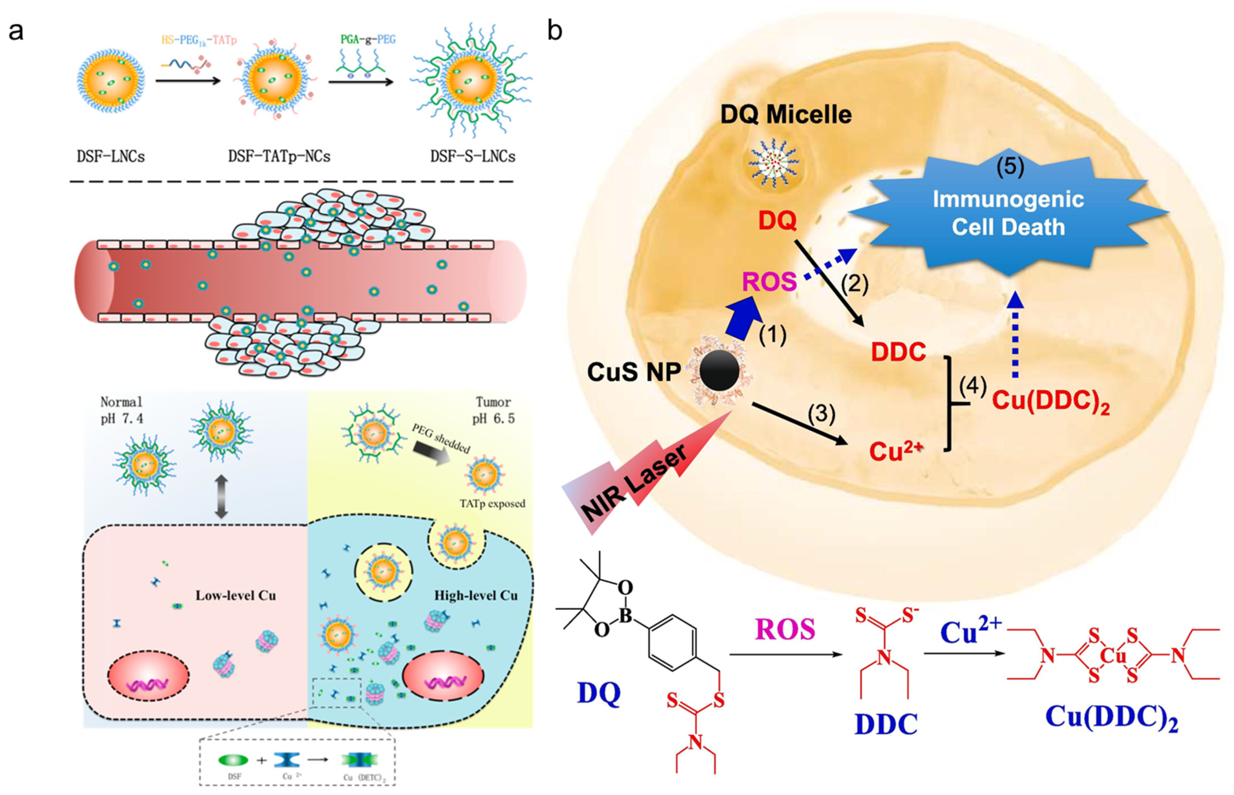
| DDS for DDC prodrug | DSF prodrug and copper sulfide nanoparticles+ near Infrared laser combination therapy | Breast cancer (4T1 cells) | This combination therapy effectively increased the intra-tumor ROS levels, which efficiently activated DQ prodrug. This combination also induced immunogenic cell death, thereby being a inducer for eliciting antitumor immunity. | [101] |
| H2O2-responsive diethyldithiocarbamate-based prodrug | Breast cancer (4T1 cells) | DQ showed much lower cytotoxicity (IC50 > 100 µM) to normal cells than DSF (IC50 of 12.5 µM), suggesting the advantage of DQ. | [167] | |
| Dithiocarbamate releasing prochelator GGT-DTC, which requires activation by γ-glutamyl transferase (GGT) | Prostate cancer (22Rv1, LNCaP, PC3 prostate cancer cells, as well as PWR-1E prostate epithelial cells) | GGT-DTC shown favorable stability against non-specific degradation in both normal and prostate cancer cells. and GGT-DTC selectively released diethyldithiocarbamate only in cells with measurable GGT activity. | [168] | |
| Β-D-galactose receptor targeted disulfiram-loaded nanoparticles | Ovarian cancer (SKOV-3 ovarian cancer cells and NCI-Adr-Res drug-resistant ovarian cancer cells) | These nanoparticles, upon internalization by cells, degrade and release diethyldithiocarbamate due to cleavage of disulfide bonds and form Copper (II)DDTC complex, which showed much greater tumor mass penetrating and destructive capacity. In addition, these NPs exhibited greater tumor growth inhibition capacity than the dosage form used in clinical trials (DSF in combination with copper gluconate). | [169] | |
| DDS for Copper | N-Oxide polymer-cupric ion nanogels | Breast cancer (MDA-MB-231 and 4T1 cancer cells) | These neutral and water soluble zwitterionic N-oxide polymer, poly [2-(N-oxide-N,N-dimethylamino)ethyl methacrylate/Cu nanogels efficiently delivered copper ions to tumor cells both in vitro and in vivo levels. The effective delivery of copper potentiated antitumor activity of DSF. | [150] |
| Ferritin-albumin-Cu nanoparticle in combination with disulfiram | Breast cancer (4T1, MDA-MB-231 cells) | These nanoparticles shown favorable accumulation in the tumor and demonstrated targeting capacity towards cancer cells In vivo assays, the NPs also shown more potent anti-tumor efficacy compared to DSF or nanoparticle alone. | [170] | |
| Copper-cysteamine nanoparticles | Esophageal cancer (Human ESCC KYSE-30 cells) | These nanoparticles showed greater inhibition of tumor growth compared to DSF and Cu-Cy alone, and resulted in ROS accumulation, and blocked nuclear translocation of NF-kB in esophageal cancer cells. | [171] | |
| DDS for combined DSF/Cu | Glutathione-responsive coordination nanoparticles (Cu-IXZ@DSF) | Breast cancer (4T1 cells) | These nanoparticles showed good biosafety and excellent antitumor activity via the increase of endoplasmic reticulum (ER) stress. | [172] |
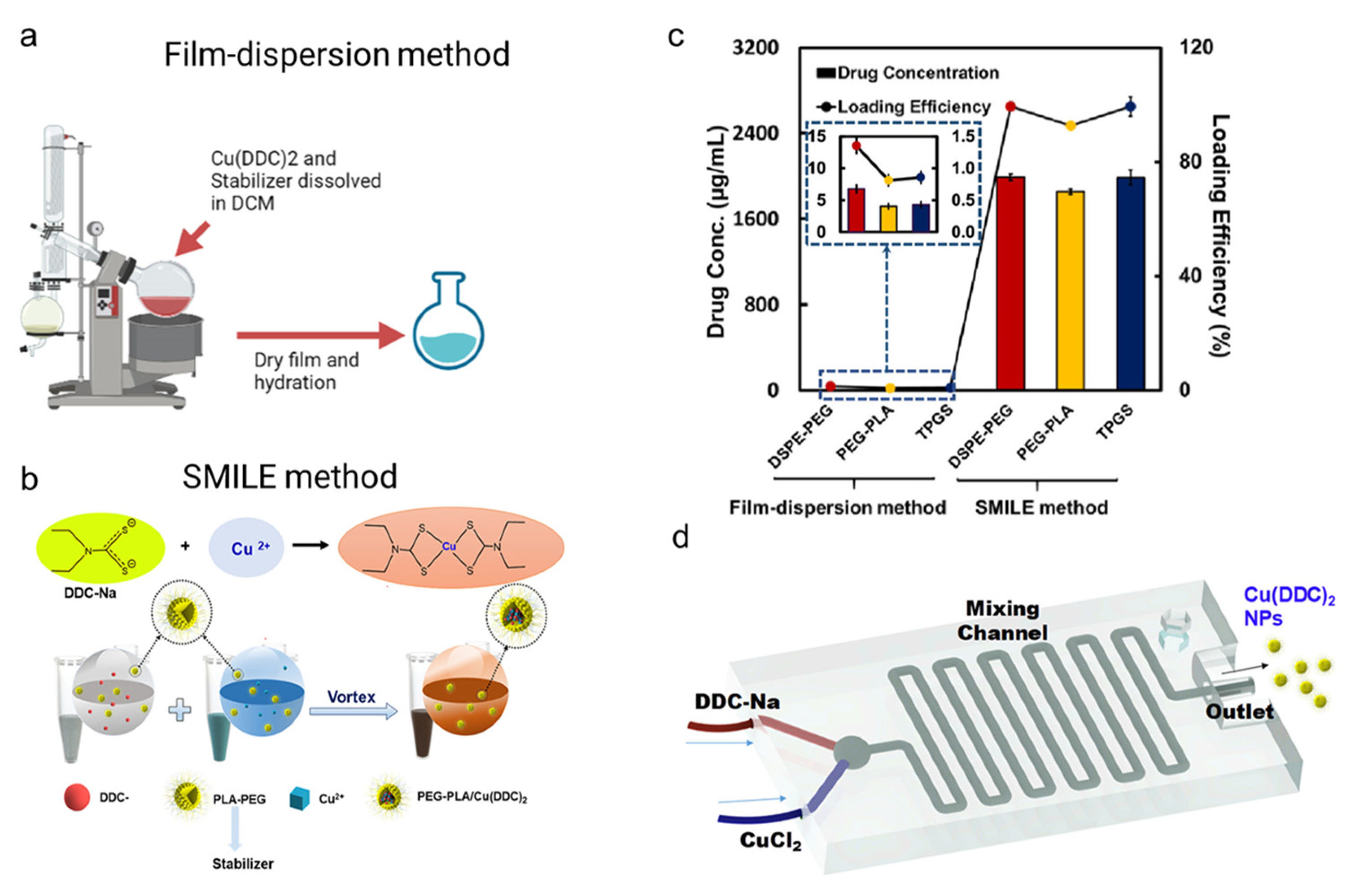
| BSA/Cu(DDC)2 metal organic nanoparticles | Breast cancer (4T1 cells) | This study shown the scale-production using 3D printing device, the NPs generated by the device showed potent antitumor activity and effectively inhibited growth of tumors in orthotopic 4T1 breast cancer mice model. | [32] | |
| Fe3O4@mSiO2 magnetic mesoporous silica nanoparticles | Breast cancer (MCF-7 cells) | The cytotoxicity of these DSF-loaded carrier systems was improved by adding copper and/or sodium nitroprusside, and cytotoxicity of NPs was greater in MCF-7 cells compared to non-tumorigenic MCF-10A cells. | [173] | |
| Disulfiram in combination with bacterially synthesized copper oxide nanoparticles | Breast cancer (MDA-MB-231 cells) | These combination nanoparticles showed higher pro-oxidant effect-mediated apoptosis and anti-metastatic potential via inhibition of antioxidant defenders and elevation of cellular reactive oxygen species. | [157] | |
| Copper-doped DSF-loaded hollow mesoporous silica nanoparticles | Breast cancer (4T1 cells) | These nanoparticles showed high chemotherapeutic efficacy with tumor growth inhibition (TGI) values as high as 71.4% compared to free DSF (which did not show antitumor effect). | [174] | |
| pH-responsive metal organic framework nanoparticles (DSF/DOX@ZIF-8@Cu-TA) | Breast cancer (MDA-MB-231 cells) | These nanoparticles significantly enhanced therapeutic efficiency of DSF and DOX both in vitro and in vivo. Accumulation of DSF and Cu (II) resulted in rapid formation of highly cytotoxic complexes accompanied with the generation of ROS. | [175] | |
| Mannosylated albumin nanoparticles with co-encapsulation of DSF/Cu and regorafenib | Colorectal cancer (Human colon cancer drug-resistant HCT8/ADR cell line, mouse fibroblast L929 cells) | This combination therapy greatly suppressed the growth of drug-resistant colon tumors, enhanced apoptosis, and upregulated intracellular ROS and anti-angiogenesis. | [176] | |
| Disulfiram-loaded hollow copper sulfide nanoparticles | Colorectal cancer (CT26 cells) | These nanoparticles +NIR laser significantly induced apoptosis with72% in vitro and 100% in vivo. Furthermore, the treatment approach effectively promoted tumor elimination in vivo. | [177] | |
| Disulfiram- and copper-loaded pH-responsive lipid-coated calcium phosphate nanoparticles | Colon cancer (CT26 murine colon cancer cells) | These nanoparticles effectively induced the immunogenic cell death of cancer cells, thereby contributing to enhancement immune checkpoint blockade therapy. | [178] | |
| Copper-ion- and disulfiram-loaded hyaluronic acid (HA)/polyethyleneimine (PEI) nanoparticles | Esophageal cancer (Eca109 and TE1 Esophageal squamous cell carcinoma cells) | These nanoparticles showed higher apoptosis than 5-FU (a conventional therapeutics), DSF/Cu and control and inhibited tumor proliferation with no toxicity on normal tissues. | [179] | |
| Combination of Disulfiram with copper oleate PEGylated liposome | Liver cancer (H-22 cells) | These nanoparticles demonstrated prolonged circulation, increased area under curve (AUC) and an increase in tumor inhibition rates by producing synergistic antitumor effect. | [180] | |
| Hyaluronic-acid-decorated liposomes containing Cu(DDC)2 | Pancreas cancer (Human Panc1 pancreatic adenocarcinoma cells) | These nanoparticles produced high ROS-mediated anticancer efficacy and increased anti-proliferative activity on pancreatic cancer stem cells, compared to DSF, Zn(DDC)2 and Fe(DDC)2. | [181] | |
| DDS for CuET | Copper–drug complexes in liposomes | Breast cancer (MDA231-BR cells) | Synthesis pf Cu(DDC)2 in lipid vesicles enhanced the stability and addressed the solubility issues related to each agent. | [182] |
| CuET/DIR (near infrared dye) nanoparticles | Breast cancer (MCF-7, 4T1, and 4T1 subline-LG12 cells) | These nanoparticles showed enhanced tumor killing efficacy through nuclear targeting compared to CuET alone and showed optimal biocompatibility. | [183] | |
| Copper(II)-disulfiram-loaded melanin dots | Breast cancer (4T1 and mouse fibroblast NIH/3T3 cells) | These nanodots showed good tumor accumulation, excellent tumor inhibition capacity, and higher tumor growth inhibition rate of 45.1%; The combination with photothermal the produce a higher tumor growth inhibition rate (78.6%) compared to nanodots without irradiation. | [184] | |
| DTC–copper complex@hyaluronic acid nanoparticles | Breast cancer (M231 cells) | These nanoparticles produced significant cytotoxicity towards cancer cells, The nanoparticles accumulated in tumors, and elicited tumor growth inhibition at a dose of 1mg/kg without toxic side effects. | [185] | |
| Stabilized metal ion ligand complex (SMILE) to prepare Cu(DDC)2 nanoparticles | Prostate cancer (DU145-TXR cells) | These nanoparticles induced cell death in drug-resistant prostate cancer cell lines through paraptosis (Paraptosis is a type of programmed cell death, distinct from apoptosis or necrosis, paraptosis involves the dilation of mitochondria, formation of vacuoles in the cytoplasm, and swelling of organelles, leading to cell death.). | [25] | |
| Injectable copper diethyldithiocarbamate formulation | Brain cancer (F98 glioblastoma cells) | This formulation showed ~50% reduction in tumor volume at its respective maximum tolerated dose compared to vehicle- and copper-treated animals. | [186] |
Disclaimer/Publisher’s Note: The statements, opinions and data contained in all publications are solely those of the individual author(s) and contributor(s) and not of MDPI and/or the editor(s). MDPI and/or the editor(s) disclaim responsibility for any injury to people or property resulting from any ideas, methods, instructions or products referred to in the content. |
© 2023 by the authors. Licensee MDPI, Basel, Switzerland. This article is an open access article distributed under the terms and conditions of the Creative Commons Attribution (CC BY) license (https://creativecommons.org/licenses/by/4.0/).
Share and Cite
Kang, X.; Jadhav, S.; Annaji, M.; Huang, C.-H.; Amin, R.; Shen, J.; Ashby, C.R., Jr.; Tiwari, A.K.; Babu, R.J.; Chen, P. Advancing Cancer Therapy with Copper/Disulfiram Nanomedicines and Drug Delivery Systems. Pharmaceutics 2023, 15, 1567. https://doi.org/10.3390/pharmaceutics15061567
Kang X, Jadhav S, Annaji M, Huang C-H, Amin R, Shen J, Ashby CR Jr., Tiwari AK, Babu RJ, Chen P. Advancing Cancer Therapy with Copper/Disulfiram Nanomedicines and Drug Delivery Systems. Pharmaceutics. 2023; 15(6):1567. https://doi.org/10.3390/pharmaceutics15061567
Chicago/Turabian StyleKang, Xuejia, Sanika Jadhav, Manjusha Annaji, Chung-Hui Huang, Rajesh Amin, Jianzhong Shen, Charles R. Ashby, Jr., Amit K. Tiwari, R. Jayachandra Babu, and Pengyu Chen. 2023. "Advancing Cancer Therapy with Copper/Disulfiram Nanomedicines and Drug Delivery Systems" Pharmaceutics 15, no. 6: 1567. https://doi.org/10.3390/pharmaceutics15061567