
Cationic Polymers as Transfection Reagents for Nucleic Acid Delivery


Abstract
:1. Introduction
2. Cationic Polymers
2.1. Synthetic Polymers
2.1.1. Conventional Polymers
- DEAE-dextran
- Poly-amino acids
- PAMAM
- PPI (poly-(propylenimine))
- PEI (polyethylenimine)
- PDMAEMA (polymethacrylic acid N,N-dimethylaminoethyl ester)
2.1.2. Polyesters
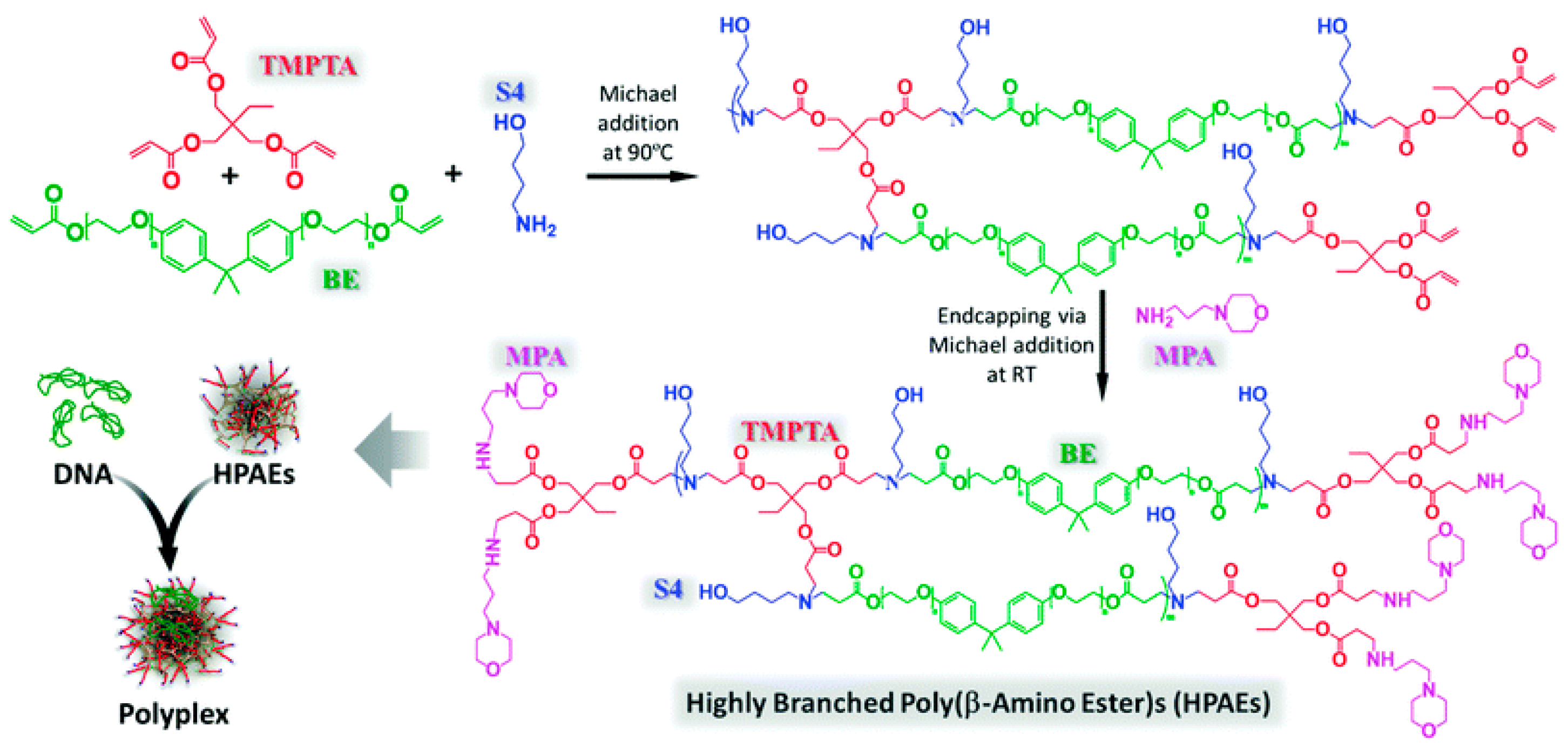
- Poly(β-amino ester)
- PHP (Poly(4-hydroxy-L-proline ester))
- PAGA (poly[α-(4-aminobutyl)-l-glycolic acid])
- PVL poly(δ-valerolactone)
- Aminated PAHA (aminated poly(α-hydroxy acids))
- PPE (polyphosphoester)
2.1.3. Polylactide(Poly(lactic acid), PLA)
2.1.4. Polycarbonates
2.1.5. Polyurethanes (PUs)
2.1.6. Conjugated Polymers (CPs)
2.2. Natural Polymers
2.2.1. Protein
- Histone
- Gelatin
- Protamine
2.2.2. Polysaccharides
- Cyclodextrins
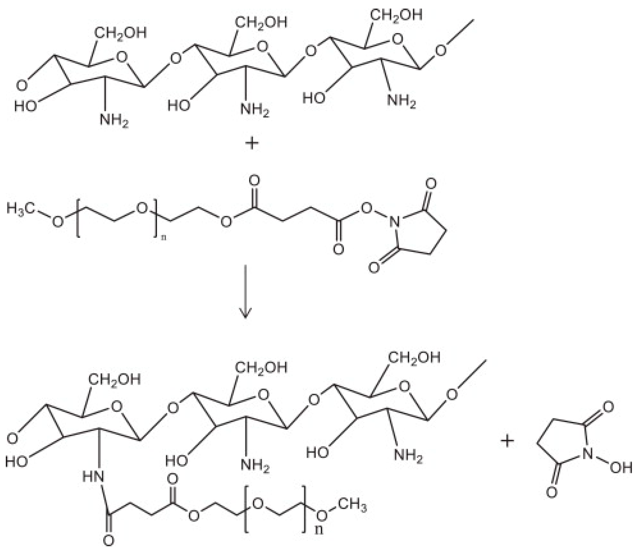
- Chitosan
3. The Applications of Cationic Polymers
3.1. Gene Augmentation
- Protein replacement therapies
- Vaccine
3.2. Gene Suppression
- Small interfering RNA (siRNA)
- Short hairpin RNA (shRNA)
3.3. Genome Editing
- CRISPR/Cas
4. Concluding Remarks and Perspectives
Supplementary Materials
Author Contributions
Funding
Institutional Review Board Statement
Informed Consent Statement
Data Availability Statement
Acknowledgments
Conflicts of Interest
References
- Chong, Z.X.; Yeap, S.K.; Ho, W.Y. Transfection types, methods and strategies: A technical review. PeerJ 2021, 9, e11165. [Google Scholar] [CrossRef] [PubMed]
- Dana, H.; Chalbatani, G.M.; Mahmoodzadeh, H.; Karimloo, R.; Rezaiean, O.; Moradzadeh, A.; Mehmandoost, N.; Moazzen, F.; Mazraeh, A.; Marmari, V.; et al. Molecular Mechanisms and Biological Functions of siRNA. Int. J. Biomed. Sci. 2017, 13, 48–57. [Google Scholar] [PubMed]
- Oh, Y.K.; Park, T.G. siRNA delivery systems for cancer treatment. Adv. Drug Deliv. Rev. 2009, 61, 850–862. [Google Scholar] [CrossRef] [PubMed]
- Soutschek, J.; Akinc, A.; Bramlage, B.; Charisse, K.; Constien, R.; Donoghue, M.; Elbashir, S.; Geick, A.; Hadwiger, P.; Harborth, J.; et al. Therapeutic silencing of an endogenous gene by systemic administration of modified siRNAs. Nature 2004, 432, 173–178. [Google Scholar] [CrossRef]
- Mottaghitalab, F.; Rastegari, A.; Farokhi, M.; Dinarvand, R.; Hosseinkhani, H.; Ou, K.L.; Pack, D.W.; Mao, C.; Dinarvand, M.; Fatahi, Y.; et al. Prospects of siRNA applications in regenerative medicine. Int. J. Pharm. 2017, 524, 312–329. [Google Scholar] [CrossRef]
- Pardi, N.; Hogan, M.J.; Porter, F.W.; Weissman, D. mRNA vaccines—A new era in vaccinology. Nat. Rev. Drug Discov. 2018, 17, 261–279. [Google Scholar] [CrossRef] [Green Version]
- Dolgin, E. The tangled history of mRNA vaccines. Nature 2021, 597, 318–324. [Google Scholar] [CrossRef]
- Wang, H.; Feng, Z.; Xu, B. Supramolecular Assemblies of Peptides or Nucleopeptides for Gene Delivery. Theranostics 2019, 9, 3213–3222. [Google Scholar] [CrossRef]
- Lundstrom, K. Viral Vectors in Gene Therapy. Diseases 2018, 6, 42. [Google Scholar] [CrossRef] [Green Version]
- Bessis, N.; GarciaCozar, F.J.; Boissier, M.C. Immune responses to gene therapy vectors: Influence on vector function and effector mechanisms. Gene Ther. 2004, 11 (Suppl. S1), S10–S17. [Google Scholar] [CrossRef] [Green Version]
- Zu, H.; Gao, D. Non-viral Vectors in Gene Therapy: Recent Development, Challenges, and Prospects. AAPS J. 2021, 23, 78. [Google Scholar] [CrossRef]
- Pack, D.W.; Hoffman, A.S.; Pun, S.; Stayton, P.S. Design and development of polymers for gene delivery. Nat. Rev. Drug Discov. 2005, 4, 581–593. [Google Scholar] [CrossRef]
- Degors, I.M.S.; Wang, C.; Rehman, Z.U.; Zuhorn, I.S. Carriers Break Barriers in Drug Delivery: Endocytosis and Endosomal Escape of Gene Delivery Vectors. Acc. Chem. Res. 2019, 52, 1750–1760. [Google Scholar] [CrossRef] [Green Version]
- Vermeulen, L.M.P.; De Smedt, S.C.; Remaut, K.; Braeckmans, K. The proton sponge hypothesis: Fable or fact? Eur. J. Pharm. Biopharm. 2018, 129, 184–190. [Google Scholar] [CrossRef] [Green Version]
- Shim, M.S.; Wang, X.; Ragan, R.; Kwon, Y.J. Dynamics of nucleic acid/cationic polymer complexation and disassembly under biologically simulated conditions using in situ atomic force microscopy. Microsc. Res. Tech. 2010, 73, 845–856. [Google Scholar] [CrossRef] [Green Version]
- Chen, C.; Yang, Z.; Tang, X. Chemical modifications of nucleic acid drugs and their delivery systems for gene-based therapy. Med. Res. Rev. 2018, 38, 829–869. [Google Scholar] [CrossRef]
- Li, J.; Liu, L.; Feng, Z.; Wang, X.; Huang, Y.; Dai, H.; Zhang, L.; Song, F.; Wang, D.; Zhang, P.; et al. Tumor markers CA15-3, CA125, CEA and breast cancer survival by molecular subtype: A cohort study. Breast Cancer 2020, 27, 621–630. [Google Scholar] [CrossRef]
- McCutchan, J.H.; Pagano, J.S. Enchancement of the infectivity of simian virus 40 deoxyribonucleic acid with diethylaminoethyl-dextran. J. Natl. Cancer Inst. 1968, 41, 351–357. [Google Scholar]
- Vaheri, A.; Pagano, J.S. Infectious poliovirus RNA: A sensitive method of assay. Virology 1965, 27, 434–436. [Google Scholar] [CrossRef]
- Siewert, C.; Haas, H.; Nawroth, T.; Ziller, A.; Nogueira, S.S.; Schroer, M.A.; Blanchet, C.E.; Svergun, D.I.; Radulescu, A.; Bates, F.; et al. Investigation of charge ratio variation in mRNA-DEAE-dextran polyplex delivery systems. Biomaterials 2019, 192, 612–620. [Google Scholar] [CrossRef]
- Uchiumi, F.; Watanabe, T.; Tanuma, S. Characterization of various promoter regions of the human DNA helicase-encoding genes and identification of duplicated ets (GGAA) motifs as an essential transcription regulatory element. Exp. Cell Res. 2010, 316, 1523–1534. [Google Scholar] [CrossRef]
- Felgner, P.L.; Gadek, T.R.; Holm, M.; Roman, R.; Chan, H.W.; Wenz, M.; Northrop, J.P.; Ringold, G.M.; Danielsen, M. Lipofection: A highly efficient, lipid-mediated DNA-transfection procedure. Proc. Natl. Acad. Sci. USA 1987, 84, 7413–7417. [Google Scholar] [CrossRef] [PubMed] [Green Version]
- Thompson, C.D.; Frazier-Jessen, M.R.; Rawat, R.; Nordan, R.P.; Brown, R.T. Evaluation of methods for transient transfection of a murine macrophage cell line, RAW 264.7. Biotechniques 1999, 27, 824–826, 828–830, 832. [Google Scholar] [CrossRef] [PubMed]
- Fischer, D.; Li, Y.; Ahlemeyer, B.; Krieglstein, J.; Kissel, T. In vitro cytotoxicity testing of polycations: Influence of polymer structure on cell viability and hemolysis. Biomaterials 2003, 24, 1121–1131. [Google Scholar] [CrossRef] [PubMed]
- Vizcarra, J.A.; Karges, S.L.; Wettemann, R.P. Immunization of beef heifers against gonadotropin-releasing hormone prevents luteal activity and pregnancy: Effect of conjugation to different proteins and effectiveness of adjuvants. J. Anim. Sci. 2012, 90, 1479–1488. [Google Scholar] [CrossRef]
- Piedrafita, D.; Preston, S.; Kemp, J.; de Veer, M.; Sherrard, J.; Kraska, T.; Elhay, M.; Meeusen, E. The effect of different adjuvants on immune parameters and protection following vaccination of sheep with a larval-specific antigen of the gastrointestinal nematode, Haemonchus contortus. PLoS ONE 2013, 8, e78357. [Google Scholar] [CrossRef]
- Kwoh, D.Y.; Coffin, C.C.; Lollo, C.P.; Jovenal, J.; Banaszczyk, M.G.; Mullen, P.; Phillips, A.; Amini, A.; Fabrycki, J.; Bartholomew, R.M.; et al. Stabilization of poly-L-lysine/DNA polyplexes for in vivo gene delivery to the liver. Biochim. Biophys. Acta 1999, 1444, 171–190. [Google Scholar] [CrossRef]
- Wu, G.Y.; Wu, C.H. Receptor-mediated in vitro gene transformation by a soluble DNA carrier system. J. Biol. Chem. 1987, 262, 4429–4432. [Google Scholar] [CrossRef]
- Cotten, M.; Langle-Rouault, F.; Kirlappos, H.; Wagner, E.; Mechtler, K.; Zenke, M.; Beug, H.; Birnstiel, M.L. Transferrin-polycation-mediated introduction of DNA into human leukemic cells: Stimulation by agents that affect the survival of transfected DNA or modulate transferrin receptor levels. Proc. Natl. Acad. Sci. USA 1990, 87, 4033–4037. [Google Scholar] [CrossRef] [Green Version]
- Mislick, K.A.; Baldeschwieler, J.D.; Kayyem, J.F.; Meade, T.J. Transfection of folate-polylysine DNA complexes: Evidence for lysosomal delivery. Bioconjug. Chem. 1995, 6, 512–515. [Google Scholar] [CrossRef]
- Farber, F.E.; Melnick, J.L.; Butel, J.S. Optimal Conditions for Uptake of Exogenous DNA by Chinese-Hamster Lung-Cells Deficient in Hypoxanthine-Guanine Phosphoribosyltransferase. Biochim. Et Biophys. Acta 1975, 390, 298–311. [Google Scholar] [CrossRef]
- Ramsay, E.; Gumbleton, M. Polylysine and polyornithine gene transfer complexes: A study of complex stability and cellular uptake as a basis for their differential in-vitro transfection efficiency. J. Drug Target. 2002, 10, 1–9. [Google Scholar] [CrossRef]
- Li, J.; Liang, H.; Liu, J.; Wang, Z. Poly (amidoamine) (PAMAM) dendrimer mediated delivery of drug and pDNA/siRNA for cancer therapy. Int. J. Pharm. 2018, 546, 215–225. [Google Scholar] [CrossRef]
- Wu, J.; Huang, W.; He, Z. Dendrimers as carriers for siRNA delivery and gene silencing: A review. Sci. World J. 2013, 2013, 630654. [Google Scholar] [CrossRef] [Green Version]
- Boussif, O.; Lezoualc’h, F.; Zanta, M.A.; Mergny, M.D.; Scherman, D.; Demeneix, B.; Behr, J.P. A versatile vector for gene and oligonucleotide transfer into cells in culture and in vivo: Polyethylenimine. Proc. Natl. Acad. Sci. USA 1995, 92, 7297–7301. [Google Scholar] [CrossRef] [Green Version]
- Haensler, J.; Szoka, F.C., Jr. Polyamidoamine cascade polymers mediate efficient transfection of cells in culture. Bioconjug. Chem. 1993, 4, 372–379. [Google Scholar] [CrossRef]
- Kukowska-Latallo, J.F.; Bielinska, A.U.; Johnson, J.; Spindler, R.; Tomalia, D.A.; Baker, J.R., Jr. Efficient transfer of genetic material into mammalian cells using Starburst polyamidoamine dendrimers. Proc. Natl. Acad. Sci. USA 1996, 93, 4897–4902. [Google Scholar] [CrossRef] [Green Version]
- Monteagudo, S.; Perez-Martinez, F.C.; Perez-Carrion, M.D.; Guerra, J.; Merino, S.; Sanchez-Verdu, M.P.; Cena, V. Inhibition of p42 MAPK using a nonviral vector-delivered siRNA potentiates the anti-tumor effect of metformin in prostate cancer cells. Nanomedicine 2012, 7, 493–506. [Google Scholar] [CrossRef]
- Yemul, O.; Imae, T. Covalent-bonded immobilization of lipase on poly(phenylene sulfide) dendrimers and their hydrolysis ability. Biomacromolecules 2005, 6, 2809–2814. [Google Scholar] [CrossRef]
- Hollins, A.J.; Omidi, Y.; Benter, I.F.; Akhtar, S. Toxicogenomics of drug delivery systems: Exploiting delivery system-induced changes in target gene expression to enhance siRNA activity. J. Drug Target. 2007, 15, 83–88. [Google Scholar] [CrossRef]
- Bugno, J.; Hsu, H.J.; Pearson, R.M.; Noh, H.; Hong, S. Size and Surface Charge of Engineered Poly(amidoamine) Dendrimers Modulate Tumor Accumulation and Penetration: A Model Study Using Multicellular Tumor Spheroids. Mol. Pharm. 2016, 13, 2155–2163. [Google Scholar] [CrossRef] [PubMed]
- Kaur, D.; Jain, K.; Mehra, N.K.; Kesharwani, P.; Jain, N.K. A review on comparative study of PPI and PAMAM dendrimers. J. Nanopart. Res. 2016, 18, 146. [Google Scholar] [CrossRef]
- Hashemi, M.; Tabatabai, S.M.; Parhiz, H.; Milanizadeh, S.; Amel Farzad, S.; Abnous, K.; Ramezani, M. Gene delivery efficiency and cytotoxicity of heterocyclic amine-modified PAMAM and PPI dendrimers. Mater. Sci. Eng. C Mater. Biol. Appl. 2016, 61, 791–800. [Google Scholar] [CrossRef] [PubMed]
- Liu, H.; Wang, Y.; Wang, M.; Xiao, J.; Cheng, Y. Fluorinated poly(propylenimine) dendrimers as gene vectors. Biomaterials 2014, 35, 5407–5413. [Google Scholar] [CrossRef] [PubMed]
- Yue, Y.; Jin, F.; Deng, R.; Cai, J.; Dai, Z.; Lin, M.C.; Kung, H.F.; Mattebjerg, M.A.; Andresen, T.L.; Wu, C. Revisit complexation between DNA and polyethylenimine--effect of length of free polycationic chains on gene transfection. J. Control. Release 2011, 152, 143–151. [Google Scholar] [CrossRef]
- Choudhury, C.K.; Kumar, A.; Roy, S. Characterization of conformation and interaction of gene delivery vector polyethylenimine with phospholipid bilayer at different protonation state. Biomacromolecules 2013, 14, 3759–3768. [Google Scholar] [CrossRef]
- Nguyen, J.; Szoka, F.C. Nucleic acid delivery: The missing pieces of the puzzle? Acc. Chem. Res. 2012, 45, 1153–1162. [Google Scholar] [CrossRef] [Green Version]
- Ziebarth, J.D.; Wang, Y. Understanding the protonation behavior of linear polyethylenimine in solutions through Monte Carlo simulations. Biomacromolecules 2010, 11, 29–38. [Google Scholar] [CrossRef] [Green Version]
- Itaka, K.; Harada, A.; Yamasaki, Y.; Nakamura, K.; Kawaguchi, H.; Kataoka, K. In situ single cell observation by fluorescence resonance energy transfer reveals fast intra-cytoplasmic delivery and easy release of plasmid DNA complexed with linear polyethylenimine. J. Gene Med. 2004, 6, 76–84. [Google Scholar] [CrossRef]
- Wightman, L.; Kircheis, R.; Rossler, V.; Carotta, S.; Ruzicka, R.; Kursa, M.; Wagner, E. Different behavior of branched and linear polyethylenimine for gene delivery in vitro and in vivo. J. Gene Med. 2001, 3, 362–372. [Google Scholar] [CrossRef]
- Zhang, P.; Wagner, E. History of Polymeric Gene Delivery Systems. Top. Curr. Chem. 2017, 375, 26. [Google Scholar] [CrossRef]
- Wen, Y.; Pan, S.; Luo, X.; Zhang, X.; Zhang, W.; Feng, M. A biodegradable low molecular weight polyethylenimine derivative as low toxicity and efficient gene vector. Bioconjug. Chem. 2009, 20, 322–332. [Google Scholar] [CrossRef]
- Teo, P.Y.; Yang, C.; Hedrick, J.L.; Engler, A.C.; Coady, D.J.; Ghaem-Maghami, S.; George, A.J.; Yang, Y.Y. Hydrophobic modification of low molecular weight polyethylenimine for improved gene transfection. Biomaterials 2013, 34, 7971–7979. [Google Scholar] [CrossRef]
- Zintchenko, A.; Philipp, A.; Dehshahri, A.; Wagner, E. Simple modifications of branched PEI lead to highly efficient siRNA carriers with low toxicity. Bioconjug. Chem. 2008, 19, 1448–1455. [Google Scholar] [CrossRef] [Green Version]
- Jiang, H.L.; Islam, M.A.; Xing, L.; Firdous, J.; Cao, W.; He, Y.J.; Zhu, Y.; Cho, K.H.; Li, H.S.; Cho, C.S. Degradable Polyethylenimine-Based Gene Carriers for Cancer Therapy. Top. Curr. Chem. 2017, 375, 34. [Google Scholar] [CrossRef]
- Cherng, J.Y.; van de Wetering, P.; Talsma, H.; Crommelin, D.J.; Hennink, W.E. Effect of size and serum proteins on transfection efficiency of poly ((2-dimethylamino)ethyl methacrylate)-plasmid nanoparticles. Pharm. Res. 1996, 13, 1038–1042. [Google Scholar] [CrossRef]
- van de Wetering, P.; Cherng, J.Y.; Talsma, H.; Crommelin, D.J.A.; Hennink, W.E. 2-(dimethylamino)ethyl methacrylate based (co)polymers as gene transfer agents. J. Control. Release 1998, 53, 145–153. [Google Scholar] [CrossRef]
- Jones, R.A.; Poniris, M.H.; Wilson, M.R. pDMAEMA is internalised by endocytosis but does not physically disrupt endosomes. J. Control. Release 2004, 96, 379–391. [Google Scholar] [CrossRef]
- Zuidam, N.J.; Posthuma, G.; de Vries, E.T.; Crommelin, D.J.; Hennink, W.E.; Storm, G. Effects of physicochemical characteristics of poly(2-(dimethylamino)ethyl methacrylate)-based polyplexes on cellular association and internalization. J. Drug Target 2000, 8, 51–66. [Google Scholar] [CrossRef]
- Funhoff, A.M.; van Nostrum, C.F.; Lok, M.C.; Kruijtzer, J.A.; Crommelin, D.J.; Hennink, W.E. Cationic polymethacrylates with covalently linked membrane destabilizing peptides as gene delivery vectors. J. Control. Release 2005, 101, 233–246. [Google Scholar] [CrossRef]
- Lynn, D.M.; Anderson, D.G.; Putnam, D.; Langer, R. Accelerated discovery of synthetic transfection vectors: Parallel synthesis and screening of a degradable polymer library. J. Am. Chem. Soc. 2001, 123, 8155–8156. [Google Scholar] [CrossRef] [PubMed]
- Lynn, D.M.; Langer, R. Degradable Poly(β-amino esters): Synthesis, Characterization, and Self-Assembly with Plasmid DNA. J. Am. Chem. Soc. 2000, 122, 10761–10768. [Google Scholar] [CrossRef]
- Gong, J.H.; Wang, Y.; Xing, L.; Cui, P.F.; Qiao, J.B.; He, Y.J.; Jiang, H.L. Biocompatible fluorinated poly(beta-amino ester)s for safe and efficient gene therapy. Int. J. Pharm. 2018, 535, 180–193. [Google Scholar] [CrossRef] [PubMed]
- Song, W.; Tang, Z.; Li, M.; Lv, S.; Yu, H.; Ma, L.; Zhuang, X.; Huang, Y.; Chen, X. Tunable pH-sensitive poly(beta-amino ester)s synthesized from primary amines and diacrylates for intracellular drug delivery. Macromol. Biosci. 2012, 12, 1375–1383. [Google Scholar] [CrossRef]
- Chen, J.; Ouyang, J.; Kong, J.; Zhong, W.; Xing, M.M. Photo-cross-linked and pH-sensitive biodegradable micelles for doxorubicin delivery. ACS Appl. Mater. Interfaces 2013, 5, 3108–3117. [Google Scholar] [CrossRef]
- Liu, M.; Chen, J.; Xue, Y.N.; Liu, W.M.; Zhuo, R.X.; Huang, S.W. Poly(beta-aminoester)s with pendant primary amines for efficient gene delivery. Bioconjug. Chem. 2009, 20, 2317–2323. [Google Scholar] [CrossRef]
- Chen, J.; Huang, S.W.; Liu, M.; Zhuo, R.X. Synthesis and degradation of poly(beta-aminoester) with pendant primary amine. Polymer 2007, 48, 675–681. [Google Scholar] [CrossRef]
- Cutlar, L.; Zhou, D.; Gao, Y.; Zhao, T.; Greiser, U.; Wang, W.; Wang, W. Highly Branched Poly(beta-Amino Esters): Synthesis and Application in Gene Delivery. Biomacromolecules 2015, 16, 2609–2617. [Google Scholar] [CrossRef]
- Chen, J.; Wu, C.; Oupicky, D. Bioreducible hyperbranched poly(amido amine)s for gene delivery. Biomacromolecules 2009, 10, 2921–2927. [Google Scholar] [CrossRef] [Green Version]
- Chen, J.; Zehtabi, F.; Ouyang, J.; Kong, J.M.; Zhong, W.; Xing, M.M.Q. Reducible self-assembled micelles for enhanced intracellular delivery of doxorubicin. J. Mater. Chem. 2012, 22, 7121–7129. [Google Scholar] [CrossRef]
- Tang, S.; Yin, Q.; Su, J.; Sun, H.; Meng, Q.; Chen, Y.; Chen, L.; Huang, Y.; Gu, W.; Xu, M.; et al. Inhibition of metastasis and growth of breast cancer by pH-sensitive poly (beta-amino ester) nanoparticles co-delivering two siRNA and paclitaxel. Biomaterials 2015, 48, 1–15. [Google Scholar] [CrossRef]
- Tzeng, S.Y.; Green, J.J. Subtle changes to polymer structure and degradation mechanism enable highly effective nanoparticles for siRNA and DNA delivery to human brain cancer. Adv. Healthc. Mater. 2013, 2, 468–480. [Google Scholar] [CrossRef] [Green Version]
- Huang, J.Y.; Gao, Y.; Cutlar, L.; O’Keeffe-Ahern, J.; Zhao, T.; Lin, F.H.; Zhou, D.; McMahon, S.; Greiser, U.; Wang, W.; et al. Tailoring highly branched poly(beta-amino ester)s: A synthetic platform for epidermal gene therapy. Chem. Commun. 2015, 51, 8473–8476. [Google Scholar] [CrossRef]
- Cordeiro, R.A.; Serra, A.; Coelho, J.F.J.; Faneca, H. Poly(beta-amino ester)-based gene delivery systems: From discovery to therapeutic applications. J. Control. Release 2019, 310, 155–187. [Google Scholar] [CrossRef]
- Fornaguera, C.; Guerra-Rebollo, M.; Angel Lazaro, M.; Castells-Sala, C.; Meca-Cortes, O.; Ramos-Perez, V.; Cascante, A.; Rubio, N.; Blanco, J.; Borros, S. mRNA Delivery System for Targeting Antigen-Presenting Cells In Vivo. Adv. Healthc. Mater. 2018, 7, e1800335. [Google Scholar] [CrossRef]
- Tzeng, S.Y.; Hung, B.P.; Grayson, W.L.; Green, J.J. Cystamine-terminated poly(beta-amino ester)s for siRNA delivery to human mesenchymal stem cells and enhancement of osteogenic differentiation. Biomaterials 2012, 33, 8142–8151. [Google Scholar] [CrossRef] [Green Version]
- Liu, S.; Gao, Y.; Zhou, D.; Zeng, M.; Alshehri, F.; Newland, B.; Lyu, J.; O’Keeffe-Ahern, J.; Greiser, U.; Guo, T.; et al. Highly branched poly(beta-amino ester) delivery of minicircle DNA for transfection of neurodegenerative disease related cells. Nat. Commun. 2019, 10, 3307. [Google Scholar] [CrossRef] [Green Version]
- Putnam, D.; Langer, R. Poly(4-hydroxy-L-proline ester): Low-temperature polycondensation and plasmid DNA complexation. Macromolecules 1999, 32, 3658–3662. [Google Scholar] [CrossRef]
- Lim, Y.B.; Choi, Y.H.; Park, J.S. A self-destroying polycationic polymer: Biodegradable poly(4-hydroxy-L-proline ester). J. Am. Chem. Soc. 1999, 121, 5633–5639. [Google Scholar] [CrossRef]
- Lim, Y.B.; Han, S.O.; Kong, H.U.; Lee, Y.; Park, J.S.; Jeong, B.; Kim, S.W. Biodegradable polyester, poly[alpha-(4-aminobutyl)-L-glycolic acid], as a non-toxic gene carrier. Pharm. Res. 2000, 17, 811–816. [Google Scholar] [CrossRef]
- Maheshwari, A.; Han, S.; Mahato, R.I.; Kim, S.W. Biodegradable polymer-based interleukin-12 gene delivery: Role of induced cytokines, tumor infiltrating cells and nitric oxide in anti-tumor activity. Gene Ther. 2002, 9, 1075–1084. [Google Scholar] [CrossRef] [PubMed] [Green Version]
- Koh, J.J.; Ko, K.S.; Lee, M.; Han, S.; Park, J.S.; Kim, S.W. Degradable polymeric carrier for the delivery of IL-10 plasmid DNA to prevent autoimmune insulitis of NOD mice. Gene Ther. 2000, 7, 2099–2104. [Google Scholar] [CrossRef] [PubMed] [Green Version]
- Song, L.; Ding, A.X.; Zhang, K.X.; Gong, B.; Lu, Z.L.; He, L. Degradable polyesters via ring-opening polymerization of functional valerolactones for efficient gene delivery. Org. Biomol. Chem. 2017, 15, 6567–6574. [Google Scholar] [CrossRef] [PubMed]
- Basu, A.; Kunduru, K.R.; Katzhendler, J.; Domb, A.J. Poly(alpha-hydroxy acid)s and poly(alpha-hydroxy acid-co-alpha-amino acid)s derived from amino acid. Adv. Drug Deliv. Rev. 2016, 107, 82–96. [Google Scholar] [CrossRef] [PubMed]
- Zhang, Z.; Yin, L.; Xu, Y.; Tong, R.; Lu, Y.; Ren, J.; Cheng, J. Facile functionalization of polyesters through thiol-yne chemistry for the design of degradable, cell-penetrating and gene delivery dual-functional agents. Biomacromolecules 2012, 13, 3456–3462. [Google Scholar] [CrossRef] [Green Version]
- Yilmaz, Z.E.; Jerome, C. Polyphosphoesters: New Trends in Synthesis and Drug Delivery Applications. Macromol. Biosci. 2016, 16, 1745–1761. [Google Scholar] [CrossRef]
- Lu, Z.Z.; Wu, J.; Sun, T.M.; Ji, J.; Yan, L.F.; Wang, J. Biodegradable polycation and plasmid DNA multilayer film for prolonged gene delivery to mouse osteoblasts. Biomaterials 2008, 29, 733–741. [Google Scholar] [CrossRef]
- Sun, T.M.; Du, J.Z.; Yao, Y.D.; Mao, C.Q.; Dou, S.; Huang, S.Y.; Zhang, P.Z.; Leong, K.W.; Song, E.W.; Wang, J. Simultaneous delivery of siRNA and paclitaxel via a “two-in-one” micelleplex promotes synergistic tumor suppression. ACS Nano 2011, 5, 1483–1494. [Google Scholar] [CrossRef]
- Mao, C.Q.; Xiong, M.H.; Liu, Y.; Shen, S.; Du, X.J.; Yang, X.Z.; Dou, S.; Zhang, P.Z.; Wang, J. Synthetic lethal therapy for KRAS mutant non-small-cell lung carcinoma with nanoparticle-mediated CDK4 siRNA delivery. Mol. Ther. 2014, 22, 964–973. [Google Scholar] [CrossRef] [Green Version]
- Tyler, B.; Gullotti, D.; Mangraviti, A.; Utsuki, T.; Brem, H. Polylactic acid (PLA) controlled delivery carriers for biomedical applications. Adv. Drug Deliv. Rev. 2016, 107, 163–175. [Google Scholar] [CrossRef]
- Lacroix, C.; Humanes, A.; Coiffier, C.; Gigmes, D.; Verrier, B.; Trimaille, T. Polylactide-Based Reactive Micelles as a Robust Platform for mRNA Delivery. Pharm. Res. 2020, 37, 30. [Google Scholar] [CrossRef]
- Chen, C.K.; Law, W.C.; Aalinkeel, R.; Nair, B.; Kopwitthaya, A.; Mahajan, S.D.; Reynolds, J.L.; Zou, J.; Schwartz, S.A.; Prasad, P.N.; et al. Well-defined degradable cationic polylactide as nanocarrier for the delivery of siRNA to silence angiogenesis in prostate cancer. Adv. Healthc. Mater. 2012, 1, 751–761. [Google Scholar] [CrossRef]
- Jones, C.H.; Chen, C.K.; Jiang, M.; Fang, L.; Cheng, C.; Pfeifer, B.A. Synthesis of cationic polylactides with tunable charge densities as nanocarriers for effective gene delivery. Mol. Pharm. 2013, 10, 1138–1145. [Google Scholar] [CrossRef]
- Ong, Z.Y.; Fukushima, K.; Coady, D.J.; Yang, Y.Y.; Ee, P.L.; Hedrick, J.L. Rational design of biodegradable cationic polycarbonates for gene delivery. J. Control. Release 2011, 152, 120–126. [Google Scholar] [CrossRef]
- Wang, C.F.; Lin, Y.X.; Jiang, T.; He, F.; Zhuo, R.X. Polyethylenimine-grafted polycarbonates as biodegradable polycations for gene delivery. Biomaterials 2009, 30, 4824–4832. [Google Scholar] [CrossRef]
- Segel, M.; Lash, B.; Song, J.; Ladha, A.; Liu, C.C.; Jin, X.; Mekhedov, S.L.; Macrae, R.K.; Koonin, E.V.; Zhang, F. Mammalian retrovirus-like protein PEG10 packages its own mRNA and can be pseudotyped for mRNA delivery. Science 2021, 373, 882–889. [Google Scholar] [CrossRef]
- Cherng, J.Y.; Hou, T.Y.; Shih, M.F.; Talsma, H.; Hennink, W.E. Polyurethane-based drug delivery systems. Int. J. Pharm. 2013, 450, 145–162. [Google Scholar] [CrossRef]
- Yang, T.F.; Chin, W.K.; Cherng, J.Y.; Shau, M.D. Synthesis of novel biodegradable cationic polymer: N,N-diethylethylenediamine polyurethane as a gene carrier. Biomacromolecules 2004, 5, 1926–1932. [Google Scholar] [CrossRef]
- Chien, C.S.; Wang, C.Y.; Yang, Y.P.; Chou, S.J.; Ko, Y.L.; Tsai, F.T.; Yu, W.C.; Chang, C.C.; Cherng, J.Y.; Yang, M.Y. Using cationic polyurethane-short branch PEI as microRNA-driven nano-delivery system for stem cell differentiation. J. Chin. Med. Assoc. 2020, 83, 367–370. [Google Scholar] [CrossRef]
- Yang, G.; Lv, F.; Wang, B.; Liu, L.; Yang, Q.; Wang, S. Multifunctional non-viral delivery systems based on conjugated polymers. Macromol. Biosci. 2012, 12, 1600–1614. [Google Scholar] [CrossRef]
- Song, G.; Lv, F.; Huang, Y.; Bai, H.; Wang, S. Conjugated Polymers for Gene Delivery and Photothermal Gene Expression. Chempluschem 2022, 87, e202200073. [Google Scholar] [CrossRef] [PubMed]
- Wang, X.; He, F.; Li, L.; Wang, H.; Yan, R.; Li, L. Conjugated oligomer-based fluorescent nanoparticles as functional nanocarriers for nucleic acids delivery. ACS Appl. Mater. Interfaces 2013, 5, 5700–5708. [Google Scholar] [CrossRef] [PubMed]
- Ahmed, M.S.; Dutta, R.K.; Manandhar, P.; Li, X.; Torabi, H.; Barrios, A.; Wang, P.; Chinnapaiyan, S.; Unwalla, H.J.; Moon, J.H. A guanylurea-functionalized conjugated polymer enables RNA interference in ex vivo human airway epithelium. Chem. Commun. 2019, 55, 7804–7807. [Google Scholar] [CrossRef] [PubMed]
- Wang, G.; Yin, H.; Ng, J.C.Y.; Cai, L.P.; Li, J.; Tang, B.Z.; Liu, B. Polyethyleneimine-grafted hyperbranched conjugated polyelectrolytes: Synthesis and imaging of gene delivery. Polym. Chem. 2013, 4, 5297–5304. [Google Scholar] [CrossRef]
- Kornberg, R.D.; Lorch, Y. Twenty-five years of the nucleosome, fundamental particle of the eukaryote chromosome. Cell 1999, 98, 285–294. [Google Scholar] [CrossRef] [Green Version]
- Palau, J.; Climent, F.; Aviles, F.J.; Morros, A.; Soliva, M. Interactions of histones and histone peptides with DNA Thermal denaturation and solubility studies. Biochim. Biophys. Acta 1977, 476, 108–121. [Google Scholar] [CrossRef]
- Baake, M.; Doenecke, D.; Albig, W. Characterisation of nuclear localisation signals of the four human core histones. J. Cell. Biochem. 2001, 81, 333–346. [Google Scholar] [CrossRef]
- Haberland, A.; Bottger, M. Nuclear proteins as gene-transfer vectors. Biotechnol. Appl. Biochem. 2005, 42, 97–106. [Google Scholar] [CrossRef]
- Hariton-Gazal, E.; Rosenbluh, J.; Graessmann, A.; Gilon, C.; Loyter, A. Direct translocation of histone molecules across cell membranes. J. Cell. Sci. 2003, 116, 4577–4586. [Google Scholar] [CrossRef] [Green Version]
- Budker, V.; Hagstrom, J.E.; Lapina, O.; Eifrig, D.; Fritz, J.; Wolff, J.A. Protein/amphipathic polyamine complexes enable highly efficient transfection with minimal toxicity. Biotechniques 1997, 23, 139–142. [Google Scholar] [CrossRef]
- Zaitsev, S.V.; Haberland, A.; Otto, A.; Vorob’ev, V.I.; Haller, H.; Bottger, M. H1 and HMG17 extracted from calf thymus nuclei are efficient DNA carriers in gene transfer. Gene Ther. 1997, 4, 586–592. [Google Scholar] [CrossRef] [Green Version]
- Lorch, Y.; LaPointe, J.W.; Kornberg, R.D. Nucleosomes inhibit the initiation of transcription but allow chain elongation with the displacement of histones. Cell 1987, 49, 203–210. [Google Scholar] [CrossRef]
- Kaouass, M.; Beaulieu, R.; Balicki, D. Histonefection: Novel and potent non-viral gene delivery. J. Control. Release 2006, 113, 245–254. [Google Scholar] [CrossRef]
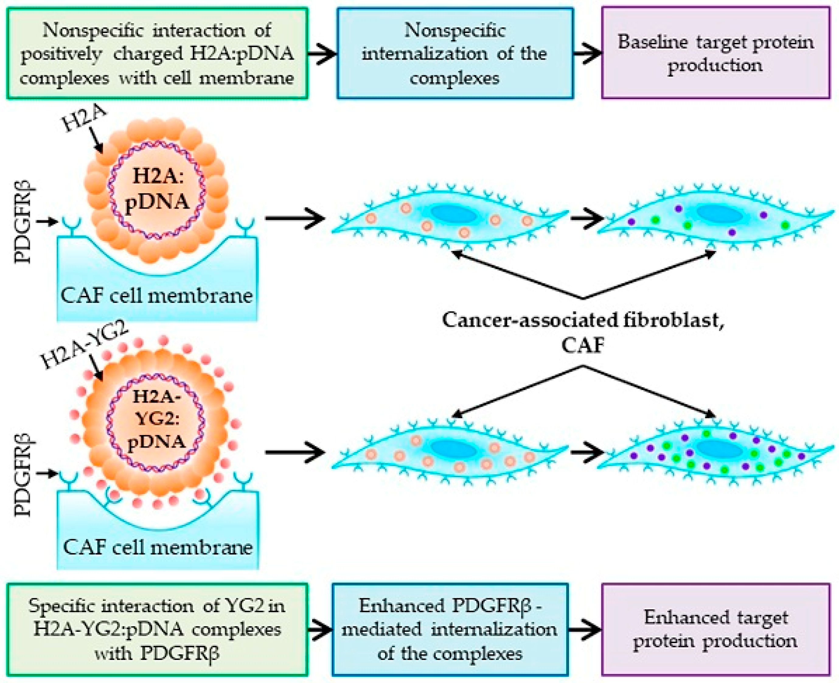
- Kuzmich, A.; Rakitina, O.; Didych, D.; Potapov, V.; Zinovyeva, M.; Alekseenko, I.; Sverdlov, E. Novel Histone-Based DNA Carrier Targeting Cancer-Associated Fibroblasts. Polymers 2020, 12, 1695. [Google Scholar] [CrossRef]
- Rong, J.; Li, P.; Ge, Y.; Chen, H.; Wu, J.; Zhang, R.; Lao, J.; Lou, D.; Zhang, Y. Histone H2A-peptide-hybrided upconversion mesoporous silica nanoparticles for bortezomib/p53 delivery and apoptosis induction. Colloids Surf. B 2020, 186, 110674. [Google Scholar] [CrossRef]
- Elzoghby, A.O. Gelatin-based nanoparticles as drug and gene delivery systems: Reviewing three decades of research. J. Control. Release 2013, 172, 1075–1091. [Google Scholar] [CrossRef]
- Wang, H.; Boerman, O.C.; Sariibrahimoglu, K.; Li, Y.; Jansen, J.A.; Leeuwenburgh, S.C. Comparison of micro- vs. nanostructured colloidal gelatin gels for sustained delivery of osteogenic proteins: Bone morphogenetic protein-2 and alkaline phosphatase. Biomaterials 2012, 33, 8695–8703. [Google Scholar] [CrossRef]
- Zwiorek, K.; Kloeckner, J.; Wagner, E.; Coester, C. Gelatin nanoparticles as a new and simple gene delivery system. J. Pharm. Pharm. Sci. 2005, 7, 22–28. [Google Scholar]
- Matsumoto, G.; Kushibiki, T.; Kinoshita, Y.; Lee, U.; Omi, Y.; Kubota, E.; Tabata, Y. Cationized gelatin delivery of a plasmid DNA expressing small interference RNA for VEGF inhibits murine squamous cell carcinoma. Cancer Sci. 2006, 97, 313–321. [Google Scholar] [CrossRef]
- Xu, X.; Capito, R.M.; Spector, M. Delivery of plasmid IGF-1 to chondrocytes via cationized gelatin nanoparticles. J. Biomed. Mater. Res. A 2008, 84, 73–83. [Google Scholar] [CrossRef]
- Jarzebska, N.T.; Mellett, M.; Frei, J.; Kundig, T.M.; Pascolo, S. Protamine-Based Strategies for RNA Transfection. Pharmaceutics 2021, 13, 877. [Google Scholar] [CrossRef] [PubMed]
- Amos, H. Protamine Enhancement of Rna Uptake by Cultured Chick Cells. Biochem. Biophys. Res. Commun. 1961, 5, 1–4. [Google Scholar] [CrossRef]
- Tusup, M.; Pascolo, S. Generation of Immunostimulating 130 nm Protamine-RNA nanoparticles. Methods Mol. Biol. 2017, 1499, 155–163. [Google Scholar] [CrossRef] [PubMed]
- Yoo, H.; Mok, H. Evaluation of multimeric siRNA conjugates for efficient protamine-based delivery into breast cancer cells. Arch. Pharm. Res. 2015, 38, 129–136. [Google Scholar] [CrossRef]
- Mai, Y.; Guo, J.; Zhao, Y.; Ma, S.; Hou, Y.; Yang, J. Intranasal delivery of cationic liposome-protamine complex mRNA vaccine elicits effective anti-tumor immunity. Cell. Immunol. 2020, 354, 104143. [Google Scholar] [CrossRef]
- Crini, G.; Fourmentin, S.; Fenyvesi, É.; Torri, G.; Fourmentin, M.; Morin-Crini, N. Cyclodextrins, from molecules to applications. Environ. Chem. Lett. 2018, 16, 1361–1375. [Google Scholar] [CrossRef]
- Cryan, S.A.; Holohan, A.; Donohue, R.; Darcy, R.; O’Driscoll, C.M. Cell transfection with polycationic cyclodextrin vectors. Eur. J. Pharm. Sci. 2004, 21, 625–633. [Google Scholar] [CrossRef]
- Hwang, S.J.; Bellocq, N.C.; Davis, M.E. Effects of structure of beta-cyclodextrin-containing polymers on gene delivery. Bioconjug. Chem. 2001, 12, 280–290. [Google Scholar] [CrossRef]
- Liu, J.; Tian, B.; Liu, Y.; Wan, J.B. Cyclodextrin-Containing Hydrogels: A Review of Preparation Method, Drug Delivery, and Degradation Behavior. Int. J. Mol. Sci. 2021, 22, 3516. [Google Scholar] [CrossRef]
- Liu, X.; Li, Z.; Loh, X.J.; Chen, K.; Li, Z.; Wu, Y.L. Targeted and Sustained Corelease of Chemotherapeutics and Gene by Injectable Supramolecular Hydrogel for Drug-Resistant Cancer Therapy. Macromol. Rapid Commun. 2019, 40, e1800117. [Google Scholar] [CrossRef]
- Elieh-Ali-Komi, D.; Hamblin, M.R. Chitin and Chitosan: Production and Application of Versatile Biomedical Nanomaterials. Int. J. Adv. Res. 2016, 4, 411–427. [Google Scholar]
- Anraku, M.; Hiraga, A.; Iohara, D.; Uekama, K.; Tomida, H.; Otagiri, M.; Hirayama, F. Preparation and antioxidant activity of PEGylated chitosans with different particle sizes. Int. J. Biol. Macromol. 2014, 70, 64–69. [Google Scholar] [CrossRef]
- TM, M.W.; Lau, W.M.; Khutoryanskiy, V.V. Chitosan and Its Derivatives for Application in Mucoadhesive Drug Delivery Systems. Polymers 2018, 10, 267. [Google Scholar] [CrossRef] [Green Version]
- Lara-Velazquez, M.; Alkharboosh, R.; Norton, E.S.; Ramirez-Loera, C.; Freeman, W.D.; Guerrero-Cazares, H.; Forte, A.J.; Quinones-Hinojosa, A.; Sarabia-Estrada, R. Chitosan-Based Non-viral Gene and Drug Delivery Systems for Brain Cancer. Front. Neurol. 2020, 11, 740. [Google Scholar] [CrossRef]
- Islam, S.U.; Shehzad, A.; Ahmed, M.B.; Lee, Y.S. Intranasal Delivery of Nanoformulations: A Potential Way of Treatment for Neurological Disorders. Molecules 2020, 25, 1929. [Google Scholar] [CrossRef] [Green Version]
- Posadas, I.; Monteagudo, S.; Cena, V. Nanoparticles for brain-specific drug and genetic material delivery, imaging and diagnosis. Nanomedicine 2016, 11, 833–849. [Google Scholar] [CrossRef] [Green Version]
- Wang, G.H.; Cai, Y.Y.; Du, J.K.; Li, L.; Li, Q.; Yang, H.K.; Lin, J.T. TAT-conjugated chitosan cationic micelle for nuclear-targeted drug and gene co-delivery. Colloids Surf. B Biointerfaces 2018, 162, 326–334. [Google Scholar] [CrossRef]
- Lin, P.Y.; Chiu, Y.L.; Huang, J.H.; Chuang, E.Y.; Mi, F.L.; Lin, K.J.; Juang, J.H.; Sung, H.W.; Leong, K.W. Oral Nonviral Gene Delivery for Chronic Protein Replacement Therapy. Adv. Sci. 2018, 5, 1701079. [Google Scholar] [CrossRef]
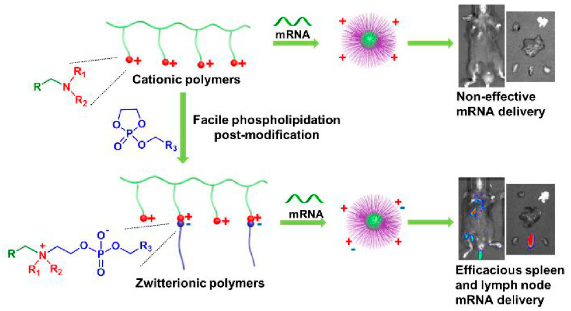
- Liu, S.; Wang, X.; Yu, X.; Cheng, Q.; Johnson, L.T.; Chatterjee, S.; Zhang, D.; Lee, S.M.; Sun, Y.; Lin, T.C.; et al. Zwitterionic Phospholipidation of Cationic Polymers Facilitates Systemic mRNA Delivery to Spleen and Lymph Nodes. J. Am. Chem. Soc. 2021, 143, 21321–21330. [Google Scholar] [CrossRef]
- Chen, H.; Li, P.; Yin, Y.; Cai, X.; Huang, Z.; Chen, J.; Dong, L.; Zhang, J. The promotion of type 1 T helper cell responses to cationic polymers in vivo via toll-like receptor-4 mediated IL-12 secretion. Biomaterials 2010, 31, 8172–8180. [Google Scholar] [CrossRef]
- Karpenko, L.I.; Apartsin, E.K.; Dudko, S.G.; Starostina, E.V.; Kaplina, O.N.; Antonets, D.V.; Volosnikova, E.A.; Zaitsev, B.N.; Bakulina, A.Y.; Venyaminova, A.G.; et al. Cationic Polymers for the Delivery of the Ebola DNA Vaccine Encoding Artificial T-Cell Immunogen. Vaccines 2020, 8, 718. [Google Scholar] [CrossRef] [PubMed]
- Sashital, D.G.; Doudna, J.A. Structural insights into RNA interference. Curr. Opin. Struct. Biol. 2010, 20, 90–97. [Google Scholar] [CrossRef] [PubMed] [Green Version]
- Piombo, E.; Vetukuri, R.R.; Broberg, A.; Kalyandurg, P.B.; Kushwaha, S.; Funck Jensen, D.; Karlsson, M.; Dubey, M. Role of Dicer-Dependent RNA Interference in Regulating Mycoparasitic Interactions. Microbiol. Spectr. 2021, 9, e0109921. [Google Scholar] [CrossRef] [PubMed]
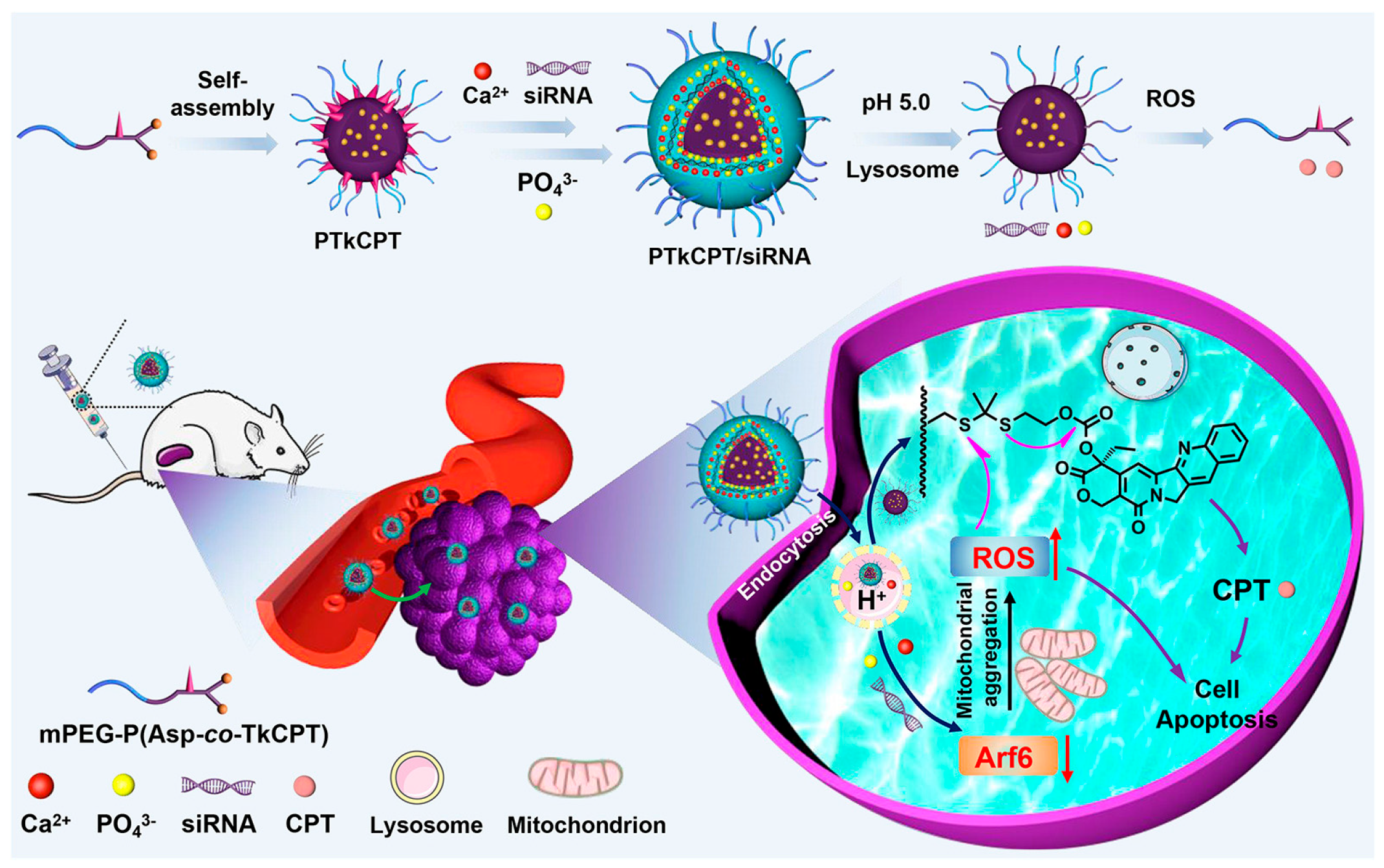
- Huang, J.; Zheng, C.; Xiao, H.; Huang, H.; Wang, Y.; Lin, M.; Pang, J.; Wang, Y.; Yuan, Y.; Shuai, X. A polymer--calcium phosphate nanocapsule for RNAi-induced oxidative stress and cascaded chemotherapy. J. Control. Release 2021, 340, 259–270. [Google Scholar] [CrossRef]
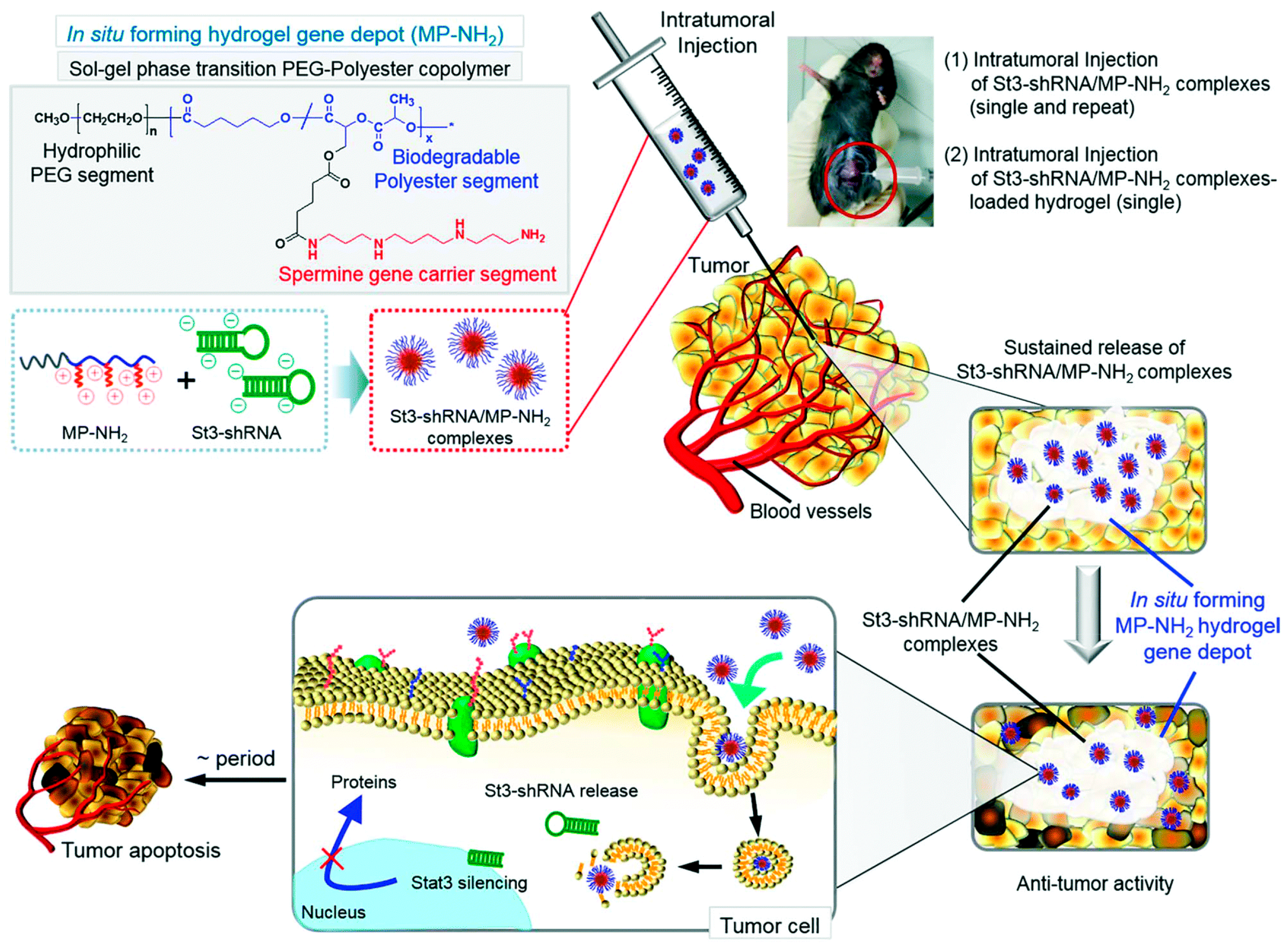
- Kim, D.Y.; Ju, H.J.; Kim, J.H.; Choi, S.; Kim, M.S. Injectable in situ forming hydrogel gene depot to improve the therapeutic effect of STAT3 shRNA. Biomater. Sci. 2021, 9, 4459–4472. [Google Scholar] [CrossRef]
- Zhang, Z.; Wan, T.; Chen, Y.; Chen, Y.; Sun, H.; Cao, T.; Songyang, Z.; Tang, G.; Wu, C.; Ping, Y.; et al. Cationic Polymer-Mediated CRISPR/Cas9 Plasmid Delivery for Genome Editing. Macromol. Rapid Commun. 2019, 40, e1800068. [Google Scholar] [CrossRef]
- Luo, Y.L.; Xu, C.F.; Li, H.J.; Cao, Z.T.; Liu, J.; Wang, J.L.; Du, X.J.; Yang, X.Z.; Gu, Z.; Wang, J. Macrophage-Specific in Vivo Gene Editing Using Cationic Lipid-Assisted Polymeric Nanoparticles. ACS Nano 2018, 12, 994–1005. [Google Scholar] [CrossRef]
- Cox, D.B.T.; Gootenberg, J.S.; Abudayyeh, O.O.; Franklin, B.; Kellner, M.J.; Joung, J.; Zhang, F. RNA editing with CRISPR-Cas13. Science 2017, 358, 1019–1027. [Google Scholar] [CrossRef] [Green Version]
- Blanchard, E.L.; Vanover, D.; Bawage, S.S.; Tiwari, P.M.; Rotolo, L.; Beyersdorf, J.; Peck, H.E.; Bruno, N.C.; Hincapie, R.; Michel, F.; et al. Treatment of influenza and SARS-CoV-2 infections via mRNA-encoded Cas13a in rodents. Nat. Biotechnol. 2021, 39, 717–726. [Google Scholar] [CrossRef]
- Wang, H.; Wan, G.; Liu, Y.; Chen, B.; Chen, H.; Zhang, S.; Wang, D.; Xiong, Q.; Zhang, N.; Wang, Y. Dual-responsive nanoparticles based on oxidized pullulan and a disulfide-containing poly(β-amino) ester for efficient delivery of genes and chemotherapeutic agents targeting hepatoma. Polym. Chem. 2016, 7, 6340–6353. [Google Scholar] [CrossRef]
- Zhang, S.; Wang, D.; Li, Y.; Li, L.; Chen, H.; Xiong, Q.; Liu, Y.; Wang, Y. pH- and redox-responsive nanoparticles composed of charge-reversible pullulan-based shells and disulfide-containing poly(ss-amino ester) cores for co-delivery of a gene and chemotherapeutic agent. Nanotechnology 2018, 29, 325101. [Google Scholar] [CrossRef]
- Wang, Y.; Chen, H.; Liu, Y.; Wu, J.; Zhou, P.; Wang, Y.; Li, R.; Yang, X.; Zhang, N. pH-sensitive pullulan-based nanoparticle carrier of methotrexate and combretastatin A4 for the combination therapy against hepatocellular carcinoma. Biomaterials 2013, 34, 7181–7190. [Google Scholar] [CrossRef]
- Kaneo, Y.; Tanaka, T.; Nakano, T.; Yamaguchi, Y. Evidence for receptor-mediated hepatic uptake of pullulan in rats. J. Control. Release 2001, 70, 365–373. [Google Scholar] [CrossRef]
- Jones, C.H.; Chen, M.; Gollakota, A.; Ravikrishnan, A.; Zhang, G.; Lin, S.; Tan, M.; Cheng, C.; Lin, H.; Pfeifer, B.A. Structure-Function Assessment of Mannosylated Poly(beta-amino esters) upon Targeted Antigen Presenting Cell Gene Delivery. Biomacromolecules 2015, 16, 1534–1541. [Google Scholar] [CrossRef]
- Liu, Z.H.; Zhang, Z.Y.; Zhou, C.R.; Jiao, Y.P. Hydrophobic modifications of cationic polymers for gene delivery. Prog. Polym. Sci. 2010, 35, 1144–1162. [Google Scholar] [CrossRef]
- Vaidyanathan, S.; Anderson, K.B.; Merzel, R.L.; Jacobovitz, B.; Kaushik, M.P.; Kelly, C.N.; van Dongen, M.A.; Dougherty, C.A.; Orr, B.G.; Banaszak Holl, M.M. Quantitative Measurement of Cationic Polymer Vector and Polymer-pDNA Polyplex Intercalation into the Cell Plasma Membrane. ACS Nano 2015, 9, 6097–6109. [Google Scholar] [CrossRef] [Green Version]


Disclaimer/Publisher’s Note: The statements, opinions and data contained in all publications are solely those of the individual author(s) and contributor(s) and not of MDPI and/or the editor(s). MDPI and/or the editor(s) disclaim responsibility for any injury to people or property resulting from any ideas, methods, instructions or products referred to in the content. |
© 2023 by the authors. Licensee MDPI, Basel, Switzerland. This article is an open access article distributed under the terms and conditions of the Creative Commons Attribution (CC BY) license (https://creativecommons.org/licenses/by/4.0/).
Share and Cite
Cai, X.; Dou, R.; Guo, C.; Tang, J.; Li, X.; Chen, J.; Zhang, J. Cationic Polymers as Transfection Reagents for Nucleic Acid Delivery. Pharmaceutics 2023, 15, 1502. https://doi.org/10.3390/pharmaceutics15051502
Cai X, Dou R, Guo C, Tang J, Li X, Chen J, Zhang J. Cationic Polymers as Transfection Reagents for Nucleic Acid Delivery. Pharmaceutics. 2023; 15(5):1502. https://doi.org/10.3390/pharmaceutics15051502
Chicago/Turabian StyleCai, Xiaomeng, Rui Dou, Chen Guo, Jiaruo Tang, Xiajuan Li, Jun Chen, and Jiayu Zhang. 2023. "Cationic Polymers as Transfection Reagents for Nucleic Acid Delivery" Pharmaceutics 15, no. 5: 1502. https://doi.org/10.3390/pharmaceutics15051502