
Current Trends in Gelatin-Based Drug Delivery Systems
 , and
, and 
Abstract
:1. Introduction
2. Methodology
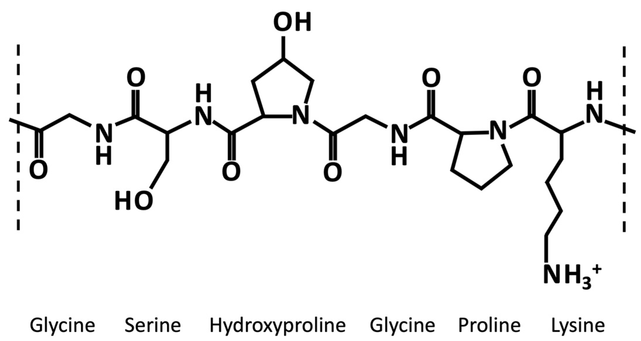
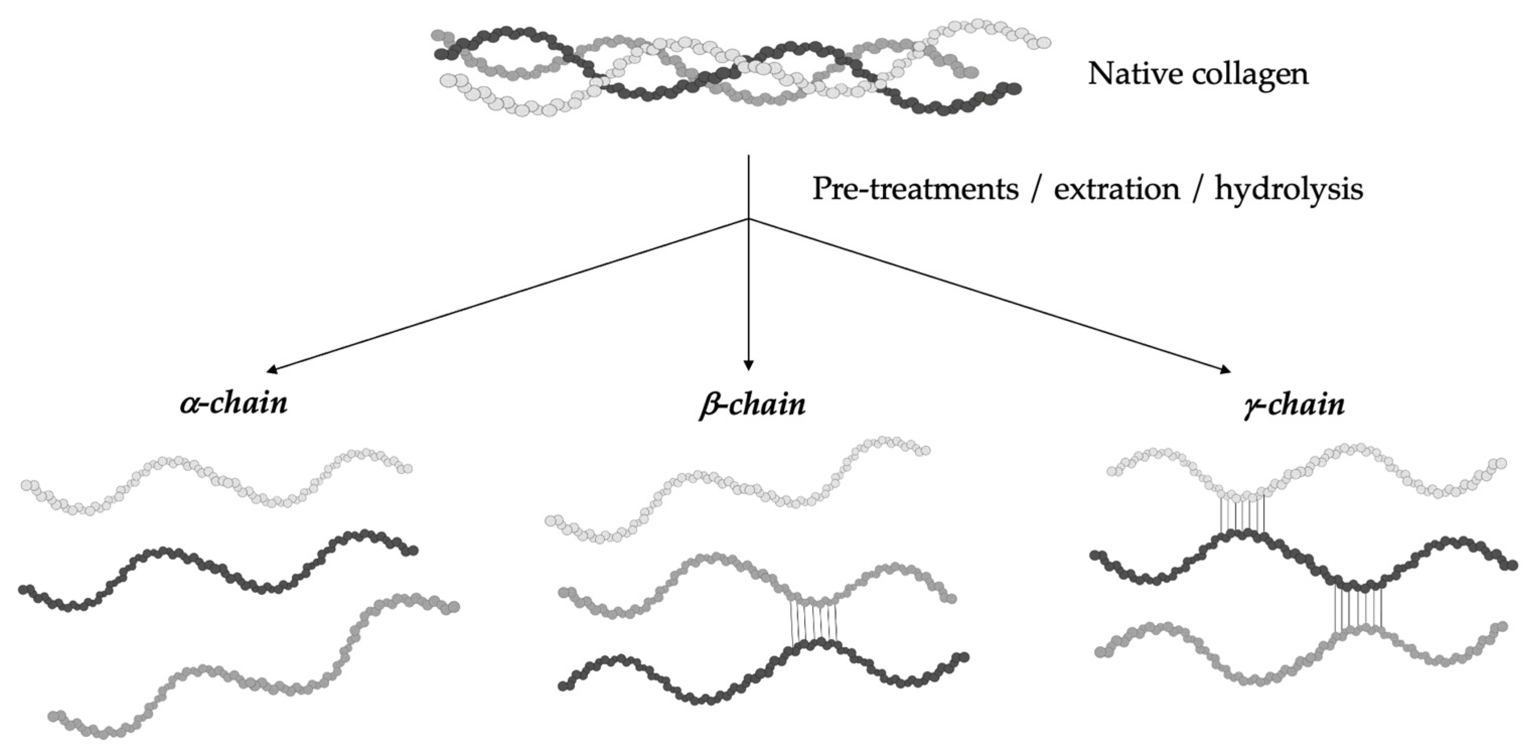
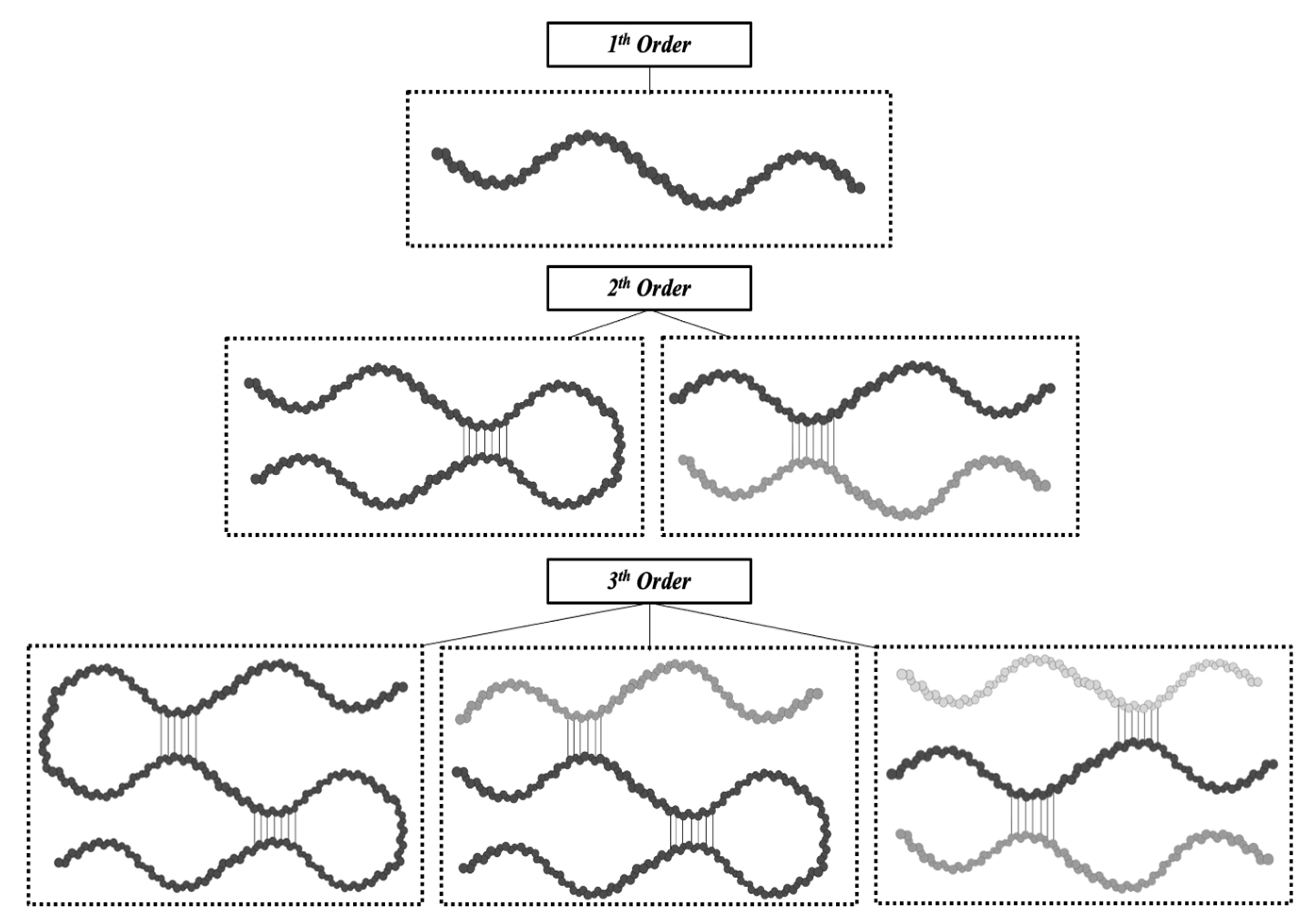
3. Gelatin: Structure and Properties
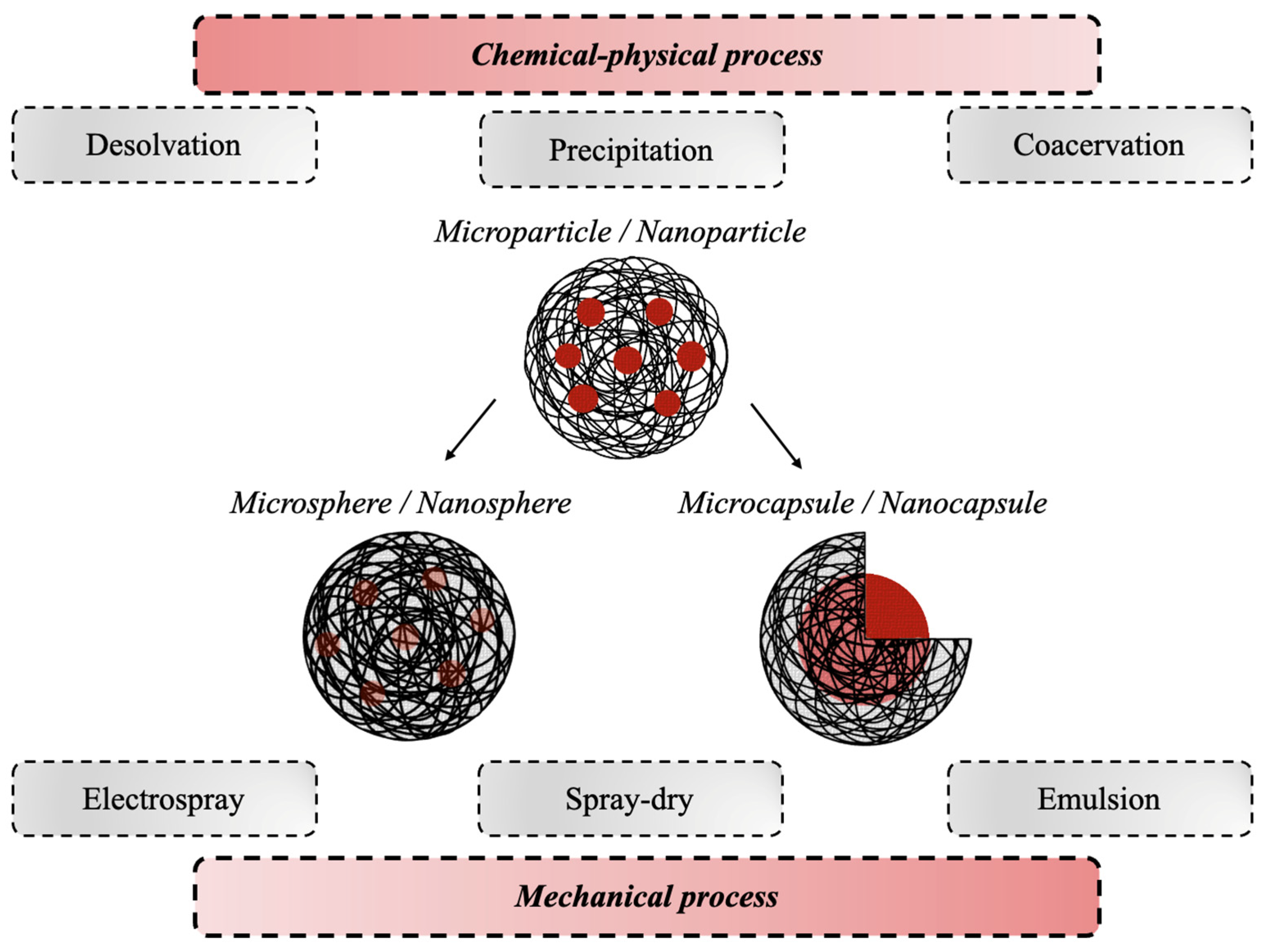
4. Gelatin-Based DDSs

| DDS Synthesis Technique | DDS Type | Type of Gelatin | Gelatin Source | Encapsulated Bioactive Compound | Crosslinker | Ref. |
|---|---|---|---|---|---|---|
| Desolvation | Nanoparticles | Type A | Porcine skin | Ibuprofen sodium | CaCl2 | [105] |
| Didanosine | GA | [81] | ||||
| Moxifloxacin | GA | [106] | ||||
| Timol maleate | GA | [107] | ||||
| - | GA | [108] | ||||
| Type B | Bovine skin | Rutin | GA | [109] | ||
| Rosiglitazone | GA | [84] | ||||
| Type A and B | Porcine skin and bovine skin | BMP-2, bFGF | GA | [100] | ||
| Texas Red | GA | [76] | ||||
| Cardamom | GA | [77] | ||||
| Amphotericin B | GA | [78] | ||||
| Porcine skin and beef nails | Fluorescein-5-isothiocynate | GA | [79] | |||
| N. d. | Bovine skin | Bovine serum albumin | GA | [110] | ||
| Fish skin | - | GA | [111] | |||
| Camel skin | - | GA | [86] | |||
| Nanoprecipitation | Nanoparticles | Type B | Bovine skin | Lysozyme | DIC | [95] |
| - | – | [112] | ||||
| - | GA | [113] | ||||
| Fluorescein-5-isothiocynate | GA | [114] | ||||
| Dextran | GA | [70] | ||||
| Tizadine hydrochloride gatifloxacin | GA | [113] | ||||
| Metoprolol | GA | [90] | ||||
| N. d. | Porcine | Erythromycin | GA | [115] | ||
| N. d. | Cocoa-derived polyphenolic extract | GA | [102] | |||
| Type A | Porcine skin | Zaltoprofen | GA | [82] | ||
| Type A and B | Porcine skin and bovine skin | Non-steroidal anti-inflammatory drugs | GA | [80] | ||
| Coacervation | Microcapsules/nanocapsules | Type A | Porcine skin | α-Tocopherol | GA | [83] |
| Vitamin D3 | TG | [99] | ||||
| Capsaicin | GA | [116] | ||||
| N. d. | N. d. | Capsaicin | GA | [103] | ||
| N. d. | N. d. | Zeaxanthin | TG | [117] | ||
| N. d. | N. d. | Phenacetin | Formalin | [94] | ||
| N. d. | N. d. | Berberine hydrochloride Gallic acid | – | [118] | ||
| Type B | Bovine skin | Geraniol oil | GA | [85] | ||
| N. d. | N. d. | Moxa oil | FA | [119] | ||
| N. d. | Fish | Fish oil | CaCl2 | [120] | ||
| Emulsion | Microspheres/nanospheres | Type B | N. d. | Mitomycin C-dextran conjugate | FA | [121] |
| Bovine skin | Sodium fluoride | GA | [88] | |||
| Type A | Porcine skin | TGF-β1 | Genepin | [27] | ||
| L929 fibroblasts | MBA | [93] | ||||
| Type A and B | N. d. | bFGF | GA | [122] | ||
| Porcine skin and bovine skin | BMP-2, VEGF | Genepin | [91] | |||
| N. d. | N. d. | Cefquinome sulfate | GA | [123] | ||
| N. d. | N. d. | Tetracycline hydrochloride | GA | [124] | ||
| N. d. | N. d. | Amoxicillin | GA | [125] | ||
| N. d. | N. d. | Phyllanthus urinaria extract | – | [101] | ||
| Microparticles/nanoparticles | Type B | Bovine skin | Bovine serum albumin | – | [126] | |
| BMP-4, bFGF | Heat | [98] | ||||
| Methotrexate | GA | [127] | ||||
| Type A | Porcine skin | Tramadol hydrochloride | GA | [128] | ||
| Type A and B | N. d. | BMP-2 | GA | [33] | ||
| Spray-dry | Microcapsules | Type A | N. d. | Revaprazan | – | [129] |
| N. d. | Curcumin | DCMC | [92] | |||
| N. d. | Nifedipine | – | [130] | |||
| N. d. | Valsaran | – | [131] | |||
| N. d. | Fenofibrate | – | [132] | |||
| N. d. | Ibuprofen | – | [133] | |||
| N. d. | Ibuprofen | GA | [134] | |||
| N. d. | Piroxicam | – | [135] | |||
| Electrospray | Nanocapsules | N. d. | Tilapia fish skin | Moringa oleifera | – | [136] |
| Microparticles/nanoparticles | N. d. | N. d. | Piroxicam | – | [87] | |
| N. d. | Tilapia fish skin | Bitter gourd | – | [137] | ||
| Type A | Porcine skin | Epigallocatechin 3-gallate | GA | [89] | ||
| Microspheres | Type B | Bovine skin | Human bone marrow stromal cell | CaCl2 | [96] | |
| N. d. | Human-adipose-derived stem cells | – | [104] |
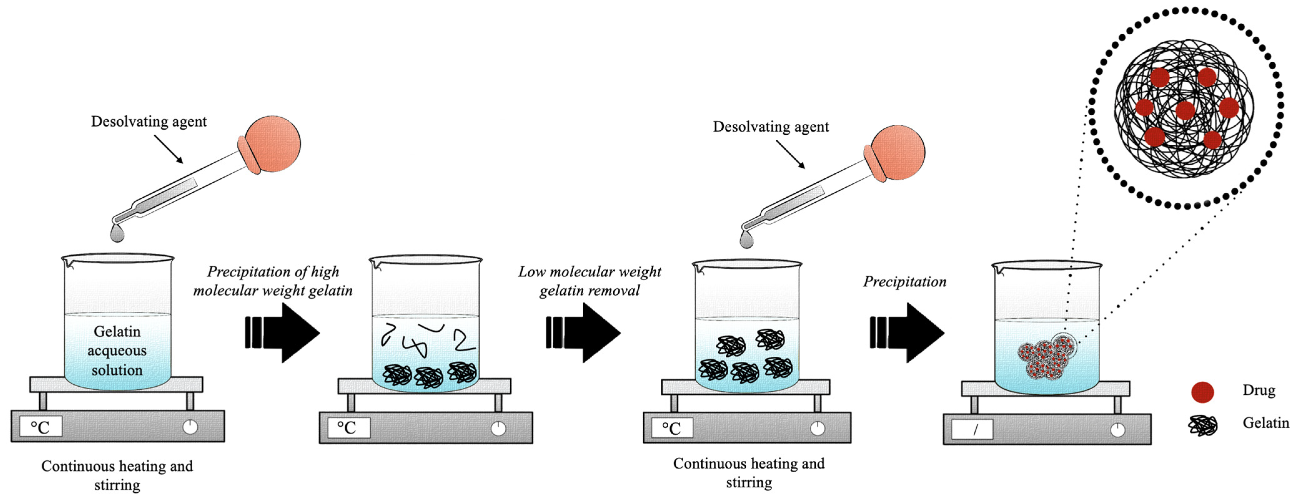
4.1. Desolvation
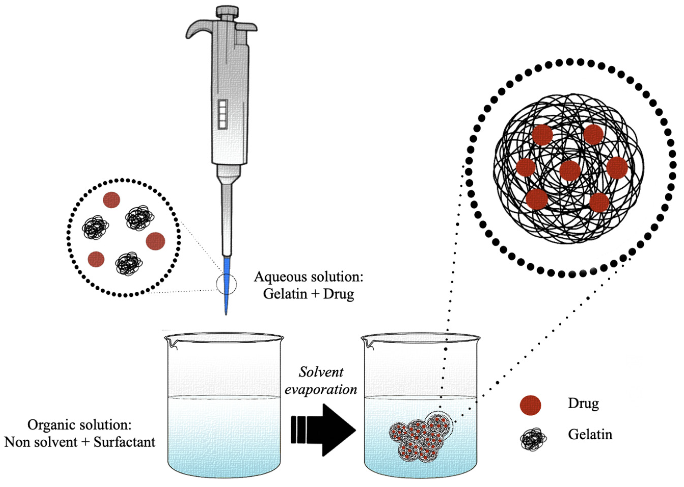
4.2. Nanoprecipitation
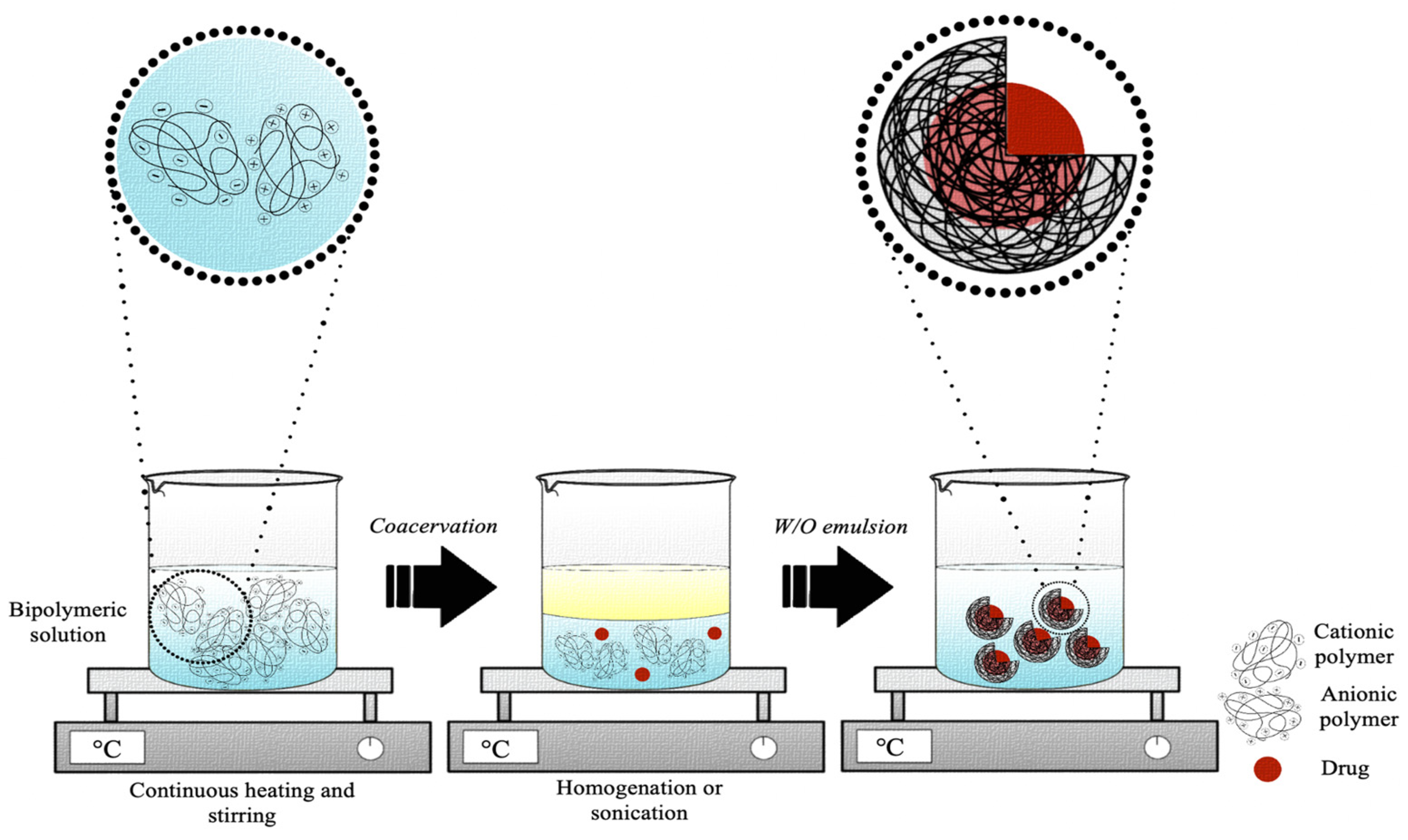
4.3. Coacervation
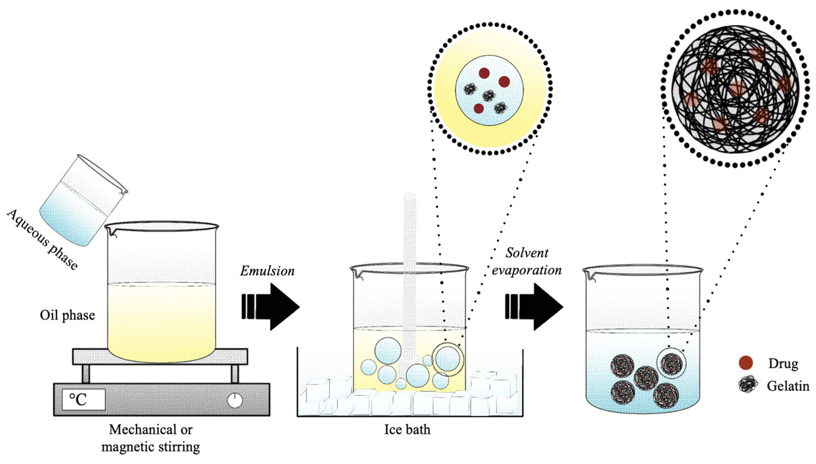
4.4. Emulsion
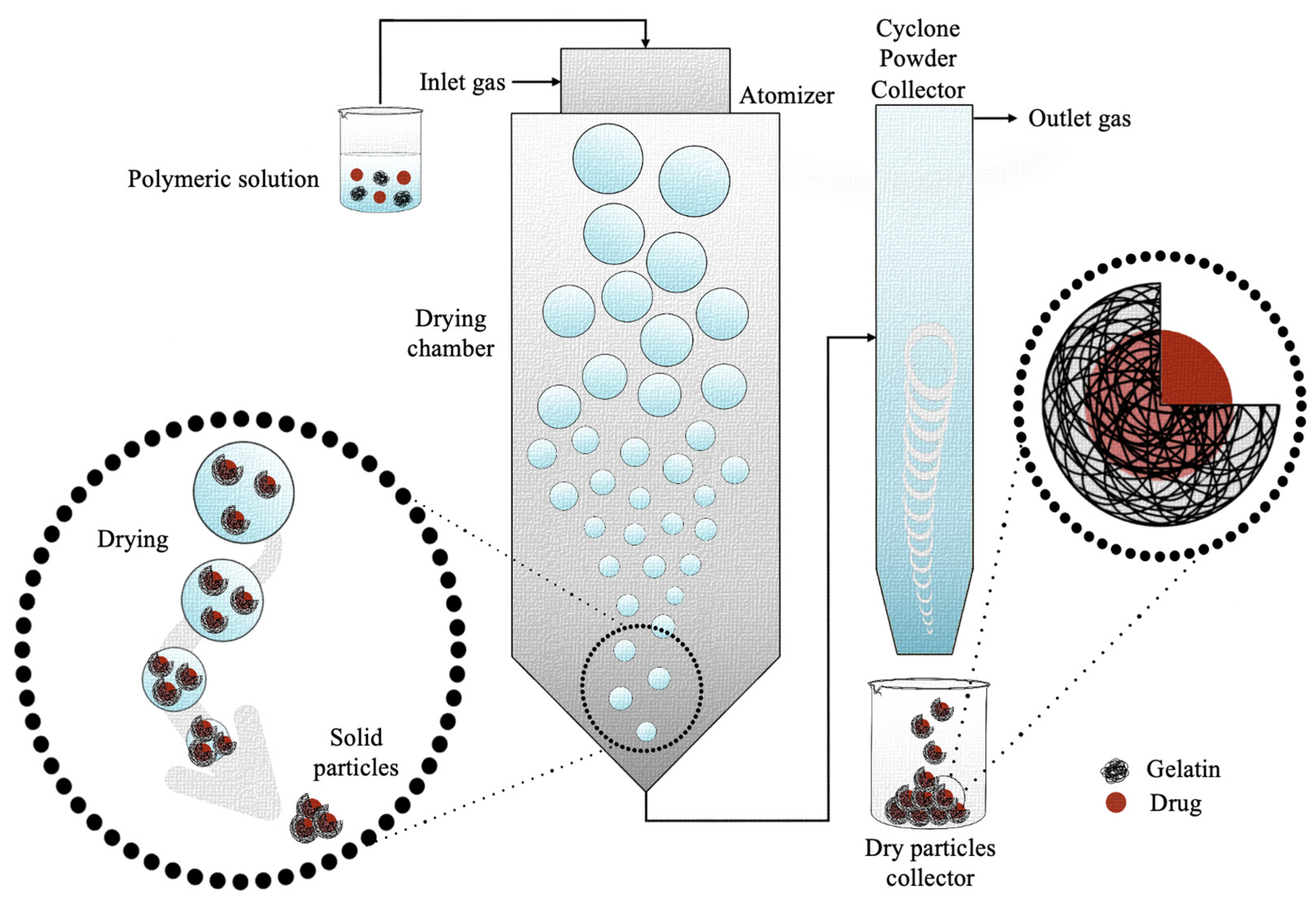
4.5. Spray Drying
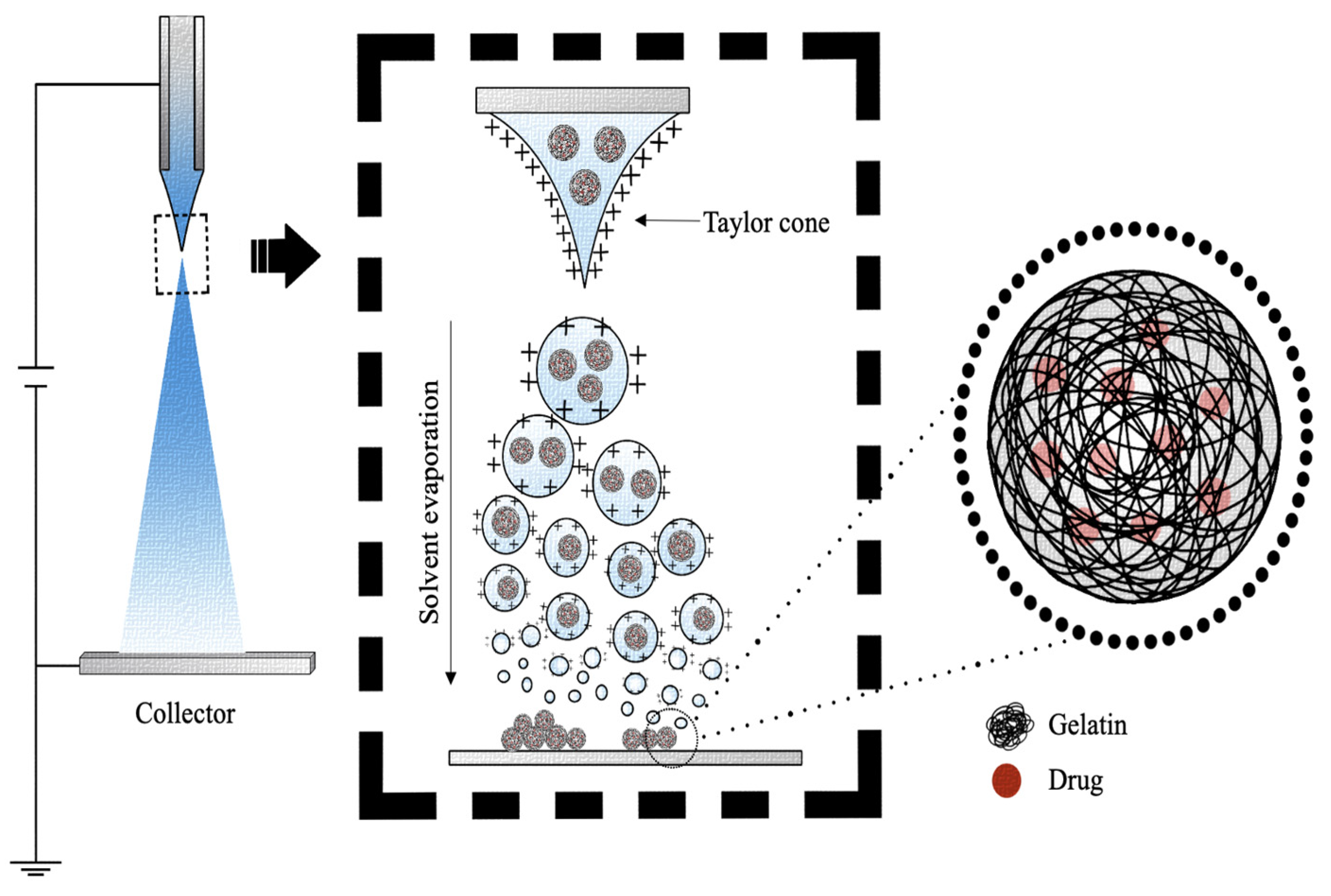
4.6. Electrospray
5. Preclinical and Clinical Outcomes
5.1. Preclinical Studies
5.2. Clinical Studies
| Issue | Bioactive Compound | Outcome(s) | Ref. |
|---|---|---|---|
| Chronic venous leg ulcers | Keratinocytes | Fast healing and complete regeneration. | [200] |
| Vitiligo | Melanocytes | Complete repigmentation, no adverse events. | [201] |
| Sun protection | Ruthin | Increased free-radical-scavenging rate. | [109] |
| Metastatic liver tumors | Cisplatin | Reduction in tumor size, no serious side effects. | [194] |
| Hepatocellular carcinoma | Cisplatin | No serious side effects, 100% success rate, reduced abdominal pain. | [195] |
| Limb ischemia | bFGF | Complete or partial regression of ischemic ulcers, no local and systemic effects. | [196] |
| Peripheral arterial disease | bFGF | Improvement in symptoms and no serious complications, incomplete necrosis or ulcer healing. | [198] |
| Fingertip amputation | bFGF | No statistically significative improvements. | [199] |
| Limb ischemia | bFGF | No serious adverse events. | [197] |
6. Conclusions and Future Perspectives
Author Contributions
Funding
Institutional Review Board Statement
Informed Consent Statement
Data Availability Statement
Conflicts of Interest
References
- Liu, D.; Yang, F.; Xiong, F.; Gu, N. The Smart Drug Delivery System and Its Clinical Potential. Theranostics 2016, 6, 1306–1323. [Google Scholar] [CrossRef]
- Adepu, S.; Ramakrishna, S. Controlled Drug Delivery Systems: Current Status and Future Directions. Molecules 2021, 26, 5905. [Google Scholar] [CrossRef]
- Langer, R. Drug Delivery and Targeting. Nature 1998, 392, 5–10. [Google Scholar] [PubMed]
- Jain, K.K. (Ed.) Drug Delivery System. In Methods in Molecular Biology; Humana: New York, NY, USA, 2020. [Google Scholar]
- Hoffman, A.S. The Origins and Evolution of “Controlled” Drug Delivery Systems. J. Control. Release 2008, 132, 153–163. [Google Scholar] [CrossRef] [PubMed]
- Langer, R. New Methods of Drug Delivery. Science 1990, 249, 1527–1533. [Google Scholar] [CrossRef] [PubMed]
- Elzoghby, A.O.; Samy, W.M.; Elgindy, N.A. Protein-Based Nanocarriers as Promising Drug and Gene Delivery Systems. J. Control. Release 2012, 161, 38–49. [Google Scholar] [CrossRef]
- Gorgieva, S.; Kokol, V. Collagen-vs. Gelatine-Based Biomaterials and Their Biocompatibility: Review and Perspectives. Biomater. Appl. Nanomed. 2011, 2, 18–52. [Google Scholar] [CrossRef]
- Pal, A.; Bajpai, J.; Bajpai, A.K. Easy Fabrication and Characterization of Gelatin Nanocarriers and in Vitro Investigation of Swelling Controlled Release Dynamics of Paclitaxel. Polym. Bull. 2018, 75, 4691–4711. [Google Scholar] [CrossRef]
- Tan, H.; Tu, Z.; Jia, H.; Gou, X.; Ngai, T. Hierarchical Porous Protein Scaffold Templated from High Internal Phase Emulsion Costabilized by Gelatin and Gelatin Nanoparticles. Langmuir 2018, 34, 4820–4829. [Google Scholar] [CrossRef]
- Yang, C.; Wang, J. Preparation and Characterization of Collagen Microspheres for Sustained Release of Steroidal Saponins. Mater. Res. 2014, 17, 1644–1650. [Google Scholar] [CrossRef]
- Rossler, B.; Scherer, D. Collagen Microparticles: Preparation and Properties. J. Microencapsul. 1995, 12, 49–57. [Google Scholar] [CrossRef]
- Rathore, P.; Arora, I.; Rastogi, S.; Akhtar, M.; Singh, S.; Samim, M. Collagen Nanoparticle-Mediated Brain Silymarin Delivery: An Approach for Treating Cerebral Ischemia and Reperfusion-Induced Brain Injury. Front. Neurosci. 2020, 14, 538404. [Google Scholar] [CrossRef] [PubMed]
- Seong, Y.J.; Song, E.H.; Park, C.; Lee, H.; Kang, I.G.; Kim, H.E.; Jeong, S.H. Porous Calcium Phosphate–Collagen Composite Microspheres for Effective Growth Factor Delivery and Bone Tissue Regeneration. Mater. Sci. Eng. C. 2020, 109, 110480. [Google Scholar] [CrossRef]
- Calejo, M.T.; Almeida, A.J.; Fernandes, A.I. Exploring a New Jellyfish Collagen in the Production of Microparticles for Protein Delivery. J. Microencapsul. 2012, 29, 520–531. [Google Scholar] [CrossRef] [PubMed]
- Yeung, P.; Sin, H.S.; Chan, S.; Chan, G.C.F.; Chan, B.P. Microencapsulation of Neuroblastoma Cells and Mesenchymal Stromal Cells in Collagen Microspheres: A 3D Model for Cancer Cell Niche Study. PLoS ONE 2015, 10, e0144139. [Google Scholar] [CrossRef]
- Kozlowska, J.; Stachowiak, N.; Prus, W. Stability Studies of Collagen-Based Microspheres with Calendula Officinalis Flower Extract. Polym. Degrad. Stab. 2019, 163, 214–219. [Google Scholar] [CrossRef]
- Zhang, W.; Wang, X.-C.; Wang, J.-J.; Zhang, L.-l. Drugs Adsorption and Release Behavior of Collagen/Bacterial Cellulose Porous Microspheres. Int. J. Biol. Macromol. 2019, 140, 196–205. [Google Scholar] [CrossRef] [PubMed]
- Zhang, Z.; Li, X.; Li, Z.; Bai, Y.; Liao, G.; Pan, J.; Zhang, C. Collagen/Nano-Sized β-Tricalcium Phosphate Conduits Combined with Collagen Filaments and Nerve Growth Factor Promote Facial Nerve Regeneration in Miniature Swine: An in Vivo Study. Oral. Surg. Oral. Med. Oral. Pathol. Oral. Radiol. 2019, 128, 472–478. [Google Scholar] [CrossRef]
- Berthold, A.; Cremer, K.; Rg Kreuter, J. Collagen Microparticles: Carriers for Glucocorticosteroids. Eur. J. Pharm. Biopharm. 1998, 45, 23–29. [Google Scholar] [CrossRef]
- Doi, N.; Jo, J.I.; Tabata, Y. Preparation of Biodegradable Gelatin Nanospheres with a Narrow Size Distribution for Carrier of Cellular Internalization of Plasmid DNA. J. Biomater. Sci. Polym. Ed. 2012, 23, 991–1004. [Google Scholar] [CrossRef]
- Krause, H.J.; Rohdewald, P. Preparation of gelatin nanocapsules and their pharmaceutical characterization. Pharm. Res. 1985, 2, 239–243. [Google Scholar] [CrossRef] [PubMed]
- Zhou, S.; Li, L.; Chen, C.; Chen, Y.; Zhou, L.; Zhou, F.H.; Dong, J.; Wang, L. Injectable Gelatin Microspheres Loaded with Platelet Rich Plasma Improve Wound Healing by Regulating Early Inflammation. Int. J. Med. Sci. 2021, 18, 1910–1920. [Google Scholar] [CrossRef] [PubMed]
- Wang, H.; Boerman, O.C.; Sariibrahimoglu, K.; Li, Y.; Jansen, J.A.; Leeuwenburgh, S.C.G. Comparison of Micro- vs. Nanostructured Colloidal Gelatin Gels for Sustained Delivery of Osteogenic Proteins: Bone Morphogenetic Protein-2 and Alkaline Phosphatase. Biomaterials 2012, 33, 8695–8703. [Google Scholar] [CrossRef]
- Leong, W.; Lau, T.T.; Wang, D.A. A Temperature-Cured Dissolvable Gelatin Microsphere-Based Cell Carrier for Chondrocyte Delivery in a Hydrogel Scaffolding System. Acta Biomater. 2013, 9, 6459–6467. [Google Scholar] [CrossRef] [PubMed]
- Kawadkar, J.; Jain, R.; Kishore, R.; Pathak, A.; Chauhan, M.K. Formulation and Evaluation of Flurbiprofen-Loaded Genipin Cross-Linked Gelatin Microspheres for Intra-Articular Delivery. J. Drug. Target. 2013, 21, 200–210. [Google Scholar] [CrossRef] [PubMed]
- Kudva, A.K.; Dikina, A.D.; Luyten, F.P.; Alsberg, E.; Patterson, J. Gelatin Microspheres Releasing Transforming Growth Factor Drive in Vitro Chondrogenesis of Human Periosteum Derived Cells in Micromass Culture. Acta Biomater. 2019, 90, 287–299. [Google Scholar] [CrossRef]
- Ramshaw, J.A.M.; Werkmeister, J.A.; Glattauer, V. Collagen-Based Biomaterials. Biotechnol. Genet. Eng. Rev. 1996, 13, 335–382. [Google Scholar] [CrossRef]
- Lee, C.H.; Singla, A.; Lee, Y. Biomedical Applications of Collagen. Int. J. Pharm. 2001, 221, 1–22. [Google Scholar] [CrossRef]
- Zwiorek, K.; Kloeckner, J.; Wagner, E.; Coester, C. Gelatin nanoparticles as a new and simple gene delivery system. J. Pharm. Pharm. Sci. 2005, 7, 22–28. [Google Scholar]
- Lukin, I.; Erezuma, I.; Maeso, L.; Zarate, J.; Desimone, M.F.; Al-Tel, T.H.; Dolatshahi-Pirouz, A.; Orive, G. Progress in Gelatin as Biomaterial for Tissue Engineering. Pharmaceutics 2022, 14, 1177. [Google Scholar] [CrossRef]
- Johnston-Banks, F.A. Gelatine. In Food Gels; Elsevier Applied Food Science Series; Springer: Dordrecht, The Netherlands, 1990; Chapter 7; pp. 233–289. [Google Scholar]
- Patel, Z.S.; Yamamoto, M.; Ueda, H.; Tabata, Y.; Mikos, A.G. Biodegradable Gelatin Microparticles as Delivery Systems for the Controlled Release of Bone Morphogenetic Protein-2. Acta Biomater. 2008, 4, 1126–1138. [Google Scholar] [CrossRef]
- Samal, S.K.; Dash, M.; Van Vlierberghe, S.; Kaplan, D.L.; Chiellini, E.; van Blitterswijk, C.; Moroni, L.; Dubruel, P. Cationic Polymers and Their Therapeutic Potential. Chem. Soc. Rev. 2012, 41, 7147–7194. [Google Scholar] [CrossRef]
- Madkhali, O.; Mekhail, G.; Wettig, S.D. Modified Gelatin Nanoparticles for Gene Delivery. Int. J. Pharm. 2019, 554, 224–234. [Google Scholar] [CrossRef]
- Singh, S.; Rao, R.K.V.; Venugopal, K.; Manikandan, R. Alteration in Dissolution Characteristic of Gelatin Containing Formulations: A Review of the Problem, Test Methods, and Solutions. Pharm. Technol. 2002, 26, 36–58. [Google Scholar]
- Taheri, A.; Abedian Kenari, A.M.; Gildberg, A.; Behnam, S. Extraction and Physicochemical Characterization of Greater Lizardfish (Saurida Tumbil) Skin and Bone Gelatin. J. Food Sci. 2009, 74, E160–E165. [Google Scholar] [CrossRef] [PubMed]
- Zhou, P.; Regenstein, J.M. Determination of Total Protein Content in Gelatin Solutions with the Lowry or Biuret Assay. J. Food Sci. 2006, 71, C474–C479. [Google Scholar] [CrossRef]
- Poppe, J. Gelatin. In Thickening and Gelling Agents for Food; Imeson, A.P., Ed.; Springer: New York, NY, USA, 1997; pp. 144–168. [Google Scholar] [CrossRef]
- Elzoghby, A.O. Gelatin-Based Nanoparticles as Drug and Gene Delivery Systems: Reviewing Three Decades of Research. J. Control. Release 2013, 172, 1075–1091. [Google Scholar] [CrossRef]
- Babel, W. Gelatine-Ein Vielseitiges Biopolymer. Tech. Chem. 1996, 30, 86–95. [Google Scholar] [CrossRef]
- Haug, I.J.; Draget, K.I.; Smidsrød, O. Physical and Rheological Properties of Fish Gelatin Compared to Mammalian Gelatin. Food Hydrocoll. 2004, 18, 203–213. [Google Scholar] [CrossRef]
- Gudipati, V. Fish Gelatin: A Versatile Ingredient for the Food and Pharmaceutical Industries. In Marine Proteins and Peptides: Biological Activities and Aplications; Wiley: Hoboken, NJ, USA, 2013; pp. 271–295. [Google Scholar] [CrossRef]
- Duthen, S.; Rochat, C.; Kleiber, D.; Violleau, F.; Daydé, J.; Raynaud, C.; Levasseur-Garcia, C. Physicochemical characterization and study of molar mass of industrial gelatins by AsFlFFF-UV/MALS and chemometric approach. PLoS ONE 2018, 13, e0203595. [Google Scholar] [CrossRef] [PubMed]
- Gomez-Guillen, M.C.; Turnay, J.; Fernandez-Diaz, M.D.; Ulmo, N.; Lizarbe, M.A.; Montero, P. Structural and physical properties of gelatin extracted from different marine species: A comparative study. Food Hydrocoll. 2002, 16, 25–34. [Google Scholar] [CrossRef]
- Duconseille, A.; Astruc, T.; Quintana, N.; Meersman, F.; Sante-Lhoutellier, V. Gelatin Structure and Composition Linked to Hard Capsule Dissolution: A Review. Food Hydrocoll. 2015, 43, 360–376. [Google Scholar] [CrossRef]
- Guo, L.; Colby, R.H.; Lusignan, C.P.; Whitesides, T.H. Kinetics of Triple Helix Formation in Semidilute Gelatin Solutions. Macromolecules 2003, 36, 9999–10008. [Google Scholar] [CrossRef]
- Karim, A.A.; Bhat, R. Fish Gelatin: Properties, Challenges, and Prospects as an Alternative to Mammalian Gelatins. Food Hydrocoll. 2009, 23, 563–576. [Google Scholar] [CrossRef]
- Wainewright, F.W. Physical Tests for Gelatin and Gelatin Products. In The Science and Technology of Gelatin; Ward, A.G., Courts, A., Eds.; Academic Press: London, UK, 1977; Chapter 16; pp. 508–557. [Google Scholar]
- Schrieber, R.; Gareis, H. Gelatine Handbook: Theory and Industrial Practice; Wiley-VCH: Weinheim, Germany, 2007. [Google Scholar]
- Gomez-Guillen, M.C.; Gimenez, B.; Lopez-Caballero, M.E.; Montero, M.P. Functional and Bioactive Properties of Collagen and Gelatin from Alternative Sources: A Review. Food Hydrocoll. 2011, 25, 1813–1827. [Google Scholar] [CrossRef]
- Stainsby, G. The Physical Chemistry of Gelatin in Solution. In The Science and Technology of Gelatin; Ward, A.G., Courts, A., Eds.; Academic Press: London, UK, 1977; Chapter 4; pp. 109–135. [Google Scholar]
- Alfaro, A. da T.; Balbinot, E.; Weber, C.I.; Tonial, I.B.; Machado-Lunkes, A. Fish Gelatin: Characteristics, Functional Properties, Applications and Future Potentials. Food Eng. Rev. 2015, 7, 33–44. [Google Scholar] [CrossRef]
- Michon, C.; Cuvelier, G.; Relkin, P.; Launay, B. Influence of Thermal History on the Stability of Gelatin Gels. Int. J. Biol. Macromol. 1997, 20, 259–264. [Google Scholar] [CrossRef]
- Borchard, W.; Burg, B. Molecular mechanisms during the thermoreversible gelation of gelatin-water-systems. Interfaces Condens. Syst. 2007, 83, 200–210. [Google Scholar] [CrossRef]
- Wang, R.; Hartel, R.W. Confectionery Gels: Gelling Behavior and Gel Properties of Gelatin in Concentrated Sugar Solutions. Food Hydrocoll. 2022, 124, 107132. [Google Scholar] [CrossRef]
- Alipal, J.; Mohd Pu’ad, N.A.S.; Lee, T.C.; Nayan, N.H.M.; Sahari, N.; Basri, H.; Idris, M.I.; Abdullah, H.Z. A Review of Gelatin: Properties, Sources, Process, Applications, and Commercialisation. Mater. Today Proc. 2021, 42, 240–250. [Google Scholar] [CrossRef]
- Mahmoudi Saber, M. Strategies for surface modification of gelatin-based nanoparticles. Colloids Surf. B. Biointerfaces 2019, 183, 110407. [Google Scholar] [CrossRef] [PubMed]
- Su, K.; Wang, C. Recent Advances in the Use of Gelatin in Biomedical Research. Biotechnol. Lett. 2015, 37, 2139–2145. [Google Scholar] [CrossRef] [PubMed]
- Karim, A.A.; Bhat, R. Gelatin Alternatives for the Food Industry: Recent Developments, Challenges and Prospects. Trends Food Sci. Technol. 2008, 19, 644–656. [Google Scholar] [CrossRef]
- Igoe, R.S. Dictionary of Food Ingredients; Van Nostrand Reinhold: New York, NY, USA, 1983. [Google Scholar]
- Clark, A.H.; Ross-Murphy, S.B. Structural and Mechanical Properties of Biopolymer Gels. Adv. Polym. Sci. 1987, 83, 57. [Google Scholar] [CrossRef]
- Sionkowska, A.; Skrzyński, S.; Śmiechowski, K.; Kołodziejczak, A. The Review of Versatile Application of Collagen. Polym. Adv. Technol. 2016, 28, 4–9. [Google Scholar] [CrossRef]
- Elgadir, M.A.; Mirghani, M.E.S.; Adam, A. Fish gelatin and its applications in selected pharmaceutical aspects as alternative source to pork gelatin. J. Food Agric. Environ. 2013, 11, 73–79. [Google Scholar]
- Fan, J.; Zhuang, Y.; Li, B. Effects of Collagen and Collagen Hydrolysate from Jellyfish Umbrella on Histological and Immunity Changes of Mice Photoaging. Nutrients 2013, 5, 223–233. [Google Scholar] [CrossRef]
- Young, S.; Wong, M.; Tabata, Y.; Mikos, A.G. Gelatin as a Delivery Vehicle for the Controlled Release of Bioactive Molecules. J. Control. Release 2005, 109, 256–274. [Google Scholar] [CrossRef]
- Lee, E.J.; Lim, K.H. Hardly Water-Soluble Drug-Loaded Gelatin Nanoparticles Sustaining a Slow Release: Preparation by Novel Single-Step O/W/O Emulsion Accompanying Solvent Diffusion. Bioprocess. Biosyst. Eng. 2017, 40, 1701–1712. [Google Scholar] [CrossRef]
- Nur Hanani, Z.A.; Roos, Y.H.; Kerry, J.P. Use and Application of Gelatin as Potential Biodegradable Packaging Materials for Food Products. Int. J. Biol. Macromol. 2014, 71, 94–102. [Google Scholar] [CrossRef]
- Weiss, A.V.; Fischer, T.; Iturri, J.; Benitez, R.; Toca-Herrera, J.L.; Schneider, M. Mechanical Properties of Gelatin Nanoparticles in Dependency of Crosslinking Time and Storage. Colloids Surf. B. Biointerfaces 2019, 175, 713–720. [Google Scholar] [CrossRef] [PubMed]
- Hathout, R.M.; Omran, M.K. Gelatin-Based Particulate Systems in Ocular Drug Delivery. Pharm. Dev. Technol. 2016, 21, 379–386. [Google Scholar] [CrossRef]
- Slomkowski, S.; Alemán, J.V.; Gilbert, R.G.; Hess, M.; Horie, K.; Jones, R.G.; Kubisa, P.; Meisel, I.; Mormann, W.; Penczek, S.; et al. Terminology of Polymers and Polymerization Processes in Dispersed Systems (IUPAC Recommendations 2011). Pure Appl. Chem. 2011, 83, 2229–2259. [Google Scholar] [CrossRef]
- Oliveira, M.B.; Mano, J.F. Polymer-Based Microparticles in Tissue Engineering and Regenerative Medicine. Biotechnol. Prog. 2011, 27, 897–912. [Google Scholar] [CrossRef]
- Letícia Braz, A.; Ahmed, I. Manufacturing Processes for Polymeric Micro and Nanoparticles and Their Biomedical Applications. AIMS Bioeng. 2017, 4, 46–72. [Google Scholar] [CrossRef]
- Stevanović, M. Polymeric Micro- and Nanoparticles for Controlled and Targeted Drug Delivery. In Nanostructures for Drug Delivery; Elsevier: Amsterdam, The Netherlands, 2017; pp. 355–378. [Google Scholar] [CrossRef]
- Bruschi, L.M. Drug Delivery Systems. In Strategies to Modify the Drug. Release from Pharmaceutical Systems; Elsevier: Amsterdam, The Netherlands, 2015; pp. 87–194. [Google Scholar] [CrossRef]
- Azarmi, S.; Huang, Y.; Chen, H.; Mcquarrie, S.; Abrams, D.; Roa, W.; Finlay, W.H.; Miller, G.G.; Löbenberg, R. Optimization of a Two-Step Desolvation Method for Preparing Gelatin Nanoparticles and Cell Uptake Studies in 143B Osteosarcoma Cancer Cells. J. Pharm. Pharm. Sci. 2006, 9, 124–132. [Google Scholar] [CrossRef]
- Nejat, H.; Rabiee, M.; Varshochian, R.; Tahriri, M.; Jazayeri, H.E.; Rajadas, J.; Ye, H.; Cui, Z.; Tayebi, L. Preparation and Characterization of Cardamom Extract-Loaded Gelatin Nanoparticles as Effective Targeted Drug Delivery System to Treat Glioblastoma. React. Funct. Polym. 2017, 120, 46–56. [Google Scholar] [CrossRef]
- Nahar, M.; Mishra, D.; Dubey, V.; Jain, N.K. Development, Characterization, and Toxicity Evaluation of Amphotericin B-Loaded Gelatin Nanoparticles. Nanomedicine 2008, 4, 252–261. [Google Scholar] [CrossRef]
- Hathout, R.M.; Metwally, A.A. Gelatin Nanoparticles. Methods Mol. Biol. 2019, 2000, 71–78. [Google Scholar] [CrossRef]
- Koletti, A.E.; Tsarouchi, E.; Kapourani, A.; Kontogiannopoulos, K.N.; Assimopoulou, A.N.; Barmpalexis, P. Gelatin Nanoparticles for NSAID Systemic Administration: Quality by Design and Artificial Neural Networks Implementation. Int. J. Pharm. 2020, 578, 119118. [Google Scholar] [CrossRef]
- Kaur, A.; Jain, S.; Tiwary, A.K. Mannan-Coated Gelatin Nanoparticles for Sustained and Targeted Delivery of Didanosine: In Vitro and in Vivo Evaluation. Acta Pharm. 2008, 58, 61–74. [Google Scholar] [CrossRef] [PubMed]
- Pande, V. Studies on the Characteristics of Zaltoprofen Loaded Gelatin Nanoparticles by Nanoprecipitation. Inven. Rapid NDDS 2015, 3, 1–7. [Google Scholar]
- Sharifi, F.; Hadizadeh, F.; Sadeghi, F.; Hamed Mosavian, M.T.; Zarei, C. Process Optimization, Physical Properties, and Environmental Stability of an α-Tocopherol Nanocapsule Preparation Using Complex Coacervation Method and Full Factorial Design. Chem. Eng. Commun. 2016, 203, 64–74. [Google Scholar] [CrossRef]
- Singh, V.; Chaudhary, A.K. Development and characterization of Rosiglitazone loaded gelatin nanoparticles using two step desolvation method. Int. J. Pharm. Sci. Rev. Res. 2010, 5, 100–103. [Google Scholar]
- Ogilvie-Battersby, J.D.; Nagarajan, R.; Mosurkal, R.; Orbey, N. Microencapsulation and Controlled Release of Insect Repellent Geraniol in Gelatin/Gum Arabic Microcapsules. Colloids Surf. Physicochem. Eng. Asp. 2022, 640, 128494. [Google Scholar] [CrossRef]
- Ahmed, M.A.; Al-Kahtani, H.A.; Jaswir, I.; AbuTarboush, H.; Ismail, E.A. Extraction and Characterization of Gelatin from Camel Skin (Potential Halal Gelatin) and Production of Gelatin Nanoparticles. Saudi J. Biol. Sci. 2020, 27, 1596–1601. [Google Scholar] [CrossRef]
- Zhao, L.; Mustapha, O.; Shafique, S.; Jamshaid, T.; Din, F.U.; Mehmood, Y.; Anwer, K.; Yousafi, Q.U.A.; Hussain, T.; Khan, I.U.; et al. Electrospun Gelatin Nanocontainers for Enhanced Biopharmaceutical Performance of Piroxicam: In Vivo and in Vitro Investigations. Int. J. Nanomed. 2020, 15, 8819–8828. [Google Scholar] [CrossRef] [PubMed]
- Wu, H.; Zhang, Z.X.; Zhao, H.P.; Wu, D.C.; Wu, B.L.; Cong, R. Preparation of Sodium Fluoride-Loaded Gelatin Microspheres, Characterization and Cariostatic Studies. J. Microencapsul. 2004, 21, 889–903. [Google Scholar] [CrossRef]
- Loepfe, M.; Duss, A.; Zafeiropoulou, K.A.; Björgvinsdóttir, O.; D’Este, M.; Eglin, D.; Fortunato, G.; Klasen, J.; Ferguson, S.J.; Wuertz-Kozak, K.; et al. Electrospray-Based Microencapsulation of Epigallocatechin 3-Gallate for Local Delivery into the Intervertebral Disc. Pharmaceutics 2019, 11, 435. [Google Scholar] [CrossRef]
- Naskar, S.; Sharma, S.; Kuotsu, K. A Smart Gelatin Nanoparticle for Delivery of Metoprolol Succinate: A Strategy for Enhancing the Therapeutic Efficacy by Improving Bioavailability. J. Drug. Deliv. Sci. Technol. 2019, 53, 101214. [Google Scholar] [CrossRef]
- Turner, P.A.; Thiele, J.S.; Stegemann, J.P. Growth Factor Sequestration and Enzyme-Mediated Release from Genipin-Crosslinked Gelatin Microspheres. J. Biomater. Sci. Polym. Ed. 2017, 28, 1826–1846. [Google Scholar] [CrossRef] [PubMed]
- Kocer, Z.; Aru, B.; Sezer, U.A.; Demirel, G.Y.; Beker, U.; Sezer, S. Process Optimisation, Biocompatibility and Anti-Cancer Efficacy of Curcumin Loaded Gelatine Microparticles Cross-Linked with Dialdeyhde Carboxymethyl Cellulose. J. Microencapsul. 2019, 36, 485–499. [Google Scholar] [CrossRef] [PubMed]
- Contessi Negrini, N.; Lipreri, M.V.; Tanzi, M.C.; Farè, S. In Vitro Cell Delivery by Gelatin Microspheres Prepared in Water-in-Oil Emulsion. J. Mater. Sci. Mater. Med. 2020, 31, 26. [Google Scholar] [CrossRef]
- Shimokawa, K.I.; Saegusa, K.; Wada, Y.; Ishii, F. Physicochemical Properties and Controlled Drug Release of Microcapsules Prepared by Simple Coacervation. Colloids Surf. Biointerfaces 2013, 104, 1–4. [Google Scholar] [CrossRef]
- Baseer, A.; Koenneke, A.; Zapp, J.; Khan, S.A.; Schneider, M. Design and Characterization of Surface-Crosslinked Gelatin Nanoparticles for the Delivery of Hydrophilic Macromolecular Drugs. Macromol. Chem. Phys. 2019, 220, 1900260. [Google Scholar] [CrossRef]
- Xu, Y.; Peng, J.; Richards, G.; Lu, S.; Eglin, D. Optimization of Electrospray Fabrication of Stem Cell–Embedded Alginate–Gelatin Microspheres and Their Assembly in 3D-Printed Poly(ε-Caprolactone) Scaffold for Cartilage Tissue Engineering. J. Orthop. Translat. 2019, 18, 128–141. [Google Scholar] [CrossRef] [PubMed]
- Liao, S.; Meng, H.; Zhao, J.; Lin, W.; Liu, X.; Tian, Z.; Lan, L.; Yang, H.; Zou, Y.; Xu, Y.; et al. Injectable Adipose-Derived Stem Cells-Embedded Alginate-Gelatin Microspheres Prepared by Electrospray for Cartilage Tissue Regeneration. J. Orthop. Translat. 2022, 33, 174–185. [Google Scholar] [CrossRef]
- Nguyen, A.H.; McKinney, J.; Miller, T.; Bongiorno, T.; McDevitt, T.C. Gelatin Methacrylate Microspheres for Controlled Growth Factor Release. Acta Biomater. 2015, 13, 101–110. [Google Scholar] [CrossRef]
- Santos, M.B.; de Carvalho, C.W.P.; Garcia-Rojas, E.E. Microencapsulation of Vitamin D3 by Complex Coacervation Using Carboxymethyl Tara Gum (Caesalpinia Spinosa) and Gelatin A. Food Chem. 2021, 343, 128529. [Google Scholar] [CrossRef]
- Wang, H.; Zou, Q.; Boerman, O.C.; Nijhuis, A.W.G.; Jansen, J.A.; Li, Y.; Leeuwenburgh, S.C.G. Combined Delivery of BMP-2 and BFGF from Nanostructured Colloidal Gelatin Gels and Its Effect on Bone Regeneration in Vivo. J. Control. Release 2013, 166, 172–181. [Google Scholar] [CrossRef]
- Lam, P.L.; Kok, S.H.L.; Ho, Y.W.; Wong, R.S.M.; Cheng, G.Y.M.; Cheng, C.H.; Lam, K.H.; Gambari, R.; Lee, K.K.H.; Chui, C.H. A Novel Green Gelatin-Agar Microencapsulation System with P. Urinaria as an Improved Anti-A. Niger Model. Carbohydr. Polym. 2013, 92, 877–880. [Google Scholar] [CrossRef]
- Quiroz-Reyes, C.N.; Ronquillo-De Jesús, E.; Duran-Caballero, N.E.; Aguilar-Méndez, M.Á. Development and Characterization of Gelatin Nanoparticles Loaded with a Cocoa-Derived Polyphenolic Extract. Fruits 2014, 69, 481–489. [Google Scholar] [CrossRef]
- Xing, F.; Cheng, G.; Yi, K.; Ma, L. Nanoencapsulation of Capsaicin by Complex Coacervation of Gelatin, Acacia, and Tannins. J. Appl. Polym. Sci. 2005, 96, 2225–2229. [Google Scholar] [CrossRef]
- Yao, R.; Zhang, R.; Luan, J.; Lin, F. Alginate and Alginate/Gelatin Microspheres for Human Adipose-Derived Stem Cell Encapsulation and Differentiation. Biofabrication 2012, 4, 025007. [Google Scholar] [CrossRef]
- Narayanan, D.; Geena, M.G.; Lakshmi, H.; Koyakutty, M.; Nair, S.; Menon, D. Poly-(Ethylene Glycol) Modified Gelatin Nanoparticles for Sustained Delivery of the Anti-Inflammatory Drug Ibuprofen-Sodium: An in Vitro and in Vivo Analysis. Nanomedicine 2013, 9, 818–828. [Google Scholar] [CrossRef] [PubMed]
- Mahor, A.; Prajapati, S.K.; Verma, A.; Gupta, R.; Iyer, A.K.; Kesharwani, P. Moxifloxacin Loaded Gelatin Nanoparticles for Ocular Delivery: Formulation and in-Vitro, in-Vivo Evaluation. J. Colloid. Interface Sci. 2016, 483, 132–138. [Google Scholar] [CrossRef]
- Shokry, M.; Hathout, R.M.; Mansour, S. Exploring Gelatin Nanoparticles as Novel Nanocarriers for Timolol Maleate: Augmented in-Vivo Efficacy and Safe Histological Profile. Int. J. Pharm. 2018, 545, 229–239. [Google Scholar] [CrossRef]
- Coester, C.J.; Langer, K.; Von Briesen, H.; Kreuter, J. Gelatin nanoparticles by two step desolvation- a new preparation method, surface modifications and cell uptake. J. Microencapsul. 2000, 17, 187–193. [Google Scholar] [CrossRef] [PubMed]
- de Oliveira, C.A.; Peres, D.D.A.; Graziola, F.; Chacra, N.A.B.; de Araújo, G.L.B.; Flórido, A.C.; Mota, J.; Rosado, C.; Velasco, M.V.R.; Rodrigues, L.M.; et al. Cutaneous Biocompatible Rutin-Loaded Gelatin-Based Nanoparticles Increase the SPF of the Association of UVA and UVB Filters. Eur. J. Pharm. Sci. 2016, 81, 1–9. [Google Scholar] [CrossRef]
- Azimi, B.; Nourpanah, P.; Rabiee, M.; Arbab, S. Producing Gelatin Nanoparticles as Delivery System for Bovine Serum Albumin. Iran. Biomed. J. 2013, 18, 34–40. [Google Scholar] [CrossRef]
- Subara, D.; Jaswir, I.; Alkhatib; Noorbatcha, I.A.N. Process Optimization for the Production of Fish Gelatin Nanoparticles. Int. Food Res. J. 2017, 24, S501–S507. [Google Scholar]
- Khan, S.A.; Schneider, M. Stabilization of Gelatin Nanoparticles without Crosslinking. Macromol. Biosci. 2014, 14, 1627–1638. [Google Scholar] [CrossRef] [PubMed]
- Lee, E.J.; Khan, S.A.; Lim, K.H. Gelatin Nanoparticle Preparation by Nanoprecipitation. J. Biomater. Sci. Polym. Ed. 2011, 22, 753–771. [Google Scholar] [CrossRef] [PubMed]
- Khan, S.A.; Schneider, M. Improvement of Nanoprecipitation Technique for Preparation of Gelatin Nanoparticles and Potential Macromolecular Drug Loading. Macromol. Biosci. 2013, 13, 455–463. [Google Scholar] [CrossRef] [PubMed]
- Fathollahipour, S.; Abouei Mehrizi, A.; Ghaee, A. Fabrication and Characterization of Gelatin Nanoparticles by Nanoprecipitation as a Delivery System for Erythromycin. In Proceedings of the 11th International Seminar on Polymer Science and Technology, Tehran, Iran, 6–9 October 2014. [Google Scholar]
- Wang, J.; Chen, S.H.; Xu, Z.C. Synthesis and Properties Research on the Nanocapsulated Capsaicin by Simple Coacervation Method. J. Dispers. Sci. Technol. 2008, 29, 687–695. [Google Scholar] [CrossRef]
- Zhang, J.; Jia, G.; Wanbin, Z.; Minghao, J.; Wei, Y.; Hao, J.; Liu, X.; Gan, Z.; Sun, A. Nanoencapsulation of Zeaxanthin Extracted from Lycium Barbarum L. by Complex Coacervation with Gelatin and CMC. Food Hydrocoll. 2021, 112, 106280. [Google Scholar] [CrossRef]
- Lam, P.L.; Lee, K.K.H.; Kok, S.H.L.; Cheng, G.Y.M.; Tao, X.M.; Hau, D.K.P.; Yuen, M.C.W.; Lam, K.H.; Gambari, R.; Chui, C.H.; et al. Development of Formaldehyde-Free Agar/Gelatin Microcapsules Containing Berberine HCl and Gallic Acid and Their Topical and Oral Applications. Soft Matter 2012, 8, 5027–5037. [Google Scholar] [CrossRef]
- Li, L.; Au, W.; Hua, T.; Zhao, D.; Wong, K. Improvement in Antibacterial Activity of Moxa Oil Containing Gelatin-Arabic Gum Microcapsules. Text. Res. J. 2013, 83, 1236–1241. [Google Scholar] [CrossRef]
- Esfahani, R.; Jafari, S.M.; Jafarpour, A.; Dehnad, D. Loading of Fish Oil into Nanocarriers Prepared through Gelatin-Gum Arabic Complexation. Food Hydrocoll. 2019, 90, 291–298. [Google Scholar] [CrossRef]
- Yoshioka, T.; Hashida, M.; Muranishi, S.; Sezaki, H. Specific delivery of mitomycin c to the liver spleen and lung: Nano- and microspherical carriers of gelatin. Int. J. Pharm. 1981, 8, 131–141. [Google Scholar] [CrossRef]
- Tabata, Y.; Hijikata, S.; Muniruzzaman, M.; Ikadaf, Y. Neovascularization Effect of Biodegradable Gelatin Microspheres Incorporating Basic Fibroblast Growth Factor. J. Biomater. Sci. Polym. Ed. 1999, 10, 79–94. [Google Scholar] [CrossRef]
- Zhang, S.; Dai, W.; Lu, Z.; Lei, Z.; Yang, B.; He, B.; Zhou, H.; Cao, J. Preparation and Evaluation of Cefquinome-Loaded Gelatin Microspheres and the Pharmacokinetics in Pigs. J. Vet. Pharm. 2018, 41, 117–124. [Google Scholar] [CrossRef]
- Chen, H.; Xing, X.; Tan, H.; Jia, Y.; Zhou, T.; Chen, Y.; Ling, Z.; Hu, X. Covalently Antibacterial Alginate-Chitosan Hydrogel Dressing Integrated Gelatin Microspheres Containing Tetracycline Hydrochloride for Wound Healing. Mater. Sci. Eng. C. 2017, 70, 287–295. [Google Scholar] [CrossRef] [PubMed]
- Wang, J.; Tauchi, Y.; Deguchi, Y.; Morimoto, K.; Tabata, Y.; Ikada, Y. Positively Charged Gelatin Microspheres as Gastric Mucoadhesive Drug Delivery System for Eradication of H. Pylori. Drug. Deliv. J. Deliv. Target. Ther. Agents 2000, 7, 237–243. [Google Scholar] [CrossRef]
- Li, A.K.; Wu, X.S. Gelatin Nanoencapsulation of Protein/Peptide Drugs Using an Emulsifier-Free Emulsion Method. J. Microencapsul. 1998, 15, 163–172. [Google Scholar] [CrossRef] [PubMed]
- Cascone, M.G.; Lazzeri, L. Gelatin Nanoparticles Produced by a Simple W/O Emulsion as Delivery System for Methotrexate. J. Mater. Sci. Mater. Med. 2002, 13, 523–526. [Google Scholar] [CrossRef]
- Houshyari, A.; Heydari, M.; Bagheri, M.; Nezafati, N. Preparation of Gelatin Nanoparticles by a Water-in-Oil Emulsion Method for Water-Soluble Model Drug Encapsulation. Mater. Today Proc. 2018, 5, 15800–15805. [Google Scholar] [CrossRef]
- Kim, J.S.; Park, J.H.; Jeong, S.C.; Kim, D.S.; Yousaf, A.M.; Din, F.U.; Kim, J.O.; Yong, C.S.; Youn, Y.S.; Oh, K.T.; et al. Novel Revaprazan-Loaded Gelatin Microsphere with Enhanced Drug Solubility and Oral Bioavailability. J. Microencapsul. 2018, 35, 421–427. [Google Scholar] [CrossRef]
- Li, D.X.; Kim, J.O.; Oh, D.H.; Lee, W.S.; Hong, M.J.; Kang, J.Y.; Choi, J.S.; Woo, J.S.; Yong, C.S.; Choi, H.G. Development of Nifedipine-Loaded Coated Gelatin Microcapsule as a Long Acting Oral Delivery. Arch. Pharm. Res. 2009, 32, 127–132. [Google Scholar] [CrossRef]
- Li, D.X.; Yan, Y.D.; Oh, D.H.; Yang, K.Y.; Seo, Y.G.; Kim, J.O.; Kim, Y.I.; Yong, C.S.; Choi, H.G. Development of Valsartan-Loaded Gelatin Microcapsule without Crystal Change Using Hydroxypropylmethylcellulose as a Stabilizer. Drug. Deliv. 2010, 17, 322–329. [Google Scholar] [CrossRef]
- Yousaf, A.M.; Kim, D.W.; Kim, J.K.; Kim, J.O.; Yong, C.S.; Choi, H.G. Novel Fenofibrate-Loaded Gelatin Microcapsules with Enhanced Solubility and Excellent Flowability: Preparation and Physicochemical Characterization. Powder Technol. 2015, 275, 257–262. [Google Scholar] [CrossRef]
- Li, D.X.; Oh, Y.K.; Lim, S.J.; Kim, J.O.; Yang, H.J.; Sung, J.H.; Yong, C.S.; Choi, H.G. Novel Gelatin Microcapsule with Bioavailability Enhancement of Ibuprofen Using Spray-Drying Technique. Int. J. Pharm. 2008, 355, 277–284. [Google Scholar] [CrossRef]
- Yong, C.-S.; Li, D.-X.; Oh, D.-H.; Kim, J.-A.; Yoo, B.-K.; Woo, J.-S.; Rhee, J.-D.; Choi, H.-G. Retarded Dissolution of Ibuprofen in Gelatin Microcapsule by Cross-Linking with Glutaradehyde. Arch. Pharm. Res. 2006, 29, 520–524. [Google Scholar] [CrossRef]
- Piao, M.G.; Yang, C.W.; Li, D.X.; Kim, J.O.; Jang, K.-Y.; Yoo, B.K.; Kim, J.A.; Woo, J.S.; Lyoo, W.S.; Han, S.S.; et al. Preparation and in vivo evaluation of piroxicam-loaded gelatin microcapsule by spray drying technique. Biol. Pharm. Bull. 2008, 31, 1284–1287. [Google Scholar] [CrossRef]
- Hani, N.; Azarian, M.H.; Torkamani, A.E.; Kamil Mahmood, W.A. Characterisation of Gelatin Nanoparticles Encapsulated with Moringa Oleifera Bioactive Extract. Int. J. Food Sci. Technol. 2016, 51, 2327–2337. [Google Scholar] [CrossRef]
- Torkamani, A.E.; Syahariza, Z.A.; Norziah, M.H.; Mahmood, W.A.K.; Juliano, P. Production and Characterization of Gelatin Spherical Particles Formed via Electrospraying and Encapsulated with Polyphenolic Antioxidants from Momordica Charantia. Food Bioproc. Tech. 2018, 11, 1943–1954. [Google Scholar] [CrossRef]
- Fessi, C.; Devissaguet, J.P.; Puisieux, F.; Thies, C. Process for the Preparation of Dispersible Colloidal Systems of a Substance in the Form of Nanoparticles. U.S. Patent US5118528A, 4 October 1990. [Google Scholar]
- Minost, A.; Delaveau, J.; Bolzinger, M.A.; Fessi, H.; Elaissari, A.H. Nanoparticles via Nanoprecipitation Process. Recent. Pat. Drug. Deliv. Formul. 2012, 6, 250–258. [Google Scholar] [CrossRef]
- Martínez Rivas, C.J.; Tarhini, M.; Badri, W.; Miladi, K.; Greige-Gerges, H.; Nazari, Q.A.; Galindo Rodríguez, S.A.; Román, R.Á.; Fessi, H.; Elaissari, A. Nanoprecipitation Process: From Encapsulation to Drug Delivery. Int. J. Pharm. 2017, 532, 66–81. [Google Scholar] [CrossRef]
- Kruyt, H.R.; Bungenberg de Jong, H.G. Koazervation. Proc. K. Ned. Akad. Wet. 1929, 32, 849–856. [Google Scholar]
- Nairm, J.G. Coacervation-phase separation Technology. Adv. Pharm. Sci. 1995, 7, 93–219. [Google Scholar]
- Phares, R., Jr.; Sperandio, G.J. Coating Pharmaceuticals by Coacervation. J. Pharm. Sci. 1964, 53, 515–518. [Google Scholar] [CrossRef] [PubMed]
- Arshady, R. Microspheres and Microcapsules, a Survey of Manufacturing Techniques Part II: Coacervation. Polym. Eng. Sci. 1990, 30, 905–914. [Google Scholar] [CrossRef]
- Swider, E.; Koshkina, O.; Tel, J.; Cruz, L.J.; de Vries, I.J.M.; Srinivas, M. Customizing Poly(Lactic-Co-Glycolic Acid) Particles for Biomedical Applications. Acta Biomater. 2018, 73, 38–51. [Google Scholar] [CrossRef]
- Everett, D.H. Manual of Symbols and Terminology for Physicochemical Quantities and Units, Appendix II: Definitions, Terminology and Symbols in Colloid and Surface Chemistry. Pure Appl. Chem. 1972, 30, 577–638. [Google Scholar] [CrossRef]
- Khan, A.Y.; Talegaonkar, S.; Iqbal, Z.; Ahmed, J.; Krishan Khar, R. Multiple Emulsions: An Overview. Curr. Drug. Deliv. 2006, 3, 429–443. [Google Scholar] [CrossRef] [PubMed]
- Schramm, L.L. (Ed.) Applied surfactants Principles and Applications. In Emulsions, Foams, and Suspensions; Wiley-VCH: Weinheim, Germany, 2005; Chapter 1; pp. 1–9. [Google Scholar]
- Percy, S.R. Improvement in Drying and Concentrating Liquid Substances by Atomizing. U.S. Patent US125406A, 9 April 1872. [Google Scholar]
- Cal, K.; Sollohub, K. Spray Drying Technique. I: Hardware and Process Parameters. J. Pharm. Sci. 2010, 99, 575–586. [Google Scholar] [CrossRef]
- Santos, D.; Maurício, A.C.; Sencadas, V.; Santos, J.D.; Fernandes, M.H.; Gomes, P.S. Spray Drying: An Overview. In Biomaterials-Physics and Chemistry–New Edition; InTech Open: London, UK, 2017; Chapter 2; pp. 9–35. [Google Scholar]
- Morais, A.Í.S.; Vieira, E.G.; Afewerki, S.; Sousa, R.B.; Honorio, L.M.C.; Cambrussi, A.N.C.O.; Santos, J.A.; Bezerra, R.D.S.; Furtini, J.A.O.; Silva-Filho, E.C.; et al. Fabrication of Polymeric Microparticles by Electrospray: The Impact of Experimental Parameters. J. Funct. Biomater. 2020, 11, 4. [Google Scholar] [CrossRef]
- Taylor, I.G. Disintegration of Water Drops in an Electric Field. Proc. R. Soc. Lond. A Math. Phys. Sci. 1964, 280, 383–397. [Google Scholar] [CrossRef]
- Lord Rayleigh, F.R.S. On the equilibrium of liquid conducting masses charged with electricity. Lond. Edinb. Dublin Philos. Mag. J. Sci. 2009, 5, 184–186. [Google Scholar] [CrossRef]
- Xie, J.; Lim, L.K.; Phua, Y.; Hua, J.; Wang, C.H. Electrohydrodynamic Atomization for Biodegradable Polymeric Particle Production. J. Colloid. Interface Sci. 2006, 302, 103–112. [Google Scholar] [CrossRef]
- Bock, N.; Woodruff, M.A.; Hutmacher, D.W.; Dargaville, T.R. Electrospraying, a Reproducible Method for Production of Polymeric Microspheres for Biomedical Applications. Polymers 2011, 3, 131–149. [Google Scholar] [CrossRef]
- Vonnegut, B.; Neubauer, R.L.; Geller, M.; Maynard, K. Production of monodisperse liquid particles by electrical atomization. J. Colloid. Sci. 1952, 7, 616–622. [Google Scholar] [CrossRef]
- Tapia-Hernández, J.A.; Torres-Chávez, P.I.; Ramírez-Wong, B.; Rascón-Chu, A.; Plascencia-Jatomea, M.; Barreras-Urbina, C.G.; Rangel-Vázquez, N.A.; Rodríguez-Félix, F. Micro- and Nanoparticles by Electrospray: Advances and Applications in Foods. J. Agric. Food Chem. 2015, 63, 4699–4707. [Google Scholar] [CrossRef] [PubMed]
- Oe, S.; Fukunaka, Y.; Hirose, T.; Yamaoka, Y.; Tabata, Y. A Trial on Regeneration Therapy of Rat Liver Cirrhosis by Controlled Release of Hepatocyte Growth Factor. J. Control. Release 2003, 88, 193–200. [Google Scholar] [CrossRef] [PubMed]
- Nitta, N.; Sonoda, A.; Seko, A.; Ohta, S.; Nagatani, Y.; Tsuchiya, K.; Otani, H.; Tanaka, T.; Kanasaki, S.; Takahashi, M.; et al. Combination of Cisplatin-Eluting Gelatin Microspheres and Flavopiridol Enhances Anti-Tumour Effects in a Rabbit VX2 Liver Tumour Model. Br. J. Radiol. 2010, 83, 428–432. [Google Scholar] [CrossRef] [PubMed]
- Ohta, S.; Nitta, N.; Sonoda, A.; Seko, A.; Tanaka, T.; Takahashi, M.; Takemura, S.; Tabata, Y.; Murata, K. Prolonged Local Persistence of Cisplatin-Loaded Gelatin Microspheres and Their Chemoembolic Anti-Cancer Effect in Rabbits. Eur. J. Radiol. 2009, 72, 534–540. [Google Scholar] [CrossRef]
- Gunji, S.; Obama, K.; Matsui, M.; Tabata, Y.; Sakai, Y. A Novel Drug Delivery System of Intraperitoneal Chemotherapy for Peritoneal Carcinomatosis Using Gelatin Microspheres Incorporating Cisplatin. Surgery 2013, 154, 991–999. [Google Scholar] [CrossRef]
- Obata, Y.; Nishino, T.; Kushibiki, T.; Tomoshige, R.; Xia, Z.; Miyazaki, M.; Abe, K.; Koji, T.; Tabata, Y.; Kohno, S. HSP47 SiRNA Conjugated with Cationized Gelatin Microspheres Suppresses Peritoneal Fibrosis in Mice. Acta Biomater. 2012, 8, 2688–2696. [Google Scholar] [CrossRef]
- Singh, A.; Xu, J.; Mattheolabakis, G.; Amiji, M. EGFR-Targeted Gelatin Nanoparticles for Systemic Administration of Gemcitabine in an Orthotopic Pancreatic Cancer Model. Nanomedicine 2016, 12, 589–600. [Google Scholar] [CrossRef]
- Kushibiki, T.; Matsumoto, K.; Nakamura, T.; Tabata, Y. Suppression of the progress of disseminated pancreatic cancer cells by NK4 plasmid DNA released from cationized gelatin microspheres. Pharm. Res. 2004, 21, 1109–1118. [Google Scholar] [CrossRef]
- Hirose, F.; Kiryu, J.; Tabata, Y.; Tamura, H.; Musashi, K.; Takase, N.; Usui, H.; Kuwayama, S.; Kato, A.; Yoshimura, N.; et al. Experimental Proliferative Vitreoretinopathy in Rabbits by Delivery of Bioactive Proteins with Gelatin Microspheres. Eur. J. Pharm. Biopharm. 2018, 129, 267–272. [Google Scholar] [CrossRef] [PubMed]
- Chuang, Y.L.; Fang, H.W.; Ajitsaria, A.; Chen, K.H.; Su, C.Y.; Liu, G.S.; Tseng, C.L. Development of Kaempferol-Loaded Gelatin Nanoparticles for the Treatment of Corneal Neovascularization in Mice. Pharmaceutics 2019, 11, 635. [Google Scholar] [CrossRef] [PubMed]
- Luo, Y.; Li, D.; Xie, X.; Kang, P.D. Porous, Lithium-Doped Calcium Polyphosphate Composite Scaffolds Containing Vascular Endothelial Growth Factor (VEGF)-Loaded Gelatin Microspheres for Treating Glucocorticoid-Induced Osteonecrosis of the Femoral Head. Biomed. Mater. 2019, 14, 035013. [Google Scholar] [CrossRef]
- Saravanan, M.; Bhaskar, K.; Maharajan, G.; Pillai, K.S. Ultrasonically Controlled Release and Targeted Delivery of Diclofenac Sodium via Gelatin Magnetic Microspheres. Int. J. Pharm. 2004, 283, 71–82. [Google Scholar] [CrossRef]
- Kumar, R.; Nagarwal, R.C.; Dhanawat, M.; Pandit, J.K. In-Vitro and In-Vivo Study of Indomethacin Loaded Gelatin Nanoparticles. J. Biomed. Nanotechnol. 2011, 7, 325–333. [Google Scholar] [CrossRef] [PubMed]
- Lu, Z.; Yeh, T.-K.; Tsai, M.; Jessie, L.-S.; Au, J.; Guill Wientjes, M. Paclitaxel-Loaded Gelatin Nanoparticles for Intravesical Bladder Cancer Therapy. Clin. Cancer Res. 2004, 10, 7677–7684. [Google Scholar] [CrossRef] [PubMed]
- De Clercq, K.; Xie, F.; de Wever, O.; Descamps, B.; Hoorens, A.; Vermeulen, A.; Ceelen, W.; Vervaet, C. Preclinical Evaluation of Local Prolonged Release of Paclitaxel from Gelatin Microspheres for the Prevention of Recurrence of Peritoneal Carcinomatosis in Advanced Ovarian Cancer. Sci. Rep. 2019, 9, 14881. [Google Scholar] [CrossRef]
- Tseng, C.L.; Su, W.Y.; Yen, K.C.; Yang, K.C.; Lin, F.H. The Use of Biotinylated-EGF-Modified Gelatin Nanoparticle Carrier to Enhance Cisplatin Accumulation in Cancerous Lungs via Inhalation. Biomaterials 2009, 30, 3476–3485. [Google Scholar] [CrossRef] [PubMed]
- Kaul, G.; Amiji, M. Tumor-Targeted Gene Delivery Using Poly(Ethylene Glycol)-Modified Gelatin Nanoparticles: In Vitro and In Vivo Studies. Pharm. Res. 2005, 22, 951–961. [Google Scholar] [CrossRef]
- Nakase, H.; Okazaki, K.; Tabata, Y.; Ozeki, M.; Watanabe, N.; Ohana, M.; Uose, S.; Uchida, K.; Nishi, T.; Mastuura, M.; et al. New cytokine delivery system using gelatin microspheres containing interleukin-10 for experimental inflammatory bowel disease. J. Pharm. Exp. 2002, 301, 59–65. [Google Scholar] [CrossRef]
- Matsumine, H.; Sasaki, R.; Tabata, Y.; Matsui, M.; Yamato, M.; Okano, T.; Sakurai, H. Facial Nerve Regeneration Using Basic Fibroblast Growth Factor-Impregnated Gelatin Microspheres in a Rat Model. J. Tissue Eng. Regen. Med. 2016, 10, E559–E567. [Google Scholar] [CrossRef] [PubMed]
- Ramamoorth, M.; Narvekar, A. Non Viral Vectors in Gene Therapy—An Overview. J. Clin. Diagn. Res. 2015, 9, GE01–GE06. [Google Scholar] [CrossRef] [PubMed]
- My Beaujeux, R.; Laurent, A.; Wassef, M.; Casasco, A.; Gobin, Y.P.; Aymard, A.; Rüfenacht, D.; Merland, J.J. Trisacryl Gelatin Microspheres for Therapeutic Embolization, II: Preliminary Clinical Evaluation in Tumors and Arteriovenous Malformations. AJNR Am. J. Neuroradiol. 1996, 17, 541–548. [Google Scholar]
- Bendszus, M.; Klein, R.; Burger, R.; Warmuth-Metz, M.; Hofmann, E.; Solymosi, L. Efficacy of Trisacryl Gelatin Microspheres versus Polyvinyl Alcohol Particles in the Preoperative Embolization of Meningiomas. AJNR Am. J. Neuroradiol. 2000, 21, 255–261. [Google Scholar]
- Küçükay, M.B.; Küçükay, F. Superior Rectal Artery Embolization with Tris-Acryl Gelatin Microspheres: A Randomized Comparison of Particle Size. J. Vasc. Interv. Radiol. 2021, 32, 819–825. [Google Scholar] [CrossRef]
- Basile, A.; Rand, T.; Lomoschitz, F.; Toma, C.; Lupattelli, T.; Kettenbach, J.; Lammer, J. Trisacryl Gelatin Microspheres versus Polyvinyl Alcohol Particles in the Preoperative Embolization of Bone Neoplasms. Cardiovasc. Interv. Radiol. 2004, 27, 495–502. [Google Scholar] [CrossRef]
- Spies, J.B.; Benenati, J.F.; Worthington-Kirsch, R.L.; Pelage, J.P. Initial Experience with Use of Tris-Acryl Gelatin Microspheres for Uterine Artery Embolization for Leiomyomata. J. Vasc. Interv. Radiol. 2001, 12, 1059–1063. [Google Scholar] [CrossRef]
- Pelage, J.P.; le Dref, O.; Beregi, J.P.; Nonent, M.; Robert, Y.; Cosson, M.; Jacob, D.; Truc, J.B.; Laurent, A.; Rymer, R. Limited Uterine Artery Embolization with Tris-Acryl Gelatin Microspheres for Uterine Fibroids. J. Vasc. Interv. Radiol. 2003, 14, 15–20. [Google Scholar] [CrossRef]
- Ryu, R.K.; Omary, R.A.; Sichlau, M.J.; Siddiqi, A.; Chrisman, H.B.; Nemcek, A.A.; Vogelzang, R.L. Comparison of Pain after Uterine Artery Embolization Using Tris-Acryl Gelatin Microspheres versus Polyvinyl Alcohol Particles. Cardiovasc. Interv. Radiol. 2003, 26, 375–378. [Google Scholar] [CrossRef] [PubMed]
- Chua, G.C.; Wilsher, M.; Young, M.P.A.; Manyonda, I.; Morgan, R.; Belli, A.M. Comparison of Particle Penetration with Non-Spherical Polyvinyl Alcohol versus Trisacryl Gelatin Microspheres in Women Undergoing Premyomectomy Uterine Artery Embolization. Clin. Radiol. 2005, 60, 116–122. [Google Scholar] [CrossRef]
- Joffre, F.; Tubiana, J.M.; Pelage, J.P. FEMIC (Fibromes Embolisés Aux MICrosphères Calibrées): Uterine Fibroid Embolization Using Tris-Acryl Microspheres. A French Multicenter Study. Cardiovasc. Interv. Radiol. 2004, 27, 600–606. [Google Scholar] [CrossRef] [PubMed]
- Spies, J.B.; Cornell, C.; Worthington-Kirsch, R.; Lipman, J.C.; Benenati, J.F. Long-Term Outcome from Uterine Fibroid Embolization with Tris-Acryl Gelatin Microspheres: Results of a Multicenter Study. J. Vasc. Interv. Radiol. 2007, 18, 203–207. [Google Scholar] [CrossRef] [PubMed]
- Siskin, G.P.; Beck, A.; Schuster, M.; Mandato, K.; Englander, M.; Herr, A. Leiomyoma Infarction after Uterine Artery Embolization: A Prospective Randomized Study Comparing Tris-Acryl Gelatin Microspheres versus Polyvinyl Alcohol Microspheres. J. Vasc. Interv. Radiol. 2008, 19, 58–65. [Google Scholar] [CrossRef] [PubMed]
- Worthington-Kirsch, R.L.; Siskin, G.P.; Hegener, P.; Chesnick, R. Comparison of the Efficacy of the Embolic Agents Acrylamido Polyvinyl Alcohol Microspheres and Tris-Acryl Gelatin Microspheres for Uterine Artery Embolization for Leiomyomas: A Prospective Randomized Controlled Trial. Cardiovasc. Interv. Radiol. 2011, 34, 493–501. [Google Scholar] [CrossRef]
- Yu, S.C.H.; Lok, I.; Ho, S.S.Y.; Tong, M.M.B.; Hui, J.W.Y. Comparison of Clinical Outcomes of Tris-Acryl Microspheres versus Polyvinyl Alcohol Microspheres for Uterine Artery Embolization for Leiomyomas: Results of a Randomized Trial. J. Vasc. Interv. Radiol. 2011, 22, 1229–1235. [Google Scholar] [CrossRef] [PubMed]
- Shlansky-Goldberg, R.D.; Rosen, M.A.; Mondschein, J.I.; Stavropoulos, S.W.; Trerotola, S.O.; Diaz-Cartelle, J. Comparison of Polyvinyl Alcohol Microspheres and Tris-Acryl Gelatin Microspheres for Uterine Fibroid Embolization: Results of a Single-Center Randomized Study. J. Vasc. Interv. Radiol. 2014, 25, 823–832. [Google Scholar] [CrossRef] [PubMed]
- Katsumori, T.; Miura, H.; Yoshikawa, T.; Seri, S.; Kotera, Y.; Asato, A. Intra-Arterial Lidocaine Administration for Anesthesia after Uterine Artery Embolization with Trisacryl Gelatin Microspheres for Leiomyoma. J. Vasc. Interv. Radiol. 2020, 31, 114–120. [Google Scholar] [CrossRef]
- Han, K.; Kim, S.Y.; Kim, H.J.; Kwon, J.H.; Kim, G.M.; Lee, J.; Won, J.Y.; Shin, H.J.; Yoon, E.J.; Kim, M.D. Nonspherical Polyvinyl Alcohol Particles versus Tris-Acryl Microspheres: Randomized Controlled Trial Comparing Pain after Uterine Artery Embolization for Symptomatic Fibroids. Radiology 2021, 298, 458–465. [Google Scholar] [CrossRef]
- Nitta, N.; Ohta, S.; Tanaka, T.; Takazakura, R.; Toyama, T.; Sonoda, A.; Seko, A.; Furukawa, A.; Takahashi, M.; Murata, K.; et al. An Initial Clinical Study on the Efficacy of Cisplatin-Releasing Gelatin Microspheres for Metastatic Liver Tumors. Eur. J. Radiol. 2009, 71, 519–526. [Google Scholar] [CrossRef]
- Toyama, T.; Nitta, N.; Ohta, S.; Tanaka, T.; Nagatani, Y.; Takahashi, M.; Murata, K.; Shiomi, H.; Naka, S.; Kurumi, Y.; et al. Clinical Trial of Cisplatin-Conjugated Gelatin Microspheres for Patients with Hepatocellular Carcinoma. Jpn. J. Radiol. 2012, 30, 62–68. [Google Scholar] [CrossRef]
- Marui, A.; Tabata, Y.; Kojima, S.; Yamamoto, M.; Tambara, K.; Nishina, T.; Saji, Y.; Inui, K.-i.; Hashida, T.; Yokoyama, S.; et al. A novel approach to therapeutic angiogenesis for patients with critical limb ischemia by sustained release of basic fibroblast growth factor using biodegradable gelatin hydrogel: An initial report of the phase I-IIa study. Circ. J. 2007, 71, 1181–1186. [Google Scholar] [CrossRef]
- Kumagai, M.; Marui, A.; Tabata, Y.; Takeda, T.; Yamamoto, M.; Yonezawa, A.; Tanaka, S.; Yanagi, S.; Ito-Ihara, T.; Ikeda, T.; et al. Safety and Efficacy of Sustained Release of Basic Fibroblast Growth Factor Using Gelatin Hydrogel in Patients with Critical Limb Ischemia. Heart Vessel. 2016, 31, 713–721. [Google Scholar] [CrossRef] [PubMed]
- Hashimoto, T.; Koyama, H.; Miyata, T.; Hosaka, A.; Tabata, Y.; Takato, T.; Nagawa, H. Selective and Sustained Delivery of Basic Fibroblast Growth Factor (BFGF) for Treatment of Peripheral Arterial Disease: Results of a Phase I Trial. Eur. J. Vasc. Endovasc. Surg. 2009, 38, 71–75. [Google Scholar] [CrossRef] [PubMed]
- Kusuhara, H.; Itani, Y.; Isogai, N.; Tabata, Y. Randomized Controlled Trial of the Application of Topical B-FGF-Impregnated Gelatin Microspheres to Improve Tissue Survival in Subzone II Fingertip Amputations. J. Hand Surg. Eur. Vol. 2011, 36, 455–460. [Google Scholar] [CrossRef] [PubMed]
- Liu, J.Y.; Hafner, J.; Dragieva, G.; Seifert, B.; Burg, G. Autologous Cultured Keratinocytes on Porcine Gelatin Microbeads Effectively Heal Chronic Venous Leg Ulcers. Wound Repair. Regen. 2004, 12, 148–156. [Google Scholar] [CrossRef] [PubMed]
- Liu, J.Y.; Hafner, J.; Dragieva, G.; Burg, G. Bioreactor Microcarrier Cell Culture System (Bio-MCCS) for Large-Scale Production of Autologous Melanocytes. Cell. Transpl. 2004, 13, 809–816. [Google Scholar] [CrossRef] [PubMed]


| Polymer % (w/v) | Temperature (°C) | Solvent:Non-Solvent Ratio | pH | Crosslinker % (v/v) | Ref. |
|---|---|---|---|---|---|
| 0.06 | 40 | N. d. | 2.5 | GA, 25% | [106] |
| 0.8 | 40 | N. d. | 4.0 | GA, 8% | [81] |
| 2.0 | 40 | 1:3 | 3.0–11.0 | GA, 25% | [78] |
| 2.0 | 40 | 1:3 | <4.8, 9.2–9.4 | GA, 25% | [77] |
| 2.0 | 40 | 1:3 | 3.0 | GA, 25% | [110] |
| 3.0 | 40 | N. d. | 8.5 | CaCl2, 1 M | [105] |
| 5.0 | 40 | 1:1.6 | 3.0 | GA, 25% | [86] |
| 5.0 | N. d. | 1:1.6 | 2.5 | GA, 8% | [108] |
| 5.0 | 40 | 1:3 | 2.5 | GA, 25% | [107] |
| 5.0 | 50 | 1:3 | 2.5 | GA, 25% | [100] |
| 5.0 | 50 | 1:3 | 2.5–12.0 | GA, 25% | [79] |
| 5.0 | 35–37 | 1:1 | N. d. | GA, 25% | [84] |
| 5.0 | 40 | 1:3 | 2.5–12.0 | GA, 25% | [76] |
| 7.7 | 40 | 1:3 | 11.0 | GA, 25% | [109] |
| 9.0 | 40 | 1:3 | 3.0 | GA, 25% | [111] |
| Polymer % (w/v) | Solvent:Non-Solvent Ratio | Non-Solvent | Stabilizer % (w/v) | Crosslinker % (w/v) | Ref. |
|---|---|---|---|---|---|
| 0.80–3.41 | 1:15 | Ethanol | 0.80–3.41 Tween 80 | GA, 5.0% | [102] |
| 0.90 | 1:1.5 | Ethanol | 0.03 Lutrol F68 | GA, 0.5% | [90] |
| 1.25 | 1:20 | Ethanol | 2.00 Pluronic F-127 | GA, 5.0% | [113] |
| 1.25 | 1:20 | Ethanol | 2.00 Pluronic F-127 | GA, 5.0% | [82] |
| 1.25 | 1:10 | Acetone | 4.00 Poloxamer 407 | GA, 2.0% | [80] |
| 2.00 | 1:15 | Acetone | 3.00 Poloxamer 188 | Diisopropylcarbodiimide, 1.50% | [95] |
| 2.00 | 1:15 | Acetone | 2.8 Poloxamer 188 | GA, 2.0% | [69] |
| 2.00 | 1:15 | Ethanol | 4.27 Pluronic F-127 | GA, 5.0% | [113] |
| 0.2 | 1:10 | Ethanol | 7.00 Lutrol F127 | GA, 2.0% | [114] |
| 4.00 | 1:6 | Acetone-dimethyl formamide | – | – | [111] |
| Type | Polymer % (w/v) | Gelatin:Polymer Ratio | Surfactant | Coacervant Agent | Crosslinker % (w/v) | Coacervate pH | Ref. |
|---|---|---|---|---|---|---|---|
| Simple | 1.0–2.0 | – | Tween 60 | Ethanol | GA, 5.0% | – | [116] |
| 10.0 | – | – | Propanol | Formalin, 30.0% | – | [94] | |
| Complex | 0.9 | 1:1 | – | Geraniol | GA, 25.0% | 4.45 | [85] |
| 30.0 | 1:1 | Span 80 | Fish ω3 fatty acid | CaCl2, 1.0% | 7.00 | [120] | |
| 7.5 | 1:1 | Span 80 | Moxa oil | FA, 25.0% | 4.00 | [119] | |
| 0.2 | 1:1 | HEC | - | GA, 0.5% | 4.20 | [103] | |
| 1.0 | 1:1 | Tween 80 | - | GA, 25.0% | 4.50 | [83] | |
| 1.0 | 9:1 | Span 80 Tween 80 | Sunflower seed oil | TG, 20 U/g | 4.50 | [117] | |
| 1.0 | 6:1 | – | Canola oil | TG, 30 U/g | 4.00 | [99] | |
| 1.0 | 1:2 | Span 80 | Olive oil | – | – | [118] |
| Polymer % | Oil | Temperature (°C) | Surfactant | Crosslinker % | Ref. |
|---|---|---|---|---|---|
| 30 | Sesame oil | 70–80 | HCO-60 SO-15 | FA, 10.00% | [121] |
| 40 | PMMA in Chloroform/toluene | 4 | – | GA, 25.00–8.00% | [127] |
| N. d. | Olive oil | 40 | – | GA, 0.06–0.12% | [125] |
| N. d. | Olive oil | 50–70 | Span 80 | – | [101] |
| 11.1 | Olive oil | N. d. | – | GA, 10–40 mM | [33] |
| 11.1 | Olive oil | 45 | – | Genipin, 2.0% | [27] |
| 10 | Olive oil | 45 | – | GA, 0.002–0.010 M | [122] |
| 10 | Soybean oil | 60 | Span 80 | GA, 50.00% | [124] |
| 10 | Liquid paraffin | 60 | Span 80 | GA, 1.30% | [125] |
| 10 | Corn oil | 37, 4 | Polysorbate 20 | GA, 10 mM | [98] |
| 2.5 | Corn oil | 40 | – | – | [126] |
| 6 | PDMS oil | 40 | – | Genipin, 1.00% | [91] |
| 15 | Soybean oil | 50 | – | MBA, 0.015 M | [93] |
| 1 | Ethyl acetate | 50 | Span 80 Tween 80 | GA, 25.00% | [128] |
| 25 | Liquid paraffin | 50 | Span 80 | GA, 25.00% | [88] |
| Solvent | Polymer % (w/v) | Inlet T (°C) | Outlet T (°C) | Flow Rate (mL/min) | Pressure of Spray Air (kg/cm2) | Surfactant % (w/v) | Crosslinker % | Ref. |
|---|---|---|---|---|---|---|---|---|
| Water | 0.004 | 140 | N. d. | 3 | N. d. | Tween 80, 0.05% | DCMC, 4.5 × 10⁻⁴% | [92] |
| 2 | 130 | 80 | 10 | 5 | HPMC, 2.00% | – | [131] | |
| Water-Ethanol | 4 | 100–120 | N. d. | 5 | 4 | SLS, 0.60% | – | [133] |
| 5.7 | 105 | N. d. | 5 | 4 | SLS, 0.60% | GA, 25% | [134] | |
| 5.7 | 105 | N. d. | 5 | 4 | SLS, 0.60% | – | [130] | |
| 8 | 120 | 65–70 | 7 | 4 | – | – | [132] | |
| 13.33 | 105 | N. d. | 5 | 5 | SLS, 0.60% | – | [135] | |
| Water-Methanol | 0.25 | 100 | 65 | 5 | 4 | – | – | [129] |
| Solvent | Polymer % (w/v) | Flow Rate (mL/h) | Voltage (kV) | Distance (cm) | Crosslinker % (w/v) | Ref. |
|---|---|---|---|---|---|---|
| Acetic acid | 8.5 | 0.5 | 20 | N. d. | – | [137] |
| 7.5 | 0.5 | 20 | 10 | – | [136] | |
| 4 | 14.4 | 21 | 10 | – | [89] | |
| 4 | 0.12 | 20 | 10 | GA, 5% | ||
| Water | 0.9 | 1 | 12.5 | N. d. | – | [87] |
| 0.5 | N. d. | 8 | N. d. | CaCl2, 3% | [98] | |
| 0.5 | 20 | 6.5 | 3 | CaCl2, 3% | [97] | |
| 2.0–4.0 | 20 | 6.0–9.0 | 3 | – | [104] |
| Body Apparatus | Application | DDS | Drug | Animal Model | Outcome(s) | Ref. |
|---|---|---|---|---|---|---|
| Digestive | Hepatocellular carcinoma | M | Cisplatin | Rabbit | No adverse systemic effects; more pronounced antitumor effect. | [161] |
| Liver tumor | M | Cisplatin | Rabbit | Tumor proliferation almost 6-times lower. | [160] | |
| Peritoneal carcinomatosis | M | Cisplatin | Mouse | Longer survival time. | [162] | |
| Peritoneal fibrosis | M | Cisplatin | Mouse | Peritoneal fibrosis progression suppression. | [163] | |
| Liver cirrhosis | M | HGF | Rat | Enhanced tissue regeneration. | [161] | |
| Pancreatic cancer | N | Gemcitabine | Mouse | Enhanced antitumor efficiency. | [164] | |
| Pancreatic cancer | M | NK4 plasmid DNA | Mouse | Angiogenic inhibition and tumor suppression, limit of the route of administration. | [165] | |
| Visual | Eye infection | N | Moxifloxacin | Rabbit | Non-irritant to the ocular tissues, safe, antibacterial power more effective than commercial products. | [106] |
| Glaucoma | N | Timolol maleate | Rabbit | Enhanced effectiveness compared to commercial products, non-irritating. | [107] | |
| Proliferative vitreoretinopathy | M | b-FGF/IFNβ | Rabbit | No side effects. | [166] | |
| Corneal neovascularization | N | Kaempferol | Mouse | Anti-angiogenic effect, enhanced drug bioavailability. | [167] | |
| Musculoskeletal | Osteonecrosis | M | VEGF | Rabbit | Effective promotion of new bone formation. | [168] |
| Osteoarthritis | M | Diclofenac | Rabbit | Specific targeting with external stimuli. | [169] | |
| Osteoarthritis | N | Indomethacin | Rat | Side effects’ reduction, drug bioavailability increases of 500%. | [170] | |
| Urogenital | Bladder cancer | N | Paclitaxel | Dog | Rapid release, significant increase in antitumor activity, higher tissue concentrations than the commercial formulation. | [172] |
| Ovarian cancer | M | Paclitaxel | Mouse | Tumor size reduction. | [172] | |
| Respiratory | Lung cancer | N | EGF | Mouse | Increment of site-specific drug concentration, lower toxicity. | [174] |
| Lung cancer | N | pCMV-b | Mouse | Transfection efficiency increase. | [174] | |
| Immunitory | Inflammation | N | Ibuprofen Sodium | Rat | No side effects, increase of drug bioavailability. | [105] |
| Inflammatory bowel disease | M | Cytokine | Mouse | Side effects’ reduction. | [175] | |
| Peripheral nervous | Facial nerve regeneration | M | bFGF | Rat | Improved nerve axon maturation and increase of nerve regeneration rate. | [176] |
| Application | Outcome(s) | Ref. |
|---|---|---|
| Meningioma embolization | Greater penetration into the intra-tumoral vascular bed, reduction of intraoperative blood loss. | [179] |
| Rectal artery embolization for hemorrhoids | Low incidence of postoperative pain, low rate of ischemic complications. | [180] |
| Bone neoplasms’ embolization | Slowed tumor revascularization. | [181] |
| Uterine artery embolization for uterine fibroids | Pelvic pain and discomfort reduction of 92%, no severe complications. | [182] |
| Resolution of 85% menorrhagia. | [183] | |
| Post-procedural pain comparable to PVA-based microparticles. | [184] | |
| Higher affinity to target the fibroid than PVA-based microparticles. | [185] | |
| Lower post-procedural pain and complications. | [186] | |
| Improved health-related quality of life and patient satisfaction. | [187] | |
| Greater degree of tumor infarction in patients treated with gelatin microspheres compared to patients treated with PVA-based microparticles. | [188] | |
| Efficacy comparable to PVA-based microparticles. | [189] | |
| Lower incidence of post-treatment tumor enlargement. | [190] | |
| Efficacy comparable to PVA-based microparticles. | [191] | |
| No significant reduction in pain or in the volume of administered narcotic. | [192] | |
| Similar pain scores and fentanyl dose of PVA-based particles. Less inflammatory response of PVA-based microparticles. | [193] |
Disclaimer/Publisher’s Note: The statements, opinions and data contained in all publications are solely those of the individual author(s) and contributor(s) and not of MDPI and/or the editor(s). MDPI and/or the editor(s) disclaim responsibility for any injury to people or property resulting from any ideas, methods, instructions or products referred to in the content. |
© 2023 by the authors. Licensee MDPI, Basel, Switzerland. This article is an open access article distributed under the terms and conditions of the Creative Commons Attribution (CC BY) license (https://creativecommons.org/licenses/by/4.0/).
Share and Cite
Milano, F.; Masi, A.; Madaghiele, M.; Sannino, A.; Salvatore, L.; Gallo, N. Current Trends in Gelatin-Based Drug Delivery Systems. Pharmaceutics 2023, 15, 1499. https://doi.org/10.3390/pharmaceutics15051499
Milano F, Masi A, Madaghiele M, Sannino A, Salvatore L, Gallo N. Current Trends in Gelatin-Based Drug Delivery Systems. Pharmaceutics. 2023; 15(5):1499. https://doi.org/10.3390/pharmaceutics15051499
Chicago/Turabian StyleMilano, Francesca, Annalia Masi, Marta Madaghiele, Alessandro Sannino, Luca Salvatore, and Nunzia Gallo. 2023. "Current Trends in Gelatin-Based Drug Delivery Systems" Pharmaceutics 15, no. 5: 1499. https://doi.org/10.3390/pharmaceutics15051499