
Design and Development of Sublingual Printlets Containing Domperidone Nanocrystals Using 3D Melting Solidification Printing Process (MESO-PP)
 , ,
, ,
Abstract
:1. Introduction
2. Materials and Methods
2.1. Materials
2.2. Methods
2.2.1. Obtaining Domperidone Nanocrystals
Nanomilling
Freeze-Drying
2.2.2. Particle Size and Polydispersity Index
2.2.3. Nanoparticles’ Stability
2.2.4. Physical Mixture
2.2.5. Saturated Solubility
2.2.6. NC-Loaded Ink Formulation and Control Inks
2.2.7. Printlet Setup
2.2.8. Printing Process
2.2.9. Printing Efficiency
2.2.10. Determination of the pH of the Printlets
2.2.11. Weight/Volume Ratio and Weight Variation
2.2.12. Microscopy
Scanning Electron Microscopy (SEM) and Energy Dispersive X-ray Spectroscopy (EDS)
Hot Stage Microscopy
2.2.13. Infrared Spectroscopy
2.2.14. X-ray Diffraction
2.2.15. Thermal Analysis
2.2.16. Disintegration Test
2.2.17. In Vitro Dissolution Test
2.2.18. Statistical Analysis
3. Results and Discussion
3.1. Domperidone Nanocrystals
3.1.1. Media Milling and Freeze-Drying
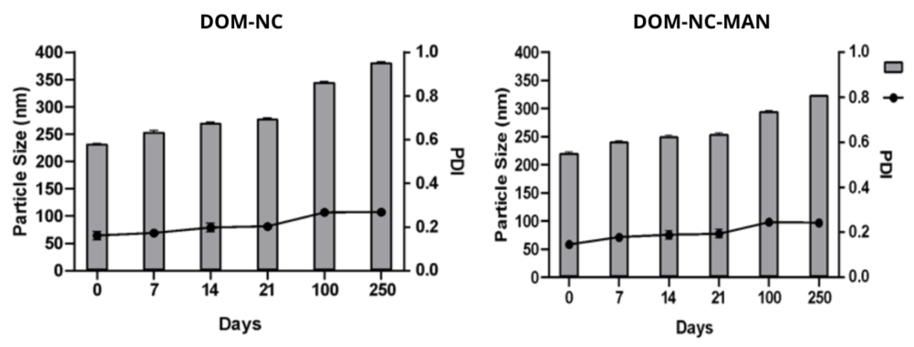
3.1.2. Storage Stability
3.1.3. Solid State Characterization
Infrared Spectroscopy
X-ray Analysis
Differential Scanning Calorimetry and Thermogravimetric Analysis
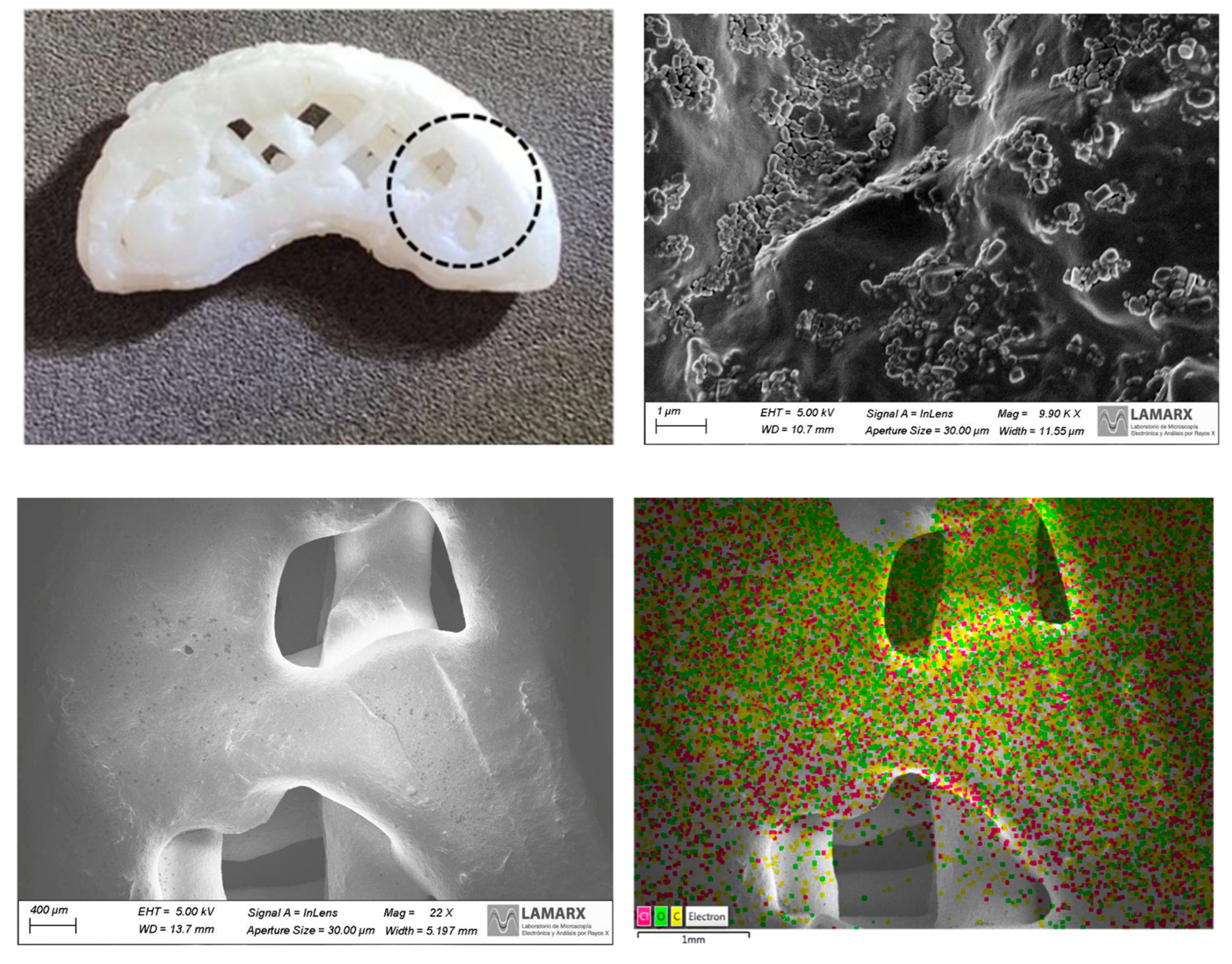
Scanning Electron Microscopy
3.1.4. Solubility Test
3.2. Ultra-Fast Ink Release
3.2.1. Ink Formulation
3.2.2. Ink pH
3.2.3. Solid State Characterization
Hot Stage Microscopy
Differential Scanning Calorimetry and Thermogravimetric Analysis
FT-IR Analysis
3.3. DOM-NC 3D Printlets
3.3.1. Design of the Dosage Form
3.3.2. Printing of the SDF
3.3.3. Solid-State Characterization
Scanning Electron Microscopy
3.4. Disintegration Test
3.5. Dissolution Study
4. Conclusions
Author Contributions
Funding
Institutional Review Board Statement
Informed Consent Statement
Data Availability Statement
Acknowledgments
Conflicts of Interest
References
- Tullberg, S. Domperidone. In xPharm: The Comprehensive Pharmacology Reference; Enna, S.J., Bylund, D.B., Eds.; Elsevier: Amsterdam, The Netherlands, 2007; pp. 1–4. [Google Scholar] [CrossRef]
- Greenstein, G.R. The Merck Index: An Encyclopedia of Chemicals, Drugs, and Biologicals (14th Edition). Ref. Rev. 2007, 21, 40. [Google Scholar] [CrossRef]
- Ortiz, A.; Cooper, C.J.; Gomez, Y.; Sarosiek, I.; McCallum, R.W.; Alvarez, A. Cardiovascular Safety Profile and Clinical Experience With High-Dose Domperidone Therapy for Nausea and Vomiting. Am. J. Med. Sci. 2015, 349, 421–424. [Google Scholar] [CrossRef] [PubMed]
- Daihom, B.A.; Bendas, E.R.; Mohamed, M.I.; Badawi, A.A. Domperidone resinate complex as new formulation for gastroretentive drug delivery. J. Drug Deliv. Sci. Technol. 2020, 58, 101868. [Google Scholar] [CrossRef]
- McGuckin, M.B.; Wang, J.; Ghanma, R.; Qin, N.; Palma, S.D.; Donnelly, R.F.; Paredes, A.J. Nanocrystals as a master key to deliver hydrophobic drugs via multiple administration routes. J. Control. Release 2022, 345, 334–353. [Google Scholar] [CrossRef] [PubMed]
- Reddymasu, S.C.; Soykan, I.; McCallum, R.W. Domperidone: Review of Pharmacology and Clinical Applications in Gastroenterology. Off. J. Am. Coll. Gastroenterol. ACG 2007, 102, 2036. [Google Scholar] [CrossRef]
- Saritha, D.; Sathish, D.; Rao, Y.M. Formulation and Evaluation of Gastroretentive Floating Tablets of Domperidone Maleate. J. Appl. Pharm. Sci. 2012, 2, 68–73. [Google Scholar]
- Aboutaleb, A.E.; Abdel-Rahman, S.I.; Ahmed, M.O.; Younis, M.A. Design and evaluation of domperidone sublingual tablets. Int. J. Pharm. Pharm. Sci. 2016, 8, 195–201. [Google Scholar]
- Rao, N.G.R.; Shravani, B.; Reddy, M.S. Overview on Buccal Drug Delivery Systems. J. Pharm. Sci. 2013, 5, 80. [Google Scholar]
- Narang, N.; Sharma, J. Sublingual mucosa as a route for systemic drug delivery. Int. J. Pharm. Pharm. Sci. 2011, 3, 18–22. [Google Scholar]
- Aboutaleb, A.; Abdel-Rahman, S.; Ahmed, M.; Younis, M. Improvement of Domperidone Solubility and Dissolution Rate by Dispersion in Various Hydrophilic Carriers. J. Appl. Pharm. Sci. 2016, 6, 133–139. [Google Scholar] [CrossRef]
- Paredes, A.J.; Camacho, N.M.; Schofs, L.; Dib, A.; del Pilar Zarazaga, M.; Litterio, N.; Allemandi, D.A.; Bruni, S.S.; Lanusse, C.; Palma, S.D. Ricobendazole nanocrystals obtained by media milling and spray drying: Pharmacokinetic comparison with the micronized form of the drug. Int. J. Pharm. 2020, 585, 119501. [Google Scholar] [CrossRef] [PubMed]
- Lopez-Vidal, L.; Real, J.P.; Real, D.A.; Camacho, N.; Kogan, M.J.; Paredes, A.J.; Palma, S.D. Nanocrystal-based 3D-printed tablets: Semi-solid extrusion using melting solidification printing process (MESO-PP) for oral administration of poorly soluble drugs. Int. J. Pharm. 2022, 611, 121311. [Google Scholar] [CrossRef]
- Beckett, A.H.; Hossie, R.D. Buccal Absorption of Drugs. In Concepts in Biochemical Pharmacology; Brodie, B.B., Gillette, J.R., Ackerman, H.S., Eds.; Springer: Berlin/Heidelberg, Germany, 1971; pp. 25–46. [Google Scholar] [CrossRef]
- Formica, M.L.; Awde Alfonso, H.G.; Paredes, A.J.; Melian, M.E.; Camacho, N.M.; Faccio, R.; Tártara, L.I.; Palma, S.D. Development of Triamcinolone Acetonide Nanocrystals for Ocular Administration. Pharmaceutics 2023, 15, 683. [Google Scholar] [CrossRef]
- Malamatari, M.; Taylor, K.M.G.; Malamataris, S.; Douroumis, D.; Kachrimanis, K. Pharmaceutical nanocrystals: Production by wet milling and applications. Drug Discov. Today 2018, 23, 534–547. [Google Scholar] [CrossRef] [PubMed]
- Duchêne, D.; Ponchel, G. Bioadhesion of solid oral dosage forms, why and how? Eur. J. Pharm. Biopharm. 1997, 44, 15–23. [Google Scholar] [CrossRef]
- Liu, J.; Tu, L.; Cheng, M.; Feng, J.; Jin, Y. Mechanisms for oral absorption enhancement of drugs by nanocrystals. J. Drug Deliv. Sci. Technol. 2020, 56, 101607. [Google Scholar] [CrossRef]
- Lopez-Vidal, L.; Real, D.A.; Paredes, A.J.; Real, J.P.; Daniel Palma, S. 3D-Printed Nanocrystals for Oral Administration of the Drugs. In Drug Delivery Using Nanomaterials; Shahzad, Y., Rizvi, S.A.A., Yousaf, A.M., Eds.; Taylor and Francis: Germantown, NY, USA, 2022. [Google Scholar] [CrossRef]
- Hotze, E.M.; Phenrat, T.; Lowry, G.V. Nanoparticle Aggregation: Challenges to Understanding Transport and Reactivity in the Environment. J. Environ. Qual. 2010, 39, 1909–1924. [Google Scholar] [CrossRef]
- Khaled, S.A.; Burley, J.C.; Alexander, M.R.; Roberts, C.J. Desktop 3D printing of controlled release pharmaceutical bilayer tablets. Int. J. Pharm. 2014, 461, 105–111. [Google Scholar] [CrossRef]
- Erzengin, S.; Guler, E.; Eser, E.; Polat, E.B.; Gunduz, O.; Cam, M.E. In vitro and in vivo evaluation of 3D printed sodium alginate/polyethylene glycol scaffolds for sublingual delivery of insulin: Preparation, characterization, and pharmacokinetics. Int. J. Biol. Macromol. 2022, 204, 429–440. [Google Scholar] [CrossRef]
- Abdella, S.; Afinjuomo, F.; Song, Y.; Upton, R.; Garg, S. 3D printed bilayer mucoadhesive buccal film of estradiol: Impact of design on film properties, release kinetics and predicted in vivo performance. Int. J. Pharm. 2022, 628, 122324. [Google Scholar] [CrossRef]
- Charoenying, T.; Patrojanasophon, P.; Ngawhirunpat, T.; Rojanarata, T.; Akkaramongkolporn, P.; Opanasopit, P. Three-dimensional (3D)-printed devices composed of hydrophilic cap and hydrophobic body for improving buoyancy and gastric retention of domperidone tablets. Eur. J. Pharm. Sci. 2020, 155, 105555. [Google Scholar] [CrossRef] [PubMed]
- Chai, X.; Chai, H.; Wang, X.; Yang, J.; Li, J.; Zhao, Y.; Cai, W.; Tao, T.; Xiang, X. Fused Deposition Modeling (FDM) 3D Printed Tablets for Intragastric Floating Delivery of Domperidone. Sci. Rep. 2017, 7, 2829. [Google Scholar] [CrossRef] [PubMed]
- Skowyra, J.; Pietrzak, K.; Alhnan, M.A. Fabrication of extended-release patient-tailored prednisolone tablets via fused deposition modelling (FDM) 3D printing. Eur. J. Pharm. Sci. 2015, 68, 11–17. [Google Scholar] [CrossRef] [PubMed]
- Real, J.P.; Barberis, M.E.; Camacho, N.M.; Sánchez Bruni, S.; Palma, S.D. Design of novel oral ricobendazole formulation applying melting solidification printing process (MESO-PP): An innovative solvent-free alternative method for 3D printing using a simplified concept and low temperature. Int. J. Pharm. 2020, 587, 119653. [Google Scholar] [CrossRef]
- Barberis, M.E.; Palma, S.D.; Gonzo, E.E.; Bermúdez, J.M.; Lorier, M.; Ibarra, M.; Real, J.P. Mathematical and Pharmacokinetic Approaches for the Design of New 3D Printing Inks Using Ricobendazole. Pharm. Res. 2022, 39, 2277–2290. [Google Scholar] [CrossRef]
- Paredes, A.J.; Llabot, J.M.; Sánchez Bruni, S.; Allemandi, D.; Palma, S.D. Self-dispersible nanocrystals of albendazole produced by high pressure homogenization and spray-drying. Drug Dev. Ind. Pharm. 2016, 42, 1564–1570. [Google Scholar] [CrossRef]
- US Pharmacopeia (USP). Available online: https://www.usp.org/ (accessed on 8 December 2022).
- Ndlovu, S.T.; Ullah, N.; Khan, S.; Ramharack, P.; Soliman, M.; de Matas, M.; Shahid, M.; Sohail, M.; Imran, M.; Shah, S.W.A.; et al. Domperidone nanocrystals with boosted oral bioavailability: Fabrication, evaluation and molecular insight into the polymer-domperidone nanocrystal interaction. Drug Deliv. Transl. Res. 2019, 9, 284–297. [Google Scholar] [CrossRef]
- Al-lami, M.S.; Oudah, M.H.; Rahi, F.A. Preparation and characterization of domperidone nanoparticles for dissolution improvement. Iraqi J. Pharm. Sci. 2018, 27, 39–52. [Google Scholar] [CrossRef]
- Choi, J.-Y.; Yoo, J.Y.; Kwak, H.-S.; Uk Nam, B.; Lee, J. Role of polymeric stabilizers for drug nanocrystal dispersions. Curr. Appl. Phys. 2005, 5, 472–474. [Google Scholar] [CrossRef]
- Fontana, F.; Figueiredo, P.; Zhang, P.; Hirvonen, J.T.; Liu, D.; Santos, H.A. Production of pure drug nanocrystals and nano co-crystals by confinement methods. Adv. Drug Deliv. Rev. 2018, 131, 3–21. Available online: https://www.sciencedirect.com/science/article/pii/S0169409X18300838?casa_token=Nhbq4kDi6ksAAAAA:YlxOVCgXHxJ9ZI9nsuk_ZHMs9CfxopSAyCx1UEke3ouyXi2kMczJXNIgJeJjzhWOxpX-iuS9qQ (accessed on 8 December 2022). [CrossRef]
- Voorhees, P.W. The theory of Ostwald ripening. J. Stat. Phys. 1985, 38, 231–252. [Google Scholar] [CrossRef]
- Al-Saif, F.A.; El-Habeeb, A.A.; Refat, M.S.; Eldaroti, H.H.; Adam, A.M.A.; Fetooh, H.; Saad, H.A. Chemical and physical properties of the charge transfer complexes of domperidone antiemetic agent with π-acceptors. J. Mol. Liq. 2019, 293, 111517. [Google Scholar] [CrossRef]
- Pontremoli, C.; Boffito, M.; Fiorilli, S.; Laurano, R.; Torchio, A.; Bari, A.; Tonda-Turo, C.; Ciardelli, G.; Vitale-Brovarone, C. Hybrid injectable platforms for the in situ delivery of therapeutic ions from mesoporous glasses. Chem. Eng. J. 2018, 340, 103–113. [Google Scholar] [CrossRef]
- Willart, J.F.; Descamps, M. Solid state amorphization of pharmaceuticals. Mol. Pharm. 2008, 5, 905–920. Available online: https://pubs.acs.org/doi/full/10.1021/mp800092t (accessed on 8 December 2022). [CrossRef]
- Kluge, J.; Muhrer, G.; Mazzotti, M. High pressure homogenization of pharmaceutical solids. J. Supercrit. Fluids 2012, 66, 380–388. [Google Scholar] [CrossRef]
- Rowe, R.C.; Sheskey, P.J.; Quinn, M.E. Handbook of Pharmaceutical Excipients; Pharmaceutical Press: London, UK, 2008. [Google Scholar]
- Ginés, J.M.; Arias, M.J.; Rabasco, A.M.; Novák, C.; Ruiz-Conde, A.; Sánchez-Soto, P.J. Thermal characterization of polyethylene glycols applied in the pharmaceutical technology using differential scanning calorimetry and hot stage microscopy. J. Therm. Anal. Calorim. 2005, 46, 291–304. [Google Scholar] [CrossRef]
- Bácskay, I.; Ujhelyi, Z.; Fehér, P.; Arany, P. The Evolution of the 3D-Printed Drug Delivery Systems: A Review. Pharmaceutics 2022, 14, 1312. [Google Scholar] [CrossRef]
- Song, Q.; Shen, C.; Shen, B.; Lian, W.; Liu, X.; Dai, B.; Yuan, H. Development of a fast dissolving sublingual film containing meloxicam nanocrystals for enhanced dissolution and earlier absorption. J. Drug Deliv. Sci. Technol. 2018, 43, 243–252. [Google Scholar] [CrossRef]
- Zayed, G.M.; Rasoul, S.A.-E.; Ibrahim, M.A.; Saddik, M.S.; Alshora, D.H. In vitro and in vivo characterization of domperidone-loaded fast dissolving buccal films. Saudi Pharm. J. 2020, 28, 266–273. [Google Scholar] [CrossRef]


| Time (min) | Z-Average (nm) | PI |
|---|---|---|
| 0 | 11,980 ± 132 | 0.83 |
| 30 | 248.8 ± 1.4 | 0.147 |
| 60 | 200.6 ± 2.0 | 0.149 |
| 90 | 185.8 ± 0.7 | 0.156 |
| 120 | 183.2 ± 0.9 | 0.14 |
| NC | Cryoprotector | Particle Size (nm) | PI | Performance (%) |
|---|---|---|---|---|
| 1 | - | 233.3 ± 0.6 | 0.161 ± 0.01 | 93.55 |
| 2 | Mannitol (3% P/V) | 221.2 ± 1.8 | 0.145 ± 0.01 | 90.2 |
| 3 | Sucrose (3% P/V) | 222.5 ± 1.8 | 0.225 ± 0.01 | 87.9 |
| Medium | DOM | PM | DOM-NCs |
|---|---|---|---|
| Water | 0.008 ± 0.002 mg/mL | 0.015 ± 0.007 mg/mL | 0.031 ± 0.003 mg/mL |
| Simulated Saliva | 0.0026 ± 0.0006 mg/mL | 0.014 ± 0.001 mg/mL | 0.042 ± 0.003 mg/mL |
| Components | Proportion of DOM-NC-3D Components (%) | Components | Proportion of 1 DOM-3D Ink Components (%) | Proportion of 2 DOM-3D Ink Components (%) |
|---|---|---|---|---|
| CCS | 5 | CCS | 5 | 5 |
| SSG | 5 | SSG | 5 | 5 |
| SC | 3 | SC | 3 | 3 |
| PEG 1500 | 70 | PEG 1500 | 70 | 70 |
| Propylene glycol | 7 | Propylene glycol | 7 | 7 |
| (DOM-NCs) DOM | 7.5 | DOM | 10 | 7.5 |
| P407 | 2.4 | P407 | 0 | 2.4 |
| SLS | 0.1 | SLS | 0 | 0.1 |
| Form | Weight (mg) | Dimensions (mm) | Volume (mm3) | DOM (mg) | |||
|---|---|---|---|---|---|---|---|
| A | B | C | D | ||||
| A | 343.5 | 19.65 | 9.6 | 7.65 | 2.9 | 824.2 | 25.7 |
| B | 322.8 | 18.05 | 9.4 | 7.1 | 2.9 | 724.3 | 24.2 |
| C | 266.8 | 15.9 | 9 | 6.5 | 2.75 | 563.8 | 20 |
| D | 167.3 | 13.5 | 7.3 | 5.4 | 2.6 | 372.8 | 12.5 |
| E | 132.5 | 12.4 | 6.6 | 5.3 | 2.4 | 295.6 | 9.9 |
| F | 101.3 | 11.7 | 6.35 | 5 | 1.75 | 194.1 | 7.6 |
Disclaimer/Publisher’s Note: The statements, opinions and data contained in all publications are solely those of the individual author(s) and contributor(s) and not of MDPI and/or the editor(s). MDPI and/or the editor(s) disclaim responsibility for any injury to people or property resulting from any ideas, methods, instructions or products referred to in the content. |
© 2023 by the authors. Licensee MDPI, Basel, Switzerland. This article is an open access article distributed under the terms and conditions of the Creative Commons Attribution (CC BY) license (https://creativecommons.org/licenses/by/4.0/).
Share and Cite
Lopez-Vidal, L.; Paredes, A.J.; Palma, S.D.; Real, J.P. Design and Development of Sublingual Printlets Containing Domperidone Nanocrystals Using 3D Melting Solidification Printing Process (MESO-PP). Pharmaceutics 2023, 15, 1459. https://doi.org/10.3390/pharmaceutics15051459
Lopez-Vidal L, Paredes AJ, Palma SD, Real JP. Design and Development of Sublingual Printlets Containing Domperidone Nanocrystals Using 3D Melting Solidification Printing Process (MESO-PP). Pharmaceutics. 2023; 15(5):1459. https://doi.org/10.3390/pharmaceutics15051459
Chicago/Turabian StyleLopez-Vidal, Lucía, Alejandro J. Paredes, Santiago Daniel Palma, and Juan Pablo Real. 2023. "Design and Development of Sublingual Printlets Containing Domperidone Nanocrystals Using 3D Melting Solidification Printing Process (MESO-PP)" Pharmaceutics 15, no. 5: 1459. https://doi.org/10.3390/pharmaceutics15051459