
Discovery of Novel Thiophene/Hydrazones: In Vitro and In Silico Studies against Pancreatic Cancer
 , and
, and
Abstract
:1. Introduction
2. Materials and Methods
2.1. General
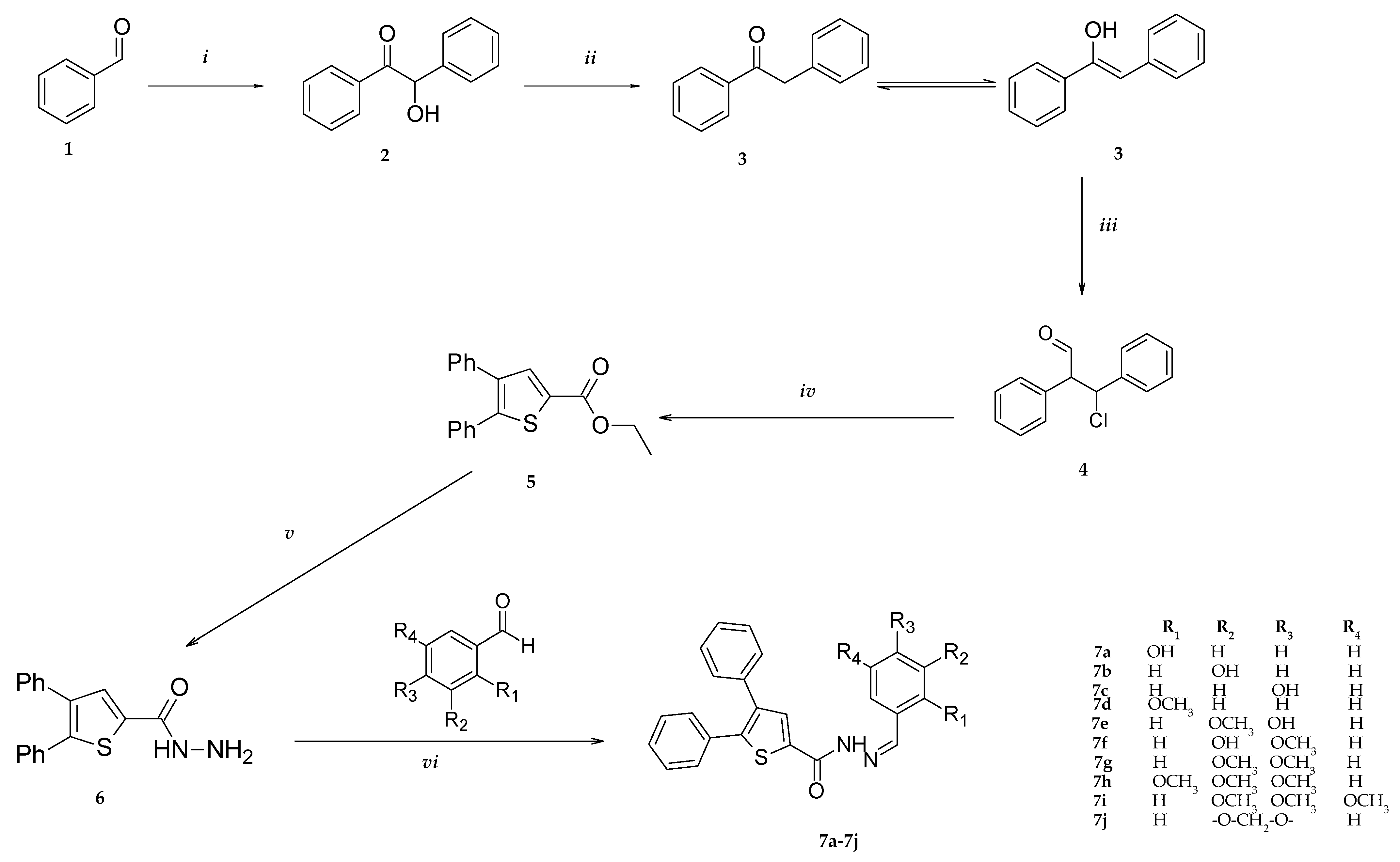
2.2. Synthesis
Hydrazone Synthesis
2.3. Cell Culture Studies
2.3.1. 2D Anticancer Activity
2.3.2. Cell Viability
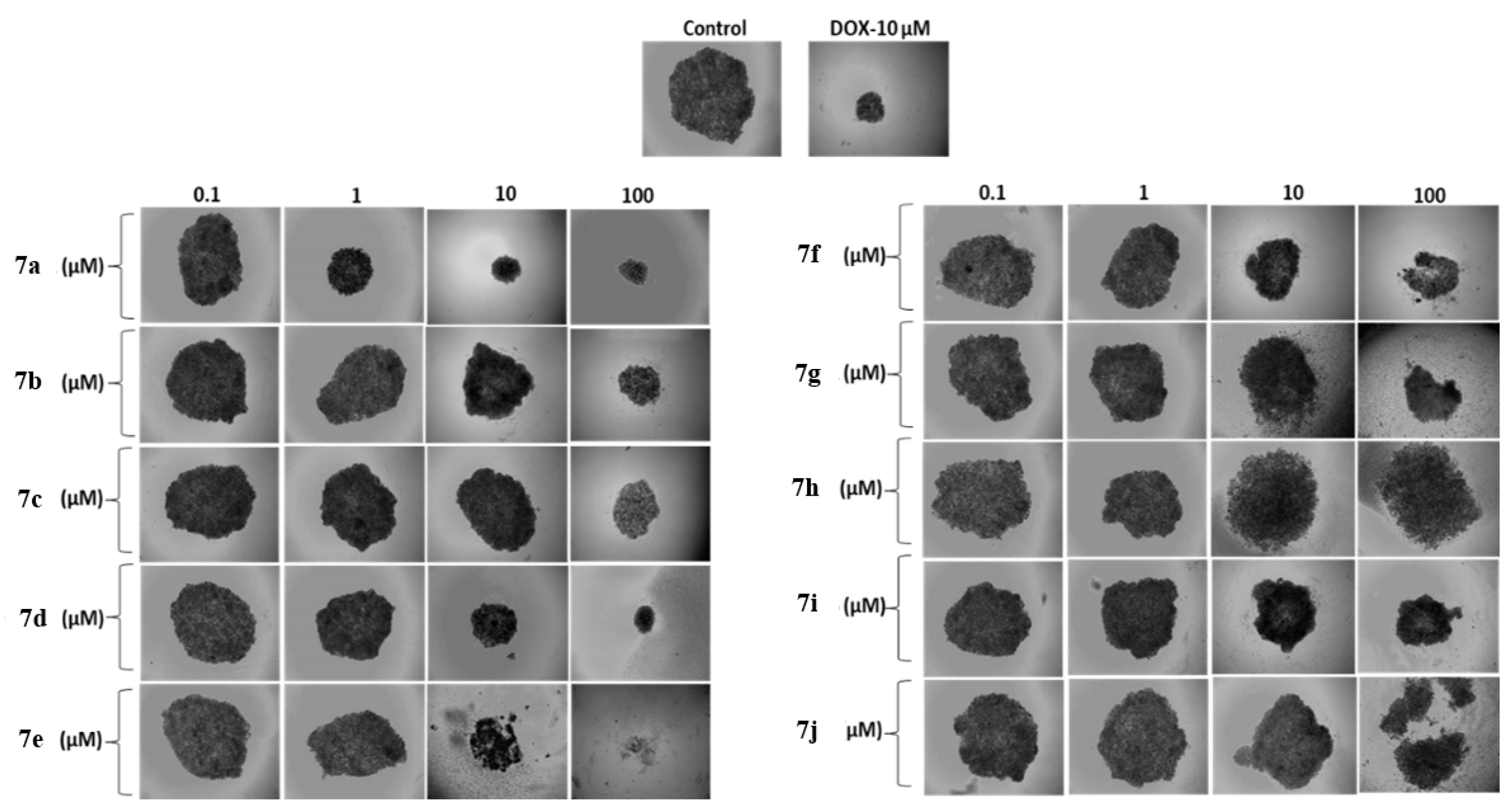
2.3.3. 3D Spheroid Formation/Growth Assay
2.4. Enzyme Inhibition Assay
2.5. Molecular Docking Study
3. Results and Discussion
3.1. Chemistry
3.2. 2D Anticancer Activity
3.3. Anticancer Activity in 3D Cell Cultures (Spheroids)
3.4. Enzyme Inhibition Results
3.5. Molecular Docking Study
3.5.1. Molecular Docking into COX-2
3.5.2. Molecular Docking into 5-LOX
4. Conclusions
Supplementary Materials
Author Contributions
Funding
Institutional Review Board Statement
Informed Consent Statement
Data Availability Statement
Conflicts of Interest
References
- Zhao, Z.; Liu, W. Pancreatic Cancer: A Review of Risk Factors, Diagnosis, and Treatment. Technol. Cancer Res. Treat. 2020, 19, 1533033820962117. [Google Scholar] [CrossRef] [PubMed]
- Bocci, G.; Danesi, R.; Marangoni, G.; Fioravanti, A.; Boggi, U.; Esposito, I.; Fasciani, A.; Boschi, E.; Campani, D.; Bevilacqua, G.; et al. Antiangiogenic versus cytotoxic therapeutic approaches to human pancreas cancer: An experimental study with a vascular endothelial growth factor receptor-2 tyrosine kinase inhibitor and gemcitabine. Eur. J. Pharmacol. 2004, 498, 9–18. [Google Scholar] [CrossRef] [PubMed]
- Goral, V. Pancreatic Cancer: Pathogenesis and Diagnosis. Asian Pac. J. Cancer Prev. 2015, 16, 5619–5624. [Google Scholar] [CrossRef] [PubMed]
- Wang, H.; Wang, J.; Wang, J.; Cui, W.; Sun, M.; Liu, Y. Relationships of pain in pancreatic cancer patients with pathological stage and expressions of NF-κB and COX-2. J. BUON Off. J. Balk. Union Oncol. 2020, 25, 448–453. [Google Scholar] [PubMed]
- Ding, X.; Zhu, C.; Qiang, H.; Zhou, X.; Zhou, G. Enhancing antitumor effects in pancreatic cancer cells by combined use of COX-2 and 5-LOX inhibitors. Biomed. Pharmacother. 2011, 65, 486–490. [Google Scholar] [CrossRef]
- Hill, R.; Li, Y.; Tran, L.M.; Dry, S.; Calvopina, J.H.; Garcia, A.; Kim, C.; Wang, Y.; Donahue, T.R.; Herschman, H.R.; et al. Cell intrinsic role of COX-2 in pancreatic cancer development. Mol. Cancer Ther. 2012, 11, 2127–2137. [Google Scholar] [CrossRef]
- Yip-Schneider, M.T.; Barnard, D.S.; Billings, S.D.; Cheng, L.; Heilman, D.K.; Lin, A.; Marshall, S.J.; Crowell, P.L.; Marshall, M.S.; Sweeney, C.J. Cyclooxygenase-2 expression in human pancreatic adenocarcinomas. Carcinogenesis 2000, 21, 139–146. [Google Scholar] [CrossRef]
- Şahin, Z.; Kalkan, M.; Berk, B.; Yurttaş, L.; Bender, C.; Kaleli, S.N.B.; Demirayak, Ş. Synthesis, characterization, COX1/2 inhibition and molecular modeling studies on novel 2-thio-diarylimidazoles. Turk. J. Chem. 2021, 45, 15. [Google Scholar] [CrossRef]
- Yurttaş, L.; Şahin, Z.; Çiftçi, G.A.; Temel, H.E.; Demirayak, S. Synthesis of novel 3, 5, 6-trisubstituted triazine derivatives and their biological activity evaluation as potential antitumor and anti-inflammatory agents. Acta Pharm. Sci. 2016, 54, 83–92. [Google Scholar] [CrossRef]
- Sahin, Z.; Özhan, Y.; Sipahi, H.; Biltekin, S.N.; Yurttaş, L.; Berk, B.; Demirayak, Ş. Novel benzofurane-pyrazole derivatives with anti-inflammatory, cyclooxygenase inhibitory and cytotoxicity evaluation. Z. Für Nat. C. 2022, 77, 279–285. [Google Scholar] [CrossRef]
- Rao, C.V.; Janakiram, N.B.; Madka, V.; Devarkonda, V.; Brewer, M.; Biddick, L.; Lightfoot, S.; Steele, V.E.; Mohammed, A. Simultaneous targeting of 5-LOX-COX and EGFR blocks progression of pancreatic ductal adenocarcinoma. Oncotarget 2015, 6, 33290–33305. [Google Scholar] [CrossRef] [PubMed]
- Abdelgawad, M.A.; Labib, M.B.; Abdel-Latif, M. Pyrazole-hydrazone derivatives as anti-inflammatory agents: Design, synthesis, biological evaluation, COX-1,2/5-LOX inhibition and docking study. Bioorganic Chem. 2017, 74, 212–220. [Google Scholar] [CrossRef] [PubMed]
- Sardar, A.; Abid, O.R.; Daud, S.; Shah, B.A.; Shahid, W.; Ashraf, M.; Fatima, M.; Ezzine, S.; Wadood, A.; Shareef, A.; et al. Identification of novel diclofenac acid and naproxen bearing hydrazones as 15-LOX inhibitors: Design, synthesis, in vitro evaluation, cytotoxicity, and in silico studies. Arab. J. Chem. 2022, 15, 104300. [Google Scholar] [CrossRef]
- Masashi, H.; Yoichi, K.; Nobuo, M.; Shigeyuki, N. Pharmaceutical Compositions Containing Novel Amidopropionic Acids as Sphingosine-1-Phosphate (S1P) Receptor Agonists. Patent No. JP2009040702, 26 February 2009. China. Available online: https://scifinder-n.cas.org/searchDetail/reference/6384934f5aefd615b687e831/referenceDetails (accessed on 30 March 2023).
- Čeponytė, U.; Paškevičiūtė, M.; Petrikaitė, V. Comparison of NSAIDs activity in COX-2 expressing and non-expressing 2D and 3D pancreatic cancer cell cultures. Cancer Manag. Res. 2018, 10, 1543. [Google Scholar] [CrossRef]
- Guzelmeric, E.; Reis, R.; Sen, N.B.; Celik, C.; Ozhan, Y.; Petrikaite, V.; Sipahi, H.; Aydın, A.; Yesilada, E. Insights into the Anti-Inflammatory, Analgesic, and Anti-Cancer Potentials of the Standardized Extracts from Three cistus L. Species. HERMED-D-22-00448. 2022. Available online: https://ssrn.com/abstract=4122153 (accessed on 30 April 2023).
- COX2 Inhibitor Screening Kit. Available online: https://www.abcam.com/products/assay-kits/cox2-inhibitor-screening-kit-fluorometric-ab283401.html (accessed on 30 April 2023).
- COX1 Inhibitor Assay Kit. Available online: https://www.abcam.com/cyclooxygenase-1-cox1-inhibitor-assay-kit-fluorometric-ab204698.html (accessed on 30 April 2023).
- ALOX5 Assay Kit. Available online: https://www.mybiosource.com/human-assay-kits/5-lipoxygenase/846911 (accessed on 30 April 2023).
- Protein Data Bank. Available online: http://www.rcsb.org/ (accessed on 30 April 2023).
- MAKE Receptor 3.2.0.2: OpenEye Scientific Software, Santa Fe, NM. Available online: https://www.eyesopen.com/ (accessed on 30 April 2023).
- FRED 3.2.0.2: OpenEye Scientific Software, Santa Fe, NM. Available online: https://www.eyesopen.com/ (accessed on 30 April 2023).
- McGann, M. FRED pose prediction and virtual screening accuracy. J. Chem. Inf. Model 2011, 51, 578–596. [Google Scholar] [CrossRef] [PubMed]
- McGann, M. FRED and HYBRID docking performance on standardized datasets. J. Comput. Aided Mol. Des. 2012, 26, 897–906. [Google Scholar] [CrossRef]
- Carugo, O.; Pongor, S. A normalized root-mean-spuare distance for comparing protein three-dimensional structures. Protein Sci. 2001, 10, 1470–1473. [Google Scholar] [CrossRef]
- Coşkun, G.P.; Aklamuz, A.; İnce, U.; Ülgen, M. Synthesis, Structure Elucidation and Biological Activity of New Hybrid Hydrazone-Amide Compounds. Cumhur. Sci. J. 2022, 43, 384–390. [Google Scholar] [CrossRef]
- Šermukšnytė, A.; Kantminienė, K.; Jonuškienė, I.; Tumosienė, I.; Petrikaitė, V. The Effect of 1, 2, 4-Triazole-3-thiol Derivatives Bearing Hydrazone Moiety on Cancer Cell Migration and Growth of Melanoma, Breast, and Pancreatic Cancer Spheroids. Pharmaceuticals 2022, 15, 1026. [Google Scholar] [CrossRef]
- Mohsin, N.U.A.; Aslam, S.; Ahmad, M.; Irfan, M.; Al-Hussain, S.A.; Zaki, M.E. Cyclooxygenase-2 (COX-2) as a Target of Anticancer Agents: A Review of Novel Synthesized Scaffolds Having Anticancer and COX-2 Inhibitory Potentialities. Pharmaceuticals 2022, 15, 1471. [Google Scholar] [CrossRef]
- Fischer, A.S.; Metzner, J.; Steinbrink, S.D.; Ulrich, S.; Angioni, C.; Geisslinger, G.; Steinhilber, D.; Maier, T.J. 5-Lipoxygenase inhibitors induce potent anti-proliferative and cytotoxic effects in human tumour cells independently of suppression of 5-lipoxygenase activity. Br. J. Pharmacol. 2010, 161, 936–949. [Google Scholar] [CrossRef] [PubMed]
- Goradel, N.H.; Najafi, M.; Salehi, E.; Farhood, B.; Mortezaee, K. Cyclooxygenase-2 in cancer: A review. J. Cell. Physiol. 2019, 234, 5683–5699. [Google Scholar] [CrossRef] [PubMed]
- Kahnt, A.S.; Angioni, C.; Göbel, T.; Hofmann, B.; Roos, J.; Steinbrink, S.D.; Rörsch, F.; Thomas, D.; Geisslinger, G.; Zacharowski, K.; et al. Inhibitors of human 5-lipoxygenase potently interfere with prostaglandin transport. Front. Pharmacol. 2022, 12, 782584. [Google Scholar] [CrossRef]
- Gilbert, N.C.; Gerstmeier, J.; Schexnaydre, E.E.; Börner, F.; Garscha, U.; Neau, D.B.; Werz, O.; Newcomer, M.E. Structural and mechanistic insights into 5-lipoxygenase inhibition by natural products. Nat. Chem. Biol. 2020, 16, 783–790. [Google Scholar] [CrossRef] [PubMed]


| Tested Compound | IC50 (µM) |
|---|---|
| 7a | 7.51 ± 0.08 |
| 7b | 9.09 ± 1.11 |
| 7c | 42.77 ± 3.59 |
| 7d | 9.09 ± 1.04 |
| 7e | >100 µM |
| 7f | 4.86 ± 0.19 |
| 7g | 24.23 ± 3.76 |
| 7h | >100 µM |
| 7i | >100 µM |
| 7j | 9.65 ± 0.22 |
| DOX | 0.35 ± 0.18 |
| Tested Compound | IC50 (µM) |
|---|---|
| 7a | 0.88 ± 0.06 |
| 7d | >100 µM |
| 7f | 0.67 ± 0.13 |
| DOX | 0.11 ± 0.08 |
| Sample Concentrations | Spheroid Diameter (Square Millimeters) | ||
|---|---|---|---|
| Day 0 | Day 9 | ||
| Control | - | 1111.8 ± 95.5 | 7240.2 ± 37.1 |
| DOX | 10 µM | 1194.0 ± 63.3 | 910.0 ± 29.3 |
| 7a | |||
| 0.1 µM | 1156.5 ± 14.2 | 5050.5 ± 36.9 | |
| 1 µM | 1186.5 ± 91.9 | 1755.0 ± 58.9 | |
| 10 µM | 1176.0 ± 31.1 | 636.0 ± 12.7 | |
| 100 µM | 1183.0 ± 36.1 | 432.0 ± 17.4 | |
| 7b | |||
| 0.1 µM | 1193.0 ± 13.1 | 5922.5 ± 39.8 | |
| 1 µM | 1185.5 ± 82.7 | 6461.5 ± 59.6 | |
| 10 µM | 969.3 ± 28.9 | 4192.0 ± 44.8 | |
| 100 µM | 644.7 ± 26.9 | 1050.5 ± 18.9 | |
| 7c | |||
| 0.1 µM | 1118.0 ± 31.1 | 5415.0 ± 12.7 | |
| 1 µM | 1168.0 ± 52.7 | 6174.0 ± 106.1 | |
| 10 µM | 989.5 ± 54.1 | 3729.5 ± 12.8 | |
| 100 µM | 940.5 ± 33.9 | 2207.0 ± 47.1 | |
| 7d | |||
| 0.1 µM | 1127.5 ± 36.1 | 4911.0 ± 117.4 | |
| 1 µM | 1211.0 ± 17.9 | 6312.5 ± 24.8 | |
| 10 µM | 1297.5 ± 55.1 | 1708.0 ± 87.1 | |
| 100 µM | 974.0 ± 32.2 | 445.7 ± 64.1 | |
| 7e | |||
| 0.1 µM | 1181.5 ± 33.2 | 5607.5 ± 44.55 | |
| 1 µM | 1153.0 ± 73.5 | 6396.0 ± 16.1 | |
| 10 µM | - | - | |
| 100 µM | - | - | |
| 7f | |||
| 0.1 µM | 1110.0 ± 113.1 | 5823.0 ± 39.8 | |
| 1 µM | 1108.0 ± 17.6 | 4336.0 ± 48.1 | |
| 10 µM | 1215.0 ± 12.7 | 2806.0 ± 31.5 | |
| 100 µM | - | - | |
| 7g | |||
| 0.1 µM | 1222.5 ± 28.73 | 5969.0 ± 59.6 | |
| 1 µM | 985.5 ± 65.7 | 3318.0 ± 35.4 | |
| 10 µM | - | - | |
| 100 µM | - | - | |
| 7h | |||
| 0.1 µM | 1125.5 ± 28.9 | 5215 ± 18.3 | |
| 1 µM | 1220.5 ± 31.5 | 6682.5 ± 7.1 | |
| 10 µM | 1192.0 ± 24.7 | 8165.5 ±18.1 | |
| 100 µM | 942.0 ± 58.9 | 7936.5 ± 48.6 | |
| 7i | |||
| 0.1 µM | 1135.5 ± 2.5 | 5523.0 ± 15.2 | |
| 1 µM | 943.0 ± 12.1 | 3539.0 ± 25.4 | |
| 10 µM | 118.6 ± 24.7 | 2764.0 ± 37.9 | |
| 100 µM | 1023.0 ± 59.5 | 2776.5 ± 85.2 | |
| 7j | |||
| 0.1 µM | 1112.0 ± 35.4 | 6291.5 ± 45.87 | |
| 1 µM | 998.0 ± 11.5 | 5934.0 ± 96.3 | |
| 10 µM | 1068.0 ± 68.1 | 5217.4 ± 25.7 | |
| 100 µM | - | - | |
| Compound | COX-2 IC50 (µM) | COX-2 Percent of İnhibition | COX-1 IC50 (µM) | 5-LOX IC50 (µM) | 5-LOX Percent of İnhibition |
|---|---|---|---|---|---|
| 7a | - | 13.57 ± 0.38 | Not tested | 3.78 ± 0.39 | 64.38 ± 0.46 |
| 7b | - | 24.13 ± 1.55 | Not tested | 93.23 ± 9.89 | 25.77 ± 2.54 |
| 7c | 10.13 ± 0.25 | 51.97 ± 2.51 | ˃100 | 2.60 ± 0.11 | 55.05 ± 1.37 |
| 7d | - | 16.74 ± 3.36 | Not tested | - | 15.61 ± 0.66 |
| 7e | 13.86 ± 0.76 | 49.41±1.12 | ˃100 | 3.30 ± 0.07 | 60.69 ± 0.54 |
| 7f | 39.05 ± 3.41 | 38.41 ± 0.85 | ˃100 | 2.94 ± 0.20 | 60.56 ± 2.49 |
| 7g | - | 11.41 ± 3.40 | Not tested | - | 20.38 ± 1.98 |
| 7h | - | 8.35 ± 0.93 | Not tested | - | 19.87 ± 1.62 |
| 7i | - | 13.27 ± 0.73 | Not tested | - | 27.23 ± 0.64 |
| 7j | - | 16.64 ± 1.11 | Not tested | - | 36.10 ± 3.92 |
| Celecoxib | 0.07 ± 0.01 | 85.02 ± 0.31 | ˃100 | Not tested | Not tested |
| Zileuton | Not tested | Not tested | Not tested | 0.36 ± 0.10 | 64.85 ± 0.39 |
Disclaimer/Publisher’s Note: The statements, opinions and data contained in all publications are solely those of the individual author(s) and contributor(s) and not of MDPI and/or the editor(s). MDPI and/or the editor(s) disclaim responsibility for any injury to people or property resulting from any ideas, methods, instructions or products referred to in the content. |
© 2023 by the authors. Licensee MDPI, Basel, Switzerland. This article is an open access article distributed under the terms and conditions of the Creative Commons Attribution (CC BY) license (https://creativecommons.org/licenses/by/4.0/).
Share and Cite
Coskun, G.P.; Ozhan, Y.; Dobričić, V.; Bošković, J.; Reis, R.; Sipahi, H.; Sahin, Z.; Demirayak, S. Discovery of Novel Thiophene/Hydrazones: In Vitro and In Silico Studies against Pancreatic Cancer. Pharmaceutics 2023, 15, 1441. https://doi.org/10.3390/pharmaceutics15051441
Coskun GP, Ozhan Y, Dobričić V, Bošković J, Reis R, Sipahi H, Sahin Z, Demirayak S. Discovery of Novel Thiophene/Hydrazones: In Vitro and In Silico Studies against Pancreatic Cancer. Pharmaceutics. 2023; 15(5):1441. https://doi.org/10.3390/pharmaceutics15051441
Chicago/Turabian StyleCoskun, Goknil Pelin, Yagmur Ozhan, Vladimir Dobričić, Jelena Bošković, Rengin Reis, Hande Sipahi, Zafer Sahin, and Seref Demirayak. 2023. "Discovery of Novel Thiophene/Hydrazones: In Vitro and In Silico Studies against Pancreatic Cancer" Pharmaceutics 15, no. 5: 1441. https://doi.org/10.3390/pharmaceutics15051441