Article

Repurposing Disulfiram as an Antifungal Agent: Development of a New Disulfiram Vaginal Mucoadhesive Gel

by
Maria Lajarin-Reinares
1,2,*,
Iria Naveira-Souto
2,
Mireia Mallandrich
3,
Joaquim Suñer-Carbó
3,
Montserrat Llagostera Casas
1,
Maria Angels Calvo
4 and
Francisco Fernandez-Campos
5
1
Department of Genetics and Microbiology, Campus Microbiology Unit, Autonomous University of Barcelona, 08193 Bellaterra, Spain
2
R & D Development, Reig Jofre Laboratories, 08970 Sant Joan Despí, Spain
3
Department of Pharmacy and Pharmaceutical Technology and Physical Chemistry, School of Pharmacy and Food Sciences, University of Barcelona, 08028 Barcelona, Spain
4
Department of Animal Health and Anatomy, Faculty of Veterinary, Autonomous University of Barcelona, 08193 Bellaterra, Spain
5
R & D Department, Labiana Pharmaceuticals, 08757 Corbera Llobregat, Spain
*
Author to whom correspondence should be addressed.
Pharmaceutics 2023, 15(5), 1436; https://doi.org/10.3390/pharmaceutics15051436
Submission received: 21 March 2023 / Revised: 2 May 2023 / Accepted: 3 May 2023 / Published: 8 May 2023

Abstract

:
Alternative formulations need to be developed to improve the efficacy of treatments administered via the vaginal route. Mucoadhesive gels with disulfiram, a molecule that was originally approved as an antialcoholism drug, offer an attractive alternative to treat vaginal candidiasis. The aim of the current study was to develop and optimize a mucoadhesive drug delivery system for the local administration of disulfiram. Such formulations were composed of polyethylene glycol and carrageenan to improve the mucoadhesive and mechanical properties and to prolong the residence time in the vaginal cavity. Microdilution susceptibility testing showed that these gels had antifungal activity against Candida albicans, Candida parapsilosis, and Nakaseomyces glabratus. The physicochemical properties of the gels were characterized, and the in vitro release and permeation profiles were investigated with vertical diffusion Franz cells. After quantification, it was determined that the amount of the drug retained in the pig vaginal epithelium was sufficient to treat candidiasis infection. Together, our findings suggest that mucoadhesive disulfiram gels have the potential to be an effective alternative treatment for vaginal candidiasis.

Graphical Abstract

1. Introduction

Vaginal candidiasis (VC), often referred to as vulvovaginal candidiasis, is mainly caused by the Candida species and is the second most prevalent infection of the vaginal mucosa after bacterial vaginosis [1]. VC affects approximately 75% of women of reproductive age at least once during their lives, and it is estimated that 40–50% will experience recurrence and 8% will develop chronic VC [2,3,4].
Candida spp. are commensal fungi in the human microflora that are found in the lower genital tract in 10–20% of healthy women. Although part of the normal microflora, Candida can become opportunistic agents with a tendency to overgrow [2]. The most common Candida species involved in VC is C. albicans (85–90%), followed by Nakaseomyces glabratus (C. glabrata), C. tropicalis, Pichia kudriavzevii (C. krusei), and C. parapsilosis [5]. Approximately 25 to 40% of women who are culture-positive for Candida are asymptomatic. The natural evolution of asymptomatic colonization to symptomatic infection is not clear. There are multiple mechanisms that contribute to the colonization of Candida and its symptomatic infection, such as genetic host factors (polymorphisms in the blood group), hormones, the use of antibacterial agents, age, sexual activity, certain pathologies (diabetes mellitus and immunodepression), idiopathic causes, pregnancy, and an imbalance in the vaginal microbiota [4,6,7]. The most common clinical manifestations of VC are pruritus, hyperemia, vaginal discomfort, leucorrhea, burning, soreness, dyspareunia, and vaginal erythema [7]. Furthermore, colonization of the vaginal epithelium contributes to the deterioration of mucosal surfaces, facilitates the formation of yeast reservoirs, or becomes a source for future infections, leading to chronic infections and serious consequences (i.e., infertility and sterility) [8].
The treatment of noncomplicated VC involves the use of short-term local therapy (up to 3–7 days) or single-dose oral antimycotics, mainly those in the azole family, which is effective in 90% of cases. The most common locally applied azoles are clotrimazole, butoconazole, and miconazole. Fluconazole is usually given orally between 1 and 3 days [9]. Topical azoles may provide more immediate relief; however, some patients experience hypersensitivity, local itching, or burning [10]. For vaginal drug delivery, several pharmaceutical formulations are available for consumers, including creams, ovules, tablets, and gels [8,11,12]. These treatments exhibit some drawbacks, such as low permeation in the underlying epithelium (to remove reservoirs) and low residence time owing to the self-cleaning action of the vaginal tract. (It is recommended to be administered at night before lying in bed.) These factors could lead to low drug exposure and the development of resistance. Many studies have documented the ability of Candida to develop high-level resistance to antifungal therapy [13,14,15,16]. Resistance mechanisms include overexpression of or mutations to the target enzyme of azoles, lanosterol 14a-demethylase, and overexpression of ATP-binding cassette (ABC) efflux proteins [17]. Among ABC transporters, Cdr1p plays a key role in azole resistance in C. albicans [18].
Disulfiram (D) is a dithiocarbamate used to treat alcoholism that was approved by the US Food and Drug Administration (FDA) in 1951. D produces discomfort in patients who drink alcohol as a strategy for their dishabituation, inhibiting aldehyde dehydrogenase (ALDH), which results in the specific accumulation of acetaldehyde [19]. In addition, D is characterized by a low molecular weight (296 Da), logP value of 3.88, melting point under 200 °C (71.5 °C), and fewer than five hydrogen bond donors and acceptors with a strong ability to chelate metals [20]. In accordance with its physicochemical properties, a good permeation profile is expected. Different in vitro and in vivo studies have shown that D could be an active substance against scabies, lice, and bacteria [21,22]. Furthermore, the effectiveness of D against fungi and cancer was reported [23,24]. Different mechanisms of action have been proposed to explain the antimicrobial activity of D: metal ion chelation, which affects cell homeostasis, and the presence of reactive thiol groups, which can impact the functions of proteins by forming incorrect disulfide bonds [22]. D was found to be potentially useful by inhibiting the function of glycoprotein P (P-gp) and has also been shown to act as a modulator of other multidrug transporters, such as MRP1 and MRP4 [25]. Because of the functional similarities between Cdr1p and human P-gp, D is a potent modulator of Cdr1p [17]. Therefore, D is an attractive agent to develop for candidiasis treatment.
The aim of this work is to develop and optimize a mucoadhesive drug delivery system for the local administration of D. The in vitro antifungal efficacy of D was studied with different Candida species, and the mucoadhesion of the formulation was enhanced using polymers with different adhesive mechanisms. The proposed formulation represents an alternative to the commercially available treatments for VC due to the selected novel drug and mucoadhesive polymers that would increase the residence time of the treatment and yeast drug exposure.

2. Materials and Methods

2.1. Materials

Sodium chloride (0.35% w/v) (Quimivita, Barcelona, Spain), potassium hydroxide (0.14% w/v), calcium hydroxide (0.02% w/v), acetic acid (0.1% w/v) (Scharlab, S.L., Sentmenat, Spain), bovine serum albumin 0.002% (w/v) (Merck Life Science, S.L.U., Madrid, Spain), lactic acid (0.2% w/v) (Escuder, S.L., Rubi, Spain), glycerol 0.02% (w/v) (Caila & Pares, S.A., Barcelona, Spain), urea (0.04% w/v) (Quality Chemicals, S.L., Esparraguera, Spain) and glucose (0.5% w/v) (Quimidroga, S.A., Barcelona, Spain) were used to prepare simulated vaginal fluid (SVF) [26].
Mucoadhesive gels were produced with polyethylene glycol (PEG-90M) (Colorcon Limited, Kent, UK) and carrageenan (DuPont, Wilmington, DE, USA); benzylic alcohol (Scharlab, S.L., Sentmenat, Spain) was chosen as a preservative; and hydroxypropyl-beta-cyclodextrin (HPCD) (Pracofar, Martorelles, Spain) was used as an active solubilizing agent. D (Farchemia S.r.l., Treviglio, Italy) was the active pharmaceutical ingredient, and the selected solvent was water purified in-house. Sodium fluorescein (Scharlau, Barcelona, Spain) was used in the mucoadhesive study as a marker.

2.2. Antifungal Susceptibility Testing

The fungal strains used in this study were purchased from the Spanish Collection of Type Cultures (CECT). C. albicans CECT 1002 (which corresponds to ATCC 18804), C. parapsilosis CECT 10434 (MCYC 2031), and C. glabrata CECT 1448 (ATCC 2001) were used for the antifungal susceptibility test.
The minimum inhibitory concentration (MIC) of D was determined by the broth microdilution method in 96-well microplates according to guidelines M27-A3 of the Clinical and Laboratory Standards Institute (CLSI) standard [27]. A 24 h culture of Candida adjusted to a 0.5 McFarland was used to prepare the initial inoculum. A working suspension was generated by making a 1:100 dilution followed by a 1:20 dilution of the stock suspension with RPMI 1640 broth medium buffered with MOPS (3-(N morpholino)propanesulfonic acid) (Thermo Fisher Scientific, Waltham, MA, USA) at pH 7.0. Stock solutions (10 mg/mL) of D were prepared in ethanol, and D was tested from 64 (the highest soluble concentration) to 0.125 µg/mL. The final ethanol concentration was below 0.5% to not alter yeast growth. The negative control was the respective medium, and a control with the same ethanol concentration as that in the samples with D was evaluated. Furthermore, each fungal strain in medium was used as a positive control, and amphotericin B was used as a positive control of inhibition. The plates were incubated at 35 °C for 48 h, and then, the MIC values were determined. The M27 methodology gave reproducible results when testing Candida, and the establishment of interpretive breakpoints has begun. However, it is becoming clear that the M27 methodology may be inadequate for certain organisms and antifungal agents. For example, determining the susceptibility of Candida isolates to amphotericin B may be a concern [28]. D is not described in these guidelines, and the suitability of the M27 method is not known. To resolve the inherent possible variability of MIC determination, MICs were also determined an alternative medium (Sabouraud dextrose broth).

2.3. Preparation and Optimization of the Mucoadhesive Gel

For the mucoadhesive placebo gel preparation, purified water (q.s., 100% w/w) was heated to 50 °C with HPCD (20% w/w). PEG-90M was added, it was stirred at 750 rpm with a mixing paddle, and then stirring was reduced to 150 rpm until complete dissolution. Finally, carrageenan was added with stirring at 75 rpm until complete homogenization. Disulfiram gels were produced in the same way by adding D (0.5% or 1% w/w) to purified water with HPCD. Fluorescein (0.01% w/w) mucoadhesive gels were produced to study the in vitro mucoadhesive properties.
The properties of formulations were influenced by different experimental variables, such as the amounts of the polymers. To obtain the most suitable mucoadhesive gel for vaginal application, the influence of the concentrations of PEG-90M and carrageenan on the dependent variables (mucoadhesion, fluorescein adhesion, and viscosity) was studied (Table 1) with placebo formulations. Central composite design was employed to analyze the main and combined effects of these variables on the responses. For this, 5 central points were selected, and a default α value of 1.41 was used to determine the axial points. A significance level (α) of 0.05 was established. The choice of the model was made based on the values of R2, adjusted R2, and AIC (Akaike information criterion). In addition, the production order of the formulations was randomized to reduce the chances of external design factors affecting our results.
Statistical analysis of the variables studied was carried out using Minitab 17 statistical software (Minitab, Inc., 2010, State College, PA, USA) to obtain mathematical equations for each model. Then, optimization of each model was performed (Section 2.7).

2.4. Adhesive Strength of the Gels

The adhesive strengths of the placebo gels were evaluated by modifying the devices from Mei et al. [11] (Figure 1). To simulate the vaginal epithelium, agar hydrogels (4% w/w) (Scharlab, Barcelona, Spain) were prepared with type II pig mucin (0.34% w/w) (Sigma–Aldrich, St. Louis, MI, USA) [29]. This gel was attached to two planks, as shown in Figure 1. The formulation (0.5 g) was placed on the bottom plank, and to evaluate the effect of vaginal fluid on the formulations, SVF (0.25 mL) was applied to the formulation before the upper plank was pressed onto the bottom plank and the formulation.
One milliliter of water was added every 15 s to a tared beaker fixed on the opposite side of the balance. Once the two planks were separated, the weight of the water (W1) was used to calculate the tensile stress (σ; mN/cm2) according to Equation (1):
σ = (W1 × g)/s
where g is gravity (m/s2), and s is the agar surface (cm2).

2.5. Viscosity of the Mucoadhesive Gels

As a screening step in the selection of the final formulation composition, the viscosities of the mucoadhesive gels were evaluated using a rotatory viscosimeter (Brookfield, Manassas, VA, USA). The viscosities of the samples were measured 24 h after production of each formulation. Moreover, viscosities of the same samples with SVF in a ratio of 1:6.67 (SVF:gel) were measured after mixing and resting for 48 h. Ten milliliters of each sample was applied to the sample container at 25.0 ± 0.5 °C. Viscosity measurements were conducted for 30 min at 10 rpm (spindle SC4-29). The ratio of SVF to gel used was considered to allow approximately 0.5–0.75 mL of SVF to be present at any one time [30].

2.6. Mucoadhesive Properties

For the mucoadhesive studies, a vagina and its physiological conditions were simulated using a tube with a portion of agar (8 × 2 cm) with 0.34% w/w mucin, on which 1 g of mucoadhesive gel with fluorescein (0.01% w/w) was placed on top of the tube. The tubes were mounted at an approximate angle of 70° under physiological conditions. SVF at 1 rpm was passed through the tube using a peristaltic pump (Shenchen Pump YZ1515x, Hebei, China) at 0.0133 mL/s, the lowest flow of the peristaltic pump. Friedl et al. used a total of 6 mL of SVF over 24 h (v = 0.00006944 mL/s), taking samples at 4 h, 8 h, 12 h, and 24 h [31]. Extrapolating these sampling times to the flow of our system, the determined sampling times were as follows: 1.15 min, 2.30 min, 3.45 min, and 7.30 min. The whole experimental setup is illustrated in Figure 2.
At the end of the study, the concentration of fluorescein released (F2) at each time point was determined by measuring the fluorescence at a wavelength of 485 nm using a Victor Nivo multimode plate reader (Perkin Elmer, Waltham, MA, USA). Before sample measurement, a fluorescein calibration curve was prepared by measuring the fluorescence of fluorescein solutions in water from 7.6 × 10⁻⁴ μg/mL to 2 × 10⁻⁵ μg/mL (R2 = 0.999928).
Once the samples were quantified, the fluorescent dose remaining in the agar (F3) was determined by calculating the difference from the fluorescein dose initially seeded (F1) following the equation
F3 = F2 − F1
The F3 value was used as an indirect parameter of the mucoadhesion of the formulation.

2.7. Optimization of the Mucoadhesive Gels

Once the mathematical models for each variable were obtained, the two factors (PEG-90M and carrageenan concentration) were examined to optimize the mucoadhesive properties and viscosity of the formulation. The concentration of polymers giving the formulation the highest mucoadhesion and lowest viscosity was determined.
The mucoadhesive properties and viscosity of the optimized final formulation placebo and the formulations with 0.5 and 1% D (w/w) were studied. Furthermore, from the mathematical model obtained during the statistical study, the bias was calculated (Equation (2)) to determine the predictive capacity of the model.
% Bias = R e f Z 1 R e f × 100
where Ref is the theoretical reference value of the model, and Z1 is the mean of the experimental value obtained.
Additionally, the rheological properties (Section 2.8) and syringeability (Section 2.9) of the placebo-optimized formulation and the release (Section 2.10) and permeation (Section 2.11) profile of the final D formulation were characterized.

2.8. Final Rheological Properties

Placebo-optimized formulation rheology, thixotropy, and viscoelasticity measurements were made by performing rotational and oscillatory tests using a Haake Rheostress® 1 rheometer (Thermo Fisher, Karlsruhe, Germany).
Rotational measurements. Steady-state measurements were made with a parallel pate and plate geometry (P35Ti L: 60 mm diameter, 2° angle). The shear stress (τ) was measured as a function of the shear rate ( γ ˙ ). Viscosity curves (η = f(γ)) and flow curves (τ = f( γ ˙ )) were recorded at 37 ± 0.1 °C. The shear rate ramp program included a 3 min ramp-up period from 0 to 100 s−1, a 1 min constant shear rate period at 100 s−1, and a 3 min ramp-down period from 100 to 0 s−1. Representative mathematical models were fit to the flow curves to search for the best descriptive model (Table 2) [32]. Selection of the best-fitting model was based on the correlation coefficient (observed vs. predicted) and Chi-square value. The apparent thixotropy (Pa/s) was estimated as the area of the hysteresis loop. Steady-state viscosity (η, Pa s) was determined from the constant shear section at 100 s−1.
Oscillatory test. The oscillatory test was performed with parallel plate–plate geometry (P35Ti L: 60 mm diameter, 2° angle). A strain sweep test was performed with a strain range of 0.01 to 500 Pa and an oscillation rate of 1 s−1 to determine the linear viscoelastic region (LVR) of the samples. During each sweep stress test, the changes in the storage and loss moduli and the phase angle (G′, G″, and δ, respectively) were plotted as a function of shear stress. Afterward, a frequency sweep test was carried out between 0.1 and 10 s−1 at a constant shear rate within the LVR to determine the related variations in the G′, G″, and complex viscosity (η*), which were used for sample characterization.
All obtained data were analyzed with HAAKE RheoWin® Data Manager v. 4.88.

2.9. Syringeability of the Formulations

Syringeability was determined by calculating the time needed for 5 g of the product to fall from a cannula with the application of a constant force of 9.688 N (applied weight of 987.6 g) [33].

2.10. In Vitro Release Tests of the Disulfiram Gel

The in vitro release of D from the mucoadhesive gels was studied using vertical Franz cells (Vidrafoc, Barcelona, Spain) with a 12 mL receptor compartment and an effective diffusion area of 1.54 cm2. A 15% w/w HPCD solution in PBS (pH 5.5) was used as the receptor medium (RM) at 37 °C with stirring at 500 rpm to maintain sink conditions throughout the experiment due to the poor water solubility of D (4.09 mg/L) [20] and according to previous studies reported [21,22]. A total of 0.0545 g/cm2 gel was applied in the donor compartment, corresponding to 420 μg of D for the 0.5% formulation (n = 5) and 840 μg of D for the 1% formulation (n = 5). A membrane with a 0.45 μm pore diameter was used (Supor PES membrane, Thermo Fisher, Waltham, MA, USA). Aliquots of 300 µL were taken at certain times (0, 0.5, 1, 1.5, 3, 4, 5, and 6 h). The samples were analyzed using High-Performance Liquid Chromatography (HPLC) with a method previously reported for similar studies [21] to quantify the amount of D that had diffused through the membrane.
Kinetic modeling of the release data was carried out with the DD-solver Excel Add-on [34]. The mean values of the release curve were adjusted to nonlinear models, i.e., first-order, Higuchi, Korsmeyer–Peppas, and Weibull (Table 3), to select the population behavior. Then, individual data were adjusted according to the selected population model. Model selection was based on the AIC, which reflected the lowest deviation of the model with respect to the empirical data [35]. The mean and standard deviation of the parameters were reported.
In Table 3, if n is between 0.43 and 0.85, then the release mechanism follows an anomalous transport mechanism. In the case of β, for values lower than 0.75, the release follows Fickian diffusion, either in Euclidian (0.69 < β < 0.75) or fractal (β < 0.69) spaces. Values (β) in the range of 0.75–1.0 indicate a combined mechanism, which is frequently encountered in release studies [36].

2.11. Pig Vagina Permeation

Pig vaginas were obtained at the time of sacrifice from a local abattoir (Barcelona, Spain). The full thickness vaginal mucosa (approximately 6.2 mm) was carefully debrided, cleaned with sterile saline solution, and transported to the laboratory at 4 °C in saline solution. For this study, tissues were frozen by placing them in containers with a PBS mixture containing 4% albumin and 10% DMSO (as cryoprotective agents) and stored (for a maximum of 1 month) at −80 °C in a mechanical freezer. DMSO produces adverse effects at room temperature; therefore, the addition of DMSO prior to freezing was performed at 4 °C, whilst thawing involved immersion in a water bath filled with PBS at 37 ± 1 °C and gentle shaking for 30 min, until total elimination of DMSO was achieved [37]. On the day of the experiment, the vaginal pieces were thawed and mounted on Franz cells (n = 4 for each dose) with an effective diffusion area of 0.64 cm2 and approximately 5 mL of receptor volume capacity (HPCD 15% in PBS, pH = 5.5 with stirring at 600 rpm). A total of 0.0545 g/cm2 gel (0.5% or 1%) was administered in infinite doses under nonoccluded conditions. Samples from the receptor compartment (300 µL) were taken at regular time intervals of 1.5, 2.5, 3.5, 4.5, and 6 h. Samples were analyzed and quantified using the HPLC method described in the previous section.
After drug quantification, the following permeation parameters were calculated: transmucosal flux (J, μg/cm2 h) (Equation (3)), permeability coefficient (Kp, cm/h) (Equation (4)), lag time (Tlag, h) (obtained by linear extrapolation of the x-axis of the points at steady state), diffusion parameter (Dif, 1/h) (Equation (5)), and partition parameter (P, cm/h2) (Equation (6)).
J = d Q d T × S ,
where J is the transdermal flux, dQ is the difference in the amount permeated, dT is the time differential, and S is the membrane diffusion surface.
K p = J C d ,
where Cd (μg/mL) is the concentration of the drug in the donor compartment.
D i f = 1 6 T l a g .
P = K p D i f .

2.12. Determination of the Concentration Retained in the Pig Vagina

After the permeation studies, the retained D was extracted from the mucosal sections. The mucosa that had come in contact with the formulation was cut into 4 equal pieces (approximately 50 mg each) per Franz cell for introduction into the MagNa Lyser instrument (Roche, Sant Cugat del Valles, Spain) with 1 mL of mobile phase. Homogenization of the tissue was carried out with 5 cycles of 90 s each at 6500 rpm. Finally, the samples were analyzed using the HPLC method [21].

3. Results and Discussion

3.1. Antifungal Susceptibility Testing

The susceptibility of different species of Candida to D was determined in vitro by the microdilution broth method after 48 h of treatment in two different media. The results are presented in Table 4.
RPMI 1640 with MOPS is the medium proposed by CLSI, but the suitability of this medium for D is not well known. To determine if RPMI is adequate, we compared the results with this medium with the results with Sabouraud dextrose, a medium classically used for yeast testing [38,39]. In general, good agreement was obtained between the MIC values with the two different media, confirming the suitability of RPMI 1640.
Regarding the MIC values obtained with RPMI 1640, D was effective against the three species tested at concentrations ranging from 2 to 8 µg/mL. These results correlate well with published data, reporting MIC values ranging from 2 to 8 µg/mL for C. albicans (ATCC 90028 and 36082), 16 µg/mL for C. parapsilosis (ATCC 22019), and 4 µg/mL for N. glabrata (ATCC 90030) [40,41]. The differences in the results could be due to the different strains used from different isolation sources. Furthermore, conventional antifungal (amphotericin B) was used as the control for the yeast susceptibility method. CLSI guidelines suggest that comparing the MICs of different antifungals should not be based solely on the numerical value but rather on how far the MIC is from the breakpoint [27]. Regarding D, there are no defined clinical breakpoints because there are no published data on natural mutants resistant to it. Therefore, it was difficult to use the classical approach to compare D activity with other antifungals.
The key aspects of the disulfiram mode of action that could explain its antifungal activity can be understood by two effects. First, disulfiram is a chelator agent that can sequester copper and consequently alter various metabolic pathways in cells; and second, D has an affinity for the thiol groups of cysteine residues in several cellular targets [21,22]. D may be considered a potential agent for the treatment of candidiasis. Moreover, it has been noted that D inhibits the activities of ABC drug transport proteins that are associated with antifungal resistance [17]. Therefore, its synergism with other antifungals could be an option, and inhibition of ATP pumps would probably result in an increase in the intracellular concentration of the second drug, thus making the second drug, such as an azole, more effective in candidiasis therapy. Additional studies should be carried out to examine this possible effect in clinically relevant strains.

3.2. Preparation and Statistical Design to Evaluate the Effect of Experimental Variables on the Mucoadhesive Gel Properties

Once the interesting antifungal properties of D were examined, a novel vaginal placebo formulation was developed to increase mucoadhesion with the vaginal epithelium. Mucoadhesion is defined as the interaction between a synthetic or natural polymer and a mucin surface. It can be affected by different factors, including molecular weight, hydrophilicity, crosslinking, swelling, pH, and polymer concentration [42]. Polymer concentration is an important factor, as there is an optimal concentration at which mucoadhesion reaches a maximum [43]. Additionally, polymers affect the mechanical and rheological properties of the formulation; therefore, controlling the concentration of the polymer combination is important to allow the formulation to adapt to its site of application and ensure appropriate efficacy [44].
PEG-90M was selected as the polymer because of its mucoadhesive properties, its biocompatibility, and its approval by different pharmacopoeias. In addition, PEG-90M presents controlled release properties, especially when used in solid oral dosage formulations [45]. Carrageenan is an anionic polymer with sulfate functional groups that can crosslink with monovalent cations (i.e., sodium and potassium) [46,47]. Carrageenan was selected because it can interact with these ions in the vaginal fluid and increase the hydrogel strength and the residence time in situ, avoiding possible loss of the formulation. Both components have been widely used for vaginal administration of other antimicrobial agents [48,49,50,51,52], including antimicrobial properties for carrageenan [53,54]. This combination has not been described and is of great interest due to the different and complementary mucoadhesion mechanisms of both polymers. Furthermore, due to the hydrophobic nature of D, 20% w/w HPCD was added to the formulation.
To study the effects of the polymers on the formulation properties (viscosity and mucoadhesion), the percentages of PEG-90M and carrageenan were modified between 0.50% w/w and 1.50% w/w and 1.00% w/w and 2.00% w/w, respectively. As shown in Table 5, thirteen formulation batches were produced, and the viscosity, mucoadhesion, and amount of adhered fluorescein were characterized for each of them. Fluorescein was selected as the model drug, representing the opposite case compared with D because fluorescein is a hydrophilic compound that would be more easily removed from the formulation when SVF passes over. The viscosity and mucoadhesion were measured with and without SVF to evaluate the effect of carrageenan crosslinking.
As shown in Table 5, the addition of SVF reduced the formulation viscosity in most cases, probably due to a dilution effect, and the effect of the crosslinking effect was now observed. In contrast, the addition of SVF increased the mucoadhesion of the formulation, although the dilution effect (seen with viscosity), mucoadhesion, and viscosity under simulated physiological conditions (in the presence of vaginal fluid) were not correlated.
First, the relationships among the different variables were studied by means of a correlation matrix (Figure 3). This study also tried to reduce the variables in the experimental design and remove highly correlated variables to avoid statistical redundancy.
As Figure 3 shows, mucoadhesion and mucoadhesion with SVF presented a linear relationship. Therefore, because the formulation was applied to the vaginal epithelium, which contains vaginal fluid, the mucoadhesion data without SVF were discarded. The same effect was observed with viscosity, but in this case, viscosity without SVF was chosen because viscosity is related to the administration device (syringe or canula), and as previously seen, there was no relationship between the viscosity and mucoadhesion of the formulation. Finally, fluorescein adhesion did not present a linear relationship.
Surface response methodology was applied using Minitab software (with a significance level of α = 0.05 for the level terms) to evaluate the effects of the tested experimental variables (mucoadhesion with SVF, viscosity, and fluorescein adhesion). The fitted response surface model equations are shown in Table 6. Model selection was based on the adjusted R2, which takes into account the different parameters of the nested models. The high R2 value of the final equation indicates that it described most of the experimental variability [55].
The amounts of PEG-90M and carrageenan were significant with respect to mucoadhesion with SVF (p = 0.017 and p = 0.003, respectively). By increasing the amount of polymer, the mucoadhesion increased (Figure 4), as expected. As more polymer was included in the formulation, there were more interactions with mucin, probably due to the formation of hydrogen bonds. The coefficient X2 is higher than X1, as shown in Table 6, so the effect of carrageenan on mucoadhesion is more important than that of PEG, probably due to the crosslinking effect with monovalent ions in SVF.
Regarding viscosity, a similar effect was observed. The levels of PEG-90M and carrageenan and the interaction between the two variables were significant (p = 0.000, p = 0.000, and p = 0.006, respectively). By increasing the polymer concentrations, the viscosity increased (Figure 5A). In this case, the concentration of PEG-90M had a more significant effect on the viscosity values. Both PEG-90M and carrageenan can form hydrogen bonds; so, the interaction between them influences viscosity. As Figure 5B shows, even though the interaction between the polymers is significant, the viscosity is proportional to the range of polymer concentrations used.
In the case of fluorescein adhesion, a quadratic relationship was obtained (Figure 6A), and a significant interaction between the studied variables was found (Figure 6B). There is a maximum level of adherence (expressed as the fluorescein remaining in the formulation) that decreases at high concentrations of polymer. There is an optimal concentration at which mucoadhesion is maximized. This observed effect may be due to diffusion theory; when the concentration of polymer is high (especially carrageenan because it has a larger spatial conformation than PEG-90M), the interpenetration of the polymeric chains with mucin is hindered, establishing weaker mucoadhesion.

3.3. Optimization of the Mucoadhesive Gels

Considering the high R2 values from the obtained model, statistical optimization of the formulation was carried out to obtain maximum mucoadhesion and fluorescein adhesion effect and minimal viscosity (to facilitate administration). To obtain maximum mucoadhesion and minimum viscosity (to facilitate syringeability), within the design space limits, the independent variables were optimized. As shown in Table 7, to obtain a mucoadhesion value of >70 mN/cm2, viscosity of 33,824 cP, and fluorescein adherence of 98.42%, it is necessary to use 0.866% PEG-90M and 2% carrageenan.
The quality by design study was carried out with the placebo. To check if the inclusion of the drug affected the dependent variables, three additional batches (placebo, 0.5%, and 1% D gel) were produced with the optimized formula (0.866% PEG-90M and 2% carrageenan). The results, as well as model predictability and bias from the theoretical predictions, are reported in Table 8. All obtained results were in the 95% confidence interval (CI), and the bias was <10% in all cases.

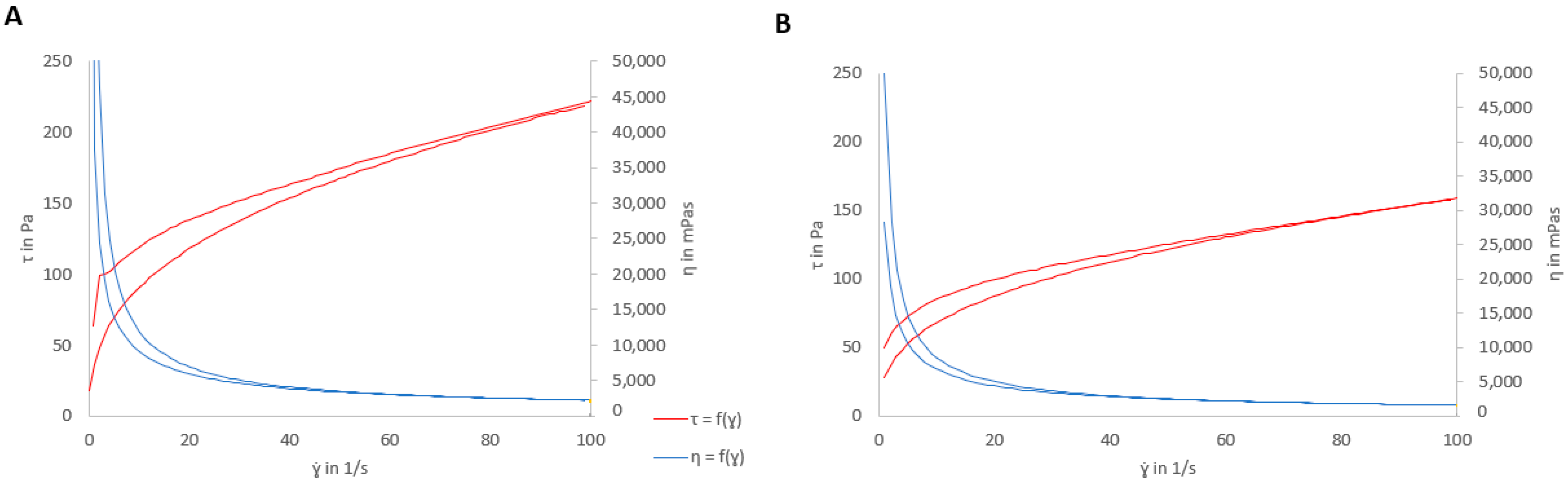
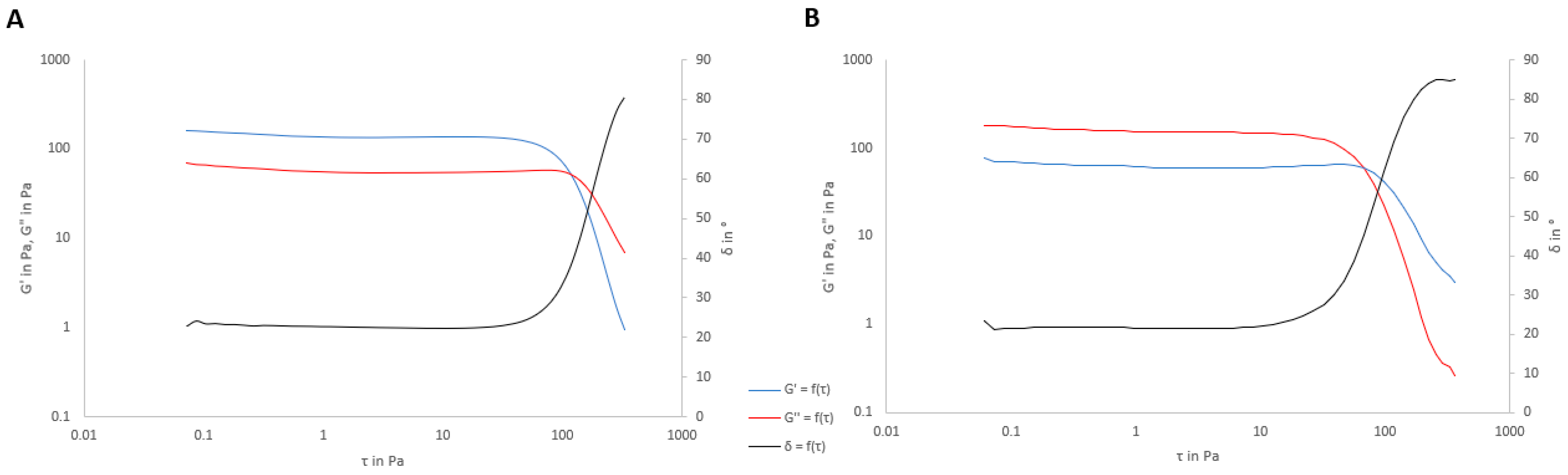
3.4. Rheological Properties

As there are small differences in viscosity between the optimized placebo formula and the formulations with D, the following characterization was carried out with the placebo. The rheological behavior was studied with and without SVF (Figure 7).
As Figure 7 shows, the formulation presented thixotropy (area of 29.56 Pa/s), and thus the system required more time to recover its original internal structure. The same characteristics were observed for the placebo gel with SVF but with a lower thixotropic area (17.95 Pa/s). Additionally, the viscosity values under the experimental conditions were 2212 ± 10.22 mPa×s and 1586 ± 2.75 mPa×s with and without SVF, respectively. The reduction in viscosity and apparent thixotropy were probably due to the dilution effect of SVF.
Figure 7 shows the possible pseudoplastic behavior of the tested formulation (with and without SVF) due to the apparent viscosity decrease with an increasing shear rate. To confirm the rheological behavior, the experimental data were fit to different equations, as shown in Table 9. A lower Chi2 and a higher correlation factor represent the best fit. The model that best fits the experimental data is the Cross equation, which is usually employed to describe the rheological behavior of pseudoplastic materials with yield stress values [56].
The Cross equation is a versatile equation that can estimate different models that converge into simpler models based on different assumptions. Table 10 shows the Cross equation parameters, where for shear-thinning materials, the value of n is between zero and one. In this case, the n values of the gels with and without SVF were lower than one, which confirms the pseudoplastic profile [56]. The placebo gel without SVF showed a higher viscosity at zero shear than with SVF due to the dilution effect of SVF. In contrast, the η in both cases were very similar, indicating that the gels converged toward similar viscosities at high flow rates with extremely small values compared to the viscosities described previously (2212 and 1586 mPa × s). According to ɣ ˙ value, the placebo gels without SVF were less shear-thinning than they were with SVF. The rheological flow properties of polymers depend on the structural parameters; in this case, SVF crosslinks with carrageenan, affecting the rheological behavior of the gel.
In addition, a viscoelasticity study was carried out to evaluate the internal structure of the formulation and the effect of SVF on it. Figure 8 and Table 11 show the viscoelasticity parameters (storage modulus G′ and loss modulus G″).
The formulations behaved as solid-like products (G′ > G″), and this value was confirmed by tan(δ) < 1. The loss tangent (tan δ) is a measure of the energy lost to stored energy during cyclic deformation (tan δ = G″/G′). A value of tan δ < 1 indicates prevalent elastic behavior [57]. In this case, the same value of tan δ was obtained for both formulations, and it was <1. The values of both the storage and complex moduli increased in the presence of SVF. When SVF was added to the gel, G″ remained essentially the same as that without SVF, but G′ increased. Carrageenan crosslinking with the monovalent ions of SVF forms a more rigid structure, and therefore, the storage modulus is higher than that without SVF. Despite the increasing values with SVF, the proportion was maintained, and therefore, the value of δ was the same in both cases.
Under an external force, molecular chain orientation is caused by internal friction. When the molecular weight is lower than a certain value, there is a crossover point between the G″ and G″ curves, which means that there is a balance between the states of internal friction and disorientation. With an increase in molecular weight, the crossover point moves to a lower frequency because of restricted disorientation. This lower frequency allows sufficient time for molecular orientation. Therefore, the higher molecular weight molecules need more time for molecular orientation. In summary, the crossover point data demonstrate that the molecular weight decreases with an increase in frequency and polydispersity increases with a decrease in frequency [58]. In this case, as Figure 8 shows, the crossing point is higher without SVF (approximately τ = 140 Pa) than with SVF (approximately 75 Pa). With SVF, sodium ions intercalated into the carrageenan chains, increasing their rigidity and producing a more restricted orientation, so the value of τ was lower.

3.5. Syringeability of the Formulations

Syringeability (the force required to extrude a formulation though a syringe) is another important parameter for practical administration with a syringe [50]. In this case, the time needed to empty the syringe was determined with the placebo formulation, which was 3.04 ± 0.0058 s.
The time and force needed to empty the syringe were acceptable and would allow for the easy application of the product to the vaginal area.

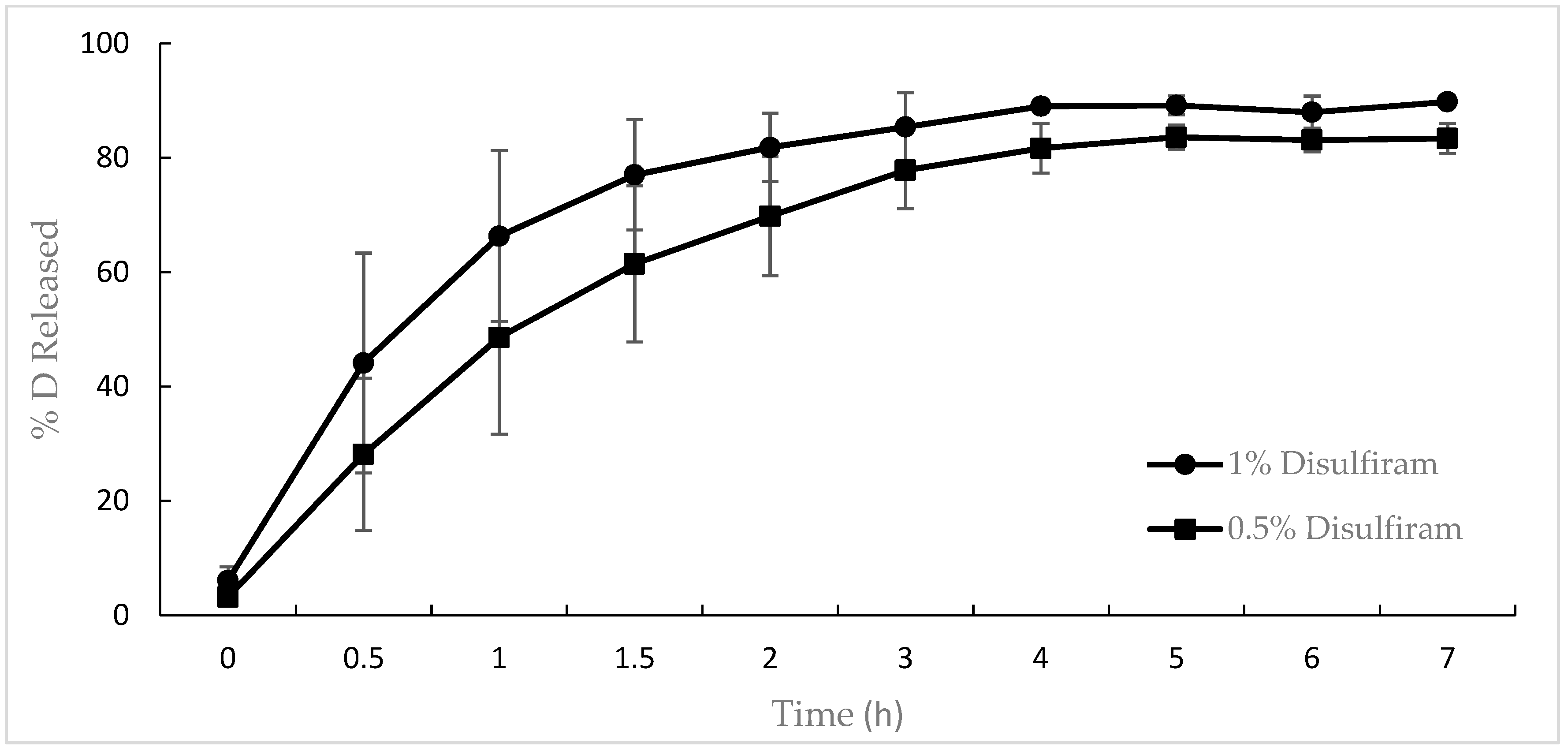
3.6. In Vitro Release of Disulfiram from the Gel

The release of disulfiram from the 0.5% and 1% mucoadhesive gels is depicted in Figure 9. After 6 h, 80% of D was released from the 0.5% gel, while more than 90% was released from the 1% gel.
After fitting the mean release data to the different mathematical models (Table 12), it was observed that the first-order model provided the best fit (AIC 30.16, adj R2 99.78%) of the experimental data in the case of the 0.5% (w/w) gel formulation, whereas the Weibull model best fit the data from the 1% (w/w) D gel (AIC 55.19, adj R2 97.33%).
Once the mean release kinetic behaviors were determined, the means and standard deviations of the individual release data for both gels were calculated, as reported in Table 12.
The first-order model confirmed that the D release mechanism from the 0.5% (w/w) gel was diffusion, according to Fick’s Law [33,51]. However, the Weibull model was a nonmechanistic equation. Papadopoulou et al. [51] determined a relationship between the shape parameter β and the release mechanism. In the case of the 1% (w/w) D gel, the value of β was 0.73, which corresponds to the Fick diffusion release mechanism (β < 0.75). The release from both formulations, despite being explained by different models, present the same release mechanism.

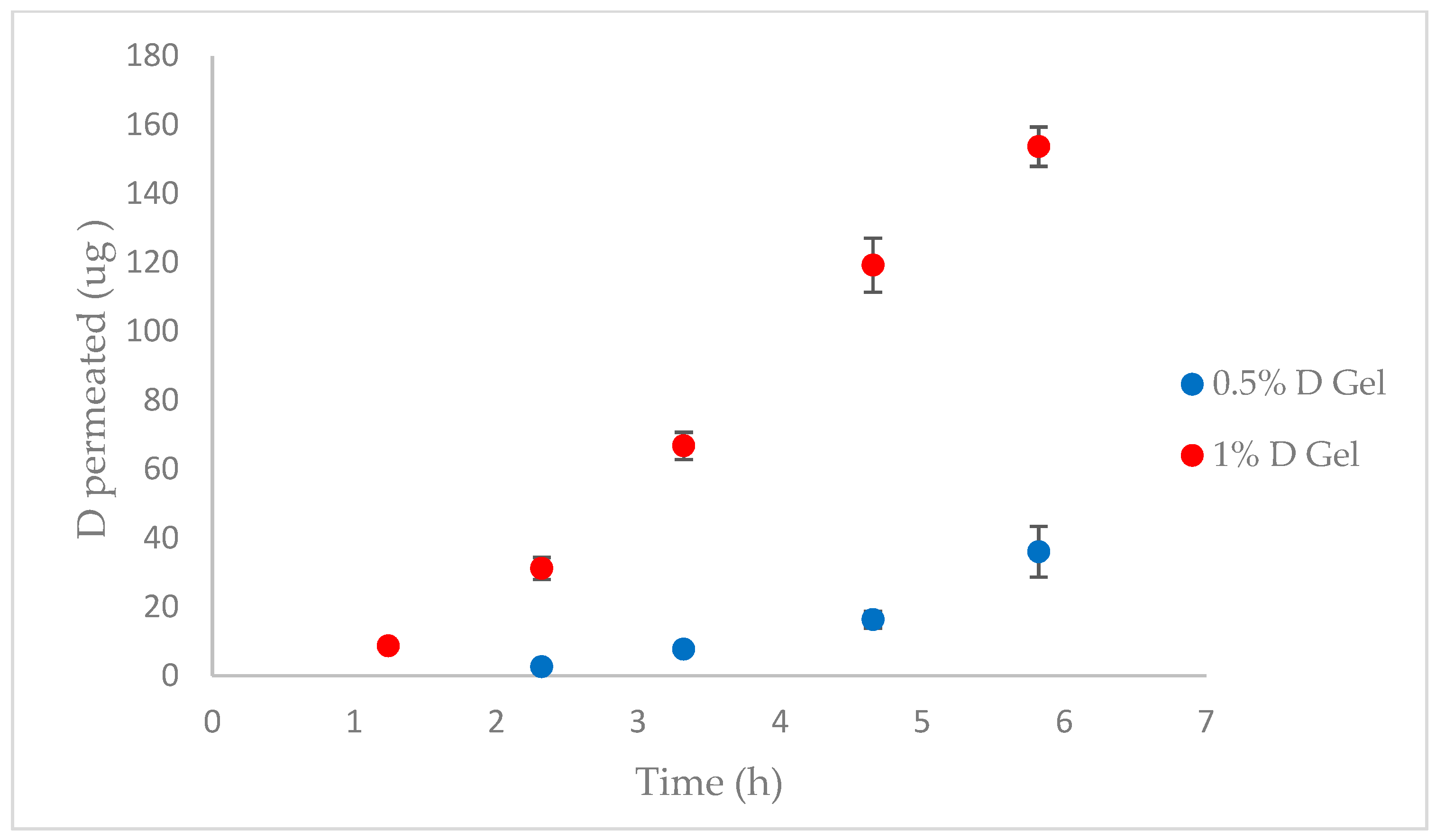
3.7. Pig Vagina Permeation

In vivo permeation tests were performed with pig vagina and the 0.5% (w/w) and 1% (w/w) D gels (n = 4). Figure 10 shows the permeation profiles of the tested formulations, and the permeation parameters are listed in Table 13.
D had a good permeation profile, achieving a steady state within the first hour after administration of the 1% D gel (Tlag = 1.17 h) and within the second hour for the 0.5% D gel (Tlag = 2.28 h). The observed mucosal absorption was probably caused by the favorable physicochemical properties of the compound according to Lipinski’s rule of five [59]: low molecular weight (296 Da), logp value of 3.88, melting point under 200 °C (71.5 °C), and fewer than five hydrogen bond donors and acceptors [20]. The partition parameter (P) of the API between the gel and the vaginal mucosa revealed that this is the factor that affects the vaginal permeation of both formulations the most. The p value obtained was very similar for the 0.5 and 1% gels because it is an independent factor of the D concentration. In this case, the diffusion coefficient had a low contribution to D permeability, and the concentration effect was more noticeable for the 1% formulation according to Fick’s Law. Even though D permeates, as previously noted, D has classically been used orally as a treatment for alcoholism and has been reported to have a good safety profile during clinical use.
In other permeation studies involving human and pig skin, D showed a lack of permeability [21,22]. These differences are due to the nonkeratinization of the vaginal epithelium, and the increase in surface area provided by the transversal rugae of the mucosa would also increase drug absorption across this tissue [60]. Generally, porcine vaginal tissue seems to be a good in vivo permeability model for extrapolation to human vaginal tissue due to substantial histological similarities (stratified squamous epithelium supported by connective tissue) [61,62,63].

3.8. Disulfiram Concentration Retained in the Pig Vagina

Candida affects the vaginal epithelium and has the capacity to adhere to epithelial cells and penetrate into the tissue. Especially important, in recurrent candidiasis, vaginal relapse following incomplete organism eradication after treatment is a mechanism by which chronification occurs. Furthermore, colonization of the vaginal epithelium facilitates the formation of a yeast reservoir or a source for future infections [8]. Given that our target is the vaginal epithelium, the drug content in the vagina was determined. After permeation studies, the tissue was cut into 50 mg pieces, and these pieces were homogenized with mobile phase using a MagNa Lyser instrument. After HPLC analysis, the mean amount of drug detected in the tissue after administration of the 0.5% D gel was 36.48 ± 2.50 µg and was 87.73 ± 3.12 µg for the 1% D gel. Considering the vaginal density (1 g/mL due to the high water content), the mean D concentrations obtained were 138.17 µg/mL and 268.67 µg/mL, respectively. These concentrations are still higher than the MIC values (2–8 µg/mL), confirming the suitability of D for treating vaginal infections, including those in the deeper mucosal layers or recurrent cases.

4. Conclusions

Disulfiram was revealed to be effective against different Candida species and is a potential alternative to classic antifungals for the treatment of fungal infections. In this study, disulfiram mucoadhesive gels were prepared successfully for the treatment of vaginal candidiasis. Formula optimization has allowed the development of gels with suitable physicochemical characteristics that facilitate vaginal administration, improving the residence time, patient compliance, appearance of resistance, and typical limitations of conventional vaginal drug delivery systems and traditional antifungals. Furthermore, after gel characterization, the optimized formulation was shown to have appropriate adhesiveness, syringeability, and mucoadhesiveness. Additionally, the rheological behavior revealed the pseudoplastic flow of the gels. The developed gel showed promising in vitro release, with more than 70% release of it in the first 5 h. According to in vitro distribution studies, the drug concentration in vaginal tissue was higher than the MIC value, making D potentially effective for the treatment of candidiasis. However, the clinical implementation of D has encountered some challenges. Further research should be carried out, and an exploration of the molecular mechanism of disulfiram as an antifungal agent is needed. Taken together, these results suggest that disulfiram mucoadhesive gels could be a good option as an alternative treatment for vaginal candidiasis.

Author Contributions

Conceptualization, F.F.-C., M.L.C. and M.A.C.; methodology, M.L.-R., I.N.-S., F.F.-C., M.L.C., J.S.-C. and M.M.; validation, M.L.-R. and I.N.-S.; formal analysis, M.L.-R. and I.N.-S.; investigation, M.L.-R.; resources, F.F.-C., M.L.C. and M.M.; data curation, M.L.-R., F.F.-C., M.L.C. and M.A.C.; writing—original draft preparation, M.L.-R.; writing—review and editing, M.L.-R., F.F.-C., M.L.C. and M.A.C.; visualization, M.L.-R. and M.A.C.; supervision, F.F.-C., M.L.C. and M.A.C.; project administration, F.F.-C., M.L.C. and M.A.C.; funding acquisition, F.F.-C. All authors have read and agreed to the published version of the manuscript.

Funding

This research was funded by the Generalitat of Catalonia, Industrial Doctorate Program, grant number 2020 DI 97.

Institutional Review Board Statement

Not applicable.

Informed Consent Statement

Not applicable.

Data Availability Statement

Data are available upon request due to intellectual property.

Acknowledgments

We thank the valuable contribution of Mari Carmen Moreno (Laboratory Reig Jofre, Sant Joan Despí, Barcelona, Spain).

Conflicts of Interest

M.L.-R. and I.N.-S. are employees of Laboratory Reig Jofre. They belong to the R&D department and do not participate in marketing or commercial activities. The data presented were conducted under general good laboratory practices. The other authors do not declare any conflicts of interest.

References

  1. Johal, H.S.; Garg, T.; Rath, G.; Goyal, A.K. Advanced topical drug delivery system for the management of vaginal candidiasis. Drug Deliv. 2016, 23, 550–563. [Google Scholar] [CrossRef] [PubMed]
  2. Ceccarani, C.; Foschi, C.; Parolin, C.; D’antuono, A.; Gaspari, V.; Consolandi, C.; Laghi, L.; Camboni, T.; Vitali, B.; Severgnini, M.; et al. Diversity of vaginal microbiome and metabolome during genital infections. Sci. Rep. 2019, 9, 14095. [Google Scholar] [CrossRef] [PubMed]
  3. Pérez-González, N.; Febrer, N.B.-D.; Calpena-Campmany, A.C.; Nardi-Ricart, A.; Rodríguez-Lagunas, M.J.; Morales-Molina, J.A.; Soriano-Ruiz, J.L.; Fernández-Campos, F.; Clares-Naveros, B. New formulations loading caspofungin for topical therapy of vulvovaginal candidiasis. Gels 2021, 7, 259. [Google Scholar] [CrossRef] [PubMed]
  4. Rodríguez-Cerdeira, C.; Gregorio, M.C.; Molares-Vila, A.; López-Barcenas, A.; Fabbrocini, G.; Bardhi, B.; Sinani, A.; Sánchez-Blanco, E.; Arenas-Guzmán, R.; Hernandez-Castro, R. Biofilms and vulvovaginal candidiasis. Colloids Surf. B Biointerfaces 2019, 174, 110–125. [Google Scholar] [CrossRef]
  5. Felix, T.C.; de Brito Röder, D.V.D.; dos Santos Pedroso, R. Alternative and complementary therapies for vulvovaginal candidiasis. Folia Microbiol. 2018, 64, 133–141. [Google Scholar] [CrossRef]
  6. Gonçalves, B.; Ferreira, C.; Alves, C.T.; Henriques, M.; Azeredo, J.; Silva, S. Vulvovaginal candidiasis: Epidemiology, microbiology and risk factors. Crit. Rev. Microbiol. 2015, 42, 905–927. [Google Scholar] [CrossRef]
  7. Mtibaa, L.; Fakhfakh, N.; Kallel, A.; Belhadj, S.; Salah, N.B.; Bada, N.; Kallel, K. Les candidoses vulvovaginales: Étiologies, symptômes et facteurs de risque. J. Mycol. Med. 2017, 27, 153–158. [Google Scholar] [CrossRef]
  8. Ravani, L.; Esposito, E.; Bories, C.; Moal, V.L.-L.; Loiseau, P.M.; Djabourov, M.; Cortesi, R.; Bouchemal, K. Clotrimazole-loaded nanostructured lipid carrier hydrogels: Thermal analysis and in vitro studies. Int. J. Pharm. 2013, 454, 695–702. [Google Scholar] [CrossRef]
  9. Sobel, J. Current treatment options for vulvovaginal candidiasis. Women’s Health 2005, 1, 253–261. [Google Scholar] [CrossRef]
  10. Azie, N.; Angulo, D.; Dehn, B.; Sobel, J.D. Oral Ibrexafungerp: An investigational agent for the treatment of vulvovaginal candidiasis. Expert. Opin. Investig. Drugs 2020, 29, 893–900. [Google Scholar] [CrossRef]
  11. Mei, L.; Chen, J.; Yu, S.; Huang, Y.; Xie, Y.; Wang, H.; Pan, X.; Wu, C. Expansible thermal gelling foam aerosol for vaginal drug delivery. Drug Deliv. 2017, 24, 1325–1337. [Google Scholar] [CrossRef] [PubMed]
  12. Sauna, Z.E.; Shukla, S.; Ambudkar, S.V. Disulfiram, an old drug with new potential therapeutic uses for human cancers and fungal infections. Mol. Biosyst. 2005, 1, 127–134. [Google Scholar] [CrossRef] [PubMed]
  13. Sobel, J.D.; Sobel, R. Current treatment options for vulvovaginal candidiasis caused by azole-resistant Candida species. Expert Opin. Pharmacother. 2018, 19, 971–977. [Google Scholar] [CrossRef] [PubMed]
  14. Prasad, R.; Nair, R.; Banerjee, A. Multidrug transporters of Candida species in clinical azole resistance. Fungal Genet. Biol. 2019, 132, 103252. [Google Scholar] [CrossRef] [PubMed]
  15. Bhattacharya, S.; Sae-Tia, S.; Fries, B.C. Candidiasis and Mechanisms of Antifungal Resistance. Antibiotics 2020, 9, 312. [Google Scholar] [CrossRef]
  16. Pristov, K.E.; Ghannoum, M.A. Resistance of Candida to azoles and echinocandins worldwide. Clin. Microbiol. Infect. 2019, 25, 792–798. [Google Scholar] [CrossRef]
  17. Shukla, S.; Sauna, Z.E.; Prasad, R.; Ambudkar, S.V. Disulfiram is a potent modulator of multidrug transporter Cdr1p of Candida albicans. Biochem. Biophys. Res. Commun. 2004, 322, 520–525. [Google Scholar] [CrossRef]
  18. Sanglard, D.; Ischer, F.; Monod, M.; Bille, J. Susceptibilities of Candida albicans multidrug transporter mutants to various antifungal agents and other metabolic inhibitors. Antimicrob. Agents Chemother. 1996, 40, 2300–2305. [Google Scholar] [CrossRef]
  19. Lu, C.; Li, X.; Ren, Y.; Zhang, X. Disulfiram: A Novel Repurposed Drug for Cancer Therapy. Cancer Chemother. Pharmacol. 2021, 87, 159–172. [Google Scholar] [CrossRef]
  20. National Center for Biotechnology Information. Disulfiram: Chemical and Physical Properties. PubChem Compound Summary CID 3117. Available online: https://pubchem.ncbi.nlm.nih.gov/compound/Disulfiram#section=Chemical-and-Physical-Properties (accessed on 20 March 2023).
  21. Lajarin-Reinares, M.; Martinez-Esteve, E.; Pena-Rodr, E.; Cañellas-Santos, M.; Fernandez-Campos, F. The Efficacy and Biopharmaceutical Properties of a Fixed-Dose Combination of Disulfiram and Benzyl Benzoate. Int. J. Mol. Sci. 2022, 23, 10969. [Google Scholar] [CrossRef]
  22. Lajarin-Reinares, M.; Pena-Rodríguez, E.; Cañellas-Santos, M.; Rosell-Vives, E.; Cortés, P.; Casas, M.L.; Calvo, M.À.; Fernandez-Campos, F. Repurposing Disulfiram as an Antimicrobial Agent in Topical Infections. Antibiotics 2022, 11, 1752. [Google Scholar] [CrossRef] [PubMed]
  23. Li, H.; Wang, J.; Wu, C.; Wang, L.; Chen, Z.S.; Cui, W. The combination of disulfiram and copper for cancer treatment. Drug Discov. Today 2020, 25, 1099–1108. [Google Scholar] [CrossRef] [PubMed]
  24. Das, S.; Garg, T.; Chopra, S.; Dasgupta, A. Repurposing disulfiram to target infections caused by non-tuberculous mycobacteria. J. Antimicrob. Chemother. 2019, 74, 1317–1322. [Google Scholar] [CrossRef] [PubMed]
  25. Sauna, Z.E.; Peng, X.H.; Nandigama, K.; Tekle, S.; Ambudkar, S.V. The Molecular Basis of the Action of Disulfiram as a Modulator of the Multidrug Resistance-Linked ATP Binding Cassette Transporters MDR1 (ABCB1) and MRP1 (ABCC1). Mol. Pharmacol. 2004, 65, 675–684. [Google Scholar] [CrossRef]
  26. Owen, D.H.; Katz, D.F. A vaginal fluid simulant. Contraception 1999, 59, 91–95. [Google Scholar] [CrossRef]
  27. M27-A3 Reference Method for Broth Dilution Antifungal Susceptibility Testing of Yeasts; Approved Standard-Third Edition. Published online 2008. Available online: www.clsi.org (accessed on 7 September 2022).
  28. Rex, J.H.; Cooper, C.R.; Merz, W.G.; Galgiani, J.N.; Anaissie, E.J. Detection of amphotericin B-resistant Candida isolates in a broth-based system. Antimicrob. Agents Chemother. 1995, 39, 906–909. [Google Scholar] [CrossRef]
  29. Villegas, I.; Rosillo, M.; Alarcón-De-La-Lastra, C.; Vázquez-Román, V.; Llorente, M.; Sánchez, S.; Gil, A.G.; Alcalde, P.; González, E.; Rosell, E.; et al. Amoxicillin and clarithromycin mucoadhesive delivery system for helicobacter pylori infection in a mouse model: Characterization, pharmacokinetics, and efficacy. Pharmaceutics 2021, 13, 153. [Google Scholar] [CrossRef]
  30. Geonnotti, A.R.; Katz, D.F. Compartmental transport model of microbicide delivery by an intravaginal ring. J. Pharm. Sci. 2010, 99, 3514. [Google Scholar] [CrossRef] [PubMed]
  31. Friedl, H.E.; Dünnhaupt, S.; Waldner, C.; Bernkop-Schnürch, A. Preactivated thiomers for vaginal drug delivery vehicles. Biomaterials 2013, 34, 7811–7818. [Google Scholar] [CrossRef]
  32. Park, E.K.; Song, K.W. Rheological evaluation of petroleum jelly as a base material in ointment and cream formulations: Steady shear flow behavior. Arch. Pharmacal. Res. 2010, 33, 141–150. [Google Scholar] [CrossRef]
  33. Venkatesh, M.P.; Kumar, T.P.; Pai, D.R. Targeted drug delivery of Methotrexate in situ gels for the treatment of Rheumatoid Arthritis. Saudi Pharm. J. 2020, 28, 1548–1557. [Google Scholar] [CrossRef] [PubMed]
  34. Zhang, Y.; Huo, M.; Zhou, J.; Zou, A.; Li, W.; Yao, C.; Xie, S. DDSolver: An add-in program for modeling and comparison of drug dissolution profiles. AAPS J. 2010, 12, 263–271. [Google Scholar] [CrossRef] [PubMed]
  35. Monica, L.L.; Jordi, G.; Francisco, F.C. In situ bioadhesive film-forming system for topical delivery of mometasone furoate: Characterization and biopharmaceutical properties. J. Drug Deliv. Sci. Technol. 2020, 59, 101852. [Google Scholar] [CrossRef]
  36. Pena-Rodríguez, E.; Lajarin-Reinares, M.; Mata-Ventosa, A.; Pérez-Torras, S.; Fernández-Campos, F. Dexamethasone-loaded lipomers: Development, characterization, and skin biodistribution studies. Pharmaceutics 2021, 13, 533. [Google Scholar] [CrossRef] [PubMed]
  37. Amores, S.; Domenech, J.; Colom, H.; Calpena, A.C.; Clares, B.; Gimeno, Á.; Lauroba, J. An improved cryopreservation method for porcine buccal mucosa in ex vivo drug permeation studies using Franz diffusion cells. Eur. J. Pharm. Sci. 2014, 60, 49–54. [Google Scholar] [CrossRef] [PubMed]
  38. Kaomongkolgit, R.; Jamdee, K.; Wongnoi, S.; Chimnoi, N.; Techasakul, S. Antifungal activity of coronarin D against Candida albicans. Oral Surg. Oral Med. Oral Pathol. Oral Radiol. 2012, 114, 61–66. [Google Scholar] [CrossRef]
  39. Radetsky, M.; Wheeler, R.C.; Roe, M.H.; Todd, J.K. Microtiter broth dilution method for yeast susceptibility testing with validation by clinical outcome. J. Clin. Microbiol. 1986, 24, 600–606. [Google Scholar] [CrossRef]
  40. Khan, S.; Singhal, S.; Mathur, T.; Upadhyay, D.J.; Rattan, A. Antifungal potential of disulfiram. Jpn. J. Med. Mycol. 2007, 48, 109–113. [Google Scholar] [CrossRef]
  41. Hao, W.; Qiao, D.; Han, Y.; Du, N.; Li, X.; Fan, Y.; Ge, X.; Zhang, H. Identification of disulfiram as a potential antifungal drug by screening small molecular libraries. J. Infect. Chemother. 2020, 27, 696–701. [Google Scholar] [CrossRef]
  42. Shaikh, R.; Raj Singh, T.; Garland, M.; Woolfson, A.; Donnelly, R. Mucoadhesive drug delivery systems. J. Pharm. Bioallied. Sci. 2011, 3, 89–100. [Google Scholar] [CrossRef]
  43. Ahuja, A.; Khar, R.K.; Ali, J. Mucoadhesive Drug Delivery Systems. Drug. Dev. Ind. Pharm. 1997, 23, 489–515. [Google Scholar] [CrossRef]
  44. De Souza Ferreira, S.B.; Da Silva, J.B.; Volpato Junqueira, M.; Belincanta Borghi-Pangoni, F.; Guttierres Gomes, R.; Luciano Bruschi, M. The importance of the relationship between mechanical analyses and rheometry of mucoadhesive thermoresponsive polymeric materials for biomedical applications. J. Mech. Behav. Biomed. Mater. 2017, 74, 142–153. [Google Scholar] [CrossRef] [PubMed]
  45. Resins, W. POLYOX® Water-Soluble Resins. Chem. Eng. News Arch. 1995, 73, 40. [Google Scholar] [CrossRef]
  46. Da Silva, J.B.; de Ferreira, S.B.S.; Reis, A.V.; Cook, M.T.; Bruschi, M.L. Assessing Mucoadhesion in Polymer Gels: The Effect of Method Type and Instrument Variables. Polymers 2018, 10, 254. [Google Scholar] [CrossRef]
  47. Vigani, B.; Rossi, S.; Gentile, M.; Sandri, G.; Bonferoni, M.C.; Cavalloro, V.; Martino, E.; Collina, S.; Ferrari, F. Development of a Mucoadhesive and an in Situ Gelling Formulation Based on κ-Carrageenan for Application on Oral Mucosa and Esophagus Walls. I.I. Loading of a Bioactive Hydroalcoholic Extract. Mar. Drugs 2019, 17, 153. [Google Scholar] [CrossRef]
  48. Khazaeli, P.; Mehrabani, M.; Mosadegh, A.; Bios, S.; Zareshahi, R.; Hasan Moshafi, M. Formulation, Physiochemical, and Microbial Assay of Henna Oil Vaginal Suppository Formulated with Polyethylene Glycol Bases. Iran. J. Med. Sci. 2020, 45, 207. [Google Scholar] [CrossRef]
  49. Kaewnopparat, S.; Kaewnopparat, N. Formulation and Evaluation of Vaginal Suppositories Containing Lactobacillus. World Acad. Sci. Eng. Technol. 2009, 55, 117–120. [Google Scholar]
  50. Pacheco-Quito, E.M.; Ruiz-Caro, R.; Rubio, J.; Tamayo, A.; Veiga, M.D. Carrageenan-Based Acyclovir Mucoadhesive Vaginal Tablets for Prevention of Genital Herpes. Mar. Drugs 2020, 18, 249. [Google Scholar] [CrossRef]
  51. Elias, C.J.; Coggins, C.; Alvarez, F.; Brache, V.; Fraser, I.S.; Lacarra, M.; Lähteenmäkl, P.; Massai, R.; Mishell, D.R.; Phillips, D.M.; et al. Colposcopic evaluation of a vaginal gel formulation of iota-carrageenan. Contraception 1997, 56, 387–389. [Google Scholar] [CrossRef]
  52. Valenta, C.; Schultz, K. Influence of carrageenan on the rheology and skin permeation of microemulsion formulations. J. Control. Release 2004, 95, 257–265. [Google Scholar] [CrossRef]
  53. Cha, D.S.; Choi, J.H.; Chinnan, M.S.; Park, H.J. Antimicrobial Films Based on Na-alginate and κ-carrageenan. LWT 2002, 35, 715–719. [Google Scholar] [CrossRef]
  54. Seol, K.H.; Lim, D.G.; Jang, A.; Jo, C.; Lee, M. Antimicrobial effect of κ-carrageenan-based edible film containing ovotransferrin in fresh chicken breast stored at 5 °C. Meat Sci. 2009, 83, 479–483. [Google Scholar] [CrossRef] [PubMed]
  55. Kim, S.; Jeong, C.; Cho, S.; Kim, S.B. Effects of thermal treatment on the physical properties of edible calcium alginate gel beads: Response surface methodological approach. Foods 2019, 8, 578. [Google Scholar] [CrossRef]
  56. Osswald, T.A.; Book, N.R. Polymer Rheology Fundamentals and Applications; Hanser Publications: Munich, Germany, 2015. [Google Scholar]
  57. Madsen, F.; Eberth, K.; Smart, J.D. A rheological examination of the mucoadhesive/mucus interaction: The effect of mucoadhesive type and concentration. J. Control Release. 1998, 50, 167–178. [Google Scholar] [CrossRef] [PubMed]
  58. Gao, T.; Xie, R.; Zhang, L.; Gui, H.; Huang, M. Use of Rubber Process Analyzer for Characterizing the Molecular Weight Parameters of Natural Rubber. Int. J. Polym. Sci. 2015, 2015, 517260. [Google Scholar] [CrossRef]
  59. Lipinski, C.A. Drug-like properties and the causes of poor solubility and poor permeability. J. Pharmacol. Toxicol. Methods 2000, 44, 235–249. [Google Scholar] [CrossRef] [PubMed]
  60. Mohammed, Y.; Holmes, A.; Kwok, P.C.L.; Kumeria, T.; Namjoshi, S.; Imran, M.; Matteucci, L.; Ali, M.; Tai, W.; Benson, H.A.; et al. Advances and future perspectives in epithelial drug delivery. Adv. Drug Deliv. Rev. 2022, 186, 1–38. [Google Scholar] [CrossRef]
  61. Machado, R.M.; Palmeira-de-Oliveira, A.; Gaspar, C.; Martinez-de-Oliveira, J.; Palmeira-de-Oliveira, R. Studies and methodologies on vaginal drug permeation. Adv. Drug Deliv. Rev. 2015, 92, 14–26. [Google Scholar] [CrossRef]
  62. Squier, C.A.; Mantz, M.J.; Schlievert, P.M.; Davis, C.C. Porcine vagina Ex Vivo as a model for studying permeability and pathogenesis in mucosa. J. Pharm. Sci. 2008, 97, 9–21. [Google Scholar] [CrossRef]
  63. Van Eyk, A.D.; Van Der Bijl, P. Porcine vaginal mucosa as an in vitro permeability model for human vaginal mucosa. Int. J. Pharm. 2005, 305, 105–111. [Google Scholar] [CrossRef]
Figure 1. Schematic illustration of the device for evaluating adhesive strength.
Pharmaceutics 15 01436 g001
Figure 2. Schematic of the device used to measure the mucoadhesive properties.
Pharmaceutics 15 01436 g002
Figure 3. Matrix plots explaining the relationships between the different variables.
Pharmaceutics 15 01436 g003
Figure 4. Plots of the main effects of PEG-90M and carrageenan on the mucoadhesive properties of the gels.
Pharmaceutics 15 01436 g004
Figure 5. (A) Plots of the main effects of PEG-90M and carrageenan on the viscosity (mPa×s) of the gels. (B) Viscosity interaction point.
Pharmaceutics 15 01436 g005
Figure 6. (A) Plots of the main effects of PEG-90M and carrageenan on fluorescein adhesion. (B) Fluorescein adhesion interaction plot.
Pharmaceutics 15 01436 g006
Figure 7. (A) Shear stress vs. shear rate (red) and viscosity vs. shear rate (blue) curves of the placebo formulation. (B) Shear stress vs. shear rate (red) and viscosity vs. shear rate (blue) curves of the placebo formulation with SVF.
Pharmaceutics 15 01436 g007
Figure 8. Storage modulus, loss modulus, and phase angle during the sweep stress test on the mucoadhesive gel 24 h after preparation at a frequency of 1 s−1. (A) Placebo Gel. (B) Placebo Gel with SVF.
Pharmaceutics 15 01436 g008
Figure 9. Release of disulfiram from the 0.5% gel and from the 1% gel. Mean of the quantities released of D expressed as a percentage respect total amount seeded from the 0.5% gel and from the 1% gel after 6 h in vitro.
Pharmaceutics 15 01436 g009
Figure 10. Pig vagina permeation profiles after administration of the 0.5% and 1% D gels (n = 4).
Pharmaceutics 15 01436 g010
Table 1. Levels studied for surface response design.
FactorLower LevelHigher Level
% PEG-90M (w/w)0.51.5
% Carrageenan (w/w)1.02.0
Table 2. Rheological equations used to evaluate the prepared formulations.
Rheological ModelEquation
Newton τ = η × γ ˙
Bingham τ = τ 0 + ( η 0 × γ ˙ )
Ostwald–de Waele τ = K × γ ˙ n
Herschel–Bulkley τ = τ 0 + K × γ ˙ n
Casson τ = τ 0 n + η 0 × γ ˙ n n
Cross τ = γ ˙ × ( η + ( η 0 η ) / ( 1 + ( γ ˙ / γ ˙ 0 ) n )
τ : Shear stress (Pa), η : viscosity (cP or Pa-s), γ ˙ : shear rate (s−1), τ 0 : minimum yield stress (Pa), η 0 : zero shear viscosity (cP or Pa-s), K: consistency factor (Pa sn), η : infinite shear viscosity (cP or Pa-s), γ ˙ 0 : zero shear rate (s−1).
Table 3. Different kinetic models and equations tested.
Kinetic ModelEquationParameter(s)
First-Order F = F m a x 1 e ( K 1 t ) F m a x , K1
Higuchi F = K H × t 1 / 2 KH
Korsmeyer–PeppasF = KKP × tnKKP, n
Weibull F = F m a x × 1 e ( t T d ) β F m a x , α, β
F: Fraction of drug released, t: time, F m a x : maximum fraction of drug released, K1: first-order constant, K H : Higuchi constant, KKP: Korsmeyer–Peppas constant, n: diffusional exponent, Td: the time at which 63.2% of the drug had been released, β: Weibull shape parameter.
Table 4. Summary of the MICs (n = 3) for disulfiram with three different species of Candida using two different test media.
MIC (µg/mL)
Test Media
Candida spp.RPMI 1640Sabouraud Dextrose
C. albicans22
C. glabrata42
C. parapsilosis88
Table 5. Influences of PEG-90M and carrageenan on the physicochemical parameters.
BatchPEG-90M
(% w/w)
Carrageenan
(% w/w)
Viscosity
(cP)
Viscosity with SVF
(cP)
Mucoadhesion (mN/cm2)Mucoadhesion with SVF
(mN/cm2)
Adhered Fluorescein
(%)
LP-830.501.5019,91016,17050.660.591.40
LP-842.001.5061,66039,35065.379.399.64
LP-861.252.0052,84031,36071.980.999.50
LP-871.251.5038,00027,07067.370.899.52
LP-881.251.5033,17022,21057.458.199.60
LP-891.251.5027,89021,73059.461.899.59
LP-901.251.5033,37025,07065.568.299.43
LP-911.251.5032,26023,25053.663.399.60
LP-921.781.1429,75026,18047.950.397.33
LP-930.721.8520,10016,79048.351.998.10
LP-940.721.14472010,49034.938.890.28
LP-951.781.8587,28040,59064.874.999.54
LP-961.251.0010,00013,69038.447.892.42
Table 6. Response surface model equations for monitoring the effect of polymer concentration on the properties of mucoadhesive gels.
Quadratic Polynomial Model EquationR2Adj R2
Y1 = −0.8 + 14.40 X1 + 29.88 X20.7020.642
Y2 = 24,704 − 48,649 X1 − 23,052 X2 + 56,200 X1 X20.9450.927
Y3 = 28.85 + 32.60 X1 + 56.06 X2 − 6.652 X12 − 13.21 X22 − 7.48 X1X20.9870.978
X1: Concentration (% w/w) of PEG-90M; X2: concentration (% w/w) of carrageenan. Y1: mucoadhesion with SVF (mN/cm2), Y2: Viscosity (cPs), Y3: fluorescein adhered (%).
Table 7. Prediction of polymer concentrations for a formulation with maximum mucoadhesion and fluorescein adhesion and minimal viscosity.
PEG-90MCarrageenanFluorescein Adhesion FitMucoadhesion with SVF FitViscosity FitComposite Desirability
0.866298.4271.4633,8240.76
Table 8. Results after characterization of the final formulations with bias with respect to the theoretical value.
PropertyTheoretical Value
(95% CI)
Placebo Gel0.5% D Gel1% D GelBias (%)
Mucoadhesion
(mN/cm2)
71.5 (60.85; 82.07)72.968.269.41.86
Viscosity
(cP)
33,824 (22,885; 44,763)29,83530,40032,7808.30
Fluorescein adhered (%)98.42 (97.23; 99.62)98.6798.5898.810.27
Table 9. Rheological models with Chi2 and r values for the placebo gel and placebo gel with SVF.
Rheological ModelPlacebo GelPlacebo Gel with SVF
Chi²rChi²r
Newton1.773 × 1050.3261.004 × 105−0.137
Bingham1.147 × 1040.97061270.968
Ostwald–de Waele8.9971.00013.180.999
Herschel–Bulkley7.0201.00012.9000.999
Casson9.2200.98817910.991
Cross1.1641.0001.3591.000
Table 10. Rheological model fitting of the placebo gels with and without SVF.
Cross Equation ParameterPlacebo GelPlacebo Gel with SVF
η 318.7141.9
η 0.15170.1600
ɣ ˙ 0.04220.1235
N0.64880.6853
Table 11. Viscoelasticity parameters of the Placebo Gel and Placebo Gel with SVF (mean ± standard deviation (SD)).
ParameterFormulationMean ± SD
G′Placebo gel129.92 ± 25.20 Pa
Placebo gel with SVF149.66 ± 25.33 Pa
G″Placebo gel57.90 ± 4.60 Pa
Placebo gel with SVF63.67 ± 4.01 Pa
η*Placebo gel22,709 ± 3657 mPa
Placebo gel with SVF25,947 ± 3649 mPa
δPlacebo gel24.20 ± 4.46°
Placebo gel with SVF23.67 ± 5.07°
tan(δ)Placebo gel0.45 ± 0.11
Placebo gel with SVF0.44 ± 0.08
Table 12. Individual models and parameters for the release of D from the 0.5 and 1% gels.
FormulationModelParametersValue
0.5% D gelFirst-OrderK1 (h−1)
Fmax (%)
0.95 ± 0.48
85.79 ± 2.13
1% D gelWeibullα
β
Fmax (%)
0.88 ± 0.43
0.73 ± 0.23
93.42 ± 3.21
Table 13. Mean parameters of D permeation after gel application to pig vagina.
FormulationParameterMeanSD
0.5% D gelJsup (µg/h·cm2)9.01060.6562
R20.91130.0445
Kp (cm/h)0.24810.0181
Tlag (h)2.28340.6721
P (cm/h2)3.43761.2485
Dif (1/h)0.07630.0225
1% D gelJsup (µg/h·cm2)32.97691.1737
R20.99130.0001
Kp (cm/h)0.45420.0162
Tlag (h)1.16980.0514
P (cm/h2)3.18510.0265
Dif (1/h)0.14260.0063
Disclaimer/Publisher’s Note: The statements, opinions and data contained in all publications are solely those of the individual author(s) and contributor(s) and not of MDPI and/or the editor(s). MDPI and/or the editor(s) disclaim responsibility for any injury to people or property resulting from any ideas, methods, instructions or products referred to in the content.

