
Intranasal Polymeric and Lipid-Based Nanocarriers for CNS Drug Delivery

Abstract
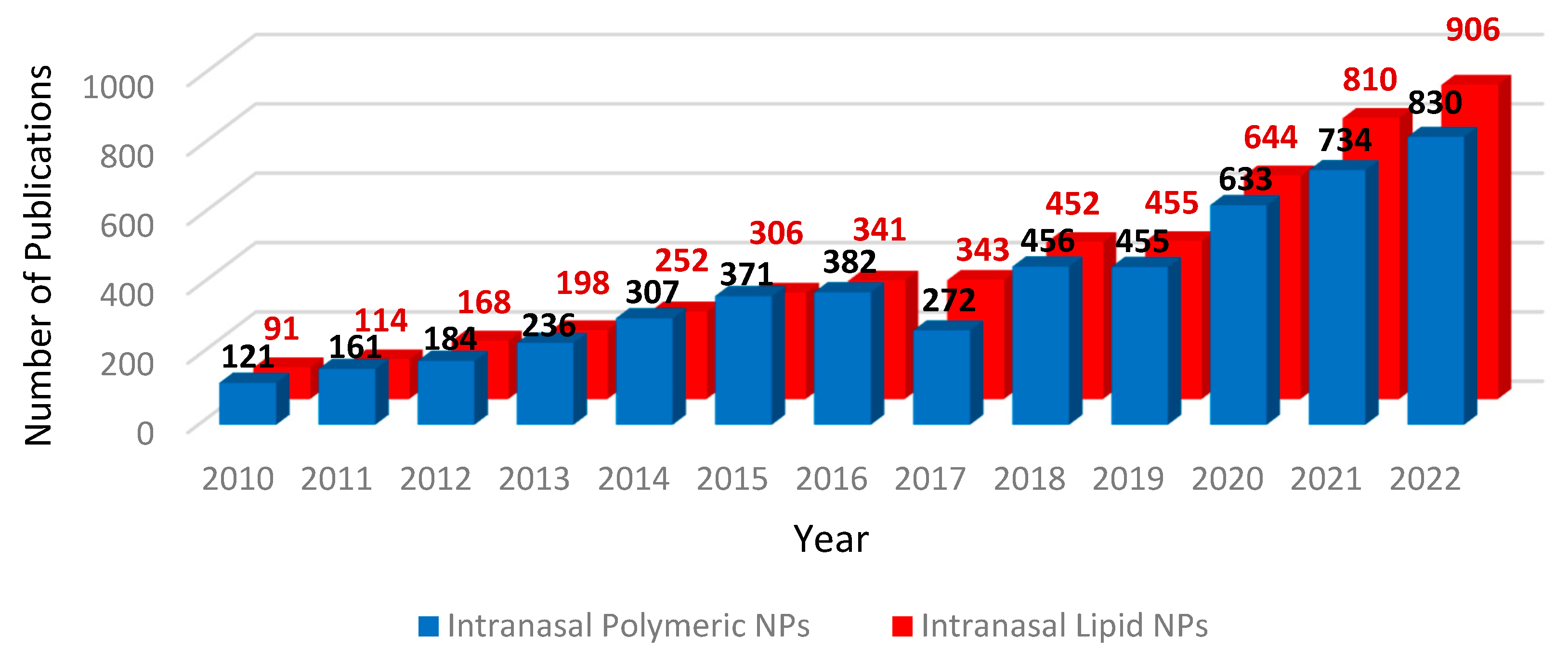
:1. Introduction
2. Nanotechnology for BBB Crossing
2.1. Polymeric NPs
2.2. Solid Lipid Nanoparticles
2.3. Surface Charge
2.4. Surface Modification
2.5. PLGA NPs and SLNs Are Compatible with Brain Cells In Vitro
2.6. Permeation of In Vitro BBB Models
2.7. PLGA NP and SLN Drug Delivery to In Vitro CNS Disease Models
2.7.1. Neurodegenerative Disease
2.7.2. Brain Cancer
3. PLGA NP and SLN-Mediated Drug Delivery In Vivo
3.1. Biocompatibility and Brain Distribution of PLGA NPs and SLNs In Vivo
3.2. PLGA NPs and SLNs as Drug Delivery Vehicles for CNS Disease: Preclinical Evidence

| Disease | Nanocarrier | Disease Model | Drug Loaded | Mode of Action | Size (nm) | PDI | Zeta Potential (mV) | ROA | Drug Conc. Administered by NP | Outcomes | Ref. |
|---|---|---|---|---|---|---|---|---|---|---|---|
| Alzheimer’s disease | |||||||||||
| SLN | Isoproterenol-induced rat model | Galantamine | AChE inhibitor | 88 ± 1.89–221.4 ± 1.34 | 0.275 ± 0.12–0.380 ±0.16 | −10.04 ± 1.9–−18.75 ± 1.7 | Oral | 5 mg/kg for 3 weeks | Galantamine-loaded SLNs protected against memory impairments | [91] | |
| PLGA NP | Scopolamine-induced rat model | Phytol | Antioxidant, anti-inflammatory, anti-amyloid | 177.4 ± 5.9 | 0.2 ± 0.06 | −32.8 ± 2.2. | Oral | 100 mg/kg or 200 mg/kg | Improved spatial & short memory, prevented acetylcholine breakdown and regulated neuronal death, reduced oxidative stress | [101,105] | |
| PLGA-PEG NP | Transgenic mouse model | Pioglitazone | Neuroprotection | 155 ± 1.8 | 0.1 | −13 ± 0.5 | Oral | 10 mg/kg; 5 days a week for 4 weeks | Reduced memory impairment and fewer cortical Aβ deposits | [103] | |
| PLGA-PEG NP | Transgenic mouse model | ECGC | Antioxidant, neuroprotection | 124.8 ± 5.2 | 0.054 ± 0.013 | −15 | Oral | 40 mg/kg daily for 3 months | Improved spatial learning and memory, increased number of synapses, reduced neuroinflammation and Aβ burden | [100] | |
| PLGA NP | Transgenic mouse model | Vitamin D-binding protein | Anti-amyloid | 226.6 ± 44.4 | 0.039 ± 0.013 | −0.144 | i.v. | 2.5 mg/kg of NPs daily for 4 weeks | Inhibited Aβ aggregation, neuroinflammation, neuronal death and cognitive deficits | [59] | |
| SLN | Streptozotocin rat model | Nicotinamide | Cognitive enhancer | 124 ± 0.8 | 0.831 | −12.5 ± 0.7 | i.p. and i.v. | 60, 30, 15 mg/kg every other day | Improved cognition, neuroprotection and reduced tau hyperphosphorylation | [99] | |
| Vascular Dementia | |||||||||||
| SLN | Homocysteine rat model | Curcumin | Antioxidant, anti-inflammatory | 154.8 | 0.928 | −10.9 | Oral | 25 mg/kg daily for 2 weeks | Improved memory, reduced oxidative stress biomarkers, reduced AChE activity, increased GABA, decreased glutamate and exerted neuroprotection in the cortex and hippocampus | [98] | |
| Parkinson’s disease | |||||||||||
| Lactoferrin-PLGA NP | MPTP mouse model | Resveratrol | Antioxidant, anti-inflammatory, neuroprotective | 148.2 ± 4.2 | 0.12 ± 0.18 | −23.1 ± 3.0 | i.v. | 5 mg/kg every other day for 15 days | Improved motor functions, protected against DA depletion, neuroprotective and reduced glial activation and neuroinflammation in the SN | [57] | |
| PLGA NP | Rotenone rat model | Tolcapone | Reduces dopamine metabolism | 182.59 ± 23.94 | Not stated | −26.32 ± 0.48 | i.p. | 3 mg/kg every 3 days for 45 days | Improved motor functions, prevented nigral cell death, reduced glial activation | [79] | |
| Albumin-PLGA NP | 6-OHDA mouse model | Dopamine | Dopamine replenishment | 353 | 0.5 | −37 | i.p. | 0.05 mg/μL or 0.1 mg/μL | Increased dopamine, improved motor coordination, balance and sensorimotor functions | [82] | |
| SLN | Rotenone mouse model | Curcumin | Antioxidant, anti-inflammatory | 134.5 ± 0.85 | 0.39 ±0.04 | −18.56 ± 0.55 | Transdermal | 85 mg/kg | Decreased bradykinesia, improved coordination and balance | [97] | |
| Huntington’s disease | |||||||||||
| PLGA-PEG NP | 3-nitropropionic acid mouse model | EGCG | Antioxidant, neuroprotection | 124.8 ± 5.2 | 0.054 ± 0.013 | −15.7 ± 1.7 | i.p. | 50 mg/kg daily for 5 days | Relieved motor symptoms, neuroprotective and reduced neuroinflammation | [55] | |
| Glyco-protein7 -PLGA NP | Transgenic mouse model | Cholesterol | Slows disease progression | 249 ± 38 | 0.29 ± 0.05 | −30 ± 7 | i.p. | 1.7 mg NPs/mouse twice weekly for 5 weeks | Delayed onset of symptoms in pre-symptomatic mice, rescued cognitive decline in symptomatic mice, improved motor recovery, reduced muHTT aggregation | [102] | |
| GBM | |||||||||||
| SPIO-PLGA NP | Orthotopic U87MG tumour mouse model | Paclitaxel | Prevents cancer cell growth and induces cell death | 250 ± 20 | 0.11 | −18 ± 5 | i.v. | 5 mg/kg every 4 days for 16 days starting 8 days post tumour inoculation | Improved survival time | [92] | |
| PLGA-PEG NP | Orthotopic U87MG tumour mouse model | siRNA targeting hepatocyte growth factor receptor | Reduces tumour cell proliferation | 117.4 ± 11.7 | Not stated | 37.3 ± 2.3 | i.v. | 0.125, 0.5 or 2 mg/kg three times a week for 3 weeks, two weeks post tumour inoculation | Reduced tumour volume | [104] | |
| Epilepsy | |||||||||||
| PLGA NP | Pentylene- tetrazole induced seizure rat model | Carbamaze-pine and levetiracetam | Reduces epileptic activity | 180.62 ± 6.26 | 0.107 ± 0.03 | −27.08 ± 3.11 | i.p. | 30 mg/kg carbamazepine & 1.2 mg/kg levetiracetam | Decreased seizure activity | [93] | |
| Depression | |||||||||||
| SLN | LPS rat model | Duloxetine | Reduces symptoms of depression | 114.5 ± 2 | 0.29 ± 0.03 | −18.2 ± 1.8 | i.p. | 30 mg/kg daily for 14 days | Decreased immobility time, reduced TNFα and COX-2 expression | [94] | |
| Schizophrenia | |||||||||||
| SLN | Dizocilpine rat model | Lurasidone hydrochloride | Reduces symptoms of psychosis | 139.8 ± 5.5. | 0.118 ± 0.002 | −30.8 ± 3.5 | oral | 2.066 mg/kg for 3 weeks | Improved cognition and reduced EPS effects | [95] | |
| Substance abuse disorder | |||||||||||
| PLGA NP | Fentanyl-dependent rat | Naloxone | Opioid receptor antagonist | 263 | 0.2 | Not stated | i.m. | 10 mg/kg | Prevented fentanyl induced antinociception and respiratory depression | [106] | |
4. Intranasal Drug Delivery
4.1. Nasal Drug Delivery Bypasses the BBB
4.2. Intranasal Formulations Reduce Side Effects
4.3. Strategies for Improving Nose-to-Brain Transport
5. Intranasal Delivery of Experimental Therapeutics to the CNS via PLGA NPs and SLNs
5.1. Brain Distribution and Drug Bioavailability of Intranasal PLGA NPs and SLNs
5.2. Proof of Concept: Efficacy of Drug-Loaded Intranasal PLGA NPs and SLNs in Animal Models of CNS Disease
5.3. Concerns Regarding Intranasal Delivery of Nanomedicine
| Disease | Nanocarrier | Disease Model | Drug Loaded | Mode of Action | Size (nm) | PDI | Zeta Potential (mV) | Drug conc. Administered by NP | Outcomes | Ref. |
|---|---|---|---|---|---|---|---|---|---|---|
| Alzheimer’s disease | ||||||||||
| PLGA NP | Scopolamine rat model | Memantine | NMDA antagonist; cognition enhancer | 58.04 | 0.204 | −23 | 0.1 mg/kg in 20 μL daily for 9 days | Improved spatial memory | [54] | |
| Chitosan-SLN | Streptozotocin rat model | Ferulic acid | Antioxidant, neuroprotective properties | 184.9 | 0.277 | 12.4 | 80 mg/kg for 28 days | Enhanced cognition, reduced oxidative stress and AChE activity in the cortex and hippocampus | [166] | |
| Parkinson’s Disease | ||||||||||
| Lactoferrin PLGA-PEG NP | 6-OHDA rat model | Rotigotine | DA agonist; improves dopaminergic neurotransmision | 118 ± 12.14 | Not stated | −21.94 ± 2.83 | 2 mg/kg in 200 μL twice daily for 1 week | Improved dopaminergic transmission, reduced nigro-striatal neurodegeneration | [173] | |
| WGA-PLGA NP | MPTP mouse model | L-Dopa | DA precursor; increases brain levels of DA and transmission | 329 ± 188.3 | 0.384 ± 0.113 | −4.47 ± 0.576 | 16 mg/kg in 20 μL for 7 days | Improved locomotor activity | [174] | |
| GBM | ||||||||||
| PLGA NP | U87 luciferase tumour bearing nude mouse model | Bevacizumab | Anti-VEGF; anti-angiogenesis and tumour cell death | 185.0 ± 3.0 | 0.056 ± 0.016 | −2.50 ± 0.27 | 5 mg/kg in 5 μL weekly for 24 days | Reduced tumour growth and reduced VEGF expression and synthesis | [181] | |
| Anti-EphA3 PLGA NP | T98G tumour bearing nude mouse model | Temozolomide derivative | Cell cycle arrest; tumour cell death | 135.1 ± 2.4 | 0.085 ± 0.037 | −28.65 ± 1.2 | 5 mg/kg when tumour reached 5 mm for 15 days | Increased survival time, increased apoptosis of tumour cells | [58] | |
| PLGA NP | U87MG tumour bearing mouse model | Paclitaxel | Cell cycle arrest; tumour cell death | 154 ± 22.19 | 0.232 | −23.7 ± 2.71 | 7.5 mg/kg twice, one week apart | Reduced tumour growth | [163] | |
| Stroke | ||||||||||
| RVG29-PLGA-PEG NP | Rat model of cerebral ischemia | Baicalin | Neuroprotection | 120 | 0.18 | −3 | 9 mg/mL 3 days before modeling | Reduced neuroinflammation | [182] | |
| Epilepsy | ||||||||||
| PLGA NP | Pentylene- tetrazole-induced seizure rat model | Lamotrigine | Reduces neuronal excitation to suppress seizure activity | 170 ± 2.8 | 0.191 ± 0.035 | −16.6 ± 2.96 | 0.833 mg/kg 15 min before induction of seizure activity | Delayed seizure onset | [164] | |
| Chitosan-PLGA NP | Pentylene- tetrazole- and increasing current electroshock-induced seizure rat model | Catechin hydrate | Antioxidant, anti-inflammatory properties | 93.46 ± 3.94 | 0.106 ± 0.01 | −12.63 ± 0.08 | 10 mg/kg | Increased seizure latency and threshold | [170] | |
| Depression | ||||||||||
| Chitosan-PLGA NP | Stress and reserpine induced rat model | Desvenlafaxine succinate | Inhibition of serotonin and noradrenaline re-uptake | 172.5 ± 10.2 | 0.254 | 35.63 ± 8.25 | 5 mg/kg daily in 50 μL per nostril for 16 days | Reduced symptoms of depression, increased levels of serotonin, noradrenaline and dopamine | [161] | |
6. Conclusions and Future Perspectives
Author Contributions
Funding
Institutional Review Board Statement
Informed Consent Statement
Data Availability Statement
Conflicts of Interest
References
- Batta, A.; Kalra, B.S.; Khirasaria, R. Trends in FDA drug approvals over last 2 decades: An observational study. J. Fam. Med. Prim. Care 2020, 9, 105–114. [Google Scholar] [CrossRef]
- Correale, J.; Villa, A. Cellular Elements of the Blood-Brain Barrier. Neurochem. Res. 2009, 34, 2067–2077. [Google Scholar] [CrossRef]
- Harilal, S.; Jose, J.; Parambi, D.G.T.; Kumar, R.; Unnikrishnan, M.K.; Uddin, M.S.; Mathew, G.E.; Pratap, R.; Marathakam, A.; Mathew, B. Revisiting the blood-brain barrier: A hard nut to crack in the transportation of drug molecules. Brain Res. Bull. 2020, 160, 121–140. [Google Scholar] [CrossRef]
- Kinoshita, M.; McDannold, N.; Jolesz, F.A.; Hynynen, K. Noninvasive localized delivery of Herceptin to the mouse brain by MRI-guided focused ultrasound-induced blood-brain barrier disruption. Proc. Natl. Acad. Sci. USA 2006, 103, 11719–11723. [Google Scholar] [CrossRef] [PubMed] [Green Version]
- Aryal, M.; Arvanitis, C.D.; Alexander, P.M.; McDannold, N. Ultrasound-mediated blood-brain barrier disruption for targeted drug delivery in the central nervous system. Adv. Drug Deliv. Rev. 2014, 72, 94–109. [Google Scholar] [CrossRef] [PubMed] [Green Version]
- Teleanu, D.M.; Negut, I.; Grumezescu, V.; Grumezescu, A.M.; Teleanu, R.I. Nanomaterials for Drug Delivery to the Central Nervous System. Nanomaterials 2019, 9, 371. [Google Scholar] [CrossRef] [Green Version]
- Shankar, R.; Joshi, M.; Pathak, K. Lipid Nanoparticles: A Novel Approach for Brain Targeting. Pharm. Nanotechnol. 2018, 6, 81–93. [Google Scholar] [CrossRef] [PubMed]
- Sartaj, A.; Qamar, Z.; Md, S.; Alhakamy, N.A.; Baboota, S.; Ali, J. An Insight to Brain Targeting Utilizing Polymeric Nanoparticles: Effective Treatment Modalities for Neurological Disorders and Brain Tumor. Front. Bioeng. Biotechnol. 2022, 10, 788128. [Google Scholar] [CrossRef]
- Satapathy, M.K.; Yen, T.L.; Jan, J.S.; Tang, R.D.; Wang, J.Y.; Taliyan, R.; Yang, C.H. Solid Lipid Nanoparticles (SLNs): An Advanced Drug Delivery System Targeting Brain through BBB. Pharmaceutics 2021, 13, 1183. [Google Scholar] [CrossRef]
- Zhi, K.; Raji, B.; Nookala, A.R.; Khan, M.M.; Nguyen, X.H.; Sakshi, S.; Pourmotabbed, T.; Yallapu, M.M.; Kochat, H.; Tadrous, E.; et al. PLGA Nanoparticle-Based Formulations to Cross the Blood-Brain Barrier for Drug Delivery: From R&D to cGMP. Pharmaceutics 2021, 13, 500. [Google Scholar] [CrossRef]
- Boyuklieva, R.; Pilicheva, B. Micro- and Nanosized Carriers for Nose-to-Brain Drug Delivery in Neurodegenerative Disorders. Biomedicines 2022, 10, 1706. [Google Scholar] [CrossRef]
- Bahr, R.; Lopez, A.; Rey, J.A. Intranasal Esketamine (Spravato(TM)) for Use in Treatment-Resistant Depression In Conjunction With an Oral Antidepressant. Pharm. Ther. 2019, 44, 340–375. [Google Scholar]
- Romeo, V.D.; deMeireles, J.; Sileno, A.P.; Pimplaskar, H.K.; Behl, C.R. Effects of physicochemical properties and other factors on systemic nasal drug delivery. Adv. Drug Deliv. Rev. 1998, 29, 89–116. [Google Scholar] [CrossRef] [PubMed]
- Pinheiro, R.G.R.; Coutinho, A.J.; Pinheiro, M.; Neves, A.R. Nanoparticles for Targeted Brain Drug Delivery: What Do We Know? Int. J. Mol. Sci. 2021, 22, 11654. [Google Scholar] [CrossRef]
- Anselmo, A.C.; Mitragotri, S. Nanoparticles in the clinic: An update post COVID-19 vaccines. Bioeng. Transl. Med. 2021, 6, e10246. [Google Scholar] [CrossRef]
- Chan, J.M.; Valencia, P.M.; Zhang, L.; Langer, R.; Farokhzad, O.C. Polymeric nanoparticles for drug delivery. Methods Mol. Biol. 2010, 624, 163–175. [Google Scholar]
- Spandana, K.A.; Bhaskaran, M.; Karri, V.R.; Natarajan, J. A comprehensive review of nano drug delivery system in the treatment of CNS disorders. J. Drug Deliv. Sci. Technol. 2020, 57, 101628. [Google Scholar] [CrossRef]
- Patel, T.; Zhou, J.; Piepmeier, J.M.; Saltzman, W.M. Polymeric nanoparticles for drug delivery to the central nervous system. Adv. Drug Deliv. Rev. 2012, 64, 701–705. [Google Scholar] [CrossRef] [PubMed] [Green Version]
- Neha, B.; Ganesh, B.; Preeti, K. Drug delivery to the brain using polymeric nanoparticles: A review. Int. J. Pharm. Life Sci. 2013, 2, 107–132. [Google Scholar] [CrossRef] [Green Version]
- Begines, B.; Ortiz, T.; Pérez-Aranda, M.; Martínez, G.; Merinero, M.; Argüelles-Arias, F.; Alcudia, A. Polymeric nanoparticles for drug delivery: Recent developments and future prospects. Nanomaterials 2020, 10, 1403. [Google Scholar] [CrossRef] [PubMed]
- Kumari, A.; Yadav, S.K.; Yadav, S.C. Biodegradable polymeric nanoparticles based drug delivery systems. Colloids Surf. B Biointerfaces 2010, 75, 1–18. [Google Scholar] [CrossRef] [PubMed]
- Makadia, H.K.; Siegel, S.J. Poly Lactic-co-Glycolic Acid (PLGA) as Biodegradable Controlled Drug Delivery Carrier. Polymers 2011, 3, 1377–1397. [Google Scholar] [CrossRef]
- De, R.; Mahata, M.K.; Kim, K.T. Structure-Based Varieties of Polymeric Nanocarriers and Influences of Their Physicochemical Properties on Drug Delivery Profiles. Adv. Sci. 2022, 9, 2105373. [Google Scholar] [CrossRef]
- Mukherjee, S.; Ray, S.; Thakur, R.S. Solid lipid nanoparticles: A modern formulation approach in drug delivery system. Indian J. Pharm. Sci. 2009, 71, 349–358. [Google Scholar] [CrossRef] [Green Version]
- Müller, R.H.; Mäder, K.; Gohla, S. Solid lipid nanoparticles (SLN) for controlled drug delivery—A review of the state of the art. Eur. J. Pharm. Biopharm. 2000, 50, 161–177. [Google Scholar] [CrossRef]
- Mishra, V.; Bansal, K.K.; Verma, A.; Yadav, N.; Thakur, S.; Sudhakar, K.; Rosenholm, J.M. Solid Lipid Nanoparticles: Emerging Colloidal Nano Drug Delivery Systems. Pharmaceutics 2018, 10, 191. [Google Scholar] [CrossRef] [PubMed] [Green Version]
- Thi, T.T.H.; Suys, E.J.A.; Lee, J.S.; Nguyen, D.H.; Park, K.D.; Truong, N.P. Lipid-Based Nanoparticles in the Clinic and Clinical Trials: From Cancer Nanomedicine to COVID-19 Vaccines. Vaccines 2021, 9, 359. [Google Scholar] [CrossRef]
- Neves, A.R.; Queiroz, J.F.; Weksler, B.; Romero, I.A.; Couraud, P.O.; Reis, S. Solid lipid nanoparticles as a vehicle for brain-targeted drug delivery: Two new strategies of functionalization with apolipoprotein E. Nanotechnology 2015, 26, 495103. [Google Scholar] [CrossRef]
- Blasi, P.; Giovagnoli, S.; Schoubben, A.; Ricci, M.; Rossi, C. Solid lipid nanoparticles for targeted brain drug delivery. Adv. Drug Deliv. Rev. 2007, 59, 454–477. [Google Scholar] [CrossRef] [PubMed]
- Paliwal, R.; Paliwal, S.R.; Kenwat, R.; Kurmi, B.D.; Sahu, M.K. Solid lipid nanoparticles: A review on recent perspectives and patents. Expert Opin. Ther. Pat. 2020, 30, 179–194. [Google Scholar] [CrossRef] [PubMed]
- Jo, D.H.; Kim, J.H.; Lee, T.G.; Kim, J.H. Size, surface charge, and shape determine therapeutic effects of nanoparticles on brain and retinal diseases. Nanomedicine 2015, 11, 1603–1611. [Google Scholar] [CrossRef] [PubMed]
- He, C.; Hu, Y.; Yin, L.; Tang, C.; Yin, C. Effects of particle size and surface charge on cellular uptake and biodistribution of polymeric nanoparticles. Biomaterials 2010, 31, 3657–3666. [Google Scholar] [CrossRef] [PubMed]
- Hersh, A.M.; Alomari, S.; Tyler, B.M. Crossing the Blood-Brain Barrier: Advances in Nanoparticle Technology for Drug Delivery in Neuro-Oncology. Int. J. Mol. Sci. 2022, 23, 4153. [Google Scholar] [CrossRef] [PubMed]
- Di, J.; Gao, X.; Du, Y.; Zhang, H.; Gao, J.; Zheng, A. Size, shape, charge and “stealthy” surface: Carrier properties affect the drug circulation time In Vivo. Asian J. Pharm. Sci. 2021, 16, 444–458. [Google Scholar] [CrossRef] [PubMed]
- Lockman, P.R.; Koziara, J.M.; Mumper, R.J.; Allen, D.D. Nanoparticle surface charges alter blood-brain barrier integrity and permeability. J. Drug Target. 2004, 12, 635–641. [Google Scholar] [CrossRef]
- Priya, S.; Desai, V.M.; Singhvi, G. Surface Modification of Lipid-Based Nanocarriers: A Potential Approach to Enhance Targeted Drug Delivery. ACS Omega 2023, 8, 74–86. [Google Scholar] [CrossRef]
- Nance, E.A.; Woodworth, G.F.; Sailor, K.A.; Shih, T.Y.; Xu, Q.; Swaminathan, G.; Xiang, D.; Eberhart, C.; Hanes, J. A dense poly(ethylene glycol) coating improves penetration of large polymeric nanoparticles within brain tissue. Sci. Transl. Med. 2012, 4, 149ra119. [Google Scholar] [CrossRef] [Green Version]
- Sánchez-López, E.; Ettcheto, M.; Egea, M.A.; Espina, M.; Cano, A.; Calpena, A.C.; Camins, A.; Carmona, N.; Silva, A.M.; Souto, E.B.; et al. Memantine loaded PLGA PEGylated nanoparticles for Alzheimer’s disease: In Vitro and In Vivo characterization. J. Nanobiotechnol. 2018, 16, 32. [Google Scholar] [CrossRef]
- Baek, J.S.; Cho, C.W. Surface modification of solid lipid nanoparticles for oral delivery of curcumin: Improvement of bioavailability through enhanced cellular uptake, and lymphatic uptake. Eur. J. Pharm. Biopharm. 2017, 117, 132–140. [Google Scholar] [CrossRef]
- Zhang, Y.; Guo, P.; Ma, Z.; Lu, P.; Kebebe, D.; Liu, Z. Combination of cell-penetrating peptides with nanomaterials for the potential therapeutics of central nervous system disorders: A review. J. Nanobiotechnol. 2021, 19, 255. [Google Scholar] [CrossRef]
- Halder, J.; Pradhan, D.; Kar, B.; Ghosh, G.; Rath, G. Nanotherapeutics approaches to overcome P-glycoprotein-mediated multi-drug resistance in cancer. Nanomedicine 2022, 40, 102494. [Google Scholar] [CrossRef]
- Aderibigbe, B.A.; Naki, T. Chitosan-Based Nanocarriers for Nose to Brain Delivery. Appl. Sci. 2019, 9, 2219. [Google Scholar] [CrossRef] [Green Version]
- Mura, P.; Maestrelli, F.; Cirri, M.; Mennini, N. Multiple Roles of Chitosan in Mucosal Drug Delivery: An Updated Review. Mar. Drugs 2022, 20, 335. [Google Scholar] [CrossRef] [PubMed]
- Gänger, S.; Schindowski, K. Tailoring Formulations for Intranasal Nose-to-Brain Delivery: A Review on Architecture, Physico-Chemical Characteristics and Mucociliary Clearance of the Nasal Olfactory Mucosa. Pharmaceutics 2018, 10, 116. [Google Scholar] [CrossRef] [PubMed] [Green Version]
- Bravo-Osuna, I.; Vauthier, C.; Farabollini, A.; Palmieri, G.F.; Ponchel, G. Mucoadhesion mechanism of chitosan and thiolated chitosan-poly (isobutyl cyanoacrylate) core-shell nanoparticles. Biomaterials 2007, 28, 2233–2243. [Google Scholar] [CrossRef]
- Casettari, L.; Illum, L. Chitosan in nasal delivery systems for therapeutic drugs. J. Control. Release 2014, 190, 189–200. [Google Scholar] [CrossRef]
- Rassu, G.; Soddu, E.; Cossu, M.; Gavini, E.; Giunchedi, P.; Dalpiaz, A. Particulate formulations based on chitosan for nose-to-brain delivery of drugs. A review. J. Drug Deliv. Sci. Technol. 2016, 32, 77–87. [Google Scholar] [CrossRef]
- Ways, T.M.; Lau, W.M.; Khutoryanskiy, V.V. Chitosan and its derivatives for application in mucoadhesive drug delivery systems. Polymers 2018, 10, 267. [Google Scholar] [CrossRef] [Green Version]
- Cassano, R.; Trapani, A.; Di Gioia, M.L.; Mandracchia, D.; Pellitteri, R.; Tripodo, G.; Trombino, S.; Di Gioia, S.; Conese, M. Synthesis and characterization of novel chitosan-dopamine or chitosan-tyrosine conjugates for potential nose-to-brain delivery. Int. J. Pharm. 2020, 589, 119829. [Google Scholar] [CrossRef]
- Zhong, M.; Kou, H.; Zhao, P.; Zheng, W.; Xu, H.; Zhang, X.; Lan, W.; Guo, C.; Wang, T.; Guo, F.; et al. Nasal Delivery of D-Penicillamine Hydrogel Upregulates a Disintegrin and Metalloprotease 10 Expression via Melatonin Receptor 1 in Alzheimer’s Disease Models. Front. Aging Neurosci. 2021, 13, 660249. [Google Scholar] [CrossRef] [PubMed]
- Cirri, M.; Maestrelli, F.; Nerli, G.; Mennini, N.; D’Ambrosio, M.; Luceri, C.; Mura, P.A. Development of a Cyclodextrin-Based Mucoadhesive-Thermosensitive In Situ Gel for Clonazepam Intranasal Delivery. Pharmaceutics 2021, 13, 969. [Google Scholar] [CrossRef]
- Diedrich, C.; Camargo Zittlau, I.; Schineider Machado, C.; Taise Fin, M.; Maissar Khalil, N.; Badea, I.; Mara Mainardes, R. Mucoadhesive nanoemulsion enhances brain bioavailability of luteolin after intranasal administration and induces apoptosis to SH-SY5Y neuroblastoma cells. Int. J. Pharm. 2022, 626, 122142. [Google Scholar] [CrossRef]
- Ahmad, N.; Ahmad, R.; Ahmad, F.J.; Ahmad, W.; Alam, M.A.; Amir, M.; Ali, A. Poloxamer-chitosan-based Naringenin nanoformulation used in brain targeting for the treatment of cerebral ischemia. Saudi J. Biol. Sci. 2020, 27, 500–517. [Google Scholar] [CrossRef]
- Kaur, A.; Nigam, K.; Tyagi, A.; Dang, S. A Preliminary Pharmacodynamic Study for the Management of Alzheimer’s Disease Using Memantine-Loaded PLGA Nanoparticles. AAPS PharmSciTech 2022, 23, 298. [Google Scholar] [CrossRef]
- Cano, A.; Ettcheto, M.; Espina, M.; Auladell, C.; Folch, J.; Kühne, B.A.; Barenys, M.; Sánchez-López, E.; Souto, E.B.; García, M.L. Epigallocatechin-3-gallate PEGylated poly (lactic-co-glycolic) acid nanoparticles mitigate striatal pathology and motor deficits in 3-nitropropionic acid intoxicated mice. Nanomedicine 2020, 16, 19–35. [Google Scholar] [CrossRef]
- Chatzitaki, A.T.; Jesus, S.; Karavasili, C.; Andreadis, D.; Fatouros, D.G.; Borges, O. Chitosan-coated PLGA nanoparticles for the nasal delivery of ropinirole hydrochloride: In Vitro and Ex Vivo evaluation of efficacy and safety. Int. J. Pharm. 2020, 589, 119776. [Google Scholar] [CrossRef]
- Katila, N.; Duwa, R.; Bhurtel, S.; Khanal, S.; Maharjan, S.; Jeong, J.H.; Lee, S.; Choi, D.Y.; Yook, S. Enhancement of blood-brain barrier penetration and the neuroprotective effect of resveratrol. J. Control. Release 2022, 346, 1–19. [Google Scholar] [CrossRef]
- Wang, S.; Yu, Y.; Wang, A.; Duan, X.; Sun, Y.; Wang, L.; Chu, L.; Lv, Y.; Cui, N.; Fan, X.; et al. Temozolomide hexadecyl ester targeted plga nanoparticles for drug-resistant glioblastoma therapy via intranasal administration. Front. Pharmacol. 2022, 13, 965789. [Google Scholar] [CrossRef]
- Jeon, S.G.; Cha, M.Y.; Kim, J.I.; Hwang, T.W.; Kim, K.A.; Kim, T.H.; Song, K.C.; Kim, J.J.; Moon, M. Vitamin D-binding protein-loaded PLGA nanoparticles suppress Alzheimer’s disease-related pathology in 5XFAD mice. Nanomedicine 2019, 17, 297–307. [Google Scholar] [CrossRef]
- Vanden-Hehir, S.; Cairns, S.A.; Lee, M.; Zoupi, L.; Shaver, M.P.; Brunton, V.G.; Williams, A.; Hulme, A.N. Alkyne-Tagged PLGA Allows Direct Visualization of Nanoparticles In Vitro and Ex Vivo by Stimulated Raman Scattering Microscopy. Biomacromolecules 2019, 20, 4008–4014. [Google Scholar] [CrossRef] [Green Version]
- Grabrucker, A.M.; Garner, C.C.; Boeckers, T.M.; Bondioli, L.; Ruozi, B.; Forni, F.; Vandelli, M.A.; Tosi, G. Development of novel Zn2+ loaded nanoparticles designed for cell-type targeted drug release in CNS neurons: In Vitro evidences. PLoS ONE 2011, 6, e17851. [Google Scholar] [CrossRef] [Green Version]
- Meng, Q.; Wang, A.; Hua, H.; Jiang, Y.; Wang, Y.; Mu, H.; Wu, Z.; Sun, K. Intranasal delivery of Huperzine A to the brain using lactoferrin-conjugated N-trimethylated chitosan surface-modified PLGA nanoparticles for treatment of Alzheimer’s disease. Int. J. Nanomed. 2018, 13, 705–718. [Google Scholar] [CrossRef] [Green Version]
- Pinheiro, R.G.R.; Granja, A.; Loureiro, J.A.; Pereira, M.C.; Pinheiro, M.; Neves, A.R.; Reis, S. Quercetin lipid nanoparticles functionalized with transferrin for Alzheimer’s disease. Eur. J. Pharm. Sci. 2020, 148, 105314. [Google Scholar] [CrossRef]
- Trapani, A.; Guerra, L.; Corbo, F.; Castellani, S.; Sanna, E.; Capobianco, L.; Monteduro, A.G.; Manno, D.E.; Mandracchia, D.; Di Gioia, S.; et al. Cyto/Biocompatibility of Dopamine Combined with the Antioxidant Grape Seed-Derived Polyphenol Compounds in Solid Lipid Nanoparticles. Molecules 2021, 26, 916. [Google Scholar] [CrossRef]
- Ganesan, P.; Kim, B.; Ramalaingam, P.; Karthivashan, G.; Revuri, V.; Park, S.; Kim, J.S.; Ko, Y.T.; Choi, D.K. Antineuroinflammatory Activities and Neurotoxicological Assessment of Curcumin Loaded Solid Lipid Nanoparticles on LPS-Stimulated BV-2 Microglia Cell Models. Molecules 2019, 24, 1170. [Google Scholar] [CrossRef] [Green Version]
- Topal, G.R.; Mészáros, M.; Porkoláb, G.; Szecskó, A.; Polgár, T.F.; Siklós, L.; Deli, M.A.; Veszelka, S.; Bozkir, A. ApoE-Targeting Increases the Transfer of Solid Lipid Nanoparticles with Donepezil Cargo across a Culture Model of the Blood-Brain Barrier. Pharmaceutics 2020, 13, 38. [Google Scholar] [CrossRef]
- Montenegro, L.; Campisi, A.; Sarpietro, M.G.; Carbone, C.; Acquaviva, R.; Raciti, G.; Puglisi, G. In Vitro evaluation of idebenone-loaded solid lipid nanoparticles for drug delivery to the brain. Drug Dev. Ind. Pharm. 2011, 37, 737–746. [Google Scholar] [CrossRef]
- Zhou, L.; Tu, J.; Fang, G.; Deng, L.; Gao, X.; Guo, K.; Kong, J.; Lv, J.; Guan, W.; Yang, C. Combining PLGA Scaffold and MSCs for Brain Tissue Engineering: A Potential Tool for Treatment of Brain Injury. Stem. Cells Int. 2018, 2018, 5024175. [Google Scholar] [CrossRef]
- Kuo, Y.C.; Rajesh, R. Nerve growth factor-loaded heparinized cationic solid lipid nanoparticles for regulating membrane charge of induced pluripotent stem cells during differentiation. Mater. Sci. Eng. C Mater. Biol. Appl. 2017, 77, 680–689. [Google Scholar] [CrossRef]
- Onyema, H.N.; Berger, M.; Musyanovych, A.; Bantz, C.; Maskos, M.; Freese, C. Uptake of polymeric nanoparticles in a human induced pluripotent stem cell-based blood–brain barrier model: Impact of size, material, and protein corona. Biointerphases 2021, 16, 021004. [Google Scholar] [CrossRef]
- Williams-Medina, A.; Deblock, M.; Janigro, D. In Vitro Models of the Blood-Brain Barrier: Tools in Translational Medicine. Front. Med. Technol. 2020, 2, 623950. [Google Scholar] [CrossRef]
- Loureiro, J.A.; Andrade, S.; Duarte, A.; Neves, A.R.; Queiroz, J.F.; Nunes, C.; Sevin, E.; Fenart, L.; Gosselet, F.; Coelho, M.A.; et al. Resveratrol and Grape Extract-loaded Solid Lipid Nanoparticles for the Treatment of Alzheimer’s Disease. Molecules 2017, 22, 277. [Google Scholar] [CrossRef] [Green Version]
- Yang, M.; Jin, L.; Wu, Z.; Xie, Y.; Zhang, P.; Wang, Q.; Yan, S.; Chen, B.; Liang, H.; Naman, C.B.; et al. PLGA-PEG Nanoparticles Facilitate In Vivo Anti-Alzheimer’s Effects of Fucoxanthin, a Marine Carotenoid Derived from Edible Brown Algae. J. Agric. Food Chem. 2021, 69, 9764–9777. [Google Scholar] [CrossRef]
- Kuo, Y.-C.; Lee, I.-H. Delivery of doxorubicin to glioblastoma multiforme In Vitro using solid lipid nanoparticles with surface aprotinin and melanotransferrin antibody for enhanced chemotherapy. J. Taiwan Inst. Chem. Eng. 2016, 61, 32–45. [Google Scholar] [CrossRef]
- Jamali, Z.; Khoobi, M.; Hejazi, S.M.; Eivazi, N.; Abdolahpour, S.; Imanparast, F.; Moradi-Sardareh, H.; Paknejad, M. Evaluation of targeted curcumin (CUR) loaded PLGA nanoparticles for In Vitro photodynamic therapy on human glioblastoma cell line. Photodiagnosis Photodyn. Ther. 2018, 23, 190–201. [Google Scholar] [CrossRef]
- Guido, C.; Baldari, C.; Maiorano, G.; Mastronuzzi, A.; Carai, A.; Quintarelli, C.; De Angelis, B.; Cortese, B.; Gigli, G.; Palamà, I.E. Nanoparticles for Diagnosis and Target Therapy in Pediatric Brain Cancers. Diagnostics 2022, 12, 173. [Google Scholar] [CrossRef]
- Doktorovová, S.; Kovačević, A.B.; Garcia, M.L.; Souto, E.B. Preclinical safety of solid lipid nanoparticles and nanostructured lipid carriers: Current evidence from in vitro and in vivo evaluation. Eur. J. Pharm. Biopharm. 2016, 108, 235–252. [Google Scholar] [CrossRef]
- Fonseca-Gomes, J.; Loureiro, J.A.; Tanqueiro, S.R.; Mouro, F.M.; Ruivo, P.; Carvalho, T.; Sebastião, A.M.; Diógenes, M.J.; Pereira, M.C. In vivo Bio-Distribution and Toxicity Evaluation of Polymeric and Lipid-Based Nanoparticles: A Potential Approach for Chronic Diseases Treatment. Int. J. Nanomed. 2020, 15, 8609–8621. [Google Scholar] [CrossRef]
- Casanova, Y.; Negro, S.; Slowing, K.; García-García, L.; Fernández-Carballido, A.; Rahmani, M.; Barcia, E. Micro- and Nano-Systems Developed for Tolcapone in Parkinson’s Disease. Pharmaceutics 2022, 14, 1080. [Google Scholar] [CrossRef]
- Cruz, L.J.; Stammes, M.A.; Que, I.; van Beek, E.R.; Knol-Blankevoort, V.T.; Snoeks, T.J.A.; Chan, A.; Kaijzel, E.L.; Löwik, C. Effect of PLGA NP size on efficiency to target traumatic brain injury. J. Control. Release 2016, 223, 31–41. [Google Scholar] [CrossRef]
- Chiu, H.I.; Samad, N.A.; Fang, L.; Lim, V. Cytotoxicity of targeted PLGA nanoparticles: A systematic review. RSC Adv. 2021, 11, 9433–9449. [Google Scholar] [CrossRef]
- Monge-Fuentes, V.; Biolchi Mayer, A.; Lima, M.R.; Geraldes, L.R.; Zanotto, L.N.; Moreira, K.G.; Martins, O.P.; Piva, H.L.; Felipe, M.S.S.; Amaral, A.C. Dopamine-loaded nanoparticle systems circumvent the blood–brain barrier restoring motor function in mouse model for Parkinson’s Disease. Sci. Rep. 2021, 11, 15185. [Google Scholar] [CrossRef]
- Graverini, G.; Piazzini, V.; Landucci, E.; Pantano, D.; Nardiello, P.; Casamenti, F.; Pellegrini-Giampietro, D.E.; Bilia, A.R.; Bergonzi, M.C. Solid lipid nanoparticles for delivery of andrographolide across the blood-brain barrier: In vitro and in vivo evaluation. Colloids Surf. B Biointerfaces 2018, 161, 302–313. [Google Scholar] [CrossRef]
- Sadegh Malvajerd, S.; Azadi, A.; Izadi, Z.; Kurd, M.; Dara, T.; Dibaei, M.; Sharif Zadeh, M.; Akbari Javar, H.; Hamidi, M. Brain Delivery of Curcumin Using Solid Lipid Nanoparticles and Nanostructured Lipid Carriers: Preparation, Optimization, and Pharmacokinetic Evaluation. ACS Chem. Neurosci. 2019, 10, 728–739. [Google Scholar] [CrossRef]
- Hoyos-Ceballos, G.P.; Ruozi, B.; Ottonelli, I.; Da Ros, F.; Vandelli, M.A.; Forni, F.; Daini, E.; Vilella, A.; Zoli, M.; Tosi, G.; et al. PLGA-PEG-ANG-2 Nanoparticles for Blood-Brain Barrier Crossing: Proof-of-Concept Study. Pharmaceutics 2020, 12, 72. [Google Scholar] [CrossRef] [Green Version]
- Tran, T.H.; Ramasamy, T.; Cho, H.J.; Kim, Y.I.; Poudel, B.K.; Choi, H.G.; Yong, C.S.; Kim, J.O. Formulation and optimization of raloxifene-loaded solid lipid nanoparticles to enhance oral bioavailability. J. Nanosci. Nanotechnol. 2014, 14, 4820–4831. [Google Scholar] [CrossRef]
- Kakkar, V.; Muppu, S.K.; Chopra, K.; Kaur, I.P. Curcumin loaded solid lipid nanoparticles: An efficient formulation approach for cerebral ischemic reperfusion injury in rats. Eur. J. Pharm. Biopharm. 2013, 85, 339–345. [Google Scholar] [CrossRef]
- Jeong, S.H.; Jang, J.H.; Lee, Y.B. Oral delivery of topotecan in polymeric nanoparticles: Lymphatic distribution and pharmacokinetics. J. Control. Release 2021, 335, 86–102. [Google Scholar] [CrossRef]
- Householder, K.T.; DiPerna, D.M.; Chung, E.P.; Wohlleb, G.M.; Dhruv, H.D.; Berens, M.E.; Sirianni, R.W. Intravenous delivery of camptothecin-loaded PLGA nanoparticles for the treatment of intracranial glioma. Int. J. Pharm. 2015, 479, 374–380. [Google Scholar] [CrossRef] [Green Version]
- Jose, S.; Anju, S.S.; Cinu, T.A.; Aleykutty, N.A.; Thomas, S.; Souto, E.B. In vivo pharmacokinetics and biodistribution of resveratrol-loaded solid lipid nanoparticles for brain delivery. Int. J. Pharm. 2014, 474, 6–13. [Google Scholar] [CrossRef]
- Misra, S.; Chopra, K.; Sinha, V.R.; Medhi, B. Galantamine-loaded solid–lipid nanoparticles for enhanced brain delivery: Preparation, characterization, in vitro and in vivo evaluations. Drug Deliv. 2016, 23, 1434–1443. [Google Scholar] [CrossRef] [PubMed] [Green Version]
- Ganipineni, L.P.; Ucakar, B.; Joudiou, N.; Bianco, J.; Danhier, P.; Zhao, M.; Bastiancich, C.; Gallez, B.; Danhier, F.; Préat, V. Magnetic targeting of paclitaxel-loaded poly(lactic-co-glycolic acid)-based nanoparticles for the treatment of glioblastoma. Int. J. Nanomed. 2018, 13, 4509–4521. [Google Scholar] [CrossRef] [Green Version]
- Kandilli, B.; Ugur Kaplan, A.B.; Cetin, M.; Taspinar, N.; Ertugrul, M.S.; Aydin, I.C.; Hacimuftuoglu, A. Carbamazepine and levetiracetam-loaded PLGA nanoparticles prepared by nanoprecipitation method: In vitro and in vivo studies. Drug Dev. Ind. Pharm. 2020, 46, 1063–1072. [Google Scholar] [CrossRef] [PubMed]
- Rana, I.; Khan, N.; Ansari, M.M.; Shah, F.A.; Din, F.U.; Sarwar, S.; Imran, M.; Qureshi, O.S.; Choi, H.I.; Lee, C.H.; et al. Solid lipid nanoparticles-mediated enhanced antidepressant activity of duloxetine in lipopolysaccharide-induced depressive model. Colloids Surf. B Biointerfaces 2020, 194, 111209. [Google Scholar] [CrossRef] [PubMed]
- Patel, M.H.; Mundada, V.P.; Sawant, K.K. Fabrication of solid lipid nanoparticles of lurasidone HCl for oral delivery: Optimization, in vitro characterization, cell line studies andin vivoefficacy in schizophrenia. Drug Dev. Ind. Pharm. 2019, 45, 1242–1257. [Google Scholar] [CrossRef]
- Madison, C.A.; Arora, M.; Kumar, M.; Eitan, S. Novel Oral Nanoparticle Formulation of Sustained Release Naloxone with Mild Withdrawal Symptoms in Mice. ACS Chem. Neurosci. 2020, 11, 1955–1964. [Google Scholar] [CrossRef]
- Prabhu, A.; Jose, J.; Kumar, L.; Salwa, S.; Vijay Kumar, M.; Nabavi, S.M. Transdermal Delivery of Curcumin-Loaded Solid Lipid Nanoparticles as Microneedle Patch: An In Vitro and In Vivo Study. AAPS PharmSciTech 2022, 23, 23. [Google Scholar] [CrossRef]
- Prathipati, B.; Rohini, P.; Kola, P.K.; Danduga, R.C.S.R. Neuroprotective effects of curcumin loaded solid lipid nanoparticles on homocysteine induced oxidative stress in vascular dementia. Curr. Res. Behav. Sci. 2021, 2, 100029. [Google Scholar] [CrossRef]
- Vakilinezhad, M.A.; Amini, A.; Javar, H.A.; Zarandi, B.F.B.B.; Montaseri, H.; Dinarvand, R. Nicotinamide loaded functionalized solid lipid nanoparticles improves cognition in Alzheimer’s disease animal model by reducing Tau hyperphosphorylation. Daru 2018, 26, 165–177. [Google Scholar] [CrossRef]
- Cano, A.; Ettcheto, M.; Chang, J.-H.; Barroso, E.; Espina, M.; Kühne, B.A.; Barenys, M.; Auladell, C.; Folch, J.; Souto, E.B. Dual-drug loaded nanoparticles of Epigallocatechin-3-gallate (EGCG)/Ascorbic acid enhance therapeutic efficacy of EGCG in a APPswe/PS1dE9 Alzheimer’s disease mice model. J. Control. Release 2019, 301, 62–75. [Google Scholar] [CrossRef]
- Sathya, S.; Manogari, B.G.; Thamaraiselvi, K.; Vaidevi, S.; Ruckmani, K.; Devi, K.P. Phytol loaded PLGA nanoparticles ameliorate scopolamine-induced cognitive dysfunction by attenuating cholinesterase activity, oxidative stress and apoptosis in Wistar rat. Nutr. Neurosci. 2022, 25, 485–501. [Google Scholar] [CrossRef] [PubMed]
- Birolini, G.; Valenza, M.; Ottonelli, I.; Talpo, F.; Minoli, L.; Cappelleri, A.; Bombaci, M.; Caccia, C.; Leoni, V.; Passoni, A. Cholesterol-laden brain-permeable nanoparticles support long-lasting cognitive recovery and motor amelioration in the slow-progressing zQ175DN mouse model of Huntington’s Disease. bioRxiv 2022. [Google Scholar] [CrossRef]
- Silva-Abreu, M.; Calpena, A.C.; Andrés-Benito, P.; Aso, E.; Romero, I.A.; Roig-Carles, D.; Gromnicova, R.; Espina, M.; Ferrer, I.; García, M.L.; et al. PPARγ agonist-loaded PLGA-PEG nanocarriers as a potential treatment for Alzheimer’s disease: In vitro and in vivo studies. Int. J. Nanomed. 2018, 13, 5577–5590. [Google Scholar] [CrossRef] [Green Version]
- Jin, J.; Bae, K.H.; Yang, H.; Lee, S.J.; Kim, H.; Kim, Y.; Joo, K.M.; Seo, S.W.; Park, T.G.; Nam, D.-H. In Vivo Specific Delivery of c-Met siRNA to Glioblastoma Using Cationic Solid Lipid Nanoparticles. Bioconjugate Chem. 2011, 22, 2568–2572. [Google Scholar] [CrossRef] [PubMed]
- Sathya, S.; Shanmuganathan, B.; Saranya, S.; Vaidevi, S.; Ruckmani, K.; Pandima Devi, K. Phytol-loaded PLGA nanoparticle as a modulator of Alzheimer’s toxic Aβ peptide aggregation and fibrillation associated with impaired neuronal cell function. Artif. Cells Nanomed. Biotechnol. 2018, 46, 1719–1730. [Google Scholar] [CrossRef] [Green Version]
- Kassick, A.J.; Wu, M.; Luengas, D.; Ebqa’Ai, M.; Tharika Nirmani, L.P.; Tomycz, N.; Nelson, T.L.; Pravetoni, M.; Raleigh, M.D.; Averick, S. Covalently Loaded Naloxone Nanoparticles as a Long-Acting Medical Countermeasure to Opioid Poisoning. ACS Pharmacol. Transl. Sci. 2021, 4, 1654–1664. [Google Scholar] [CrossRef] [PubMed]
- Crowe, T.P.; Hsu, W.H. Evaluation of Recent Intranasal Drug Delivery Systems to the Central Nervous System. Pharmaceutics 2022, 14, 629. [Google Scholar] [CrossRef] [PubMed]
- Jeong, S.H.; Jang, J.H.; Lee, Y.B. Drug delivery to the brain via the nasal route of administration: Exploration of key targets and major consideration factors. J. Pharm. Investig. 2023, 53, 119–152. [Google Scholar] [CrossRef]
- Canuso, C.M.; Singh, J.B.; Fedgchin, M.; Alphs, L.; Lane, R.; Lim, P.; Pinter, C.; Hough, D.; Sanacora, G.; Manji, H. Efficacy and safety of intranasal esketamine for the rapid reduction of symptoms of depression and suicidality in patients at imminent risk for suicide: Results of a double-blind, randomized, placebo-controlled study. Am. J. Psychiatry 2018, 175, 620–630. [Google Scholar] [CrossRef] [Green Version]
- Daly, E.J.; Singh, J.B.; Fedgchin, M.; Cooper, K.; Lim, P.; Shelton, R.C.; Thase, M.E.; Winokur, A.; Van Nueten, L.; Manji, H. Efficacy and safety of intranasal esketamine adjunctive to oral antidepressant therapy in treatment-resistant depression: A randomized clinical trial. JAMA Psychiatry 2018, 75, 139–148. [Google Scholar] [CrossRef]
- Daly, E.J.; Trivedi, M.H.; Janik, A.; Li, H.; Zhang, Y.; Li, X.; Lane, R.; Lim, P.; Duca, A.R.; Hough, D. Efficacy of esketamine nasal spray plus oral antidepressant treatment for relapse prevention in patients with treatment-resistant depression: A randomized clinical trial. JAMA Psychiatry 2019, 76, 893–903. [Google Scholar] [CrossRef] [Green Version]
- Popova, V.; Daly, E.J.; Trivedi, M.; Cooper, K.; Lane, R.; Lim, P.; Mazzucco, C.; Hough, D.; Thase, M.E.; Shelton, R.C. Efficacy and safety of flexibly dosed esketamine nasal spray combined with a newly initiated oral antidepressant in treatment-resistant depression: A randomized double-blind active-controlled study. Am. J. Psychiatry 2019, 176, 428–438. [Google Scholar] [CrossRef] [PubMed]
- Skriptshak, C.; Reich, A. Intranasal esketamine use in bipolar disorder: A case report. Ment. Health Clin. 2021, 11, 259–262. [Google Scholar] [CrossRef] [PubMed]
- Rothärmel, M.; Benosman, C.; El-Hage, W.; Berjamin, C.; Ribayrol, D.; Guillin, O.; Gaillard, R.; Berkovitch, L.; Moulier, V. Efficacy and Safety of Intranasal Esketamine in Patients With Treatment-Resistant Depression and Comorbid Chronic Post-traumatic Stress Disorder: Open-Label Single-Arm Pilot Study. Front. Psychiatry 2022, 13, 865466. [Google Scholar] [CrossRef] [PubMed]
- Souza-Marques, B.; Telles, M.; Leal, G.C.; Faria-Guimarães, D.; Correia-Melo, F.S.; Jesus-Nunes, A.P.; Vieira, F.; Souza, L.; Lins-Silva, D.; Mello, R.P. Esketamine for unipolar major depression with psychotic features: A retrospective chart review and comparison with nonpsychotic depression. J. Clin. Psychopharmacol. 2022, 42, 408–412. [Google Scholar] [CrossRef]
- Takahashi, J.; Yamada, D.; Ueta, Y.; Iwai, T.; Koga, E.; Tanabe, M.; Oka, J.I.; Saitoh, A. Oxytocin reverses Aβ-induced impairment of hippocampal synaptic plasticity in mice. Biochem. Biophys. Res. Commun. 2020, 528, 174–178. [Google Scholar] [CrossRef]
- Takahashi, J.; Ueta, Y.; Yamada, D.; Sasaki-Hamada, S.; Iwai, T.; Akita, T.; Yamashita, C.; Saitoh, A.; Oka, J.I. Intracerebroventricular administration of oxytocin and intranasal administration of the oxytocin derivative improve β-amyloid peptide (25-35)-induced memory impairment in mice. Neuropsychopharmacol. Rep. 2022, 42, 492–501. [Google Scholar] [CrossRef]
- Matsuo, K.; Shinoda, Y.; Abolhassani, N.; Nakabeppu, Y.; Fukunaga, K. Transcriptome Analysis in Hippocampus of Rats Prenatally Exposed to Valproic Acid and Effects of Intranasal Treatment of Oxytocin. Front. Psychiatry 2022, 13, 859198. [Google Scholar] [CrossRef]
- Zheng, W.; Zhu, X.M.; Zhang, Q.E.; Yang, X.H.; Cai, D.B.; Li, L.; Li, X.B.; Ng, C.H.; Ungvari, G.S.; Ning, Y.P.; et al. Adjunctive intranasal oxytocin for schizophrenia: A meta-analysis of randomized, double-blind, placebo-controlled trials. Schizophr. Res. 2019, 206, 13–20. [Google Scholar] [CrossRef]
- Borroto-Escuela, D.O.; Fores, R.; Pita, M.; Barbancho, M.A.; Zamorano-Gonzalez, P.; Casares, N.G.; Fuxe, K.; Narváez, M. Intranasal Delivery of Galanin 2 and Neuropeptide Y1 Agonists Enhanced Spatial Memory Performance and Neuronal Precursor Cells Proliferation in the Dorsal Hippocampus in Rats. Front. Pharmacol. 2022, 13, 820210. [Google Scholar] [CrossRef]
- Beck, K.D.; Valverde, J.; Alexi, T.; Poulsen, K.; Moffat, B.; Vandlen, R.A.; Rosenthal, A.; Hefti, F. Mesencephalic dopaminergic neurons protected by GDNF from axotomy-induced degeneration in the adult brain. Nature 1995, 373, 339–341. [Google Scholar] [CrossRef] [PubMed]
- Cohen, A.D.; Zigmond, M.J.; Smith, A.D. Effects of intrastriatal GDNF on the response of dopamine neurons to 6-hydroxydopamine: Time course of protection and neurorestoration. Brain Res. 2011, 1370, 80–88. [Google Scholar] [CrossRef] [PubMed] [Green Version]
- Migliore, M.M.; Ortiz, R.; Dye, S.; Campbell, R.B.; Amiji, M.M.; Waszczak, B.L. Neurotrophic and neuroprotective efficacy of intranasal GDNF in a rat model of Parkinson’s disease. Neuroscience 2014, 274, 11–23. [Google Scholar] [CrossRef]
- Zhou, Z.; Chen, H.; Zhang, K.; Yang, H.; Liu, J.; Huang, Q. Protective effect of nerve growth factor on neurons after traumatic brain injury. J. Basic Clin. Physiol. Pharmacol. 2003, 14, 217–224. [Google Scholar] [CrossRef] [PubMed]
- Chiaretti, A.; Genovese, O.; Riccardi, R.; Di Rocco, C.; Di Giuda, D.; Mariotti, P.; Pulitanò, S.; Piastra, M.; Polidori, G.; Colafati, G.S. Intraventricular nerve growth factor infusion: A possible treatment for neurological deficits following hypoxic–ischemic brain injury in infants. Neurol. Res. 2005, 27, 741–746. [Google Scholar] [CrossRef]
- Chiaretti, A.; Conti, G.; Falsini, B.; Buonsenso, D.; Crasti, M.; Manni, L.; Soligo, M.; Fantacci, C.; Genovese, O.; Calcagni, M.L. Intranasal nerve growth factor administration improves cerebral functions in a child with severe traumatic brain injury: A case report. Brain Inj. 2017, 31, 1538–1547. [Google Scholar] [CrossRef] [Green Version]
- Kosten, T.R.; Domingo, C.B.; Shorter, D.; Orson, F.; Green, C.; Somoza, E.; Sekerka, R.; Levin, F.R.; Mariani, J.J.; Stitzer, M. Vaccine for cocaine dependence: A randomized double-blind placebo-controlled efficacy trial. Drug Alcohol. Depend. 2014, 140, 42–47. [Google Scholar] [CrossRef] [Green Version]
- Havlicek, D.F.; Rosenberg, J.B.; De, B.P.; Hicks, M.J.; Sondhi, D.; Kaminsky, S.M.; Crystal, R.G. Cocaine vaccine dAd5GNE protects against moderate daily and high-dose “binge” cocaine use. PLoS ONE 2020, 15, e0239780. [Google Scholar] [CrossRef]
- Lin, M.; Marin, A.; Ellis, B.; Eubanks, L.M.; Andrianov, A.K.; Janda, K.D. Polyphosphazene: A New Adjuvant Platform for Cocaine Vaccine Development. Mol. Pharm. 2022, 19, 3358–3366. [Google Scholar] [CrossRef]
- Reitz, M.; Demestre, M.; Sedlacik, J.; Meissner, H.; Fiehler, J.; Kim, S.U.; Westphal, M.; Schmidt, N.O. Intranasal delivery of neural stem/progenitor cells: A noninvasive passage to target intracerebral glioma. Stem. Cells Transl. Med. 2012, 1, 866–873. [Google Scholar] [CrossRef]
- Weidner, L.D.; Kannan, P.; Mitsios, N.; Kang, S.J.; Hall, M.D.; Theodore, W.H.; Innis, R.B.; Mulder, J. The expression of inflammatory markers and their potential influence on efflux transporters in drug-resistant mesial temporal lobe epilepsy tissue. Epilepsia 2018, 59, 1507–1517. [Google Scholar] [CrossRef] [PubMed] [Green Version]
- Goncalves, J.; Silva, S.; Gouveia, F.; Bicker, J.; Falcão, A.; Alves, G.; Fortuna, A. A combo-strategy to improve brain delivery of antiepileptic drugs: Focus on BCRP and intranasal administration. Int. J. Pharm. 2021, 593, 120161. [Google Scholar] [CrossRef] [PubMed]
- Moroo, I.; Yamada, T.; Makino, H.; Tooyama, I.; McGeer, P.L.; McGeer, E.G.; Hirayama, K. Loss of insulin receptor immunoreactivity from the substantia nigra pars compacta neurons in Parkinson’s disease. Acta Neuropathol. 1994, 87, 343–348. [Google Scholar] [CrossRef] [PubMed]
- Pang, Y.; Lin, S.; Wright, C.; Shen, J.; Carter, K.; Bhatt, A.; Fan, L.-W. Intranasal insulin protects against substantia nigra dopaminergic neuronal loss and alleviates motor deficits induced by 6-OHDA in rats. Neuroscience 2016, 318, 157–165. [Google Scholar] [CrossRef] [PubMed] [Green Version]
- Fine, J.M.; Stroebel, B.M.; Faltesek, K.A.; Terai, K.; Haase, L.; Knutzen, K.E.; Kosyakovsky, J.; Bowe, T.J.; Fuller, A.K.; Frey, W.H. Intranasal delivery of low-dose insulin ameliorates motor dysfunction and dopaminergic cell death in a 6-OHDA rat model of Parkinson’s Disease. Neurosci. Lett. 2020, 714, 134567. [Google Scholar] [CrossRef] [PubMed]
- Novak, P.; Pimentel Maldonado, D.A.; Novak, V. Safety and preliminary efficacy of intranasal insulin for cognitive impairment in Parkinson disease and multiple system atrophy: A double-blinded placebo-controlled pilot study. PLoS ONE 2019, 14, e0214364. [Google Scholar] [CrossRef] [Green Version]
- Cornett, E.M.; Amarasinghe, S.N.; Angelette, A.; Abubakar, T.; Kaye, A.M.; Kaye, A.D.; Neuchat, E.E.; Urits, I.; Viswanath, O. VALTOCO® (Diazepam Nasal Spray) for the Acute Treatment of Intermittent Stereotypic Episodes of Frequent Seizure Activity. Neurol. Int. 2021, 13, 64–78. [Google Scholar] [CrossRef]
- Cornett, E.M.; Nemomsa, M.A.; Turbeville, B.; Busby, M.A.; Kaye, J.S.; Kaye, A.J.; Choi, J.; Ramírez, G.F.; Varrassi, G.; Kaye, A.M. Midazolam nasal spray to treat intermittent, stereotypic episodes of frequent seizure activity: Pharmacology and clinical role, a comprehensive review. Health Psychol. Res. 2022, 10, 38536. [Google Scholar] [CrossRef]
- Detyniecki, K.; Van Ess, P.J.; Sequeira, D.J.; Wheless, J.W.; Meng, T.C.; Pullman, W.E. Safety and efficacy of midazolam nasal spray in the outpatient treatment of patients with seizure clusters—A randomized, double-blind, placebo-controlled trial. Epilepsia 2019, 60, 1797–1808. [Google Scholar] [CrossRef]
- Chhabra, R.; Gupta, R.; Gupta, L.K. Intranasal midazolam versus intravenous/rectal benzodiazepines for acute seizure control in children: A systematic review and meta-analysis. Epilepsy Behav. 2021, 125, 108390. [Google Scholar] [CrossRef]
- Cramer, J.A.; Faught, E.; Davis, C.; Misra, S.N.; Carrazana, E.; Rabinowicz, A.L. Quality-of-life results in adults with epilepsy using diazepam nasal spray for seizure clusters from a long-term, open-label safety study. Epilepsy Behav. 2022, 134, 108811. [Google Scholar] [CrossRef] [PubMed]
- Tarquinio, D.; Dlugos, D.; Wheless, J.W.; Desai, J.; Carrazana, E.; Rabinowicz, A.L. Safety of Diazepam Nasal Spray in Children and Adolescents With Epilepsy: Results From a Long-Term Phase 3 Safety Study. Pediatr. Neurol. 2022, 132, 50–55. [Google Scholar] [CrossRef] [PubMed]
- Perri, R.G.B.; Mantello, A.G.; Rosa, D.S.; Beleboni, R.O. Silencing of the GluN1-NMDA Glutamate Receptor Subunit by Intranasal siRNA Increases the Latency Time for Seizures in the Pilocarpine Rodent Model of Epilepsy. Pharmaceuticals 2022, 15, 1470. [Google Scholar] [CrossRef] [PubMed]
- Bruinsmann, F.A.; Richter Vaz, G.; de Cristo Soares Alves, A.; Aguirre, T.; Raffin Pohlmann, A.; Stanisçuaski Guterres, S.; Sonvico, F. Nasal drug delivery of anticancer drugs for the treatment of glioblastoma: Preclinical and clinical trials. Molecules 2019, 24, 4312. [Google Scholar] [CrossRef] [Green Version]
- Gadhave, D.; Gorain, B.; Tagalpallewar, A.; Kokare, C. Intranasal teriflunomide microemulsion: An improved chemotherapeutic approach in glioblastoma. J. Drug Deliv. Sci. Technol. 2019, 51, 276–289. [Google Scholar] [CrossRef]
- Ma, L.; Zhao, Y.; Li, T.; Piao, J.; Piao, M. Study on the nasal drug delivery system of PPX microcapsules in situ thermosensitive gel. Pak. J. Pharm. Sci. 2022, 35, 1423–1436. [Google Scholar]
- Lin, H.; Xie, L.; Lv, L.; Chen, J.; Feng, F.; Liu, W.; Han, L.; Liu, F. Intranasally administered thermosensitive gel for brain-targeted delivery of rhynchophylline to treat Parkinson’s disease. Colloids Surf. B Biointerfaces 2022, 222, 113065. [Google Scholar] [CrossRef]
- Zhu, S.; Zhang, Y.; Li, Q.; Pang, L.; Ma, J.; Wang, C.; Zhang, S.; Wang, X.; Jin, Y.; Ma, S.; et al. Nasal Caffeine Thermo-Sensitive In Situ Gel for Enhanced Cognition after Sleep-Deprivation. Curr. Drug Deliv. 2022, 20, 98–109. [Google Scholar] [CrossRef]
- Gu, F.; Fan, H.; Cong, Z.; Li, S.; Wang, Y.; Wu, C. Preparation, characterization, and in vivo pharmacokinetics of thermosensitive in situ nasal gel of donepezil hydrochloride. Acta Pharm. 2020, 70, 411–422. [Google Scholar] [CrossRef] [Green Version]
- Chen, Y.; Cheng, G.; Hu, R.; Chen, S.; Lu, W.; Gao, S.; Xia, H.; Wang, B.; Sun, C.; Nie, X.; et al. A Nasal Temperature and pH Dual-Responsive In Situ Gel Delivery System Based on Microemulsion of Huperzine A: Formulation, Evaluation, and In Vivo Pharmacokinetic Study. AAPS PharmSciTech 2019, 20, 301. [Google Scholar] [CrossRef]
- Tan, M.S.A.; Pandey, P.; Lohman, R.J.; Falconer, J.R.; Siskind, D.J.; Parekh, H.S. Fabrication and Characterization of Clozapine Nanoemulsion Sol-Gel for Intranasal Administration. Mol. Pharm. 2022, 19, 4055–4066. [Google Scholar] [CrossRef]
- Bachhav, S.S.; Dighe, V.; Mali, N.; Gogtay, N.J.; Thatte, U.M.; Devarajan, P.V. Nose-to-Brain Delivery of Diazepam from an Intranasal Aqua-Triggered In-Situ (ATIS) Gelling Microemulsion: Monitoring Brain Uptake by Microdialysis. Eur. J. Drug Metab. Pharm. 2020, 45, 785–799. [Google Scholar] [CrossRef] [PubMed]
- Pires, P.C.; Santos, L.T.; Rodrigues, M.; Alves, G.; Santos, A.O. Intranasal fosphenytoin: The promise of phosphate esters in nose-to-brain delivery of poorly soluble drugs. Int. J. Pharm. 2021, 592, 120040. [Google Scholar] [CrossRef] [PubMed]
- Gonçalves, J.; Bicker, J.; Gouveia, F.; Liberal, J.; Oliveira, R.C.; Alves, G.; Falcão, A.; Fortuna, A. Nose-to-brain delivery of levetiracetam after intranasal administration to mice. Int. J. Pharm. 2019, 564, 329–339. [Google Scholar] [CrossRef] [PubMed]
- Patel, M.S.; Mandal, S.D.; Mandal, S.; Faldu, S.; Patel, J. Nasotransmucosal Delivery of Curcumin-Loaded Mucoadhesive Microemulsions for Treating Inflammation-Related CNS Disorders. Turk. J. Pharm. Sci. 2022, 19, 560–571. [Google Scholar] [CrossRef]
- Qu, Y.; Li, A.; Ma, L.; Iqbal, S.; Sun, X.; Ma, W.; Li, C.; Zheng, D.; Xu, Z.; Zhao, Z.; et al. Nose-to-brain delivery of disulfiram nanoemulsion in situ gel formulation for glioblastoma targeting therapy. Int. J. Pharm. 2021, 597, 120250. [Google Scholar] [CrossRef]
- Thakkar, H.; Vaghela, D.; Patel, B.P. Brain targeted intranasal in-situ gelling spray of paroxetine: Formulation, characterization and in-vivo evaluation. J. Drug Deliv. Sci. Technol. 2021, 62, 102317. [Google Scholar] [CrossRef]
- Qi, J.; Zhuang, J.; Lu, Y.; Dong, X.; Zhao, W.; Wu, W. In vivo fate of lipid-based nanoparticles. Drug Discov. Today 2017, 22, 166–172. [Google Scholar] [CrossRef]
- Hasan, N.; Imran, M.; Kesharwani, P.; Khanna, K.; Karwasra, R.; Sharma, N.; Rawat, S.; Sharma, D.; Ahmad, F.J.; Jain, G.K.; et al. Intranasal delivery of Naloxone-loaded solid lipid nanoparticles as a promising simple and non-invasive approach for the management of opioid overdose. Int. J. Pharm. 2021, 599, 120428. [Google Scholar] [CrossRef]
- Youssef, N.; Kassem, A.A.; Farid, R.M.; Ismail, F.A.; El-Massik, M.A.E.; Boraie, N.A. A novel nasal almotriptan loaded solid lipid nanoparticles in mucoadhesive in situ gel formulation for brain targeting: Preparation, characterization and in vivo evaluation. Int. J. Pharm. 2018, 548, 609–624. [Google Scholar] [CrossRef]
- Tong, G.F.; Qin, N.; Sun, L.W. Development and evaluation of Desvenlafaxine loaded PLGA-chitosan nanoparticles for brain delivery. Saudi. Pharm. J. 2017, 25, 844–851. [Google Scholar] [CrossRef] [PubMed]
- Ahmad, S.; Khan, I.; Pandit, J.; Emad, N.A.; Bano, S.; Dar, K.I.; Rizvi, M.M.A.; Ansari, M.D.; Aqil, M.; Sultana, Y. Brain targeted delivery of carmustine using chitosan coated nanoparticles via nasal route for glioblastoma treatment. Int. J. Biol. Macromol. 2022, 221, 435–445. [Google Scholar] [CrossRef] [PubMed]
- Zhang, Y.; Sun, C.; Zhang, Q.; Deng, Y.; Hu, X.; Chen, P. Intranasal delivery of Paclitaxel encapsulated nanoparticles for brain injury due to Glioblastoma. J. Appl. Biomater. Funct. Mater. 2020, 18, 2280800020977170. [Google Scholar] [CrossRef]
- Shah, P.; Dubey, P.; Vyas, B.; Kaul, A.; Mishra, A.K.; Chopra, D.; Patel, P. Lamotrigine loaded PLGA nanoparticles intended for direct nose to brain delivery in epilepsy: Pharmacokinetic, pharmacodynamic and scintigraphy study. Artif. Cells Nanomed. Biotechnol. 2021, 49, 511–522. [Google Scholar] [CrossRef] [PubMed]
- Wang, L.; Zhao, X.; Du, J.; Liu, M.; Feng, J.; Hu, K. Improved brain delivery of pueraria flavones via intranasal administration of borneol-modified solid lipid nanoparticles. Nanomedicine 2019, 14, 2105–2119. [Google Scholar] [CrossRef]
- Saini, S.; Sharma, T.; Jain, A.; Kaur, H.; Katare, O.; Singh, B. Systematically designed chitosan-coated solid lipid nanoparticles of ferulic acid for effective management of Alzheimer’s disease: A preclinical evidence. Colloids Surf. B Biointerfaces 2021, 205, 111838. [Google Scholar] [CrossRef] [PubMed]
- Arya, R.K.K.; Juyal, V.; Bisht, D.; Rashid, M.; Alfawaz Altamimi, A.S.; Afzal, O.; Sethiya, N.K. Enhanced Brain Delivery via Intranasal Administration of Carbamazepine Loaded Solid Lipid Nanoparticles: Optimization, Pharmacokinetic analysis, In-Vitro and In-Vivo Drug Release Study. Curr. Drug Deliv. 2022. ahead of print. [Google Scholar] [CrossRef]
- Ahmad, M.Z.; Sabri, A.H.B.; Anjani, Q.K.; Domínguez-Robles, J.; Abdul Latip, N.; Hamid, K.A. Design and Development of Levodopa Loaded Polymeric Nanoparticles for Intranasal Delivery. Pharmaceuticals 2022, 15, 370. [Google Scholar] [CrossRef]
- Musumeci, T.; Di Benedetto, G.; Carbone, C.; Bonaccorso, A.; Amato, G.; Lo Faro, M.J.; Burgaletto, C.; Puglisi, G.; Bernardini, R.; Cantarella, G. Intranasal Administration of a TRAIL Neutralizing Monoclonal Antibody Adsorbed in PLGA Nanoparticles and NLC Nanosystems: An In Vivo Study on a Mouse Model of Alzheimer’s Disease. Biomedicines 2022, 10, 985. [Google Scholar] [CrossRef]
- Ahmad, N.; Ahmad, R.; Alrasheed, R.; Almatar, H.; Al-Ramadan, A.; Amir, M.; Sarafroz, M. Quantification and Evaluations of Catechin Hydrate Polymeric Nanoparticles Used in Brain Targeting for the Treatment of Epilepsy. Pharmaceutics 2020, 12, 203. [Google Scholar] [CrossRef] [Green Version]
- Cayero-Otero, M.D.; Gomes, M.J.; Martins, C.; Álvarez-Fuentes, J.; Fernández-Arévalo, M.; Sarmento, B.; Martín-Banderas, L. In vivo biodistribution of venlafaxine-PLGA nanoparticles for brain delivery: Plain vs. functionalized nanoparticles. Expert Opin. Drug Deliv. 2019, 16, 1413–1427. [Google Scholar] [CrossRef] [PubMed]
- Bi, C.; Wang, A.; Chu, Y.; Liu, S.; Mu, H.; Liu, W.; Wu, Z.; Sun, K.; Li, Y. Intranasal delivery of rotigotine to the brain with lactoferrin-modified PEG-PLGA nanoparticles for Parkinson’s disease treatment. Int. J. Nanomed. 2016, 11, 6547–6559. [Google Scholar] [CrossRef] [PubMed] [Green Version]
- Yan, X.; Xu, L.; Bi, C.; Duan, D.; Chu, L.; Yu, X.; Wu, Z.; Wang, A.; Sun, K. Lactoferrin-modified rotigotine nanoparticles for enhanced nose-to-brain delivery: LESA-MS/MS-based drug biodistribution, pharmacodynamics, and neuroprotective effects. Int. J. Nanomed. 2018, 13, 273–281. [Google Scholar] [CrossRef] [PubMed] [Green Version]
- Arisoy, S.; Sayiner, O.; Comoglu, T.; Onal, D.; Atalay, O.; Pehlivanoglu, B. In Vitro and In Vivo evaluation of levodopa-loaded nanoparticles for nose to brain delivery. Pharm. Dev. Technol. 2020, 25, 735–747. [Google Scholar] [CrossRef]
- Ostrowitzki, S.; Lasser, R.A.; Dorflinger, E.; Scheltens, P.; Barkhof, F.; Nikolcheva, T.; Ashford, E.; Retout, S.; Hofmann, C.; Delmar, P.; et al. A phase III randomized trial of gantenerumab in prodromal Alzheimer’s disease. Alzheimer’s Res. Ther. 2017, 9, 95. [Google Scholar] [CrossRef] [Green Version]
- Al-Karagholi, M.A.; Ghanizada, H.; Nielsen, C.A.W.; Skandarioon, C.; Snellman, J.; Lopez Lopez, C.; Hansen, J.M.; Ashina, M. Opening of BK(Ca) channels alters cerebral hemodynamic and causes headache in healthy volunteers. Cephalalgia 2020, 40, 1145–1154. [Google Scholar] [CrossRef]
- Husain, M.I.; Chaudhry, I.B.; Khoso, A.B.; Husain, M.O.; Hodsoll, J.; Ansari, M.A.; Naqvi, H.A.; Minhas, F.A.; Carvalho, A.F.; Meyer, J.H.; et al. Minocycline and celecoxib as adjunctive treatments for bipolar depression: A multicentre, factorial design randomised controlled trial. Lancet Psychiatry 2020, 7, 515–527. [Google Scholar] [CrossRef]
- Freiesleben, S.D.; Furczyk, K. A systematic review of agomelatine-induced liver injury. J. Mol. Psychiatry 2015, 3, 4. [Google Scholar] [CrossRef] [Green Version]
- Fatouh, A.; Elshafeey, A.; Abdelbary, A. Intranasal agomelatine solid lipid nanoparticles to enhance brain delivery: Formulation, optimization and in vivo pharmacokinetics. Drug Des. Dev. Ther. 2017, 11, 1815–1825. [Google Scholar] [CrossRef] [Green Version]
- Nigam, K.; Kaur, A.; Tyagi, A.; Nematullah, M.; Khan, F.; Gabrani, R.; Dang, S. Nose-to-brain delivery of lamotrigine-loaded PLGA nanoparticles. Drug Deliv. Transl. Res. 2019, 9, 879–890. [Google Scholar] [CrossRef]
- Sousa, F.; Dhaliwal, H.K.; Gattacceca, F.; Sarmento, B.; Amiji, M.M. Enhanced anti-angiogenic effects of bevacizumab in glioblastoma treatment upon intranasal administration in polymeric nanoparticles. J. Control. Release 2019, 309, 37–47. [Google Scholar] [CrossRef] [PubMed]
- Li, X.; Li, S.; Ma, C.; Li, T.; Yang, L. Preparation of baicalin-loaded ligand-modified nanoparticles for nose-to-brain delivery for neuroprotection in cerebral ischemia. Drug Deliv. 2022, 29, 1282–1298. [Google Scholar] [CrossRef]
- Tan, N.C.; Drilling, A.J.; Jardeleza, C.; Wormald, P.J. Is nasal steroid spray bottle contamination a potential issue in chronic rhinosinusitis? J. Laryngol. Otol. 2014, 128 (Suppl. S1), S28–S33. [Google Scholar] [CrossRef] [PubMed] [Green Version]
- Escada, P. Localization and distribution of human olfactory mucosa in the nasal cavities. Acta Med. Port. 2013, 26, 200–207. [Google Scholar]
- Gizurarson, S. Anatomical and histological factors affecting intranasal drug and vaccine delivery. Curr. Drug Deliv. 2012, 9, 566–582. [Google Scholar] [CrossRef] [Green Version]
- Ahsanuddin, S.; Povolotskiy, R.; Tayyab, R.; Nasser, W.; Barinsky, G.L.; Grube, J.G.; Paskhover, B. Adverse Events Associated with Intranasal Sprays: An Analysis of the Food and Drug Administration Database and Literature Review. Ann. Otol. Rhinol. Laryngol. 2021, 130, 1292–1301. [Google Scholar] [CrossRef]
- Lofts, A.; Abu-Hijleh, F.; Rigg, N.; Mishra, R.K.; Hoare, T. Using the Intranasal Route to Administer Drugs to Treat Neurological and Psychiatric Illnesses: Rationale, Successes, and Future Needs. CNS Drugs 2022, 36, 739–770. [Google Scholar] [CrossRef]
- Doty, R.L.; Popova, V.; Wylie, C.; Fedgchin, M.; Daly, E.; Janik, A.; Ochs-Ross, R.; Lane, R.; Lim, P.; Cooper, K.; et al. Effect of Esketamine Nasal Spray on Olfactory Function and Nasal Tolerability in Patients with Treatment-Resistant Depression: Results from Four Multicenter, Randomized, Double-Blind, Placebo-Controlled, Phase III Studies. CNS Drugs 2021, 35, 781–794. [Google Scholar] [CrossRef] [PubMed]


Disclaimer/Publisher’s Note: The statements, opinions and data contained in all publications are solely those of the individual author(s) and contributor(s) and not of MDPI and/or the editor(s). MDPI and/or the editor(s) disclaim responsibility for any injury to people or property resulting from any ideas, methods, instructions or products referred to in the content. |
© 2023 by the authors. Licensee MDPI, Basel, Switzerland. This article is an open access article distributed under the terms and conditions of the Creative Commons Attribution (CC BY) license (https://creativecommons.org/licenses/by/4.0/).
Share and Cite
Maher, R.; Moreno-Borrallo, A.; Jindal, D.; Mai, B.T.; Ruiz-Hernandez, E.; Harkin, A. Intranasal Polymeric and Lipid-Based Nanocarriers for CNS Drug Delivery. Pharmaceutics 2023, 15, 746. https://doi.org/10.3390/pharmaceutics15030746
Maher R, Moreno-Borrallo A, Jindal D, Mai BT, Ruiz-Hernandez E, Harkin A. Intranasal Polymeric and Lipid-Based Nanocarriers for CNS Drug Delivery. Pharmaceutics. 2023; 15(3):746. https://doi.org/10.3390/pharmaceutics15030746
Chicago/Turabian StyleMaher, Rebecca, Almudena Moreno-Borrallo, Dhruvi Jindal, Binh T. Mai, Eduardo Ruiz-Hernandez, and Andrew Harkin. 2023. "Intranasal Polymeric and Lipid-Based Nanocarriers for CNS Drug Delivery" Pharmaceutics 15, no. 3: 746. https://doi.org/10.3390/pharmaceutics15030746