
Recent Advances in Polymer-Based Nanomaterials for Non-Invasive Photothermal Therapy of Arthritis
 , and
, and
Abstract
:1. Introduction
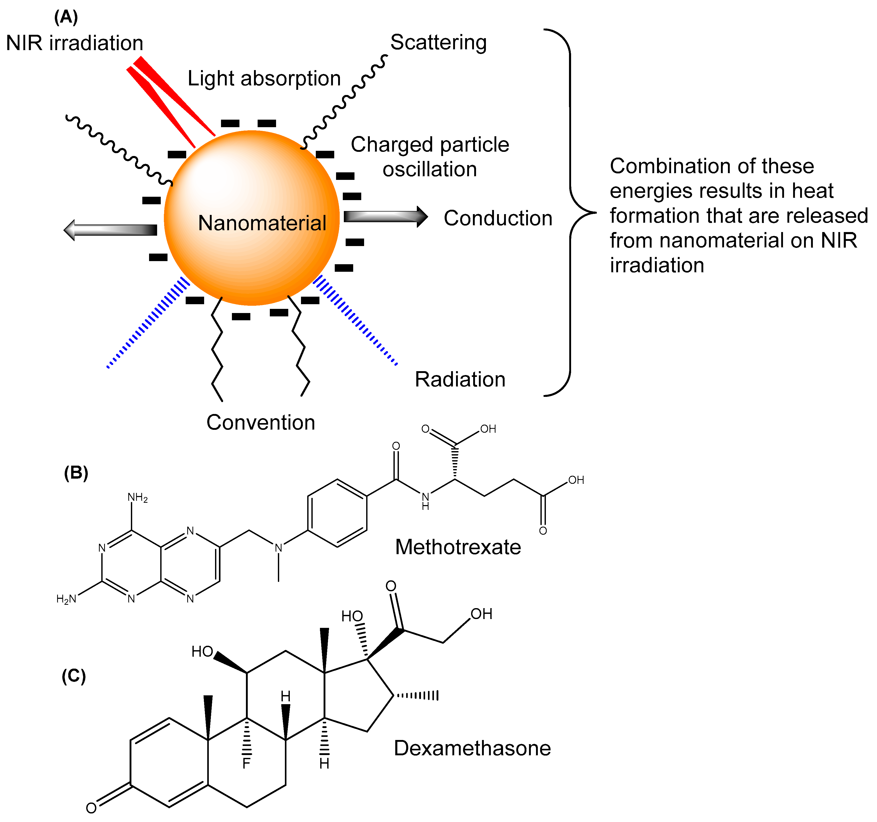
2. Fundamentals of Photothermal Therapy
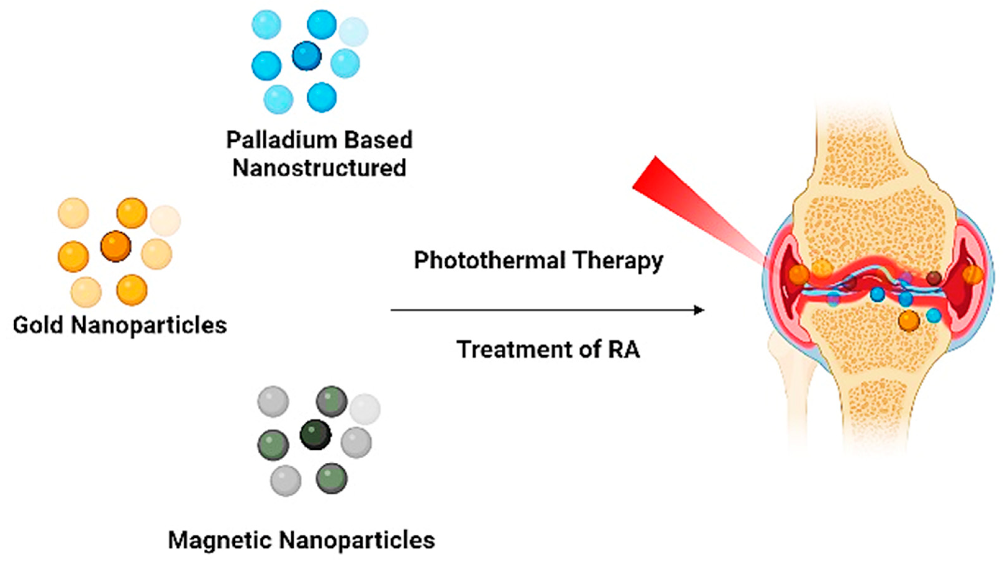
3. Various Polymer-Based Nanomaterials for Non-Invasive Photothermal Therapy of Arthritis
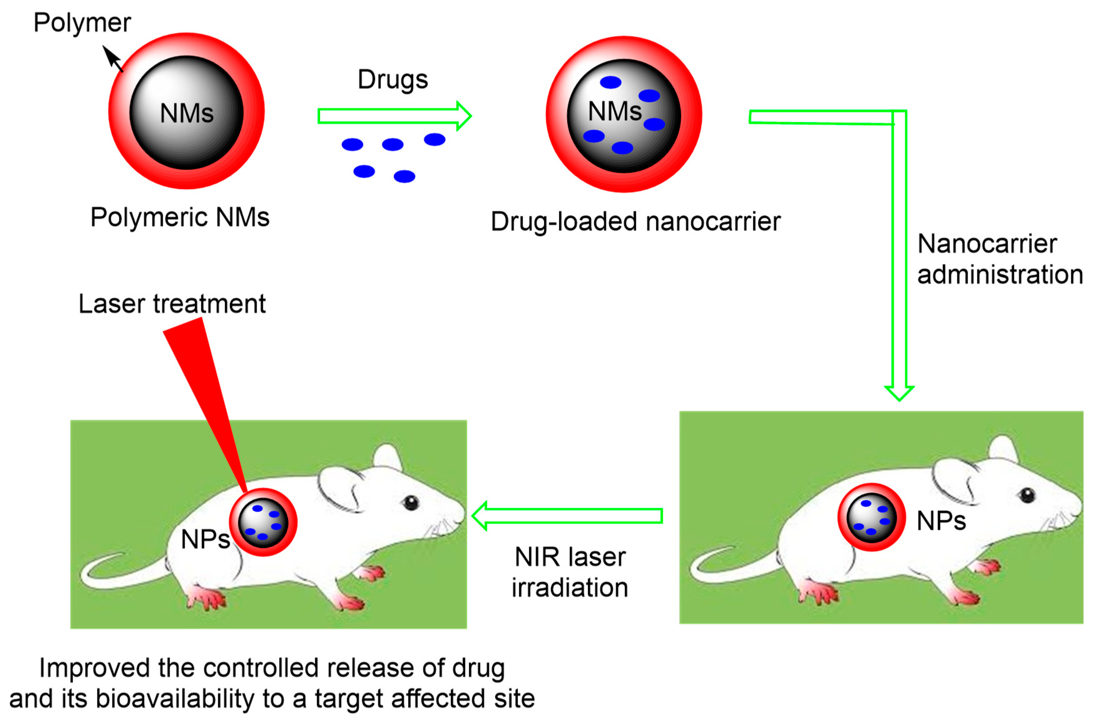
3.1. Polymer-Based Nanomaterials
3.2. Polymer-NIR780 Bioconjugate
3.3. Polymer-Based Gold Nanomaterials
3.4. Polymer-Based Copper Nanomaterials

| Photothermal Therapy | Objective of Investigation | Method of Synthesis | Drug Used | In Vitro Model Used | Animal Model Investigated | Outcome of Investigation | Ref |
|---|---|---|---|---|---|---|---|
| Polymer-based nanomaterials | Establish the controlled release of drug at specific arthritic sites using PCL-PEG micelles | Film dispersion | Dexamethasone | Murine macrophage Raw264.7 cell line and human umbilical vein endothelial cell | Rats with adjuvant-induced arthritis | Reduced joint swelling, bone erosion, and inflammatory cytokine expression in both joint tissue and serum | [12] |
| Methotrexate-loaded PCL–PEG–PCL) triblock copolymer against RA | Copolymerization reaction and precipitation | Methotrexate | Fresh human blood and non-activated and lipopolysaccharide-activated macrophages | Mouse with RA | MTX-loaded nanomicelles proved to be a promising agent against RA | [13] | |
| To evaluate the influential role of PLGA NPs coated with anti- cyclooxygenase-2 (COX2) siRNA in arthritis treatment | Water-in-oil-in-water solvent evaporation technique | Dexamethasone | Human chondrocyte cell line (C28/I2) | - | Synergistic action of dexamethasone and COX-2 siRNA treatment reduced the expression of inflammatory and apoptosis-related factors produced in C28/I2 cells | [23] | |
| Evaluation of QRu-PLGA-RES-DS NPs) for effective arthritis therapy | Precipitation and sonication technique | Resveratrol | RAW 264.7 and HUVECs cells | CIA mice | QRu-PLGA-RES-DS NPs effectively treated RA after eliminating the inflammatory response | [24] | |
| NO-Hb@siRNA@PLGA-PEG (NHsPP) was utilised for osteoarthritis therapy | Minor medication, precipitation, and conjugation processes | Nitric oxide | RAW 264.7 cells and HEK 293T) | Mice | The therapeutic effect of the NHsPP NPs was significantly enhanced compared to the treatment groups using only NO, siRNA, or PTT. | [14] | |
| Polymer-NIR780 bioconjugate | To investigate nanogold-core multifunctional dendrimer to establish photothermal therapy of RA | Citrate reduction method and conjugation process | Methotrexate | Mouse Macrophage RAW264.7 cells | - | Multifunctional targeted NPs proved to be potential therapeutics for the improved treatment of RA | [16] |
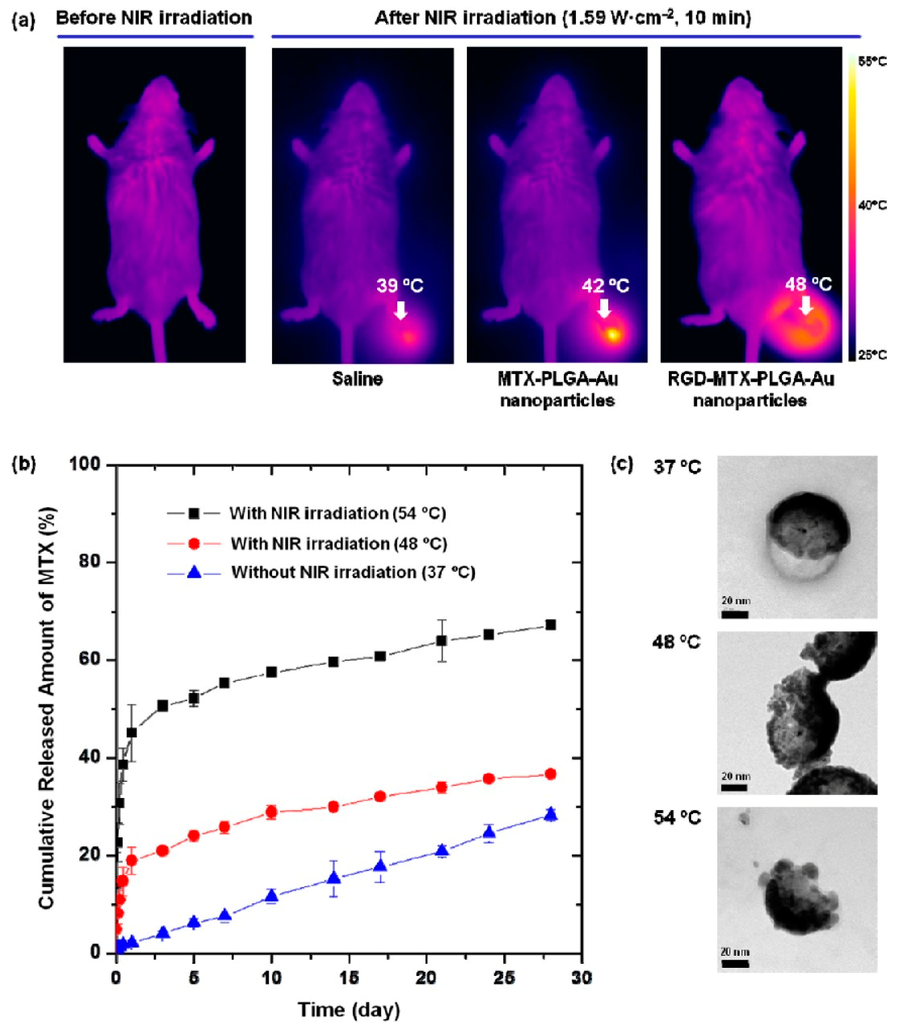
| Gold nanomaterial | Development of MTX-loaded poly(DL-lactic-co-glycolic acid) Au half-shell NPs (MTX-PLGA-Au) for arthritis treatment | Turkevich process for Au synthesis followed by MTX-PLGA conjugation | Methotrexate | Joint tissues were extracted for histological study | CIA Mice | This drug delivery system proved to be effective and minimised dosage-related MTX side effects in the treatment of RA | [25] |
| Hyaluronate–gold nanoparticle/Tocilizumab (HA-AuNP/TCZ) complex was prepared for RA | Thiolated HA (HA-SH) was synthesised by reductive amination and conjugated with Au prepared using the Turkevich method | Tocilizumab | HUVECs cells | CIA Mice | Ha/Au/TCZ complex can be used for RA as well as other therapeutic applications | [15] | |
| Improvement of (MTX)-loaded PLGA) gold (Au)/iron (Fe)/gold (Au) half-shell nanoparticles conjugated with arginine–glycine–aspartic acid (RGD) for magnetic targeted chemo-photothermal treatment of RA | MTX-PLGA was prepared by a solvent evaporation method, and RGD was conjugated with MTX-PLGA Au/Fe/Au NPs | Methotrexate | - | CIA Mice | The combined effect of NIR irradiation and external magnetic field enhanced the therapeutic effects of NPs with an MTX dosage of only 0.05% dosage compared to free MTX therapy for the treatment of RA | [20] | |
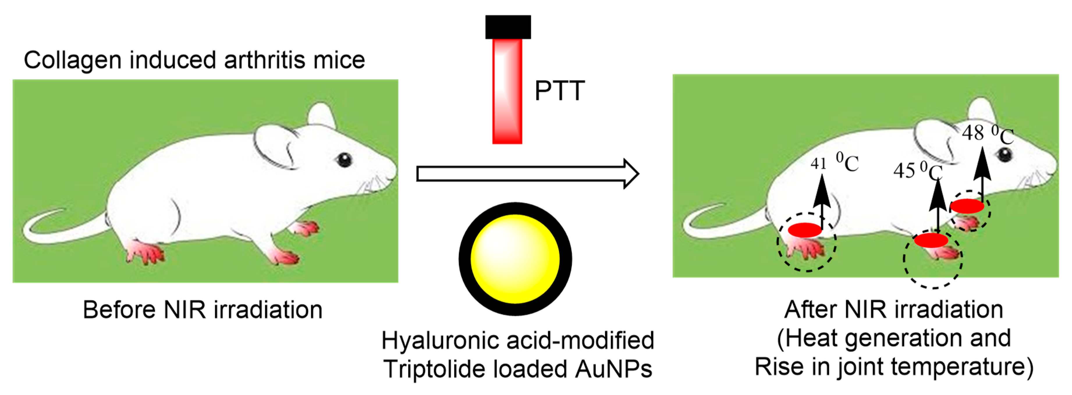
| Hydrogels of hyaluronic acid hybridised with triptolide/gold nanoparticles for targeted delivery to rheumatoid arthritis-affected regions combined with photothermal therapy | Hyaluronic acid (HA) hydrogels loaded onto an RGD-attached gold nanoshell containing TP are prepared | Triptolide (TP) | - | CIA mice | Targeted photothermal-chemotherapy using hybrid hydrogels for the treatment of RA is an effective strategy that can maximize the therapeutic effects and reduce dose-related side effects. | [19] | |
| Copper nanomaterial | To investigate combined effect of Au, CuS, and PTT on RA therapy | Seed-mediated and conjugated process | MTX | Murine fibroblast-like synovial cells (FLS) and mouse fibroblast (L929) | CIA rats | VIP-HA-Au NR@CuS-MTX revealed excellent therapeutic effects in vivo and higher NIR absorption than Au NR@CuS. Both HA and VIP played a significant part in controlling or regulating the release of MTX | [9] |
| Iron oxide nanomaterials | Synthesis of superparamagnetic IONPs to evaluate the efficacy of photothermal effect in arthritis treatment. | Solvothermal method | - | Cytotoxicity study (MTT assay) -RAW 264.7 cells | (CIA) mouse model | The prepared Fe3O4 nanoparticles showed promising results, with a size of 220 nm, high photothermal efficiency, and better targeting in the inflamed joint, thereby alleviating the symptoms of RA. | [26] |
| Black phosphorus nanosheets | Preparation of BPNs/Chitosan/PRP to study their synergistic effect with PTT and PDT therapy for improving osteogenesis in the treatment of RA. | Liquid exfoliation method | Methotrexate | Cytotoxicity assay using CCK-8 standard method (RAW264.7 cells, L929 cells and MSC cells) | (CIA) mouse model | Based on the distinguished concurrent PTT and PDT attributes of BPNs, chitosan/PRP thermos-responsive hydrogel effectively removed proliferating synoviocytes. In addition, BPNs accomplished calcium-extracted biomineralization via the in situ phosphorus-driven activation of BPNs in the target physiological microenvironment, which affected the entire course of treatment and provided improved therapeutic outcome. | [27] |
| Quantum dots | Synthesis of FAGM involved the following steps: 1. Formation of CTAB-coated gold nanorods (GNR) 2. GNR and methotrexate were coated with mesoporous silica shell 3. Synthesis of folic acid-functionalised GM. | 1. Formation of CTAB-coated gold nanorods (GNR). The GNRs were prepared via the reduction of HAuCl4 with sodium borohydride. Afterward, CTAB, HauCl4, AgNO3, and H2SO4 were added to the above solution in a certain amount. The solution was stirred at 28 °C for 30 min. Then, ascorbic acid was added dropwise, and a colour change was observed from light yellow to a state of colourlessness. The solution was kept overnight and stirred at 28 °C. 2. The prepared CTAB-GNR was again centrifuged and then resuspended. TEOS was added as a silica source and stirred for 30 min. Centrifuged and removed excess CTAB and FAGMs were collected. 3. Methotrexate was mixed with the prepared FAGM, and MTX-FAGM was formed. | Methotrexate | Cytotoxicity assay RAW 246.7 cells | Adjuvant-induced arthritis (AIA) rat model | In conclusion, for the therapy of RA, nanoscale MTX-FAGMs were prepared and their precisely targeted cytotoxicity towards active macrophages was confirmed under NIR laser irradiation. Synergistic action was observed between the photothermal therapy and chemotherapy | [28] |
3.5. Iron Oxide Nanomaterial
3.6. Black Phosphorus Nanosheets
3.7. Use of Quantum Dots Together with Photothermal Therapy of Arthritis
4. Biological Applications of Polymer-Based Nanomaterials for PTT of Arthritis
4.1. Photothermal Therapy of Arthritis
4.2. Chemophotothermal Therapy of Arthritis
4.3. Image-Guided Photothermal Therapy of Arthritis
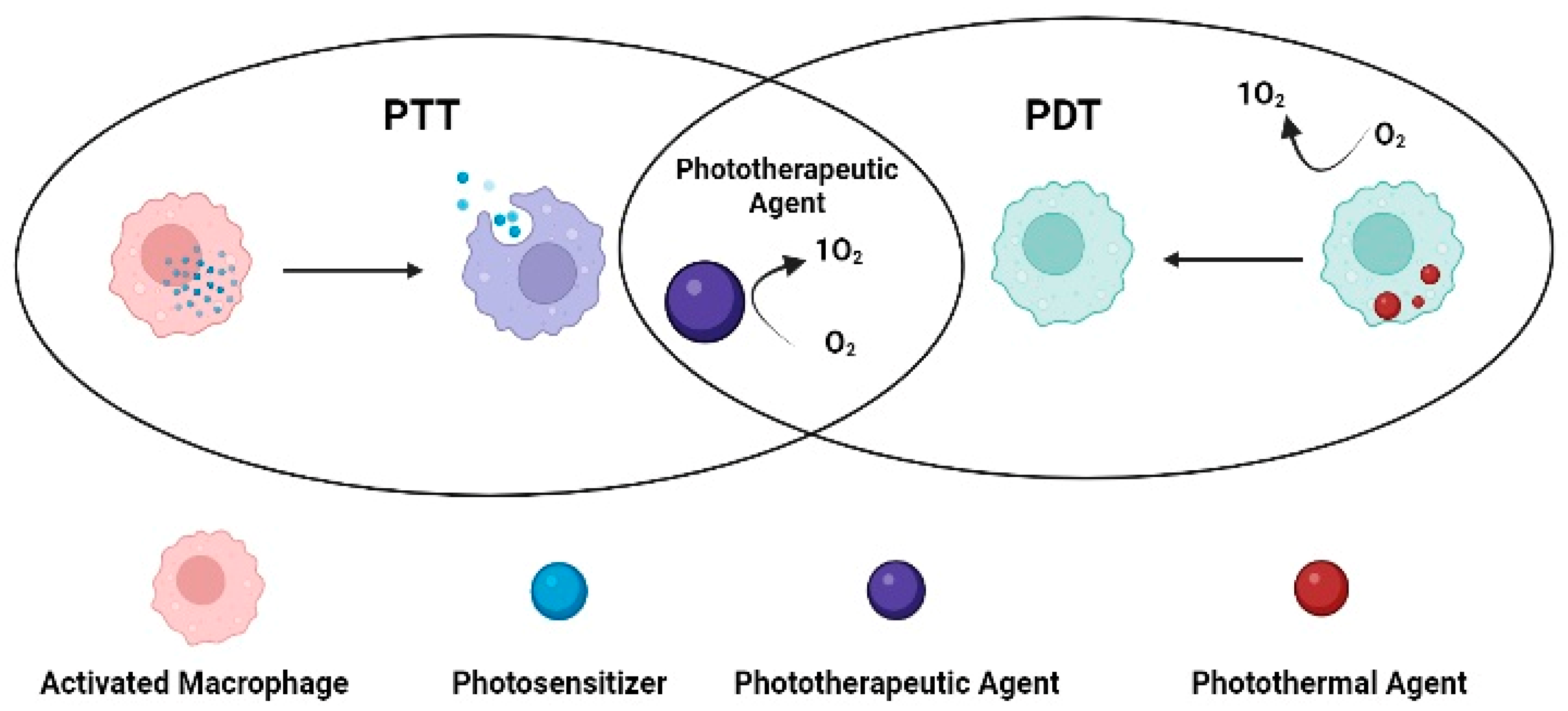
4.4. PDT Combined Photothermal Therapy of Arthritis
4.5. Gene Therapy-Combined Photothermal Therapy of Arthritis
5. Conclusions
Author Contributions
Funding
Institutional Review Board Statement
Informed Consent Statement
Data Availability Statement
Acknowledgments
Conflicts of Interest
Abbreviations
| AuSPIONs | Gold-coated superparamagnetic IONPs |
| BPNs | Black phosphorous nanosheets |
| CIA | Collagen-induced mouse model |
| CTAB | Cetrimonium bromide |
| IONPs | Iron oxide nanoparticles |
| MRI | Magnetic resonance imaging |
| PRP | Platelet-rich plasma |
| PTT | Photothermal therapy |
| RA | Rheumatoid arthritis |
| ROS | Reactive oxygen species |
| NMs | Nanomaterials |
| NPs | Nanoparticles |
| MTX | Methotrexate |
| DMARDs | Disease-modifying anti-rheumatic drugs |
| NIR | Near infra-red |
| PEG | Polyethylene glycol |
| RGD | Arginine–glycine–aspartic acid |
| PDT | Photodynamic therapy |
| HA | Hyaluronic acid |
| TNF-α | Tumour necrosis factor-α |
| NSAID | Non-steroidal anti-inflammatory medications |
| VIP | Vasoactive intestinal peptide |
| ENCs | Engineered nanocarriers |
| PMI | Photoacoustic molecular imaging |
| TCZ | Tocilizumab |
| Dex | Dexamethasone |
| PA | Photoacoustic |
| RNAi | RNA interference |
| siRNA | Small interference RNA |
References
- Smolen, J.S.; Aletaha, D.; McInnes, I.B. Rheumatoid arthritis. Lancet 2016, 388, 2023–2038. [Google Scholar] [CrossRef]
- Liang, W.; Yu, Y.; Liu, Z.; Ming, W.; Lin, H.; Long, H.; Zhao, J. The Therapeutic Potential of Targeted Nanoparticulate Systems to Treat Rheumatoid Arthritis. J. Nanomater. 2022, 2022, 8900658. [Google Scholar] [CrossRef]
- Dolati, S.; Sadreddini, S.; Rostamzadeh, D.; Ahmadi, M.; Jadidi-Niaragh, F.; Yousefi, M. Utilization of nanoparticle technology in rheumatoid arthritis treatment. Biomed. Pharmacother. 2016, 80, 30–41. [Google Scholar] [CrossRef]
- Shang, H.; Gu, H.; Zhang, N. From traditional to novel treatment of arthritis: A review of recent advances in nanotechnology-based thermal therapy. Nanomedicine 2021, 16, 2117–2132. [Google Scholar] [CrossRef]
- Zhang, X.; Koo, S.; Kim, J.H.; Huang, X.; Kong, N.; Zhang, L.; Zhou, J.; Xue, J.; Harris, M.B.; Tao, W. Nanoscale materials-based platforms for the treatment of bone-related diseases. Matter 2021, 4, 2727–2764. [Google Scholar] [CrossRef]
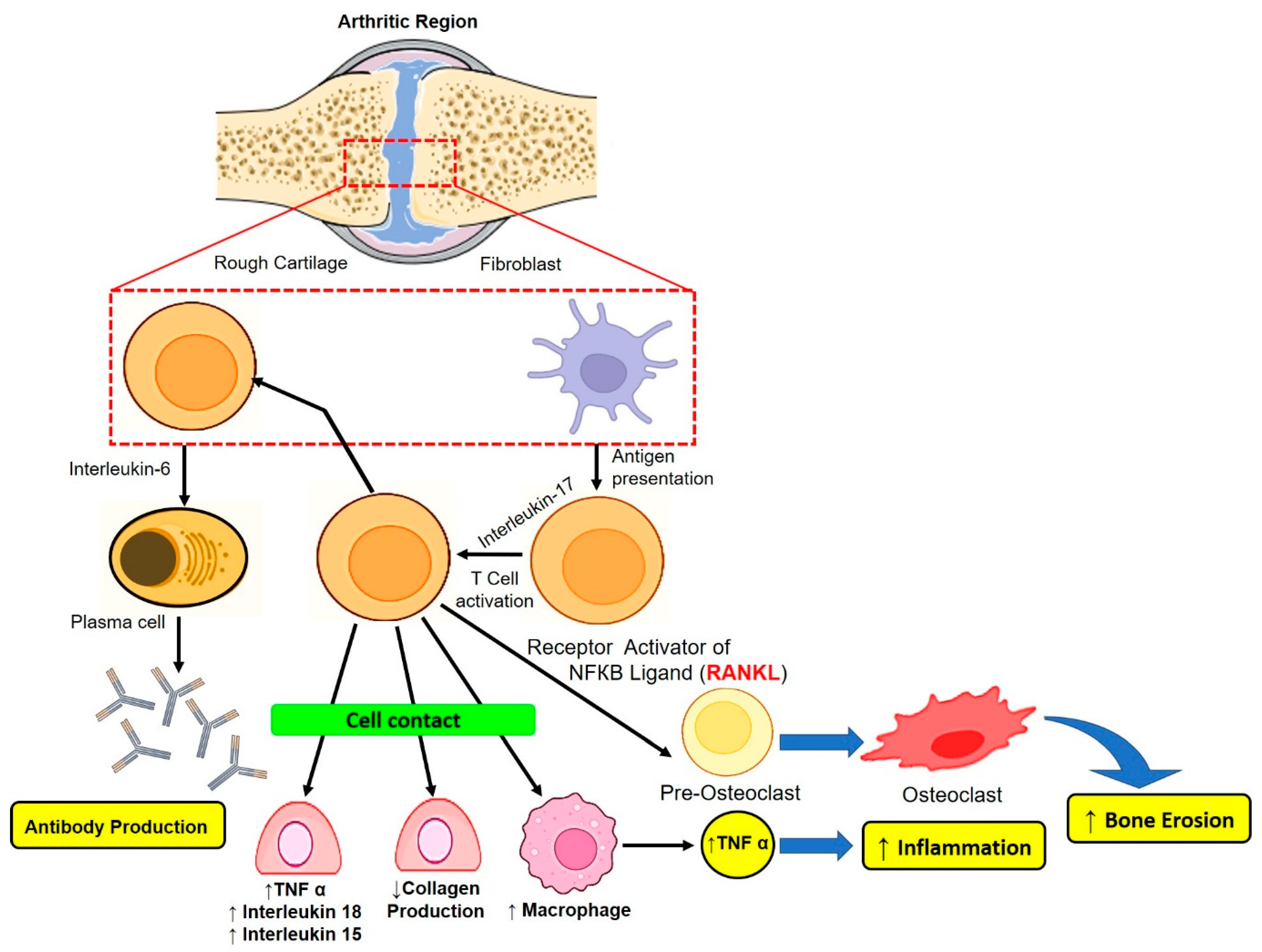
- Münz, C.; Lünemann, J.D.; Getts, M.T.; Miller, S.D. Antiviral immune responses: Triggers of or triggered by autoimmunity? Nat. Rev. Immunol. 2009, 9, 246–258. [Google Scholar] [CrossRef] [Green Version]
- Beech, J.T.; Andreakos, E.; Ciesielski, C.J.; Green, P.; Foxwell, B.M.; Brennan, F.M. T-cell contact-dependent regulation of CC and CXC chemokine production in monocytes through differential involvement of NFκB: Implications for rheumatoid arthritis. Arthritis Res. Ther. 2006, 8, 1–10. [Google Scholar] [CrossRef] [Green Version]
- Tran, C.N.; Lundy, S.K.; White, P.T.; Endres, J.L.; Motyl, C.D.; Gupta, R.; Wilke, C.M.; Shelden, E.A.; Chung, K.C.; Urquhart, A.G. Molecular interactions between T cells and fibroblast-like synoviocytes: Role of membrane tumor necrosis factor-α on cytokine-activated T cells. Am. J. Pathol. 2007, 171, 1588–1598. [Google Scholar] [CrossRef] [Green Version]
- Huang, Q.-Q.; Pope, R.M. The role of toll-like receptors in rheumatoid arthritis. Curr. Rheumatol. Rep. 2009, 11, 357–364. [Google Scholar] [CrossRef] [Green Version]
- Mavers, M.; Ruderman, E.M.; Perlman, H. Intracellular signal pathways: Potential for therapies. Curr. Rheumatol. Rep. 2009, 11, 378–385. [Google Scholar] [CrossRef] [Green Version]
- Gibofsky, A. Epidemiology, pathophysiology, and diagnosis of rheumatoid arthritis: A Synopsis. Am. J. Manag. Care 2014, 20, S128–S135. [Google Scholar]
- Wang, Q.; Jiang, J.; Chen, W.; Jiang, H.; Zhang, Z.; Sun, X. Targeted delivery of low-dose dexamethasone using PCL–PEG micelles for effective treatment of rheumatoid arthritis. J. Control. Release 2016, 230, 64–72. [Google Scholar] [CrossRef]
- Qindeel, M.; Khan, D.; Ahmed, N.; Khan, S.; Rehman, A.U. Surfactant-free, self-assembled nanomicelles-based transdermal hydrogel for safe and targeted delivery of methotrexate against rheumatoid arthritis. ACS Nano 2020, 14, 4662–4681. [Google Scholar] [CrossRef]
- Chen, X.; Liu, Y.; Wen, Y.; Yu, Q.; Liu, J.; Zhao, Y.; Liu, J.; Ye, G. A photothermal-triggered nitric oxide nanogenerator combined with siRNA for precise therapy of osteoarthritis by suppressing macrophage inflammation. Nanoscale 2019, 11, 6693–6709. [Google Scholar] [CrossRef]
- Lee, S.-M.; Kim, H.J.; Ha, Y.-J.; Park, Y.N.; Lee, S.-K.; Park, Y.-B.; Yoo, K.-H. Targeted chemo-photothermal treatments of rheumatoid arthritis using gold half-shell multifunctional nanoparticles. ACS Nano 2013, 7, 50–57. [Google Scholar] [CrossRef]
- Pandey, P.K.; Maheshwari, R.; Raval, N.; Gondaliya, P.; Kalia, K.; Tekade, R.K. Nanogold-core multifunctional dendrimer for pulsatile chemo-, photothermal-and photodynamic-therapy of rheumatoid arthritis. J. Colloid Interface Sci. 2019, 544, 61–77. [Google Scholar] [CrossRef]
- Gadeval, A.; Chaudhari, S.; Bollampally, S.P.; Polaka, S.; Kalyane, D.; Sengupta, P.; Kalia, K.; Tekade, R.K. Integrated nanomaterials for non-invasive photothermal therapy of rheumatoid arthritis. Drug Discov. Today 2021, 26, 2315–2328. [Google Scholar] [CrossRef]
- Sibuyi, N.R.S.; Moabelo, K.L.; Fadaka, A.O.; Meyer, S.; Onani, M.O.; Madiehe, A.M.; Meyer, M. Multifunctional Gold Nanoparticles for Improved Diagnostic and Therapeutic Applications: A Review. Nanoscale Res. Lett. 2021, 16, 174. [Google Scholar] [CrossRef]
- Li, C.; Liu, R.; Song, Y.; Zhu, D.; Yu, L.; Huang, Q.; Zhang, Z.; Xue, Z.; Hua, Z.; Lu, C. Intra-articular Administrated Hydrogels of Hyaluronic Acid Hybridized with Triptolide/Gold Nanoparticles for Targeted Delivery to Rheumatoid Arthritis Combined with Photothermal-chemo Therapy. Res. Sq. 2021. [Google Scholar] [CrossRef]
- Kim, H.J.; Lee, S.-M.; Park, K.-H.; Mun, C.H.; Park, Y.-B.; Yoo, K.-H. Drug-loaded gold/iron/gold plasmonic nanoparticles for magnetic targeted chemo-photothermal treatment of rheumatoid arthritis. Biomaterials 2015, 61, 95–102. [Google Scholar] [CrossRef]
- Xu, C.; Chen, J.; Li, L.; Pu, X.; Chu, X.; Wang, X.; Li, M.; Lu, Y.; Zheng, X. Promotion of chondrogenic differentiation of mesenchymal stem cells by copper: Implications for new cartilage repair biomaterials. Mater. Sci. Eng. C 2018, 93, 106–114. [Google Scholar] [CrossRef]
- Huang, R.; Zhang, C.; Bu, Y.; Li, Z.; Zheng, X.; Qiu, S.; Machuki, J.O.A.; Zhang, L.; Yang, Y.; Guo, K. A multifunctional nano-therapeutic platform based on octahedral yolk-shell Au NR@ CuS: Photothermal/photodynamic and targeted drug delivery tri-combined therapy for rheumatoid arthritis. Biomaterials 2021, 277, 121088. [Google Scholar] [CrossRef]
- Park, J.S.; Yang, H.N.; Jeon, S.Y.; Woo, D.G.; Kim, M.S.; Park, K.-H. The use of anti-COX2 siRNA coated onto PLGA nanoparticles loading dexamethasone in the treatment of rheumatoid arthritis. Biomaterials 2012, 33, 8600–8612. [Google Scholar] [CrossRef]
- Chen, X.; Zhu, X.; Ma, L.; Lin, A.; Gong, Y.; Yuan, G.; Liu, J. A core–shell structure QRu-PLGA-RES-DS NP nanocomposite with photothermal response-induced M2 macrophage polarization for rheumatoid arthritis therapy. Nanoscale 2019, 11, 18209–18223. [Google Scholar] [CrossRef]
- Lee, H.; Lee, M.-Y.; Bhang, S.H.; Kim, B.-S.; Kim, Y.S.; Ju, J.H.; Kim, K.S.; Hahn, S.K. Hyaluronate–gold nanoparticle/tocilizumab complex for the treatment of rheumatoid arthritis. ACS Nano 2014, 8, 4790–4798. [Google Scholar] [CrossRef]
- Zhang, S.; Wu, L.; Cao, J.; Wang, K.; Ge, Y.; Ma, W.; Qi, X.; Shen, S. Effect of magnetic nanoparticles size on rheumatoid arthritis targeting and photothermal therapy. Colloids Surf. B Biointerfaces 2018, 170, 224–232. [Google Scholar] [CrossRef]
- Pan, W.; Dai, C.; Li, Y.; Yin, Y.; Gong, L.; Machuki, J.O.A.; Yang, Y.; Qiu, S.; Guo, K.; Gao, F. PRP-chitosan thermoresponsive hydrogel combined with black phosphorus nanosheets as injectable biomaterial for biotherapy and phototherapy treatment of rheumatoid arthritis. Biomaterials 2020, 239, 119851. [Google Scholar] [CrossRef]
- Li, X.; Zhang, S.; Zhang, X.; Hou, Y.; Meng, X.; Li, G.; Xu, F.; Teng, L.; Qi, Y.; Sun, F.; et al. Folate receptor-targeting semiconducting polymer dots hybrid mesoporous silica nanoparticles against rheumatoid arthritis through synergistic photothermal therapy, photodynamic therapy, and chemotherapy. Int. J. Pharm. 2021, 607, 120947. [Google Scholar] [CrossRef]
- Xiao, L.; Wu, Z.; Zhang, J.; Wang, G.; Ma, Y.; Ding, Y.; He, X.; Zhang, S.; Zhang, Z. Synthesis, photothermal effect and cytotoxicity of Fe3O4@Au nanocomposites. J. Nanosci. Nanotechnol. 2019, 19, 2467–2473. [Google Scholar] [CrossRef]
- Chen, C.-L.; Siow, T.Y.; Chou, C.-H.; Lin, C.-H.; Lin, M.-H.; Chen, Y.-C.; Hsieh, W.-Y.; Wang, S.-J.; Chang, C. Targeted superparamagnetic iron oxide nanoparticles for in vivo magnetic resonance imaging of T-cells in rheumatoid arthritis. Mol. Imaging Biol. 2017, 19, 233–244. [Google Scholar] [CrossRef]
- Carneiro, M.F.H.; Machado, A.R.T.; Antunes, L.M.; Souza, T.E.; Freitas, V.A.; Oliveira, L.C.; Rodrigues, J.L.; Pereira, M.C.; Barbosa, F. Gold-coated superparamagnetic iron oxide nanoparticles attenuate collagen-induced arthritis after magnetic targeting. Biol. Trace Elem. Res. 2020, 194, 502–513. [Google Scholar] [CrossRef]
- Liu, J.; Du, P.; Mao, H.; Zhang, L.; Ju, H.; Lei, J. Dual-triggered oxygen self-supply black phosphorus nanosystem for enhanced photodynamic therapy. Biomaterials 2018, 172, 83–91. [Google Scholar] [CrossRef]
- Yang, X.; Wang, D.; Shi, Y.; Zou, J.; Zhao, Q.; Zhang, Q.; Huang, W.; Shao, J.; Xie, X.; Dong, X. Black phosphorus nanosheets immobilizing Ce6 for imaging-guided photothermal/photodynamic cancer therapy. ACS Appl. Mater. Interfaces 2018, 10, 12431–12440. [Google Scholar] [CrossRef]
- Itkonen, S.T.; Rita, H.J.; Saarnio, E.M.; Kemi, V.E.; Karp, H.J.; Kärkkäinen, M.U.; Pekkinen, M.H.; Laitinen, E.K.; Risteli, J.; Koivula, M.-K. Dietary phosphorus intake is negatively associated with bone formation among women and positively associated with some bone traits among men—A cross-sectional study in middle-aged Caucasians. Nutr. Res. 2017, 37, 58–66. [Google Scholar] [CrossRef] [Green Version]
- Lu, Y.; Li, L.; Lin, Z.; Wang, L.; Lin, L.; Li, M.; Zhang, Y.; Yin, Q.; Li, Q.; Xia, H. A new treatment modality for rheumatoid arthritis: Combined photothermal and photodynamic therapy using Cu7.2S4 nanoparticles. Adv. Healthc. Mater. 2018, 7, 1800013. [Google Scholar] [CrossRef]
- Simón-Vázquez, R.; Tsapis, N.; Lorscheider, M.; Rodríguez, A.; Calleja, P.; Mousnier, L.; de Miguel Villegas, E.; González-Fernández, Á.; Fattal, E. Improving dexamethasone drug loading and efficacy in treating arthritis through a lipophilic prodrug entrapped into PLGA-PEG nanoparticles. Drug Deliv. Transl. Res. 2022, 12, 1270–1284. [Google Scholar] [CrossRef]
- Zheng, C.; Wu, A.; Zhai, X.; Ji, H.; Chen, Z.; Chen, X.; Yu, X. The cellular immunotherapy of integrated photothermal anti-oxidation Pd–Se nanoparticles in inhibition of the macrophage inflammatory response in rheumatoid arthritis. Acta Pharm. Sin. B 2021, 11, 1993–2003. [Google Scholar] [CrossRef]
- Zhang, S.; Zhang, M.; Li, X.; Li, G.; Yang, B.; Lu, X.; Gao, Y.; Sun, F. Nano-Based Co-Delivery System for Treatment of Rheumatoid Arthritis. Molecules 2022, 27, 5973. [Google Scholar] [CrossRef]
- Ren, H.; He, Y.; Liang, J.; Cheng, Z.; Zhang, M.; Zhu, Y.; Hong, C.; Qin, J.; Xu, X.; Wang, J. Role of liposome size, surface charge, and PEGylation on rheumatoid arthritis targeting therapy. ACS Appl. Mater. Interfaces 2019, 11, 20304–20315. [Google Scholar] [CrossRef]
- Pirmardvand Chegini, S.; Varshosaz, J.; Taymouri, S. Recent approaches for targeted drug delivery in rheumatoid arthritis diagnosis and treatment. Artif. Cells Nanomed. Biotechnol. 2018, 46 (Suppl. 2), 502–514. [Google Scholar] [CrossRef] [Green Version]
- Hashemkhani, M.; Muti, A.; Sennaroğlu, A.; Acar, H.Y. Multimodal image-guided folic acid targeted Ag-based quantum dots for the combination of selective methotrexate delivery and photothermal therapy. J. Photochem. Photobiol. B Biol. 2020, 213, 112082. [Google Scholar] [CrossRef]
- Castano, A.P.; Demidova, T.N.; Hamblin, M.R. Mechanisms in photodynamic therapy: Part one-photosensitizers, photochemistry and cellular localization. Photodiagnosis Photodyn. Ther. 2004, 1, 279–293. [Google Scholar] [CrossRef] [Green Version]
- Hou, Y.-J.; Yang, X.-X.; Liu, R.-Q.; Zhao, D.; Guo, C.-X.; Zhu, A.-C.; Wen, M.-N.; Liu, Z.; Qu, G.-F.; Meng, H.-X. Pathological mechanism of photodynamic therapy and photothermal therapy based on nanoparticles. Int. J. Nanomed. 2020, 15, 6827. [Google Scholar] [CrossRef]
- Kwiatkowski, S.; Knap, B.; Przystupski, D.; Saczko, J.; Kędzierska, E.; Knap-Czop, K.; Kotlińska, J.; Michel, O.; Kotowski, K.; Kulbacka, J. Photodynamic therapy—Mechanisms, photosensitizers and combinations. Biomed. Pharmacother. 2018, 106, 1098–1107. [Google Scholar] [CrossRef]
- Calixto, G.M.F.; Bernegossi, J.; de Freitas, L.M.; Fontana, C.R.; Chorilli, M. Nanotechnology-based drug delivery systems for photodynamic therapy of cancer: A review. Molecules 2016, 21, 342. [Google Scholar] [CrossRef]
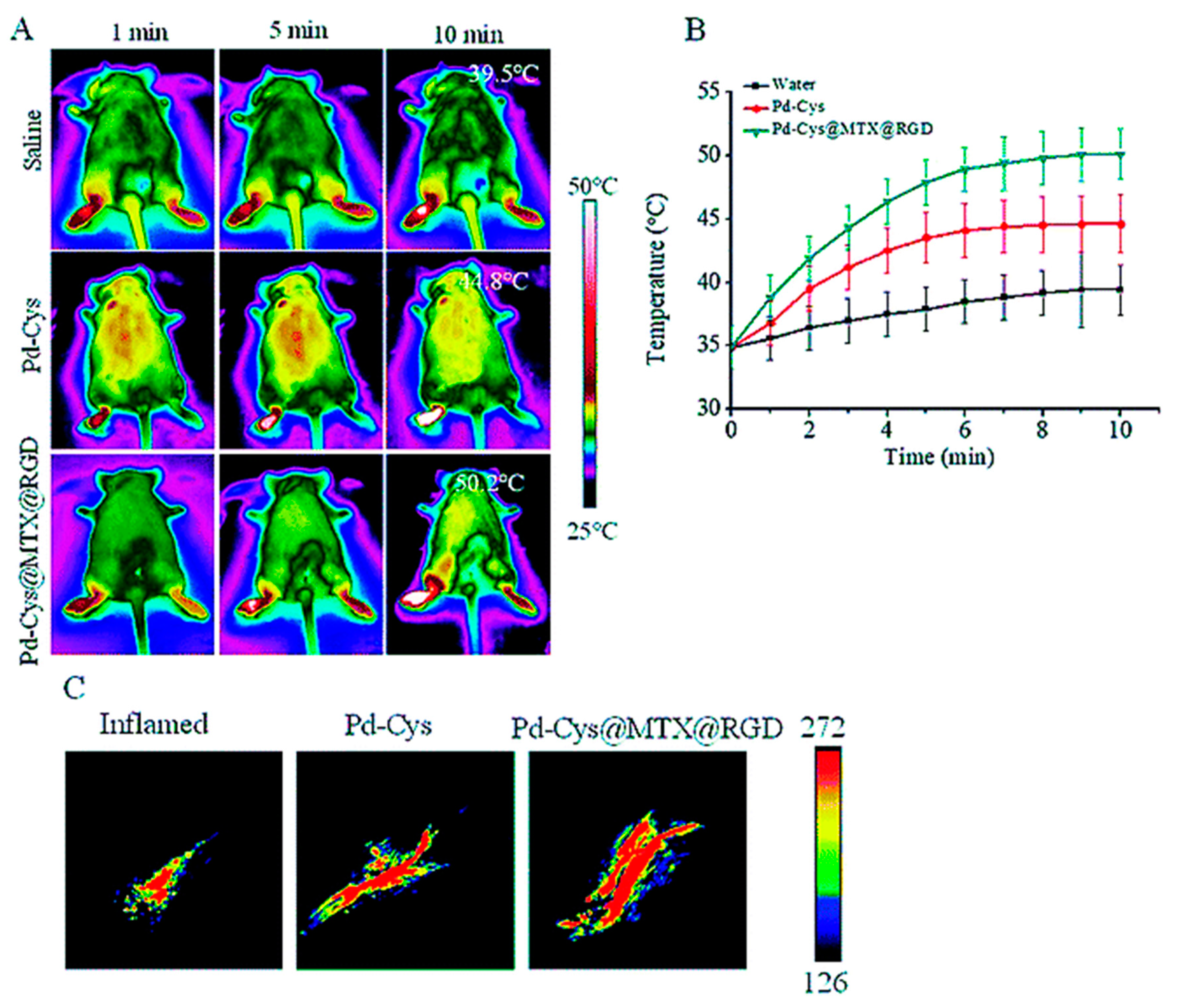
- Chen, X.; Zhu, X.; Xu, T.; Xu, M.; Wen, Y.; Liu, Y.; Liu, J.; Qin, X. Targeted hexagonal Pd nanosheet combination therapy for rheumatoid arthritis via the photothermal controlled release of MTX. J. Mater. Chem. B 2019, 7, 112–122. [Google Scholar] [CrossRef]
- Wang, Y.; Meng, H.-M.; Song, G.; Li, Z.; Zhang, X.-B. Conjugated-polymer-based nanomaterials for photothermal therapy. ACS Appl. Polym. Mater. 2020, 2, 4258–4272. [Google Scholar] [CrossRef]
- Scheinman, R.I.; Trivedi, R.; Vermillion, S.; Kompella, U.B. Functionalized STAT1 siRNA nanoparticles regress rheumatoid arthritis in a mouse model. Nanomedicine 2011, 6, 1669–1682. [Google Scholar] [CrossRef]
- Desai, P.R.; Marepally, S.; Patel, A.R.; Voshavar, C.; Chaudhuri, A.; Singh, M. Topical delivery of anti-TNFα siRNA and capsaicin via novel lipid-polymer hybrid nanoparticles efficiently inhibits skin inflammation in vivo. J. Control. Release 2013, 170, 51–63. [Google Scholar] [CrossRef] [Green Version]


| Photothermal Therapy | Nanomaterial Used | Combination Therapy | In Vitro Model Used | Animal Model Investigated | Outcome of Investigation | Ref |
|---|---|---|---|---|---|---|
| Photothermal Therapy alone | Palladium nanosheets | - | CIA Mouse | Pd-MTX nanosheets reduced the toxicity of MTX and prevented VEGF from proliferating as well as the generation of pro-inflammatory cytokines such as TNF-a and COX-2. | [17] | |
| Gold nanorods | PTT combined with chemotherapy | - | CIA mouse model | Aggregation of NPs increased in the presence of an external magnetic field, which raised the temperature in the exposed region and expedited the release of MTX. | [17] | |
| Magnetic iron oxide (Fe3O4) NPs | PTT combined with chemotherapy | CIA mouse model | Since NPs’ small size allowed them to enter inflammatory cells, more of them accumulated in inflamed joints. | [17] | ||
| Chemo-photothermal therapy | Gold half-shelled Nanoparticles | Chemotherapy with photothermal therapy | CIA mouse model | In the inflammatory joints, nanoparticle accumulation was elevated. | [15] | |
| PDT combined Photothermal therapy | Palladium nanosheets | PTT combined with photothermal therapy | Fibroblast cells | CIA Mouse | Pd-MTX nanosheets reduced the toxicity of MTX and prevented VEGF from proliferating as well as the generation of pro-inflammatory cytokines such as TNF-a and COX-2. | [14] |
| Copper-based Nanoparticles | - | CIA Mouse | Cu7.2S4 NPs act on joints that showed good bone retention and resembled normal joints | [35] | ||
| Gene therapy combined Photothermal therapy | Poly (lactide-co-glycolide) nanoparticles (NPs) | Gene therapy combined with photothermal therapy | - | CIA Mice | RGD functionalised PLGA nanoparticles encapsulating STAT1-targeted siRNAs may be more efficient, presumably by selectively inhibiting macrophage and dendritic cell activation. | [36] |
| Dexamethasone-loaded PLGA nanospheres | Gene therapy combined with photothermal therapy | - | CIA Mouse Human TNF Transgene model | The effectiveness of PLGA-PEG NPs for the delivery of a therapeutic medicine in the affected tissues in rheumatoid arthritis was low, perhaps because of the leaky vasculature, angiogenesis, and associated ELVIS effect that occur in the affected joints. | [36] |
Disclaimer/Publisher’s Note: The statements, opinions and data contained in all publications are solely those of the individual author(s) and contributor(s) and not of MDPI and/or the editor(s). MDPI and/or the editor(s) disclaim responsibility for any injury to people or property resulting from any ideas, methods, instructions or products referred to in the content. |
© 2023 by the authors. Licensee MDPI, Basel, Switzerland. This article is an open access article distributed under the terms and conditions of the Creative Commons Attribution (CC BY) license (https://creativecommons.org/licenses/by/4.0/).
Share and Cite
Tekade, M.; Pingale, P.; Gupta, R.; Pawar, B.; Tekade, R.K.; Sharma, M.C. Recent Advances in Polymer-Based Nanomaterials for Non-Invasive Photothermal Therapy of Arthritis. Pharmaceutics 2023, 15, 735. https://doi.org/10.3390/pharmaceutics15030735
Tekade M, Pingale P, Gupta R, Pawar B, Tekade RK, Sharma MC. Recent Advances in Polymer-Based Nanomaterials for Non-Invasive Photothermal Therapy of Arthritis. Pharmaceutics. 2023; 15(3):735. https://doi.org/10.3390/pharmaceutics15030735
Chicago/Turabian StyleTekade, Muktika, Prashant Pingale, Rachna Gupta, Bhakti Pawar, Rakesh Kumar Tekade, and Mukesh Chandra Sharma. 2023. "Recent Advances in Polymer-Based Nanomaterials for Non-Invasive Photothermal Therapy of Arthritis" Pharmaceutics 15, no. 3: 735. https://doi.org/10.3390/pharmaceutics15030735