
Updates on Biodegradable Formulations for Ocular Drug Delivery

Abstract
:1. Introduction
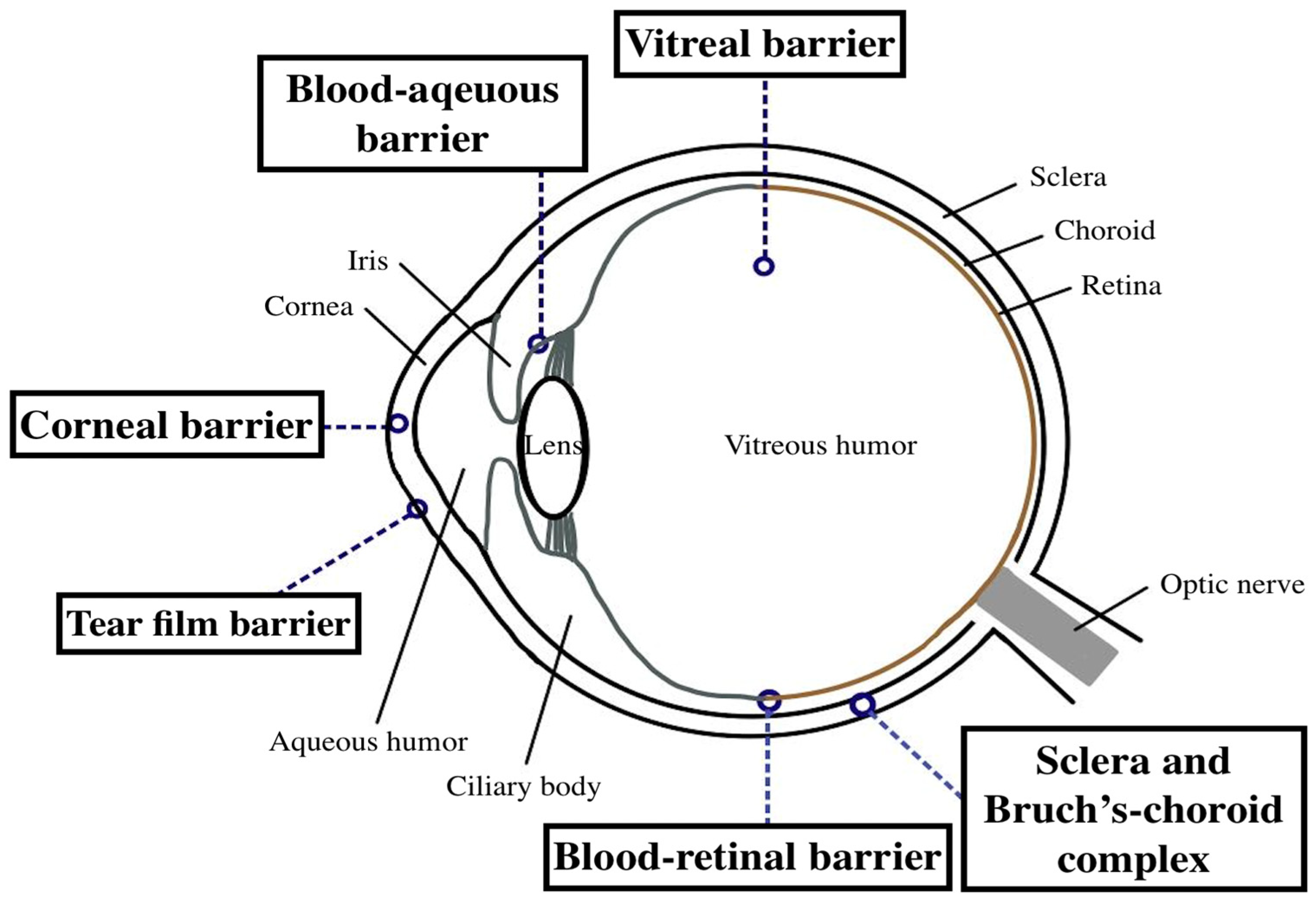
2. Barriers to Effective Ocular Drug Delivery
2.1. Barriers in the Anterior Segment
2.1.1. Tear Film
2.1.2. Cornea
2.1.3. Blood–Aqueous Barrier
2.2. Barriers in the Posterior Segment
2.2.1. Vitreous Humor
2.2.2. Sclera and Bruch’s–Choroid Complex
2.2.3. Blood-Retinal Barrier

3. Conventional Routes of Ocular Drug Delivery
3.1. Topical Administration
3.2. Systemic Administration
3.3. Periocular and Intraocular Injections
4. Novel Biodegradable Ocular Drug Delivery Systems
4.1. Nanotechnology-Based Systems
4.1.1. Nanoparticles
4.1.2. Liposomes and Niosomes
4.1.3. Dendrimers
4.1.4. Microemulsions
4.1.5. Nanosuspensions
4.1.6. Polymeric Micelles
4.2. Biodegradable Microneedles
4.3. Hydrogels
4.3.1. In Situ Gels
4.3.2. Hydrogel Implants
4.4. Biodegradable Implants
5. Application of Different Biodegradable Ocular Drug Delivery Systems on Ocular Diseases
5.1. Dry Eye Disease

| Study | Drug and System | Experimental Models | Description |
|---|---|---|---|
| Chen et al., 2022 [113] | Tacrolimus-loaded cationic liposomes | Male New Zealand rabbits | The use of cationic liposomes to encapsulate FK506 prolonged ocular retention, increased corneal FK506 levels, and reduced reactive oxygen species and dry eye–related inflammation factors. |
| Han et al., 2022 [114] | Polyhedral oligomeric silsesquioxane hybrid thermoresponsive FK506 hydrogel | Female C57BL/6 mice | This hydrogel possesses good biocompatibility, prolonged ocular retention and enhanced therapeutic efficiency in comparison with Commercial FK506 in dry eye. |
| Mirgorodskaya et al., 2022 [107] | Indomethacin-loaded MEs and nanoemulsions | A carrageenan-induced edema rat model | The Indomethacin nanoemulsion showed a prolonged release and slowed down the progression of carrageenan-induced edema compared to the unencapsulated drug. |
| Akbari et al., 2021 [97] | Hyaluronic acid-loaded chitosan nanoparticle-containing ring-implanted PVA contact lens | In vitro | The ring-implanted contact lens showed sustained release of hyaluronic acid for up to 14 days, and a cellular study indicated no corneal epithelial cell toxicity. |
| Eldesouky et al., 2021 [112] | CsA lipid nanocapsules as thermoresponsive in situ gel | Male New Zealand colored rabbits | This drug delivery form extends the ocular stay of CsA and improves its tissue penetration capacity. A subsequent pharmacodynamic study showed it was more effective than the commercially available cyclosporine nanoemulsion in increasing tear production in rabbits. |
| Ma et al., 2021 [115] | Levocarnitine thermosensitive in situ gel | New Zealand rabbits | The formulation can significantly increase the amount of tear secretion and the number of conjunctival goblet cells, improve the degree of corneal damage and the pathological morphology of the lacrimal gland, and down-regulate the apoptosis rate of corneal epithelial cells. |
| Yan et al., 2021 [111] | CsA loaded cationic NSs | Male adult New Zealand albino rabbits | The cationic NSs can deliver CsA to anterior ocular tissues in effective therapeutic concentrations (10–20 μg/g) with topical drop instillation. |
| Nagai et al., 2020 [116] | Rebamipide solid nanoparticle-based sustained-release formulations | Adult rabbits | The rebamipide formulation showed sustained release compared to the commercial suspension, and improved mucin levels and tear film breakup in an N-acetylcysteine treated rabbit model. |
| Sánchez-López et al., 2020 [41] | Dexibuprofen-loaded PLGA NPs | New Zealand albino rabbits | These formulations were able to release dexibuprofen more effectively for the treatment of ocular inflammation. |
| Hanafy et al., 2019 [42] | Prednisolone acetate loaded chitosan-deoxycholate self-assembled NPs | Female guinea pig eyes | The formulation achieved a twofold increase in the prednisolone release after 24 h when compared with the commercial micronized drug-loaded gel. |
| Wang et al., 2019 [43] | Dexamethasone sodium phosphate-loaded NPs using zinc ion bridging with dense coatings of polyethylene glycol | A corneal neovascularization rat model | A single subconjunctival administration inhibited corneal angiogenesis in rats within 2 weeks, without increasing IOP or causing toxicity at the injection site, which could be an effective strategy for preventing and treating corneal neovascularization. |
| Ryu et al., 2019 [44] | Dry tablets dexamethasone-loaded micelles | Male New Zealand white rabbits | This formulation increased 2.6-fold the ocular drug bioavailability when compared to Maxidex®. |
| Gonzalez-Pizarro et al., 2019 [117] | Fluorometholone loaded PLGA NPs in situ forming gels | New Zealand albino male rabbits | The formulation administration improved precorneal residency time, leading to increased ocular bioavailability and penetration into deep tissues such as aqueous humor and crystalline. |
| Tatke et al., 2018 [106] | Ion-sensitive in situ gelling system containing triamcinolone acetonide–loaded solid lipid NPs | New Zealand albino rabbits | The ex vivo on rabbit corneas showed improved permeability with the formulation compared to simple solutions. The gelation increased drug absorption and prolonged drug residence time on the ocular surface and in the conjunctiva sac, leading to sustained release and minimal pre-corneal drug loss. |
| Ren et al., 2018 [118] | Azithromycin–cholesteryl hemisuccinate ion pair in liposome | A dry eye rat model | Azithromycin liposomes showed enhanced corneal permeation compared to the azithromycin solution. |
| Huang et al., 2018 [119] | Gelatin–epigallocatechin gallate NPs with hyaluronic acid | New Zealand white rabbits | The eye drops effectively prolonging drug retention on the ocular surface and effectively inhibiting ocular inflammation in dry eye rabbits. |
| Vaidehi et al., 2017 [120] | Tacrolimus loaded modified liposomes with propylene glycol | New Zealand albino rabbits | Topical application of the formulation in rabbits showed prolonged precorneal retention, improved corneal and non-corneal penetration, and increased intraocular drug levels compared to a drug solution. |
| García-Millán et al., 2017 [103] | Triamcinolone acetonide-loaded nanosuspensions with Poloxamer 407 and PVA as stabilizing agents | Polyhydroxyethyl methacrylate soft contact lenses | The NSs significantly improved drug loading and release in soft contact lenses compared to a drug-saturated solution. |
| Soiberman et al., 2017 [104] | Subconjunctival injectable dendrimer-dexamethasone gel | Rats and rabbits | A subconjunctival injection of dexamethasone gel prolonged efficacy for 2 weeks and showed improved outcomes with reduced central corneal thickness, improved corneal clarity, and no elevation in IOP. |
| Tan et al., 2017 [121] | Nanostructured lipid carriers-based chitosan thermosensitive hydrogel with dexamethasone | In vitro | This formulation can be administered in the eye in solution state by dropping, and will transform to hydrogel when it contacts with the conjunctival sac due to its thermosensitivity. The results of the release study showed a sustained release of dexamethasone in vitro. |
| Zeng et al., 2016 [122] | Tacrolimus loaded hyaluronic acid-coated niosomes | New Zealand albino rabbits | Hyaluronic acid-coating improved the adhesion to mucin, and the formulation resulted in increased precorneal retention, pharmacokinetics, and ocular bioavailability of tacrolimus. |
| Chen et al., 2016 [123] | Flurbiprofen-loaded chitosan liposomes | New Zealand albino rabbits | The formulation could prolong pre-corneal retention and improve transcorneal penetration compared to flurbiprofen-solution without ocular damage or abnormal clinical signs. |
| Cholkar et al., 2015 [124] | CsA-loaded nanomicelles | New Zealand White albino rabbits | The instillation of nanomicelles resulted in the highest concentration of CsA in the anterior chamber, but a higher level was detected in the retina, indicating their potential to deliver drugs to the posterior segment through a conjunctival-scleral pathway. |
| Addo et al., 2015 [125] | Albumin–chitosan microparticles with 0.66% atropine sulfate | Rabbits | The formulation had longer contact time and superior effects on mydriasis in rabbits than the standard 1% atropine sulfate solution. |
| Abrego et al., 2014 [126] | Pranoprofen-loaded PLGA nanoparticles with PVA as a stabilizer | In Vitro | The formulation showed sustained release of the drug compared to commercial eye drops and free drug, with optimal ocular tolerance as no irritation reactions were detected within 5 min of the assay. |
| Yu et al., 2014 [127] | Injectable in situ polyethylene glycol hydrogels for bevacizumab | In Vitro | The developed hydrogel showed no cytotoxicity in vitro after 7 days and sustained the release of encapsulated bevacizumab for 14 days, which might have potential to treat the corneal neovascularization. |
| Kesavan et al., 2013 [128] | Mucoadhesive chitosan-coated cationic microemulsion of dexamethasone | New Zealand White rabbits | The formulation was developed to treat chronic uveitis and showed stability for 3 months in vitro, with improved therapeutic effect of the incorporated steroid in vivo. |
| Luschmann et al., 2013 [110] | CsA-loaded nanosuspensions and micelle | the rabbit cornea | The formulation had significantly higher drug concentrations in the corneal tissues of rabbits compared to the commercially available Restasis® group. |
5.2. Conjunctivitis and Keratitis
| Study | Drug and System | Experimental Models | Description |
|---|---|---|---|
| Al-Joufi et al., 2022 [132] | Ciprofloxacin loaded liposome | Male New Zealand white albino rabbits | This formulation exhibited superior performance in comparison to the commercially available product, Ciloxan®, in terms of peak aqueous humor concentration, time to reach peak aqueous humor concentration, elimination rate constant, corneal permeability, and relative bioavailability. |
| Abbas et al., 2022 [135] | Oxytetracycline-loaded gelatin-polyacrylic acid NPs laden in situ gelling solution | White albino rabbits | The optimized formulation was tested for its ability to combat Pseudomonas aeruginosa, a common cause of keratitis, both in vitro and in vivo on a rabbit eye conjunctivitis model. The results showed sustained effectiveness against keratitis and comparable antibacterial activity to a commercial product. |
| Mahboobian et al., 2020 [136] | thermosensitive in situ gel nanoemulsions containing acyclovir | Male New Zealand albino rabbits | The sustained release pattern of the drug was observed in the formulation compared to the control solution. The drug permeation of the optimal formulation was about 2.8 times higher than the control solution. |
| Gugleva et al., 2019 [137] | Doxycycline hyclate niosomes | In vitro | In vitro release studies revealed a sustained release profile. Additionally, the encapsulation efficiency and particle size of the niosomes were found to be physically stable after being stored for 2 months at 4 °C. |
| Roy et al., 2019 [134] | Amphotericin B containing polymeric microneedle ocular patch with PVA and PVP | New Zealand White male rabbits infected with Candida albicans | The microneedles resulted in a significant reduction of the Candida albicans load within the cornea, as determined through both ex vivo and in vivo infection models. |
| Ameeduzzafar et al., 2018 [133] | Levofloxacin loaded chitosan NPs | New Zealand albino rabbits | The formulated levofloxacin possessed superior antibacterial activity against P. aeruginosa and S. aureus, as well as reduced corneal clearance and nasolacrimal drainage, resulting in increased retention of the drug in comparison to a simple solution. |
| Xie et al., 2017 [93] | A hyaluronic acid-based in situ punctal plug containing ofloxacin-loaded microcapsules | In vitro | The development of a one-step in-situ drug-encapsulation process was revealed in the study, which enables the creation of a resorbable hydrogel punctal plug with extended drug release. |
| Silva et al., 2017 [138] | Chitosan/sodium TPP-hyaluronic acid-based NPs containing ceftazidime | In vitro cell lines | The produced NPs interact with mucin and increase the residence time of the NPs on the eye surface, which improves the drug absorption and reduces the frequency of administration with no toxicity. |
| Kalam et al., 2016 [139] | Gatifloxacin-loaded microemulsion | New Zealand white rabbits | The optimized microemulsions were stable and exhibited improved adherence to the cornea, leading to an increased diffusion of gatifloxacin into the anterior chamber. This resulted in a twofold increase in gatifloxacin concentration compared to a conventional dosage form. |
| Kapanigowda et al., 2015 [140] | Ganciclovir chitosan microspheres | Male and female Wistar rats | The formulation demonstrated a significant increase in maximum concentration when compared to a ganciclovir solution. The in vivo ocular pharmacokinetic studies in conjunction with the histopathology report showcased the effectiveness and tolerability of the formulation. |
| Sharma et al., 2015 [141] | Amikacin sulphate laden polymeric NPs | Male New Zealand albino rabbits | The ocular bioavailability of the formulation was greater than that of currently available eye drops, and did not cause any discomfort to the cornea for up to 12 h after administration. |
| Silva et al., 2015 [142] | Daptomycin-loaded chitosan NPs | In vitro | The in vitro release of daptomycin was found to be complete within 4 h. The bacteria remained susceptible to daptomycin encapsulated in NPs. The addition of mucin was found to enhance their mucoadhesive properties for endophthalmitis. |
| Taha et al., 2014 [131] | Ciprofloxacin-loaded liposomes | Male New Zealand white Albino rabbits | The formulation revealed superior aqueous humor concentrations and a threefold increase in bioavailability compared to the commercially available eye drops (Ciprocin®) |
| Üstündag-Okur et al., 2014 [143] | Ofloxacin-loaded microemulsion | Male New Zealand rabbits | The use of this microemulsion as a treatment for bacterial keratitis was found to be noninferior to a commercial formulation containing 0.3% Ofloxacin. |
| Mudgil et al., 2013 [144] | Moxifloxacin-loaded PLGA nanosuspension | Freshly excised goat eyes | The formulation demonstrated improved transcorneal permeation and prolonged microbial efficacy against S. aureus and P. aeruginosa, compared with the marketed eye drop Moxicip®. |
5.3. Uveitis
| Study | Drug and System | Experimental Models | Description |
|---|---|---|---|
| Tavakoli et al., 2022 [149] | Sunitinib-loaded liposomes | A laser induced CNV mouse model | Intravitreal administration of sunitinib-loaded liposomes showed an inhibitory effect on established neovascularization in a mouse model of laser-induced CNV. |
| Rudeen et al., 2022 [150] | A hydrogel DDS containing dexamethasone-loaded NPs and aflibercept-loaded microparticles | In vitro | The Combo-DDS hydrogel, consisting of both aflibercept-loaded microparticles and dexamethasone-loaded NPs, showed a sustained release time of 224 days. The swelling ratio and equilibrium water content of Combo-DDS slightly decreased compared to aflibercept-DDS and dexamethasone-DDS. |
| Wu et al., 2021 [151] | Ovalbumin-encapsulated PLGA NP loaded bilayer dissolving microneedle | Ex vivo with excised porcine sclera | This method of delivering encapsulated proteins has the potential to provide sustained release for over 2 months and effectively bypass the scleral barrier, making it a promising therapy for treating neovascular ocular diseases. |
| Mehra et al., 2021 [152] | Everolimus loaded nanomicelles prepared using a grafted polymer (Soluplus®) | Ex vivo with goat cornea | The formulation is a promising nanocarrier for topical ocular drug delivery for uveitis due to their longer duration in the circulatory system and accumulation in the inflammatory area, as well as their ability to enhance the permeation of the drug through the cornea via the topical route. |
| Xu et al., 2020 [147] | Chitosan oligosaccharide-valylvaline-stearic acid nanomicelles with dexamethasone | Male rats and male New Zealand albino rabbits | The nanomicelles showed long-lasting release, were well-tolerated, adhered well to mucosal surfaces, and improved penetration. |
| Blazaki et al., 2020 [153] | Intravitreal injection of calcein, FITC-dextran-4000 and flurbiprofen encapsulated liposome aggregate platform system | Adult pigmented rabbits | The LAP system significantly increased the retention of flurbiprofen in the ocular tissues and decreased inflammatory reactions towards calcein, compared to non-aggregated liposomes. |
| Chauhan et al., 2019 [154] | Dasatinib encapsulated spray dried PLGA particles | In vitro | The formulation showed sustained release and significant inhibition of collagen matrix contraction in an in vitro scar contraction assay, demonstrating its potential for treating proliferative vitreoretinopathy. |
| Qiu et al., 2019 [155] | Fenofibrate-loaded PLGA NPs | Male Brown Norway mice | The Feno-NP improved retinal dysfunctions, inhibited retinal leukostasis, diminished retinal vascular leakage, and regulated the over expression of VEGF at eight weeks after the application, with the therapeutic potential for the treatment of DR and nAMD with prolonged drug release and potentially reduced injection frequency. |
| Lui et al., 2019 [156] | Dexamethasone-loaded PLGA and polyethylenimine NPs with bevacizumab adsorbed onto the surfaces | Male New Zealand White rabbits and male Chinchilla rabbits | These NPs demonstrated a good anti-angiogenic effect and a strong inhibitory effect on VEGF secretion, and is a potential treatment for AMD. |
| Alami-Milani et al., 2018 [157] | Dexamethasone-loaded polycaprolactone-polyethylene glycol-polycaprolactone micelles | Ex vivo with freshly prepared bovine cornea | The micelles demonstrated improved transcorneal permeation compared to the commercial eye drop, resulting in higher dexamethasone levels in the intraocular tissues after topical administration. |
| Badiee et al., 2018 [158] | Bevacizumab-loaded chitosan nanoparticles embedded in a hyaluronic acid ocular implant | Rabbit vitreous humor | The results showed that the formulation sustained drug release for 2 months. Using bevacizumab-loaded chitosan NPs within a matrix of hyaluronic acid and zinc cation could be a promising approach for sustained bevacizumab delivery. |
| Mahaling et al., 2018 [159] | Triamcinolone acetonide-loaded NP with a hydrophobic polycaprolactone core and a hydrophilic Pluronic® F68 shell | A diabetic retinopathy rat model | The NPs decreased retinal inflammation as shown by a reduction in NF-κB, ICAM-1, and TNFα expression after 20 days of treatment. They also reduced glial cell hyperplasia with lower GFAP expression and microvascular complications evidenced by a decrease in VEGF secretion and microvascular tuft formation after 40 days of treatment. |
| Wu et al., 2016 [148] | Rapamycin-loaded polymeric micelles | A rat experimental autoimmune uveitis model | Retinal pigment epithelial cells in rats retained rapamycin-loaded micelles for at least 14 days after intravitreal injection, extending drug retention time in the retina. The micelle system improved therapeutic outcomes for autoimmune uveitis in rats compared to rapamycin suspension alone. |
| Adamson et al., 2016 [160] | anti-VEGF molecule loaded microparticles of PolyActive™ hydrogel co-polymer | Primate and rabbit models of wet AMD | The dual domain antibodies (dAb) showed high potency, with a lower IC50 than aflibercept in VEGF receptor binding assays, and retained its activity after being released from microparticles for up to 12 months in vitro. In vivo, the microparticles released functional dual dAb in the eyes of rabbits and primates for up to 6 months, providing sufficient protection against laser-induced grade IV CNV in Cynomolgus. |
| Yavuz et al., 2016 [161] | Dexamethasone- Polyamidoamine conjugated dendrimers | Male Sprague Dawley rats | Drug-loaded dendrimers enhanced the ocular permeability of dexamethasone after subconjunctival injection, as compared with the free drug. |
| Varshochianand et al., 2015 [162] | Bevacizumab-loaded albumin PLGA NPs | New Zealand albino rabbits | The prepared NPs provided a sustained-release formulation of bevacizumab with a vitreous concentration of more than 500 g/L, and were extended for about 8 weeks. |
| Vaishya et al., 2014 [146] | Dexamethasone-encapsulated polymeric nanomicelles | Ex vivo with excised rabbit sclera | Results from ex vivo permeability and rigid nanomicelle core showed that these nanomicelles may be able to deliver dexamethasone to the posterior segment through topical administration, potentially making it a viable option for treating intermediate to posterior segment uveitis. |
| Luo et al., 2013 [163] | PLGA NPs delivering recombinant Flt23k intraceptor plasmid | Rodent and primate models of CNV | The formulation offers an innovative method of ocular drug delivery through systemic administration that effectively curbs neovascularization and fibrosis in macular degeneration models while overcoming the significant disadvantages of intraocular injection of anti-VEGF agents. |
| Yandrapu et al., 2013 [164] | Bevacizumab loaded PLGA NPs | A rat model | The in vitro examination of the formulation revealed a sustained release of bevacizumab over a period of 4 months. Upon in vivo evaluation in a rat model, the detection of bevacizumab delivery was observed for a duration of 2 months in the vitreous humor. |
| Iwase et al., 2013 [165] | Doxorubicin conjugated polyethylene glycol and poly(sebacic acid) NPs | C57BL/6 mice and Dutch belted rabbits | The intraocular injection of NPs (10 μg of doxorubicin) was effective in suppressing neovascularization in transgenic mice that express VEGF in their photoreceptors, resulting in suppression for at least 35 days. The injection of NPs (2.7 mg of doxorubicinin) in rabbits resulted in sustained release with detectable levels in both aqueous humor and vitreous for up to 105 days. |
5.4. Age-Related Macular Degeneration (AMD)
5.5. Glaucoma
| Study | Drug and System | Experimental Models | Description |
|---|---|---|---|
| Pan et al., 2020 [186] | Dexamethasone and melatonin co-loaded PLGA NPs | A rabbit eye model | The NPs showed sustained release of both drugs in vitro without any burst release. The in vitro cytotoxicity study found no toxicity on R28 cells, similar to the control group. The NPs also showed improved retinal penetration and a significant reduction of IOP. |
| Roy et al., 2020 [173] | Pilocarpine-loaded microneedle ocular patch using dissolvable PVA and PVPM | Ex vivo with excised human cornea and porcine eye | The patch significantly increased the permeation of pilocarpine across the excised cornea. The availability in the aqueous humor of the porcine eye globe was greater within 30 min of the patch application than the solution formulation. |
| Agibayeva et al., 2020 [174] | Gellan gum and its 6, 14 and 49% methacrylated derivatives as in situ gelling mucoadhesive formulations of pilocarpine | Chinchilla rabbits | The formulations of pilocarpine hydrochloride that contain gellan gum and methacrylated derivatives improved the drug’s effectiveness. However, the best results were observed with the polysaccharide that had a 6% methacrylation level. |
| Bhalerao et al., 2020 [175] | Brinzolamide dimethyl sulfoxide in situ gelling solution | New Zealand white rabbits | The tested formulations were found to be safe and effective in reducing IOP, resulting in a decrease from 25–28 mmHg to 12–14 mmHg compared to control samples. Additionally, the test formulations also showed an improvement in the area under change in intraocular pressure from baseline and an extended mean residence time (7.4 to 17.7 h) compared to the commercial suspension of Azopt® (4.9 h). |
| Arranz-Romera et al., 2019 [187] | Multi-loaded PLGA-microspheres incorporating three recognized neuroprotective agents (dexamethasone, melatonin and coenzyme Q10) | A rodent model of chronic ocular hypertension | In vitro studies showed that multi-loaded microspheres were neuroprotective in a model of glutamate-induced cytotoxicity in R28 cells. In vivo studies found that this formulation provided significant neuroprotection for retinal ganglion cells compared to controls. No neuroprotective effect was observed with empty microspheres or individual single-drug-loaded microspheres. |
| Orasugh et al., 2019 [176] | Pilocarpine loaded thermo-responsive in situ gelling systems with cellulose nanocrystals | In vitro | The results showed that the formulation had a prolonged release of the drug and lower toxicity. |
| Franca et al., 2019 [188] | Chitosan/hydroxyethyl cellulose inserts for sustained release of dorzolamide | Male Wistar rats | A single administration of the ocular insert resulted in a significant decrease in IOP for two weeks, while no significant change in IOP was observed in the placebo and untreated groups. The insert also demonstrated a preventative effect on the retinal ganglion cell death. |
| Sánchez-López et al., 2018 [189] | Memantine loaded PLGA NPs | Morrison’s ocular hypertension model in Dark Agouti rats | In vitro and ex vivo studies showed that NPs provide sustained release and enhanced delivery compared to other formulations. These NPs were also well-tolerated in human retinoblastoma cells and in vivo Draize test. In the rodent model, topical application of the formulation for 3 weeks resulted in a significant reduction of RGC loss. |
| Fahmy et al., 2018 [171] | Latanoprost/Thymoquinone encapsulated liposome | White albino rabbits | The liposome samples were found to significantly reduce IOP for up to 84 h. Treatment of glaucomatous rabbits with the formulations also improved histopathological lesions in ocular tissue. |
| El-Feky et al., 2018 [177] | Timolol maleate loaded chitosan-gelatin hydrogel | Male albino rabbits | The hydrogel’s mucoadhesive properties were studied, with in vitro release profiles showing that crosslinking with oxidized sucrose slowed down the release rate of timolol. In vitro and in vivo studies confirmed that the hydrogel sustained timolol release and efficacy for a longer period compared to regular eye drops. |
| Kouchak et al., 2018 [172] | Dorzolamide loaded-nanoliposome | A randomized control trial in primary open angle glaucoma and ocular hypertension patients | The study measured the effectiveness of dorzolamide-loaded nanoliposome eye drops in reducing IOP, compared to a control group (marketed dorzolamide HCl eye drop). Results showed a significant decrease in IOP in the intervention group, with no significant adverse effects. |
| Sun et al., 2018 [178] | Gellan gum based brinzolamide ion sensitive in situ gelling system | New Zealand rabbits | The formulation was found to be safe and bioadhesive. The gel formed a strong gel upon contact with simulated tear solutions, enabling the controlled release of brinzolamide. |
| Morsi et al., 2017 [190] | Nanoemulsion-based ion-sensitive in situ gels containing acetazolamide | In vitro | The formulation demonstrated a prolonged drug release when compared to the plain nanoemulsion. These gels exhibited greater therapeutic effectiveness and a longer-lasting reduction in IOP compared to commercial eye drops and oral tablets. |
| Salama et al., 2017 [170] | Brinzolamide-loaded PLGA nanoparticles | Male New Zealand Albino rabbits | Injected subconjunctivally in normotensive Albino rabbits, this formulation was able to reduce the IOP for up to 10 days. |
| Lai et al., 2017 [179] | Intracameral pilocarpine administration with Chitosan-g-poly(N-isopropylacrylamide) in situ gelling delivery system | A rabbit model of experimental glaucoma | The formulation allowed the drug concentration to reach the minimum therapeutic level for treating glaucoma for 42 days during the study. Good ocular biocompatibility with lens epithelial cell cultures was also noted. The effectiveness of pilocarpine in reducing IOP causing miosis and preserving the corneal endothelium was found to be closely related to the drug release profiles. |
| Tan et al., 2017 [105] | Timolol maleate chitosan coated liposomes | New Zealand white rabbits | The formulation showed a better mucoadhesive effect with a prolonged retention time of the cornea, and an excellent IOP-lowering effect compared with commercial timolol maleate drops. |
| Sun et al., 2017 [191] | A layered double hydroxide nanoparticle/thermogel composite drug delivery system for sustained release of brimonidine | New Zealand rabbits | The system demonstrated biocompatibility and a lack of cytotoxicity to human corneal epithelial cells. In vivo testing showed sustained drug release from a special contact lens made of this system for at least 7 days, resulting in more effective modulation of IOP relief. |
| Lavik et al., 2016 [192] | A biodegradable microsphere formulation for timolol maleate | Male New Zealand white rabbits | The use of timolol microspheres in a subconjunctival administration method resulted in a sustained delivery of the drug and a reduction in IOP for up to 90 days in rabbits, without causing any inflammation or toxicity in either the local or systemic areas. |
| Bravo-Osuna et al., 2016 [193] | Acetazolamide loaded water-soluble mucoadhesive carbosilane dendrimers | New Zealand white rabbits | The eyedrop formulation induced a rapid (within 1 h) and extended (p to 7 h) decrease in IOP. The addition of a small amount of cationic carbosilane dendrimers to an acetazolamide solution was found to be well-tolerated and resulted in an improvement in the drug’s hypotensive effect. |
| Huang et al., 2016 [194] | Thermosensitive in situ hydrogel of betaxolol hydrochloride | A rabbit model | The in vitro study of the formulation showed an increase in viscosity and a prolonged release of betaxolol hydrochloride. The results of the in vivo study confirmed the improved bioavailability and a significant reduction in IOP. |
| Lai et al., 2015 [195] | Intracameral pilocarpine administration with gelatin-g-poly(N-isopropylacrylamide) in situ gelling delivery system | A rabbit model of experimental glaucoma | The 2-week in vitro study showed that the formulation was able to provide sustained release of pilocarpine, sufficient for therapeutic action in reducing ocular hypertension. Clinical observations in rabbits also confirmed the effectiveness of the injections through reduction of IOP and preservation of corneal endothelial cell health. |
| Yu et al., 2015 [196] | Liposome incorporated ion sensitive in situ gels for timolol maleate | New Zealand rabbits | The eye drops were found to be most effective 30 min after administration, with the effect lasting for 240 min. Compared to traditional eye drops, the in situ gels were able to more quickly and effectively lower IOP and had a longer lasting effect. |
| Mishra et al., 2014 [197] | Acetazolamide loaded poly(propylene imine) dendrimer nanoarchitectures | Normotensive adult male New Zealand albino rabbits | The study revealed that the dendrimer-based formulation prolonged the reduction in IOP to 4 h, compared to the 2-h reduction seen with the acetazolamide solution alone. |
| Wong et al., 2014 [198] | Liposomal latanoprost | An open-label, pilot study on humans with ocular hypertension or primary open-angle glaucoma | The use of liposomal latanoprost via subconjunctival injection was found to be well tolerated by all six subjects and resulted in a significant decrease in IOP of 47.43% within 1 h and lasting up to 3 months, with a statistically significant reduction observed. |
| Singh et al., 2014 [199] | Acetazolamide-loaded, pH-triggered polymeric nanoparticulate in situ gel | A rabbit model | Ex vivo study showed higher acetazolamide permeation from this formulation than eye drops and suspension. Nonirritant properties were confirmed by a modified Draize test, and no corneal toxicity was observed. The in situ gel also caused a significant decrease in IOP in rabbits compared to eye drops. |
| Li et al., 2014 [200] | A brinzolamide drug-resin thermosensitive in situ gelling system | A rabbit model | This stable, non-irritant formulation showed controlled release of brinzolamide over 8 h in vitro. In vivo evaluation revealed improved retention of the drug compared to commercial preparations. |
| Jung et al., 2013 [201] | Timolol encapsulated nanoparticle loaded silicone-hydrogel contact lenses | Beagle dogs | Incorporating nanoparticles into silicone hydrogels decreases ion and oxygen permeability, increases modulus, and these effects are proportional to the number of nanoparticles used. A gel with 5% nanoparticles can deliver therapeutic doses of timolol for a month with minimal impact on lens properties, as shown in preliminary animal studies in Beagle dogs. |
6. Conclusions
Author Contributions
Funding
Institutional Review Board Statement
Informed Consent Statement
Data Availability Statement
Acknowledgments
Conflicts of Interest
References
- Patel, A.; Cholkar, K.; Agrahari, V.; Mitra, A.K. Ocular drug delivery systems: An overview. World J. Pharmacol. 2013, 2, 47–64. [Google Scholar] [CrossRef]
- Molokhia, S.A.; Thomas, S.C.; Garff, K.J.; Mandell, K.J.; Wirostko, B.M. Anterior eye segment drug delivery systems: Current treatments and future challenges. J. Ocul. Pharmacol. Ther. 2013, 29, 92–105. [Google Scholar] [CrossRef]
- Runkle, E.A.; Antonetti, D.A. The blood-retinal barrier: Structure and functional significance. Methods Mol. Biol. 2011, 686, 133–148. [Google Scholar] [CrossRef]
- Freddo, T.F. A contemporary concept of the blood-aqueous barrier. Prog. Retin. Eye Res. 2013, 32, 181–195. [Google Scholar] [CrossRef] [Green Version]
- Ghate, D.; Edelhauser, H.F. Ocular drug delivery. Expert Opin. Drug Deliv. 2006, 3, 275–287. [Google Scholar] [CrossRef]
- Dartt, D.A.; Willcox, M.D. Complexity of the tear film: Importance in homeostasis and dysfunction during disease. Exp. Eye Res. 2013, 117, 1–3. [Google Scholar] [CrossRef] [Green Version]
- Bachu, R.D.; Chowdhury, P.; Al-Saedi, Z.H.F.; Karla, P.K.; Boddu, S.H.S. Ocular Drug Delivery Barriers-Role of Nanocarriers in the Treatment of Anterior Segment Ocular Diseases. Pharmaceutics 2018, 10, 28. [Google Scholar] [CrossRef] [Green Version]
- Mannermaa, E.; Vellonen, K.S.; Urtti, A. Drug transport in corneal epithelium and blood-retina barrier: Emerging role of transporters in ocular pharmacokinetics. Adv. Drug Deliv. Rev. 2006, 58, 1136–1163. [Google Scholar] [CrossRef]
- Gipson, I.K.; Argüeso, P. Role of mucins in the function of the corneal and conjunctival epithelia. Int. Rev. Cytol. 2003, 231, 1–49. [Google Scholar] [CrossRef]
- Sridhar, M.S. Anatomy of cornea and ocular surface. Indian J. Ophthalmol. 2018, 66, 190–194. [Google Scholar] [CrossRef]
- Mantelli, F.; Mauris, J.; Argüeso, P. The ocular surface epithelial barrier and other mechanisms of mucosal protection: From allergy to infectious diseases. Curr. Opin. Allergy Clin. Immunol. 2013, 13, 563–568. [Google Scholar] [CrossRef] [Green Version]
- Rojanasakul, Y.; Robinson, J.R. Transport mechanisms of the cornea: Characterization of barrier permselectivity. Int. J. Pharm. 1989, 55, 237–246. [Google Scholar] [CrossRef]
- Gaudana, R.; Ananthula, H.K.; Parenky, A.; Mitra, A.K. Ocular drug delivery. AAPS J. 2010, 12, 348–360. [Google Scholar] [CrossRef]
- Barar, J.; Javadzadeh, A.R.; Omidi, Y. Ocular novel drug delivery: Impacts of membranes and barriers. Expert Opin. Drug Deliv. 2008, 5, 567–581. [Google Scholar] [CrossRef]
- Tomi, M.; Hosoya, K. The role of blood-ocular barrier transporters in retinal drug disposition: An overview. Expert Opin. Drug Metab. Toxicol. 2010, 6, 1111–1124. [Google Scholar] [CrossRef]
- Murthy, K.R.; Goel, R.; Subbannayya, Y.; Jacob, H.K.C.; Murthy, P.R.; Manda, S.S.; Patil, A.H.; Sharma, R.; Sahasrabuddhe, N.A.; Parashar, A.; et al. Proteomic analysis of human vitreous humor. Clin. Proteom. 2014, 11, 29. [Google Scholar] [CrossRef] [Green Version]
- Theocharis, D.A.; Skandalis, S.S.; Noulas, A.V.; Papageorgakopoulou, N.; Theocharis, A.D.; Karamanos, N.K. Hyaluronan and chondroitin sulfate proteoglycans in the supramolecular organization of the mammalian vitreous body. Connect. Tissue Res. 2008, 49, 124–128. [Google Scholar] [CrossRef]
- Ankamah, E.; Sebag, J.; Ng, E.; Nolan, J.M. Vitreous Antioxidants, Degeneration, and Vitreo-Retinopathy: Exploring the Links. Antioxidants 2019, 9, 7. [Google Scholar] [CrossRef] [Green Version]
- Käsdorf, B.T.; Arends, F.; Lieleg, O. Diffusion Regulation in the Vitreous Humor. Biophys. J. 2015, 109, 2171–2181. [Google Scholar] [CrossRef] [Green Version]
- Olsen, T.W.; Aaberg, S.Y.; Geroski, D.H.; Edelhauser, H.F. Human sclera: Thickness and surface area. Am. J. Ophthalmol. 1998, 125, 237–241. [Google Scholar] [CrossRef]
- Rada, J.A.; Shelton, S.; Norton, T.T. The sclera and myopia. Exp. Eye Res. 2006, 82, 185–200. [Google Scholar] [CrossRef]
- Varela-Fernández, R.; Díaz-Tomé, V.; Luaces-Rodríguez, A.; Conde-Penedo, A.; García-Otero, X.; Luzardo-Álvarez, A.; Fernández-Ferreiro, A.; Otero-Espinar, F.J. Drug Delivery to the Posterior Segment of the Eye: Biopharmaceutic and Pharmacokinetic Considerations. Pharmaceutics 2020, 12, 269. [Google Scholar] [CrossRef] [Green Version]
- Nickla, D.L.; Wallman, J. The multifunctional choroid. Prog. Retin. Eye Res. 2010, 29, 144–168. [Google Scholar] [CrossRef] [Green Version]
- Hussain, A.A.; Starita, C.; Hodgetts, A.; Marshall, J. Macromolecular diffusion characteristics of ageing human Bruch’s membrane: Implications for age-related macular degeneration (AMD). Exp. Eye Res. 2010, 90, 703–710. [Google Scholar] [CrossRef]
- Cheruvu, N.P.; Amrite, A.C.; Kompella, U.B. Effect of eye pigmentation on transscleral drug delivery. Investig. Ophthalmol. Vis. Sci. 2008, 49, 333–341. [Google Scholar] [CrossRef] [Green Version]
- Cunha-Vaz, J.; Bernardes, R.; Lobo, C. Blood-retinal barrier. Eur. J. Ophthalmol. 2011, 21 (Suppl. 6), S3–S9. [Google Scholar] [CrossRef]
- Sarin, H. Physiologic upper limits of pore size of different blood capillary types and another perspective on the dual pore theory of microvascular permeability. J. Angiogenes Res. 2010, 2, 14. [Google Scholar] [CrossRef] [Green Version]
- Baranowski, P.; Karolewicz, B.; Gajda, M.; Pluta, J. Ophthalmic Drug Dosage Forms: Characterisation and Research Methods. Sci. World J. 2014, 2014, 861904. [Google Scholar] [CrossRef] [Green Version]
- Pal Kaur, I.; Kanwar, M. Ocular Preparations: The Formulation Approach. Drug Dev. Ind. Pharm. 2002, 28, 473–493. [Google Scholar] [CrossRef]
- Urtti, A.; Salminen, L. Minimizing systemic absorption of topically administered ophthalmic drugs. Surv. Ophthalmol. 1993, 37, 435–456. [Google Scholar] [CrossRef]
- Gaudana, R.; Jwala, J.; Boddu, S.H.; Mitra, A.K. Recent perspectives in ocular drug delivery. Pharm. Res. 2009, 26, 1197–1216. [Google Scholar] [CrossRef] [Green Version]
- del Amo, E.M.; Rimpelä, A.-K.; Heikkinen, E.; Kari, O.K.; Ramsay, E.; Lajunen, T.; Schmitt, M.; Pelkonen, L.; Bhattacharya, M.; Richardson, D.; et al. Pharmacokinetic aspects of retinal drug delivery. Prog. Retin. Eye Res. 2017, 57, 134–185. [Google Scholar] [CrossRef]
- Urtti, A. Challenges and obstacles of ocular pharmacokinetics and drug delivery. Adv. Drug Deliv. Rev. 2006, 58, 1131–1135. [Google Scholar] [CrossRef]
- Jager, R.D.; Aiello, L.P.; Patel, S.C.; Cunningham, E.T., Jr. Risks of intravitreous injection: A comprehensive review. Retina 2004, 24, 676–698. [Google Scholar] [CrossRef]
- Yasin, M.N.; Svirskis, D.; Seyfoddin, A.; Rupenthal, I.D. Implants for drug delivery to the posterior segment of the eye: A focus on stimuli-responsive and tunable release systems. J. Control. Release 2014, 196, 208–221. [Google Scholar] [CrossRef]
- Khiev, D.; Mohamed, Z.A.; Vichare, R.; Paulson, R.; Bhatia, S.; Mohapatra, S.; Lobo, G.P.; Valapala, M.; Kerur, N.; Passaglia, C.L.; et al. Emerging Nano-Formulations and Nanomedicines Applications for Ocular Drug Delivery. Nanomaterials 2021, 11, 173. [Google Scholar] [CrossRef]
- Davis, M.E.; Chen, Z.; Shin, D.M. Nanoparticle therapeutics: An emerging treatment modality for cancer. Nat. Rev. Drug Discov. 2008, 7, 771–782. [Google Scholar] [CrossRef]
- Zielińska, A.; Carreiró, F.; Oliveira, A.M.; Neves, A.; Pires, B.; Venkatesh, D.N.; Durazzo, A.; Lucarini, M.; Eder, P.; Silva, A.M.; et al. Polymeric Nanoparticles: Production, Characterization, Toxicology and Ecotoxicology. Molecules 2020, 25, 3731. [Google Scholar] [CrossRef]
- Tang, Z.; He, C.; Tian, H.; Ding, J.; Hsiao, B.S.; Chu, B.; Chen, X. Polymeric nanostructured materials for biomedical applications. Prog. Polym. Sci. 2016, 60, 86–128. [Google Scholar] [CrossRef] [Green Version]
- Fang, C.L.; Al-Suwayeh, S.A.; Fang, J.Y. Nanostructured lipid carriers (NLCs) for drug delivery and targeting. Recent Pat. Nanotechnol. 2013, 7, 41–55. [Google Scholar] [CrossRef]
- Sánchez-López, E.; Esteruelas, G.; Ortiz, A.; Espina, M.; Prat, J.; Muñoz, M.; Cano, A.; Calpena, A.C.; Ettcheto, M.; Camins, A.; et al. Dexibuprofen Biodegradable Nanoparticles: One Step Closer towards a Better Ocular Interaction Study. Nanomaterials 2020, 10, 720. [Google Scholar] [CrossRef] [Green Version]
- Hanafy, A.F.; Abdalla, A.M.; Guda, T.K.; Gabr, K.E.; Royall, P.G.; Alqurshi, A. Ocular anti-inflammatory activity of prednisolone acetate loaded chitosan-deoxycholate self-assembled nanoparticles. Int. J. Nanomed. 2019, 14, 3679–3689. [Google Scholar] [CrossRef] [Green Version]
- Wang, B.; Tang, Y.; Oh, Y.; Lamb, N.W.; Xia, S.; Ding, Z.; Chen, B.; Suarez, M.J.; Meng, T.; Kulkarni, V.; et al. Controlled release of dexamethasone sodium phosphate with biodegradable nanoparticles for preventing experimental corneal neovascularization. Nanomedicine 2019, 17, 119–123. [Google Scholar] [CrossRef]
- Ryu, W.M.; Kim, S.-N.; Min, C.H.; Choy, Y.B. Dry Tablet Formulation of PLGA Nanoparticles with a Preocular Applicator for Topical Drug Delivery to the Eye. Pharmaceutics 2019, 11, 651. [Google Scholar] [CrossRef] [Green Version]
- Vaneev, A.; Tikhomirova, V.; Chesnokova, N.; Popova, E.; Beznos, O.; Kost, O.; Klyachko, N. Nanotechnology for Topical Drug Delivery to the Anterior Segment of the Eye. Int. J. Mol. Sci. 2021, 22, 12368. [Google Scholar] [CrossRef]
- Mufamadi, M.S.; Pillay, V.; Choonara, Y.E.; Du Toit, L.C.; Modi, G.; Naidoo, D.; Ndesendo, V.M. A review on composite liposomal technologies for specialized drug delivery. J. Drug Deliv. 2011, 2011, 939851. [Google Scholar] [CrossRef]
- Agarwal, R.; Iezhitsa, I.; Agarwal, P.; Abdul Nasir, N.A.; Razali, N.; Alyautdin, R.; Ismail, N.M. Liposomes in topical ophthalmic drug delivery: An update. Drug Deliv. 2016, 23, 1075–1091. [Google Scholar] [CrossRef]
- Nakhaei, P.; Margiana, R.; Bokov, D.O.; Abdelbasset, W.K.; Jadidi Kouhbanani, M.A.; Varma, R.S.; Marofi, F.; Jarahian, M.; Beheshtkhoo, N. Liposomes: Structure, Biomedical Applications, and Stability Parameters With Emphasis on Cholesterol. Front. Bioeng. Biotechnol. 2021, 9, 705886. [Google Scholar] [CrossRef]
- Kazi, K.M.; Mandal, A.S.; Biswas, N.; Guha, A.; Chatterjee, S.; Behera, M.; Kuotsu, K. Niosome: A future of targeted drug delivery systems. J. Adv. Pharm. Technol. Res. 2010, 1, 374–380. [Google Scholar] [CrossRef] [Green Version]
- Alavi, M.; Karimi, N.; Safaei, M. Application of Various Types of Liposomes in Drug Delivery Systems. Adv. Pharm. Bull. 2017, 7, 3–9. [Google Scholar] [CrossRef]
- Pattni, B.S.; Chupin, V.V.; Torchilin, V.P. New Developments in Liposomal Drug Delivery. Chem. Rev. 2015, 115, 10938–10966. [Google Scholar] [CrossRef]
- Allen, T.M.; Martin, F.J. Advantages of liposomal delivery systems for anthracyclines. Semin. Oncol. 2004, 31, 5–15. [Google Scholar] [CrossRef]
- Lajunen, T.; Nurmi, R.; Kontturi, L.; Viitala, L.; Yliperttula, M.; Murtomäki, L.; Urtti, A. Light activated liposomes: Functionality and prospects in ocular drug delivery. J. Control. Release 2016, 244, 157–166. [Google Scholar] [CrossRef]
- Almeida, B.; Nag, O.K.; Rogers, K.E.; Delehanty, J.B. Recent Progress in Bioconjugation Strategies for Liposome-Mediated Drug Delivery. Molecules 2020, 25, 5672. [Google Scholar] [CrossRef]
- Sherje, A.P.; Jadhav, M.; Dravyakar, B.R.; Kadam, D. Dendrimers: A versatile nanocarrier for drug delivery and targeting. Int. J. Pharm. 2018, 548, 707–720. [Google Scholar] [CrossRef]
- Kesharwani, P.; Jain, K.; Jain, N.K. Dendrimer as nanocarrier for drug delivery. Prog. Polym. Sci. 2014, 39, 268–307. [Google Scholar] [CrossRef]
- Lancina, M.G., 3rd; Yang, H. Dendrimers for Ocular Drug Delivery. Can. J. Chem. 2017, 95, 897–902. [Google Scholar] [CrossRef]
- Janaszewska, A.; Lazniewska, J.; Trzepiński, P.; Marcinkowska, M.; Klajnert-Maculewicz, B. Cytotoxicity of Dendrimers. Biomolecules 2019, 9, 330. [Google Scholar] [CrossRef] [Green Version]
- Gote, V.; Sikder, S.; Sicotte, J.; Pal, D. Ocular Drug Delivery: Present Innovations and Future Challenges. J. Pharmacol. Exp. Ther. 2019, 370, 602–624. [Google Scholar] [CrossRef]
- Kumar, R.; Sinha, V.R. Preparation and optimization of voriconazole microemulsion for ocular delivery. Colloids Surf. B Biointerfaces 2014, 117, 82–88. [Google Scholar] [CrossRef]
- Karasulu, H.Y. Microemulsions as novel drug carriers: The formation, stability, applications and toxicity. Expert Opin. Drug Deliv. 2008, 5, 119–135. [Google Scholar] [CrossRef]
- Lawrence, M.J.; Rees, G.D. Microemulsion-based media as novel drug delivery systems. Adv. Drug Deliv. Rev. 2000, 45, 89–121. [Google Scholar] [CrossRef]
- Hegde, R.R.; Verma, A.; Ghosh, A. Microemulsion: New Insights into the Ocular Drug Delivery. ISRN Pharm. 2013, 2013, 826798. [Google Scholar] [CrossRef]
- Okur, N.Ü.; Çağlar, E.Ş.; Siafaka, P.I. Novel Ocular Drug Delivery Systems: An Update on Microemulsions. J. Ocul. Pharmacol. Ther. 2020, 36, 342–354. [Google Scholar] [CrossRef]
- Kassem, M.A.; Abdel Rahman, A.A.; Ghorab, M.M.; Ahmed, M.B.; Khalil, R.M. Nanosuspension as an ophthalmic delivery system for certain glucocorticoid drugs. Int. J. Pharm. 2007, 340, 126–133. [Google Scholar] [CrossRef]
- Sutradhar, K.B.; Khatun, S.; Luna, I.P. Increasing Possibilities of Nanosuspension. J. Nanotechnol. 2013, 2013, 346581. [Google Scholar] [CrossRef] [Green Version]
- Patel, V.R.; Agrawal, Y.K. Nanosuspension: An approach to enhance solubility of drugs. J. Adv. Pharm. Technol. Res. 2011, 2, 81–87. [Google Scholar] [CrossRef]
- Wang, X.; Wang, S.; Zhang, Y. Advance of the application of nano-controlled release system in ophthalmic drug delivery. Drug Deliv. 2016, 23, 2897–2901. [Google Scholar] [CrossRef] [Green Version]
- Kamaleddin, M.A. Nano-ophthalmology: Applications and considerations. Nanomed. Nanotechnol. Biol. Med. 2017, 13, 1459–1472. [Google Scholar] [CrossRef]
- Ghezzi, M.; Pescina, S.; Padula, C.; Santi, P.; Del Favero, E.; Cantù, L.; Nicoli, S. Polymeric micelles in drug delivery: An insight of the techniques for their characterization and assessment in biorelevant conditions. J. Control. Release 2021, 332, 312–336. [Google Scholar] [CrossRef]
- Carstens, M.G.; Rijcken, C.J.F.; van Nostrum, C.F.; Hennink, W.E. Pharmaceutical Micelles: Combining Longevity, Stability, and Stimuli Sensitivity. In Multifunctional Pharmaceutical Nanocarriers; Torchilin, V., Ed.; Springer: New York, NY, USA, 2008; pp. 263–308. [Google Scholar] [CrossRef]
- Cheung, R.C.; Ng, T.B.; Wong, J.H.; Chan, W.Y. Chitosan: An Update on Potential Biomedical and Pharmaceutical Applications. Mar. Drugs 2015, 13, 5156–5186. [Google Scholar] [CrossRef]
- Mandal, A.; Bisht, R.; Rupenthal, I.D.; Mitra, A.K. Polymeric micelles for ocular drug delivery: From structural frameworks to recent preclinical studies. J. Control. Release 2017, 248, 96–116. [Google Scholar] [CrossRef] [Green Version]
- Sahoo, S.K.; Dilnawaz, F.; Krishnakumar, S. Nanotechnology in ocular drug delivery. Drug Discov. Today 2008, 13, 144–151. [Google Scholar] [CrossRef]
- Lalu, L.; Tambe, V.; Pradhan, D.; Nayak, K.; Bagchi, S.; Maheshwari, R.; Kalia, K.; Tekade, R.K. Novel nanosystems for the treatment of ocular inflammation: Current paradigms and future research directions. J. Control. Release 2017, 268, 19–39. [Google Scholar] [CrossRef]
- Kim, Y.-C.; Park, J.-H.; Prausnitz, M.R. Microneedles for drug and vaccine delivery. Adv. Drug Deliv. Rev. 2012, 64, 1547–1568. [Google Scholar] [CrossRef] [Green Version]
- Thakur Singh, R.R.; Tekko, I.; McAvoy, K.; McMillan, H.; Jones, D.; Donnelly, R.F. Minimally invasive microneedles for ocular drug delivery. Expert Opin. Drug Deliv. 2017, 14, 525–537. [Google Scholar] [CrossRef] [Green Version]
- Park, S.H.; Jo, D.H.; Cho, C.S.; Lee, K.; Kim, J.H.; Ryu, S.; Joo, C.; Kim, J.H.; Ryu, W. Depthwise-controlled scleral insertion of microneedles for drug delivery to the back of the eye. Eur. J. Pharm. Biopharm. 2018, 133, 31–41. [Google Scholar] [CrossRef]
- Song, H.B.; Lee, K.J.; Seo, I.H.; Lee, J.Y.; Lee, S.M.; Kim, J.H.; Kim, J.H.; Ryu, W. Impact insertion of transfer-molded microneedle for localized and minimally invasive ocular drug delivery. J. Control. Release 2015, 209, 272–279. [Google Scholar] [CrossRef]
- Aldawood, F.K.; Andar, A.; Desai, S. A Comprehensive Review of Microneedles: Types, Materials, Processes, Characterizations and Applications. Polymers 2021, 13, 2815. [Google Scholar] [CrossRef]
- Thakur, R.R.; Fallows, S.J.; McMillan, H.L.; Donnelly, R.F.; Jones, D.S. Microneedle-mediated intrascleral delivery of in situ forming thermoresponsive implants for sustained ocular drug delivery. J. Pharm. Pharmacol. 2014, 66, 584–595. [Google Scholar] [CrossRef]
- Patel, S.R.; Lin, A.S.; Edelhauser, H.F.; Prausnitz, M.R. Suprachoroidal drug delivery to the back of the eye using hollow microneedles. Pharm. Res. 2011, 28, 166–176. [Google Scholar] [CrossRef]
- Chang, D.; Park, K.; Famili, A. Hydrogels for sustained delivery of biologics to the back of the eye. Drug Discov. Today 2019, 24, 1470–1482. [Google Scholar] [CrossRef]
- Lee, J.-H.; Kim, H.-W. Emerging properties of hydrogels in tissue engineering. J. Tissue Eng. 2018, 9, 2041731418768285. [Google Scholar] [CrossRef] [Green Version]
- Chowhan, A.; Giri, T.K. Polysaccharide as renewable responsive biopolymer for in situ gel in the delivery of drug through ocular route. Int. J. Biol. Macromol. 2020, 150, 559–572. [Google Scholar] [CrossRef]
- Al-Kinani, A.A.; Zidan, G.; Elsaid, N.; Seyfoddin, A.; Alani, A.W.G.; Alany, R.G. Ophthalmic gels: Past, present and future. Adv. Drug Deliv. Rev. 2018, 126, 113–126. [Google Scholar] [CrossRef] [Green Version]
- Lin, S.; Ge, C.; Wang, D.; Xie, Q.; Wu, B.; Wang, J.; Nan, K.; Zheng, Q.; Chen, W. Overcoming the Anatomical and Physiological Barriers in Topical Eye Surface Medication Using a Peptide-Decorated Polymeric Micelle. ACS Appl. Mater. Interfaces 2019, 11, 39603–39612. [Google Scholar] [CrossRef]
- Abdelkader, H.; Pierscionek, B.; Alany, R.G. Novel in situ gelling ocular films for the opioid growth factor-receptor antagonist-naltrexone hydrochloride: Fabrication, mechanical properties, mucoadhesion, tolerability and stability studies. Int. J. Pharm. 2014, 477, 631–642. [Google Scholar] [CrossRef]
- Göttel, B.; de Souza e Silva, J.M.; Santos de Oliveira, C.; Syrowatka, F.; Fiorentzis, M.; Viestenz, A.; Viestenz, A.; Mäder, K. Electrospun nanofibers – A promising solid in-situ gelling alternative for ocular drug delivery. Eur. J. Pharm. Biopharm. 2020, 146, 125–132. [Google Scholar] [CrossRef]
- Kopeček, J. Hydrogels: From Soft Contact Lenses and Implants to Self-Assembled Nanomaterials. J. Polym. Sci. Part A Polym. Chem. 2009, 47, 5929–5946. [Google Scholar] [CrossRef] [Green Version]
- Wang, T.-Z.; Liu, X.-X.; Wang, S.-Y.; Liu, Y.; Pan, X.-Y.; Wang, J.-J.; Nan, K.-H. Engineering Advanced Drug Delivery Systems for Dry Eye: A Review. Bioengineering 2022, 10, 53. [Google Scholar] [CrossRef]
- Terreni, E.; Chetoni, P.; Burgalassi, S.; Tampucci, S.; Zucchetti, E.; Chipala, E.; Alany, R.G.; Al-Kinani, A.A.; Monti, D. A hybrid ocular delivery system of cyclosporine-A comprising nanomicelle-laden polymeric inserts with improved efficacy and tolerability. Biomater. Sci. 2021, 9, 8235–8248. [Google Scholar] [CrossRef]
- Xie, J.; Wang, C.; Ning, Q.; Gao, Q.; Gao, C.; Gou, Z.; Ye, J. A new strategy to sustained release of ocular drugs by one-step drug-loaded microcapsule manufacturing in hydrogel punctal plugs. Graefes Arch. Clin. Exp. Ophthalmol. 2017, 255, 2173–2184. [Google Scholar] [CrossRef]
- Navarro-Gil, F.J.; Huete-Toral, F.; Domínguez-Godínez, C.O.; Carracedo, G.; Crooke, A. Contact Lenses Loaded with Melatonin Analogs: A Promising Therapeutic Tool against Dry Eye Disease. J. Clin. Med. 2022, 11, 3483. [Google Scholar] [CrossRef]
- Dominguez-Godinez, C.; Carracedo, G.; Pintor, J. Diquafosol Delivery from Silicone Hydrogel Contact Lenses: Improved Effect on Tear Secretion. J. Ocul. Pharmacol. Ther. 2018, 34, 170–176. [Google Scholar] [CrossRef]
- Alvarez-Lorenzo, C.; Hiratani, H.; Concheiro, A. Contact lenses for drug delivery. Am. J. Drug Deliv. 2006, 4, 131–151. [Google Scholar] [CrossRef]
- Akbari, E.; Imani, R.; Shokrollahi, P.; Heidari Keshel, S. Preparation of Nanoparticle-Containing Ring-Implanted Poly(Vinyl Alcohol) Contact Lens for Sustained Release of Hyaluronic Acid. Macromol. Biosci. 2021, 21, e2100043. [Google Scholar] [CrossRef]
- Lee, S.S.; Hughes, P.; Ross, A.D.; Robinson, M.R. Biodegradable Implants for Sustained Drug Release in the Eye. Pharm. Res. 2010, 27, 2043–2053. [Google Scholar] [CrossRef]
- Haller, J.A.; Bandello, F.; Belfort, R., Jr.; Blumenkranz, M.S.; Gillies, M.; Heier, J.; Loewenstein, A.; Yoon, Y.H.; Jiao, J.; Li, X.Y.; et al. Dexamethasone intravitreal implant in patients with macular edema related to branch or central retinal vein occlusion twelve-month study results. Ophthalmology 2011, 118, 2453–2460. [Google Scholar] [CrossRef]
- Craig, J.P.; Nichols, K.K.; Akpek, E.K.; Caffery, B.; Dua, H.S.; Joo, C.K.; Liu, Z.; Nelson, J.D.; Nichols, J.J.; Tsubota, K.; et al. TFOS DEWS II Definition and Classification Report. Ocul. Surf. 2017, 15, 276–283. [Google Scholar] [CrossRef]
- Messmer, E.M. The pathophysiology, diagnosis, and treatment of dry eye disease. Dtsch. Arztebl. Int. 2015, 112, 71–81; quiz 82. [Google Scholar] [CrossRef] [Green Version]
- O’Neil, E.C.; Henderson, M.; Massaro-Giordano, M.; Bunya, V.Y. Advances in dry eye disease treatment. Curr. Opin. Ophthalmol. 2019, 30, 166–178. [Google Scholar] [CrossRef]
- García-Millán, E.; Quintáns-Carballo, M.; Otero-Espinar, F.J. Improved release of triamcinolone acetonide from medicated soft contact lenses loaded with drug nanosuspensions. Int. J. Pharm. 2017, 525, 226–236. [Google Scholar] [CrossRef]
- Soiberman, U.; Kambhampati, S.P.; Wu, T.; Mishra, M.K.; Oh, Y.; Sharma, R.; Wang, J.; Al Towerki, A.E.; Yiu, S.; Stark, W.J.; et al. Subconjunctival injectable dendrimer-dexamethasone gel for the treatment of corneal inflammation. Biomaterials 2017, 125, 38–53. [Google Scholar] [CrossRef] [Green Version]
- Tan, G.; Yu, S.; Pan, H.; Li, J.; Liu, D.; Yuan, K.; Yang, X.; Pan, W. Bioadhesive chitosan-loaded liposomes: A more efficient and higher permeable ocular delivery platform for timolol maleate. Int. J. Biol. Macromol. 2017, 94, 355–363. [Google Scholar] [CrossRef]
- Tatke, A.; Dudhipala, N.; Janga, K.Y.; Balguri, S.P.; Avula, B.; Jablonski, M.M.; Majumdar, S. In Situ Gel of Triamcinolone Acetonide-Loaded Solid Lipid Nanoparticles for Improved Topical Ocular Delivery: Tear Kinetics and Ocular Disposition Studies. Nanomaterials 2018, 9, 33. [Google Scholar] [CrossRef] [Green Version]
- Mirgorodskaya, A.B.; Koroleva, M.Y.; Kushnazarova, R.A.; Mishchenko, E.V.; Petrov, K.A.; Lenina, O.A.; Vyshtakalyuk, A.B.; Voloshina, A.D.; Zakharova, L.Y. Microemulsions and nanoemulsions modified with cationic surfactants for improving the solubility and therapeutic efficacy of loaded drug indomethacin. Nanotechnology 2022, 33, 155103. [Google Scholar] [CrossRef]
- de Paiva, C.S.; Pflugfelder, S.C.; Ng, S.M.; Akpek, E.K. Topical cyclosporine A therapy for dry eye syndrome. Cochrane Database Syst. Rev. 2019, 9, Cd010051. [Google Scholar] [CrossRef]
- Gao, D.; Da, Z.; Yang, K.; Shi, Y. Comparison of seven cyclosporine A formulations for dry eye disease: A systematic review and network meta-analysis. Front. Pharmacol. 2022, 13, 882803. [Google Scholar] [CrossRef]
- Luschmann, C.; Herrmann, W.; Strauss, O.; Luschmann, K.; Goepferich, A. Ocular delivery systems for poorly soluble drugs: An in-vivo evaluation. Int. J. Pharm. 2013, 455, 331–337. [Google Scholar] [CrossRef]
- Yan, R.; Xu, L.; Wang, Q.; Wu, Z.; Zhang, H.; Gan, L. Cyclosporine A Nanosuspensions for Ophthalmic Delivery: A Comparative Study between Cationic Nanoparticles and Drug-Core Mucus Penetrating Nanoparticles. Mol. Pharm. 2021, 18, 4290–4298. [Google Scholar] [CrossRef]
- Eldesouky, L.M.; El-Moslemany, R.M.; Ramadan, A.A.; Morsi, M.H.; Khalafallah, N.M. Cyclosporine Lipid Nanocapsules as Thermoresponsive Gel for Dry Eye Management: Promising Corneal Mucoadhesion, Biodistribution and Preclinical Efficacy in Rabbits. Pharmaceutics 2021, 13, 360. [Google Scholar] [CrossRef]
- Chen, X.; Wu, J.; Lin, X.; Wu, X.; Yu, X.; Wang, B.; Xu, W. Tacrolimus Loaded Cationic Liposomes for Dry Eye Treatment. Front. Pharmacol. 2022, 13, 157. [Google Scholar] [CrossRef]
- Han, Y.; Jiang, L.; Shi, H.; Xu, C.; Liu, M.; Li, Q.; Zheng, L.; Chi, H.; Wang, M.; Liu, Z.; et al. Effectiveness of an ocular adhesive polyhedral oligomeric silsesquioxane hybrid thermo-responsive FK506 hydrogel in a murine model of dry eye. Bioact. Mater. 2022, 9, 77–91. [Google Scholar] [CrossRef]
- Ma, B.; Pang, L.; Huang, P.; Bai, J.; Zhang, Z.; Wu, H.; Cai, M.; Yang, J.; Xu, Y.; Yin, X.; et al. Topical Delivery of Levocarnitine to the Cornea and Anterior Eye by Thermosensitive in-situ Gel for Dry Eye Disease. Drug Des. Dev. Ther. 2021, 15, 2357–2373. [Google Scholar] [CrossRef]
- Nagai, N.; Ishii, M.; Seiriki, R.; Ogata, F.; Otake, H.; Nakazawa, Y.; Okamoto, N.; Kanai, K.; Kawasaki, N. Novel Sustained-Release Drug Delivery System for Dry Eye Therapy by Rebamipide Nanoparticles. Pharmaceutics 2020, 12, 155. [Google Scholar] [CrossRef] [Green Version]
- Gonzalez-Pizarro, R.; Carvajal-Vidal, P.; Bellowa, L.H.; Calpena, A.C.; Espina, M.; García, M.L. In-situ forming gels containing fluorometholone-loaded polymeric nanoparticles for ocular inflammatory conditions. Colloids Surf. B Biointerfaces 2019, 175, 365–374. [Google Scholar] [CrossRef]
- Ren, T.; Lin, X.; Zhang, Q.; You, D.; Liu, X.; Tao, X.; Gou, J.; Zhang, Y.; Yin, T.; He, H.; et al. Encapsulation of Azithromycin Ion Pair in Liposome for Enhancing Ocular Delivery and Therapeutic Efficacy on Dry Eye. Mol. Pharm. 2018, 15, 4862–4871. [Google Scholar] [CrossRef]
- Huang, H.Y.; Wang, M.C.; Chen, Z.Y.; Chiu, W.Y.; Chen, K.H.; Lin, I.C.; Yang, W.V.; Wu, C.C.; Tseng, C.L. Gelatin-epigallocatechin gallate nanoparticles with hyaluronic acid decoration as eye drops can treat rabbit dry-eye syndrome effectively via inflammatory relief. Int. J. Nanomed. 2018, 13, 7251–7273. [Google Scholar] [CrossRef] [Green Version]
- Garg, V.; Suri, R.; Jain, G.K.; Kohli, K. Proglycosomes: A novel nano-vesicle for ocular delivery of tacrolimus. Colloids Surf. B Biointerfaces 2017, 157, 40–47. [Google Scholar] [CrossRef]
- Tan, G.; Yu, S.; Li, J.; Pan, W. Development and characterization of nanostructured lipid carriers based chitosan thermosensitive hydrogel for delivery of dexamethasone. Int. J. Biol. Macromol. 2017, 103, 941–947. [Google Scholar] [CrossRef]
- Zeng, W.; Li, Q.; Wan, T.; Liu, C.; Pan, W.; Wu, Z.; Zhang, G.; Pan, J.; Qin, M.; Lin, Y.; et al. Hyaluronic acid-coated niosomes facilitate tacrolimus ocular delivery: Mucoadhesion, precorneal retention, aqueous humor pharmacokinetics, and transcorneal permeability. Colloids Surf. B Biointerfaces 2016, 141, 28–35. [Google Scholar] [CrossRef]
- Chen, H.; Pan, H.; Li, P.; Wang, H.; Wang, X.; Pan, W.; Yuan, Y. The potential use of novel chitosan-coated deformable liposomes in an ocular drug delivery system. Colloids Surf. B Biointerfaces 2016, 143, 455–462. [Google Scholar] [CrossRef]
- Cholkar, K.; Gilger, B.C.; Mitra, A.K. Topical, Aqueous, Clear Cyclosporine Formulation Design for Anterior and Posterior Ocular Delivery. Transl. Vis. Sci. Technol. 2015, 4, 1. [Google Scholar] [CrossRef] [Green Version]
- Addo, R.T.; Yeboah, K.G.; Siwale, R.C.; Siddig, A.; Jones, A.; Ubale, R.V.; Akande, J.; Nettey, H.; Patel, N.J.; Addo, E.; et al. Formulation and Characterization of Atropine Sulfate in Albumin–Chitosan Microparticles for In Vivo Ocular Drug Delivery. J. Pharm. Sci. 2015, 104, 1677–1690. [Google Scholar] [CrossRef]
- Abrego, G.; Alvarado, H.L.; Egea, M.A.; Gonzalez-Mira, E.; Calpena, A.C.; Garcia, M.L. Design of nanosuspensions and freeze-dried PLGA nanoparticles as a novel approach for ophthalmic delivery of pranoprofen. J. Pharm. Sci. 2014, 103, 3153–3164. [Google Scholar] [CrossRef]
- Yu, J.; Xu, X.; Yao, F.; Luo, Z.; Jin, L.; Xie, B.; Shi, S.; Ma, H.; Li, X.; Chen, H. In situ covalently cross-linked PEG hydrogel for ocular drug delivery applications. Int. J. Pharm. 2014, 470, 151–157. [Google Scholar] [CrossRef]
- Kesavan, K.; Kant, S.; Singh, P.N.; Pandit, J.K. Mucoadhesive chitosan-coated cationic microemulsion of dexamethasone for ocular delivery: In vitro and in vivo evaluation. Curr. Eye Res. 2013, 38, 342–352. [Google Scholar] [CrossRef]
- Azari, A.A.; Barney, N.P. Conjunctivitis: A systematic review of diagnosis and treatment. JAMA 2013, 310, 1721–1729. [Google Scholar] [CrossRef]
- Austin, A.; Lietman, T.; Rose-Nussbaumer, J. Update on the Management of Infectious Keratitis. Ophthalmology 2017, 124, 1678–1689. [Google Scholar] [CrossRef]
- Taha, E.I.; El-Anazi, M.H.; El-Bagory, I.M.; Bayomi, M.A. Design of liposomal colloidal systems for ocular delivery of ciprofloxacin. Saudi Pharm. J. 2014, 22, 231–239. [Google Scholar] [CrossRef] [Green Version]
- Al-Joufi, F.A.; Salem-Bekhit, M.M.; Taha, E.I.; Ibrahim, M.A.; Muharram, M.M.; Alshehri, S.; Ghoneim, M.M.; Shakeel, F. Enhancing Ocular Bioavailability of Ciprofloxacin Using Colloidal Lipid-Based Carrier for the Management of Post-Surgical Infection. Molecules 2022, 27, 733. [Google Scholar] [CrossRef]
- Ameeduzzafar; Imam, S.S.; Abbas Bukhari, S.N.; Ahmad, J.; Ali, A. Formulation and optimization of levofloxacin loaded chitosan nanoparticle for ocular delivery: In-vitro characterization, ocular tolerance and antibacterial activity. Int. J. Biol. Macromol. 2018, 108, 650–659. [Google Scholar] [CrossRef]
- Roy, G.; Galigama, R.D.; Thorat, V.S.; Mallela, L.S.; Roy, S.; Garg, P.; Venuganti, V.V.K. Amphotericin B containing microneedle ocular patch for effective treatment of fungal keratitis. Int. J. Pharm. 2019, 572, 118808. [Google Scholar] [CrossRef]
- Abbas, M.N.; Khan, S.A.; Sadozai, S.K.; Khalil, I.A.; Anter, A.; Fouly, M.E.; Osman, A.H.; Kazi, M. Nanoparticles Loaded Thermoresponsive In Situ Gel for Ocular Antibiotic Delivery against Bacterial Keratitis. Polymers 2022, 14, 1135. [Google Scholar] [CrossRef]
- Mahboobian, M.M.; Mohammadi, M.; Mansouri, Z. Development of thermosensitive in situ gel nanoemulsions for ocular delivery of acyclovir. J. Drug Deliv. Sci. Technol. 2020, 55, 101400. [Google Scholar] [CrossRef]
- Gugleva, V.; Titeva, S.; Rangelov, S.; Momekova, D. Design and in vitro evaluation of doxycycline hyclate niosomes as a potential ocular delivery system. Int. J. Pharm. 2019, 567, 118431. [Google Scholar] [CrossRef]
- Silva, M.M.; Calado, R.; Marto, J.; Bettencourt, A.; Almeida, A.J.; Gonçalves, L.M.D. Chitosan Nanoparticles as a Mucoadhesive Drug Delivery System for Ocular Administration. Mar. Drugs 2017, 15, 370. [Google Scholar] [CrossRef] [Green Version]
- Kalam, M.A.; Alshamsan, A.; Aljuffali, I.A.; Mishra, A.K.; Sultana, Y. Delivery of gatifloxacin using microemulsion as vehicle: Formulation, evaluation, transcorneal permeation and aqueous humor drug determination. Drug Deliv. 2016, 23, 896–907. [Google Scholar] [CrossRef]
- Kapanigowda, U.G.; Nagaraja, S.H.; Ramaiah, B.; Boggarapu, P.R. Improved intraocular bioavailability of ganciclovir by mucoadhesive polymer based ocular microspheres: Development and simulation process in Wistar rats. DARU J. Pharm. Sci. 2015, 23, 49. [Google Scholar] [CrossRef] [Green Version]
- Sharma, U.K.; Verma, A.; Prajapati, S.K.; Pandey, H.; Pandey, A.C. In vitro, in vivo and pharmacokinetic assessment of amikacin sulphate laden polymeric nanoparticles meant for controlled ocular drug delivery. Appl. Nanosci. 2015, 5, 143–155. [Google Scholar] [CrossRef] [Green Version]
- Silva, N.C.; Silva, S.; Sarmento, B.; Pintado, M. Chitosan nanoparticles for daptomycin delivery in ocular treatment of bacterial endophthalmitis. Drug Deliv. 2015, 22, 885–893. [Google Scholar] [CrossRef]
- Üstündag-Okur, N.; Gökçe, E.H.; Eğrilmez, S.; Özer, Ö.; Ertan, G. Novel ofloxacin-loaded microemulsion formulations for ocular delivery. J. Ocul. Pharmacol. Ther. 2014, 30, 319–332. [Google Scholar] [CrossRef]
- Mudgil, M.; Pawar, P.K. Preparation and In Vitro/Ex Vivo Evaluation of Moxifloxacin-Loaded PLGA Nanosuspensions for Ophthalmic Application. Sci. Pharm. 2013, 81, 591–606. [Google Scholar] [CrossRef] [Green Version]
- Muñoz-Fernández, S.; Martín-Mola, E. Uveitis. Best Pract. Res. Clin. Rheumatol. 2006, 20, 487–505. [Google Scholar] [CrossRef]
- Vaishya, R.D.; Gokulgandhi, M.; Patel, S.; Minocha, M.; Mitra, A.K. Novel dexamethasone-loaded nanomicelles for the intermediate and posterior segment uveitis. AAPS PharmSciTech 2014, 15, 1238–1251. [Google Scholar] [CrossRef] [Green Version]
- Xu, X.; Sun, L.; Zhou, L.; Cheng, Y.; Cao, F. Functional chitosan oligosaccharide nanomicelles for topical ocular drug delivery of dexamethasone. Carbohydr. Polym. 2020, 227, 115356. [Google Scholar] [CrossRef]
- Wu, W.; He, Z.; Zhang, Z.; Yu, X.; Song, Z.; Li, X. Intravitreal injection of rapamycin-loaded polymeric micelles for inhibition of ocular inflammation in rat model. Int. J. Pharm. 2016, 513, 238–246. [Google Scholar] [CrossRef]
- Tavakoli, S.; Puranen, J.; Bahrpeyma, S.; Lautala, V.E.; Karumo, S.; Lajunen, T.; Del Amo, E.M.; Ruponen, M.; Urtti, A. Liposomal sunitinib for ocular drug delivery: A potential treatment for choroidal neovascularization. Int. J. Pharm. 2022, 620, 121725. [Google Scholar] [CrossRef]
- Rudeen, K.M.; Liu, W.; Mieler, W.F.; Kang-Mieler, J.J. Simultaneous Release of Aflibercept and Dexamethasone from an Ocular Drug Delivery System. Curr. Eye Res. 2022, 47, 1034–1042. [Google Scholar] [CrossRef]
- Wu, Y.; Vora, L.K.; Wang, Y.; Adrianto, M.F.; Tekko, I.A.; Waite, D.; Donnelly, R.F.; Thakur, R.R.S. Long-acting nanoparticle-loaded bilayer microneedles for protein delivery to the posterior segment of the eye. Eur. J. Pharm. Biopharm. 2021, 165, 306–318. [Google Scholar] [CrossRef]
- Mehra, N.; Aqil, M.; Sultana, Y. A grafted copolymer-based nanomicelles for topical ocular delivery of everolimus: Formulation, characterization, ex-vivo permeation, in-vitro ocular toxicity, and stability study. Eur. J. Pharm. Sci. 2021, 159, 105735. [Google Scholar] [CrossRef]
- Blazaki, S.; Pachis, K.; Tzatzarakis, M.; Tsilimbaris, M.; Antimisiaris, S.G. Novel Liposome Aggregate Platform (LAP) system for sustained retention of drugs in the posterior ocular segment following intravitreal injection. Int. J. Pharm. 2020, 576, 118987. [Google Scholar] [CrossRef]
- Chauhan, R.; Balgemann, R.; Greb, C.; Nunn, B.M.; Ueda, S.; Noma, H.; McDonald, K.; Kaplan, H.J.; Tamiya, S.; O’Toole, M.G. Production of dasatinib encapsulated spray-dried poly (lactic-co-glycolic acid) particles. J. Drug Deliv. Sci. Technol. 2019, 53, 101204. [Google Scholar] [CrossRef]
- Qiu, F.; Meng, T.; Chen, Q.; Zhou, K.; Shao, Y.; Matlock, G.; Ma, X.; Wu, W.; Du, Y.; Wang, X.; et al. Fenofibrate-Loaded Biodegradable Nanoparticles for the Treatment of Experimental Diabetic Retinopathy and Neovascular Age-Related Macular Degeneration. Mol. Pharm. 2019, 16, 1958–1970. [Google Scholar] [CrossRef]
- Liu, J.; Zhang, X.; Li, G.; Xu, F.; Li, S.; Teng, L.; Li, Y.; Sun, F. Anti-Angiogenic Activity Of Bevacizumab-Bearing Dexamethasone-Loaded PLGA Nanoparticles For Potential Intravitreal Applications. Int. J. Nanomed. 2019, 14, 8819–8834. [Google Scholar] [CrossRef] [Green Version]
- Alami-Milani, M.; Zakeri-Milani, P.; Valizadeh, H.; Salehi, R.; Jelvehgari, M. Preparation and evaluation of PCL-PEG-PCL micelles as potential nanocarriers for ocular delivery of dexamethasone. Iran. J. Basic Med. Sci. 2018, 21, 153–164. [Google Scholar] [CrossRef]
- Badiee, P.; Varshochian, R.; Rafiee-Tehrani, M.; Abedin Dorkoosh, F.; Khoshayand, M.R.; Dinarvand, R. Ocular implant containing bevacizumab-loaded chitosan nanoparticles intended for choroidal neovascularization treatment. J. Biomed. Mater. Res. Part A 2018, 106, 2261–2271. [Google Scholar] [CrossRef]
- Mahaling, B.; Srinivasarao, D.A.; Raghu, G.; Kasam, R.K.; Bhanuprakash Reddy, G.; Katti, D.S. A non-invasive nanoparticle mediated delivery of triamcinolone acetonide ameliorates diabetic retinopathy in rats. Nanoscale 2018, 10, 16485–16498. [Google Scholar] [CrossRef]
- Adamson, P.; Wilde, T.; Dobrzynski, E.; Sychterz, C.; Polsky, R.; Kurali, E.; Haworth, R.; Tang, C.-M.; Korczynska, J.; Cook, F.; et al. Single ocular injection of a sustained-release anti-VEGF delivers 6months pharmacokinetics and efficacy in a primate laser CNV model. J. Control. Release 2016, 244, 1–13. [Google Scholar] [CrossRef] [Green Version]
- Yavuz, B.; Bozdağ Pehlivan, S.; Sümer Bolu, B.; Nomak Sanyal, R.; Vural, İ.; Ünlü, N. Dexamethasone – PAMAM dendrimer conjugates for retinal delivery: Preparation, characterization and in vivo evaluation. J. Pharm. Pharmacol. 2016, 68, 1010–1020. [Google Scholar] [CrossRef]
- Varshochian, R.; Riazi-Esfahani, M.; Jeddi-Tehrani, M.; Mahmoudi, A.R.; Aghazadeh, S.; Mahbod, M.; Movassat, M.; Atyabi, F.; Sabzevari, A.; Dinarvand, R. Albuminated PLGA nanoparticles containing bevacizumab intended for ocular neovascularization treatment. J. Biomed. Mater. Res. A 2015, 103, 3148–3156. [Google Scholar] [CrossRef]
- Luo, L.; Zhang, X.; Hirano, Y.; Tyagi, P.; Barabás, P.; Uehara, H.; Miya, T.R.; Singh, N.; Archer, B.; Qazi, Y.; et al. Targeted intraceptor nanoparticle therapy reduces angiogenesis and fibrosis in primate and murine macular degeneration. ACS Nano 2013, 7, 3264–3275. [Google Scholar] [CrossRef]
- Yandrapu, S.K.; Upadhyay, A.K.; Petrash, J.M.; Kompella, U.B. Nanoparticles in porous microparticles prepared by supercritical infusion and pressure quench technology for sustained delivery of bevacizumab. Mol. Pharm. 2013, 10, 4676–4686. [Google Scholar] [CrossRef] [Green Version]
- Iwase, T.; Fu, J.; Yoshida, T.; Muramatsu, D.; Miki, A.; Hashida, N.; Lu, L.; Oveson, B.; Lima e Silva, R.; Seidel, C.; et al. Sustained delivery of a HIF-1 antagonist for ocular neovascularization. J. Control. Release 2013, 172, 625–633. [Google Scholar] [CrossRef] [Green Version]
- Gehrs, K.M.; Anderson, D.H.; Johnson, L.V.; Hageman, G.S. Age-related macular degeneration--emerging pathogenetic and therapeutic concepts. Ann. Med. 2006, 38, 450–471. [Google Scholar] [CrossRef]
- Flaxman, S.R.; Bourne, R.R.A.; Resnikoff, S.; Ackland, P.; Braithwaite, T.; Cicinelli, M.V.; Das, A.; Jonas, J.B.; Keeffe, J.; Kempen, J.H.; et al. Global causes of blindness and distance vision impairment 1990-2020: A systematic review and meta-analysis. Lancet Glob. Health 2017, 5, e1221–e1234. [Google Scholar] [CrossRef] [Green Version]
- Jonas, J.B.; Aung, T.; Bourne, R.R.; Bron, A.M.; Ritch, R.; Panda-Jonas, S. Glaucoma. Lancet 2017, 390, 2183–2193. [Google Scholar] [CrossRef]
- Lavik, E.; Kuehn, M.H.; Kwon, Y.H. Novel drug delivery systems for glaucoma. Eye 2011, 25, 578–586. [Google Scholar] [CrossRef] [Green Version]
- Salama, H.A.; Ghorab, M.; Mahmoud, A.A.; Abdel Hady, M. PLGA Nanoparticles as Subconjunctival Injection for Management of Glaucoma. AAPS PharmSciTech 2017, 18, 2517–2528. [Google Scholar] [CrossRef]
- Fahmy, H.M.; Saad, E.; Sabra, N.M.; El-Gohary, A.A.; Mohamed, F.F.; Gaber, M.H. Treatment merits of Latanoprost/Thymoquinone—Encapsulated liposome for glaucomatus rabbits. Int. J. Pharm. 2018, 548, 597–608. [Google Scholar] [CrossRef]
- Kouchak, M.; Malekahmadi, M.; Bavarsad, N.; Saki Malehi, A.; Andishmand, L. Dorzolamide nanoliposome as a long action ophthalmic delivery system in open angle glaucoma and ocular hypertension patients. Drug Dev. Ind. Pharm. 2018, 44, 1239–1242. [Google Scholar] [CrossRef]
- Roy, G.; Galigama, R.D.; Thorat, V.S.; Garg, P.; Venuganti, V.V.K. Microneedle ocular patch: Fabrication, characterization, and ex-vivo evaluation using pilocarpine as model drug. Drug Dev. Ind. Pharm. 2020, 46, 1114–1122. [Google Scholar] [CrossRef]
- Agibayeva, L.E.; Kaldybekov, D.B.; Porfiryeva, N.N.; Garipova, V.R.; Mangazbayeva, R.A.; Moustafine, R.I.; Semina, I.I.; Mun, G.A.; Kudaibergenov, S.E.; Khutoryanskiy, V.V. Gellan gum and its methacrylated derivatives as in situ gelling mucoadhesive formulations of pilocarpine: In vitro and in vivo studies. Int. J. Pharm. 2020, 577, 119093. [Google Scholar] [CrossRef]
- Bhalerao, H.; Koteshwara, K.B.; Chandran, S. Brinzolamide Dimethyl Sulfoxide In Situ Gelling Ophthalmic Solution: Formulation Optimisation and In Vitro and In Vivo Evaluation. AAPS PharmSciTech 2020, 21, 69. [Google Scholar] [CrossRef]
- Orasugh, J.T.; Sarkar, G.; Saha, N.R.; Das, B.; Bhattacharyya, A.; Das, S.; Mishra, R.; Roy, I.; Chattoapadhyay, A.; Ghosh, S.K.; et al. Effect of cellulose nanocrystals on the performance of drug loaded in situ gelling thermo-responsive ophthalmic formulations. Int. J. Biol. Macromol. 2019, 124, 235–245. [Google Scholar] [CrossRef]
- El-Feky, G.S.; Zayed, G.M.; Elshaier, Y.A.M.M.; Alsharif, F.M. Chitosan-Gelatin Hydrogel Crosslinked With Oxidized Sucrose for the Ocular Delivery of Timolol Maleate. J. Pharm. Sci. 2018, 107, 3098–3104. [Google Scholar] [CrossRef]
- Sun, J.; Zhou, Z. A novel ocular delivery of brinzolamide based on gellan gum: In vitro and in vivo evaluation. Drug Des. Dev. Ther. 2018, 12, 383–389. [Google Scholar] [CrossRef] [Green Version]
- Lai, J.-Y.; Luo, L.-J. Chitosan-g-poly(N-isopropylacrylamide) copolymers as delivery carriers for intracameral pilocarpine administration. Eur. J. Pharm. Biopharm. 2017, 113, 140–148. [Google Scholar] [CrossRef]
- Shirley, M. Bimatoprost Implant: First Approval. Drugs Aging 2020, 37, 457–462. [Google Scholar] [CrossRef]
- Medeiros, F.A.; Walters, T.R.; Kolko, M.; Coote, M.; Bejanian, M.; Goodkin, M.L.; Guo, Q.; Zhang, J.; Robinson, M.R.; Weinreb, R.N.; et al. Phase 3, Randomized, 20-Month Study of Bimatoprost Implant in Open-Angle Glaucoma and Ocular Hypertension (ARTEMIS 1). Ophthalmology 2020, 127, 1627–1641. [Google Scholar] [CrossRef]
- Bacharach, J.; Tatham, A.; Ferguson, G.; Belalcázar, S.; Thieme, H.; Goodkin, M.L.; Chen, M.Y.; Guo, Q.; Liu, J.; Robinson, M.R.; et al. Phase 3, Randomized, 20-Month Study of the Efficacy and Safety of Bimatoprost Implant in Patients with Open-Angle Glaucoma and Ocular Hypertension (ARTEMIS 2). Drugs 2021, 81, 2017–2033. [Google Scholar] [CrossRef]
- Singh, R.B.; Ichhpujani, P.; Thakur, S.; Jindal, S. Promising therapeutic drug delivery systems for glaucoma: A comprehensive review. Ther. Adv. Ophthalmol. 2020, 12, 2515841420905740. [Google Scholar] [CrossRef] [Green Version]
- Mansberger, S.L.; Conley, J.; Verhoeven, R.S.; Blackwell, K.; Depenbusch, M.; Knox, T.; Walters, T.R.; Ahmad, I.; Yerxa, B.R.; Navrátil, T. Interim Analysis of Low Dose ENV515 Travoprost XR with 11 Month Duration Followed by Dose Escalation and 28 Day Efficacy Evaluation of High Dose ENV515. Investig. Ophthalmol. Vis. Sci. 2017, 58, 2110. [Google Scholar]
- Goldstein, M.H.; Goldberg, D.; Walters, T.R.; Vantipalli, S.; Braun, E.; Metzinger, J.L. Evaluating Safety, Tolerability and Efficacy of an Intracameral Hydrogel-Based Travoprost Implant in Subjects with Glaucoma—Phase 1 Trial. Investig. Ophthalmol. Vis. Sci. 2020, 61, 4266. [Google Scholar]
- Pan, X.; Liu, X.; Zhuang, X.; Liu, Y.; Li, S. Co-delivery of dexamethasone and melatonin by drugs laden PLGA nanoparticles for the treatment of glaucoma. J. Drug Deliv. Sci. Technol. 2020, 60, 102086. [Google Scholar] [CrossRef]
- Arranz-Romera, A.; Davis, B.M.; Bravo-Osuna, I.; Esteban-Pérez, S.; Molina-Martínez, I.T.; Shamsher, E.; Ravindran, N.; Guo, L.; Cordeiro, M.F.; Herrero-Vanrell, R. Simultaneous co-delivery of neuroprotective drugs from multi-loaded PLGA microspheres for the treatment of glaucoma. J. Control. Release 2019, 297, 26–38. [Google Scholar] [CrossRef]
- Franca, J.R.; Foureaux, G.; Fuscaldi, L.L.; Ribeiro, T.G.; Castilho, R.O.; Yoshida, M.I.; Cardoso, V.N.; Fernandes, S.O.A.; Cronemberger, S.; Nogueira, J.C.; et al. Chitosan/hydroxyethyl cellulose inserts for sustained-release of dorzolamide for glaucoma treatment: In vitro and in vivo evaluation. Int. J. Pharm. 2019, 570, 118662. [Google Scholar] [CrossRef]
- Sánchez-López, E.; Egea, M.A.; Davis, B.M.; Guo, L.; Espina, M.; Silva, A.M.; Calpena, A.C.; Souto, E.M.B.; Ravindran, N.; Ettcheto, M.; et al. Memantine-Loaded PEGylated Biodegradable Nanoparticles for the Treatment of Glaucoma. Small 2018, 14, 1701808. [Google Scholar] [CrossRef]
- Morsi, N.; Ibrahim, M.; Refai, H.; El Sorogy, H. Nanoemulsion-based electrolyte triggered in situ gel for ocular delivery of acetazolamide. Eur. J. Pharm. Sci. 2017, 104, 302–314. [Google Scholar] [CrossRef]
- Sun, J.; Lei, Y.; Dai, Z.; Liu, X.; Huang, T.; Wu, J.; Xu, Z.P.; Sun, X. Sustained Release of Brimonidine from a New Composite Drug Delivery System for Treatment of Glaucoma. ACS Appl. Mater. Interfaces 2017, 9, 7990–7999. [Google Scholar] [CrossRef]
- Lavik, E.; Kuehn, M.H.; Shoffstall, A.J.; Atkins, K.; Dumitrescu, A.V.; Kwon, Y.H. Sustained Delivery of Timolol Maleate for Over 90 Days by Subconjunctival Injection. J. Ocul. Pharmacol. Ther. 2016, 32, 642–649. [Google Scholar] [CrossRef] [Green Version]
- Bravo-Osuna, I.; Vicario-de-la-Torre, M.; Andrés-Guerrero, V.; Sánchez-Nieves, J.; Guzmán-Navarro, M.; de la Mata, F.J.; Gómez, R.; de Las Heras, B.; Argüeso, P.; Ponchel, G.; et al. Novel Water-Soluble Mucoadhesive Carbosilane Dendrimers for Ocular Administration. Mol. Pharm. 2016, 13, 2966–2976. [Google Scholar] [CrossRef]
- Huang, W.; Zhang, N.; Hua, H.; Liu, T.; Tang, Y.; Fu, L.; Yang, Y.; Ma, X.; Zhao, Y. Preparation, pharmacokinetics and pharmacodynamics of ophthalmic thermosensitive in situ hydrogel of betaxolol hydrochloride. Biomed. Pharmacother. 2016, 83, 107–113. [Google Scholar] [CrossRef]
- Lai, J.-Y.; Luo, L.-J. Antioxidant Gallic Acid-Functionalized Biodegradable in Situ Gelling Copolymers for Cytoprotective Antiglaucoma Drug Delivery Systems. Biomacromolecules 2015, 16, 2950–2963. [Google Scholar] [CrossRef]
- Yu, S.; Wang, Q.M.; Wang, X.; Liu, D.; Zhang, W.; Ye, T.; Yang, X.; Pan, W. Liposome incorporated ion sensitive in situ gels for opthalmic delivery of timolol maleate. Int. J. Pharm. 2015, 480, 128–136. [Google Scholar] [CrossRef]
- Mishra, V.; Jain, N.K. Acetazolamide encapsulated dendritic nano-architectures for effective glaucoma management in rabbits. Int. J. Pharm. 2014, 461, 380–390. [Google Scholar] [CrossRef]
- Wong, T.T.; Novack, G.D.; Natarajan, J.V.; Ho, C.L.; Htoon, H.M.; Venkatraman, S.S. Nanomedicine for glaucoma: Sustained release latanoprost offers a new therapeutic option with substantial benefits over eyedrops. Drug Deliv. Transl. Res. 2014, 4, 303–309. [Google Scholar] [CrossRef]
- Singh, J.; Chhabra, G.; Pathak, K. Development of acetazolamide-loaded, pH-triggered polymeric nanoparticulate in situ gel for sustained ocular delivery: In vitro. ex vivo evaluation and pharmacodynamic study. Drug Dev. Ind. Pharm. 2014, 40, 1223–1232. [Google Scholar] [CrossRef]
- Li, J.; Liu, H.; Liu, L.L.; Cai, C.N.; Xin, H.X.; Liu, W. Design and evaluation of a brinzolamide drug-resin in situ thermosensitive gelling system for sustained ophthalmic drug delivery. Chem. Pharm. Bull. (Tokyo) 2014, 62, 1000–1008. [Google Scholar] [CrossRef] [Green Version]
- Jung, H.J.; Abou-Jaoude, M.; Carbia, B.E.; Plummer, C.; Chauhan, A. Glaucoma therapy by extended release of timolol from nanoparticle loaded silicone-hydrogel contact lenses. J. Control. Release 2013, 165, 82–89. [Google Scholar] [CrossRef]
Disclaimer/Publisher’s Note: The statements, opinions and data contained in all publications are solely those of the individual author(s) and contributor(s) and not of MDPI and/or the editor(s). MDPI and/or the editor(s) disclaim responsibility for any injury to people or property resulting from any ideas, methods, instructions or products referred to in the content. |
© 2023 by the authors. Licensee MDPI, Basel, Switzerland. This article is an open access article distributed under the terms and conditions of the Creative Commons Attribution (CC BY) license (https://creativecommons.org/licenses/by/4.0/).
Share and Cite
Tsung, T.-H.; Chen, Y.-H.; Lu, D.-W. Updates on Biodegradable Formulations for Ocular Drug Delivery. Pharmaceutics 2023, 15, 734. https://doi.org/10.3390/pharmaceutics15030734
Tsung T-H, Chen Y-H, Lu D-W. Updates on Biodegradable Formulations for Ocular Drug Delivery. Pharmaceutics. 2023; 15(3):734. https://doi.org/10.3390/pharmaceutics15030734
Chicago/Turabian StyleTsung, Ta-Hsin, Yi-Hao Chen, and Da-Wen Lu. 2023. "Updates on Biodegradable Formulations for Ocular Drug Delivery" Pharmaceutics 15, no. 3: 734. https://doi.org/10.3390/pharmaceutics15030734