Article

Curcumin as a Stabilizer of Macrophage Polarization during Plasmodium Infection

by
Maria Clara C. Cordeiro
1,
Fernanda D. Tomé
1,
Felipe S. Arruda
1,
Simone Gonçalves da Fonseca
1,
Patrícia R. A. Nagib
2 and
Mara R. N. Celes
1,*
1
Department of Bioscience and Technology, Institute of Tropical Pathology and Public Health, Federal University of Goiás, Goiânia 74605-050, GO, Brazil
2
Department of Microbiology, Immunology and Parasitology, Biological Science Institute, Federal University of Juiz de Fora, Juiz de Fora 36036-900, MG, Brazil
*
Author to whom correspondence should be addressed.
Pharmaceutics 2023, 15(10), 2505; https://doi.org/10.3390/pharmaceutics15102505
Submission received: 21 August 2023 / Revised: 16 October 2023 / Accepted: 18 October 2023 / Published: 21 October 2023
(This article belongs to the Special Issue Curcumin Nanoparticles: Delivery and Therapy)

Abstract

:
Malaria is a parasitic infection responsible for high morbidity and mortality rates worldwide. During the disease, phagocytosis of infected red blood cells by the macrophages induces the production of reactive oxygen (ROS) and nitrogen species (RNS), culminating in parasite death. Curcumin (CUR) is a bioactive compound that has been demonstrated to reduce the production of pro-inflammatory cytokines and chemokines produced by macrophages but to reduce parasitemia in infected mice. Hence, the main purpose of this study is to investigate whether curcumin may interfere with macrophage function and polarization after Plasmodium berghei infection in vitro. In our findings, non-polarized macrophage (M0), classically activated (M1), and alternatively activated (M2) phenotypes showed significantly increased phagocytosis of infected red blood cells (iRBCs) when compared to phagocytosis of uninfected red blood cells (RBCs) 3 h after infection. After 24 h, M1 macrophages exposed to RBCs + CUR showed greater elimination capacity when compared to macrophages exposed to iRBCs + CUR, suggesting the interference of curcumin with the microbicidal activity. Additionally, curcumin increased the phagocytic activity of macrophages when used in non-inflammatory conditions (M0) and reduced the inducible nitric oxide synthase (iNOS) and arginase activities in all macrophage phenotypes infected (M0, M1, and M2), suggesting interference in arginine availability by curcumin and balance promotion in macrophage polarization in neutral phenotype (M0). These results support the view of curcumin treatment in malaria as an adjuvant, promoting a balance between pro- and anti-inflammatory responses for a better clinical outcome.

1. Introduction

Malaria is a parasitic infection caused by Plasmodium species and transmitted to humans through the bite of an infected female Anopheles mosquito [1,2]. This disease is one of the main causes of morbidity and mortality around the world [3,4], and in 2021, 247 million cases and 619,000 deaths were registered globally [5]. Additionally, the attempts to reduce coronavirus transmission during the global pandemic declared in 2020 raised concerns in malaria-endemic countries for creating difficulties to use essential services such as seasonal malaria chemoprevention and insecticide-treated bed nets distribution, increasing the vulnerability of these populations [5,6].
The first asymptomatic stage of the disease takes place in the liver, and the immune system begins to respond by the action of dendritic cells, natural killer cells, and macrophages, which act as antigen-presenting cells and perform phagocytosis [6,7]. The second, or erythrocytic, phase highlights the importance of macrophages to perform phagocytosis of red blood cells infected with Plasmodium and produce reactive oxygen species (ROS) and nitrogen species (RNS), crucial actions for parasite death that allows the regulation of parasitemia [7,8].
During infection, the malarial antigens activate the macrophages and undergo several metabolic and functional changes, culminating in their physiological alterations and giving rise to different functional phenotypes. Depending on the activating stimuli in the microenvironment, macrophages can be classically (M1) activated and are responsible for pro-inflammatory responses such as phagocytosis and microbicidal activity, while the alternatively activated (M2) macrophages act in tissue remodeling and inflammation resolution [8]. This polarization process is dependent on the cytokines produced, the duration of antigen exposure, and the presence of growth factors, fatty acids, prostaglandins, and pathogen-derived molecules. These stimuli act as signaling cascades, such as the mTOR/PI3K/Akt pathway, which is described as the central regulator of the macrophage activation phenotype. Furthermore, changes in this microenvironmental stimulus can reverse the original polarization, characterizing the plasticity of macrophages [9,10,11,12,13].
In malaria, the equilibrium between these responses is essential for the best clinical outcome; insufficient pro-inflammatory factors can lead to uncontrolled growth of Plasmodium, while their excessive quantity can cause tissue damage, anemia, and the development of more severe conditions, such as cerebral malaria [12,13,14].
Some natural compounds can promote the production of different inflammatory factors, which stimulate or inhibit the inflammatory response. Bioactive compounds, such as curcumin, obtained from the dried rhizomes of Curcuma L. Longa, have pharmacological antitumor, antimicrobial, and antiangiogenic activities and can be easily incorporated into the diet without any adverse effects [15]. Approved as a “safe compound” by the US Food and Drug Administration (FDA), curcumin generally is well tolerated, even in high doses [16]. Moreover, curcumin derivatives exhibit a wide range of effective actions at the cellular level, including the regulation of transcription factor levels, such as NF-kappaB (NF-κB), the modulation of inflammatory cytokines expression, and the production/activation of enzymes involved in the progression of infections [15,17]. Experimental studies that were performed in mice infected with Plasmodium demonstrated that curcumin reduced parasitemia, increased animal survival [18], and promoted neuroprotection during cerebral malaria [19].
Curcumin attenuates the production of pro-inflammatory cytokines and chemokines by macrophages, interfering with some important cellular pathways [15,17], including the regulation of mTOR/PI3K/Akt involved in the macrophage activation phenotype [18,19,20]. Therefore, we hypothesized that curcumin could interfere with macrophage function, inducing changes and polarization after infection with Plasmodium berghei in vitro.

2. Materials and Methods

2.1. Reagents and Animals

The following reagents and cytokines were used: RPMI-164 medium (Cultilab, Waltham, MA, USA), mouse recombinant IFNγ and mouse recombinant IL-4 (Peprotech, Rocky Hill, CT, USA), Escherichia coli LPS and zymosan from Saccharomyces cerevisiae (Sigma-Aldrich, St. Louis, MO, USA), and curcumin (Farmácia Escola from the Federal University of Goiás, BRA).
Animal studies were previously approved by the Animal Ethics Committee of the Federal University of Juiz de Fora (protocol number 019/2018). Experiments were performed in 6–8-week-old male Balb/c mice, specific pathogen-free (SPF). The animals were housed in a temperature-controlled room (23–25 °C), under a 12-h light-dark cycle, with water and food ad libitum.

2.2. Cell Culture and Polarization Treatments

The RAW 264.7 macrophage cell line (Rio de Janeiro Bank Cells, Duque de Caxias, Brazil) was cultured in the RPMI-1640 medium supplemented with 10% fetal bovine serum, 2 mM/mL Glutamine, 7.5% 12 mM bicarbonate solution, and 1% antibiotic solution composed of penicillin and streptomycin, at 37 °C with 5% CO2 in a humidified incubator. After 14 days, 3 × 10⁵ cells/well were dispensed into each well of 24-well plates and incubated at 37 °C in 5% CO2 for 4 h. Into each well, the following treatment for macrophage polarization was added: LPS (3 μg/mL) + IFNγ (15 ng/mL) for M1 phenotype; IL-4 (60 ng/mL) for M2 phenotype; or only the RPMI-1640 medium supplemented for M0 macrophages (control group). These cells were incubated for 24 h. The confirmation of cell polarization (M1 or M2 phenotypes) is detailed in Appendix A (Figure A1).

2.3. Curcumin Concentrations

To determine the best concentration of curcumin to be used in the experiments, 1 × 10⁵ cells/well were plated, polarized, and treated with filtered curcumin at 1, 5, or 10 μM, dissolved in 0.5% of dimethyl sulfoxide (DMSO) plus the RPMI-1640 medium. After 3 or 24 h, supernatant samples were collected for the NO production assay and cell samples for the arginase activity analysis (Appendix A, Figure A2 and Figure A3). The MTT assay was also performed using 1 × 10⁴ cells/well, plated following the same steps described. The results showed that curcumin filtered at 5 μM is able to inhibit the iNOS enzyme in M1 macrophages without significant stress to cells (Appendix A, Figure A4) and thus was the concentration chosen for the next steps.

2.4. Cell Infection with Plasmodium berghei NK65 and Treatment with Curcumin

Red blood cells and red blood cells infected with Plasmodium berghei NK65 were used in the next steps. After differentiation, macrophages M0, M1, and M2 (3 × 10⁵ cells/well) were cultivated in 24-well plates containing a sterilized coverslip (13 mm diameter), and each macrophage group (plated at 5 × 10⁵ cells/well) was incubated with the following treatment: zymosan A, a polysaccharide from Saccharomyces cerevisiae (10 particles/cell) for phagocytosis control, non-infected red blood cells (10 RBCs/cell) obtained from healthy BALB/c mice to control phagocytosis, infected red blood cells (10 iRBCs/cell) from P. berghei-infected mice on the 14th day of infection, curcumin (5 μM), zymosan plus curcumin, RBC plus curcumin, or iRBC plus curcumin. All of the groups were incubated for 3 or 24 h at 37 °C with 5% CO2.

2.5. Phagocytosis and Microbicidal Activity

To investigate the phagocytic and microbicidal capacity, the supernatants were collected after 3 and 24 h, and the coverslips were washed twice with PBS, removed, fixed in paraformaldehyde 4%, and stained with Romanowsky stain. Using light optical microscopy (40×), we counted 200 macrophages per well and the number of particles (zymosan, RBC, or iRBC) internalized by each of them at 3 and 24 h. The mean number of particles per cell was determined according to the following calculation: the number of phagocytized particles divided by the total infected macrophages. The infection index was calculated according to the following formula: the average of the phagocyted particles multiplied by the percentage (%) of cells containing at least one phagocyted particle.

2.6. NO Production Assay

The nitrate concentrations in the supernatant collected from the cultured medium were measured using a Griess reaction. In 96-well plates, a sample or nitrite was pipetted in duplicate with the Griess reagent (1% sulfanilamide and 0.1% naphthyl ethylenediamine) under acidic (phosphoric acid) conditions and incubated in the dark at room temperature for 10 min. The values were read on a spectrophotometer at the length of a 550 nm wave, and the calculations of the nitrite concentrations in the samples were carried out based on the known values of the standard curve.

2.7. Arginase Activity Assay

The activity of the enzyme l-arginase generates some products, among them urea. For the urea dosage, the macrophages were collected from the bottoms of the plates using PBS. Each sample was added to 1 mM MnCl2 before incubation in a thermoblock system (used for rapid heating and rotation of the sample) at 56 °C for 10 min at 400 rpm. The samples were transferred to ice and were added to L-arginine (5 M). One group was incubated on ice for 1 h to measure the basal activity, while the other was incubated for 1 h in a thermoblock at 37 °C (400 rpm). Then, an acid solution (1 H2SO4:3 H3PO4:7 H2O) was added and α-isonitrosopropiophenone for both samples and curves, followed by incubation for 45 min at 97 °C (600 rpm), followed by another incubation period in the dark at 35 °C (10 min and 600 rpm). The values were read on a spectrophotometer at the length of a 550 nm wavelength.

2.8. Statistical Analyses

The data obtained were analyzed using the GraphPad Prism 8 statistical program (Graph Pad Software Inc., San Diego, CA, USA). Student’s t-test was used to compare two normally distributed variables. Multiple comparisons were made by analysis of variance (one-way or two-way ANOVA), followed by the Tukey post-hoc test. The significance level chosen was 5%, and data are presented as mean + standard deviation (SD).

3. Results

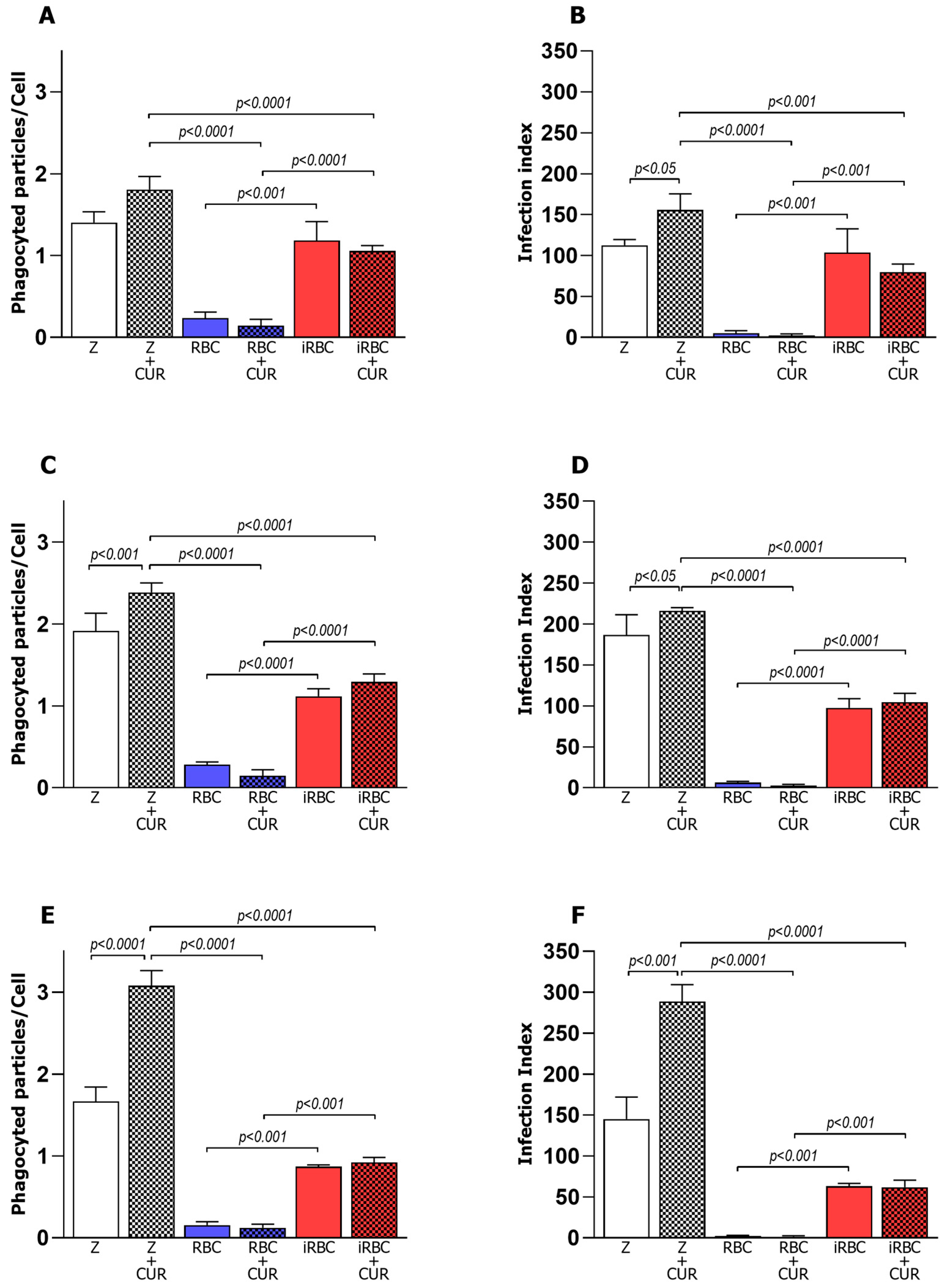
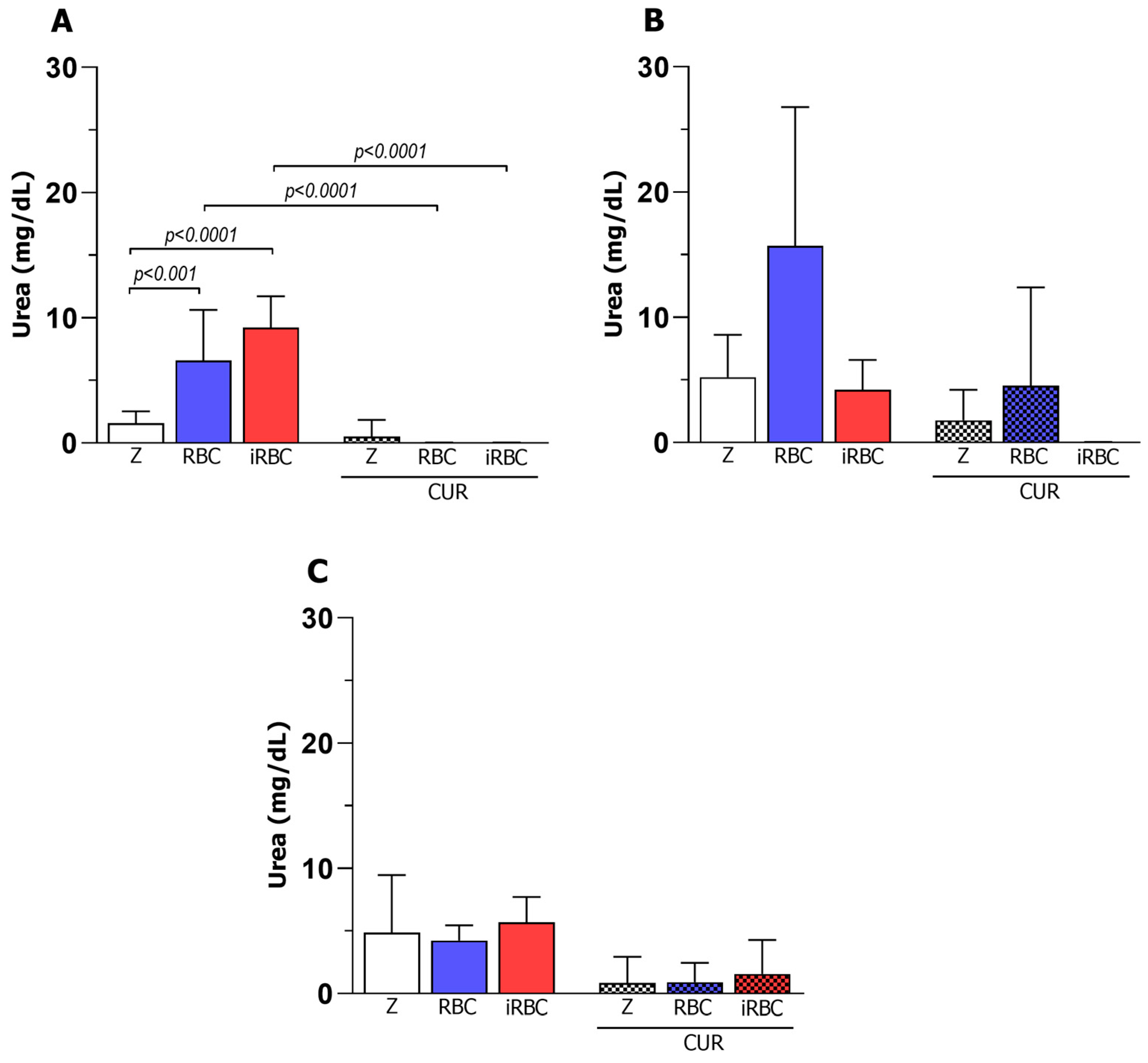
3.1. Pro-Inflammatory Macrophages Increased Phagocytosis and Microbicidal Activity of iRBCs

After in vitro polarization, the macrophages were incubated with red blood cells infected with Plasmodium berghei NK65 (iRBCs) and subjected to an analysis of the phagocytic and microbicidal activities. Macrophage phenotypes M0, M1, and M2 showed significantly increased phagocytosis of iRBCs when compared to phagocytosis of uninfected red blood cells (RBCs). Three hours after incubation with iRBCs, the mean number of phagocytosed particles (Figure 1A,C,E) and the infection index of the macrophages (phagocytic cells) (Figure 1B,D,F) increased four-fold for the pro-inflammatory M1 phenotype (Figure 1C,D). When treated with CUR, macrophage phenotypes M0, M1, and M2 presented a similar performance.
To understand whether curcumin influenced the function of polarized and infected macrophages, the phagocytic activity was evaluated three hours after the curcumin treatment. Interestingly, the treatment of polarized macrophages (M1 or M2) with curcumin did not alter the phagocytic activity of the macrophages incubated with iRBCS or RBCs. However, in polarized M1 or M2 macrophages incubated with zymosan (Figure 1), curcumin significantly increased the phagocytosis index, with an increase in the average number of phagocytosed particles per cell three hours after infection and treatment.

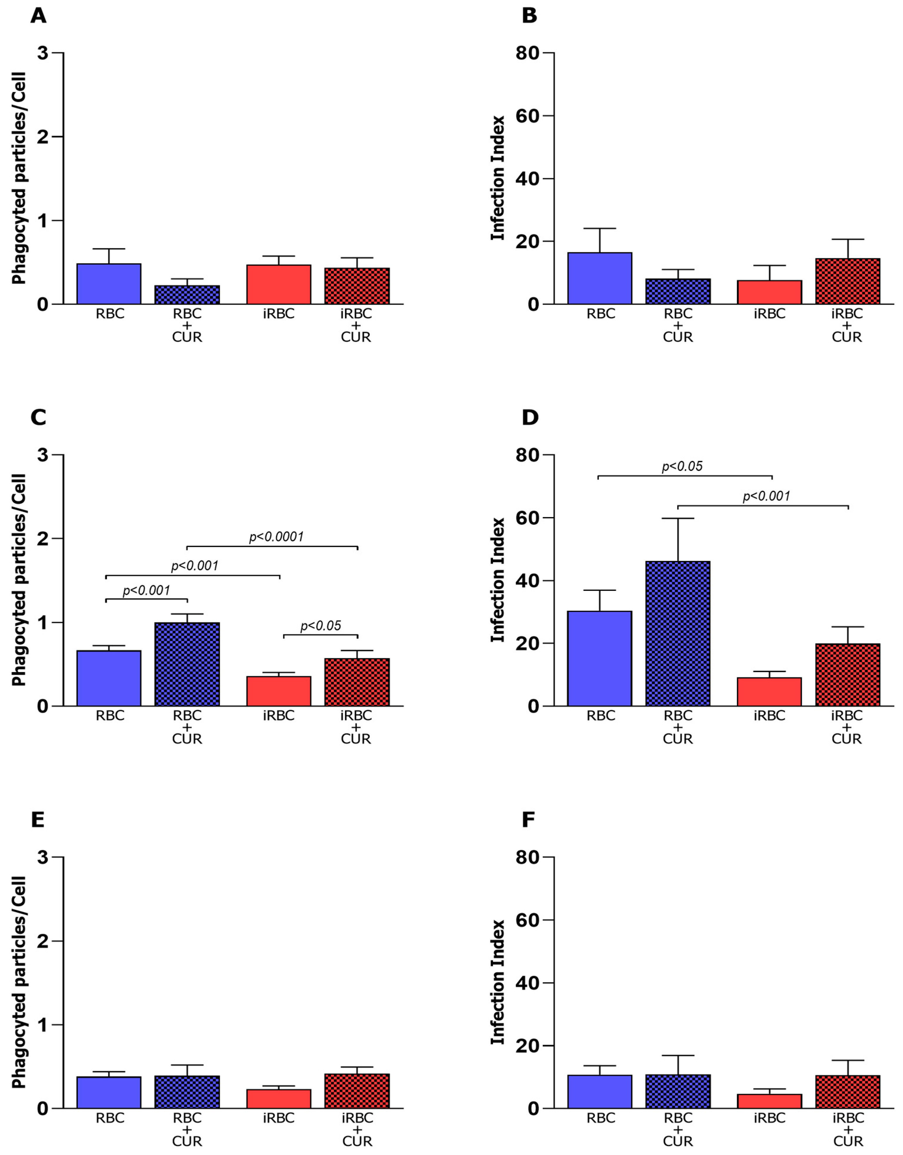
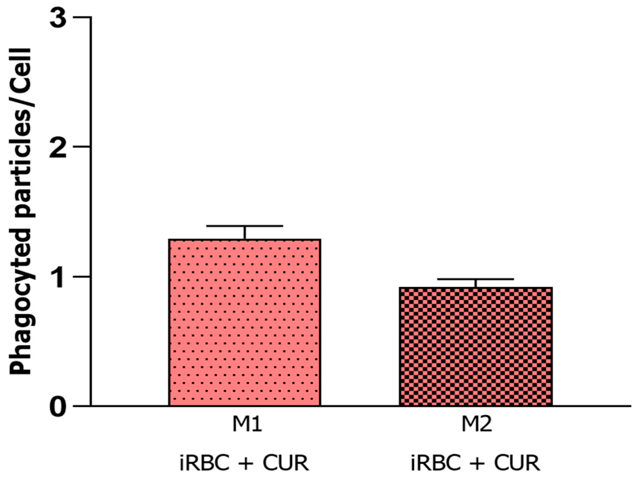
3.2. Curcumin Increased Phagocytosis in Non-Infected and Polarized Macrophages

The microbicidal activity was evaluated 24 h after infection and incubation with curcumin. The average number of particles inside non-polarized macrophages (M0) was similar in all studied groups 24 h after infection (iRBCs) and treatment with CUR (iRBCs + CUR) (Figure 2A). However, the M0 macrophages incubated with RBCs plus curcumin had a lower macrophage infection index (Figure 2B) when compared to M0 cells incubated only with red blood cells, indicating that curcumin can improve the ability of the M0 macrophages to eliminate RBCs. The M1 macrophages incubated with red blood cells plus curcumin (RBC + CUR) revealed an average number of particles (Figure 2C) and a higher macrophage infection index (Figure 2D) when compared to M1 cells incubated with infected red blood cells (iRBC + CUR). The average number of particles (Figure 2E) and macrophage infection index (Figure 2F) were similar in all studied groups 24 h after infection and treatment with CUR.

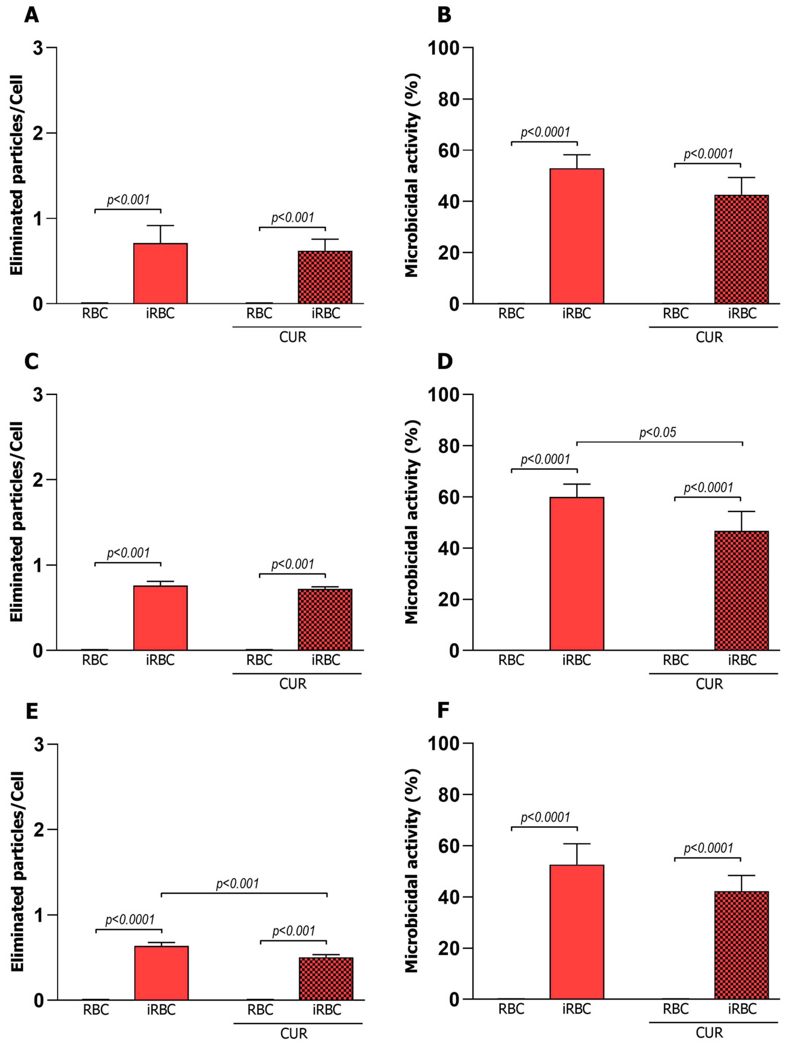
3.3. Curcumin Reduced the Microbicidal Activity of the Pro-Inflammatory Macrophages

The killing ability of the macrophages was evaluated 24 h after infection with infected or uninfected RBCs. The mean number of phagocytosed particles was significantly reduced compared to the results observed three hours after infection (Figure 2), demonstrating that the previously phagocytosed particles were eliminated by the microbicidal activity of the macrophages (Figure 3). This microbicidal activity was observed only in the cells infected with Plasmodium, with the percentage of activity higher in the M1 phenotype. Furthermore, infected M1 cells, when incubated with curcumin, showed a decreased number of cells with microbicidal activity compared to cells without curcumin treatment (Figure 3), demonstrating that in this case, curcumin was able to interfere with the elimination of particles previously internalized by the M1 phenotype.

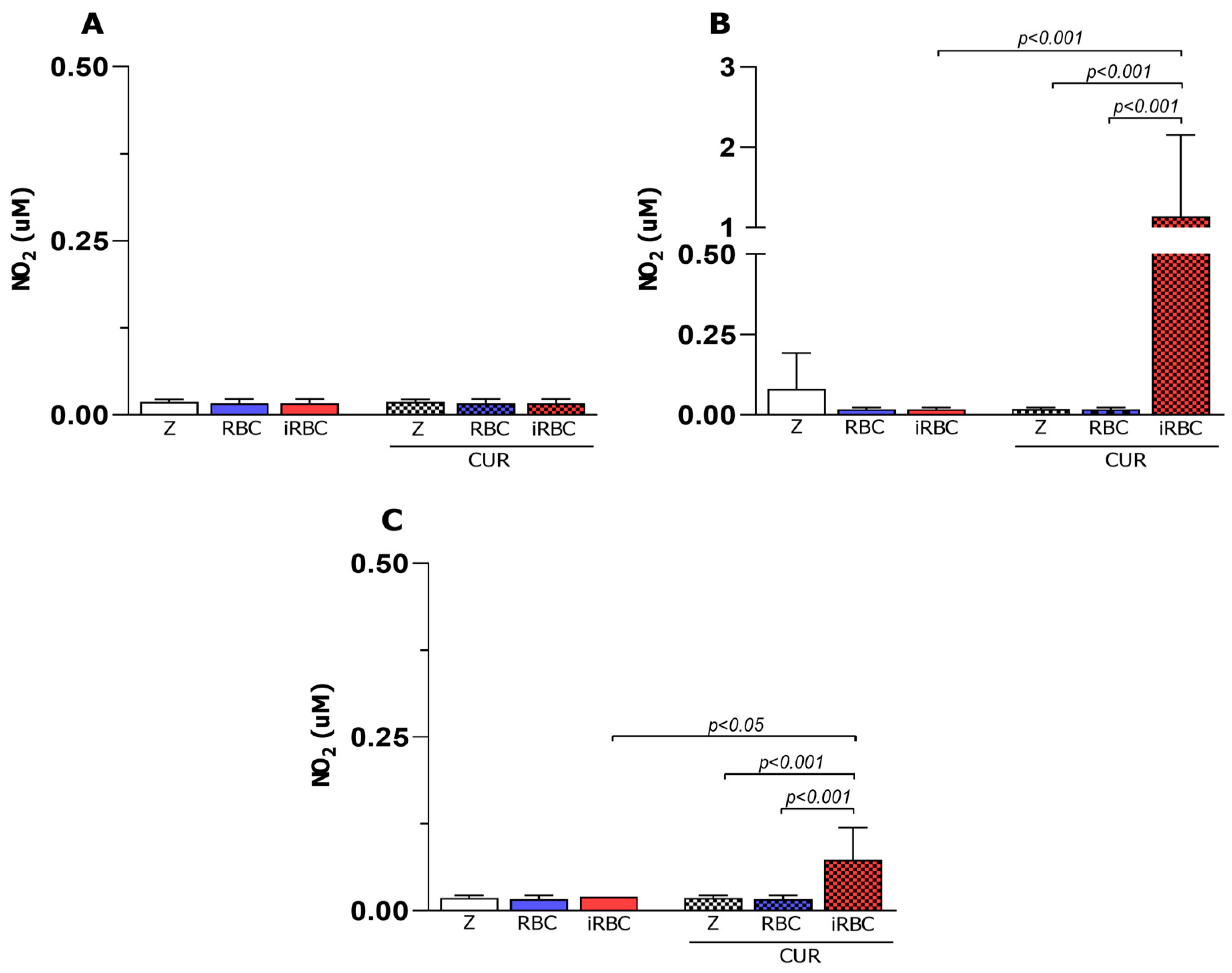
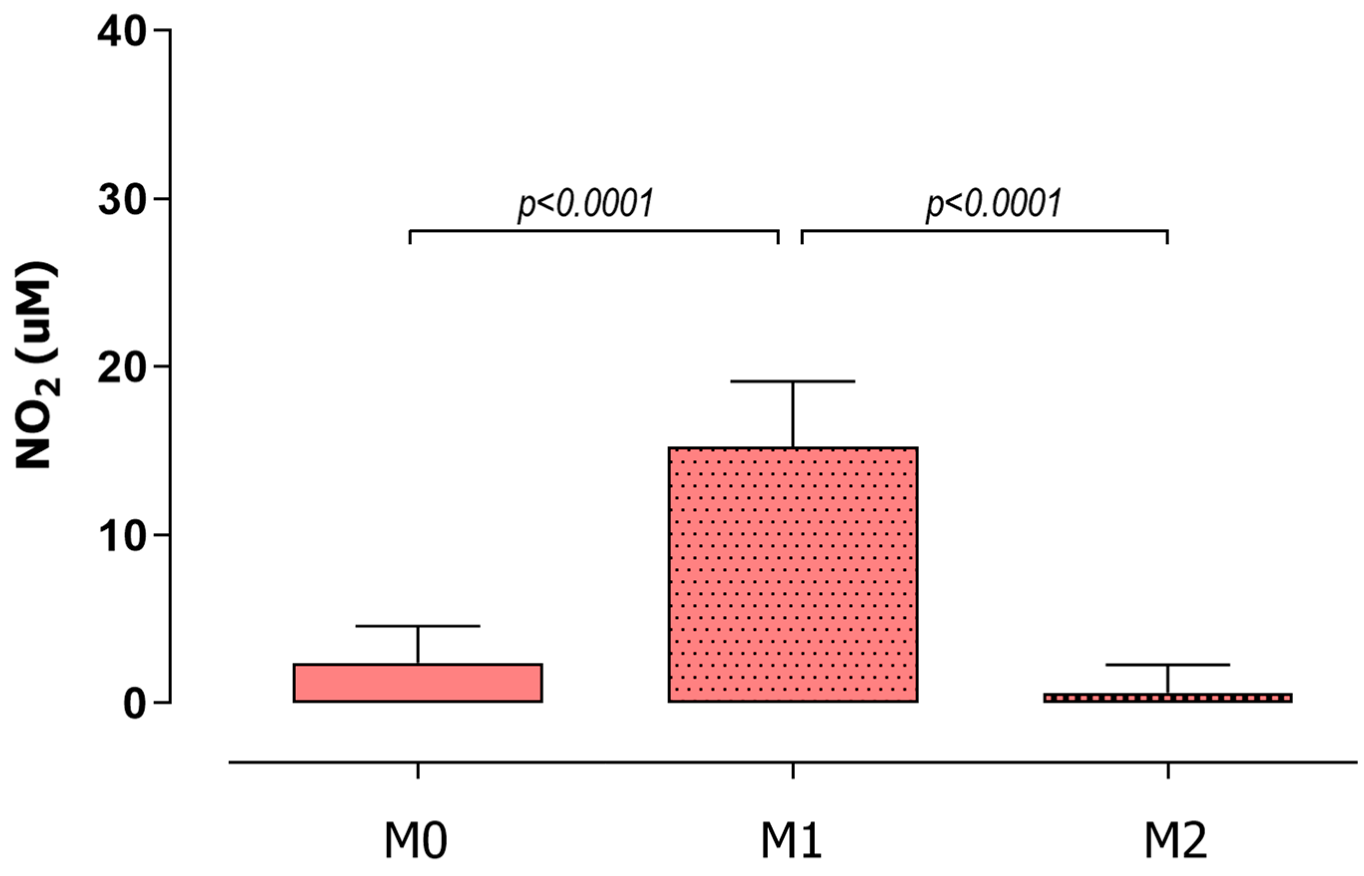
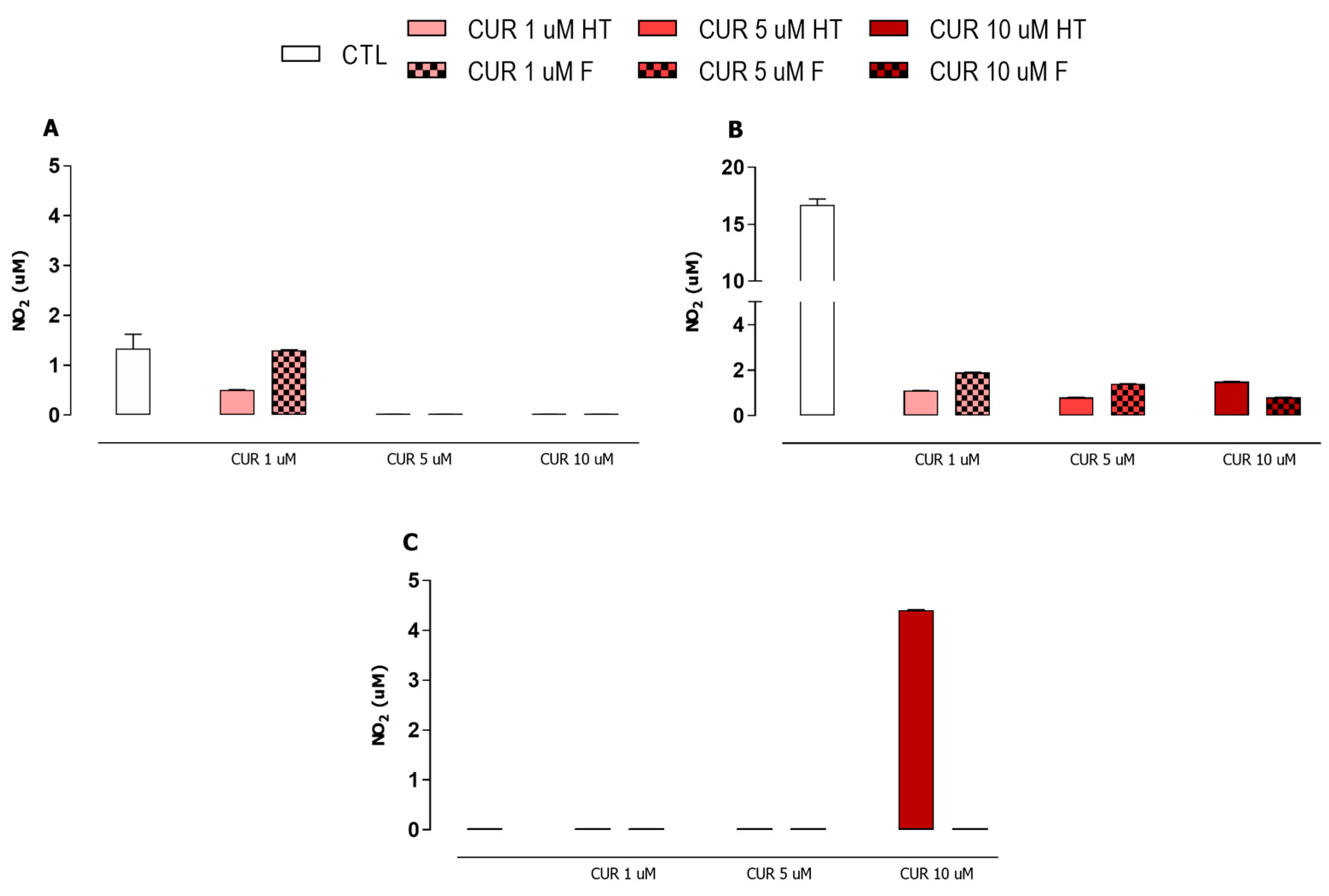
3.4. iNOS Enzyme Had No Participation in Macrophage Microbicidal Activity after Plasmodium Infection and Curcumin Treatment

Previous studies by our group (summarized in Appendix A) demonstrated that curcumin was able to inhibit the iNOS enzyme in M1 macrophages 24 h after being exposed to uninfected polarized macrophages. To clarify whether this microbicidal activity occurred in the absence of the iNOS enzyme, an NO production assay was performed 24 h after infection and treatment with curcumin.
iNOS activity is considered a hallmark marker of the M1 macrophage, and as expected, nitrite was not detected in the non-polarized macrophages (M0). Additionally, the M2 macrophages showed decreased levels of nitrite (Figure 4); the group that received iRBCs was an exception. We also observed reduced levels of nitrite in the M1 macrophages (Figure 4B), despite the increase in phagocytosis and microbicidal activity observed previously, which demonstrates the lack of participation of the iNOS enzyme in the microbicidal activity of the macrophages after 24 h of Plasmodium berghei infection and treatment with curcumin.

3.5. Curcumin Decreased Arginase Activity in Infected and Non-Infected Macrophages

As arginase is a marker of anti-inflammatory macrophages (M2) in cell culture, the arginase activity assay was performed to elucidate whether curcumin influenced urea formation and nitrite formation, which were previously observed in polarized and infected macrophages.
The results showed that curcumin interfered with urea formation. All of the macrophage phenotypes (M0, M1, and M2) demonstrated reduced urea levels 24 h after infection and curcumin treatment (Figure 5), indicating a decrease in arginase enzyme activity. Furthermore, the production of urea by macrophages that received RBCs (Figure 5), even after treatment with curcumin, demonstrates that these RBCs act as an anti-inflammatory stimulus when added to a macrophage cell culture.

3.6. Curcumin Stabilized Macrophage Polarization during Plasmodium Infection

Several studies have shown that macrophage polarization is not definitive and can be altered according to the stimuli received in the microenvironment. Considering this, we reanalyzed the phagocytic activity shown in Figure 1 and observed that the M1 and M2 cells treated with curcumin showed similar behavior. Statistical analyses showed no difference between these groups, suggesting that curcumin can avoid the most extreme polarization phenotypes, acting as a stabilizer of macrophage polarization during Plasmodium infection (Figure 6).

4. Discussion

This investigation sought to understand the in vitro effects of curcumin in the function of polarized murine macrophages exposed to red blood cells infected with Plasmodium berghei NK65 or uninfected. Firstly, the phagocytic model was reproduced in vitro and worked successfully once all of the macrophage phenotypes were able to phagocytize zymosan, which is a model of classical phagocytosis showing interaction with immunity cells [20]. Likewise, we demonstrated that iRBCs behaved like PAMPs [21], and the M0, M1, and M2 macrophages were able to recognize and phagocytose these iRBCs [22].
In this study, we demonstrated phagocytosis occurring at higher levels when macrophages were in contact with red blood cells infected by Plasmodium berghei. We observed a pattern of a higher average of phagocyted particles and an increased percentage of phagocytic cells when these were in contact with iRBCs instead of RBCs, as observed by Shear and colleagues in 1979 [23]. Additionally, the M1 phenotype, known as pro-inflammatory with high phagocytic and microbicidal activities [12], displayed higher levels of phagocytosis than the M0 and M2 phenotypes. Correlating with these results, microbicidal activity was clear in all of the macrophage phenotypes infected, as well as phagocytic activity [11]; the average number of phagocyted particles inside the macrophages decreased after 24 h when compared to 3 h after the infection. All of these findings demonstrate that the macrophages not only recognized iRBCs but also ingested and destroyed these iRBCs, with significant phagocytic and microbicidal functions. The presence of Plasmodium berghei inside erythrocytes could potentiate PAMP’s activation and related pathways, increasing cytokine production and promoting phagocytosis and digestion. In this context, there has been an increase in MHC class II and co-stimulatory molecules and an increase in cytokine production in infected macrophages after internalization of Plasmodium in RBC, involving TLR4, TLR9, and MyD88, and signaling via NF-κB [24,25].
To evaluate whether curcumin could interfere with phagocytic and microbicidal activities, the previously polarized macrophages were analyzed at 3 and 24 h after treatment with curcumin. We observed that the curcumin treatment did not change these activities in the environment in vitro with Plasmodium infection but increased phagocytosis in the non-infected and polarized macrophages. Previous in vitro and in vivo studies suggest that curcumin should be used as adjunctive therapy [26,27,28,29]. Although curcumin did not interfere positively or negatively in phagocytic and microbicidal activities when macrophages received iRBCs, our results demonstrate that curcumin significantly increased phagocytosis in polarized M1 or M2 phenotype macrophages three hours after receiving zymosan. These data agree with previous results that observed up-regulation in the phagocytic activity of RAW 264.7 macrophages after curcumin treatment under non-inflammatory conditions [27,30]. In our findings, the cells were previously stimulated with LPS, as the M1 macrophages in this study were differentiated in the presence of LPS. Thus, our results confirm that treatment with curcumin enhances the phagocytic activity of macrophages, independently of the activation received (M1 or M2), when compared with non-treated macrophages.
The M1 and M2 macrophages infected and treated with curcumin displayed similar behavior to non-polarized macrophages infected without curcumin treatment. Considering that macrophages can reverse the original polarization according to changes in the microenvironmental stimuli, it is possible that curcumin treatment could affect the polarization process; the attenuation of pro-inflammatory cytokines and chemokines produced by macrophages and influenced by this compound [14,15,31] can change the pro-inflammatory responses. This suggests that curcumin can avoid the more extreme phenotypes of M1 and M2 polarization, and these cells can behave like unpolarized and infected macrophages, still carrying out phagocytic and microbicidal activities but with the polarization stabilized in a neutral phenotype during Plasmodium infection. This action could favor the use of curcumin as an adjuvant in the treatment of malaria, since a balance between pro- and anti-inflammatory responses is essential for the best clinical outcome in this disease [32].
During phagocytic and microbicidal activities, reactive nitrogen species (RNS) are released by macrophages, like nitric oxide (NO) through the enzyme nitric oxide synthase (iNOS) [33]. This process is dependent on the availability of the amino acid arginine, which is converted into NO and citrulline in M1 cells [34]. Previous results showed that curcumin treatment inhibits iNOS enzyme production by M1 macrophages [19,27,30]. Thus, our results confirm that macrophages perform phagocytosis and microbicidal activity in the absence of the iNOS enzyme through the low nitrite values detected in all of the macrophage phenotypes, indicating that the iNOS enzyme did not participate in the microbicidal activity of the macrophages and suggesting that other RNS or ROS produced by these cells could destroy the phagocyted particles. A similar analysis occurred in a study of the killing of Leishmania braziliensis by monocytes, which concluded that NO alone is not sufficient to control the infection in patients with Cutaneous Leishmaniasis, while the production of ROS is involved in parasite killing [35].
After looking into the iNOS enzyme, we analyzed the activity of the arginase enzyme, since both make use of arginine as a substrate to produce their compounds [36]. Arginase converts arginine into ornithine and urea in the M2 cells [37]. In this study, we observed that the treatment of macrophages with curcumin after polarization reduced the levels of urea by all macrophage phenotypes, indicating a decrease in the activity of the arginase enzyme. Furthermore, a reduction in serum urea was observed in mice that received nanometric curcumin, with a protective effect on the kidneys of infected animals that is similar to chloroquine treatment [26].
Outside the malaria context, renal toxicity and carcinogenesis studies have also observed reduced arginase activity after curcumin treatment [34,38]. Unexpectedly, our results leave us without an explanation for how curcumin reduces the activity of both enzymes iNOS and arginase in the M0 or polarized M1 and M2 macrophages. Since both enzymes depend on the arginine supply, curcumin could interfere with arginine availability by hampering the enzyme’s binding site or by altering important cofactors in enzymatic reactions [38,39]. Although the mechanism is unclear, curcumin may have prevented the availability of arginine to macrophages, inhibiting the production of the markers of the M1 and M2 phenotypes (NO and urea, respectively), which may be related to the possible ability of curcumin to balance polarization, avoiding the two extreme phenotypes and consequently not reducing arginase activity in a significant way [39].

5. Conclusions

In summary, this study demonstrates that the phagocytic and microbicidal activities of macrophages, when in contact with Plasmodium berghei-infected red blood cells, are high, mainly in the pro-inflammatory M1 phenotype. In addition, curcumin stimulated phagocytic and microbicidal activity when used under non-infectious conditions, bringing new perspectives to the use of curcumin in maintaining phagocytic activity in circumstances of cell turnover and phagocytosis of altered organism particles. Our findings provide insight into curcumin treatment in malaria as an adjuvant, promoting equilibrium between pro- and anti-inflammatory responses for a better clinical outcome, once curcumin was able to reduce the NO and urea production in all of the macrophage phenotypes. More experiments should elucidate the mechanism involved in this process, clarifying its possible relationship with the arginine blockade and balance promotion in macrophage polarization in the M0 neutral phenotype.

Author Contributions

Conceptualization, F.D.T., S.G.d.F., P.R.A.N. and M.R.N.C.; formal analysis, M.C.C.C. and F.S.A.; funding acquisition, M.R.N.C.; investigation, S.G.d.F.; methodology, M.C.C.C., F.D.T. and F.S.A.; project administration, P.R.A.N. and M.R.N.C.; supervision, M.R.N.C.; validation, F.D.T., F.S.A., S.G.d.F. and P.R.A.N.; writing—original draft, M.C.C.C.; writing—review and editing, M.C.C.C., F.D.T., F.S.A., S.G.d.F., P.R.A.N. and M.R.N.C. All authors have read and agreed to the published version of the manuscript.

Funding

This research was funded by the Coordenação de Aperfeiçoamento de Pessoal de Nível Superior (CAPES), grant number AUXPE n. 88881.845026/2023-01, and Fundação de Amparo à Pesquisa do Estado de Goias (FAPEG), grant number—201810267001566-Edital FAPEG n. 06/2018.

Institutional Review Board Statement

Not applicable for studies involving cell culture.

Informed Consent Statement

Not applicable.

Data Availability Statement

All data are contained within the article.

Acknowledgments

We would like to thank Gisleine F. França for their valuable help and technical assistance during the preparation of the manuscript.

Conflicts of Interest

The authors declare no conflict of interest.

Appendix A

According to the polarization protocol, after 14 days, 3 × 10⁵ cells/well were dispensed into each well of 24-well plates and incubated at 37 °C in 5% CO2 for 4 h. Into each well was added the following treatment for macrophage polarization: LPS (3 μg/mL) + IFNγ (15 ng/mL) for M1 phenotype; IL-4 (60 ng/mL) for M2 phenotype; or only RPMI-164 medium supplemented for M0 macrophages (control group). These cells were incubated for 24 h, and confirmation of cell polarization (M1 or M2 phenotypes) was performed with the NO production assay and arginase activity analysis (Figure A1—Nitrite production 24 h after polarization).
To determine the concentration of curcumin to be used, 1 × 10⁵ cells/well were plated in 24-well plates. After, they were incubated for 4 h, and then submitted to the polarization protocol, as described above. After 24 h was added filtered curcumin at 1, 5, or 10 μM. After 3 or 24 h, supernatant samples were collected for the NO production assay (Figure A2—Nitrite production 24 h after curcumin treatment), and cell samples for the arginase activity analysis (Figure A3—Urea production 24 h after curcumin treatment).
To measure cellular metabolic activity as an indicator of cell viability or cytotoxicity during the different curcumin treatments, an MTT assay was performed, in which 1 × 10⁴ cells/well were plated in 96-well plates, following the same steps described. MTT solution was added to the wells with incubation for 2 h, and DMSO for 5 min. The reading was performed on a spectrophotometer in the 550 nm wavelength. Results showed that curcumin filtered at 5 μM was able to inhibit the iNOS enzyme in M1 macrophages without significant stress to the cells (reduced mitochondrial function) (Figure A4—Mitochondrial activity 24 h after curcumin treatment).
Figure A1. Nitrite production 24 h after polarization. Confirmation of cell polarization (M1 phenotype) was performed with NO production assay. Data were calculated from N = 3, and are presented as mean ± standard deviation. p-values are indicated and were considered statistically significant when p < 0.05.
Pharmaceutics 15 02505 g0a1
Figure A2. Nitrite production 24 h after curcumin treatment. Concentrations (μM) were detected in M0 (A), M1 (B), and M2 (C) macrophages after treatment with 1, 5, or 10 μM of curcumin (CUR), sterilized by high temperature (HT) or filtration (F). Data were calculated from N = 3, and are presented as mean ± standard deviation. p-values are indicated and were considered statistically significant when p < 0.05.
Pharmaceutics 15 02505 g0a2
Figure A3. Urea production 24 h after curcumin treatment. Concentrations (μM) were detected in M0 (A), M1 (B), and M2 (C) macrophages after treatment with 1, 5, or 10 μM of curcumin (CUR), sterilized by high temperature (HT) or filtration (F). Data were calculated from N = 3, and are presented as mean ± standard deviation. p-values are indicated and were considered statistically significant when p < 0.05.
Pharmaceutics 15 02505 g0a3
Figure A4. Mitochondrial activity 24 h after curcumin treatment. Percentage of mitochondrial activity detected in M0 (A), M1 (B), and M2 (C) macrophages after treatment with 1, 5, or 10 μM of curcumin (CUR), sterilized by high temperature (HT) or filtration (F). Data were calculated from N = 3, and are presented as mean ± standard deviation. p-values are indicated and were considered statistically significant when p < 0.05.
Pharmaceutics 15 02505 g0a4

References

  1. Fu, Y.; Ding, Y.; Wang, Q.; Zhu, F.; Tan, Y.; Lu, X.; Guo, B.; Zhang, Q.; Cao, Y.; Liu, T.; et al. Blood-stage malaria parasites manipulate host innate immune responses through the induction of sFGL2. Sci. Adv. 2020, 9, eaay9269. [Google Scholar] [CrossRef]
  2. Shahbodaghi, S.D.; Rathjen, N.A. Malaria: Prevention, Diagnosis, and Treatment. Am. Fam. Physician 2022, 106, 270–278. [Google Scholar]
  3. Milner, E.M.; Kariger, P.; Pickering, A.J.; Stewart, C.P.; Byrd, K.; Lin, A.; Rao, B.; Dentz, H.N.; Null, C.; Fernald, L.C.H. Association between Malaria Infection and Early Childhood Development Mediated by Anemia in Rural Kenya. Int. J. Environ. Res. Public Health 2020, 17, 902. [Google Scholar] [CrossRef] [PubMed]
  4. Cutts, J.C.; Agius, P.A.; Lin, Z.; Powell, R.; Moore, K.; Draper, B.; Simpson, J.A.; Fowkes, F.J.I. Pregnancy-specific malarial immunity and risk of malaria in pregnancy and adverse birth outcomes: A systematic review. BMC Med. 2020, 18, 14. [Google Scholar] [CrossRef]
  5. World Health Organization; Global Malaria Programme. World Malaria Report; Creative Commons; World Health Organization: Mountain View, CA, USA, 2022; p. 293. ISBN 978-92-4-006489-8. Available online: https://www.who.int/publications/i/item/9789240064898 (accessed on 15 October 2023).
  6. Hussein, M.I.H.; Albashir, A.A.D.; Elawad, O.A.M.A.; Homeida, A. Malaria and COVID-19: Unmasking their ties. Malar J. 2020, 19, 457. [Google Scholar] [CrossRef] [PubMed]
  7. Milner, D.A. Malaria Pathogenesis. Cold Spring Harb. Perspect. Med. 2018, 8, a025569. [Google Scholar] [CrossRef]
  8. Ozarslan, N.; Robinson, J.F.; Gaw, S.L. Circulating Monocytes, Tissue Macrophages, and Malaria. J. Trop. Med. 2019, 2019, 1–9. [Google Scholar] [CrossRef]
  9. Chua, C.L.L.; Brown, G.; Hamilton, J.A.; Rogerson, S.; Philippe, B. Monocytes and macrophages in malaria: Protection or pathology? Trends Parasitol. 2013, 29, 26–34. [Google Scholar] [CrossRef]
  10. Drewry, L.L.; Harty, J.T. Balancing in a black box: Potential immunomodulatory roles for TGF-β signaling during blood-stage malaria. Virulence 2020, 11, 159–169. [Google Scholar] [CrossRef]
  11. Kang, S.; Kumanogoh, A. The spectrum of macrophage activation by immunometabolism. Jpn. Soc. Immunol. 2020, 7, 467–473. [Google Scholar] [CrossRef]
  12. Atri, C.; Guerfali, F.Z.; Laouini, D. Role of Human Macrophage Polarization in Inflammation during Infectious Diseases. Int. J. Mol. Sci. 2018, 6, 1081. [Google Scholar] [CrossRef] [PubMed]
  13. Vergadi, E.; Ieronymaki, E.; Lyroni, K.; Vaporidi, K.; Tsatsanis, C. Akt Signaling Pathway in Macrophage Activation and M1/M2 Polarization. J. Immunol. 2017, 198, 1006–1014. [Google Scholar] [CrossRef] [PubMed]
  14. Mohammadi, A.; Blesso, C.N.; Barreto, G.; Banach, M.; Majeed, M.; Sahebkar, A. Macrophage plasticity, polarization and function in response to curcumin, a diet-derived polyphenol, as an immunomodulatory agent. J. Nutr. Biochem. 2019, 66, 1–16. [Google Scholar] [CrossRef]
  15. Jamil, S.N.H.; Ali, A.H.; Feroz, A.R.; Lam, S.D.; Agustar, H.K.; Razak, M.R.M.A.; Latip, J. Curcumin and Its Derivatives as Potential Antimalarial and Anti-Inflammatory Agents: A Review on Structure–Activity Relationship and Mechanism of Action. Pharmaceutics 2023, 16, 609. [Google Scholar] [CrossRef] [PubMed]
  16. Generally Recognized as Safe (GRAS). GRAS Notice Inventory No. 822. Available online: https://www.fda.gov/media/132575/download (accessed on 26 September 2023).
  17. Hosseini-Zare, M.S.; Sarhadi, M.; Zarei, M.; Thilagavathi, R.; Selvam, C. Synergistic effects of curcumin and its analogs with other bioactive compounds: A comprehensive review. Eur. J. Med. Chem. 2021, 15, 113072. [Google Scholar] [CrossRef]
  18. Dende, C.; Meena, J.; Nagarajan, P.; Nagaraj, V.A.; Panda, A.K.; Padmanaban, G. Nanocurcumin is superior to native curcumin in preventing degenerative changes in Experimental Cerebral Malaria. Sci. Rep. 2017, 30, 10062. [Google Scholar] [CrossRef]
  19. Manconi, M.; Manca, M.L.; Escribano-Ferrer, E.; Elisabet Martí Coma-Cros, E.M.; Biosca, A.; Lantero, E.; Fernàndez-Busquets, X.; Fadda, A.M.; Caddeo, C. Nanoformulation of curcumin-loaded eudragit-nutriosomes to counteract malaria infection by a dual strategy: Improving antioxidant intestinal activity and systemic efficacy. Int. J. Pharm. 2019, 556, 82–88. [Google Scholar] [CrossRef]
  20. Tu, Q.D.; Jin, J.; Hu, X.; Ren, Y.; Zhao, L.; He, Q. Curcumin Improves the Renal Autophagy in Rat Experimental Membranous Nephropathy via Regulating the PI3K/AKT/mTOR and Nrf2/HO-1 Signaling Pathways. BioMed Res. Int. 2020, 2020, 7069052. [Google Scholar] [CrossRef]
  21. Fuentes, A.-L.; Millis, L.; Vapenik, J.; Sigola, L. Lipopolysaccharide-mediated enhancement of zymosan phagocytosis by RAW 264.7 macrophages is independent of opsonins, laminarin, mannan, and complement receptor 3. J. Surg. Res. 2014, 189, 304–312. [Google Scholar] [CrossRef]
  22. Chan, J.-A.; Boyle, M.J.; Moore, K.A.; Reiling, L.; Lin, Z.; Hasang, W.; Avril, M.; Manning, L.; Mueller, I.; Laman, M.; et al. Antibody targets on the surface of Plasmodium falciparum–infected erythrocytes that are associated with immunity to severe malaria in young children. J. Infect. Dis. 2019, 219, 819–828. [Google Scholar] [CrossRef]
  23. Shear, H.L.; Nussenzweig, R.S.; Bianco, C. Immune phagocytosis in murine malaria. J. Exp. Med. 1979, 149, 1288–1298. [Google Scholar] [CrossRef] [PubMed]
  24. Seixas, E.; Moura Nunes, J.F.; Matos, I.; Coutinho, A. The interaction between DC and Plasmodium berghei/chabaudi-infected erythrocytes in mice involves direct cell-to-cell contact, internalization, and TLR. Eur. J. Immunol. 2009, 39, 1850–1863. [Google Scholar] [CrossRef]
  25. Stevenson, M.M.; Urban, B.C. Antigen presentation and dendritic cell biology in malaria. Parasite Immunol. 2006, 28, 5–14. [Google Scholar] [CrossRef]
  26. Jain, K.; Sood, S.; Gowthamarajan, K. Modulation of cerebral malaria by curcumin as an adjunctive therapy. Braz J. Infect. Dis. 2013, 5, 579–591. [Google Scholar] [CrossRef]
  27. Ghosh, A.; Banerjee, T.; Bhandary, S.; Surolia, A. Formulation of nanosized curcumin and demonstration of its antimalarial efficacy. Int. J. Nanomed. 2014, 9, 5373–5387. [Google Scholar] [CrossRef] [PubMed]
  28. Zendehdel, E.; Abdollahi, E.; Momtazi-Borojeni, A.A.; Korani, M.; Alavizadeh, S.H.; Sahebkar, A. The molecular mechanisms of curcumin’s inhibitory effects on cancer stem cells. J. Cell. Biochem. 2018, 4, 4739–4747. [Google Scholar] [CrossRef]
  29. Kloesch, B.; Gober, L.; Loebsch, S.; Vcelar, B.; Helson, L.; Steiner, G. In Vitro Study of a Liposomal Curcumin Formulation (Lipocurc™): Toxicity and Biological Activity in Synovial Fibroblasts and Macrophages. In Vivo 2016, 4, 413–419. [Google Scholar]
  30. Brouet, I.; Ohshima, H. Curcumin, an anti-tumor promoter, and anti-inflammatory agent, inhibits induction of nitric oxide synthase in activated macrophages. Biochem. Biophys. Res. Commun. 1995, 206, 533–540. [Google Scholar] [CrossRef]
  31. Gao, S.; Zhou, J.; Liu, N.; Wang, L.; Gao, Q.; Wu, Y.; Zhao, Q.; Liu, P.; Wang, S.; Liu, Y.; et al. Curcumin induces M2 macrophage polarization by secretion of IL-4 and/or IL-13. J. Mol. Cell. Cardiol. 2015, 85, 131–139. [Google Scholar] [CrossRef]
  32. Kalantari, P. The Emerging Role of Pattern Recognition Receptors in the Pathogenesis of Malaria. Vaccines 2018, 6, 13. [Google Scholar] [CrossRef]
  33. Bisht, K.; Choi, W.H.; Park, S.Y.; Chung, M.K.; Koh, W.S. Curcumin enhances the non-inflammatory phagocytic activity of RAW264.7 cells. Biochem. Biophys. Res. Commun. 2009, 379, 632–636. [Google Scholar] [CrossRef] [PubMed]
  34. Rath, M.; Müller, I.; Kropf, P.; Closs, E.I.; Munder, M. Metabolism via arginase or nitric oxide synthase: Two competing arginine pathways in macrophages. Front. Immunol. 2014, 5, 532. [Google Scholar] [CrossRef] [PubMed]
  35. Pemmari, A. MKP—1 Promotes Anti -inflammatory M (IL-4/IL-13) Macrophage Phenotype and Mediates the Anti-inflammatory Effects of Glucocorticoids. Basic Clin. Pharmacol. Toxicol. 2018, 124, 404–415. [Google Scholar] [CrossRef]
  36. Carneiro, P.P.; Conceição, J.; Macedo, M.; Magalhães, V.; Carvalho, E.M.; Bacellar, O. The Role of Nitric Oxide and Reactive Oxygen Species in the Killing of Leishmania braziliensis by Monocytes from Patients with Cutaneous Leishmaniasis. PLoS ONE 2016, 11, e0148084. [Google Scholar] [CrossRef]
  37. Yang, Z.; Ming, X.-F. Functions of arginase isoforms in macrophage inflammatory responses: Impact on cardiovascular diseases and metabolic disorders. Front. Immunol. 2014, 5, 533. [Google Scholar] [CrossRef]
  38. Akinyemi, A.J.; Onyebueke, N.; Faboya, O.A.; Onikanni, S.A.; Fadaka, A.; Olayide, I. Curcumin inhibits adenosine deaminase and arginase activities in cadmium-induced renal toxicity in rat kidneys. J. Food Drug Anal. 2017, 25, 438–446. [Google Scholar] [CrossRef] [PubMed]
  39. Bounaama, A.; Djerdjouri, B.; Laroche-Clary, A.; Le Morvan, V.; Robert, J. Short curcumin treatment modulates oxidative stress, arginase activity, aberrant crypt foci, and TGF-1 and HES-1 transcripts in 1,2-dimethylhydrazine-colon carcinogenesis in mice. Toxicology 2012, 302, 308–317. [Google Scholar] [CrossRef] [PubMed]
Figure 1. Macrophage phagocytic activity three hours after Plasmodium berghei NK65 infection and curcumin treatment. Average number of particles per cell by M0 (A), M1 (C), and M2 (E) macrophages and infection index of M0 (B), M1 (D), and M2 (F). CUR = curcumin, Z = zymosan, RBC = red blood cells, iRBC = red blood cells infected with Plasmodium berghei. Data were calculated from N = 3 and are presented as mean ± standard deviation. p-values are indicated and are considered statistically significant when p < 0.05.
Pharmaceutics 15 02505 g001
Figure 2. Macrophage phagocytic activity 24 h after Plasmodium berghei NK65 infection and curcumin treatment. Average number of particles per cell by M0 (A), M1 (C), and M2 (E) macrophages and infection index of M0 (B), M1 (D), and M2 (F). CUR = curcumin, RBC = red blood cells, iRBC = red blood cells infected with Plasmodium berghei. Data were calculated from N = 3 and are presented as mean ± standard deviation. p-values are indicated and are considered statistically significant when p < 0.05.
Pharmaceutics 15 02505 g002
Figure 3. Microbicidal activity of macrophages assessed 24 h after Plasmodium berghei NK65 infection and curcumin treatment. Average of eliminated particles per cell by M0 (A), M1 (C), and M2 (E) macrophages and percentages of M0 (B), M1 (D), and M2 (F) macrophages that displayed microbicidal activity. CUR = curcumin, RBC = red blood cells, iRBC = red blood cells infected with Plasmodium berghei. Data were calculated from N = 3 and are presented as mean ± standard deviation. p-values are indicated and are considered statistically significant when p < 0.05.
Pharmaceutics 15 02505 g003
Figure 4. Nitrite production 24 h after Plasmodium berghei NK65 infection and curcumin treatment. Concentrations (μM) detected in M0 (A), M1 (B), and M2 (C) macrophages were calculated from N = 6 (Z and CUR groups) and N = 3 (RBC, iRBC, RBC + CUR, and iRBC + CUR). Z = zymosan, CUR = curcumin, RBC = red blood cells, iRBC = red blood cells infected with Plasmodium berghei. Data are presented as mean ± standard deviation. p-values are indicated and are considered statistically significant when p < 0.05.
Pharmaceutics 15 02505 g004
Figure 5. Urea production 24 h after Plasmodium berghei NK65 infection and curcumin treatment. Concentrations (mg/dL) detected in M0 (A), M1 (B), and M2 (C) macrophages were calculated from N = 6 (Z and CUR groups) and N = 3 (RBC, iRBC, RBC + CUR, and iRBC + CUR). Z = zymosan, CUR = curcumin, RBC = red blood cells, iRBC = red blood cells infected with Plasmodium berghei. Data are presented as mean ± standard deviation. p-values are indicated and are considered statistically significant when p < 0.05.
Pharmaceutics 15 02505 g005
Figure 6. Macrophage phagocytic activity 3 h after Plasmodium berghei NK65 infection. Average phagocyted particles per cell by M1 and M2 macrophages after curcumin treatment. CUR = curcumin; iRBC = red blood cells infected with Plasmodium berghei. Data were calculated from N = 3 and are presented as mean ± standard deviation. p-values are indicated and are considered statistically significant when p < 0.05.
Pharmaceutics 15 02505 g006
Disclaimer/Publisher’s Note: The statements, opinions and data contained in all publications are solely those of the individual author(s) and contributor(s) and not of MDPI and/or the editor(s). MDPI and/or the editor(s) disclaim responsibility for any injury to people or property resulting from any ideas, methods, instructions or products referred to in the content.

