
Recent Advancements in Metallic Drug-Eluting Implants

Abstract
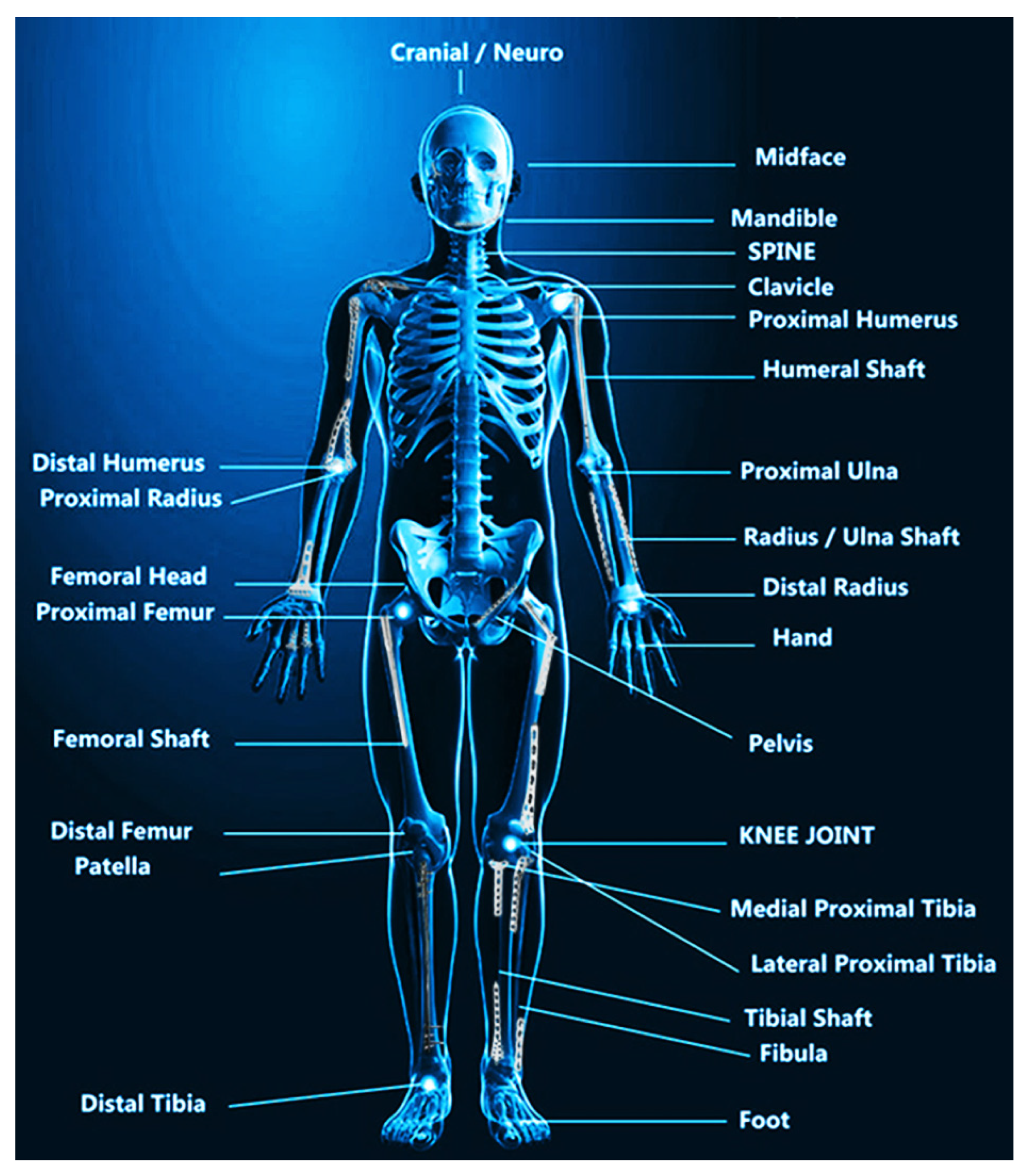
:1. Introduction
2. Conventional Drug-Eluting Implants
2.1. Anti-Inflammatory Drug-Eluting Implants
2.2. Antibiotics-Eluting Implants
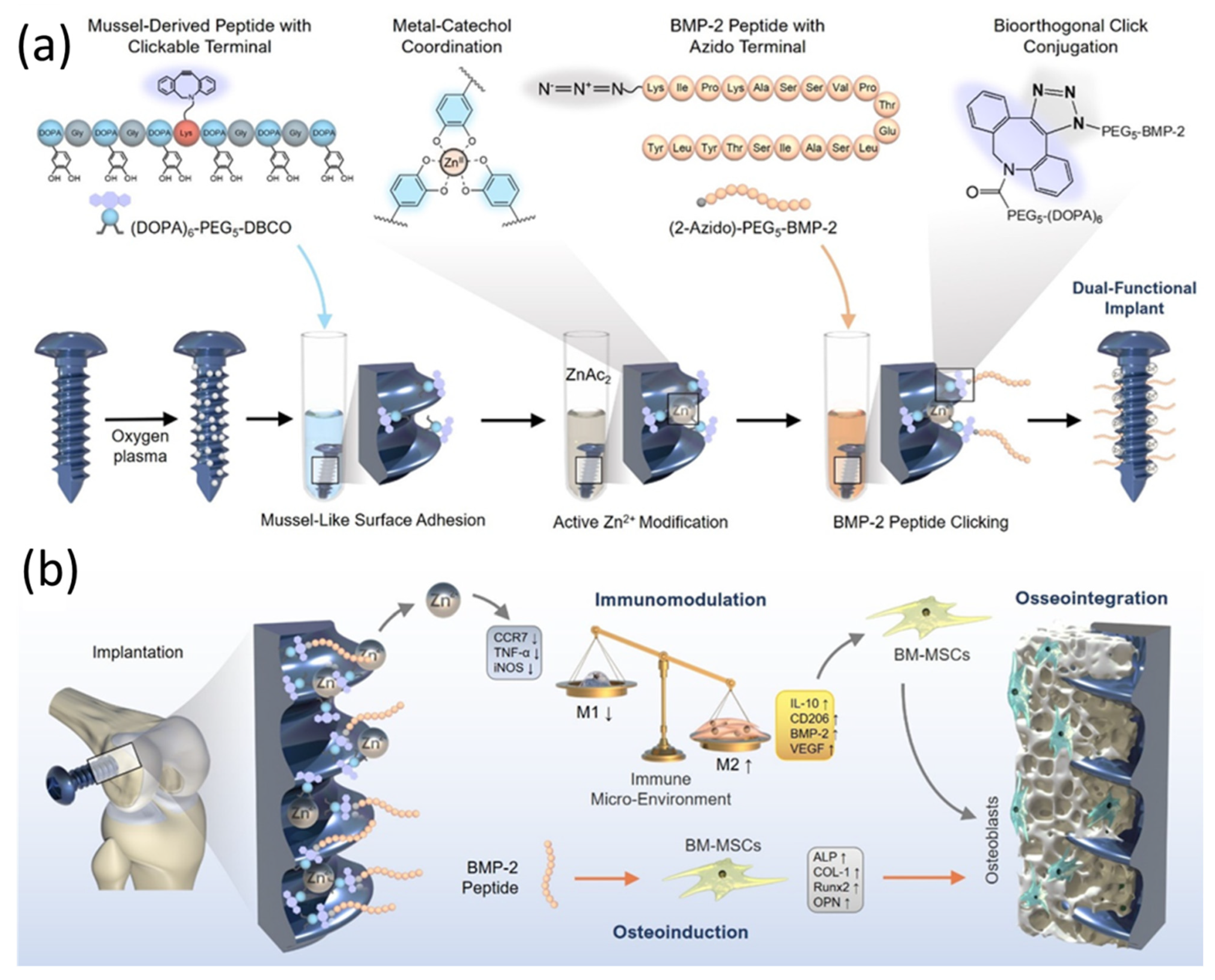
2.3. Growth Factor-Eluting Implants
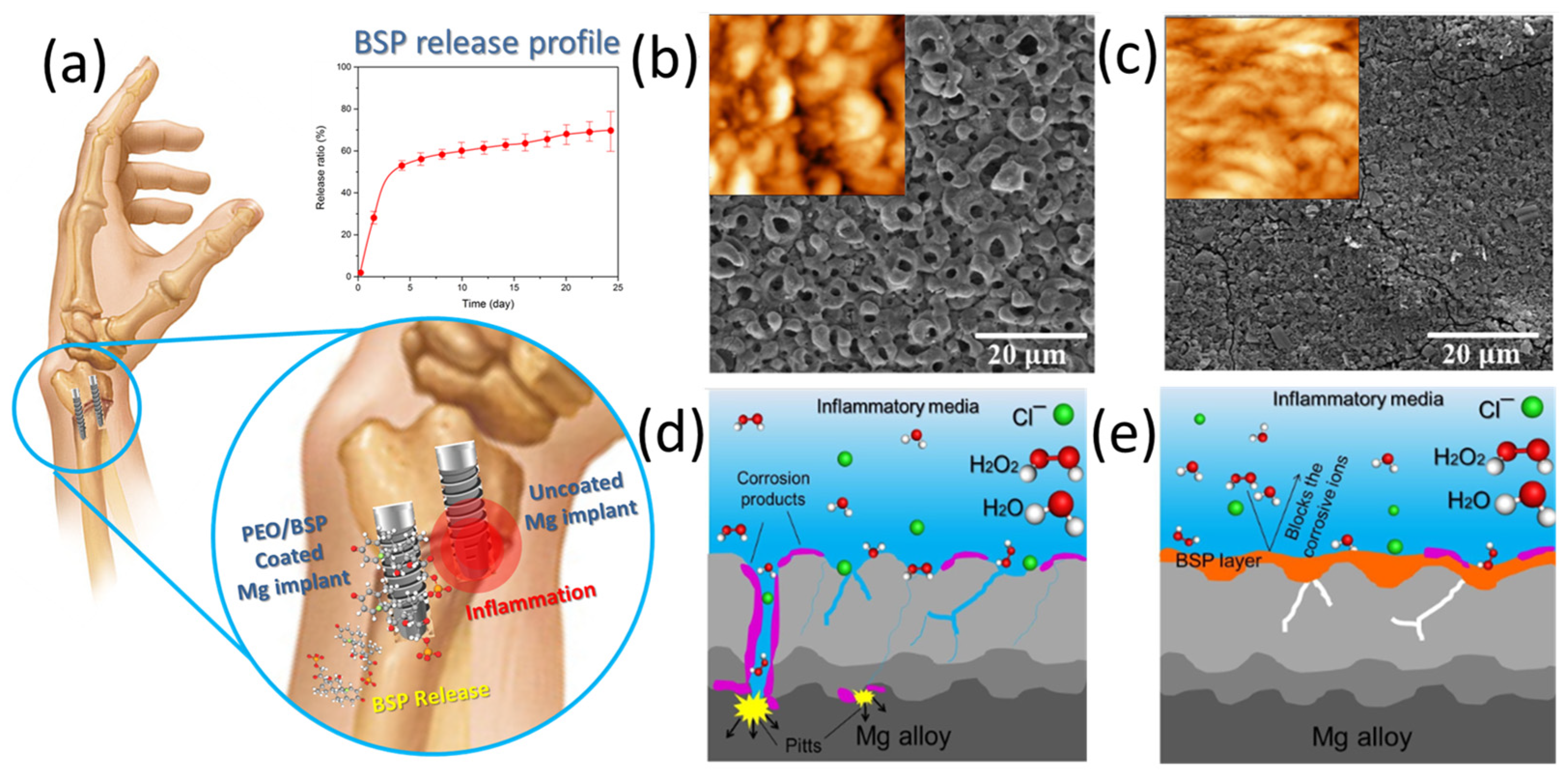
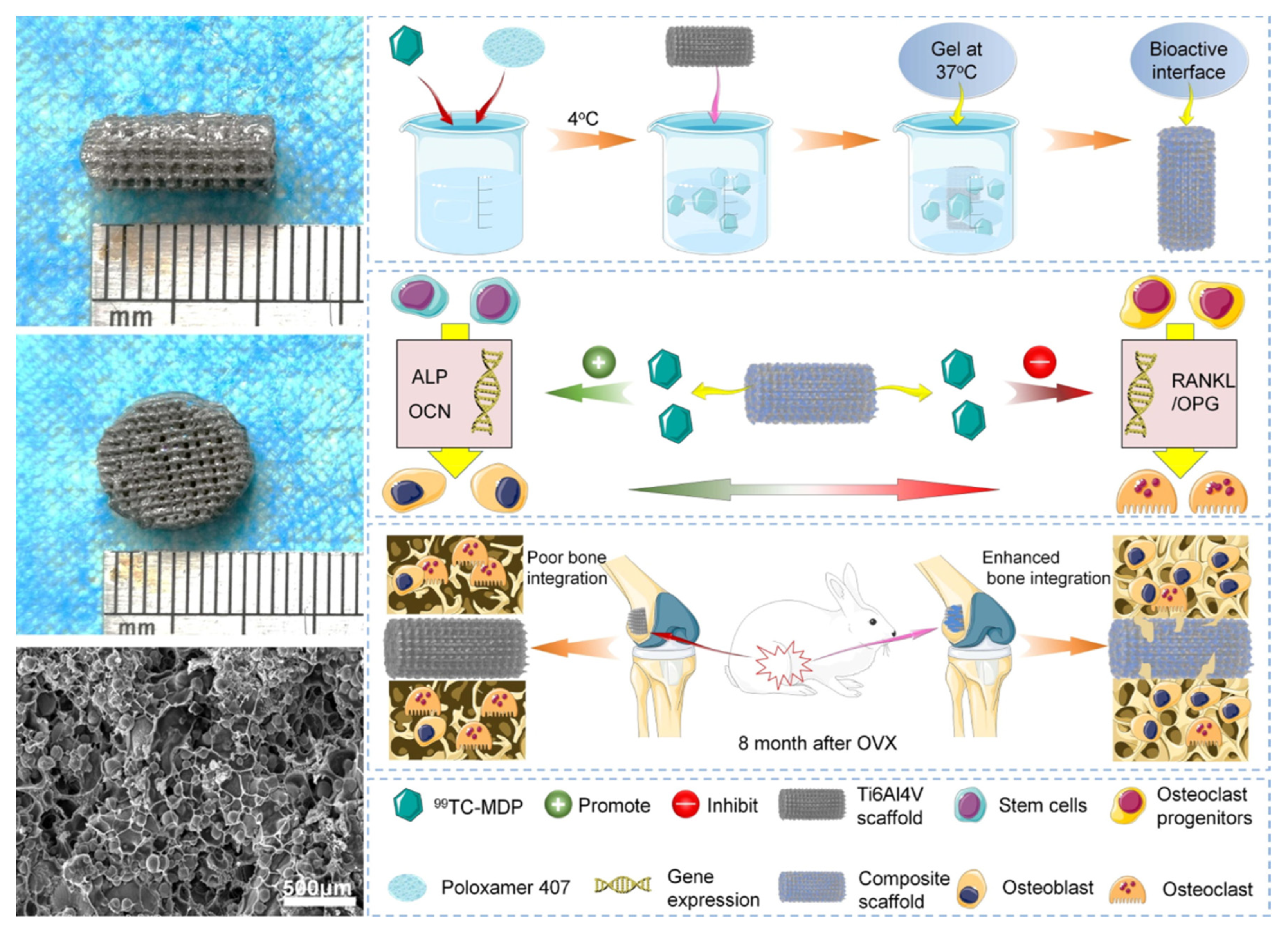
2.4. Anti-Resorptive Drug-Eluting Implants
3. Most-Studied Drug-Eluting Systems
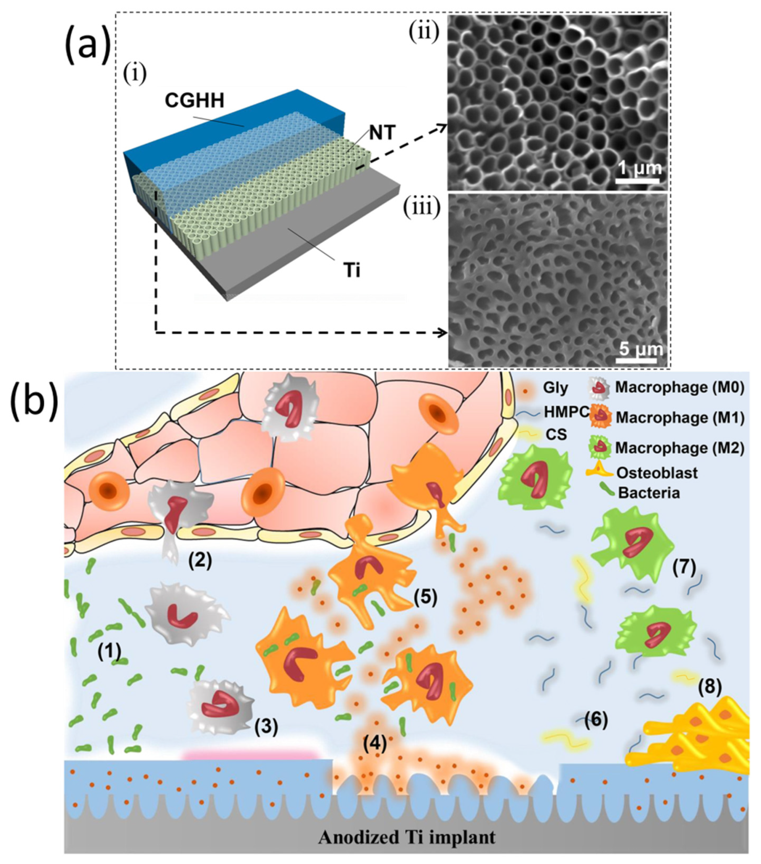
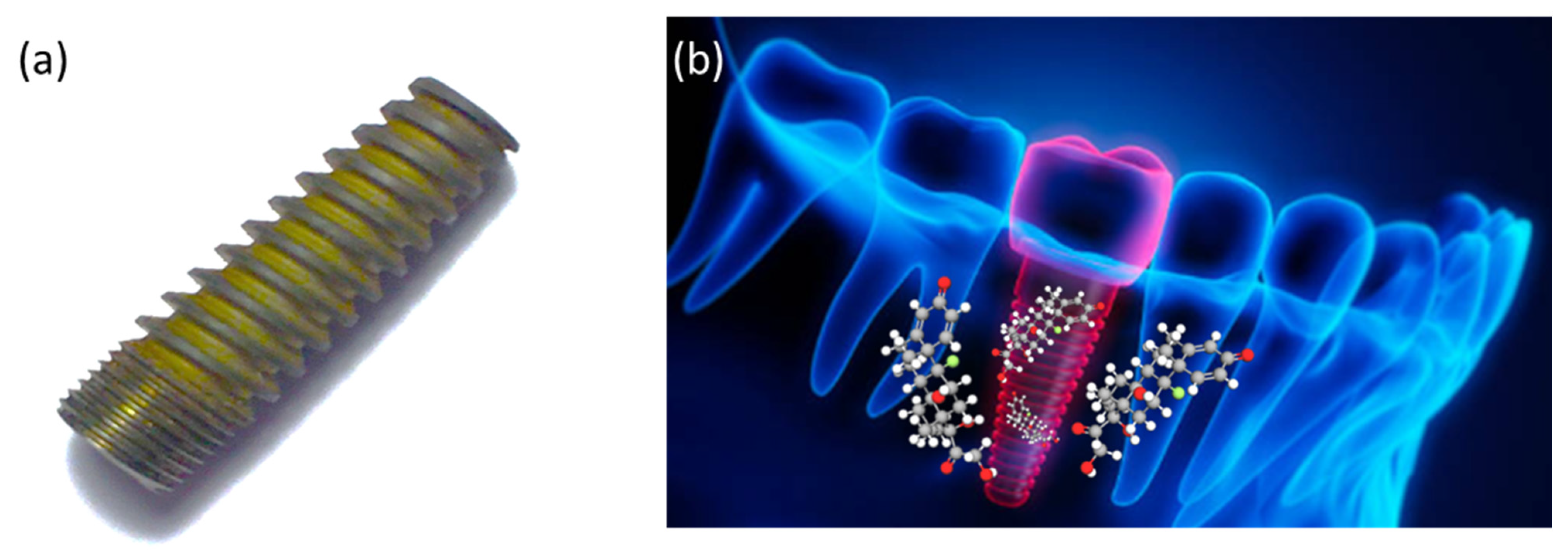
3.1. Titanium-Based Implants
3.2. Gentamicin-Eluting Implants

| Implant Material | Surface Treatment Method | Antibiotics Drug | Deposition Technology | Vivo Type | N * Number | Surgical Site | Infection Model and System | Follow-Up | Ref. |
|---|---|---|---|---|---|---|---|---|---|
| cpTi | Anodization + alkaly treatment + HA | Tobramycin | Soaking method | Rab | 5 | F | S. aureus (ATCC 6538) | 9d | [112] |
| cpTi | Machined | Vancomycin | Manual application (PH) | Rab | 9 | R | S. aureus (UAMS-1 strain) | 1w | [113] |
| cpTi | PLLA | Rifampicin + Fusidic acid | Solvent-casting | Rab | 36 | T | S. aureus (V 8189-94) | 4w | [114] |
| cpTi | Beadblasted and etched | Vancomycin | Covalent immobilization | Mic | NR | S | S. aureus (SH1000) C. albicans (SC5314) | 2d (fungal) 4d (bacterial) | [115] |
| cpTi | Machined and nanotubular anodized surface | Gentamicin | Soaking method | Rab | 36 | T | S. aureus (ATCC 25923) | 6w | [116] |
| cpTi | PDLLA | Gentamicin | NR | Rat | 30 | T | S. aureus (ATCC 49230) | 6w | [117] |
| cpTi | Machined or PDLLA | Gentamicin | PDLLA suspension | Rat | 30 | T | S. aureus (ATCC 49230) | 6w | [118] |
| cpTi | Machined + NIR light | Gentamicin | Vacuum drying process onto PEG-MoS2 coating + CS | Rat | 18 | S | S. aureus (NR strain origin) | 1d, 3d, 1w | [119] |
| cpTi | Anodized + PLEX | Doxycycline | Spraying | Rab | 28 (12 MSSA +16 MRSA) | H | S. aureus MSSA (JAR60131) S. aureus MRSA (LUH15101) | 4w | [120] |
| Ti6Al4V | Anodized | Vancomycin | Sol-gel | Rat | 11 | F | S. aureus (NR strain origin) | 1, 2, 3, 4w | [121] |
| Ti6Al4V | Machined | Rifampicin + Fosfomycin | Ink-jet | Rab | 22 (11 MSSA+ 11 MRSA) | T | S. aureus (MSSA EDCC5055) (MRSA T6625930) | 4w | [122] |
| Ti6Al4V | Machined | Vancomycin | Covalently link | Rat | 9 | F | S. aureus (ATCC 25923) | 1, 2, 3w | [123] |
| Ti6Al4V | TiO2 nanotubes | Gentamicin + Vancomycin | Drug adsorption | Rab | 20 | F | S. aureus (Human Sa5) | 4w | [124] |
| cpTi | Si-sandblasted | Clindamycin or Teicoplanin | Spraying | Rab | 30 | T | S. aureus (ATCC 29123) | 1w | [125] |
| Ti6Al4V | Porous | Ciprofloxacin | Layered double hydroxides suspension | Mic | 12 | S | P. aeruginosa (PAO1 CTX::lux) | 4h | [126] |
| cpTi | Porous Porous + CS | Vancomycin | Electrophoretic deposition | Rat | 18 | T | S. aureus (ATCC 49230) | 4w | [127] |
| Ti6Al4V | Si-sandblasted | Minocycline + Rifampin | Spraying | Rab | 25 | F | S. aureus (P1—variation of ATCC 25923) | 1w | [128] |
| Ti6Al4V | Plasma chemical oxidation | Gentamicin | Immobilization (TA or SDS) | Rat | 15 | T | S. aureus (ATCC 49230) | 4w | [129] |
| Ti6Al4V | Dopamine methacrylate + PEGDMA-Oligo HYD | Vancomycin | Covalently bond | Mic | 22 | F | S. aureus (Xen 29) | 3w | [130] |
| cpTi | Sandblasted and etched | Gentamicin | Polyelectrolyte adsorption (PEM + PGA/HEP) | Rat | 30 | T | S. aureus (ATCC 49230) | 4w | [131] |
| Ti6Al4V | Plasma-sprayed | Vancomycin | Impregnated on the plasma-sprayed coating | Rab | 20 | T | S. aureus MRSA (ATCC 43300) | 6w | [132] |
| Ti6Al4V | Machined | Vancomycin | Covalently bond | Mic | 14 | F | S. aureus (Xen29) | 3w | [133] |
| Ti6Al4V | PDLLA | Tobramycin | Impregnated on PDLLA coating | Rab | 12 | T | S. aureus (ATCC 25923) | 8w | [134] |
| cpTi | Layer-by-layer | Gentamicin | Polyelectrolyte deposition | Rab | 27 | F | S. aureus (ATCC 49230) | 4d, 1w | [135] |
| Ti6Al4V | Al-blasted + HA | Gentamicin | Spraying + PLGA | Rab | 14 | F | S. aureus (ATCC 25923) | 2d, 1w | [136] |
| Ti6Al4V | Machined | Enoxacin | Covalent immobilization | Rat | 24 | F | S. aureus (ATCC 43300) | 3w | [137] |
| Ti6Al4V | Machined | Bacitracin | Immobilization | Rat | 10 | F | S. aureus (ATCC 25923) | 3w | [138] |
| cpTi | Nanofiber | Doxycycline | Coaxial electrospinning | Rat | 48 | T | S. aureus (ATCC 49230) | 4, 8, 16w | [139] |
| cpTi | PEG-PPS | Vancomycin or Tigecycline | Encapsulation in PEG-PPS solution | Mic | 18 | F | S. aureus (Xen36) | 6w | [140] |
| TiAlNb | Ca-P | Gentamicin | Dip coating | Rat | 18 | T | S. aureus (JAR060131) | 1w | [141] |
| cpTi | Nanotubes | Gentamicin | Lyophilization + Vacuum-drying | Rat | 9 | F | S. aureus (ATCC 25923) | 6w | [142] |
| cpTi | Machined | Vancomycin | Soaking method on nanotubes coating + catechol functionalization | Rat | 6 | F | S. aureus (ATCC 25923) | 4w | [143] |
| cpTi | Machined + NIR light | Daptomycin | Immobilization with IR820 dye on PDA nanocoating | Rat | NR | T | S. aureus (ATCC 25923) | 2w | [144] |
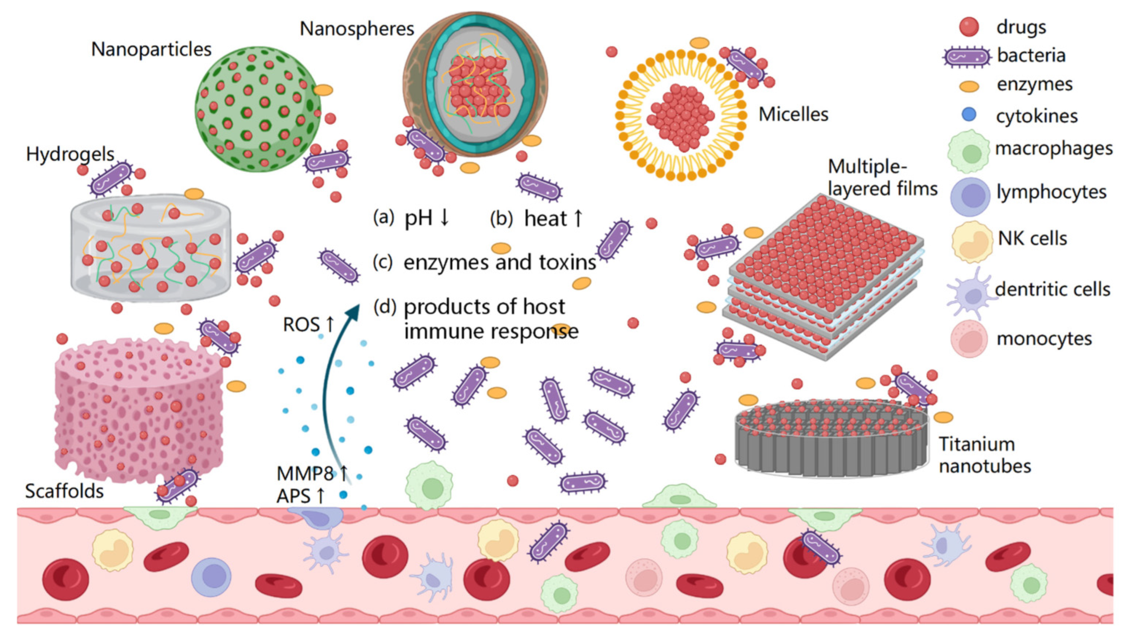
4. Novel Drug-Eluting Implant: Smart Drug Delivery System
5. Outlook and Perspectives
6. Conclusions
Author Contributions
Funding
Institutional Review Board Statement
Informed Consent Statement
Data Availability Statement
Conflicts of Interest
References
- Vajapey, S.P.; Li, M. Medical Device Recalls in Orthopedics: Recent Trends and Areas for Improvement. J. Arthroplast. 2020, 35, 2259–2266. [Google Scholar] [CrossRef] [PubMed]
- Guo, L.; Naghavi, S.A.; Wang, Z.; Varma, S.N.; Han, Z.; Yao, Z.; Wang, L.; Liu, C. On the design evolution of hip implants: A review. Mater. Des. 2022, 216, 110552. [Google Scholar] [CrossRef]
- Rohrer, F.; Farokhnia, A.; Nötzli, H.; Haubitz, F.; Hermann, T.; Gahl, B.; Limacher, A.; Brügger, J. Profit-Influencing Factors in Orthopedic Surgery: An Analysis of Costs and Reimbursements. Int. J. Environ. Res. Public Health 2022, 19, 4325. [Google Scholar] [CrossRef] [PubMed]
- Rouf, S.; Malik, A.; Raina, A.; Haq, M.I.U.; Naveed, N.; Zolfagharian, A.; Bodaghi, M. Functionally graded additive manufacturing for orthopedic applications. J. Orthop. 2022, 33, 70–80. [Google Scholar] [CrossRef] [PubMed]
- Bordbar-Khiabani, A.; Yarmand, B.; Mozafari, M. Emerging magnesium-based biomaterials for orthopedic implantation. Emerg. Mater. Res. 2019, 8, 305–319. [Google Scholar] [CrossRef] [Green Version]
- Koopaie, M.; Bordbar-Khiabani, A.; Kolahdooz, S.; Darbandsari, A.K.; Mozafari, M. Advanced surface treatment techniques counteract biofilm-associated infections on dental implants. Mater. Res. Express 2020, 7, 015417. [Google Scholar] [CrossRef]
- Fard, M.G.; Sharifianjazi, F.; Kazemi, S.S.; Rostamani, H.; Bathaei, M.S. Laser-Based Additive Manufacturing of Magnesium Alloys for Bone Tissue Engineering Applications: From Chemistry to Clinic. J. Manuf. Mater. Process. 2022, 6, 158. [Google Scholar] [CrossRef]
- Pourshadloo, M.; Rezaei, H.A.; Saeidnia, M.; Alkokab, H.; Bathaei, M.S. Effect of G-family incorporation on corrosion behavior of PEO-treated titanium alloys: A review. Surf. Innov. 2022, 40, 1–10. [Google Scholar] [CrossRef]
- Available online: https://orthopedicimplantsindia.wordpress.com/2016/05/18/implants-in-orthopedic-trauma/ (accessed on 1 October 2022).
- Kimura, M.; Kaku, N.; Kubota, Y.; Tagomori, H.; Tsumura, H. Fluorodeoxyglucose Positron-Emission Tomography/Computed Tomography and Magnetic Resonance Imaging for Adverse Local Tissue Reactions near Metal Implants after Total Hip Arthroplasty: A Preliminary Report. Clin. Orthop. Surg. 2021, 13, 320–328. [Google Scholar] [CrossRef]
- Eltit, F.; Noble, J.; Sharma, M.; Benam, N.; Haegert, A.; Bell, R.H.; Simon, F.; Duncan, C.P.; Garbuz, D.S.; Greidanus, N.V.; et al. Cobalt ions induce metabolic stress in synovial fibroblasts and secretion of cytokines/chemokines that may be diagnostic markers for adverse local tissue reactions to hip implants. Acta Biomater. 2021, 131, 581–594. [Google Scholar] [CrossRef]
- Davis, R.; Singh, A.; Jackson, M.J.; Coelho, R.T.; Prakash, D.; Charalambous, C.P.; Ahmed, W.; da Silva, L.R.R.; Lawrence, A.A. A comprehensive review on metallic implant biomaterials and their subtractive manufacturing. Int. J. Adv. Manuf. Technol. 2022, 120, 1473–1530. [Google Scholar] [CrossRef] [PubMed]
- Kaliaraj, G.S.; Siva, T.; Ramadoss, A. Surface functionalized bioceramics coated on metallic implants for biomedical and anticorrosion performance—A review. J. Mater. Chem. B 2021, 9, 9433–9460. [Google Scholar] [CrossRef] [PubMed]
- Ting, W.-T.; Chen, K.-S.; Wang, M.-J. Dense and anti-corrosion thin films prepared by plasma polymerization of hexamethyldisilazane for applications in metallic implants. Surf. Coatings Technol. 2021, 410, 126932. [Google Scholar] [CrossRef]
- Huang, P.; Xu, J.; Xie, L.; Gao, G.; Chen, S.; Gong, Z.; Lao, X.; Shan, Z.; Shi, J.; Zhou, Z.; et al. Improving hard metal implant and soft tissue integration by modulating the “inflammatory-fibrous complex” response. Bioact. Mater. 2023, 20, 42–52. [Google Scholar] [CrossRef] [PubMed]
- Bauer, T.W.; Zhang, Y.; Gao, M.A.; Lin, B.Q.; Koff, M.F. Reproducibility of pathologic scoring systems for periprosthetic adverse local tissue reactions: A cross-sectional study. Pathol.-Res. Pract. 2021, 228, 153685. [Google Scholar] [CrossRef]
- Poli, P.P.; de Miranda, F.V.; Polo, T.O.B.; Júnior, J.F.S.; Neto, T.J.L.; Rios, B.R.; Assunção, W.G.; Ervolino, E.; Maiorana, C.; Faverani, L.P. Titanium Allergy Caused by Dental Implants: A Systematic Literature Review and Case Report. Materials 2021, 14, 5239. [Google Scholar] [CrossRef]
- Montazerian, M.; Hosseinzadeh, F.; Migneco, C.; Fook, M.V.; Baino, F. Bioceramic coatings on metallic implants: An overview. Ceram. Int. 2022, 48, 8987–9005. [Google Scholar] [CrossRef]
- Komlev, A.S.; Gimaev, R.R.; Zverev, V.I. Smart magnetocaloric coatings for implants: Controlled drug release for targeted delivery. Phys. Open 2021, 7, 100063. [Google Scholar] [CrossRef]
- Santos-Coquillat, A.; Martínez-Campos, E.; Sánchez, H.M.; Moreno, L.; Arrabal, R.; Mohedano, M.; Gallardo, A.; Rodríguez-Hernández, J.; Matykina, E. Hybrid functionalized coatings on Metallic Biomaterials for Tissue Engineering. Surf. Coatings Technol. 2021, 422, 127508. [Google Scholar] [CrossRef]
- Khudhair, N.A.; Kadhim, M.M.; Khadom, A.A. Effect of Trimethoprim Drug Dose on Corrosion Behavior of Stainless Steel in Simulated Human Body Environment: Experimental and Theoretical Investigations. J. Bio-Tribo-Corrosion 2021, 7, 124. [Google Scholar] [CrossRef]
- Karacan, I.; Ben-Nissan, B.; Wang, H.A.; Juritza, A.; Swain, M.V.; Müller, W.H.; Chou, J.; Stamboulis, A.; Macha, I.J.; Taraschi, V. Mechanical testing of antimicrobial biocomposite coating on metallic medical implants as drug delivery system. Mater. Sci. Eng. C 2019, 104, 109757. [Google Scholar] [CrossRef] [PubMed]
- Sharipova, A.; Unger, R.E.; Sosnik, A.; Gutmanas, E. Dense drug-eluting biodegradable Fe-Ag nanocomposites. Mater. Des. 2021, 204, 109660. [Google Scholar] [CrossRef]
- Ghimire, A.; Song, J. Anti-Periprosthetic Infection Strategies: From Implant Surface Topographical Engineering to Smart Drug-Releasing Coatings. ACS Appl. Mater. Interfaces 2021, 13, 20921–20937. [Google Scholar] [CrossRef] [PubMed]
- Quarterman, J.C.; Geary, S.M.; Salem, A.K. Evolution of drug-eluting biomedical implants for sustained drug delivery. Eur. J. Pharm. Biopharm. 2021, 159, 21–35. [Google Scholar] [CrossRef] [PubMed]
- Negut, I.; Floroian, L.; Ristoscu, C.; Mihailescu, C.N.; Mirza Rosca, J.C.; Tozar, T.; Badea, M.; Grumezescu, V.; Hapenciuc, C.; Mihailescu, I.N. Functional Bioglass—Biopolymer Double Nanostructure for Natural Antimicrobial Drug Extracts Delivery. Nanomaterials 2020, 10, 385. [Google Scholar] [CrossRef] [Green Version]
- Singh, G.; Singh, R.P.; Jolly, S.S. Customized hydroxyapatites for bone-tissue engineering and drug delivery applications: A review. J. Sol-Gel Sci. Technol. 2020, 94, 505–530. [Google Scholar] [CrossRef]
- Nasr, A.; Gawad, S.A.; Fekry, A.M. A Sensor for Monitoring the Corrosion Behavior of Orthopedic Drug Calcium Hydrogen Phosphate on a Surgical 316L Stainless Steel Alloy as Implant. J. Bio- Tribo-Corrosion 2020, 6, 36. [Google Scholar] [CrossRef]
- Bernad, S.I.; Socoliuc, V.; Susan-Resiga, D.; Crăciunescu, I.; Turcu, R.; Tombácz, E.; Vékás, L.; Ioncica, M.C.; Bernad, E.S. Magnetoresponsive Functionalized Nanocomposite Aggregation Kinetics and Chain Formation at the Targeted Site during Magnetic Targeting. Pharmaceutics 2022, 14, 1923. [Google Scholar] [CrossRef]
- Ion, R.; Necula, M.G.; Mazare, A.; Mitran, V.; Neacsu, P.; Schmuki, P.; Cimpean, A. Drug Delivery Systems Based on Titania Nanotubes and Active Agents for Enhanced Osseointegration of Bone Implants. Curr. Med. Chem. 2020, 27, 854–902. [Google Scholar] [CrossRef]
- Liu, C.; Zhang, W.; Gao, M.; Yang, K.; Tan, L.; Zhao, W. A Degradable and Osteogenic Mg-Based MAO-MT-PLGA Drug/Ion Delivery System for Treating an Osteoporotic Fracture. Pharmaceutics 2022, 14, 1481. [Google Scholar] [CrossRef]
- Finšgar, M.; Kovač, J.; Maver, U. The development and characterization of bioactive coatings for local drug delivery in orthopedic applications. Prog. Org. Coat. 2021, 158, 106350. [Google Scholar] [CrossRef]
- Zafar, M.S.; Fareed, M.A.; Riaz, S.; Latif, M.; Habib, S.R.; Khurshid, Z. Customized Therapeutic Surface Coatings for Dental Implants. Coatings 2020, 10, 568. [Google Scholar] [CrossRef]
- Ghasali, E.; Bordbar-Khiabani, A.; Alizadeh, M.; Mozafari, M.; Niazmand, M.; Kazemzadeh, H.; Ebadzadeh, T. Corrosion behavior and in-vitro bioactivity of porous Mg/Al2O3 and Mg/Si3N4 metal matrix composites fabricated using microwave sintering process. Mater. Chem. Phys. 2019, 225, 331–339. [Google Scholar] [CrossRef]
- Türk, S.; Yılmaz, E. An innovative layer-by-layer coated titanium hydroxide-(gentamicin-polydopamine) as a hybrid drug delivery platform. J. Drug Deliv. Sci. Technol. 2022, 67, 102943. [Google Scholar] [CrossRef]
- Bácskay, I.; Ujhelyi, Z.; Fehér, P.; Arany, P. The Evolution of the 3D-Printed Drug Delivery Systems: A Review. Pharmaceutics 2022, 14, 1312. [Google Scholar] [CrossRef]
- Kunrath, M.F.; Muradás, T.C.; Penha, N.; Campos, M.M. Innovative surfaces and alloys for dental implants: What about biointerface-safety concerns? Dent. Mater. 2021, 37, 1447–1462. [Google Scholar] [CrossRef]
- Tiwari, A.; Sharma, P.; Vishwamitra, B.; Singh, G. Review on Surface Treatment for Implant Infection via Gentamicin and Antibiotic Releasing Coatings. Coatings 2021, 11, 1006. [Google Scholar] [CrossRef]
- Halim, N.A.A.; Hussein, M.Z.; Kandar, M.K. Nanomaterials-Upconverted Hydroxyapatite for Bone Tissue Engineering and a Platform for Drug Delivery. Int. J. Nanomed. 2021, 16, 6477–6496. [Google Scholar] [CrossRef]
- Chu, Y.S.; Wong, P.-C.; Jang, J.S.-C.; Chen, C.-H.; Wu, S.-H. Combining Mg–Zn–Ca Bulk Metallic Glass with a Mesoporous Silica Nanocomposite for Bone Tissue Engineering. Pharmaceutics 2022, 14, 1078. [Google Scholar] [CrossRef]
- Losic, D. Advancing of titanium medical implants by surface engineering: Recent progress and challenges. Expert Opin. Drug Deliv. 2021, 18, 1355–1378. [Google Scholar] [CrossRef]
- Majewska, P.; Oledzka, E.; Sobczak, M. Overview of the latest developments in the field of drug-eluting stent technology. Biomater. Sci. 2019, 8, 544–551. [Google Scholar] [CrossRef] [PubMed]
- Hauck, M.; Dittmann, J.; Zeller-Plumhoff, B.; Madurawala, R.; Hellmold, D.; Kubelt, C.; Synowitz, M.; Held-Feindt, J.; Adelung, R.; Wulfinghoff, S.; et al. Fabrication and Modelling of a Reservoir-Based Drug Delivery System for Customizable Release. Pharmaceutics 2022, 14, 777. [Google Scholar] [CrossRef] [PubMed]
- Wang, K.; Jin, H.; Song, Q.; Huo, J.; Zhang, J.; Li, P. Titanium dioxide nanotubes as drug carriers for infection control and osteogenesis of bone implants. Drug Deliv. Transl. Res. 2021, 11, 1456–1474. [Google Scholar] [CrossRef] [PubMed]
- Jaworska, J.; Jelonek, K.; Jaworska-Kik, M.; Musiał-Kulik, M.; Marcinkowski, A.; Szewczenko, J.; Kajzer, W.; Pastusiak, M.; Kasperczyk, J. Development of antibacterial, ciprofloxacin-eluting biodegradable coatings on Ti6Al7Nb implants to prevent peri-implant infections. J. Biomed. Mater. Res. Part A 2020, 108, 1006–1015. [Google Scholar] [CrossRef] [PubMed]
- Lee, K.; Goudie, M.J.; Tebon, P.; Sun, W.; Luo, Z.; Lee, J.; Zhang, S.; Fetah, K.; Kim, H.-J.; Xue, Y.; et al. Non-transdermal microneedles for advanced drug delivery. Adv. Drug Deliv. Rev. 2019, 165-166, 41–59. [Google Scholar] [CrossRef]
- Major, I.; Lastakchi, S.; Dalton, M.; McConville, C. Implantable drug delivery systems. Engineering Drug Delivery Systems. Woodhead Publ. 2020, 111–146. [Google Scholar] [CrossRef]
- Bordbar-Khiabani, A.; Yarmand, B.; Mozafari, M. Functional PEO layers on magnesium alloys: Innovative polymer-free drug-eluting stents. Surf. Innov. 2018, 6, 237–243. [Google Scholar] [CrossRef]
- Liu, W.; Pan, Y.; Zhong, Y.; Li, B.; Ding, Q.; Xu, H.; Qiu, Y.; Ren, F.; Li, B.; Muddassir, M.; et al. A multifunctional aminated UiO-67 metal-organic framework for enhancing antitumor cytotoxicity through bimodal drug delivery. Chem. Eng. J. 2021, 412, 127899. [Google Scholar] [CrossRef]
- Bordbar-Khiabani, A.; Yarmand, B.; Mozafari, M. Effect of ZnO pore-sealing layer on anti-corrosion and in-vitro bioactivity behavior of plasma electrolytic oxidized AZ91 magnesium alloy. Mater. Lett. 2020, 258, 126779. [Google Scholar] [CrossRef]
- Su, Y.; Cockerill, I.; Zheng, Y.; Tang, L.; Qin, Y.-X.; Zhu, D. Biofunctionalization of metallic implants by calcium phosphate coatings. Bioact. Mater. 2019, 4, 196–206. [Google Scholar] [CrossRef]
- Bordbar-Khiabani, A.; Ebrahimi, S.; Yarmand, B. In-vitro corrosion and bioactivity behavior of tailored calcium phosphate-containing zinc oxide coating prepared by plasma electrolytic oxidation. Corros. Sci. 2020, 173. [Google Scholar] [CrossRef]
- Bordbar-Khiabani, A.; Yarmand, B.; Sharifi-Asl, S.; Mozafari, M. Improved corrosion performance of biodegradable magnesium in simulated inflammatory condition via drug-loaded plasma electrolytic oxidation coatings. Mater. Chem. Phys. 2020, 239, 122003. [Google Scholar] [CrossRef]
- Mohanta, M.; Thirugnanam, A. Development of Multifunctional Commercial Pure Titanium-Polyethylene Glycol Drug-Eluting Substrates with Enhanced Optical and Antithrombotic Properties. Cardiovasc. Eng. Technol. 2022, 1–15. [Google Scholar] [CrossRef] [PubMed]
- Saberi, A.; Bakhsheshi-Rad, H.; Abazari, S.; Ismail, A.; Sharif, S.; Ramakrishna, S.; Daroonparvar, M.; Berto, F. A Comprehensive Review on Surface Modifications of Biodegradable Magnesium-Based Implant Alloy: Polymer Coatings Opportunities and Challenges. Coatings 2021, 11, 747. [Google Scholar] [CrossRef]
- Tan, G.; Zhong, Y.; Yang, L.; Jiang, Y.; Liu, J.; Ren, F. A multifunctional MOF-based nanohybrid as injectable implant platform for drug synergistic oral cancer therapy. Chem. Eng. J. 2020, 390, 124446. [Google Scholar] [CrossRef]
- Rothe, R.; Hauser, S.; Neuber, C.; Laube, M.; Schulze, S.; Rammelt, S.; Pietzsch, J. Adjuvant Drug-Assisted Bone Healing: Advances and Challenges in Drug Delivery Approaches. Pharmaceutics 2020, 12, 428. [Google Scholar] [CrossRef]
- Cheng, X.; Deng, D.; Chen, L.; Jansen, J.A.; Leeuwenburgh, S.G.C.; Yang, F. Electrodeposited Assembly of Additive-Free Silk Fibroin Coating from Pre-Assembled Nanospheres for Drug Delivery. ACS Appl. Mater. Interfaces 2020, 12, 12018–12029. [Google Scholar] [CrossRef] [Green Version]
- Biegański, P.; Szczupak, Ł.; Arruebo, M.; Kowalski, K. Brief survey on organometalated antibacterial drugs and metal-based materials with antibacterial activity. RSC Chem. Biol. 2021, 2, 368–386. [Google Scholar] [CrossRef]
- Bahaa, M.; Daily, Z.A.; Alsharbaty, M.H.M.H.; Abullais, S.S.; Arora, S.; Lafta, H.A.; Turki Jalil, A.; Almulla, A.F.; Ramírez-Mohammed, N.B.; Daily, Z.A.; et al. Effect of PMMA sealing treatment on the corrosion behavior of plasma electrolytic oxidized titanium dental implants in fluoride-containing saliva solution. Mater. Res. Express 2022, 9, 125401. [Google Scholar] [CrossRef]
- He, X.; Deng, Y.; Yu, Y.; Lyu, H.; Liao, L. Drug-loaded/grafted peptide-modified porous PEEK to promote bone tissue repair and eliminate bacteria. Colloids Surfaces B: Biointerfaces 2019, 181, 767–777. [Google Scholar] [CrossRef]
- Yoon, S.-W.; Kim, M.-J.; Paeng, K.-W.; Yu, K.A.; Lee, C.-K.; Song, Y.W.; Cha, J.-K.; Sanz, M.; Jung, U.-W. Locally Applied Slow-Release of Minocycline Microspheres in the Treatment of Peri-Implant Mucositis: An Experimental In Vivo Study. Pharmaceutics 2020, 12, 668. [Google Scholar] [CrossRef] [PubMed]
- Gholipourmalekabadi, M.; Joghataei, M.T.; Urbanska, A.M.; Aghabarari, B.; Khiabani, A.B.; Samadikuchaksaraei, A.; Mozafari, M. 13 Use of Nanotechnology for Viable Applications in the Field of Medicine. In Manufacturing Techniques for Materials, 1st ed.; Srivatsan, T.S., Sudarshan, T.S., Manigandan, K., Eds.; CRC Press, Taylor & Francis: Boca Raton, FL, USA, 2018; pp. 393–431. [Google Scholar] [CrossRef]
- Nelson, J.; Jain, S.; Pal, P.; Johnson, H.A.; Nobles, K.P.; Janorkar, A.V.; Williamson, R.S.; Roach, M.D. Anodized titanium with calcium and phosphorus surface enhancements for dental and orthopedic implant applications. Thin Solid Films 2022, 745, 139117. [Google Scholar] [CrossRef]
- Guo, T.; Ivanovski, S.; Gulati, K. Optimizing titanium implant nano-engineering via anodization. Mater. Des. 2022, 223, 111110. [Google Scholar] [CrossRef]
- Khoee, M.H.; Khoee, S.; Lotfi, M. Synthesis of titanium dioxide nanotubes with liposomal covers for carrying and extended release of 5-FU as anticancer drug in the treatment of HeLa cells. Anal. Biochem. 2019, 572, 16–24. [Google Scholar] [CrossRef] [PubMed]
- Esmaeilnejad, A.; Mahmoudi, P.; Zamanian, A.; Mozafari, M. Synthesis of titanium oxide nanotubes and their decoration by MnO nanoparticles for biomedical applications. Ceram. Int. 2019, 45, 19275–19282. [Google Scholar] [CrossRef]
- Li, B.; Zhang, L.; Wang, D.; Liu, X.; Li, H.; Liang, C.; Zhao, X. Thermo-sensitive hydrogel on anodized titanium surface to regulate immune response. Surf. Coatings Technol. 2021, 405, 126624. [Google Scholar] [CrossRef]
- Murr, L.E. Strategies for creating living, additively manufactured, open-cellular metal and alloy implants by promoting osseointegration, osteoinduction and vascularization: An overview. J. Mater. Sci. Technol. 2019, 35, 231–241. [Google Scholar] [CrossRef]
- Kalinichenko, S.G.; Matveeva, N.Y.; Kostiv, R.Y.; Edranov, S.S. The topography and proliferative activity of cells immunoreactive to various growth factors in rat femoral bone tissues after experimental fracture and implantation of titanium implants with bioactive biodegradable coatings. Bio-Medical Mater. Eng. 2019, 30, 85–95. [Google Scholar] [CrossRef]
- Zhang, B.; Su, Y.; Zhou, J.; Zheng, Y.; Zhu, D. Toward a Better Regeneration through Implant-Mediated Immunomodulation: Harnessing the Immune Responses. Adv. Sci. 2021, 8, e2100446. [Google Scholar] [CrossRef]
- Alghamdi, H.S.; Jansen, J.A. The development and future of dental implants. Dent. Mater. J. 2020, 39, 167–172. [Google Scholar] [CrossRef] [PubMed] [Green Version]
- Youn, Y.H.; Lee, S.J.; Choi, G.R.; Lee, H.R.; Lee, D.; Heo, D.N.; Kim, B.-S.; Bang, J.B.; Hwang, Y.-S.; Correlo, V.M.; et al. Simple and facile preparation of recombinant human bone morphogenetic protein-2 immobilized titanium implant via initiated chemical vapor deposition technique to promote osteogenesis for bone tissue engineering application. Mater. Sci. Eng. C 2019, 100, 949–958. [Google Scholar] [CrossRef] [PubMed]
- Zhou, W.; Shi, P.; Dong, J.; Li, S.; Lv, P.; Liu, C. Scaffolds of bioactive glass (Bioglass®) combined with recombinant human bone morphogenetic protein -9 (rhBMP-9) for tooth extraction site preservation. Heliyon 2022, 8, e08796. [Google Scholar] [CrossRef] [PubMed]
- Zhou, D.; Cheng, L.; Xu, D.; Xu, Z.; Sun, M.; Chen, L.; Liu, Y.; Sun, J. Formulation and performance of bioactive hydrogel scaffold carrying chlorhexidine and bone morphogenetic protein. Mater. Lett. 2022, 313, 131788. [Google Scholar] [CrossRef]
- Echave, M.; Erezuma, I.; Golafshan, N.; Castilho, M.; Kadumudi, F.B.; Pimenta-Lopes, C.; Ventura, F.; Pujol, A.; Jimenez, J.; Camara, J.; et al. Bioinspired gelatin/bioceramic composites loaded with bone morphogenetic protein-2 (BMP-2) promote osteoporotic bone repair. Biomater. Adv. 2022, 134, 112539. [Google Scholar] [CrossRef]
- Bordbar-Khiabani, A.; Yarmand, B.; Mozafari, M. Enhanced corrosion resistance and in-vitro biodegradation of plasma electrolytic oxidation coatings prepared on AZ91 Mg alloy using ZnO nanoparticles-incorporated electrolyte. Surf. Coatings Technol. 2019, 360, 153–171. [Google Scholar] [CrossRef]
- Zhu, L.; Tong, X.; Ye, Z.; Lin, Z.; Zhou, T.; Huang, S.; Li, Y.; Lin, J.; Wen, C.; Ma, J. Zinc phosphate, zinc oxide, and their dual-phase coatings on pure Zn foam with good corrosion resistance, cytocompatibility, and antibacterial ability for potential biodegradable bone-implant applications. Chem. Eng. J. 2022, 450, 137946. [Google Scholar] [CrossRef]
- Poli, V.; Ma, V.P.-Y.; Di Gioia, M.; Broggi, A.; Benamar, M.; Chen, Q.; Mazitschek, R.; Haggarty, S.J.; Chatila, T.A.; Karp, J.M.; et al. Zinc-dependent histone deacetylases drive neutrophil extracellular trap formation and potentiate local and systemic inflammation. iScience 2021, 24, 103256. [Google Scholar] [CrossRef]
- Wang, T.; Bai, J.; Lu, M.; Huang, C.; Geng, D.; Chen, G.; Wang, L.; Qi, J.; Cui, W.; Deng, L. Engineering immunomodulatory and osteoinductive implant surfaces via mussel adhesion-mediated ion coordination and molecular clicking. Nat. Commun. 2022, 13, 160. [Google Scholar] [CrossRef]
- Teng, C.; Jia, J.; Wang, Z.; Sharma, V.K.; Yan, B. Size-dependent maternal-fetal transfer and fetal developmental toxicity of ZnO nanoparticles after oral exposures in pregnant mice. Ecotoxicol. Environ. Saf. 2019, 182, 109439. [Google Scholar] [CrossRef]
- Song, W.; Xiao, Y. Sequential drug delivery of vancomycin and rhBMP-2 via pore-closed PLGA microparticles embedded photo-crosslinked chitosan hydrogel for enhanced osteointegration. Int. J. Biol. Macromol. 2021, 182, 612–625. [Google Scholar] [CrossRef] [PubMed]
- Kumar, G.; Dhamangaonkar, A.C. Bisphosphonate associated femoral stress fracture distal to an orthopaedic implant: They are predictable! J. Clin. Orthop. Trauma 2019, 10, S112–S114. [Google Scholar] [CrossRef] [PubMed]
- McDonald, C.L.; Lemme, N.J.; Testa, E.J.; Aaron, R.; Hartnett, D.A.; Cohen, E.M. Bisphosphonates in Total Joint Arthroplasty: A Review of Their Use and Complications. Arthroplast. Today 2022, 14, 133–139. [Google Scholar] [CrossRef] [PubMed]
- Zhang, X.; Koo, S.; Kim, J.H.; Huang, X.; Kong, N.; Zhang, L.; Zhou, J.; Xue, J.; Harris, M.B.; Tao, W.; et al. Nanoscale materials-based platforms for the treatment of bone-related diseases. Matter 2021, 4, 2727–2764. [Google Scholar] [CrossRef]
- Chen, M.; Huang, L.; Shen, X.; Li, M.; Luo, Z.; Cai, K.; Hu, Y. Construction of multilayered molecular reservoirs on a titanium alloy implant for combinational drug delivery to promote osseointegration in osteoporotic conditions. Acta Biomater. 2020, 105, 304–318. [Google Scholar] [CrossRef] [PubMed]
- Zeng, J.; Guo, J.; Sun, Z.; Deng, F.; Ning, C.; Xie, Y. Osteoblastic and anti-osteoclastic activities of strontium-substituted silicocarnotite ceramics: In vitro and in vivo studies. Bioact. Mater. 2020, 5, 435–446. [Google Scholar] [CrossRef] [PubMed]
- Cui, Y.; Wang, Z.; Li, Z.; Ji, X.; Yuan, B.; Sun, Y.; Peng, C.; Leng, Y.; Dou, M.; Wang, J.; et al. Functionalized anti-osteoporosis drug delivery system enhances osseointegration of an inorganic–organic bioactive interface in osteoporotic microenvironment. Mater. Des. 2021, 206, 109753. [Google Scholar] [CrossRef]
- Skalická, B.; Matzick, K.; Komersová, A.; Svoboda, R.; Bartoš, M.; Hromádko, L. 3D-Printed Coating of Extended-Release Matrix Tablets: Effective Tool for Prevention of Alcohol-Induced Dose Dumping Effect. Pharmaceutics 2021, 13, 2123. [Google Scholar] [CrossRef]
- Janczura, M.; Sip, S.; Cielecka-Piontek, J. The Development of Innovative Dosage Forms of the Fixed-Dose Combination of Active Pharmaceutical Ingredients. Pharmaceutics 2022, 14, 834. [Google Scholar] [CrossRef]
- Wan, P.; Wang, W.; Zheng, L.; Qin, L.; Yang, K. One-step electrodeposition synthesis of bisphosphonate loaded magnesium implant: A strategy to modulate drug release for osteoporotic fracture healing. J. Mater. Sci. Technol. 2021, 78, 92–99. [Google Scholar] [CrossRef]
- Singh, I.; Devi, G.; Barik, B.R.; Sharma, A.; Kaur, L. Mucoadhesive Pellets for Drug Delivery Applications: A Critical Review. Rev. Adhes. Adhes. 2020, 8, 153–167. [Google Scholar] [CrossRef]
- Hassan, S.S.M.; Kamel, A.H.; Hashem, H.M.; Bary, E.M.A. Drug delivery systems between metal, liposome, and polymer-based nanomedicine: A review. Eur. Chem. Bull. 2020, 9, 91–102. [Google Scholar] [CrossRef]
- Wani, S.D.; Mundada, S.A. A Review: Emerging Trends in Bionanocomposites. Int. J. Pharm. Res. Technol. 2021, 11, 1–8. [Google Scholar] [CrossRef]
- Bordbar-Khiabani, A.; Bahrampour, S.; Mozafari, M.; Gasik, M. Surface functionalization of anodized tantalum with Mn3O4 nanoparticles for effective corrosion protection in simulated inflammatory condition. Ceram. Int. 2022, 48, 3148–3156. [Google Scholar] [CrossRef]
- Jamali, R.; Bordbar-Khiabani, A.; Yarmand, B.; Mozafari, M.; Kolahi, A. Effects of co-incorporated ternary elements on biocorrosion stability, antibacterial efficacy, and cytotoxicity of plasma electrolytic oxidized titanium for implant dentistry. Mater. Chem. Phys. 2022, 276, 125436. [Google Scholar] [CrossRef]
- Fugate Ajay, R.; Nagoba Shivappa, N.; Hyam, S.R. Formulation Development and evaluation of Liposomal Drug Delivery System Containing Etoposide. J. Complement. Med. Res. 2021, 12, 7–20. [Google Scholar] [CrossRef]
- Shizari, L.N.; Dounighi, N.M.; Bayat, M.; Mosavari, N. A New Amphotericin B-loaded Trimethyl Chitosan Nanoparticles as a Drug Delivery System and Antifungal Activity on Candida albicans Biofilm. Arch. Razi Inst. 2021, 76, 575–590. [Google Scholar] [CrossRef]
- Mary, S.K.; Thomas, M.S.; Koshy, R.R.; Pillai, P.K.; Pothan, L.A.; Thomas, S. Adhesion in Biocomposites: A Critical Review. Rev. Adhes. Adhes. 2020, 8, 527–553. [Google Scholar] [CrossRef]
- Al Hamad, K.Q.; Abu Al-Addous, A.M.; Al-Wahadni, A.M.; Baba, N.Z.; Goodacre, B.J. Surface roughness of monolithic and layered zirconia restorations at different stages of finishing and polishing: An in vitro study. J. Prosthodont. 2019, 28, 818–825. [Google Scholar] [CrossRef]
- Al Hamad, K.Q.; Al-Rashdan, R.B.; Al-Rashdan, B.A.; Baba, N.Z. Effect of Milling Protocols on Trueness and Precision of Ceramic Crowns. J. Prosthodont. 2021, 30, 171–176. [Google Scholar] [CrossRef] [PubMed]
- Nemati, E.; Gholami, A. Cell Membrane Coated Nanoparticles for Biomedical Applications. Adv. Appl. NanoBio-Technol. 2022, 3, 49–59. [Google Scholar] [CrossRef]
- Aghili, A.; Kamrani, M.R. Modeling of the thermal degradation of poly (methyl methacrylate) and its nanocomposite with multi-walled carbon nanotubes. Adv. Appl. NanoBio-Technol. 2021, 2, 22–34. [Google Scholar] [CrossRef]
- K Al-Saffar, A.; M Abbas, A.; Salman, D. Synthesis of New Glycine Cephalexin Condensed Polymer as Peptide Biopolymer for Controlled Release of Cephalexin. J. Chem. Health Risks 2021, 11, 339–344. [Google Scholar] [CrossRef]
- Masoumzadeh, R. Polyethyleneimine-based materials for gene therapy, bioimaging and drug delivery systems applications. Adv. Appl. NanoBio-Technol. 2021, 2, 13–16. [Google Scholar] [CrossRef]
- Seyyedi, M.; Molajou, A. Nanohydroxyapatite loaded-acrylated polyurethane nanofibrous scaffolds for controlled release of paclitaxel anticancer drug. J. Res. Sci. Eng. Technol. 2021, 9, 50–61. [Google Scholar]
- Goudarzian, N.; Samiei, S.; Safari, F.; Mousavi, S.M.; Hashemi, S.A.; Mazraedoost, S. Evalouation of Styrene Acrylo Nitrile (SAN), Butadiene Rubber (BR), Nano-silica (Nano SiO2) Blend and Nanocomposite in the Presence of Oxoperoxidant Study. J. Environ. Treat. Technol. 2020, 9, 24–32. [Google Scholar]
- Lloyd, A.W.; Faragher, R.G.; Denyer, S.P. Ocular biomaterials and implants. Biomaterials 2001, 22, 769–785. [Google Scholar] [CrossRef]
- Ren, X.; van der Mei, H.C.; Ren, Y.; Busscher, H.J.; Peterson, B.W. Antimicrobial loading of nanotubular titanium surfaces favoring surface coverage by mammalian cells over bacterial colonization. Mater. Sci. Eng. C 2021, 123, 112021. [Google Scholar] [CrossRef]
- Tian, B.; Tang, S.; Wang, C.D.; Wang, W.G.; Wu, C.L.; Guo, Y.J.; Guo, Y.P.; Zhu, Z.A. Bactericidal properties and biocompatibility of a gentamicin-loaded Fe3O4/carbonated hydroxyapatite coating. Colloids Surf. B: Biointerfaces 2014, 123, 403–412. [Google Scholar] [CrossRef]
- Rumian, Ł.; Tiainen, H.; Cibor, U.; Krok-Borkowicz, M.; Brzychczy-Włoch, M.; Haugen, H.J.; Pamuła, E. Ceramic scaffolds enriched with gentamicin loaded poly(lactide- co -glycolide) microparticles for prevention and treatment of bone tissue infections. Mater. Sci. Eng. C 2016, 69, 856–864. [Google Scholar] [CrossRef]
- Janson, O.; Sörensen, J.H.; Strømme, M.; Engqvist, H.; Procter, P.; Welch, K. Evaluation of an alkali-treated and hydroxyapatite-coated orthopedic implant loaded with tobramycin. J. Biomater. Appl. 2019, 34, 699–720. [Google Scholar] [CrossRef]
- Jennings, J.A.; E Beenken, K.; A Skinner, R.; Meeker, D.G.; Smeltzer, M.S.; O Haggard, W.; Troxel, K.S. Antibiotic-loaded phosphatidylcholine inhibits staphylococcal bone infection. World J. Orthop. 2016, 7, 467–474. [Google Scholar] [CrossRef] [PubMed]
- Kälicke, T.; Schierholz, J.; Schlegel, U.; Frangen, T.M.; Köller, M.; Printzen, G.; Seybold, D.; Klöckner, S.; Muhr, G.; Arens, S. Effect on infection resistance of a local antiseptic and antibiotic coating on osteosynthesis implants: An in vitro and in vivo study. J. Orthop. Res. 2006, 24, 1622–1640. [Google Scholar] [CrossRef] [PubMed]
- Kucharíková, S.; Gerits, E.; De Brucker, K.; Braem, A.; Ceh, K.; Majdič, G.; Španič, T.; Pogorevc, E.; Verstraeten, N.; Tournu, H.; et al. Covalent immobilization of antimicrobial agents on titanium prevents Staphylococcus aureus and Candida albicans colonization and biofilm formation. J. Antimicrob. Chemother. 2016, 71, 936–945. [Google Scholar] [CrossRef] [PubMed] [Green Version]
- Liu, D.; He, C.; Liu, Z.; Xu, W. Gentamicin coating of nanotubular anodized titanium implant reduces implant-related osteomyelitis and enhances bone biocompatibility in rabbits. Int. J. Nanomed. 2017, 12, 5461. [Google Scholar] [CrossRef] [PubMed] [Green Version]
- Lucke, M.; Schmidmaier, G.; Sadoni, S.; Wildemann, B.; Schiller, R.; Haas, N.; Raschke, M. Gentamicin coating of metallic implants reduces implant-related osteomyelitis in rats. Bone 2003, 32, 521–531. [Google Scholar] [CrossRef]
- Lucke, M.; Wildemann, B.; Sadoni, S.; Surke, C.; Schiller, R.; Stemberger, A.; Raschke, M.; Haas, N.P.; Schmidmaier, G. Systemic versus local application of gentamicin in prophylaxis of implant-related osteomyelitis in a rat model. Bone 2005, 36, 770–778. [Google Scholar] [CrossRef]
- Ma, K.; Cai, X.; Zhou, Y.; Wang, Y.; Jiang, T. In Vitro and In Vivo Evaluation of Tetracycline Loaded Chitosan-Gelatin Nanosphere Coatings for Titanium Surface Functionalization. Macromol. Biosci. 2017, 17, 1600130. [Google Scholar] [CrossRef]
- Metsemakers, W.-J.; Emanuel, N.; Cohen, O.; Reichart, M.; Potapova, I.; Schmid, T.; Segal, D.; Riool, M.; Kwakman, P.H.; de Boer, L.; et al. A doxycycline-loaded polymer-lipid encapsulation matrix coating for the prevention of implant-related osteomyelitis due to doxycycline-resistant methicillin-resistant Staphylococcus aureus. J. Control Release 2015, 209, 47–56. [Google Scholar] [CrossRef]
- Adams, C.S.; Antoci, V.; Harrison, G.; Patal, P.; Freeman, T.A.; Shapiro, I.M.; Parvizi, J.; Hickok, N.J.; Radin, S.; Ducheyne, P. Controlled release of vancomycin from thin sol-gel films on implant surfaces successfully controls osteomyelitis. J. Orthop. Res. 2009, 27, 701–709. [Google Scholar] [CrossRef]
- Alt, V.; Kirchhof, K.; Seim, F.; Hrubesch, I.; Lips, K.S.; Mannel, H.; Domann, E.; Schnettler, R. Rifampicin–fosfomycin coating for cementless endoprostheses: Antimicrobial effects against methicillin-sensitive Staphylococcus aureus (MSSA) and methicillin-resistant Staphylococcus aureus (MRSA). Acta Biomater. 2014, 10, 4518–4524. [Google Scholar] [CrossRef]
- Antoci, V., Jr.; Adams, C.S.; Hickok, N.J.; Shapiro, I.M.; Parvizi, J. Vancomycin bound to Ti rods reduces periprosthetic infection: Preliminary study. Clin. Orthop. Relat. Res. 2007, 461, 88–95. [Google Scholar] [CrossRef] [PubMed]
- Auñón, Á.; Esteban, J.; Doadrio, A.L.; Boiza-Sánchez, M.; Mediero, A.; Eguibar-Blázquez, D.; Cordero-Ampuero, J.; Conde, A.; Arenas, M.; de-Damborenea, J.; et al. Staphylococcus aureus Prosthetic Joint Infection Is Prevented by a Fluorine-and Phosphorus-Doped Nanostructured Ti–6Al–4V Alloy Loaded with Gentamicin and Vancomycin. J. Orthop. Res. 2020, 38, 588–597. [Google Scholar] [CrossRef] [PubMed]
- Aykut, S.; Öztürk, A.; Özkan, Y.; Yanik, K.; Ilman, A.A.; Özdemir, R.M. Evaluation and comparison of the antimicrobial efficacy of teicoplanin-and clindamycin-coated titanium implants: An experimental study. J. Bone Jt. Surg. 2010, 92, 159–163. [Google Scholar] [CrossRef] [Green Version]
- Badar, M.; Rahim, M.I.; Kieke, M.; Ebel, T.; Rohde, M.; Hauser, H.; Behrens, P.; Mueller, P.P. Controlled drug release from antibiotic-loaded layered double hydroxide coatings on porous titanium implants in a mouse model. J. Biomed. Mater. Res. Part A 2014, 103, 2141–2149. [Google Scholar] [CrossRef] [PubMed]
- Croes, M.; Bakhshandeh, S.; van Hengel, I.; Lietaert, K.; van Kessel, K.; Pouran, B.; van der Wal, B.; Vogely, H.; Van Hecke, W.; Fluit, A.; et al. Antibacterial and immunogenic behavior of silver coatings on additively manufactured porous titanium. Acta Biomater. 2018, 81, 315–327. [Google Scholar] [CrossRef]
- Darouiche, R.O.; Mansouri, M.D.; Zakarevicz, D.; AlSharif, A.; Landon, G.C. In vivo efficacy of antimicrobial-coated devices. J. Bone Joint Surg. 2007, 89, 792–797. [Google Scholar] [CrossRef]
- Diefenbeck, M.; Schrader, C.; Gras, F.; Mückley, T.; Schmidt, J.; Zankovych, S.; Bossert, J.; Jandt, K.; Völpel, A.; Sigusch, B.; et al. Gentamicin coating of plasma chemical oxidized titanium alloy prevents implant-related osteomyelitis in rats. Biomaterials 2016, 101, 156–164. [Google Scholar] [CrossRef]
- Ghimire, A.; Skelly, J.D.; Song, J. Micrococcal-Nuclease-Triggered On-Demand Release of Vancomycin from Intramedullary Implant Coating Eradicates Staphylococcus aureus Infection in Mouse Femoral Canals. ACS Central Sci. 2019, 5, 1929–1936. [Google Scholar] [CrossRef] [Green Version]
- Grohmann, S.; Menne, M.; Hesse, D.; Bischoff, S.; Schiffner, R.; Diefenbeck, M.; Liefeith, K. Biomimetic multilayer coatings deliver gentamicin and reduce implant-related osteomyelitis in rats. Biomed. Eng. Biomed. Technol. 2019, 64, 383–395. [Google Scholar] [CrossRef]
- Zhang, H.; Wang, G.; Liu, P.; Tong, D.; Ding, C.; Zhang, Z.; Xie, Y.; Tang, H.; Ji, F. Vancomycin-loaded titanium coatings with an interconnected micro-patterned structure for prophylaxis of infections: An in vivo study. RSC Adv. 2018, 8, 9223–9231. [Google Scholar] [CrossRef] [Green Version]
- Zhang, B.; Braun, B.M.; Skelly, J.D.; Ayers, D.C.; Song, J. Significant Suppression of Staphylococcus aureus Colonization on Intramedullary Ti6Al4V Implants Surface-Grafted with Vancomycin-Bearing Polymer Brushes. ACS Appl. Mater. Interfaces 2019, 11, 28641–28647. [Google Scholar] [CrossRef] [PubMed]
- Zhou, L.; Liu, Q.; Zhou, Z.; Lu, W.; Tao, J. Efficacy of tobramycin-loaded coating K-wire in an open-fracture rabbit model contaminated by staphylococcus aureus. Int. J. Clin. Exp. Med. 2017, 10, 6004–6016. [Google Scholar]
- Moskowitz, J.S.; Blaisse, M.R.; Samuel, R.E.; Hsu, H.-P.; Harris, M.B.; Martin, S.D.; Lee, J.C.; Spector, M.; Hammond, P.T. The effectiveness of the controlled release of gentamicin from polyelectrolyte multilayers in the treatment of Staphylococcus aureus infection in a rabbit bone model. Biomaterials 2010, 31, 6019–6030. [Google Scholar] [CrossRef] [PubMed] [Green Version]
- Neut, D.; Dijkstra, R.; Thompson, J.; Kavanagh, C.; van der Mei, H.; Busscher, H. A biodegradable gentamicin-hydroxyapatite-coating for infection prophylaxis in cementless hip prostheses. Eur. Cells Mater. 2015, 29, 42–56. [Google Scholar] [CrossRef] [PubMed]
- Nie, B.; Long, T.; Ao, H.; Zhou, J.; Tang, T.; Yue, B. Covalent Immobilization of Enoxacin onto Titanium Implant Surfaces for Inhibiting Multiple Bacterial Species Infection and In Vivo Methicillin-Resistant Staphylococcus aureus Infection Prophylaxis. Antimicrob. Agents Chemother. 2017, 61, e01766-16. [Google Scholar] [CrossRef] [Green Version]
- Nie, B.; Ao, H.; Long, T.; Zhou, J.; Tang, T.; Yue, B. Immobilizing bacitracin on titanium for prophylaxis of infections and for improving osteoinductivity: An in vivo study. Colloids Surfaces B: Biointerfaces 2017, 150, 183–191. [Google Scholar] [CrossRef]
- Song, W.; Seta, J.; Chen, L.; Bergum, C.; Zhou, Z.; Kanneganti, P.; Kast, R.E.; Auner, G.W.; Shen, M.; Markel, D.C.; et al. Doxycycline-loaded coaxial nanofiber coating of titanium implants enhances osseointegration and inhibits Staphylococcus aureus infection. Biomed. Mater. 2017, 12, 045008. [Google Scholar] [CrossRef]
- Stavrakis, A.I.; Zhu, S.; Loftin, A.H.; Weixian, X.; Niska, J.; Hegde, V.; Segura, T.; Bernthal, N.M. Controlled Release of Vancomycin and Tigecycline from an Orthopaedic Implant Coating Prevents Staphylococcus aureus Infection in an Open Fracture Animal Model. BioMed. Res. Int. 2019, 2019, 1638508. [Google Scholar] [CrossRef] [Green Version]
- Thompson, K.; Petkov, S.; Zeiter, S.; Sprecher, C.M.; Richards, R.G.; Moriarty, T.F.; Eijer, H. Intraoperative loading of calcium phosphate-coated implants with gentamicin prevents experimental Staphylococcus aureus infection in vivo. PLoS ONE 2019, 14, e0210402. [Google Scholar] [CrossRef]
- Tang, T.-T.; Ao, H.-Y.; Yang, S.-B.; Wang, Y.-G.; Lin, W.-T.; Yu, Z.-F.; Yang, Y. In vivo evaluation of the anti-infection potential of gentamicin-loaded nanotubes on titania implants. Int. J. Nanomed. 2016, 11, 2223–2234. [Google Scholar] [CrossRef] [Green Version]
- Yuan, Z.; Huang, S.; Lan, S.; Xiong, H.; Tao, B.; Ding, Y.; Liu, Y.; Liu, P.; Cai, K. Surface engineering of titanium implants with enzyme-triggered antibacterial properties and enhanced osseointegration in vivo. J. Mater. Chem. B 2018, 6, 8090–8104. [Google Scholar] [CrossRef] [PubMed]
- Zeng, J.; Wang, Y.; Sun, Z.; Chang, H.; Cao, M.; Zhao, J.; Lin, K.; Xie, Y. A novel biocompatible PDA/IR820/DAP coating for antibiotic/photodynamic/photothermal triple therapy to inhibit and eliminate Staphylococcus aureus biofilm. Chem. Eng. J. 2020, 394, 125017. [Google Scholar] [CrossRef]
- Aghion, E.; Yered, T.; Perez, Y.; Gueta, Y. The Prospects of Carrying and Releasing Drugs Via Biodegradable Magnesium Foam. Adv. Eng. Mater. 2010, 12, B374–B379. [Google Scholar] [CrossRef]
- Aydemir, T.; Liverani, L.; Pastore, J.I.; Ceré, S.M.; Goldmann, W.H.; Boccaccini, A.R.; Ballarre, J. Functional behavior of chitosan/gelatin/silica-gentamicin coatings by electrophoretic deposition on surgical grade stainless steel. Mater. Sci. Eng. C Mater. 2020, 115, 111062. [Google Scholar] [CrossRef]
- Zhao, Y.; Chen, X.; Li, S.; Zeng, R.; Zhang, F.; Wang, Z.; Guan, S. Corrosion resistance and drug release profile of gentamicin-loaded polyelectrolyte multilayers on magnesium alloys: Effects of heat treatment. J. Colloid Interface Sci. 2019, 547, 309–317. [Google Scholar] [CrossRef]
- Bordbar-Khiabani, A.; Gasik, M. Smart Hydrogels for Advanced Drug Delivery Systems. Int. J. Mol. Sci. 2022, 23, 3665. [Google Scholar] [CrossRef] [PubMed]
- Alipour, A. Virus decorated nanobiomaterials as scaffolds for tissue engineering. Adv. Appl. NanoBio-Technol. 2021, 2, 79–85. [Google Scholar]
- Yang, Y.; Jiang, X.; Lai, H.; Zhang, X. Smart Bacteria-Responsive Drug Delivery Systems in Medical Implants. J. Funct. Biomater. 2022, 13, 173. [Google Scholar] [CrossRef]
- Yu, Y.; Ran, Q.; Shen, X.; Zheng, H.; Cai, K. Enzyme responsive titanium substrates with antibacterial property and osteo/angio-genic differentiation potentials. Colloids Surfaces B: Biointerfaces 2020, 185, 110592. [Google Scholar] [CrossRef]
- Wang, T.; Liu, X.; Zhu, Y.; Cui, Z.D.; Yang, X.J.; Pan, H.; Yeung, K.W.K.; Wu, S. Metal ion coordination polymer-capped pH-triggered drug release system on titania nanotubes for enhancing self-antibacterial capability of Ti implants. ACS Biomater. Sci. Eng. 2017, 3, 816–825. [Google Scholar] [CrossRef]
- Choi, H.; Schulte, A.; Müller, M.; Park, M.; Jo, S.; Schönherr, H. Drug Release from Thermo-Responsive Polymer Brush Coatings to Control Bacterial Colonization and Biofilm Growth on Titanium Implants. Adv. Healthc. Mater. 2021, 10, e2100069. [Google Scholar] [CrossRef] [PubMed]
- Al-Hamad, K.Q.; Al-Omari, M.; Al-Wahadni, A.; Darwazeh, A. Radiographic assessment of post-retained crowns in an adult Jordanian population. J. Contemp. Dent. Pract. 2006, 7, 29–36. [Google Scholar] [CrossRef] [PubMed]


| Implant Material | Method | Test Model | Bacterial Culture | Outcomes | Ref. |
|---|---|---|---|---|---|
| Titanium | GM loaded nanotubes coated over the implant surface | In-vivo | S. aureus |
| [108] |
| Titanium | GM loaded on the surface via immersion in GM solution | In-vitro | S. aureus, P. aeruginosa and S. Epidermidis |
| [109] |
| Ti6Al4V | GM loaded Fe3O4/carbonated hydroxyapatite coating | In-vitro | S. Epidermidis cell via spread plate method |
| [110] |
| TiO2 | Porous walls of scaffold impregnated with GM loaded poly (lactide-co-glycolide) microparticles | In-vitro | S. aureus and S. Epidermidis via Agar diffusion test |
| [111] |
| Magnesium foam | Porous Mg scaffold immersion in GM solution | In-vitro | Tested under PBS solution |
| [145] |
| Stainless Steel | Coating of chitosan/gelatin/silica-GM via Electrophoretic deposition | In-vitro | E. coli and S. aureus |
| [146] |
| AZ31 Magnesium alloy | Multilayer films of poly (allylamine hydrochloride) (PAH) + poly (acrylic acid) (PAA0 + GM through spin assisted LBL assembly and heat-treated (HT) | In-vitro | S. Aureus via plate counting method |
| [147] |
Disclaimer/Publisher’s Note: The statements, opinions and data contained in all publications are solely those of the individual author(s) and contributor(s) and not of MDPI and/or the editor(s). MDPI and/or the editor(s) disclaim responsibility for any injury to people or property resulting from any ideas, methods, instructions or products referred to in the content. |
© 2023 by the authors. Licensee MDPI, Basel, Switzerland. This article is an open access article distributed under the terms and conditions of the Creative Commons Attribution (CC BY) license (https://creativecommons.org/licenses/by/4.0/).
Share and Cite
Alshimaysawee, S.; Fadhel Obaid, R.; Al-Gazally, M.E.; Alexis Ramírez-Coronel, A.; Bathaei, M.S. Recent Advancements in Metallic Drug-Eluting Implants. Pharmaceutics 2023, 15, 223. https://doi.org/10.3390/pharmaceutics15010223
Alshimaysawee S, Fadhel Obaid R, Al-Gazally ME, Alexis Ramírez-Coronel A, Bathaei MS. Recent Advancements in Metallic Drug-Eluting Implants. Pharmaceutics. 2023; 15(1):223. https://doi.org/10.3390/pharmaceutics15010223
Chicago/Turabian StyleAlshimaysawee, Sadeq, Rasha Fadhel Obaid, Moaed E. Al-Gazally, Andrés Alexis Ramírez-Coronel, and Masoud Soroush Bathaei. 2023. "Recent Advancements in Metallic Drug-Eluting Implants" Pharmaceutics 15, no. 1: 223. https://doi.org/10.3390/pharmaceutics15010223