
High Drug-Loading Nanomedicines for Tumor Chemo–Photo Combination Therapy: Advances and Perspectives

Abstract
:1. Introduction
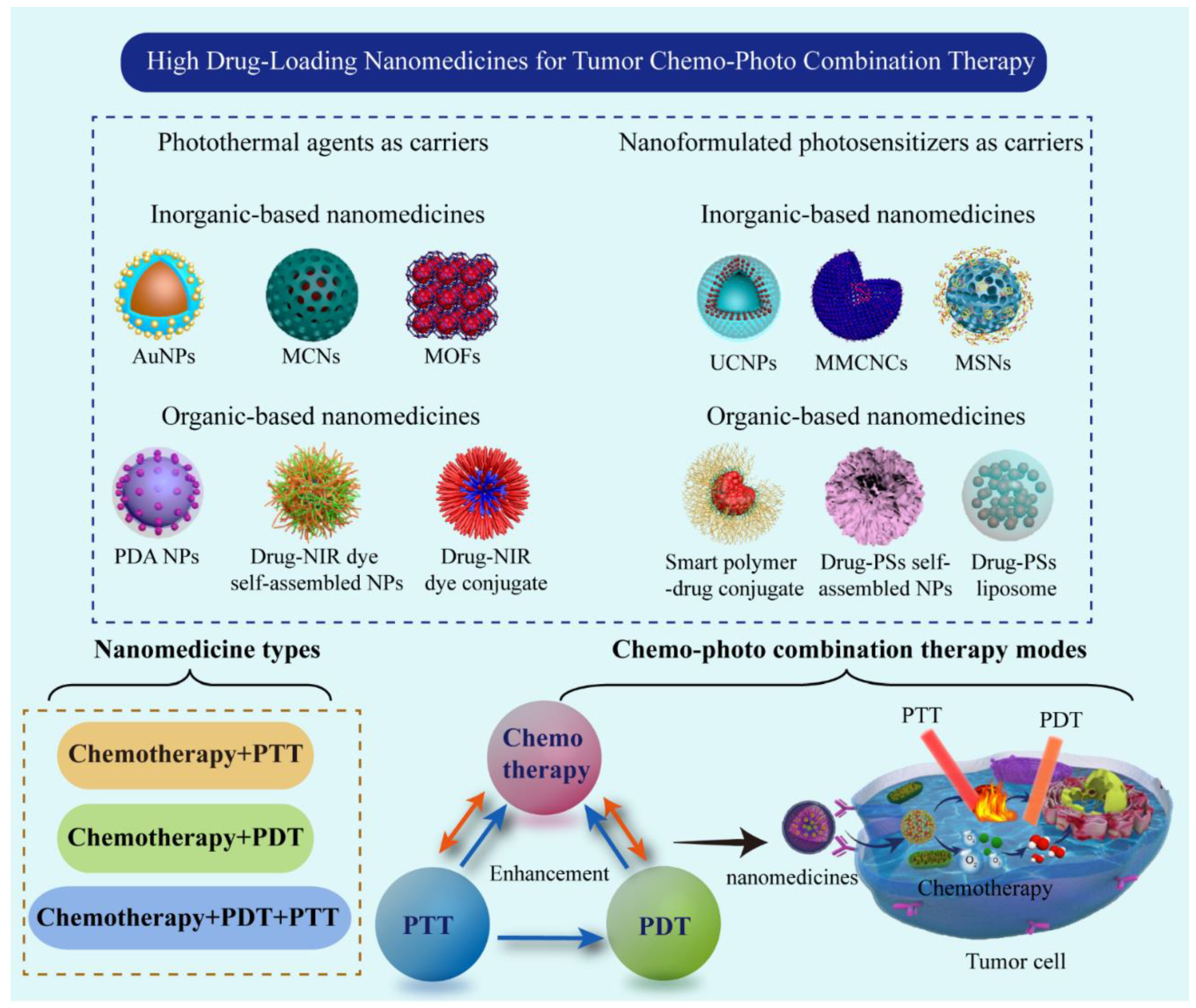
2. Phototherapeutic Agents as Carriers for Fabricating High Drug-Loading Nanomedicines
2.1. Photothermal Agents as Carriers for Fabricating High Drug-Loading Nanomedicines
2.1.1. Inorganic Materials
2.1.2. Organic Materials
2.2. Photosensitizers as Carriers for Fabricating High Drug-Loading Nanomedicine
2.2.1. Inorganic Materials


| Classes | Photosensitizer | Chemotherapeutic Drug | Nanoformulation | Drug-Loading Content (wt%) | Ref. |
|---|---|---|---|---|---|
| Inorganic | BODIPY | Doxorubicin | BODIPY-derivate MOFs | 49.7% | [92] |
| Si-Pc | Doxorubicin | Hybrid mesoporous NPs | DOX: 34.5%, Si-Pc: 51.2% | [93] | |
| PPa | Doxorubicin | UCNP@SiO2/PPa&DOX | DOX:72.8% | [94] | |
| Ir(III) complex | Cisplatin | Pt&Ir@polymer NPs | Pt: 38.9%, Ir(III): 12.9% | [95] | |
| Porphyrin | Doxorubicin | AuNP@dsDNA/Porphyrin | DOX: 75.0% | [96] | |
| PpIX | Doxorubicin | ZiF-67/8@DOX-PpIX NPs | DOX: 12.5% PpIX: 25.3%, | [97] | |
| UCNPs, Eosin Y | Camptothecin | UCNPs@CPT NPs | CPT: 53.7% | [98] | |
| Chlorin e6 | Doxorubicin | rGO-DOX-Ce6 NPs | DOX: 82.3% | [99] | |
| TCPP | Doxorubicin | porphyrin MOFs | DOX: 52.2% | [100] | |
| Organic | Chlorin e6 | GA | GA-Ce6-FA NPs | Ce6 48.5%, GA 47.79%, FA 3.71% | [101] |
| Chlorin e6 | Cabazitaxel | LNA-CTX-Ce6 NPs | Cabazitaxel: 98.87% | [102] | |
| VPF | Doxorubicin | VPF-FRRG-DOX NPs | >70.0% | [103] | |
| Zn-TPPS | Doxorubicin | Zn-TPPS-HDP NPs | Zn-TPPS: 17.0%, DOX: 31.5% | [104] | |
| Zn-TPPS | Doxorubicin | H2TPPS@DOX NPs | DOX 42.4% | [105] | |
| TPCI | Paclitaxel | TPCI-PTX liposomes | PTX: 75.0% | [106] | |
| HPPH | Camptothecin | CPT-HPPH NPs | CPT: 55.0%, HPPH: 76.0% | [107] | |
| PpIX | Doxorubicin | DOX@PpIX-RGD NPs | DOX: 34.5% | [108] | |
| ALA | Doxorubicin | HA-chitosan@DOX-ALA NPs | DOX: 29.4% ALA: 11.5% | [109] | |
| PPA | Paclitaxel | PTX-PPA NPs | PTX: 44.2 %, PPA: 27.6 % | [110] | |
| PPA | Mitoxantrone | MTX-PPA NPs | MTX: 43.5%, PPA: 56.5% | [111] | |
| Chlorine e6 | Doxorubicin | PEG-PBC-TKDOX@Ce6 NPs | DOX: 41.9% | [112] | |
| Chlorine e6 | Paclitaxel | Ce6-PEG@PTX micelles | PTX: 90.1% | [113] | |
| TCPP | Doxorubicin | PEP FA@TCPP nanotubes | DOX: 30.5% | [114] | |
| Chlorine e6 | Doxorubicin | DOX-NPs/Ce6-MBs NPs | DOX: 18.5%,Ce6: 67.1% | [115] | |
| TPC | Paclitaxel | RBC(M(TPC-PTX)) NPs | PTX: 38.0%, TPC: 13.0% | [116] |
2.2.2. Organic Materials
3. High Drug-Loading Nanomedicines for Chemo–Photothermal Combination Therapy
4. High Drug-Loading Nanomedicines for Chemo–Photodynamic Combination Therapy
5. High Drug-Loading Nanomedicines for Chemo–Photothermal–Photodynamic Combination Therapy

| Nanoformulation | Photothermal Agent | PS | Chemotherapeutic Drug | Drug-Loading Content (wt%) | In Vitro/In Vivo; Biological Model | Findings | Ref. |
|---|---|---|---|---|---|---|---|
| Bi2WO6-DOX-PEG NPs | Bi2WO6 NSs | Bi2WO6 NSs | DOX | 81.3% | In vivo and in vitro; U14 cells model | Bi2WO6 nanomaterials can produce ROS and high hyperthermia effect under low power density NIR light irradiation. | [20] |
| rGO-PEG-DOX-Ce6 NPs | rGO | Ce6 | DOX | DOX:82.3% Ce6:92.5% | In vivo and in vitro; U87 cells model | PTT was more effective than chemotherapy and PDT in the 3D spherical model of tumors. | [99] |
| Ini@PM-HA-PDA NPs | PDA NPs | Mn-TCPP MOF | Iniparib | 42.54% | In vitro and in vivo; MDA-MB-231 cells model | Biodegradable NPs for oxygen production was constructed to enhance PDT/PTT | [138] |
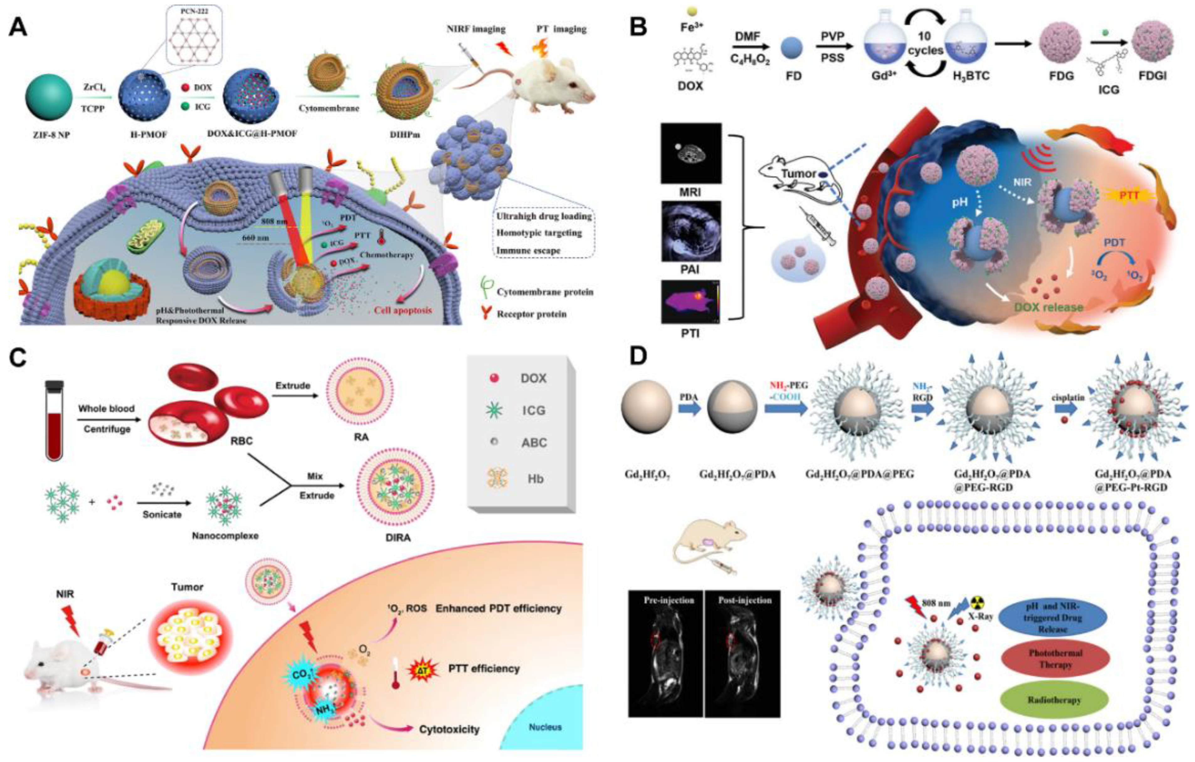
| (DOX and ICG)@H-PMOF@mem NPs | ICG | H-PMOFs | DOX | 86.4% | In vitro and in vivo; 4T1 cells model | The NPs with mesoporous spherical shells were capable of pH control and DOX release triggered by near-infrared laser. | [139] |
| Fe-DOX@Gd-MOF-ICG NPs | ICG | ICG | DOX | 71.4% | The in vitro and in vivo; 4T1 cells model | The MOFs with magnetic resonance imaging and controlled drug release were synthesized. | [140] |
| UCNPs@MIL-100(Fe)-DOX NPs | MIL-100(Fe) | UCNPs | DOX | 72.0% | In vivo and in vitro; U14 cells model | MOFs coated UCNPs were synthesized by a facile one-pot liquid-solid-solution method. | [141] |
| ICG/DOX co-loaded RBCs | ICG | ICG | DOX | 95.5% | In vitro and in vivo; 4T1 cells model | Red blood cells containing oxyHb and NH4HCO3 were prepared for co-loading and controlled release of ICG and DOX. | [142] |
| PAMAM-TMZ ICG@HA NPs | ICG | ICG | TMZ | 45.2% | In vitro and in vivo; A375 cells model | A low cytotoxic carboxyl polyamine was synthesized as a nanocarrier. | [143] |
| THMSNs@LM-DOX-ICG NPs | ICG | ICG | DOX | 37.3% | In vitro, HeLa cells | A smart subcellular organelle was designed as an effective drug delivery platform. | [144] |
| BP/UCNP-SiO2-CuS-PEG-DOX NPs | BP, CuS NPs | BP, CuS NPs | DOX | 77.4% | In vitro and in vivo; U14 cells model | The reduction in the red/green (R/G) ratio elicited by DOX release can be employed to determine the extent of DOX release. | [145] |
| BPQDs-PEG-DOX NPs | BPQDs | BPQDs | DOX | 65% | In vitro and in vivo; HEK 293T cells model | The nanoplatform can inhibit tumor growth through visualized synergistic treatment and photoacoustic and photothermal imaging | [147] |
| Fe3O4@PDA/PEG/ICG-DOX NPs | PDA NPs | ICG | DOX | 50.0% | In vitro; HeLa cells | Facilitating cell internalization of drugs under a localized magnetic field. | [148] |
| 5-FU/rGO/Bce hydrogel | rGO | Bce | 5-FU | 48.4% | In vitro; HeLa cells | Bce, a PS, participates in hydrogel crosslinking and improves biocompatibility. | [149] |
| BP-DOX nanosheets | BPs | BPs | DOX | 90.5% | In vivo and in vitro; 4T1 cells model | BP nanosheets as a multimodal treatment platform for cancer treatment. | [150] |
| Fe3O4@MnO2@PPy-DOX NPs | PPy, Fe3O4 | PPy, MnO2 | DOX | 70.0% | In vitro; HepG2 cells model | A nanocomposite with Fe3O4 as core and two layers of MnO2 and PPy as the shell was prepared to enhance PDT/PTT. | [151] |
| Pa-Hyd-DOX NPs | Pa | Pa | DOX | 53.1% | In vivo and in vitro; SCC25 cells models | Greatly promoted tumor penetration and cell internalization. | [152] |
| IONCs@Ce6-DOX/PCM NPs | IONCs | Ce6 | DOX | 41.2% | In vitro and in vivo; HeLa cells model | The designed nanomedicine can realize the combination therapy triggered by single light. | [153] |
| P(DPP-BT/DOX) NPs | DPP | DPP | DOX | 45.7% | In vitro and in vivo; HeLa cells model | The newly synthesized small-molecule dye shows strong absorption in the NIR-I and fluorescence emission in the NIR-II. | [154] |
| Mn@Au@TiO2@DOX NPs | Au@TiO2 NPs | Au@TiO2 NPs | DOX | 45.5% | In vitro and in vivo; HeLa cells model | The Au@TiO2 core-shell NPs showed stronger photodynamic properties than commercial TiO2 and Au/TiO2 composites. | [155] |
| MXene-DOX | Ti3C2 nanosheets | Ti3C2 nanosheets | DOX | 87.3% | In vitro; Hela cell | Ti3C2 nanosheets were processed into three-dimensional honeycomb structures with anti-aggregation properties as nanocarriers. | [156] |
| 5-Fu@ICG-PNIPAM nanogels | ICG | ICG | 5-Fu | 76.7% | In vitro; Hela cell | The nanogels improved drug bioavailability and achieved controlled release. | [157] |
6. Discussion and Perspectives
6.1. Discussion and Challenges
- (1)
- High drug-loading nanomedicines use as little carrier material as feasible, yet most nanomedicines still cannot be independent from the carrier. Therefore, the potential safety of carrier materials is one of the most crucial issues, especially those non-biodegradable inorganic materials, which will stay in the body for a long time after being consumed. The Au NPs may be promising in this field for their initial success in clinical trials. However, there has not been any systematic research on how to choose suitable and secure carrier materials to build high drug-loading nanomedicines.
- (2)
- Most current studies have concentrated primarily on the design of nanomedicines, but little research compares high and low drug-loading nanomedicines on the internalization mechanism, intracellular release, in vivo circulation time, and so on. For nanomedicines with drug-loading content higher than 50%, their sustained-release characteristics may be far lower than expected, while the pharmacokinetic studies in such studies are often lacking. Hence, it is urgent to conduct the research on the above-mentioned factors, which are of great significance to promote the clinical application of high drug-loading nanomedicines.
- (3)
- When constructing high drug-loading nanomedicines using photothermal materials and photosensitizers as nanocarriers, it is crucial to strike a balance between the phototherapeutic efficacy and the amount of carrier. Although this paper reviews the typical photothermal materials and novel photosensitizers employed as the carrier to manufacture high drug-loading nanomedicines in recent years, there is still a lack of systematic theories and techniques in this area. The construction of such nanomedicines is dependent on the knowledge of predecessors, and needs to be further explored and summarized.
- (4)
- Although most of the chemo–photo combination therapy nanomedicines reported so far show combination antitumor effects, whether two or three treatment modes maximize combination efficiency is uncertain. The combination therapy theory between chemotherapy and phototherapy is not clear at present, and the specific combination mechanism has not been clearly explained. Professional methods and guidance for the calculation of combination efficiency between chemotherapy and phototherapy, as well as a programmed design in this aspect, remain lacking. Moreover, given that the design of those nanomedicines is usually uneconomical or impossible through empirical methods, mathematical and computational modeling can serve as a powerful tool to allow for the controlled study of these processes [158].
- (5)
- At present, PTT and PDT have been employed in clinical treatment trials of some tumors, including basal cell carcinoma, in situ squamous cell carcinoma [159], esophageal cancer, colorectal cancer, [160] and high-grade glioma [161]. The results indicated that the treatment group achieved better antitumor effect compared with the control group and placebo group. However, the limited light penetration depth hinders the clinical application of phototherapy. Nonuniform irradiation of tumor tissue and insufficient irradiation depth make it impossible to completely eliminate the tumor cell for phototherapy, leading to following tumor recurrence and metastasis. Fortunately, chemotherapy and phototherapy can make up for each other’s shortcomings. Chemotherapeutics have a longer effect time and are more evenly distributed in the tumor tissue. After phototherapy rapidly and efficiently kills tumor cells, chemotherapy can eliminate the residual tumor cells for a long time. Therefore, chemo–phototherapy can significantly improve the treatment efficiency. Nowadays, physical therapies such as ultrasound therapy and magnetic hyperthermia are also applied in cancer treatment because of their better penetration depth. However, there are some problems in ultrasonic therapy, such as local overheating and inability to treat intestinal tumors. For magnetic hyperthermia, the large dosage of magnetic nanoparticles is often needed to achieve an ideal therapeutic effect, and the metabolism of those magnetic nanoparticles in vivo needs to be considered. In addition, such treatments require more expensive instrument costs. Although the combination of chemotherapy and phototherapy has the problem of light penetration depth, some medical equipment (such as gastroscope, endoscope, and optical fiber) can be used to transmit light to those deep lesions for the tumor that is located deep in the body. If some imaging functions can be integrated into such medical devices to track the position of phototherapy agents in real time, ensure that all tumor masses can be effectively exposed to light, and monitor the treatment response in real time, it will greatly promote the development of phototherapy in clinical cancer treatment.
- (6)
- The metabolic pathways and phagocytosis mechanisms of some phototherapeutic agents in cells and in vivo have not been clarified. The photothermal-conversion efficiency and production of ROS by existing photothermal materials and PSs can still be improved, and new highly efficient phototherapeutic agents need to be developed. The visible light used in traditional PDT is not the ideal light source, and great efforts are still required to develop new generations of PDT agents that can be more effectively excited by the NIR light.
6.2. Further Perspectivesas
- (1)
- By understanding the combination therapy mechanism between chemotherapy and phototherapy in detail, the future development of chemo–photo combination therapy nanomedicines can give full play to the combination efficiency. Due to economy and convenience, the mathematical and computational modeling method has attracted more and more attention in the clinical translation of nanomedicine. It has been used to simulate nano-sized drug delivery to solid tumors in order to investigate efficacy, understand biological phenomena, and select optimal anticancer treatment strategies through different computational models, such as quantum mechanics, molecular dynamics, and monte carlo, which are related to pharmacokinetic/pharmacodynamics and so on [162,163]. For chemo–photo combination therapy, those nanomedicines can be examined and optimized through the integration of mathematical modeling techniques with modern imaging techniques and in vitro technologies to accelerate clinical translation of those nanomedicines.
- (2)
- For the clinical application of chemo–photo combination therapy and the successful transition from laboratory to the clinic, further studying the safety and biocompatibility of nanomedicines and developing simple and inexpensive synthetic and highly repeatable methods to prepare photo combination therapy nanomedicines with higher efficiency combination therapy and lower side effects are urgent.
- (3)
- The combination of imaging, tumor targeting, controlled drug release, and other functions of high drug-loading chemo–photo combination therapy nanomedicines are expected to further improve the tumor-treatment effect without significantly increasing the system complexity and reducing the drug-loading efficiency. Therefore, determining the optimum ratio between the carrier material and the drug is necessary to realize the multiple functions of simple structures. A novel type of high drug-loading nanomedicines that integrates diagnostic and therapeutic effects should be developed and manufactured, which can realize a real-time tumor diagnosis and treatment and achieve the best therapeutic effect in their institutes; drug carriers with “all in one” functions should be developed.
- (4)
- The research of new therapeutic agents, especially photothermal and photodynamic agents, is still a very important research direction. Although there are very effective photothermal and photodynamic agents, they all have their own shortcomings, such as low solubility, low efficiency of photothermal and photodynamic treatment, and high dependence on tumor environment. The development of new photothermal and photodynamic agents can be greatly improved to enhance the effect of combined photothermal and photodynamic therapy.
- (5)
- The development and application of multifunctional carrier-free nanomedicines (such as MOFs, π-π stacking and infinte coordation polymers) is an important research direction in this field because these kinds of nanomedicines have the advantages of a simple and green preparation method, high drug-loading efficiency, and few side effects. Technologies from different kinds of fields are required for these research directions. In this review, we hope researchers from various fields will join and collaborate on high drug-loading nanomedicine research for tumor chemo–photo combination therapy.
Author Contributions
Funding
Institutional Review Board Statement
Informed Consent Statement
Data Availability Statement
Conflicts of Interest
References
- Sung, H.; Ferlay, J.; Siegel, R.L.; Laversanne, M.; Soerjomataram, I.; Jemal, A.; Bray, F. Global Cancer Statistics 2020: GLOBOCAN Estimates of Incidence and Mortality Worldwide for 36 Cancers in 185 Countries. CA Cancer J. Clin. 2021, 71, 209–224. [Google Scholar] [CrossRef] [PubMed]
- Riehemann, K.; Schneider, S.W.; Luger, T.A.; Godin, B.; Ferrari, M.; Fuchs, H. Nanomedicine—Challenge and Perspectives. Angew. Chem. Int. Ed. 2009, 48, 872–897. [Google Scholar] [CrossRef] [PubMed]
- Huang, L.; Zhao, S.; Fang, F.; Xu, T.; Lan, M.; Zhang, J. Advances and perspectives in carrier-free nanodrugs for cancer chemo-monotherapy and combination therapy. Biomaterials 2021, 268, 120557. [Google Scholar] [CrossRef] [PubMed]
- Fan, W.; Yung, B.; Huang, P.; Chen, X. Nanotechnology for Multimodal Synergistic Cancer Therapy. Chem. Rev. 2017, 117, 13566–13638. [Google Scholar] [CrossRef]
- Cao, J.; Chen, Z.; Chi, J.; Sun, Y.; Sun, Y. Recent progress in synergistic chemotherapy and phototherapy by targeted drug delivery systems for cancer treatment. Artif. Cells Nanomed. Biotechnol. 2018, 46, 817–830. [Google Scholar] [CrossRef]
- Cheng, L.; Wang, C.; Feng, L.; Yang, K.; Liu, Z. Functional Nanomaterials for Phototherapies of Cancer. Chem. Rev. 2014, 114, 10869–10939. [Google Scholar] [CrossRef]
- Lan, G.; Ni, K.; Lin, W. Nanoscale metal–organic frameworks for phototherapy of cancer. Coord. Chem. Rev. 2017, 379, 65–81. [Google Scholar] [CrossRef]
- Liu, S.; Pan, X.; Liu, H. Two-Dimensional Nanomaterials for Photothermal Therapy. Angew. Chem. Int. Ed. Engl. 2020, 59, 5890–5900. [Google Scholar] [CrossRef]
- Lucky, S.S.; Soo, K.C.; Zhang, Y. Nanoparticles in Photodynamic Therapy. Chem. Rev. 2015, 115, 1990–2042. [Google Scholar] [CrossRef]
- Zhang, A.; Li, A.; Zhao, W.; Liu, J. Recent Advances in Functional Polymer Decorated Two-Dimensional Transition-Metal Dichalcogenides Nanomaterials for Chemo–Photothermal Therapy. Chem. A Eur. J. 2018, 24, 4215–4227. [Google Scholar] [CrossRef]
- Zhao, R.; Zheng, G.; Fan, L.; Shen, Z.; Jiang, K.; Guo, Y.; Shao, J.-W. Carrier-free nanodrug by co-assembly of chemotherapeutic agent and photosensitizer for cancer imaging and chemo–photo combination therapy. Acta Biomater. 2018, 70, 197–210. [Google Scholar] [CrossRef] [PubMed]
- Sanoj Rejinold, N.; Choi, G.; Choy, J.-H. Recent trends in nano photo-chemo therapy approaches and future scopes. Coord. Chem. Rev. 2020, 411, 213252. [Google Scholar] [CrossRef]
- Li, Z.; Chen, Y.; Yang, Y.; Yu, Y.; Zhang, Y.; Zhu, D.; Yu, X.; Ouyang, X.; Xie, Z.; Zhao, Y.; et al. Recent Advances in Nanomaterials-Based Chemo–Photothermal Combination Therapy for Improving Cancer Treatment. Front. Bioeng. Biotechnol. 2019, 7, 293. [Google Scholar] [CrossRef]
- Khafaji, M.; Zamani, M.; Golizadeh, M.; Bavi, O. Inorganic nanomaterials for chemo/photothermal therapy: A promising horizon on effective cancer treatment. Biophys. Rev. 2019, 11, 335–352. [Google Scholar] [CrossRef]
- Denkova, A.G.; de Kruijff, R.M.; Serra-Crespo, P. Nanocarrier-Mediated Photochemotherapy and Photoradiotherapy. Adv. Healthc Mater. 2018, 7, e1701211. [Google Scholar] [CrossRef] [PubMed]
- Zhang, M.; Zhang, L.; Chen, Y.; Li, L.; Su, Z.; Wang, C. Precise synthesis of unique polydopamine/mesoporous calcium phosphate hollow Janus nanoparticles for imaging-guided chemo–photothermal synergistic therapy. Chem. Sci. 2017, 8, 8067–8077. [Google Scholar] [CrossRef]
- Liu, T.; Wang, C.; Gu, X.; Gong, H.; Cheng, L.; Shi, X.; Feng, L.; Sun, B.; Liu, Z. Drug Delivery with PEGylated MoS2 Nano-sheets for Combined Photothermal and Chemotherapy of Cancer. Adv. Mater. 2014, 26, 3433–3440. [Google Scholar] [CrossRef]
- Feng, L.; Yang, D.; Gai, S.; He, F.; Yang, G.; Yang, P.; Lin, J. Single bismuth tungstate nanosheets for simultaneous chemo-, photothermal, and photodynamic therapies mediated by near-infrared light. Chem. Eng. J. 2018, 351, 1147–1158. [Google Scholar] [CrossRef]
- Liu, Y.; Yang, G.; Jin, S.; Xu, L.; Zhao, C.-X. Development of High-Drug-Loading Nanoparticles. ChemPlusChem 2020, 85, 2143–2157. [Google Scholar] [CrossRef]
- Shen, S.; Wu, Y.; Liu, Y.; Wu, D. High drug-loading nanomedicines: Progress, current status, and prospects. Int. J. Nanomed. 2017, 12, 4085–4109. [Google Scholar] [CrossRef]
- Li, W.; Cao, Z.; Yu, L.; Huang, Q.; Zhu, D.; Lu, C.; Lu, A.; Liu, Y. Hierarchical drug release designed Au @PDA-PEG-MTX NPs for targeted delivery to breast cancer with combined photothermal-chemotherapy. J. Nanobiotechnol. 2021, 19, 143. [Google Scholar] [CrossRef]
- Awan, U.A.; Raza, A.; Ali, S.; Saeed, R.F.; Akhtar, N. Doxorubicin-loaded gold nanorods: A multifunctional chemo–photothermal nanoplatform for cancer management. Beilstein J. Nanotechnol. 2021, 12, 295–303. [Google Scholar] [CrossRef]
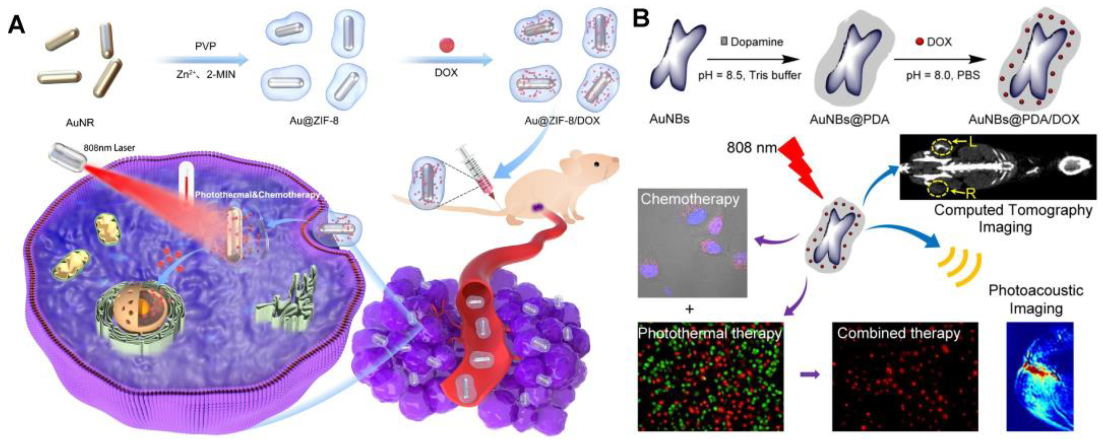
- Xu, J.; Cheng, X.; Chen, F.; Li, W.; Xiao, X.; Lai, P.; Xu, G.; Xu, L.; Pan, Y. Fabrication of multifunctional polydopamine-coated gold nanobones for PA/CT imaging and enhanced synergistic chemo–photothermal therapy. J. Mater. Sci. Technol. 2021, 63, 97–105. [Google Scholar] [CrossRef]
- Wang, J.; Sun, J.; Wang, Y.; Chou, T.; Zhang, Q.; Zhang, B.; Ren, L.; Wang, H. Gold Nanoframeworks with Mesopores for Raman–Photoacoustic Imaging and Photo-Chemo Tumor Therapy in the Second Near-Infrared Biowindow. Adv. Funct. Mater. 2020, 30, 1908825. [Google Scholar] [CrossRef]
- Song, Y.; Qu, Z.; Li, J.; Shi, L.; Zhao, W.; Wang, H.; Sun, T.; Jia, T.; Sun, Y. Fabrication of the biomimetic DOX/Au@Pt nanoparticles hybrid nanostructures for the combinational chemo/photothermal cancer therapy. J. Alloy. Compd. 2021, 881, 160592. [Google Scholar] [CrossRef]
- Sun, C.; Liu, Y.; Zhou, R.; Yao, L.; Wang, R.; Zang, W.; Meng, W. Nano Pd-Decorated Manganese Dioxide Nanosheets for Effective Photothermal Chemotherapy. ACS Appl. Bio Mater. 2019, 2, 4747–4755. [Google Scholar] [CrossRef]
- Li, X.; Yuan, H.J.; Tian, X.M.; Tang, J.; Liu, L.F.; Liu, F.Y. Biocompatible copper sulfide–based nanocomposites for artery interventional chemo–photothermal therapy of orthotropic hepatocellular carcinoma. Mater. Today Bio 2021, 12, 100128. [Google Scholar] [CrossRef]
- Xie, M.; Li, J.; Deng, T.; Yang, N.; Yang, M. Modification of magnetic molybdenum disulfide by chitosan/carboxymethylcellulose with enhanced dispersibility for targeted photothermal-/chemotherapy of cancer. J. Mater. Chem. B 2021, 9, 1833–1845. [Google Scholar] [CrossRef]
- Hou, M.; Zhong, Y.; Zhang, L.; Xu, Z.; Kang, Y.; Xue, P. Polydopamine (PDA)-activated cobalt sulfide nanospheres responsive to tumor microenvironment (TME) for chemotherapeutic-enhanced photothermal therapy. Chin. Chem. Lett. 2021, 32, 1055–1060. [Google Scholar] [CrossRef]
- Xie, M.; Yang, M.; Sun, X.; Yang, N.; Deng, T.; Li, Y.; Shen, H. WS2 nanosheets functionalized by biomimetic lipids with enhanced dispersibility for photothermal and chemo combination therapy. J. Mater. Chem. B 2020, 8, 2331–2342. [Google Scholar] [CrossRef]
- Chen, Y.; Khan, A.R.; Yu, D.; Zhai, Y.; Ji, J.; Shi, Y.; Zhai, G. Pluronic F127-functionalized molybdenum oxide nanosheets with pH-dependent degradability for chemo–photothermal cancer therapy. J. Colloid Interface Sci. 2019, 553, 567–580. [Google Scholar] [CrossRef]
- Zhan, X.; Teng, W.; Sun, K.; He, J.; Yang, J.; Tian, J.; Huang, X.; Zhou, L.; Zhou, C. CD47-mediated DTIC-loaded chitosan oligosaccharide-grafted nGO for synergistic chemo–photothermal therapy against malignant melanoma. Mater. Sci. Eng. C 2021, 123, 112014. [Google Scholar] [CrossRef]
- Xing, E.; Du, Y.; Yin, J.; Chen, M.; Zhu, M.; Wen, X.; Xu, J.; Feng, Y.; Meng, S. Multi-functional Nanodrug Based on a Three-dimensional Framework for Targeted Photo-chemo Synergetic Cancer Therapy. Adv. Heal. Mater. 2021, 10, e2001874. [Google Scholar] [CrossRef]
- Wang, Y.; Zhang, H.; Xie, J.; Liu, Y.; Wang, S.; Zhao, Q. Three dimensional mesoporous carbon nanospheres as carriers for chemo–photothermal therapy compared with two dimensional graphene oxide nanosheets. Colloids Surf. A Physicochem. Eng. Asp. 2020, 590, 124498. [Google Scholar] [CrossRef]
- Dong, X.; Sun, Z.; Wang, X.; Leng, X. An innovative MWCNTs/DOX/TC nanosystem for chemo–photothermal combination therapy of cancer. Nanomed. Nanotechnol. Biol. Med. 2017, 13, 2271–2280. [Google Scholar] [CrossRef]
- Wang, C.; Chen, Y.; Xu, Z.; Chen, B.; Zhang, Y.; Yi, X.; Li, J. Fabrication and characterization of novel cRGD modified graphene quantum dots for chemo–photothermal combination therapy. Sens. Actuators B Chem. 2020, 309, 127732. [Google Scholar] [CrossRef]
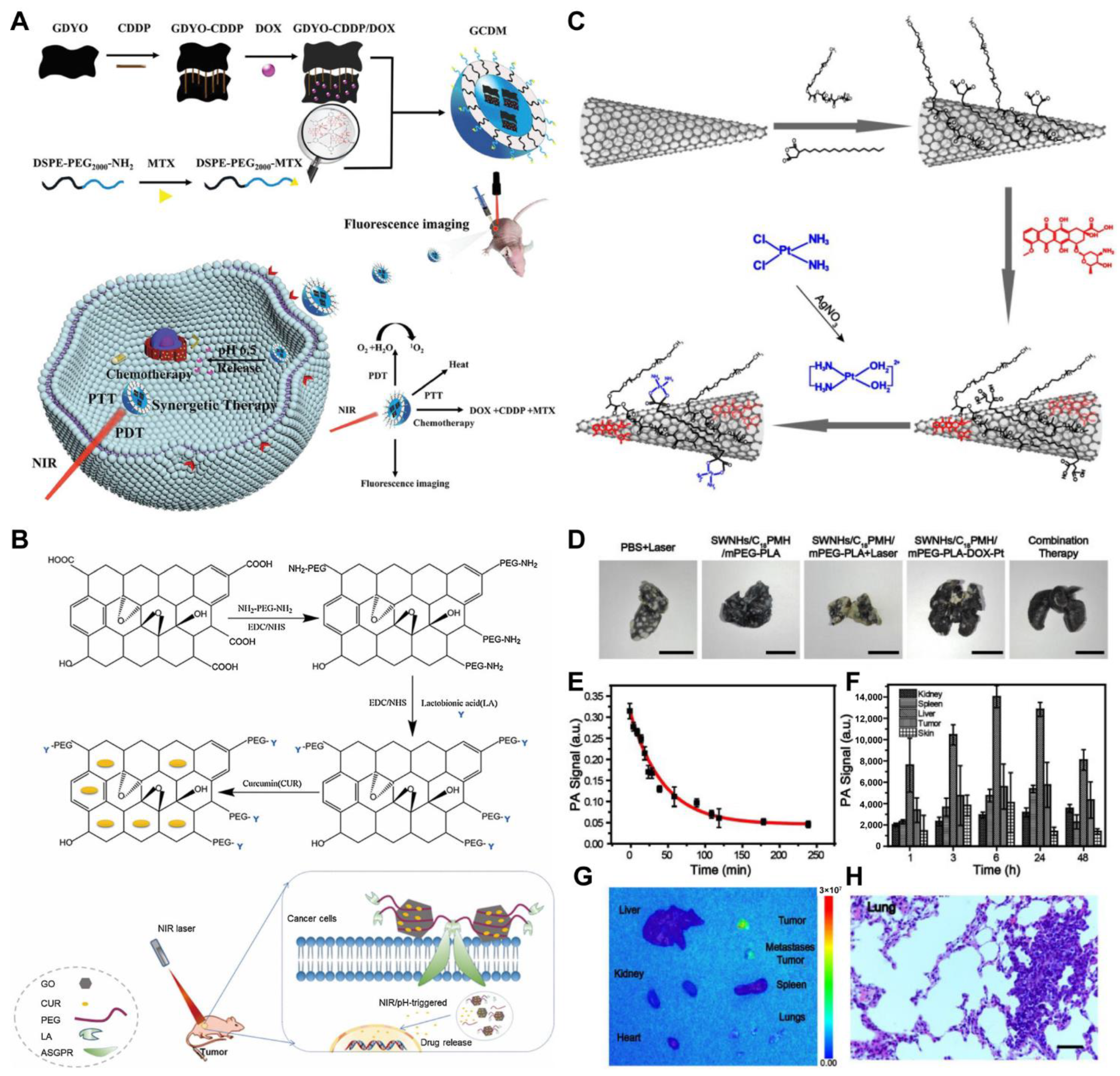
- Yang, J.; Su, H.; Sun, W.; Cai, J.; Liu, S.; Chai, Y.; Zhang, C. Dual Chemodrug-Loaded Single-Walled Carbon Nanohorns for Multimodal Imaging-Guided Chemo–photothermal Therapy of Tumors and Lung Metastases. Theranostics 2018, 8, 1966–1984. [Google Scholar] [CrossRef]
- Wei, X.; Zhang, L.; Li, S.; Chen, X.; Zhang, M.; Wang, C.; Wang, T.; Li, L. A designed synthesis of multifunctional carbon nanoframes for simultaneous imaging and synergistic chemo–photothermal cancer therapy. New J. Chem. 2018, 42, 923–929. [Google Scholar] [CrossRef]
- Lu, L.; Wang, K.; Lin, C.; Yang, W.; Duan, Q.; Li, K.; Cai, K. Constructing nanocomplexes by multicomponent self-assembly for curing orthotopic glioblastoma with synergistic chemo–photothermal therapy. Biomaterials 2021, 279, 121193. [Google Scholar] [CrossRef]
- Gao, H.; Bai, Y.; Chen, L.; Fakhri, G.E.; Wang, M. Self-Assembly Nanoparticles for Overcoming Multidrug Resistance and Imaging-Guided Chemo–photothermal Synergistic Cancer Therapy. Int. J. Nanomed. 2020, 15, 809–819. [Google Scholar] [CrossRef]
- He, L.; Qing, F.; Li, M.; Lan, D. Paclitaxel/IR1061-Co-Loaded Protein Nanoparticle for Tumor-Targeted and pH/NIR-II-Triggered Synergistic Photothermal-Chemotherapy. Int. J. Nanomed. 2020, 15, 2337–2349. [Google Scholar] [CrossRef]
- Liu, S.; Pan, J.; Liu, J.; Ma, Y.; Qiu, F.; Mei, L.; Zeng, X.; Pan, G. Dynamically PEGylated and Borate-Coordination-Polymer-Coated Polydopamine Nanoparticles for Synergetic Tumor-Targeted, Chemo–photothermal Combination Therapy. Small 2018, 14, e1703968. [Google Scholar] [CrossRef]
- Zhang, Y.; Fang, F.; Chen, Y.; Li, M.; Li, L.; Li, W.; Zhang, J. Hollow mesoporous polyaniline nanoparticles with high drug payload and robust photothermal capability for cancer combination therapy. Chin. J. Chem. Eng. 2021, 38, 221–228. [Google Scholar] [CrossRef]
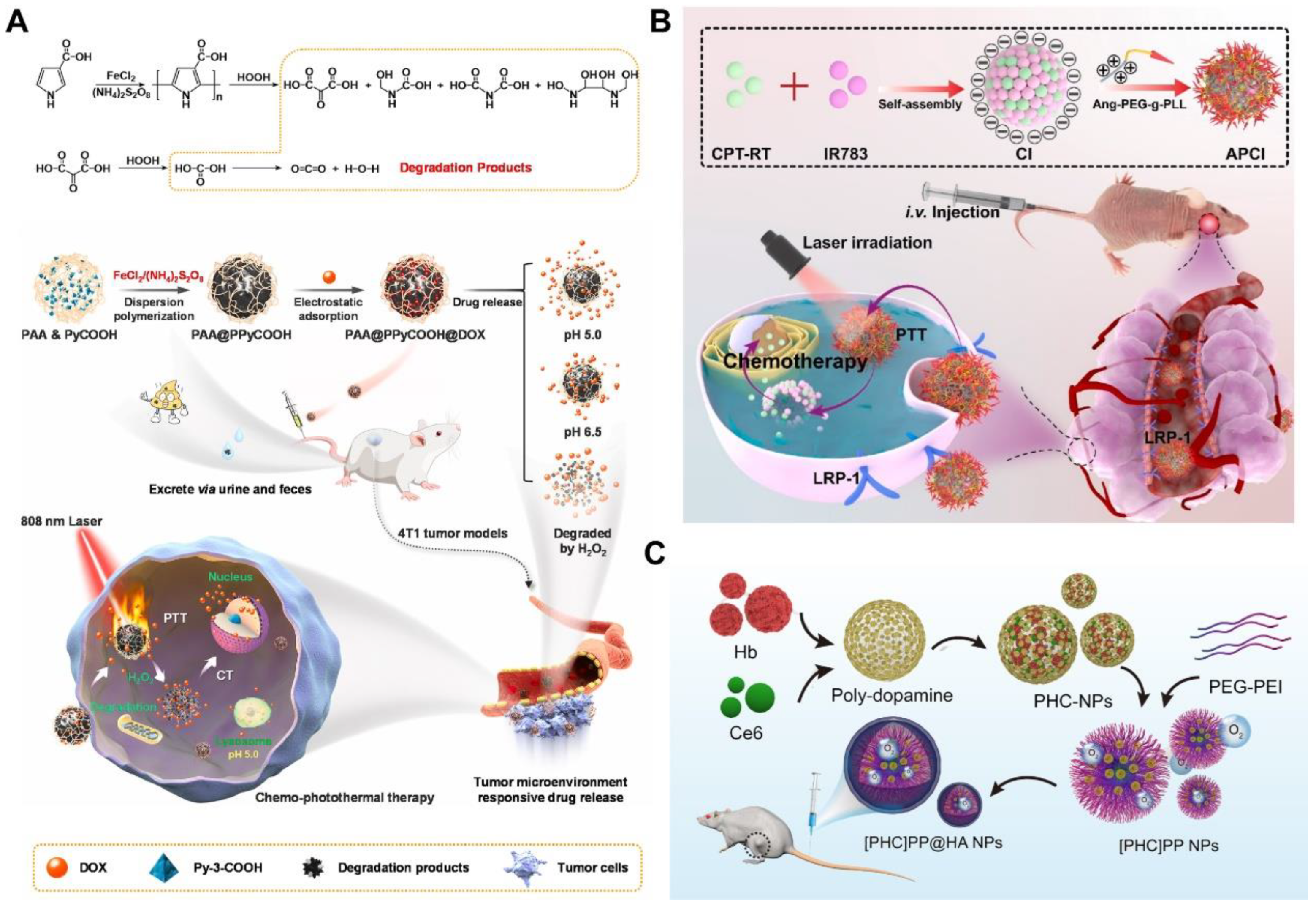
- Liu, X.; Liu, Y.; Guo, Y.; Shi, W.; Sun, Y.; He, Z.; Shen, Y.; Zhang, X.; Xiao, H.; Ge, D. Metabolizable pH/H2O2 dual-responsive conductive polymer nanoparticles for safe and precise chemo–photothermal therapy. Biomaterials 2021, 277, 121115. [Google Scholar] [CrossRef]
- Fathy, M.M.; Nasser, L.; El-Sokkary, G.; Rasheedy, M.S. Combined Chemo–photothermal Therapy of Metastatic Mammary Adenocarcinoma Using Curcumin-Coated Iron Oxide Nanoparticles. BioNanoScience 2021, 11, 447–453. [Google Scholar] [CrossRef]
- Liu, Y.; Han, Q.; Yang, W.; Gan, X.; Yang, Y.; Xie, K.; Xie, L.; Deng, Y. Two-dimensional MXene/cobalt nanowire heterojunction for controlled drug delivery and chemo–photothermal therapy. Mater. Sci. Eng. C 2020, 116, 111212. [Google Scholar] [CrossRef]
- Huang, Y.; Xue, Z.; Zeng, S. Hollow Mesoporous Bi@PEG-FA Nanoshell as a Novel Dual-Stimuli-Responsive Nanocarrier for Synergistic Chemo–photothermal Cancer Therapy. ACS Appl. Mater. Interfaces 2020, 12, 31172–31181. [Google Scholar] [CrossRef]
- Wang, J.; Wu, X.; Shen, P.; Wang, J.; Shen, Y.; Shen, Y.; Webster, T.J.; Deng, J. Applications of Inorganic Nanomaterials in Photothermal Therapy Based on Combinational Cancer Treatment. Int. J. Nanomed. 2020, 15, 1903–1914. [Google Scholar] [CrossRef]
- Connor, D.M.; Broome, A.-M. Chapter Seven—Gold Nanoparticles for the Delivery of Cancer Therapeutics. In Advances in Cancer Research; Broome, A.-M., Ed.; Academic Press: Cambridge, MA, USA, 2018; Volume 139, pp. 163–184. [Google Scholar]
- Zhang, L.; Shen, S.; Cheng, L.; You, H.; Lu, L.; Ma, C.; Dai, Y.; Fang, J. Mesoporous gold nanoparticles for photothermal controlled anticancer drug delivery. Nanomedicine 2019, 14, 1443–1454. [Google Scholar] [CrossRef]
- Lee, C.-S.; Kim, H.; Yu, J.; Yu, S.H.; Ban, S.; Oh, S.; Jeong, D.; Im, J.; Baek, M.J.; Kim, T.H. Doxorubicin-loaded oligonucleotide conjugated gold nanoparticles: A promising in vivo drug delivery system for colorectal cancer therapy. Eur. J. Med. Chem. 2017, 142, 416–423. [Google Scholar] [CrossRef]
- Song, Z.; Liu, Y.; Shi, J.; Ma, T.; Zhang, Z.; Ma, H.; Cao, S. Hydroxyapatite/mesoporous silica coated gold nanorods with improved degradability as a multi-responsive drug delivery platform. Mater. Sci. Eng. C 2018, 83, 90–98. [Google Scholar] [CrossRef] [PubMed]
- Zeng, J.-Y.; Zhang, M.-K.; Peng, M.-Y.; Gong, D.; Zhang, X.-Z. Porphyrinic Metal-Organic Frameworks Coated Gold Nanorods as a Versatile Nanoplatform for Combined Photodynamic/Photothermal/Chemotherapy of Tumor. Adv. Funct. Mater. 2017, 28, 1705451. [Google Scholar] [CrossRef]
- Xu, Q.; Wan, J.; Bie, N.; Song, X.; Yang, X.; Yong, T.; Zhao, Y.; Yang, X.; Gan, L. A Biomimetic Gold Nanocages-Based Nanoplatform for Efficient Tumor Ablation and Reduced Inflammation. Theranostics 2018, 8, 5362–5378. [Google Scholar] [CrossRef]
- Xia, Y.; Li, W.; Cobley, C.M.; Chen, J.; Xia, X.; Zhang, Q.; Yang, M.; Cho, E.C.; Brown, P.K. Gold Nanocages: From Synthesis to Theranostic Applications. Accounts Chem. Res. 2011, 44, 914–924. [Google Scholar] [CrossRef]
- Li, S.; Zhang, L.; Wang, T.; Li, L.; Wang, C.; Su, Z. The facile synthesis of hollow Au nanoflowers for synergistic chemo–photothermal cancer therapy. Chem. Commun. 2015, 51, 14338–14341. [Google Scholar] [CrossRef]
- Song, C.; Dou, Y.; Yuwen, L.; Sun, Y.; Dong, C.; Li, F.; Yang, Y.; Wang, L. A gold nanoflower-based traceable drug delivery system for intracellular SERS imaging-guided targeted chemo–phototherapy. J. Mater. Chem. B 2018, 6, 3030–3039. [Google Scholar] [CrossRef] [PubMed]
- Wang, C.; Li, J.; Amatore, C.; Chen, Y.; Jiang, H.; Wang, X.-M. Gold Nanoclusters and Graphene Nanocomposites for Drug Delivery and Imaging of Cancer Cells. Angew. Chem. Int. Ed. 2011, 50, 11644–11648. [Google Scholar] [CrossRef] [PubMed]
- Cui, H.; Shao, Z.-S.; Song, Z.; Wang, Y.-B.; Wang, H.-S. Development of gold nanoclusters: From preparation to applications in the field of biomedicine. J. Mater. Chem. C 2020, 8, 14312–14333. [Google Scholar] [CrossRef]
- Huang, J.; Xu, Z.; Jiang, Y.; Law, W.C.; Dong, B.; Zeng, X.; Ma, M.; Xu, G.; Zou, J.; Yang, C. Metal organic framework-coated gold nanorod as an on-demand drug delivery platform for chemo–photothermal cancer therapy. J. Nanobiotechnol. 2021, 19, 219. [Google Scholar] [CrossRef]
- He, J.; Dong, J.; Hu, Y.; Li, G.; Hu, Y. Design of Raman tag-bridged core–shell Au@Cu3(BTC)2 nanoparticles for Raman imaging and synergistic chemo–photothermal therapy. Nanoscale 2019, 11, 6089–6100. [Google Scholar] [CrossRef]
- Shibu, E.S.; Hamada, M.; Murase, N.; Biju, V. Nanomaterials formulations for photothermal and photodynamic therapy of cancer. J. Photochem. Photobiol. C Photochem. Rev. 2013, 15, 53–72. [Google Scholar] [CrossRef]
- Liu, Z.; Tabakman, S.M.; Chen, Z.; Dai, H. Preparation of carbon nanotube bioconjugates for biomedical applications. Nat. Protoc. 2009, 4, 1372–1382. [Google Scholar] [CrossRef] [PubMed]
- Wang, Y.; Hu, J.; Xiang, D.; Peng, X.; You, Q.; Mao, Y.; Hua, D.; Yin, J. Targeted nanosystem combined with chemo–photothermal therapy for hepatocellular carcinoma treatment. Colloids Surf. A Physicochem. Eng. Asp. 2020, 596, 124711. [Google Scholar] [CrossRef]
- Ortiz de Solorzano, I.; Prieto, M.; Mendoza, G.; Alejo, T.; Irusta, S.; Sebastian, V.; Arruebo, M. Microfluidic Synthesis and Biological Evaluation of Photothermal Biodegradable Copper Sulfide Nanoparticles. ACS Appl. Mater. Interfaces 2016, 8, 21545–21554. [Google Scholar] [CrossRef]
- Gayathri, R.; Rajalakshmi, P.S.; Thomas, A.; Rengan, A.K. Doxorubicin loaded polyvinylpyrrolidone-copper sulfide nanoparticles enabling mucoadhesiveness and chemo–photothermal synergism for effective killing of breast cancer cells. Materialia 2021, 19, 101195. [Google Scholar]
- Zhang, S.; Sun, C.; Zeng, J.; Sun, Q.; Wang, G.; Wang, Y.; Wu, Y.; Dou, S.; Gao, M.; Li, Z. Ambient Aqueous Synthesis of Ultrasmall PEGylated Cu2−xSe Nanoparticles as a Multifunctional Theranostic Agent for Multimodal Imaging Guided Photothermal Therapy of Cancer. Adv. Mater. 2016, 28, 8927–8936. [Google Scholar] [CrossRef]
- Chou, S.S.; Kaehr, B.; Kim, J.; Foley, B.M.; De, M.; Hopkins, P.E.; Huang, J.; Brinker, C.J.; Dravid, V.P. Chemically Exfoliated MoS2 as Near-Infrared Photothermal Agents. Angew. Chem. Int. Ed. 2013, 52, 4160–4164. [Google Scholar] [CrossRef]
- Wu, C.; Wang, S.; Zhao, J.; Liu, Y.; Zheng, Y.; Luo, Y.; Ye, C.; Huang, M.; Chen, H. Biodegradable Fe(III)@WS2-PVP Nanocapsules for Redox Reaction and TME-Enhanced Nanocatalytic, Photothermal, and Chemotherapy. Adv. Funct. Mater. 2019, 29, 1901722. [Google Scholar] [CrossRef]
- Fang, L.; Wang, J.; Ouyang, X.; Liang, B.; Zhao, L.; Huang, D.; Deng, D. FeSe2 nanosheets as a bifunctional platform for synergistic tumor therapy reinforced by NIR-II light. Biomater. Sci. 2021, 9, 5542–5550. [Google Scholar] [CrossRef]
- She, D.; Peng, S.; Liu, L.; Huang, H.; Zheng, Y.; Lu, Y.; Geng, D.; Yin, B. Biomimic FeS2 nanodrug with hypothermal photothermal effect by clinical approved NIR-Ⅱ light for augmented chemodynamic therapy. Chem. Eng. J. 2020, 400, 125933. [Google Scholar] [CrossRef]
- Qian, X.; Shen, S.; Liu, T.; Cheng, L.; Liu, Z. Two-dimensional TiS2 nanosheets for in vivo photoacoustic imaging and photothermal cancer therapy. Nanoscale 2015, 7, 6380–6387. [Google Scholar] [CrossRef] [PubMed]
- Chen, Y.; Gao, M.; Zhang, L.; Ha, E.; Hu, X.; Zou, R.; Yan, L.; Hu, J. Tumor Microenvironment Responsive Biodegradable Fe-Doped MoOx Nanowires for Magnetic Resonance Imaging Guided Photothermal-Enhanced Chemodynamic Synergistic Antitumor Therapy. Adv. Healthc. Mater. 2021, 10, 2001665. [Google Scholar] [CrossRef] [PubMed]
- Sharker, S.M.; Kim, S.M.; Lee, J.E.; Choi, K.H.; Shin, G.; Lee, S.; Lee, K.D.; Jeong, J.H.; Lee, H.; Park, S.Y. Functionalized biocompatible WO3 nanoparticles for triggered and targeted in vitro and in vivo photothermal therapy. J. Control. Release 2015, 217, 211–220. [Google Scholar] [CrossRef] [PubMed]
- Ou, G.; Li, Z.; Li, D.; Cheng, L.; Liu, Z.; Wu, H. Photothermal therapy by using titanium oxide nanoparticles. Nano Res. 2016, 9, 1236–1243. [Google Scholar] [CrossRef]
- Wu, M.; Mei, T.; Lin, C.; Wang, Y.; Chen, J.; Le, W.; Sun, M.; Xu, J.; Dai, H.; Zhang, Y.; et al. Melanoma Cell Membrane Biomimetic Versatile CuS Nanoprobes for Homologous Targeting Photoacoustic Imaging and Photothermal Chemotherapy. ACS Appl. Mater. Interfaces 2020, 12, 16031–16039. [Google Scholar] [CrossRef]
- Li, L.; Lu, Y.; Qian, Z.; Yang, Z.; Zong, S.; Wang, Z.; Cui, Y. A Ti2N MXene-based nanosystem with ultrahigh drug loading for dual-strategy synergistic oncotherapy. Nanoscale 2021, 13, 18546–18557. [Google Scholar] [CrossRef]
- Wang, Y.; Wu, Y.; Li, K.; Shen, S.; Liu, Z.; Wu, D. Ultralong Circulating Lollipop-Like Nanoparticles Assembled with Gossypol, Doxorubicin, and Polydopamine via π-π Stacking for Synergistic Tumor Therapy. Adv. Funct. Mater. 2019, 29, 1805582. [Google Scholar] [CrossRef]
- Wang, Y.; Luo, S.; Wu, Y.; Tang, P.; Liu, J.; Liu, Z.; Shen, S.; Ren, H.; Wu, D. Highly Penetrable and On-Demand Oxygen Release with Tumor Activity Composite Nanosystem for Photothermal/Photodynamic Synergetic Therapy. ACS Nano. 2020, 14, 17046–17062. [Google Scholar] [CrossRef]
- Lin, X.; Cao, Y.; Li, J.; Zheng, D.; Lan, S.; Xue, Y.; Yu, F.; Wu, M.; Zhu, X. Folic acid-modified Prussian blue/polydopamine nanoparticles as an MRI agent for use in targeted chemo/photothermal therapy. Biomater. Sci. 2019, 7, 2996–3006. [Google Scholar] [CrossRef]
- Chen, W.; Zeng, K.; Liu, H.; Ouyang, J.; Wang, L.; Liu, Y.; Wang, H.; Deng, L.; Liu, Y.-N. Cell Membrane Camouflaged Hollow Prussian Blue Nanoparticles for Synergistic Photothermal-/Chemotherapy of Cancer. Adv. Funct. Mater. 2017, 27, 1605795. [Google Scholar] [CrossRef]
- Tian, Y.; Yi, W.; Bai, L.; Cheng, X.; Yi, T.; Mu, M.; Zhang, P.; Si, J.; Hou, X.; Hou, J. One-step in situ growth of MoS2@lentinan as a dual-stimuli-responsive nanocarrier for synergistic chemo–photothermal therapy. New J. Chem. 2021, 45, 17966–17975. [Google Scholar] [CrossRef]
- Zhang, Y.; Guo, R.; Wang, D.; Sun, X.; Xu, Z. Pd nanoparticle-decorated hydroxy boron nitride nanosheets as a novel drug carrier for chemo–photothermal therapy. Colloids Surf. B Biointerfaces 2019, 176, 300–308. [Google Scholar] [CrossRef] [PubMed]
- Dinçer, C.A.; Getiren, B.; Gökalp, C.; Ciplak, Z.; Karakeçili, A.; Yildiz, N. An anticancer drug loading and release study to ternary GO-Fe3O4-PPy and Fe3O4 @PPy-NGQDs nanocomposites for photothermal chemotherapy. Colloids Surf. A Physicochem. Eng. Asp. 2022, 633, 127791. [Google Scholar] [CrossRef]
- Hak, A.; Ravasaheb Shinde, V.; Rengan, A.K. A review of advanced nanoformulations in phototherapy for cancer therapeutics. Photodiagnosis Photodyn. Ther. 2021, 33, 102205. [Google Scholar] [CrossRef] [PubMed]
- Kim, J.; Jo, Y.U.; Na, K. Photodynamic therapy with smart nanomedicine. Arch. Pharm. Res. 2020, 43, 22–31. [Google Scholar] [CrossRef]
- Sewid, F.A.; Skurlov, I.D.; Kurshanov, D.A.; Orlova, A.O. Singlet oxygen generation by hybrid structures based on CdSe/ZnS quantum dots and tetraphenylporphyrin in organic medium. Chem. Phys. Lett. 2021, 765, 138303. [Google Scholar] [CrossRef]
- Luo, L.; Sun, W.; Feng, Y.; Qin, R.; Zhang, J.; Ding, D.; Shi, T.; Liu, X.; Chen, X.; Chen, H. Conjugation of a Scintillator Complex and Gold Nanorods for Dual-Modal Image-Guided Photothermal and X-ray-Induced Photodynamic Therapy of Tumors. ACS Appl. Mater. Interfaces 2020, 12, 12591–12599. [Google Scholar] [CrossRef]
- Liang, J.; Chen, B.; Hu, J.; Huang, Q.; Zhang, D.; Wan, J.; Hu, Z.; Wang, B. pH and Thermal Dual-Responsive Graphene Oxide Nanocomplexes for Targeted Drug Delivery and Photothermal-Chemo/Photodynamic Synergetic Therapy. ACS Appl. Bio Mater. 2019, 2, 5859–5871. [Google Scholar] [CrossRef]
- Liu, J.; Du, P.; Liu, T.; Córdova Wong, B.J.; Wang, W.; Ju, H.; Lei, J. A black phosphorus/manganese dioxide nanoplatform: Oxygen self-supply monitoring, photodynamic therapy enhancement and feedback. Biomaterials 2019, 192, 179–188. [Google Scholar] [CrossRef]
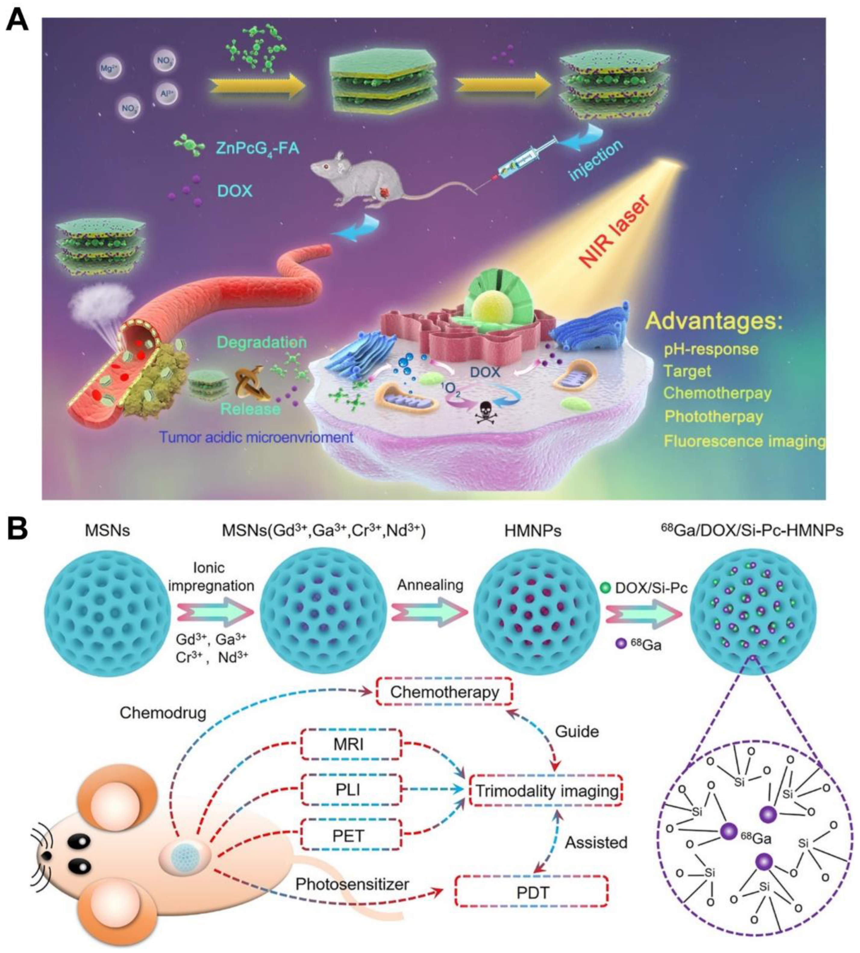
- Wang, Z.; Zhu, H.; He, F.; Yan, D.; Jia, T.; Sun, Q.; Wang, X.; Wang, X.; Yang, D.; Gai, S.; et al. Layer structured LDH_ZnPcG4-FA nanoplatform for targeted and imaging guided chemo–photodynamic therapy mediated by 650 nm light. Chem. Eng. J. 2020, 382, 122847. [Google Scholar] [CrossRef]
- Meng, Y.; Du, Y.; Lin, Y.; Su, Y.; Li, R.; Feng, Y.; Meng, S. A two-fold interpenetration pillar-layered metal-organic frameworks based on BODIPY for chemo–photodynamic therapy. Dye. Pigment. 2021, 188, 109174. [Google Scholar] [CrossRef]
- Zou, R.; Gao, Y.; Zhang, Y.; Jiao, J.; Wong, K.-L.; Wang, J. 68Ga-Labeled Magnetic-NIR Persistent Luminescent Hybrid Mesoporous Nanoparticles for Multimodal Imaging-Guided Chemotherapy and Photodynamic Therapy. ACS Appl. Mater. Interfaces 2021, 13, 9667–9680. [Google Scholar] [CrossRef] [PubMed]
- Wu, X.; Zhang, Y.; Wang, Z.; Wu, J.; Yan, R.; Guo, C.; Jin, Y. Near-Infrared Light-Initiated Upconversion Nanoplatform with Tumor Microenvironment Responsiveness for Improved Photodynamic Therapy. ACS Appl. Bio Mater. 2020, 3, 5813–5823. [Google Scholar] [CrossRef]
- Huang, Y.; Liu, D.-E.; An, J.; Liu, B.; Sun, L.; Fu, H.; Yan, S.; Sun, W.; Gao, H. Reactive Oxygen Species Self-Sufficient Multifunctional Nanoplatform for Synergistic Chemo–Photodynamic Therapy with Red/Near-Infrared Dual-Imaging. ACS Appl. Bio Mater. 2020, 3, 9135–9144. [Google Scholar] [CrossRef]
- Cui, Y.-X.; Sun, G.-Y.; Li, Y.-H.; Tang, A.-N.; Zhu, L.-N.; Kong, D.-M. DNA-Based pH-Responsive Core–Shell Drug Nanocarrier for Tumor-Targeted Chemo–Photodynamic Therapy. Adv. Mater. Interfaces 2020, 7, 2000292. [Google Scholar] [CrossRef]
- Ren, S.-Z.; Wang, B.; Zhu, X.-H.; Zhu, D.; Liu, M.; Li, S.-K.; Yang, Y.-S.; Wang, Z.-C.; Zhu, H.-L. Oxygen Self-Sufficient Core–Shell Metal–Organic Framework-Based Smart Nanoplatform for Enhanced Synergistic Chemotherapy and Photodynamic Therapy. ACS Appl. Mater. Interfaces 2020, 12, 24662–24674. [Google Scholar] [CrossRef]
- Zhu, K.; Liu, G.; Hu, J.; Liu, S. Near-Infrared Light-Activated Photochemical Internalization of Reduction-Responsive Polyprodrug Vesicles for Synergistic Photodynamic Therapy and Chemotherapy. Biomacromolecules 2017, 18, 2571–2582. [Google Scholar] [CrossRef] [PubMed]
- Liu, J.; Liu, K.; Feng, L.; Liu, Z.; Xu, L. Comparison of nanomedicine-based chemotherapy, photodynamic therapy and photothermal therapy using reduced graphene oxide for the model system. Biomater. Sci. 2017, 5, 331–340. [Google Scholar] [CrossRef]
- Liu, W.; Wang, Y.-M.; Li, Y.-H.; Cai, S.-J.; Yin, X.-B.; He, X.-W.; Zhang, Y.-K. Fluorescent Imaging-Guided Chemotherapy-and-Photodynamic Dual Therapy with Nanoscale Porphyrin Metal–Organic Framework. Small 2017, 13, 1603459. [Google Scholar] [CrossRef]
- Lan, J.-S.; Liu, L.; Zeng, R.-F.; Qin, Y.-H.; Hou, J.-W.; Xie, S.-S.; Yue, S.; Yang, J.; Ho, R.J.Y.; Ding, Y.; et al. Tumor-specific carrier-free nanodrugs with GSH depletion and enhanced ROS generation for endogenous synergistic anti-tumor by a chemotherapy-photodynamic therapy. Chem. Eng. J. 2021, 407, 127212. [Google Scholar] [CrossRef]
- Huang, L.; Wan, J.; Wu, H.; Chen, X.; Bian, Q.; Shi, L.; Jiang, X.; Yuan, A.; Gao, J.; Wang, H. Quantitative self-assembly of photoactivatable small molecular prodrug cocktails for safe and potent cancer chemo–photodynamic therapy. Nano Today 2021, 36, 101030. [Google Scholar] [CrossRef]
- Choi, J.; Shim, M.K.; Yang, S.; Hwang, H.S.; Cho, H.; Kim, J.; Yun, W.S.; Moon, Y.; Kim, J.; Yoon, H.Y.; et al. Visible-Light-Triggered Prodrug Nanoparticles Combine Chemotherapy and Photodynamic Therapy to Potentiate Checkpoint Blockade Cancer Immunotherapy. ACS Nano 2021, 15, 12086–12098. [Google Scholar] [CrossRef] [PubMed]
- Liu, L.; Bao, Y.; Zhang, Y.; Xiao, C.; Chen, L. Acid-responsive dextran-based therapeutic nanoplatforms for photodynamic-chemotherapy against multidrug resistance. Int. J. Biol. Macromol. 2020, 155, 233–240. [Google Scholar] [CrossRef]
- Liu, L.; Bao, Y.; Wang, J.; Xiao, C.; Chen, L. Construction of carrier-free porphyrin-based drug self-framed delivery system to reverse multidrug resistance through photodynamic-chemotherapy. Dye. Pigment. 2020, 177, 107922. [Google Scholar] [CrossRef]
- Wang, X.; Tong, J.; He, Z.; Yang, X.; Meng, F.; Liang, H.; Zhang, X.; Luo, L. Paclitaxel-Potentiated Photodynamic Theranostics for Synergistic Tumor Ablation and Precise Anticancer Efficacy Monitoring. ACS Appl. Mater. Interfaces 2020, 12, 5476–5487. [Google Scholar] [CrossRef]
- Jiang, M.; Mu, J.; Jacobson, O.; Wang, Z.; He, L.; Zhang, F.; Yang, W.; Lin, Q.; Zhou, Z.; Ma, Y.; et al. Reactive Oxygen Species Activatable Heterodimeric Prodrug as Tumor-Selective Nanotheranostics. ACS Nano 2020, 14, 16875–16886. [Google Scholar] [CrossRef]
- Cheng, Y.-J.; Qin, S.-Y.; Liu, W.-L.; Ma, Y.-H.; Chen, X.-S.; Zhang, A.-Q.; Zhang, X.-Z. Dual-Targeting Photosensitizer-Peptide Amphiphile Conjugate for Enzyme-Triggered Drug Delivery and Synergistic Chemo–Photodynamic Tumor Therapy. Adv. Mater. Interfaces 2020, 7, 2000935. [Google Scholar] [CrossRef]
- Wang, Y.; Yang, M.; Qian, J.; Xu, W.; Wang, J.; Hou, G.; Ji, L.; Suo, A. Sequentially self-assembled polysaccharide-based nanocomplexes for combined chemotherapy and photodynamic therapy of breast cancer. Carbohydr. Polym. 2019, 203, 203–213. [Google Scholar] [CrossRef]
- Luo, C.; Sun, B.; Wang, C.; Zhang, X.; Chen, Y.; Chen, Q.; Yu, H.; Zhao, H.; Sun, M.; Li, Z.; et al. Self-facilitated ROS-responsive nanoassembly of heterotypic dimer for synergistic chemo–photodynamic therapy. J. Control. Release 2019, 302, 79–89. [Google Scholar] [CrossRef]
- Wang, Z.; Sun, M.; Liu, T.; Tan, X.; Zhang, H.; Zhang, X.; He, Z.; Sun, J. A surfactant-like chemotherapeutic agent as a nanocarrier for delivering photosensitizers against cancer: A facile drug-delivering-drug strategy. Int. J. Pharm. 2019, 562, 313–320. [Google Scholar] [CrossRef]
- Wang, M.; Zhai, Y.; Ye, H.; Lv, Q.; Sun, B.; Luo, C.; Jiang, Q.; Zhang, H.; Xu, Y.; Jing, Y.; et al. High Co-loading Capacity and Stimuli-Responsive Release Based on Cascade Reaction of Self-Destructive Polymer for Improved Chemo–Photodynamic Therapy. ACS Nano 2019, 13, 7010–7023. [Google Scholar] [CrossRef] [PubMed]
- Liu, R.; Yu, M.; Yang, X.; Umeshappa, C.S.; Hu, C.; Yu, W.; Qin, L.; Huang, Y.; Gao, H. Linear Chimeric Triblock Molecules Self-Assembled Micelles with Controllably Transformable Property to Enhance Tumor Retention for Chemo–Photodynamic Therapy of Breast Cancer. Adv. Funct. Mater. 2019, 29, 1808462. [Google Scholar] [CrossRef]
- Luo, Y.; Song, Y.; Wang, M.; Jian, T.; Ding, S.; Mu, P.; Liao, Z.; Shi, Q.; Cai, X.; Jin, H.; et al. Bioinspired Peptoid Nanotubes for Targeted Tumor Cell Imaging and Chemo-Photodynamic Therapy. Small 2019, 15, e1902485. [Google Scholar] [CrossRef] [PubMed]
- Lee, H.; Han, J.; Shin, H.; Han, H.; Na, K.; Kim, H. Combination of chemotherapy and photodynamic therapy for cancer treatment with sonoporation effects. J. Control. Release 2018, 283, 190–199. [Google Scholar] [CrossRef] [PubMed]
- Pei, Q.; Hu, X.; Zheng, X.; Liu, S.; Li, Y.; Jing, X.; Xie, Z. Light-Activatable Red Blood Cell Membrane-Camouflaged Dimeric Prodrug Nanoparticles for Synergistic Photodynamic/Chemotherapy. ACS Nano 2018, 12, 1630–1641. [Google Scholar] [CrossRef]
- Liu, D.; Liu, L.; Liu, F.; Zhang, M.; Wei, P.; Yi, T. HOCl-Activated Aggregation of Gold Nanoparticles for Multimodality Therapy of Tumors. Adv. Sci. 2021, 8, 2100074. [Google Scholar] [CrossRef]
- Zhang, F.; Lu, G.; Wen, X.; Li, F.; Ji, X.; Li, Q.; Wu, M.; Cheng, Q.; Yu, Y.; Tang, J.; et al. Magnetic nanoparticles coated with polyphenols for spatio-temporally controlled cancer photothermal/immunotherapy. J. Control. Release 2020, 326, 131–139. [Google Scholar] [CrossRef]
- Yan, L.; Amirshaghaghi, A.; Huang, D.; Miller, J.; Stein, J.M.; Busch, T.M.; Cheng, Z.; Tsourkas, A. Protoporphyrin IX (PpIX)-Coated Superparamagnetic Iron Oxide Nanoparticle (SPION) Nanoclusters for Magnetic Resonance Imaging and Photodynamic Therapy. Adv. Funct. Mater. 2018, 28, 1707030. [Google Scholar] [CrossRef]
- Wang, L.; Shi, J.; Liu, R.; Liu, Y.; Zhang, J.; Yu, X.; Gao, J.; Zhang, C.; Zhang, Z. Photodynamic effect of functionalized single-walled carbon nanotubes: A potential sensitizer for photodynamic therapy. Nanoscale 2014, 6, 4642–4651. [Google Scholar] [CrossRef]
- Taheri, H.; Unal, M.A.; Sevim, M.; Gurcan, C.; Ekim, O.; Ceylan, A.; Syrgiannis, Z.; Christoforidis, K.C.; Bosi, S.; Ozgenç, O.; et al. Photocatalytically Active Graphitic Carbon Nitride as an Effective and Safe 2D Material for In Vitro and In Vivo Photodynamic Therapy. Small 2020, 16, 1904619. [Google Scholar] [CrossRef]
- Murali, G.; Kwon, B.; Kang, H.; Modigunta, J.K.R.; Park, S.; Lee, S.; Lee, H.; Park, Y.H.; Kim, J.; Park, S.Y.; et al. Hematoporphyrin Photosensitizer-Linked Carbon Quantum Dots for Photodynamic Therapy of Cancer Cells. ACS Appl. Nano Mater. 2022, 5, 4376–4385. [Google Scholar] [CrossRef]
- Shi, Z.; Zhang, K.; Zada, S.; Zhang, C.; Meng, X.; Yang, Z.; Dong, H. Upconversion Nanoparticle-Induced Multimode Photodynamic Therapy Based on a Metal–Organic Framework/Titanium Dioxide Nanocomposite. ACS Appl. Mater. Interfaces 2020, 12, 12600–12608. [Google Scholar] [CrossRef] [PubMed]
- Jin, F.; Qi, J.; Liu, D.; You, Y.; Shu, G.; Du, Y.; Wang, J.; Xu, X.; Ying, X.; Ji, J.; et al. Cancer-cell-biomimetic Upconversion nanoparticles combining chemo–photodynamic therapy and CD73 blockade for metastatic triple-negative breast cancer. J. Control. Release 2021, 337, 90–104. [Google Scholar] [CrossRef] [PubMed]
- Liang, S.; Sun, C.; Yang, P.; Ma, P.A.; Huang, S.; Cheng, Z.; Yu, X.; Lin, J. Core-shell structured upconversion nanocrystal-dendrimer composite as a carrier for mitochondria targeting and catalase enhanced anti-cancer photodynamic therapy. Biomaterials 2020, 240, 119850. [Google Scholar] [CrossRef]
- Zhang, N.; Wang, D.; Jing, X.; Yang, T.; Yang, H.; Meng, L. pH/ROS Dual-Responsive Polymer–Drug-Based Nanocarriers: Click-Reaction Preparation and Fluorescence Imaging-Guided Chemotherapy and Photodynamic Therapy. ACS Appl. Bio. Mater. 2021, 4, 6294–6303. [Google Scholar] [CrossRef]
- Ma, Q.; Zhao, Y.; Guan, Q.; Zhao, Y.; Zhang, H.; Ding, Z.; Wang, Q.; Wu, Y.; Liu, M.; Han, J. Amphiphilic block polymer-based self-assembly of high payload nanoparticles for efficient combinatorial chemo–photodynamic therapy. Drug Deliv. 2020, 27, 1656–1666. [Google Scholar] [CrossRef]
- Luo, R.; Zhang, Z.; Han, L.; Xue, Z.; Zhang, K.; Liu, F.; Feng, F.; Xue, J.; Liu, W.; Qu, W. An albumin-binding dimeric prodrug nanoparticle with long blood circulation and light-triggered drug release for chemo–photodynamic combination therapy against hypoxia-induced metastasis of lung cancer. Biomater. Sci. 2021, 9, 3718–3736. [Google Scholar] [CrossRef]
- Liu, X.; Du, J.; Xie, Z.; Wang, L.; Liu, X.; Hou, Z.; Wang, X.; Tang, R. Lactobionic acid-modified phycocyanin nanoparticles loaded with doxorubicin for synergistic chemo–photodynamic therapy. Int. J. Biol. Macromol. 2021, 186, 206–217. [Google Scholar] [CrossRef]
- Chibh, S.; Katoch, V.; Singh, M.; Prakash, B.; Panda, J.J. Miniatured Fluidics-Mediated Modular Self-Assembly of Anticancer Drug–Amino Acid Composite Microbowls for Combined Chemo–Photodynamic Therapy in Glioma. ACS Biomater. Sci. Eng. 2021, 7, 5654–5665. [Google Scholar] [CrossRef]
- Díaz, C.F.; Guzmán, L.; Jiménez, V.A.; Alderete, J.B. Polyamidoamine dendrimers of the third generation–chlorin e6 nanoconjugates: Nontoxic hybrid polymers with photodynamic activity. J. Appl. Polym. Sci. 2022, 139, 51835. [Google Scholar] [CrossRef]
- Conner, S.D.; Schmid, S.L. Regulated portals of entry into the cell. Nature 2003, 422, 37–44. [Google Scholar] [CrossRef] [PubMed]
- Fan, R.; Chen, C.; Hou, H.; Chuan, D.; Mu, M.; Liu, Z.; Liang, R.; Guo, G.; Xu, J. Tumor Acidity and Near-Infrared Light Responsive Dual Drug Delivery Polydopamine-Based Nanoparticles for Chemo-Photothermal Therapy. Adv. Funct. Mater. 2021, 31, 2009733. [Google Scholar] [CrossRef]
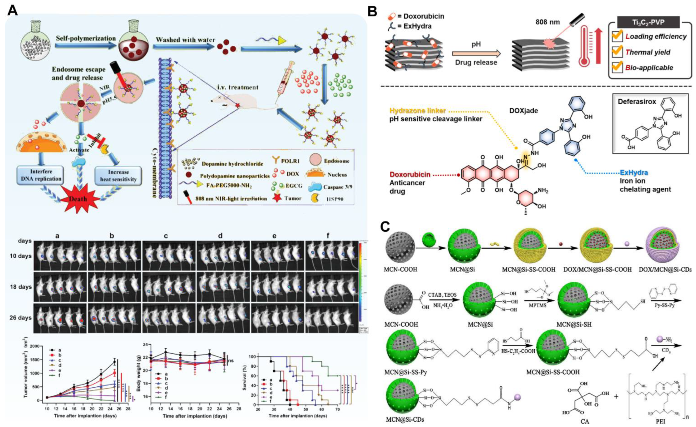
- Xu, Y.; Wang, Y.; An, J.; Sedgwick, A.C.; Li, M.; Xie, J.; Hu, W.; Kang, J.; Sen, S.; Steinbrueck, A.; et al. 2D-ultrathin MXene/DOXjade platform for iron chelation chemo–photothermal therapy. Bioact. Mater. 2022, 14, 76–85. [Google Scholar] [CrossRef] [PubMed]
- Lu, H.; Zhao, Q.; Wang, X.; Mao, Y.; Chen, C.; Gao, Y.; Sun, C.; Wang, S. Multi-stimuli responsive mesoporous silica-coated carbon nanoparticles for chemo–photothermal therapy of tumor. Colloids Surf. B Biointerfaces 2020, 190, 110941. [Google Scholar] [CrossRef] [PubMed]
- Huang, L.; Chen, X.; Bian, Q.; Zhang, F.; Wu, H.; Wang, H.; Gao, J. Photosensitizer-stabilized self-assembling nanoparticles potentiate chemo/photodynamic efficacy of patient-derived melanoma. J. Control. Release 2020, 328, 325–338. [Google Scholar] [CrossRef]
- Gao, Y.; Wang, X.; He, X.; He, Z.; Yang, X.; Tian, S.; Meng, F.; Ding, D.; Luo, L.; Tang, B.Z. A Dual-Functional Photosensitizer for Ultraefficient Photodynamic Therapy and Synchronous Anticancer Efficacy Monitoring. Adv. Funct. Mater. 2019, 29, 1902673. [Google Scholar] [CrossRef]
- Feng, L.; Chen, M.; Li, R.; Zhou, L.; Wang, C.; Ye, P.; Hu, X.; Yang, J.; Sun, Y.; Zhu, Z.; et al. Biodegradable oxygen-producing manganese-chelated metal organic frameworks for tumor-targeted synergistic chemo/photothermal/ photodynamic therapy. Acta Biomater. 2022, 138, 463–477. [Google Scholar] [CrossRef]
- Sun, X.; He, G.; Xiong, C.; Wang, C.; Lian, X.; Hu, L.; Li, Z.; Dalgarno, S.J.; Yang, Y.-W.; Tian, J. One-Pot Fabrication of Hollow Porphyrinic MOF Nanoparticles with Ultrahigh Drug Loading toward Controlled Delivery and Synergistic Cancer Therapy. ACS Appl. Mater. Interfaces 2021, 13, 3679–3693. [Google Scholar] [CrossRef]
- Zhu, Y.; Xin, N.; Qiao, Z.; Chen, S.; Zeng, L.; Zhang, Y.; Wei, D.; Sun, J.; Fan, H. Bioactive MOFs Based Theranostic Agent for Highly Effective Combination of Multimodal Imaging and Chemo–Phototherapy. Adv. Healthc. Mater. 2020, 9, 2000205. [Google Scholar] [CrossRef]
- Wan, G.; Chen, B.; Li, L.; Wang, D.; Shi, S.; Zhang, T.; Wang, Y.; Zhang, L.; Wang, Y. Nanoscaled red blood cells facilitate breast cancer treatment by combining photothermal/photodynamic therapy and chemotherapy. Biomaterials 2018, 155, 25–40. [Google Scholar] [CrossRef]
- Tang, J.; Zhou, H.; Hou, X.; Wang, L.; Li, Y.; Pang, Y.; Chen, C.; Jiang, G.; Liu, Y. Enhanced anti-tumor efficacy of temozolomide-loaded carboxylated poly(amido-amine) combined with photothermal/photodynamic therapy for melanoma treatment. Cancer Lett. 2018, 423, 16–26. [Google Scholar] [CrossRef] [PubMed]
- Shi, C.-E.; You, C.-Q.; Pan, L. Facile formulation of near-infrared light-triggered hollow mesoporous silica nanoparticles based on mitochondria targeting for on-demand chemo/photothermal/photodynamic therapy. Nanotechnology 2019, 30, 325102. [Google Scholar] [CrossRef] [PubMed]
- Xu, M.; Yang, G.; Bi, H.; Xu, J.; Dong, S.; Jia, T.; Wang, Z.; Zhao, R.; Sun, Q.; Gai, S.; et al. An intelligent nanoplatform for imaging-guided photodynamic/photothermal/chemo-therapy based on upconversion nanoparticles and CuS integrated black phosphorus. Chem. Eng. J. 2020, 382, 122822. [Google Scholar] [CrossRef]
- Kuang, Y.; Zhang, Y.; Zhao, Y.; Cao, Y.; Zhang, Y.; Chong, Y.; Pei, R. Dual-Stimuli-Responsive Multifunctional Gd2Hf2O7 Nanoparticles for MRI-Guided Combined Chemo-/Photothermal-/Radiotherapy of Resistant Tumors. ACS Appl. Mater. Interfaces 2020, 12, 35928–35939. [Google Scholar] [CrossRef]
- Wang, J.; Liang, D.; Qu, Z.; Kislyakov, I.M.; Kiselev, V.M.; Liu, J. PEGylated-folic acid–modified black phosphorus quantum dots as near-infrared agents for dual-modality imaging-guided selective cancer cell destruction. Nanophotonics 2020, 9, 2425–2435. [Google Scholar] [CrossRef]
- Sun, L.; Li, Q.; Zhang, L.; Xu, Z.; Kang, Y.; Xue, P. PEGylated Polydopamine Nanoparticles Incorporated with Indocyanine Green and Doxorubicin for Magnetically Guided Multimodal Cancer Therapy Triggered by Near-Infrared Light. ACS Appl. Nano Mater. 2018, 1, 325–336. [Google Scholar] [CrossRef]
- Yang, Y.; Zhu, L.; Xia, F.; Gong, B.; Xie, A.; Li, S.; Huang, F.; Wang, S.; Shen, Y.; Weaver, D.T. A novel 5-FU/rGO/Bce hybrid hydrogel shell on a tumor cell: One-step synthesis and synergistic chemo/photo-thermal/photodynamic effect. RSC Adv. 2017, 7, 2415–2425. [Google Scholar] [CrossRef]
- Yang, D.; Xu, J.; Yang, G.; Zhou, Y.; Ji, H.; Bi, H.; Gai, S.; He, F.; Yang, P. Metal-organic frameworks join hands to create an anti-cancer nanoplatform based on 808 nm light driving up-conversion nanoparticles. Chem. Eng. J. 2018, 344, 363–374. [Google Scholar] [CrossRef]
- Chen, W.; Ouyang, J.; Liu, H.; Chen, M.; Zeng, K.; Sheng, J.; Liu, Z.; Han, Y.; Wang, L.; Li, J.; et al. Black Phosphorus Nanosheet-Based Drug Delivery System for Synergistic Photodynamic/Photothermal/Chemotherapy of Cancer. Adv. Mater. 2017, 29, 1603864. [Google Scholar] [CrossRef]
- Yang, Y.; Wang, C.; Tian, C.; Guo, H.; Shen, Y.; Zhu, M. Fe3O4@MnO2@PPy nanocomposites overcome hypoxia: Magnetic-targeting-assisted controlled chemotherapy and enhanced photodynamic/photothermal therapy. J. Mater. Chem. B 2018, 6, 6848–6857. [Google Scholar] [CrossRef] [PubMed]
- Xue, X.; Huang, Y.; Bo, R.; Jia, B.; Wu, H.; Yuan, Y.; Wang, Z.; Ma, Z.; Jing, D.; Xu, X.; et al. Trojan Horse nanotheranostics with dual transformability and multifunctionality for highly effective cancer treatment. Nat. Commun. 2018, 9, 3653. [Google Scholar] [CrossRef] [PubMed]
- Sun, Q.; He, F.; Bi, H.; Wang, Z.; Sun, C.; Li, C.; Xu, J.; Yang, D.; Wang, X.; Gai, S.; et al. An intelligent nanoplatform for simultaneously controlled chemo-, photothermal, and photodynamic therapies mediated by a single NIR light. Chem. Eng. J. 2019, 362, 679–691. [Google Scholar] [CrossRef]
- Wang, Q.; Dai, Y.; Xu, J.; Cai, J.; Niu, X.; Zhang, L.; Chen, R.; Shen, Q.; Huang, W.; Fan, Q. All-in-One Phototheranostics: Single Laser Triggers NIR-II Fluorescence/Photoacoustic Imaging Guided Photothermal/Photodynamic/Chemo Combination Therapy. Adv. Funct. Mater. 2019, 29, 1901480. [Google Scholar] [CrossRef]
- Zheng, T.; Wang, W.; Wu, F.; Zhang, M.; Shen, J.; Sun, Y. Zwitterionic Polymer-Gated Au@TiO(2) Core-Shell Nanoparticles for Imaging-Guided Combined Cancer Therapy. Theranostics 2019, 9, 5035–5048. [Google Scholar] [CrossRef]
- Guo, Y.; Wang, H.; Feng, X.; Zhao, Y.; Liang, C.; Yang, L.; Li, M.; Zhang, Y.; Gao, W. 3D MXene microspheres with honeycomb architecture for tumor photothermal/photodynamic/chemo combination therapy. Nanotechnology 2021, 32, 195701. [Google Scholar] [CrossRef]
- Yao, S.; Jin, X.; Wang, C.; Cao, A.; Hu, J.; Chen, B.; Wang, B. ICG/5-Fu coencapsulated temperature stimulus response nanogel drug delivery platform for chemo–photothermal/photodynamic synergetic therapy. J. Biomater. Appl. 2021, 36, 565–578. [Google Scholar] [CrossRef]
- Moradi Kashkooli, F.; Soltani, M.; Souri, M.; Meaney, C.; Kohandel, M. Nexus between in silico and in vivo models to enhance clinical translation of nanomedicine. Nano Today 2021, 36, 101057. [Google Scholar] [CrossRef]
- Deng, X.; Shao, Z.; Zhao, Y. Solutions to the Drawbacks of Photothermal and Photodynamic Cancer Therapy. Adv. Sci. 2021, 8, 2002504. [Google Scholar] [CrossRef]
- Kawczyk-Krupka, A.; Bugaj, A.M.; Latos, W.; Zaremba, K.; Wawrzyniec, K.; Sieroń, A. Photodynamic therapy in colorectal cancer treatment: The state of the art in clinical trials. Photodiagnosis Photodyn. Ther. 2015, 12, 545–553. [Google Scholar] [CrossRef]
- Mahmoudi, K.; Garvey, K.L.; Bouras, A.; Cramer, G.; Stepp, H.; Jesu Raj, J.G.; Bozec, D.; Busch, T.M.; Hadjipanayis, C.G. 5-aminolevulinic acid photodynamic therapy for the treatment of high-grade gliomas. J. Neuro-Oncol. 2019, 141, 595–607. [Google Scholar] [CrossRef]
- Katiyar, R.S.; Jha, P.K. Molecular simulations in drug delivery: Opportunities and challenges. WIREs Comput. Mol. Sci. 2018, 8, e1358. [Google Scholar] [CrossRef]
- Zhan, W.; Alamer, M.; Xu, X.Y. Computational modelling of drug delivery to solid tumour: Understanding the interplay between chemotherapeutics and biological system for optimised delivery systems. Adv. Drug Deliv. Rev. 2018, 132, 81–103. [Google Scholar] [CrossRef] [PubMed]


| Photoabsobers | Photothermal Agents | Chemotherapeutic Drug | Main Drug-Loading Mechanism | Drug-Loading Efficiency (wt%) | Ref. |
|---|---|---|---|---|---|
| Noble metal–based materials | AuNPs | Methotrexate | Electrostatic interactions | 36.2% | [21] |
| AuNRs | Doxorubicin | Electrostatic interactions | 76.0% | [22] | |
| AuNBs | Doxorubicin | Electrostatic interactions | 70.0% | [23] | |
| AuNFs | Doxorubicin | Electrostatic interactions | 78.9% | [24] | |
| Au@Pt NPs | Doxorubicin | Electrostatic interactions | 32.3% | [25] | |
| Pd@MnO2 | Doxorubicin | Electrostatic interactions | 58.0% | [26] | |
| Transition metal–based materials | CuS NPs | Doxorubicin | Electrostatic interactions | 55.5% | [27] |
| MoS2 | Doxorubicin | Electrostatic interactions | 95.7% | [28] | |
| CoS, PDA | Doxorubicin | Electrostatic, π-π stacking | 44.6% | [29] | |
| WS2 nanosheets | Doxorubicin | Electrostatic, π-π stacking | 95.0% | [30] | |
| MoOx nanosheets | Doxorubicin | Electrostatic, π-π stacking | 65.0% | [31] | |
| Carbon-based material | Nano-GO | Dacarbazine | π–π stacking | 80.0% | [32] |
| GDYO | Doxorubicin, cisplatin, methotrexate | Amide reaction, π-π stacking, electrostatic interactions | 40.3% of Doxorubicin | [33] | |
| MCNs | Doxorubicin | Electrostatic, π-π stacking | 69.2% | [34] | |
| CNTs | Doxorubicin | Electrostatic, π-π stacking | 50.0% | [35] | |
| GQDs | Doxorubicin | Der Waals interaction, π-π stacking | 96.6% | [36] | |
| SWNHs | Cisplatin and doxorubicin | Hydrophobic-hydrophobic, interactions and π-π stacking | 52.4% | [37] | |
| mCNFs | 5-Fluorouracil | electrostatic adsorption | 31.0% | [38] | |
| Organic nanomaterial | IR783 | Camptothecin | Electrostatic, π-π stacking and hydrophobic interactions | 62.0% | [39] |
| ICG | Doxorubicin | Electrostatic, π-π stacking | 58.2% | [40] | |
| IR1061 | Paclitaxel | Electrostatic adsorption | 59.3% | [41] | |
| PDA NPs | Doxorubicin | Coordinate bond, electrostatic adsorption | 80.0% | [42] | |
| HMPAn NPs | Doxorubicin | Noncovalent electrostatic | 37.5% | [43] | |
| PPY NPs | Doxorubicin | electrostatic adsorption | 43.3% | [44] | |
| Others | Iron oxide NPs | Curcumin | electrostatic adsorption | 93.0% | [45] |
| Ti-WC nanowires | Doxorubicin | π-π stacking | 69.2% | [46] | |
| HM-Bi | Doxorubicin | electrostatic adsorption | 78.0% | [47] |
Publisher’s Note: MDPI stays neutral with regard to jurisdictional claims in published maps and institutional affiliations. |
© 2022 by the authors. Licensee MDPI, Basel, Switzerland. This article is an open access article distributed under the terms and conditions of the Creative Commons Attribution (CC BY) license (https://creativecommons.org/licenses/by/4.0/).
Share and Cite
Wang, Y.; Zhang, Y.; Zhang, X.; Zhang, Z.; She, J.; Wu, D.; Gao, W. High Drug-Loading Nanomedicines for Tumor Chemo–Photo Combination Therapy: Advances and Perspectives. Pharmaceutics 2022, 14, 1735. https://doi.org/10.3390/pharmaceutics14081735
Wang Y, Zhang Y, Zhang X, Zhang Z, She J, Wu D, Gao W. High Drug-Loading Nanomedicines for Tumor Chemo–Photo Combination Therapy: Advances and Perspectives. Pharmaceutics. 2022; 14(8):1735. https://doi.org/10.3390/pharmaceutics14081735
Chicago/Turabian StyleWang, Ya, Yujie Zhang, Xiaojiang Zhang, Zhe Zhang, Junjun She, Daocheng Wu, and Wei Gao. 2022. "High Drug-Loading Nanomedicines for Tumor Chemo–Photo Combination Therapy: Advances and Perspectives" Pharmaceutics 14, no. 8: 1735. https://doi.org/10.3390/pharmaceutics14081735