
Retinal Delivery of the Protein Kinase C-β Inhibitor Ruboxistaurin Using Non-Invasive Nanoparticles of Polyamidoamine Dendrimers

 , , , , , , , , and
Abstract
:1. Introduction
2. Materials and Methods
2.1. Materials
2.2. Formulation of Anionic G4.5 and Neutral G5 Complexes
2.3. Measurement of Particle Size (PS), Polydispersity Index (PDI), and ζ-Potential
2.4. Determination of Drug Loading Efficiency (DE%)
2.5. Transmission Electron Microscopy
2.6. In Vitro Drug Release of G4.5 and G5 Complexes
2.7. Stability Studies
2.7.1. Physical Stability
2.7.2. Chemical Stability
2.8. Cell Culture
2.8.1. Moorfield’s/Institute of Ophthalmology-Müller-1 (MIO-M1) Cells
2.8.2. Culture of the MIO-M1 Glial Cell Line
2.8.3. Cell Viability Studies
2.8.4. High Glucose Experiments
2.8.5. High Glucose Treatment of PAMAM Dendrimers and Complexes
2.8.6. In Vitro Permeability Study
2.9. Statistical Analysis
3. Results
3.1. Characterization of G4.5 Complexes
3.1.1. Measurement of PS and PDI
3.1.2. Measurement of ζ-Potential of G4.5 Complexes
3.1.3. Determination of DE%
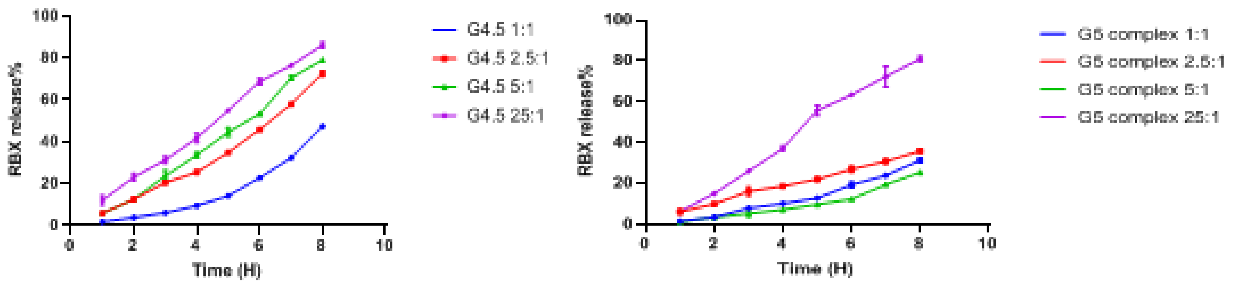
3.1.4. In Vitro Drug Release of G4.5 Complexes
3.1.5. Stability Studies of G4.5 Complex
Physical Stability
Chemical Stability
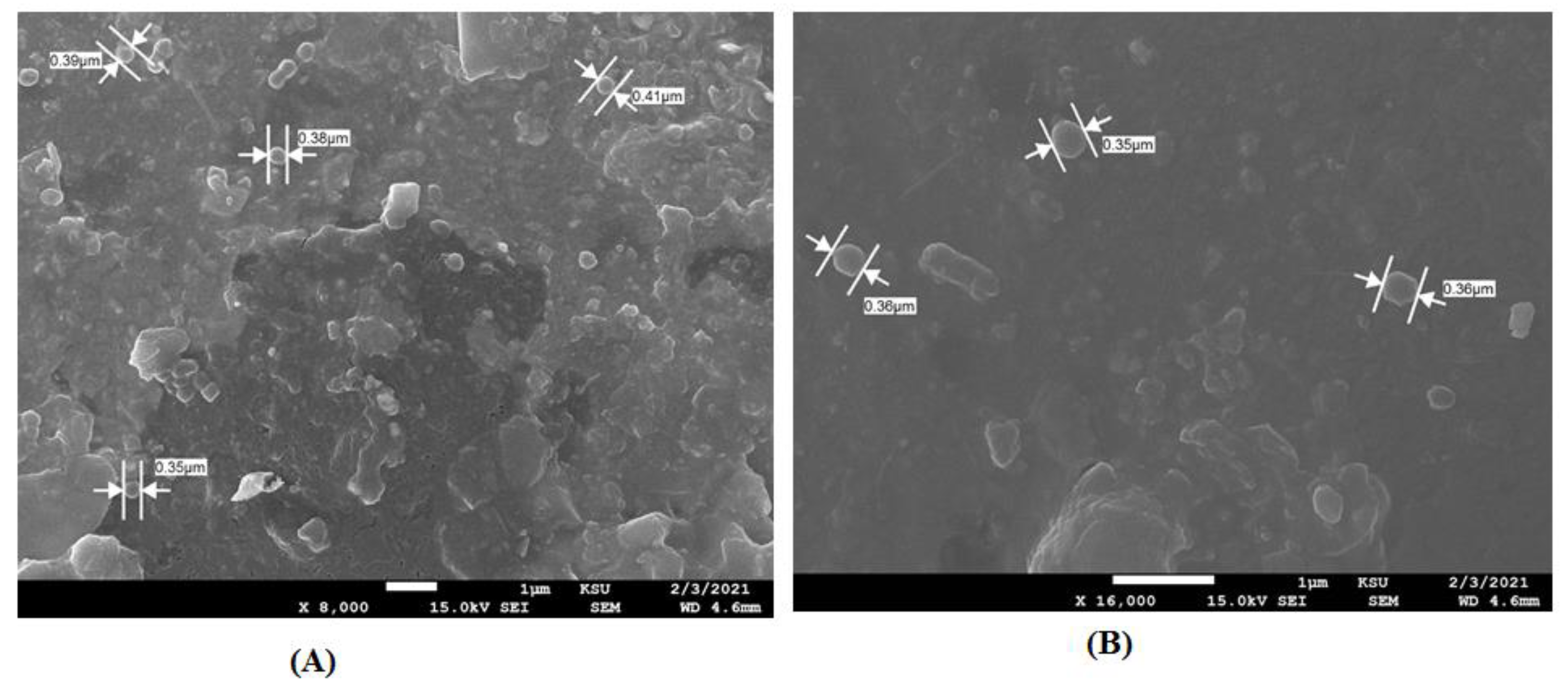
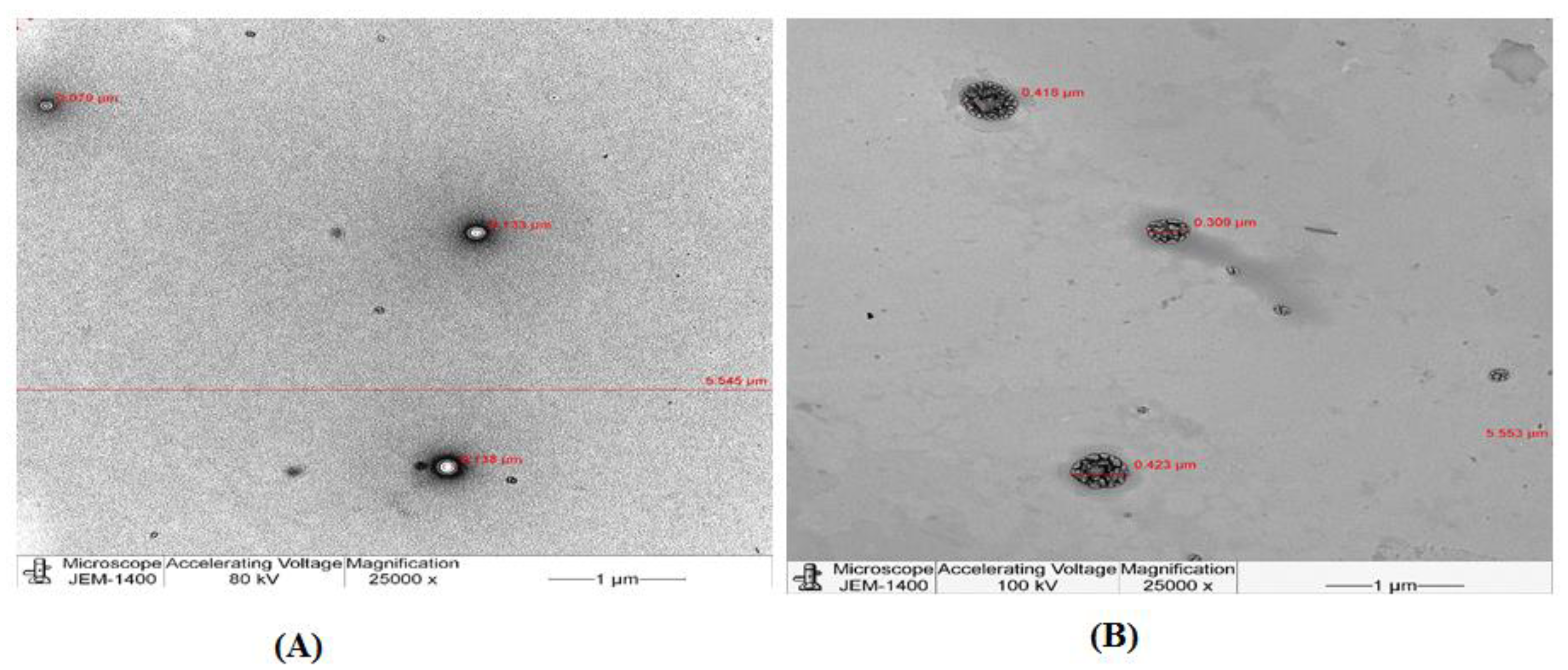
TEM
3.2. Characterization of G5 Complexes
3.2.1. Measurement of PS and PDI
3.2.2. Measurements of ζ-Potential of G5 Complexes
3.2.3. Determination of DE%
3.2.4. In Vitro Drug Release of G5 Complexes
3.2.5. Stability Studies of G5 Complex
Physical Stability
Drug Content
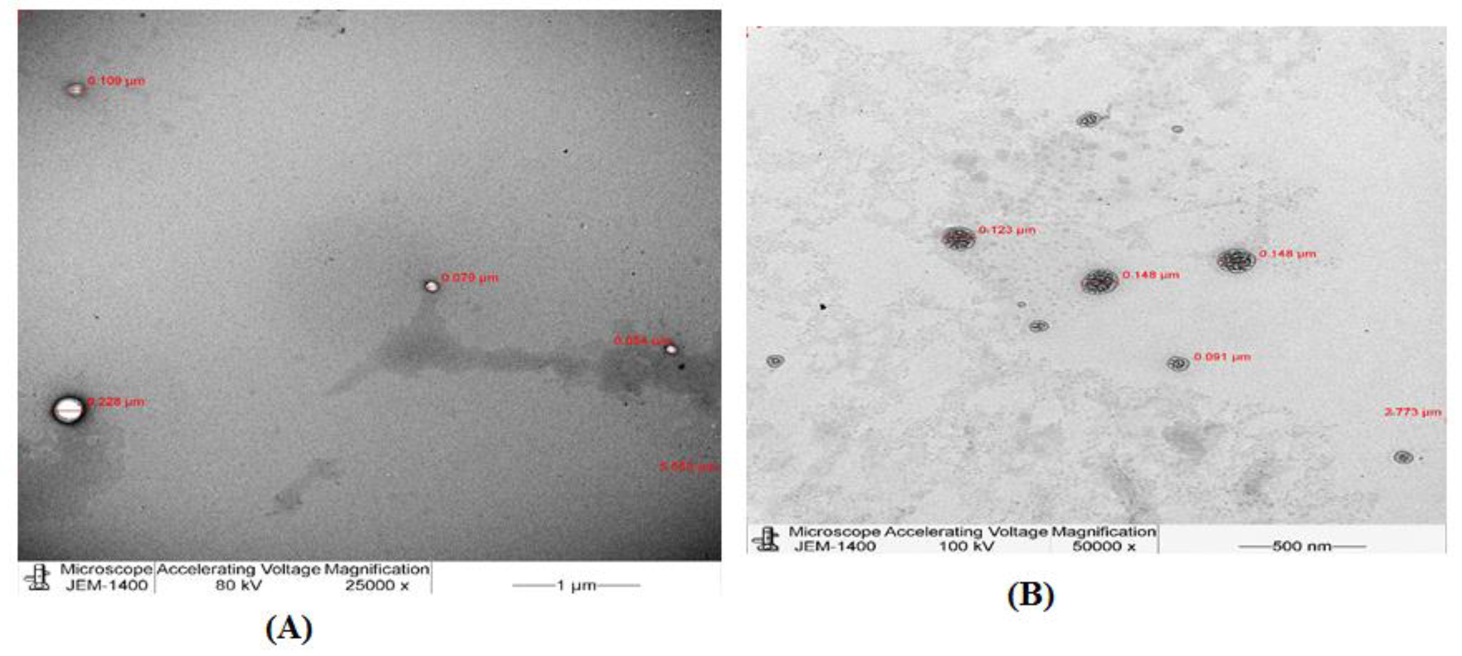
TEM
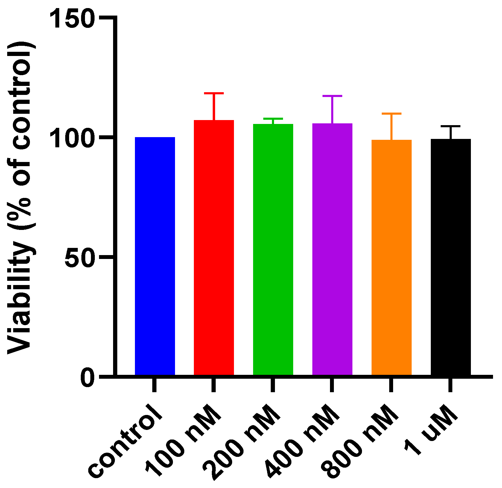
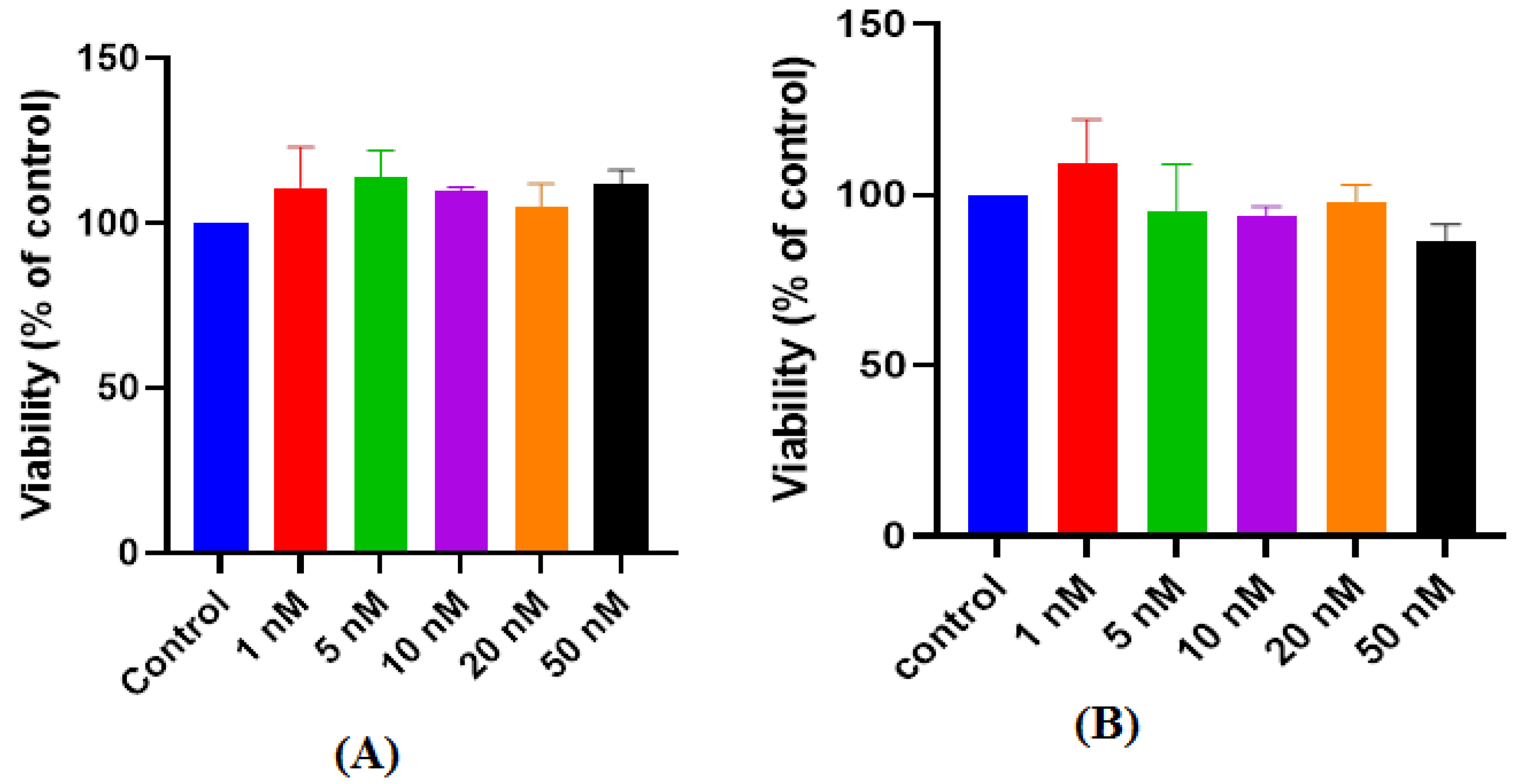
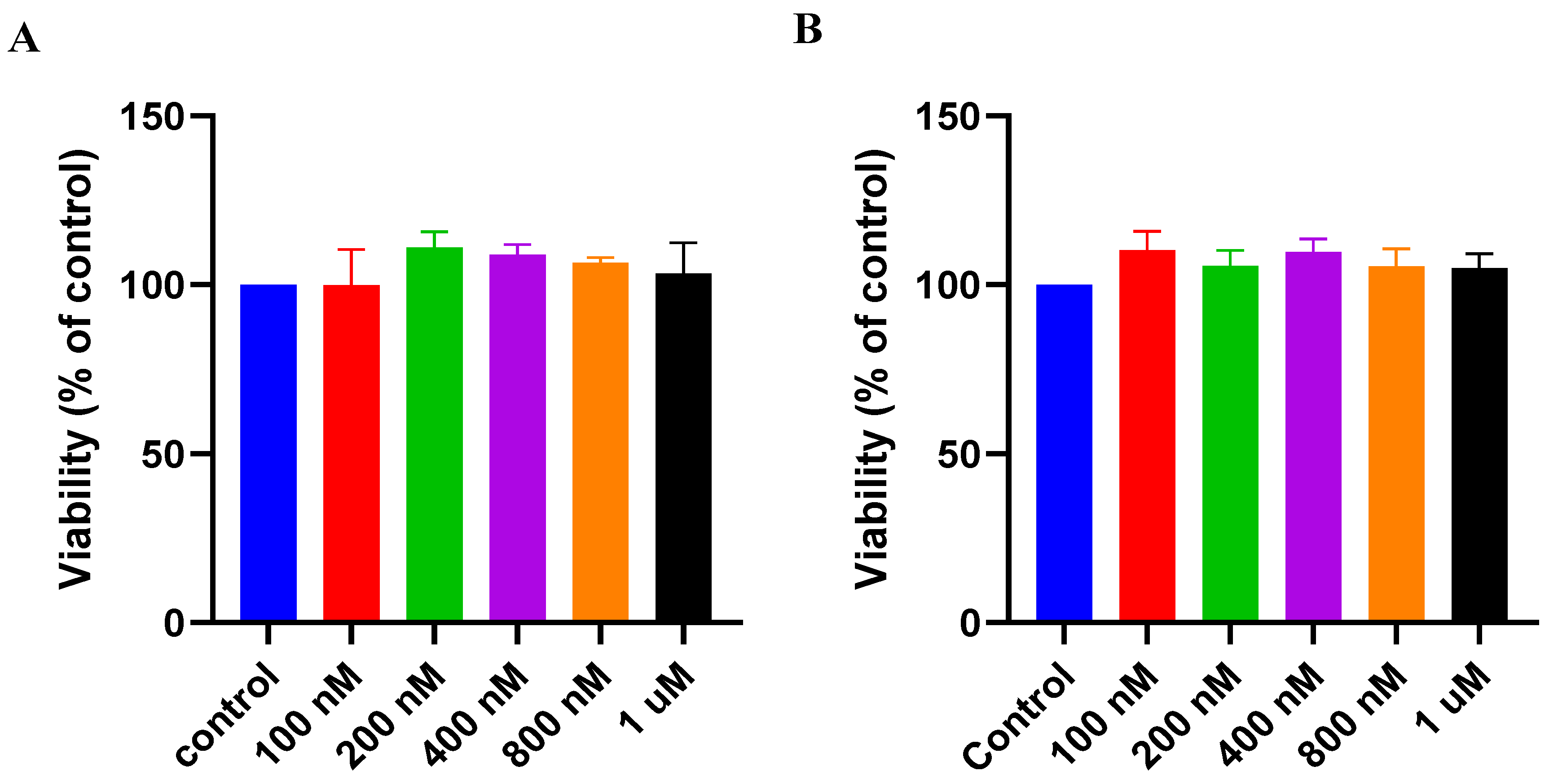
3.3. Cell Viability Studies (MTS Assay)
3.3.1. Dose–Response Studies in Controlled Mediums
Dose–Response of RBX
Dose–Response of PAMAM Dendrimers G4.5
Dose–Response of PAMAM dendrimers G5
Dose–Response of G4.5 Complex 25:1
Dose–Response of RBX G5 Complex 25:1
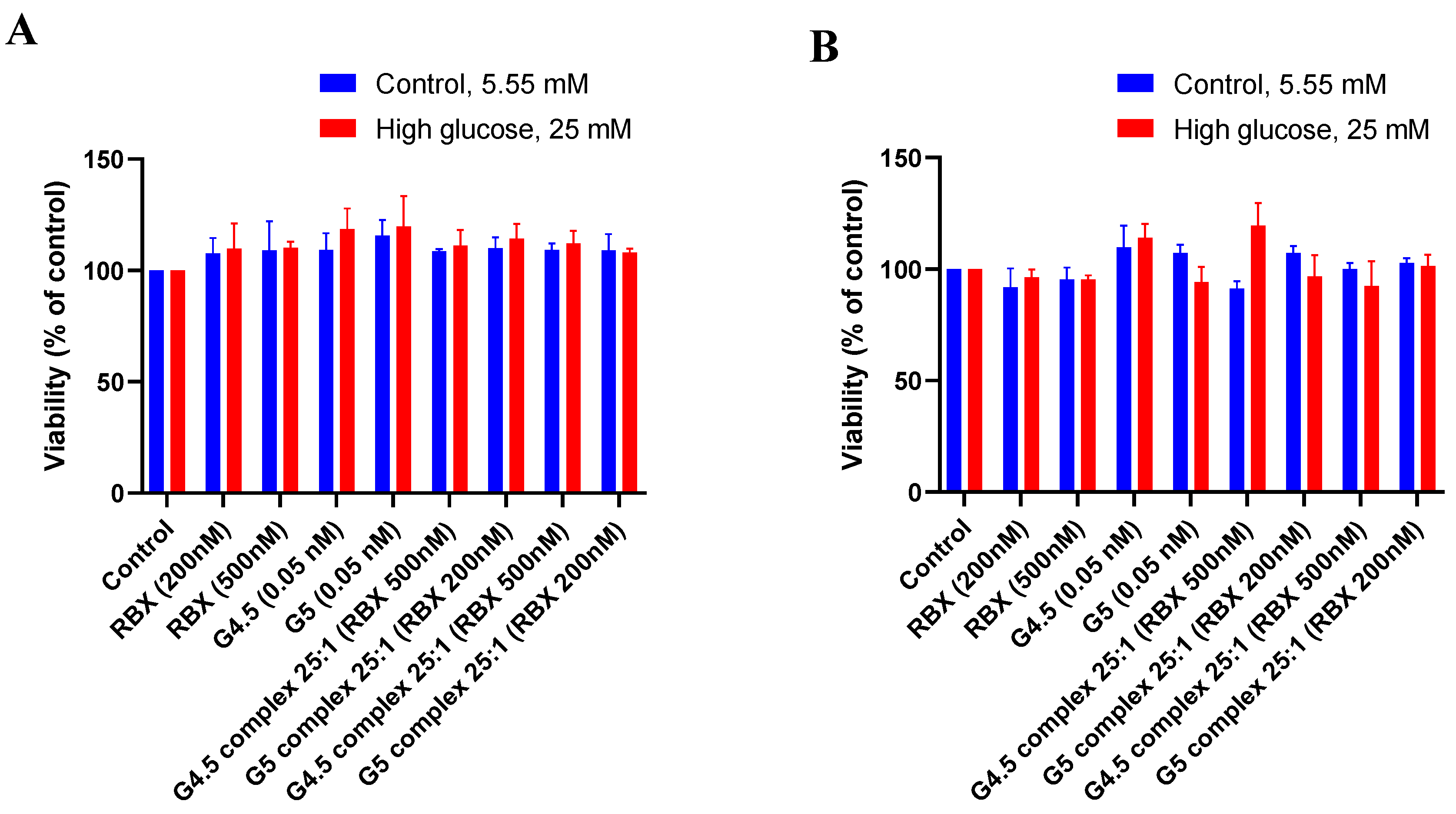
3.3.2. Effect of High Glucose Treatment on the Viability of MIO-M1 Cells
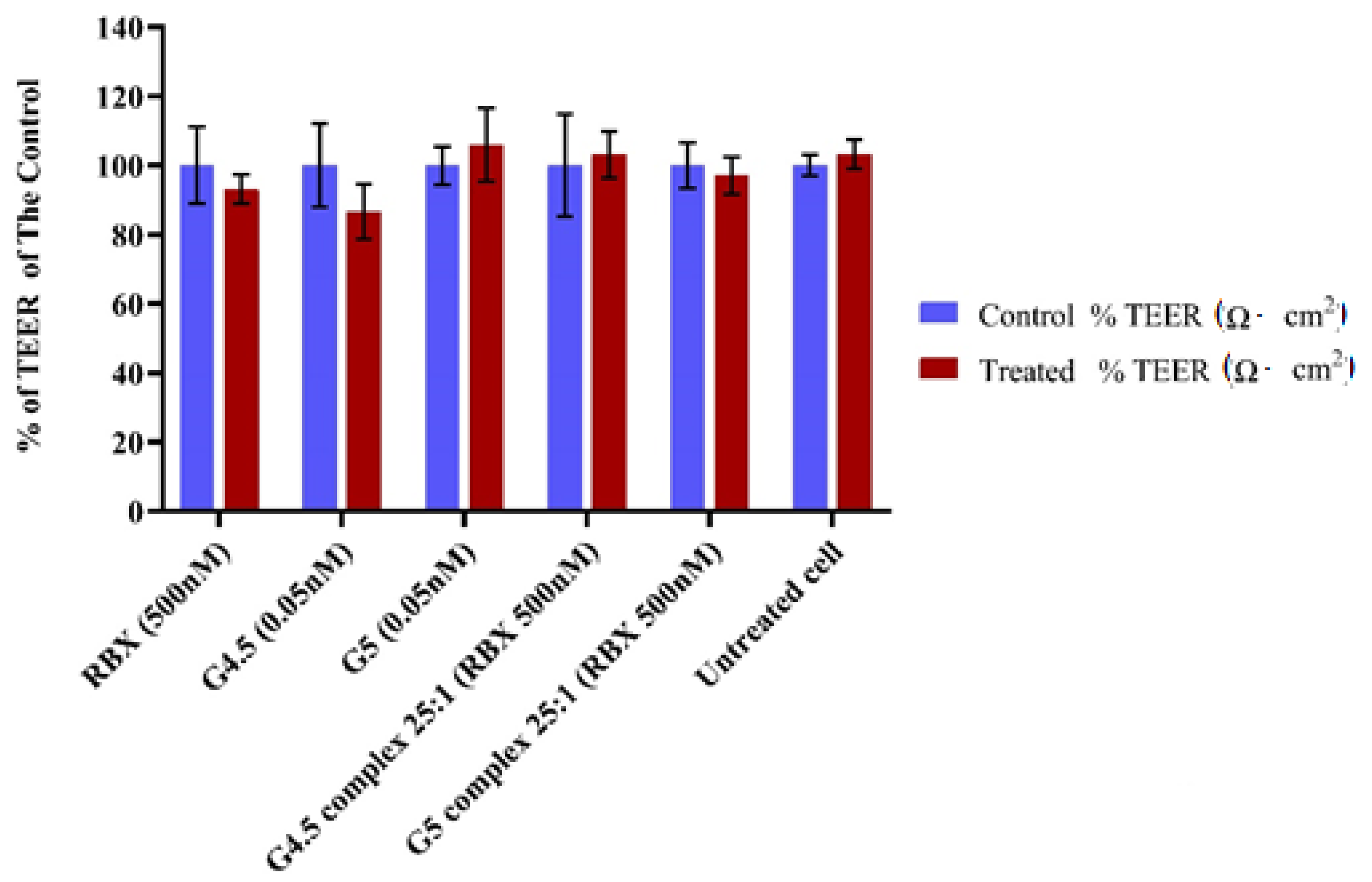
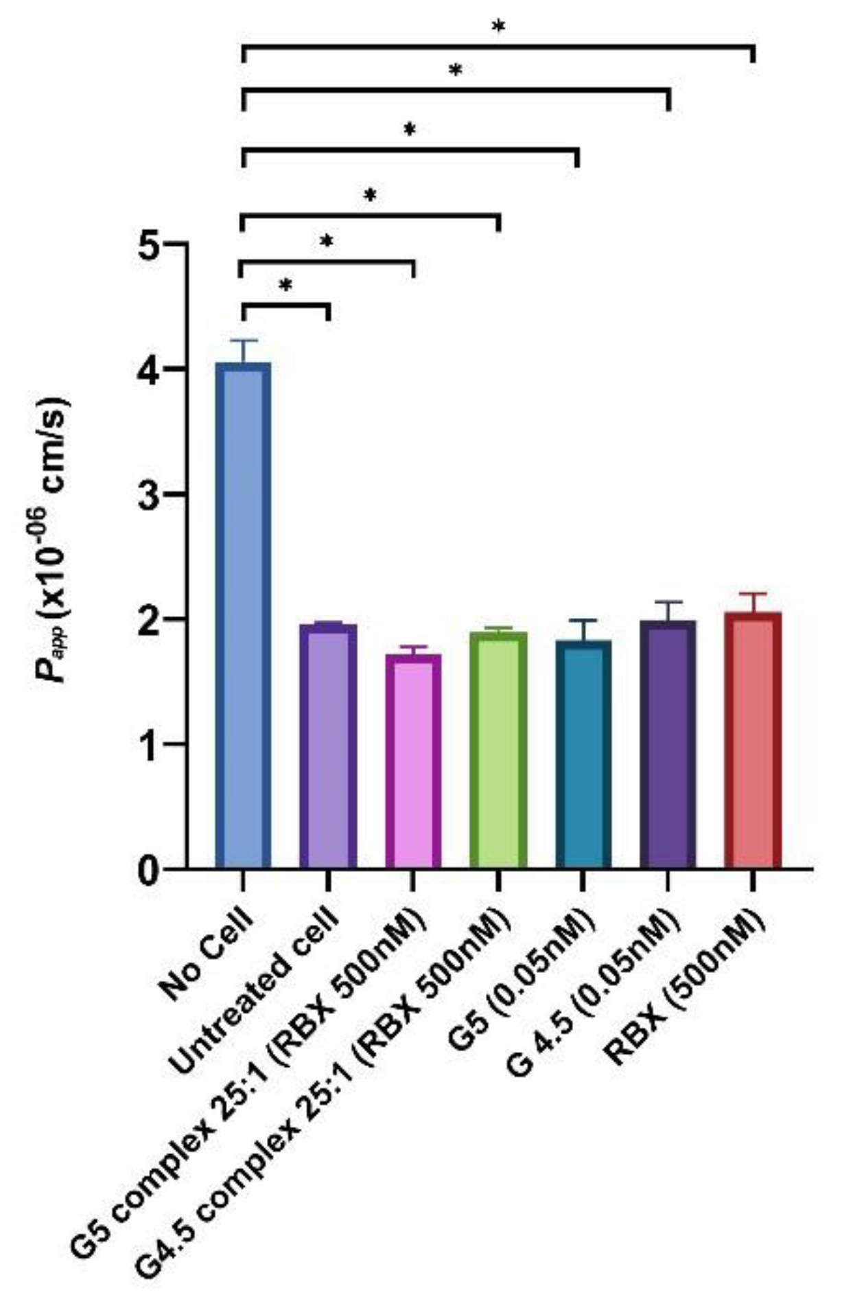
3.3.3. In Vitro Permeability Study
4. Discussion
5. Conclusions
Supplementary Materials
Author Contributions
Funding
Institutional Review Board Statement
Informed Consent Statement
Data Availability Statement
Acknowledgments
Conflicts of Interest
References
- Yavuz, B.; Pehlivan, S.B.; Bolu, B.S.; Sanyal, R.N.; Vural, I.; Unlu, N. Dexamethasone–PAMAM dendrimer conjugates for retinal delivery: Preparation, characterization and in vivo evaluation. J. Pharm. Pharmacol. 2016, 68, 1010–1020. [Google Scholar] [CrossRef]
- Gupta, N.; Mansoor, S.; Sharma, A.; Sapkal, A.; Sheth, J.; Falatoonzadeh, P.; Kuppermann, B.D.; Kenney, M.C. Diabetic retinopathy and VEGF. Open Ophthalmol. J. 2013, 7, 4–10. [Google Scholar] [CrossRef] [PubMed] [Green Version]
- Zhao, Y.; Singh, R.P. The role of anti-vascular endothelial growth factor (anti-VEGF) in the management of proliferative diabetic retinopathy. Drugs Context 2018, 7, E212532. [Google Scholar] [CrossRef] [PubMed]
- Del Amo, E.M.; Rimpelä, A.-K.; Heikkinen, E.; Kari, O.K.; Ramsay, E.; Lajunen, T.; Schmitt, M.; Pelkonen, L.; Bhattacharya, M.; Richradson, D.; et al. Pharmacokinetic aspects of retinal drug delivery. Prog. Retin. Eye Res. 2017, 57, 134–185. [Google Scholar] [CrossRef] [PubMed]
- Yau, J.W.; Rogers, S.L.; Kawasaki, R.; Lamoureux, E.L.; Kowalski, J.W.; Bek, T.; Chen, S.-J.; Dekker, J.M.; Fletcher, A.; Grauslund, J.; et al. Global prevalence and major risk factors of diabetic retinopathy. Diabetes Care 2012, 35, 556–564. [Google Scholar] [CrossRef] [Green Version]
- Xia, P.; Aiello, L.P.; Ishii, H.; Jiang, Z.Y.; Park, D.J.; Robinson, G.S.; Takagi, H.; Newsome, W.P.; Jirousek, M.R.; King, G.L. Characterization of vascular endothelial growth factor’s effect on the activation of protein kinase C, its isoforms, and endothelial cell growth. J. Clin. Investig. 1996, 98, 2018–2026. [Google Scholar] [CrossRef] [Green Version]
- Nonaka, A.; Kiryu, J.; Tsujikawa, A.; Yamashiro, K.; Miyamoto, K.; Nishiwaki, H.; Honda, Y.; Ogura, Y. PKC-β inhibitor (LY333531) attenuates leukocyte entrapment in retinal microcirculation of diabetic rats. Investig. Ophthalmol. Vis. Sci. 2000, 41, 2702–2706. [Google Scholar]
- Way, K.; Katai, N.; King, G. Protein kinase C and the development of diabetic vascular complications. Diab. Med. 2001, 18, 945–959. [Google Scholar] [CrossRef]
- Aldarwesh, A. Oxygen and Glucose Deprivation on Human Müller Cells (MIO-M1) and Human Organotypic Retinal Cultures (HORCs) in Relation to Glaucoma. Ph.D. Thesis, University of East Anglia, Norwich, UK, 2015. [Google Scholar]
- Liu, Y.; Lei, S.; Gao, X.; Mao, X.; Wang, T.; Wong, G.T.; Vanhoutte, P.; Irwin, M.G.; Xia, Z. PKCβ inhibition with ruboxistaurin reduces oxidative stress and attenuates left ventricular hypertrophy and dysfuntion in rats with streptozotocin-induced diabetes. Clin. Sci. 2012, 122, 161–173. [Google Scholar] [CrossRef]
- Edelhauser, H.F.; Rowe-Rendleman, C.L.; Robinson, M.R.; Dawson, D.G.; Chader, G.J.; Grossniklaus, H.E.; Ritetnhouse, K.D.; Wilson, C.G.; Weber, D.E.; Kuppermann, B.D.; et al. Ophthalmic drug delivery systems for the treatment of retinal diseases: Basic research to clinical applications. Investig. Ophthalmol. Vis. Sci. 2010, 51, 5403–5420. [Google Scholar] [CrossRef]
- Jiang, S.; Franco, Y.L.; Zhou, Y.; Chen, J. Nanotechnology in retinal drug delivery. Int. J. Ophthalmol. 2018, 11, 1038–1044. [Google Scholar] [PubMed]
- Patel, A.; Cholkar, K.; Agrahari, V.; Mitra, A.K. Ocular drug delivery systems: An overview. World J. Pharmacol. 2013, 2, 47–64. [Google Scholar] [CrossRef] [PubMed]
- Bessonova, J.; Meyer-Lindenberg, A.; Bäumer, W.; Kietzmann, M. Tissue distribution of dexamethasone in feline ocular structures following single topical application of dexamethasone as an ointment or suspension. Vet. Ophthalmol. 2011, 14, 109–113. [Google Scholar] [CrossRef] [PubMed]
- Gaudana, R.; Ananthula, H.K.; Parenky, A.; Mitra, A.K. Ocular drug delivery. AAPS J. 2010, 12, 348–360. [Google Scholar] [CrossRef]
- Kolhe, P.; Misra, E.; Kannan, R.M.; Kannan, S.; Lieh-Lai, M. Drug complexation, in vitro release and cellular entry of dendrimers and hyperbranched polymers. Int. J. Pharm. 2003, 259, 143–160. [Google Scholar] [CrossRef] [Green Version]
- Ma, P.; Zhang, X.; Ni, L.; Li, J.; Zhang, F.; Wang, Z.; Lian, S.; Sun, K. Targeted delivery of polyamidoamine-paclitaxel conjugate functionalized with anti-human epidermal growth factor receptor 2 trastuzumab. Int. J. Nanomed. 2015, 10, 2173–2190. [Google Scholar] [CrossRef] [Green Version]
- Wang, Y.; Guo, R.; Cao, X.; Shen, M.; Shi, X. Encapsulation of 2-methoxyestradiol within multifunctional poly (amidoamine) dendrimers for targeted cancer therapy. Biomaterials 2011, 32, 3322–3329. [Google Scholar] [CrossRef]
- Dobrovolskaia, M.A.; Patri, A.K.; Simak, J.; Hall, J.B.; Semberova, J.; Lacerda, S.H.D.P.; McNeil, S.E. Nanoparticle size and surface charge determine effects of PAMAM dendrimers on human platelets in vitro. Mol. Pharm. 2012, 9, 382–393. [Google Scholar] [CrossRef] [Green Version]
- Williams, D.B.; Carter, C.B. The transmission electron microscope. In Transmission Electron Microscopy; Springer: Berlin/Heidelberg, Germany, 1996; pp. 3–17. [Google Scholar]
- Tawfik, M.A.; Tadros, M.I.; Mohamed, M.I. Polyamidoamine (PAMAM) dendrimers as potential release modulators and oral bioavailability enhancers of vardenafil hydrochloride. Pharm. Dev. Technol. 2019, 24, 293–302. [Google Scholar] [CrossRef]
- Nabavizadeh, F.; Fanaei, H.; Imani, A.; Vahedian, J.; Amoli, F.A.; Ghorbi, J.; Sohanaki, H.; Mohammadi, S.M.; Golchoobian, R. Evaluation of nanocarrier targeted drug delivery of Capecitabine-PAMAM dendrimer complex in a mice colorectal cancer model. Acta Med. Iran. 2016, 54, 485–493. [Google Scholar]
- Klajnert, B.; Bryszewska, M. Dendrimers: Properties and applications. Acta Biochim. Pol. 2001, 48, 199–208. [Google Scholar] [CrossRef] [Green Version]
- Vandamme, T.F.; Brobeck, L. Poly (amidoamine) dendrimers as ophthalmic vehicles for ocular delivery of pilocarpine nitrate and tropicamide. J. Control. Release 2005, 102, 23–38. [Google Scholar] [CrossRef] [PubMed]
- Shadrack, D.M.; Mubofu, E.B.; Nyandoro, S.S. Synthesis of polyamidoamine dendrimer for encapsulating tetramethylscutellarein for potential bioactivity enhancement. Int. J. Mol. Sci. 2015, 16, 26363–26377. [Google Scholar] [CrossRef]
- Yeo, K.P.; Lowe, S.L.; Lim, M.T.; Voelker, J.R.; Burkey, J.L.; Wise, S.D. Pharmacokinetics of ruboxistaurin are significantly altered by rifampicin-mediated CYP3A4 induction. Br. J. Clin. Pharmacol. 2006, 61, 200–210. [Google Scholar] [CrossRef] [PubMed] [Green Version]
- Limb, G.A.; Salt, T.E.; Munro, P.M.G.; Moss, S.E.; Khaw, P.T. In vitro characterization of a spontaneously immortalized human Muller cell line (MIO-M1). Investig. Ophthalmol. Vis. Sci. 2002, 43, 864–869. [Google Scholar]
- Cory, A.H.; Owen, T.C.; Barltrop, J.A.; Cory, J.G. Use of an aqueous soluble tetrazolium/formazan assay for cell growth assays in culture. Cancer Commun. 1991, 3, 207–212. [Google Scholar] [CrossRef] [PubMed]
- Riss, T.L.; Moravec, R.A.; Niles, A.L. Cytotoxicity testing: Measuring viable cells, dead cells, and detecting mechanism of cell death. Methods Mol. Biol. 2011, 740, 103–114. [Google Scholar]
- Jafari, F.; Aghazadeh, M.; Jafari, S.; Khaki, F.; Kabiri, F. In vitro cytotoxicity comparison of MTA fillapex, AH-26 and apatite root canal sealer at different setting times. Iran. Endod. J. 2017, 12, 162–167. [Google Scholar]
- Churm, R.; Dunseath, G.J.; Prior, S.L.; Thomas, R.L.; Banerjee, S.; Owens, D.R. Development and characterization of an in vitro system of the human retina using cultured cell lines. Clin. Exp. Ophthalmol. 2019, 47, 1055–1062. [Google Scholar] [CrossRef]
- Namba, R.; Kaneko, H.; Suzumura, A.; Shimizu, H.; Kataoka, K.; Takayama, K.; Yamada, K.; Funahashi, Y.; Ito, S.; Nonobe, N.; et al. In vitro epiretinal membrane model and antibody permeability: Relationship with anti-VEGF resistance in diabetic macular edema. Investig. Ophthalmol. Vis. Sci. 2019, 60, 2942–2949. [Google Scholar] [CrossRef] [Green Version]
- Garcia-Ramírez, M.; Villarroel, M.; Corraliza, L.; Hernández, C.; Simó, R. Measuring permeability in human retinal epithelial cells (ARPE-19): Implications for the study of diabetic retinopathy. Methods Mol. Biol. 2011, 763, 179–194. [Google Scholar] [PubMed]
- Hubatsch, I.; Ragnarsson, E.G.E.; Artursson, P. Determination of drug permeability and prediction of drug absorption in Caco-2 monolayers. Nat. Prot. 2007, 2, 2111–2119. [Google Scholar] [CrossRef] [PubMed]
- Palumbo, P.; Picchini, U.; Beck, B.; Van Gelder, J.; Delbar, N.; DeGaetano, A. A general approach to the apparent permeability index. J. Pharmacokin. Pharmacodyn. 2008, 35, 235–248. [Google Scholar] [CrossRef] [PubMed]
- Helms, H.C.; Abbott, N.J.; Burek, M.; Cecchelli, R.; Couraud, P.-O.; Deli, M.A.; Forster, C.; Galla, H.J.; Romero, I.A.; Shusta, E.V.; et al. In vitro models of the blood-brain barrier: An overview of commonly used brain endothelial cell culture models and guidelines for their use. J. Cereb. Blood Flow Metab. 2016, 36, 862–890. [Google Scholar] [CrossRef] [PubMed]
- Dunn, K.C.; Aotaki-Keen, A.E.; Putkey, F.R.; Hjelmeland, L.M. ARPE-19, a human retinal pigment epithelial cell line with differentiated properties. Exp. Eye Res. 1996, 62, 155–170. [Google Scholar] [CrossRef]
- Ma, B.; Wang, J.; Sun, J.; Li, M.; Xu, H.; Sun, G.; Sun, X. Permeability of rhynchophylline across human intestinal cell in vitro. Int. J. Clin. Exp. Pathol. 2014, 7, 1957–1966. [Google Scholar]
- Yuan, S.Y.; Rigor, R.R. Regulation of Endothelial Barrier Function; Morgan & Claypool Life Sciences: San Rafael, CA, USA, 2010. Available online: https://www.ncbi.nlm.nih.gov/books/NBK54124/ (accessed on 20 December 2021).
- Eigenmann, D.E.; Dürig, C.; Jähne, E.A.; Smieško, M.; Culot, M.; Gosselet, F.; Cecchelli, R.; Helms, H.C.C.; Brodin, B.; Wimmer, L.; et al. In vitro blood-brain barrier permeability predictions for GABAA receptor modulating piperine analogs. Eur. J. Pharm. Biopharm. 2016, 103, 118–126. [Google Scholar] [CrossRef] [Green Version]
- Liu, C.; Jiang, K.; Tai, L.; Liu, Y.; Wei, G.; Lu, W.; Pan, W. Facile noninvasive retinal gene delivery enabled by penetratin. ACS Appl. Mater. Interf. 2016, 8, 19256–19267. [Google Scholar] [CrossRef]
- Mittal, N.; Kaur, G. Investigations on polymeric nanoparticles for ocular delivery. Adv. Polym. Technol. 2019, 2019, 1316249. [Google Scholar] [CrossRef]
- Gorantla, S.; Rapalli, V.K.; Waghule, T.; Singh, P.P.; Dubey, S.K.; Saha, R.N.; Singhvi, G. Nanocarriers for ocular drug delivery: Current status and translational opportunity. RSC Adv. 2020, 10, 27835–27855. [Google Scholar] [CrossRef]
- Mudalige, T.; Qu, H.; Van Haute, D.; Ansar, S.M.; Paredes, A.; Ingle, T. Characterization of nanomaterials: Tools and challenges. Nanomater. Food Appl. 2019, 313–353. [Google Scholar] [CrossRef]
- Clayton, K.N.; Salameh, J.W.; Wereley, S.T.; Kinzer-Ursem, T.L. Physical characterization of nanoparticle size and surface modification using particle scattering diffusometry. Biomicrofluidics 2016, 10, 054107. [Google Scholar] [CrossRef] [PubMed] [Green Version]
- Janagam, D.R.; Wu, L.; Lowe, T.L. Nanoparticles for drug delivery to the anterior segment of the eye. Adv. Drug Deliv. Rev. 2017, 122, 31–64. [Google Scholar] [CrossRef] [PubMed]
- Peng, J.; Qi, X.; Chen, Y.; Ma, N.; Zhang, W.; Xing, J.; Zhu, X.; Li, Z.; Wu, Z. Octreotide-conjugated PAMAM for targeted delivery to somatostatin receptors over-expressed tumor cells. J. Drug Target. 2014, 22, 428–438. [Google Scholar] [CrossRef]
- Clogston, J.D.; Patri, A.K. Zeta potential measurement. In Characterization of Nanoparticles Intended for Drug Delivery; Springer: Berlin/Heidelberg, Germany, 2011; pp. 63–70. [Google Scholar]
- Liu, Y.; Ng, Y.; Toh, M.R.; Chiu, G.N.C. Lipid-dendrimer hybrid nanosystem as a novel delivery system for paclitaxel to treat ovarian cancer. J. Control. Release 2015, 220, 438–446. [Google Scholar] [CrossRef] [PubMed]
- Gothwal, A.; Khan, I.; Kumar, P.; Raza, K.; Kaul, A.; Mishra, A.K.; Gupta, U. Bendamustine–PAMAM conjugates for improved apoptosis, efficacy, and in vivo pharmacokinetics: A sustainable delivery tactic. Mol. Pharm. 2017, 15, 2084–2097. [Google Scholar] [CrossRef]
- Yesil-Celiktas, O.; Pala, C.; Cetin-Uyanikgil, E.O.; Sevimli-Gur, C. Synthesis of silica-PAMAM dendrimer nanoparticles as promising carriers in Neuro blastoma cells. Anal. Biochem. 2017, 519, 1–7. [Google Scholar] [CrossRef]
- Kesharwani, P.; Xie, L.; Banerjee, S.; Mao, G.; Padhye, S.; Sarkar, F.H.; Iyer, A.K. Hyaluronic acid-conjugated polyamidoamine dendrimers for targeted delivery of 3, 4-difluorobenzylidene curcumin to CD44 overexpressing pancreatic cancer cells. Colloids Surf. B Biointerfaces 2015, 136, 413–423. [Google Scholar] [CrossRef]
- Prajapati, R.N.; Tekade, R.K.; Gupta, U.; Gajbhiye, V.; Jain, N.K. Dendimer-mediated solubilization, formulation development and in vitro−in vivo assessment of piroxicam. Mol. Pharm. 2009, 6, 940–950. [Google Scholar] [CrossRef]
- Tretiach, M.; Madigan, M.C.; Gillies, M.C. Conditioned medium from mixed retinal pigmented epithelium and Müller cell cultures reduces in vitro permeability of retinal vascular endothelial cells. Br. J. Ophthalmol. 2004, 88, 957–961. [Google Scholar] [CrossRef]


| Formulation | Amount of RBX (0.1% w/v) | Amount of PAMAM Dendrimers G4.5 (10% w/v) | Amount of PAMAM Dendrimers G5 (10% w/v) |
|---|---|---|---|
| G4.5 complexes | |||
| G4.5 complex 1:1 | 50 μL | 25 μL | - |
| G4.5 complex 2.5:1 | 50 μL | 10 μL | - |
| G4.5 complex 5:1 | 50 μL | 5 μL | - |
| G4.5 complex 25:1 | 50 μL | 1 μL | - |
| G5 complexes | |||
| G5 complex 1:1 | 50 μL | - | 28 μL |
| G5 complex 2.5:1 | 50 μL | - | 11.2 μL |
| G5 complex 5:1 | 50 μL | - | 5.6 μL |
| G5 complex 25:1 | 50 μL | - | 1.1 μL |
| RBX-PAMAM Nanoparticles | PS (nm) ± SD | PDI ± SD | ζ-Potential in (mV) ± SD |
|---|---|---|---|
| Empty PAMAM dendrimers G4.5 | 186.0 ± 2.3 | 0.297 ± 0.040 | −44.0 ± 2.0 |
| G4.5 complex 1:1 | 367.0 ± 13.0 | 0.335 ± 0.010 | −16.2 ± 3.1 |
| G4.5 complex 2.5:1 | 416.0 ± 4.3 | 0.337 ± 0.030 | −5.1 ± 10.4 |
| G4.5 complex 5:1 | 289.0 ± 14.9 | 0.361 ± 0.030 | −6.4 ± 1.8 |
| G4.5 complex 25:1 | 301.0 ± 7.1 | 0.304 ± 0.010 | −13.0 ± 1.6 |
| Empty PAMAM dendrimers G5 | 214.0 ± 8.5 | 0.356 ± 0.010 | −0.2 ± 0.0 |
| G5 complex 1:1 | 461.0 ± 6.4 | 0.394 ± 0.010 | 4.3 ± 1.3 |
| G5 complex 2.5:1 | 482.0 ± 9.5 | 0.388 ± 0.020 | 5.5 ± 0.2 |
| G5 complex 5:1 | 669.0 ± 2.0 | 0.587 ± 0.100 | 9.7 ± 4.9 ** |
| G5 complex 25:1 | 307.0 ± 6.9 | 0.380 ± 0.030 | −0.0 ± 0.0 |
Publisher’s Note: MDPI stays neutral with regard to jurisdictional claims in published maps and institutional affiliations. |
© 2022 by the authors. Licensee MDPI, Basel, Switzerland. This article is an open access article distributed under the terms and conditions of the Creative Commons Attribution (CC BY) license (https://creativecommons.org/licenses/by/4.0/).
Share and Cite
Alshammari, R.A.; Aleanizy, F.S.; Aldarwesh, A.; Alqahtani, F.Y.; Mahdi, W.A.; Alquadeib, B.; Alqahtani, Q.H.; Haq, N.; Shakeel, F.; Abdelhady, H.G.; et al. Retinal Delivery of the Protein Kinase C-β Inhibitor Ruboxistaurin Using Non-Invasive Nanoparticles of Polyamidoamine Dendrimers. Pharmaceutics 2022, 14, 1444. https://doi.org/10.3390/pharmaceutics14071444
Alshammari RA, Aleanizy FS, Aldarwesh A, Alqahtani FY, Mahdi WA, Alquadeib B, Alqahtani QH, Haq N, Shakeel F, Abdelhady HG, et al. Retinal Delivery of the Protein Kinase C-β Inhibitor Ruboxistaurin Using Non-Invasive Nanoparticles of Polyamidoamine Dendrimers. Pharmaceutics. 2022; 14(7):1444. https://doi.org/10.3390/pharmaceutics14071444
Chicago/Turabian StyleAlshammari, Rehab A., Fadilah S. Aleanizy, Amal Aldarwesh, Fulwah Y. Alqahtani, Wael A. Mahdi, Bushra Alquadeib, Qamraa H. Alqahtani, Nazrul Haq, Faiyaz Shakeel, Hosam G. Abdelhady, and et al. 2022. "Retinal Delivery of the Protein Kinase C-β Inhibitor Ruboxistaurin Using Non-Invasive Nanoparticles of Polyamidoamine Dendrimers" Pharmaceutics 14, no. 7: 1444. https://doi.org/10.3390/pharmaceutics14071444