Next Article in Journal
Development of Abiraterone Acetate Nanocrystal Tablets to Enhance Oral Bioavailability: Formulation Optimization, Characterization, In Vitro Dissolution and Pharmacokinetic Evaluation
Next Article in Special Issue
Localized Controlled Release of Kynurenic Acid Encapsulated in Synthetic Polymer Reduces Implant—Induced Dermal Fibrosis
Previous Article in Journal
Micro/Nanosystems for Magnetic Targeted Delivery of Bioagents
 
 
Retraction published on 23 January 2024, see Pharmaceutics 2024, 16(2), 157.
Article

RETRACTED: Fluoxetine Ecofriendly Nanoemulsion Enhances Wound Healing in Diabetic Rats: In Vivo Efficacy Assessment

1
Department of Pharmaceutics, Faculty of Pharmacy, King Abdulaziz University, Jeddah 21589, Saudi Arabia
2
Advanced Drug Delivery Research Group, Faculty of Pharmacy, King Abdulaziz University, Jeddah 21589, Saudi Arabia
3
Center of Excellence for Drug Research and Pharmaceutical Industries, King Abdulaziz University, Jeddah 21589, Saudi Arabia
4
Mohamed Saeed Tamer Chair for Pharmaceutical Industries, King Abdulaziz University, Jeddah 21589, Saudi Arabia
5
Department of Drug and Health Sciences, University of Catania, 95125 Catania, Italy
6
Unit of Neuropharmacology and Translational Neurosciences, Oasi Research Institute—IRCCS, 94018 Troina, Italy
7
Department of Natural Products and Alternative Medicine, Faculty of Pharmacy, King Abdulaziz University, Jeddah 21589, Saudi Arabia
8
Preparatory Year Program, Batterjee Medical College, Jeddah 21442, Saudi Arabia
9
Department of Pharmacognosy, Faculty of Pharmacy, Assiut University, Assiut 71526, Egypt
10
Department of Pharmacology and Toxicology, Faculty of Pharmacy, King Abdulaziz University, Jeddah 21589, Saudi Arabia
*
Author to whom correspondence should be addressed.
These authors contributed equally to this work.
Pharmaceutics 2022, 14(6), 1133; https://doi.org/10.3390/pharmaceutics14061133
Submission received: 2 May 2022 / Revised: 20 May 2022 / Accepted: 23 May 2022 / Published: 26 May 2022 / Retracted: 23 January 2024

Abstract

:
Impaired diabetic wound healing is a major concern for health care professionals worldwide, imposing an intense financial burden and reducing the quality of life of patients. A dysregulation of this process can be responsible for the development of intractable ulcers and the formation of excessive scars. Therefore, the identification of novel pharmacological strategies able to promote wound healing and restore the mechanical integrity of injured tissue becomes essential. In the present study, fluoxetine ecofriendly nanoemulsion (FLX-EFNE) was prepared and its potential efficacy in enhancing wound healing was tested in diabetic rats. The Box–Behnken response surface design was used to select the optimized formulation that was prepared by the high-shear homogenization-based technique. A Zetasizer was used for the characterization of the optimized formulation, providing a FLX-EFNE with a globule size of 199 nm. For the in vivo study, a wound was induced by surgical methods, and diabetic rats (streptozotocin-induced) were divided into five groups: untreated control, vehicle-treated, FLX, FLX-EFNE, and positive control receiving a commercially available formula. The treatment continued from the day of wound induction to day 21. Then, the animals were sacrificed and skin tissues were collected at the site of wounding and used for biochemical, histopathological, immunohistochemical, and mRNA expression assessments. In the FLX-EFNE treated group, the rate of wound contraction and signs of healing were significantly higher compared to all other groups. In addition, angiogenesis, proliferation, and collagen deposition were enhanced, while oxidative stress and inflammation decreased. The present data highlight the enhanced wound healing activity of the optimized FLX-EFNE formulation.

1. Introduction

Diabetes mellitus (DM) is the most extensively studied metabolic disorder characterized by persistent increased blood sugar level (>200 mg/dL), damaged β-cells, and reduced insulin sensitivity [1]. Various factors such as obesity, atherosclerosis, and hypertension contribute to the pathogenesis of DM [1,2]. Based on epidemiological study, 9% of the total adult population globally suffers from DM and more than 1.5 million deaths were associated with this pathology, 80% of which were reported in developing countries [3]. Among the different and numerous complications of DM, diabetic wounds and impaired wound healing represent the major challenges for health care professionals [4]. Foot and leg ulcers are commonly reported among diabetic patients, and increased blood glucose levels lead to complications in wound healing [4]. As per the published evidence, between 2.5% and 15% of total health budgets are consumed in the management and treatment of diabetic wound healing. If not appropriately managed, it will be the seventh leading cause of death worldwide by 2030 [5].
Wound healing is a complex and multifactorial process that aims to restore the skin’s structural and physiological integrity in a determined period [6]. If wound healing is not managed and treated timely, it may lead to sepsis, which is indeed fatal [7]. In the case of DM, the evolution can worsen due to impaired immunological functions, abrupt blood supply, and fibrogenesis [5,8]. Based on their healing properties, wounds are classified as acute or chronic, where acute wounds heal up within 12 weeks, while chronic wounds take more than 12 weeks and are associated with abrupt physiological functions [9]. In a healthy individual, wound healing begins with the hemostasis that stops further loss of blood and microbial invasion at the site of injury [9]. Following this stage, the inflammatory phase begins, characterized by the activation of neutrophils (pro-inflammatory cells) and the clearance of debris and pathogens by macrophages. This stage is followed by the proliferative phase, where angiogenesis and matrix construction begin filling the wounded area. The final stage is represented by the remodeling phase, where the increased tensile strength restores the damaged area [9].
Considering the wound healing in DM, impairment in the hemostasis, inflammation, proliferation, and remodeling have been reported, leading to a low quality of life [8]. Indeed, in the case of diabetic wounds, a persistent inflammatory phase with an increased level of pro-inflammatory cytokines such as tumor necrosis factor-alpha (TNF-α), damaged blood supply, reduced angiogenesis, decreased production of growth factors such as transforming growth factor-beta 1 (TGF-β1), reduced fibrosis, tensile strength, and abrupt histopathological changes have been all reported [10,11]. Oxidative stress has been described as a status characterized by an imbalance between pro-oxidants, such as reactive oxygen species (ROS), and antioxidants found in favor of pro-oxidant mediators [12,13]. Inflammation takes place when a tissue injury occurs, with immune cells including neutrophils, macrophages, and mast cells that produce pro-inflammatory mediators, such as interleukin-1 (IL-1), TNF-α, and interferon gamma (IFN-γ), as well as different growth factors, including platelet-derived growth factor subunit B (PDGF-B), epidermal growth factor (EGF), and insulin-like growth factor 1 (IGF-1), representing key mediators of the wound repair process [14,15]. In particular, it has been shown that neutrophils are characterized by an altered cytokine release pattern along with a decreased functionality, with all factors contributing to the susceptibility to wound infection [16,17]. Oxidative stress has been observed in DM, and previously published reports have shown the deleterious effect of both oxidative stress and inflammation in wound healing [11,18].
Currently, the treatment strategies for diabetic wound healing consist of, but are not limited to, wound debridement (streptodornase, maggots, streptokinase, and dextrans), revascularization approaches, and ulcer off-loading [5]. Additionally, hyperbaric oxygen therapy, cold atmospheric pressure plasma, negative-pressure wound therapy (NPWT), low-level laser therapy, growth factor administration (e.g., PDGF-B, platelet-rich plasma, granulocyte colony-stimulating factor (G-CSF), EGF), the use of semisynthetic ester of hyaluronic acid, and matrix metalloproteinase modulators have been evaluated as possible treatments [5,19].
Increased serotonin (5-HT) levels have been reported to promote tissue repair and wound healing [20]. Hence, serotonin, or selective serotonin reuptake inhibitors (SSRIs), have been examined to promote wound healing in experimental models [20]. Fluoxetine (FLX), the first antidepressant developed in the class of the SSRIs, represents one of the main first-line medications used for major depression, and is also known to reduce inflammation [21] and oxidative stress [22]. FLX, administered intraperitoneally, has also been shown to be able to enhance the wound healing process in chronically stressed Wistar rats [23]. Additionally, topical FLX has demonstrated the ability to improve wound healing in diabetic mice [24].
The ecofriendly formulation approach originated from the extension of green chemistry and green engineering principles that involve applying economical processes and designing environmentally friendly products that use minimal hazardous solvents along with safe reactants to reduce both health risks and environmental hazards [25]. Interest in integrating these green principles in pharmaceutical research has been increasing to develop ecofriendly formulations that are safe, biocompatible, and devoid of any organic solvents or surfactants aiming at reducing the adverse risks on both humans and the environment [26]. For our developed ecofriendly nanoemulsion, the surfactant has been replaced by in situ complex between natural oil and oligosaccharide (cyclodextrin) [27].
Wheat germ oil (WGO) is obtained from the flour-milling byproduct of wheat used for topical formulations. WGO possesses significant antioxidant and anti-inflammatory activity, promotes angiogenesis and the production of collagen, and exhibits significant wound healing activity [28].
Based on the above and on the fact that the currently available topic therapy for the management and treatment of diabetic wounds suffers from significant pharmacokinetic limitations such as low solubility, low dermal permeability, reduced spreadability, and minimal therapeutic outcome [29], in the present study, we designed and developed an ecofriendly FLX-based nanoemulsion (FLX-EFNE) incorporated with WGO to achieve optimum pharmacological outcomes and improve the treatment of wound healing in diabetic rats.
The specific aim of the present work was the development, characterization, and investigation of the preclinical efficacy of an optimized FLX-EFNE formulation in an in vivo model of an acute wound. Once the FLX-EFNE was prepared and characterized, it was tested for its ability to speed up the wound healing process compared to the other experimental conditions (skin samples obtained from untreated control, vehicle (VEH)-treated, FLX-treated, and positive control-treated rats).

2. Materials and Methods

2.1. Materials and Reagents

The FLX, WGO, alpha-Cyclodextrin (α-CD), and ELISA kit for hydroxyproline were supplied by R&D Systems, Inc. a Bio-Techne Brand (Minneapolis, MN, USA). Antibodies for TNF-α ((TNFA/1172); ab220210), vascular endothelial growth factor A (VEGF-A) (ab231260), PDGF-B (ab23914), and TGF-β1 ((EPR21143); ab215715) were purchased from Abcam (Cambridge, UK). When not otherwise specified, all other chemicals of the highest commercially analytical grade were supplied by Sigma-Aldrich (St. Louis, MO, USA) or Thermo Fisher Scientific Inc. (Pittsburgh, PA, USA).

2.2. Animals

Male Wistar rats (n = 50; 200–240 g) were provided by the animal facility, King Abdulaziz University (KAU, Jeddah, Saudi Arabia). The study was approved by the Research Ethics Committee, Faculty of Pharmacy, KAU approval number (PH-1443-29). The animals were kept on a 12 h light–dark cycle at a temperature of 22 ± 2 °C [30]. Diabetes in rats was induced using streptozotocin (STZ) (50 mg/kg, injected intraperitoneally) as previously described by Ahmed et al. [31]. Among several molecules used to induce diabetes, STZ has been described as the most preferred to mimic human diabetes in animals. Structural, functional, and biochemical alterations observed in STZ-induced diabetes resemble those observed in humans suffering from this pathology. Based on the above, STZ-induced diabetes represents a clinically relevant model to study the pathogenesis of diabetes as well as associated secondary complications [32,33]. Fasting blood glucose level was measured through Accu-Chek Go (Roche, Mannheim, Germany). Rats with fasting blood glucose level values in the range of 200–300 mg/100 mL (moderate diabetes) were selected for the present research study.

2.3. Experimental Design for Selection of Optimized Formula

An experimental design based on the Box–Behnken response surface design was implemented to achieve an optimized formula. Three independent variables were selected: WGO, α-CD, and homogenization time, in terms of low (−1) and high (+1), as depicted in Table 1.
The Box–Behnken response surface design produced 15 runs with different ratio of selected independent variables (Table 2), following which various FLX-EFNE were manufactured as per the software-produced runs.
The different FLX-EFNE formulations were characterized by globule size for the selection of the optimized FLX-EFNE formula. For this purpose, the data of the dependent factor were statistically analyzed using experimental software via one-way analysis of variance (ANOVA). The software also portrayed the effects of the independent factors on the dependent factor as 3D and contour plots [34,35,36].

2.4. Manufacturing Method

FLX-EFNE formulations were prepared using a high-shear homogenization-based technique [37]. Briefly, FLX (0.5% w/w) and a specified amount of α-CD, WGO, and water (according to the design) was homogenized at 20,000 rpm for 3 min at 25 °C (T25 digital Ultra-Turrax®, IKA Industrie, Staufen, Germany) in glass bottles. The quantities used for the preparation of FLX-EFNE were selected based on the experimental design runs’ composition. Next, deionized water was effused into the prepared mixture. The coarse emulsion of the different ratio of WGO and α-CD was homogenized at 20,000 rpm for various time intervals.

2.5. Characterization of FLX-EFNE Formulations’ Globule Size

The Zetasizer Nano ZSP (Nano ZSP, Malvern, Worcestershire, UK) was used to measure the globule size of the manufactured FLX-EFNE. To achieve homogeneous dispersion, the nanoemulsion samples were diluted at a 1:100 ratio with Milli-Q water. The conditions of measurement were the following: standard laser 4 mW He–Ne, 633 nm, at 25 ± 2 °C with a fixed angle of 90°. The apparatus measured the changes in the intensity of light scattered by each nanoemulsion, which was constantly moving due to Brownian motion. All of the measurements were carried out in triplicate [38].

2.6. Electron Microscope Assessment of Optimized FLX-EFNE Formulation

A transmission electron microscope (TEM) JEOL GEM-1010 (JEOL Ltd., Akishima, Tokyo, Japan) was used for the investigation of the optimized FLX-EFNE formulation at the Regional Center for Mycology and Biotechnology, Al-Azhar University, Cairo, Egypt. The optimized FLX-EFNE was stained with phosphotungistic acid on a carbon-coated grid. The stained sample was then visualized after drying.

2.7. Preparation of Tissue Homogenates

The diabetic rats were anesthetized by an intraperitoneal injection of ketamine (100 mg/kg)/xylazine (10 mg/kg). After shaving, the shaved skin was sterilized using povidone–iodine and a 1 cm circular excision was made [8]. All images of the wounds were taken from a fixed distance using a regular digital camera (Nikon DS-Fi1, Tokyo, Japan). The obtained pictures were considered 1X (actual size). After careful excision, the wounded area was cleaned and dried using a sterile solution and pads. In order to reduce the animals’ pain, lidocaine hydrochloride (2%) containing 1:80,000 epinephrine (4.4 mg/kg) was injected subcutaneously close to the wound area [8]. The animals were divided into five different groups (n = 10 each): Group 1—untreated control diabetic rats with no treatment after wound induction; Group 2—negative control diabetic rats, receiving topical daily plain VEH consisting of EFNE on the wound area; Group 3—diabetic rats, receiving raw-FLX loaded topical gel; Group 4—diabetic rats, receiving FLX-EFNE loaded topical gel; Group 5—positive control diabetic rats, receiving 0.5 g of Mebo® ointment (Gulf Pharmaceutical Industries Julphar, Ras Al Khaimah, United Arab Emirates) on the wound area. The ointment contained β-sitosterol, baicalin, and berberine as active ingredients in a base of beeswax and sesame oil. Petroleum jelly-based dressing gauze was applied on the wound and changed daily. The wounded areas were measured and photographed on day 0, 3, 10, 14, and 21. The treatment regimen continued for 21 days. The animals were sacrificed at day 10 (four animals/group) and day 21 (six animals/group), and skin specimens from the wounded areas were collected. Specimens collected at day 10 were kept in formalin for histological examination. Specimens collected at day 21 were divided into two parts; the first part was kept in formalin for histological and immunohistochemical examination, while the second part was flash frozen in liquid nitrogen for biochemical and mRNA analyses.

2.8. Measurement of Wound Contraction

Wound contraction percentage (%) was calculated through the following formula, taking into consideration the changes in wound diameter [8]:
Wound   contraction   % = Wound   diameter   at   Day   0 Wound   diameter   at   Day   10   or   Day   21 Wound   diameter   at   Day   0 × 100

2.9. Tissue Homogenate Preparation

To prepare tissue homogenates, the wounded areas were precisely removed, rinsed with normal saline, and dried using filter papers. A section of each sample of dermal tissue was preserved in formalin for histopathological analysis. The rest of each part was used for the biochemical analysis [39]. Phosphate-buffered saline (PBS) (ice-cold; 50 mM and pH 7.4) was mixed with the tissue and homogenized at 4 °C for the estimation of biochemical parameters [40].

2.10. Biochemical Estimation

The skin tissues were homogenized in a 10-fold volume of ice-cooled PBS (50 mM potassium phosphate, pH 7.4). The homogenates were then centrifuged for 15 min at 10,000× g and 4 °C. This step was followed by the collection of the supernatant, which was used for oxidative status analysis. Commercially available kits, all supplied by Biodiagnostic (Giza, Egypt), were utilized to assess the content of malondialdehyde (MDA, Cat.# MD2529) and reduced glutathione (GSH, Cat.# GR2511), as well as the activity of superoxide dismutase (SOD, Cat.# SD2521) and glutathione peroxidase (GPX, Cat.# GP2524) enzymes.

2.11. Quantitative Real-Time PCR (qRT-PCR)

For the assessment of mRNA expression of collagen, type I, alpha 1 (Col1A1), and angiopoietin 1 (Ang-1), qRT-PCR was performed. Each section of tissue was removed and immediately preserved in liquid nitrogen. The sections were then thawed, homogenized, and subjected to RNA extraction using a NucleoSpin® nucleic acid extraction kit (Macherey-Nagel GmbH and Co. KG, Duerin, Germany). Both RNA concentration and purity (A260/A280 ratio) were confirmed spectrophotometrically (Dual-Wavelength Beckman, Spectrophotometer, Brea, CA, USA). Reverse transcription was carried out using a High-Capacity cDNA Reverse Transcription Kit (Applied Biosystems, Foster City, CA, USA). PCR amplification reactions were performed by employing a Taq PCR Master Mix Kit (Qiagen, Valencia, CA, USA) coupled to the primers indicated in Table 3.
The relative quantitation of the selected target genes was performed according to the calculation of the delta–delta Ct (ΔΔCt) method [41]. The GAPDH gene was selected as the internal control, while qRT-PCR runs with primers in the absence of samples were considered as negative controls.

2.12. Histological Analysis

The wound tissues collected at days 10 and 21 were kept in neutral formalin (10%) for 24 h. This step was followed by dehydration in serial concentrations of ethanol, cleared in xylene, and inserted in paraffin [30]. Two sets of 5 μm thick sections of paraffinized tissue were cut using the microtome, rewaxed, and rehydrated. The sections were then stained with hematoxylin and eosin (H&E) or Masson’s trichrome (MT) [42]. Histological examinations were performed by a pathologist with no knowledge of the treatment groups. Based on the degree of re-epithelization, proliferation, deposition of collagens, presence of inflammatory cells, and phases of wound healing, a score ranging from “−” to “+++” was assigned.

2.13. Immunohistochemical Analysis of TNF-α, VEGF-A, PDGF-B, and TGF-β1

The tissue sections were de-paraffinized, rehydrated, and boiled in 0.1 M citrate buffer (pH 6.0) for 10 min. The sections were then kept in 5% bovine serum albumin (BSA) in Tris-buffered saline (TBS) for 2 h followed by incubation overnight at 4 °C with the selected primary antibodies (anti-TNF-α, anti-VEGF-A, anti-PDGF-B, or anti-TGF-β1) all at 1 μg/mL. A Cell and Tissue Staining Rabbit Kit, containing the blocking solution, secondary antibody i.e., Goat Anti-Rabbit, and 3,3′-diaminobenzidine (DAB) (R&D Systems, Minneapolis, MN, USA), was used for the following step as previously described [8]. A drop of the mounting solution was applied to each slide that was left to dry and then photographed using a Nikon SMZ 1000 light microscope with a digital camera (Nikon DS-Fi1, Tokyo, Japan). The images were evaluated through Image J software (1.52a, National Institutes of Health NIH, Rockville, MD, USA). A minimum of three sections per rat were considered.

2.14. Statistical Analysis

Data were expressed as mean ± standard deviation (SD). ANOVA followed by a post hoc test was used for multiple comparisons. GraphPad Prism®, version 8.0 (GraphPad, La Jolla, CA, USA) was the software selected to carry out all statistical analyses. Only two-tailed p-values < 0.05 were considered statistically significant.

3. Results

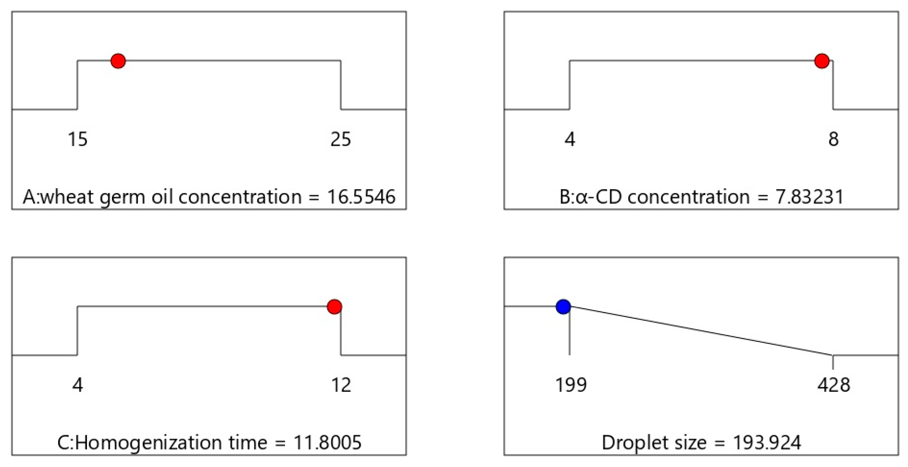
3.1. Selection of an Optimized FLX-EFNE Implementing Box–Behnken Design

As previously described, the Box–Behnken response surface design produced FLX-EFNE formulations with different ratio of the selected independent variables (Table 2). The different FLX-EFNE formulations were characterized by globule size for the selection of the optimized FLX-EFNE formula. The effects of the independent variables on the manufactured FLX-EFNE formulations are clearly depicted in Table 2 in terms of globule size (nm). The globule size of the emulsions varied as a consequence of the changes regarding the ratio of independent variables, giving a range between 199 and 428 nm. After the analysis of the response data, the experimental software exhibited a quadratic model for globule size, which carried an adjusted R2 value of 0.9921 and a predicted R2 value of 0.9583, showing a close affinity, as the difference between the adjusted and predicted R2 was < 0.2. Figure 1 also demonstrated the linear relationship between practical and predicted data produced by experimental design software with the commensurate residual plots.
Moreover, as shown in Figure 2, the response 3D plots and contour plots exhibited the effects of independent variables over the globule size of FLX-EFNE.

3.2. Optimization of FLX-EFNE

Following the software-assisted statistical analysis, an optimized FLX-EFNE with the desired globule size was obtained. The optimized FLX-EFNE formulation was characterized by 15% WGO, 8% α-CD, and 12 min of homogenization (Figure 3), with an optimized globule size of 199 nm and desirability of 1.00.

3.3. TEM Assessment of Optimized FLX-EFNE Formulation

TEM assessment of the optimized FLX-EFNE formulation (Figure 4) shows the spherical structures of the formulation with lower average size when compared with the size obtained by the light-scattering technique using the Zetasizer; this could be attributed to the drying of the formula during the handling of the sample for TEM assessment.

3.4. Assessment of Wound Healing

Figure 5A shows the wound closure in diabetic rats belonging to the five experimental groups at day 0, 3, 10, 14, and 21.
Figure 5B shows that at day 10, the treatment of diabetic rats with FLX was already able to significantly enhance the wound contraction, expressed as % of day 0, compared to both the untreated control (p < 0.001) and VEH-treated (p < 0.01) groups. The best results in terms of wound contraction were observed when diabetic rats were treated with the FLX-EFNE optimized formulation (p < 0.0001 vs. all other groups). These results were confirmed when measuring the wound contraction of animals subjected to the different experimental conditions after 21 days of treatment. In fact, as clearly depicted, at day 21, the wounds of diabetic rats receiving the local daily application of FLX-EFNE optimized preparation showed almost complete healing (>95%) (p < 0.0001 vs. all the other groups), while the wound contraction % in the animals belonging to the FLX group showed values amounting to ~78% (p < 0.0001 vs. untreated control; p < 0.05 vs. VEH-treated). It is worthy of note that the animals treated with the FLX-EFNE preparation exhibited enhanced wound-healing activity even when compared to the positive control treatment (~82%), represented by Mebo® ointment.

3.5. Histopathological Analysis

To fully understand the wound healing potential of FLX-EFNE, H&E and MT staining were performed on days 10 and 21. In the histopathological analysis, delayed healing, reduced re-epithelization, and the remodeling of epidermal tissue were observed in the samples obtained from diabetic rats belonging to the untreated or VEH-treated groups (Figure 6).
The presence of inflammatory cells such as neutrophils, along with inflamed and necrotic granulation tissue, were also observed in these samples. In the case of samples obtained from diabetic rats receiving the local daily application of FLX, mild re-epithelization and the presence of necrotic and edematous tissue on both day 10 and day 21 were observed. By comparing day 10 and day 21, an overall improvement was observed at day 21, even though the presence of inflammatory cells and vacuolations was still evident. A different outcome was obtained in the case of the animals belonging to the group receiving the topical application of FLX-EFNE in which the highest rate of healing was detected. Despite the significant improvement in the wound healing process and the absence of necrotic tissue compared to all other experimental conditions, a number of inflammatory cells were still observable. The analysis of MT-stained sections in untreated and VEH-treated groups on day 10 and 21 showed a slight presence of collages (blue color). The daily topical application of FLX also only showed mild collagen, whereas the treatment with FLX-EFNE enhanced the wound healing process even when compared to the positive control treatment, showing the formation of organized collagen fibers. The histological features, scored and reported in Table 4, strengthen the enhanced wound healing activity of the FLX-EFNE optimized formulation compared to FLX.

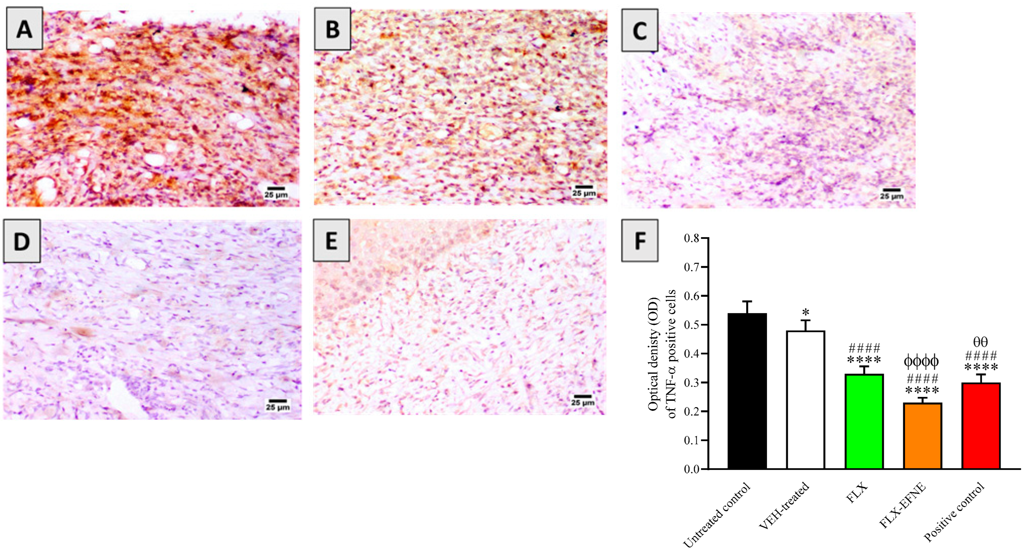
3.6. Effect of FLX or FLX-EFNE on Expression of TNF-α

In order to elucidate the mechanism of wound healing after the topical application of FLX or FLX-EFNE, the immunohistochemical analysis of the TNF-α pro-inflammatory cytokine was performed. The data reported in Figure 7 show that the topical treatment of wounded skin tissues of diabetic rats with FLX or FLX-EFNE significantly inhibited (p < 0.0001) TNF-α protein content by ~39 and ~58%, respectively, compared to the tissues obtained from animals belonging to the untreated control group (Figure 7).
Of note, the topical application of FLX-EFNE gave a decrease in TNF-α protein expression that was significantly lower compared to both FLX (p < 0.0001) and positive control (p < 0.01) groups.

3.7. Effect of FLX or FLX-EFNE on Oxidative Status

We then evaluated the ability of FLX or FLX-EFNE to modulate oxidative stress, which exerts a key role in the pathophysiology of wound healing [43]. The data reported in Table 5 indicate that wound tissues from diabetic rats treated with FLX or FLX-EFNE showed significantly lower content of MDA, amounting to 5.81 ± 0.60 and 4.35 ± 0.44, respectively, which was significantly lower than that observed in tissues from rats belonging to the untreated group (7.81 ± 0.84).
It is worth pointing out that the topical application of FLX-EFNE gave a decrease in MDA that was significantly lower than FLX or positive control. When analyzing the samples obtained from rats treated with FLX alone, a significant enhancement of GSH (4.30 ± 0.44), SOD (8.43 ± 0.78), and GPx (68.74 ± 5.95) was observed compared to the values measured in tissues obtained from untreated (control) rats. The ability of the optimized formula (FLX-EFNE) to decrease the levels of MDA, one of the final products of polyunsaturated fatty acid peroxidation in the cells [44], was paralleled by an enhancement of the antioxidant system; in fact, FLX-EFNE treatment significantly enhanced the levels of GSH as well as the activity of the antioxidant enzyme GPx compared to all other experimental conditions, including FLX alone and positive control treatments. In the case of SOD activity, FLX, FLX-EFNE, and the positive control were all able to lead to a very similar and significant enhancement.

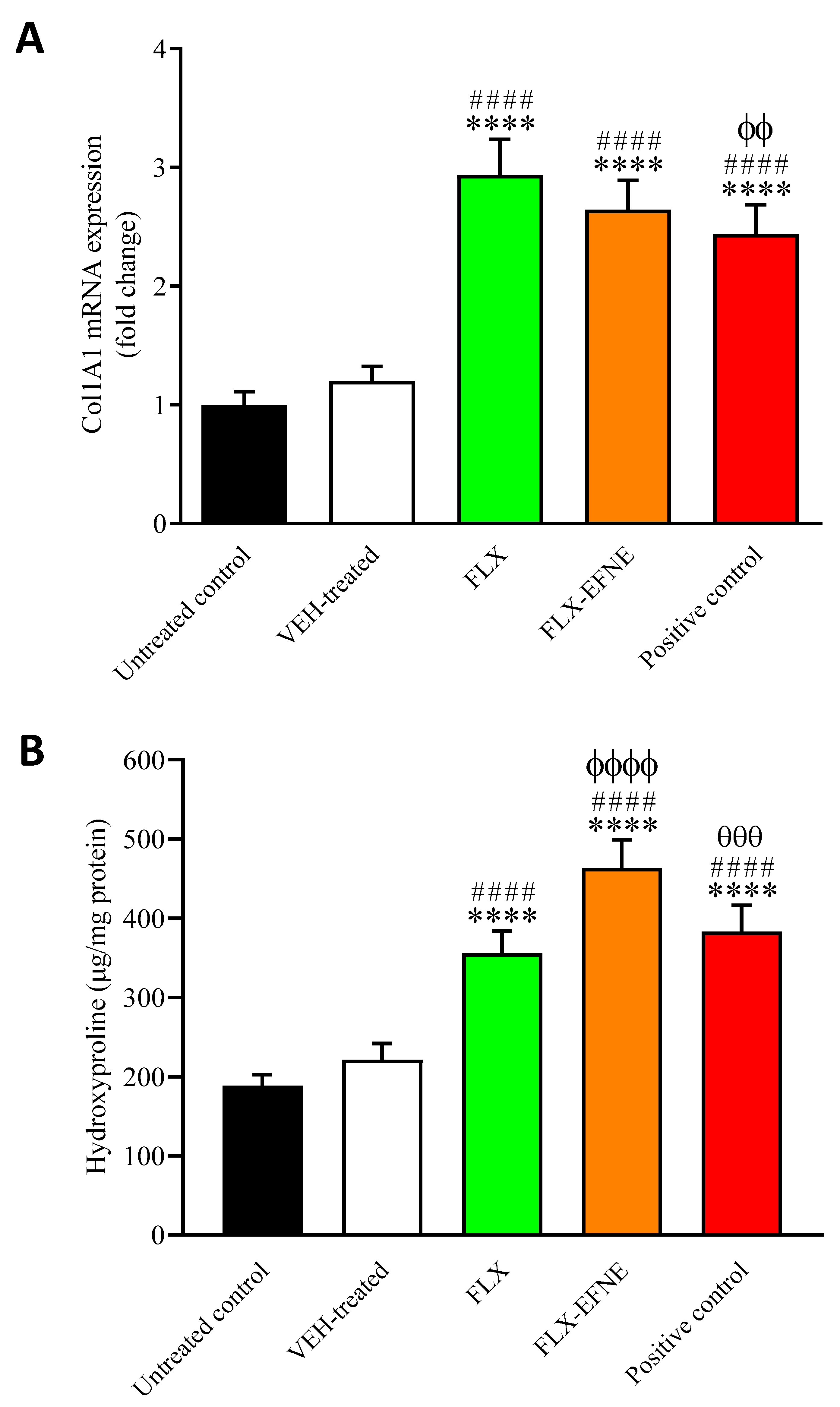
3.8. Effect of FLX or FLX-EFNE on Markers of Collagen Deposition

The daily topical application of the free drug (FLX) as well as of the optimized formulation (FLX-EFNE) significantly increased the mRNA level of Col1A1 compared to both the untreated control and VEH-treated animals (p < 0.0001) (Figure 8A).
Of note, both treatments gave an enhancement comparable to that observed in the case of animals treated with the positive control. A similar result was obtained when measuring the hydroxyproline content; in fact, the hydroxyproline levels were significantly increased due to the daily topical application of FLX or FLX-EFNE compared with the untreated control or VEH-treated animals (p < 0.0001) (Figure 8B). However, it is worth mentioning that, differently from FLX, the treatment with the FLX-EFNE optimized formulation gave an effect significantly higher than that of the positive control (p < 0.001).

3.9. Effect of FLX and FLX-EFNE on Expression of PDGF-B

The immunohistochemical analysis of PDGF-B protein in the samples obtained from diabetic rats belonging to the different groups was performed. The daily topical application of FLX was able to significantly increase the levels of PDGF-B compared to both untreated control (p < 0.0001) and VEH-treated (p < 0.001) groups (Figure 9).
Despite the significant enhancement compared to the untreated rats, FLX alone was not able to give a PDGF-B content comparable to the positive control represented by the Mebo® ointment. Once again, the maximum effect was obtained in the case of FLX-EFNE treatment, giving an increase in PDGF-B levels comparable to that observed for the positive control and significantly higher than that observed in all of the remaining experimental conditions (p < 0.0001 vs. all).

3.10. Effect of FLX and FLX-EFNE on Expression of TGF-β1 Proteins

We then evaluated the impact of FLX-EFNE on the synthesis of TGF-β1, which exerts a key role in promoting wound healing. Figure 10 shows the results obtained by performing the immunohistochemical analysis of TGF-β1.
As observed for the PDGF-B protein, the treatment with FLX significantly increased the levels of TGF-β1 compared to both untreated control (p < 0.0001) and VEH-treated (p < 0.001) groups (Figure 10). The highest levels of TGF-β1 were measured in tissues obtained from diabetic rats treated with the FLX-EFNE optimized formulation, giving an enhancement in TGF-β1 production even higher than that observed for positive control treatment (p < 0.0001 vs. untreated control, VEH-treated, or FLX; p < 0.05 vs. positive control).

3.11. Effect of FLX and FLX-EFNE on Expression of Ang-1

The enhancement of Ang-1 has been related to the improvement of the wound healing process, especially in diabetic conditions [45]. The results depicted in Figure 11 clearly show that the daily application of FLX or FLX-EFNE was able to significantly increase the mRNA levels of Ang-1 compared to both untreated control and VEH-treated animals (p < 0.0001).
Despite the significant enhancement compared to the untreated rats, FLX alone was not able to give an enhancement of Ang-1 expression comparable to that of positive controls. The maximal induction of gene expression was observed in the case of FLX-EFNE treatment, providing an increase significantly higher than that measured in the case of FLX (p < 0.001) and comparable to the positive control.

3.12. Effect of FLX and FLX-EFNE on Expression of VEGF-A

The last set of results regard the effect of the different experimental conditions on the expression of VEGF-A. Figure 12 shows the enhancement of VEGF-A levels due to the daily topical application of FLX (p < 0.0001 vs. untreated control or VEH-treated).
As expected, based on the results obtained by measuring the other markers, the highest enhancement of VEGF-A content in the wounded skin of diabetic rats was observed for the topical application of FLX-EFNE (p < 0.0001 vs. untreated control, VEH, and FLX).

4. Discussion

The dysregulation of the wound-healing process in diabetic subjects has been related to clinically relevant complications such as intractable ulcers and/or the pathological formation of scars [46,47]. Therefore, the identification of a novel and effective pharmacological strategy that can positively modulate the wound healing process, as well as rescuing the mechanical integrity of injured tissue, is of high importance [48]. In this context, a pH/glucose dual-responsive metformin release hydrogel dressing with adhesion and self-healing via dual-dynamic bonding for athletic diabetic foot wound healing has been recently described by Liang et al. [49]. In a different study, the efficacy of a new paste formulation as an alternative therapeutic agent for traumatic ulcers was presented [50]. In the study, conducted using 40 adult male rabbits, the new formulation was effective in resolving inflammation during specific phases of mucosal healing and improved the wound healing process at different stages. Additionally, it gave a better healing response in terms of reduced wound contraction.
In the present study, the wound healing properties of a novel FLX ecofriendly emulsion (FLX-EFNE) were investigated in diabetic rats in order to assess its ability to enhance the wound healing process, exerting preclinical efficacy in our in vivo model of STZ-induced diabetes.
An experimental design-based Box–Behnken response surface design was implemented in order to obtain an optimized formula (FLX-EFNE) with the desired characteristics; three independent variables (WGO, α-CD, and homogenization time) and 15 runs characterized by different ratio were produced (Table 2). Following the software-assisted statistical analysis, an optimized FLX-EFNE with the desired globule size was obtained. The optimized formulation was characterized by a minimum quantity of oil (15% WGO), maximum quantity of α-CD (8%), and 12 min of homogenization, with an optimized globule size of 199 nm and desirability of 1.00. Of note, an increase in the concentration of oil corresponded to an increase in the globule size of the emulsion (Figure 2), indicating a positive effect played by WGO. On the other hand, both α-CD quantity and homogenization time showed a negative effect on globule size, as demonstrated by the decrease of emulsion globule size due to the increase of these two factors. As described above, it is clear that the increased oil concentration enhanced the entrapment of the drug, while the maximum homogenization time broke the globules as a consequence of the high shear, producing an emulsion of minimum globule size.
After its characterization, the preclinical efficacy of the FLX-EFNE optimized formulation was tested, exhibiting, most of the time, wound healing properties equal to or better than the free drug (FLX) and/or our positive control (represented by Mebo® ointment). It is important to recall that the wound healing process consists of three different phases, identified as hemostasis and inflammation, proliferation, and remodeling [51]. As clearly illustrated in Figure 5, the daily topical application of FLX-EFNE for a total of 21 days on the induced wound strongly enhanced the wound healing process, leading to a total recovery, which was not observed in the case of FLX or positive control treatments, demonstrating an enhancement of the pharmacological activities of the optimized formula. The potential of FLX-EFNE to enhance the wound healing process was further evaluated (histologically), confirming the ability of this optimized formulation to decrease fibroblast proliferation and infiltration of inflammatory cells, as well as increasing re-epithelization and collagen deposition, therefore speeding up the recovery process (Figure 6 and Table 4).
Diabetes is a metabolic disorder characterized by high serum glucose levels or hyperglycemia responsible for, among other things, altered immune response, marked inflammation, and production of ROS and related oxidative stress [41,52]. Inflammatory mediators such as pro-inflammatory cytokines have been reported to be over-produced in injured tissues [53]; in particular, IL-1β, IL-6, and TNF-α have been shown to cause a chemotactic response in leukocytes, leading to the exacerbation of the inflammation phase [53]. In this regard, an optimized formulation such as FLX-EFNE is expected to be effective in suppressing the production and release of these pro-inflammatory mediators. In the present study, TNF-α was quantified on day 21 in the skin tissues of diabetic rats under the different experimental conditions. The levels of this pro-inflammatory cytokine were significantly reduced by FLX, FLX-EFNE, and positive control compared to the untreated diabetic rats (Figure 7). Of note, the topical application of FLX-EFNE gave a decrease in TNF-α protein expression that was significantly higher than both FLX (p < 0.0001) and positive control (p < 0.01) groups, underlining the potentiated anti-inflammatory activity of the optimized formulation.
The term “oxidative stress” refers to the imbalance between the level of pro-oxidants, such as ROS, and their elimination [54,55]. Chronic oxidative stress has been implicated in the development of non-healing wounds [56]. MDA represents a well-known index of oxidative damage [57,58], exerting a key role in the pathophysiology of wound healing [43]. It has also been demonstrated that wounded tissues are characterized by elevated oxidative stress, presenting high levels of MDA paralleled by decreased levels of antioxidants [56,59,60]. Second-generation antidepressants, such as FLX, are also known to exert an antioxidant activity both in vitro and in vivo [22,61]. Based on the evidence described above, we investigated the tissue levels of MDA, GSH, SOD, and GPx in diabetic rats subjected to our different experimental conditions. In the best scenario, the optimized formula should be able to reduce oxidative stress and enhance the wound healing process. Both FLX and FLX-EFNE were able to significantly reduce the levels of MDA and enhance the antioxidant status in comparison to untreated diabetic rats (Table 5). However, it is worth mentioning that FLX-EFNE treatment reduced the MDA tissue levels and increased GSH and GPx even more significantly than in FLX or positive control treatment. We have recently observed that FLX is able to rescue GPx levels in the central nervous system (CNS), but this is the first demonstration that this drug exerts the same effect outside the CNS in an experimental model of wound healing, with an increased efficacy of the FLX-EFNE formulation compared to FLX alone.
Remodeling represents the final stage of the wound healing process in which the formation of new epithelial cells and scar tissue takes place [62,63], with an optimum balance between degradation and synthesis processes finally leading to wound closure with minimal scarring. In this scenario, a key role is played by both collagen and hydroxyproline [64,65,66]. Our findings demonstrated the upregulation of Col1A1 gene expression due to both FLX and FLX-EFNE treatments compared to untreated diabetic rats (Figure 8A). As previously mentioned, a similar result was obtained when measuring the hydroxyproline content (Figure 8B), with FLX-EFNE giving the most marked enhancement. These findings are in strong agreement with the enhanced wound closure observed in diabetic rats subjected to the topical daily application of FLX-EFNE for 21 days (Figure 2).
We further explored the effects of the free drug (FLX) or the optimized formulation (FLX-EFNE) on the protein expression of PDGF-B, TGF-β1, and VEGF-A, and on the gene expression of Ang-1; all factors were found to play an important role in the tissue recovery linked to the wound healing process [67,68,69,70,71,72,73]. In particular, VEGF and PDGF-B regulate angiogenesis during physiological conditions [72,73], and reduced levels of these factors may result in a minimal supply of blood and nutrients, causing an impairment of the wound healing process [72]. TGF-β1 is an anti-inflammatory cytokine [74,75,76] playing an important role in all stages of wound healing, including angiogenesis and wound closure [68,77]. During the inflammatory phase, fibroblasts migrate to the wound site, and various growth factors such as TGF-β1 and Ang-1 are released from keratinocytes [71]. Ang-1 and TGF-β are also needed, as they regulate the production of collagen and ensure the availability of hydroxyproline [78]. FLX is known to induce TGF-β1 synthesis and release in vitro in glial cells [79] as well as in humans [80], but no studies have been conducted in experimental models of wound healing yet. Interestingly, we found that the daily topical application of FLX was able to significantly increase the levels of PDGF-B, TGF-β1, and VEGF-A compared to untreated diabetic rats (Figure 9, Figure 10 and Figure 12). As expected, once again, the strongest effect was observed in the case of FLX-EFNE treatment, giving an increase that was significantly higher than FLX and comparable to the positive control. Of note, in the case of TGF-β1, the inducing activity of FLX-EFNE was even higher than that of the positive control treatment. In line with the above-mentioned results, the daily application of FLX was able to significantly increase the mRNA levels of Ang-1 compared to untreated diabetic rats (Figure 11). As observed in most of the cases, the strongest effect was observed for FLX-EFNE treatment.

5. Conclusions

The present study shows the development of a new, ecofriendly nanoemulsion formulation (FLX-EFNE) and its therapeutic potential on delayed wound healing in diabetic rats. The new formulation, developed and optimized using Box–Behnken response surface design, showed enhanced wound healing properties following 21 days of daily topical application compared to the free drug (FLX) and, in most cases, to the commercially available ointment Mebo® representing the positive control. The potentiated activity of FLX-EFNE in inducing wound closure, and therefore to enhance the wound healing process, is attributable to its ability to decrease fibroblast proliferation and the infiltration of inflammatory cells, and increase re-epithelization. The daily topical application of FLX-EFNE was also characterized by anti-inflammatory (decreased TNF-α levels), antioxidant (decreased MDA and increased GSH, GPx, and SOD), and collagen-enhancing (increased Col1A1 gene expression and hydroxyproline levels) activities. The FLX-EFNE formulation was also found to significantly increase the protein expression of PDGF-B, TGF-β1, and VEGF-A, as well as the gene expression of Ang-1, with all factors playing an important role in the tissue recovery linked to the wound healing process. Overall, the present findings underline the therapeutic potential of the new FLX-EFNE formulation that is believed to represent an innovative pharmacological tool able to enhance the wound healing process in pathological conditions such as diabetes.

Author Contributions

Conceptualization, N.A.A. and G.C.; methodology, O.A.A.A., U.A.F., S.M. and A.B.A.-N.; software, G.C., B.G.E. and S.R.M.I.; validation, A.P., F.C. and A.B.A.-N.; formal analysis, A.P. and F.C.; investigation, A.P., U.A.F. and F.C.; resources, O.A.A.A., U.A.F. and S.M.; data curation, G.A.M. and S.R.M.I.; writing—original draft preparation, G.C., G.A.M. and S.R.M.I.; writing—review and editing, G.C., A.P., O.A.A.A., A.B.A.-N. and F.C.; visualization, B.G.E.; supervision, N.A.A. and G.C.; project administration, N.A.A.; funding acquisition, N.A.A. All authors have read and agreed to the published version of the manuscript.

Funding

This research was funded by the Deputyship for Research and Innovation, Ministry of Education in Saudi Arabia, through the project number IFPNC-001-166-2020 and King Abdulaziz University, DSR, Jeddah, Saudi Arabia.

Institutional Review Board Statement

The study was conducted according to the guidelines of the Declaration of Helsinki, and approved by the Committee of Research Ethics of Faculty of Pharmacy, KAU (Reference # PH-1443-29).

Data Availability Statement

The data presented in this study are available in the article.

Acknowledgments

The authors extend their appreciation to the Deputyship for Research and Innovation, Ministry of Education, Saudi Arabia and King Abdulaziz University, DSR, Jeddah, Saudi Arabia.

Conflicts of Interest

The authors declare no conflict of interest.

References

  1. Wagner, R.; Heni, M.; Tabák, A.G.; Machann, J.; Schick, F.; Randrianarisoa, E.; de Angelis, M.H.; Birkenfeld, A.L.; Stefan, N.; Peter, A. Pathophysiology-based subphenotyping of individuals at elevated risk for type 2 diabetes. Nat. Med. 2021, 27, 49–57. [Google Scholar] [CrossRef] [PubMed]
  2. Ramachandran, A. Know the signs and symptoms of diabetes. Indian J. Med. Res. 2014, 140, 579. [Google Scholar]
  3. Lachin, J.M.; Nathan, D.M. Understanding metabolic memory: The prolonged influence of glycemia during the diabetes control and complications trial (dcct) on future risks of complications during the study of the epidemiology of diabetes interventions and complications (edic). Diabetes Care 2021, 44, 2216–2224. [Google Scholar] [CrossRef] [PubMed]
  4. Patel, S.; Srivastava, S.; Singh, M.R.; Singh, D. Mechanistic insight into diabetic wounds: Pathogenesis, molecular targets and treatment strategies to pace wound healing. Biomed. Pharmacother. 2019, 112, 108615. [Google Scholar] [CrossRef]
  5. Wan, R.; Weissman, J.P.; Grundman, K.; Lang, L.; Grybowski, D.J.; Galiano, R.D. Diabetic wound healing: The impact of diabetes on myofibroblast activity and its potential therapeutic treatments. Wound Repair Regen. 2021, 29, 573–581. [Google Scholar] [CrossRef] [PubMed]
  6. Md, S.; Alhakamy, N.A.; Aldawsari, H.M.; Kotta, S.; Ahmad, J.; Akhter, S.; Shoaib Alam, M.; Khan, M.A.; Awan, Z.; Sivakumar, P.M. Improved analgesic and anti-inflammatory effect of diclofenac sodium by topical nanoemulgel: Formulation development—In vitro and in vivo studies. J. Chem. 2020, 2020, 4071818. [Google Scholar] [CrossRef]
  7. Iqubal, M.K.; Saleem, S.; Iqubal, A.; Chaudhuri, A.; Pottoo, F.H.; Ali, J.; Baboota, S. Natural, synthetic and their combinatorial nanocarriers based drug delivery system in the treatment paradigm for wound healing via dermal targeting. Curr. Pharm. Des. 2020, 26, 4551–4568. [Google Scholar] [CrossRef]
  8. Eid, B.G.; Alhakamy, N.A.; Fahmy, U.A.; Ahmed, O.A.; Md, S.; Abdel-Naim, A.B.; Caruso, G.; Caraci, F. Melittin and diclofenac synergistically promote wound healing in a pathway involving tgf-β1. Pharmacol. Res. 2022, 175, 105993. [Google Scholar] [CrossRef]
  9. Farahani, M.; Shafiee, A. Wound healing: From passive to smart dressings. Adv. Healthc. Mater. 2021, 10, 2100477. [Google Scholar] [CrossRef]
  10. Rezvanian, M.; Ng, S.-F.; Alavi, T.; Ahmad, W. In-vivo evaluation of alginate-pectin hydrogel film loaded with simvastatin for diabetic wound healing in streptozotocin-induced diabetic rats. Int. J. Biol. Macromol. 2021, 171, 308–319. [Google Scholar] [CrossRef]
  11. Li, J.; Chou, H.; Li, L.; Li, H.; Cui, Z. Wound healing activity of neferine in experimental diabetic rats through the inhibition of inflammatory cytokines and nrf-2 pathway. Artif. Cells Nanomed. Biotechnol. 2020, 48, 96–106. [Google Scholar] [CrossRef] [PubMed]
  12. Caruso, G.; Spampinato, S.F.; Cardaci, V.; Caraci, F.; Sortino, M.A.; Merlo, S. Β-amyloid and oxidative stress: Perspectives in drug development. Curr. Pharm. Des. 2019, 25, 4771–4781. [Google Scholar] [CrossRef] [PubMed]
  13. Di Pietro, V.; Yakoub, K.M.; Caruso, G.; Lazzarino, G.; Signoretti, S.; Barbey, A.K.; Tavazzi, B.; Lazzarino, G.; Belli, A.; Amorini, A.M. Antioxidant therapies in traumatic brain injury. Antioxidants 2020, 9, 260. [Google Scholar] [CrossRef] [PubMed]
  14. Chhabra, S.; Chhabra, N.; Kaur, A.; Gupta, N. Wound healing concepts in clinical practice of omfs. J. Maxillofac. Oral Surg. 2017, 16, 403–423. [Google Scholar] [CrossRef]
  15. Caruso, G.; Godos, J.; Privitera, A.; Lanza, G.; Castellano, S.; Chillemi, A.; Bruni, O.; Ferri, R.; Caraci, F.; Grosso, G. Phenolic acids and prevention of cognitive decline: Polyphenols with a neuroprotective role in cognitive disorders and Alzheimer’s disease. Nutrients 2022, 14, 819. [Google Scholar] [CrossRef]
  16. Pradhan, L.; Nabzdyk, C.; Andersen, N.D.; LoGerfo, F.W.; Veves, A. Inflammation and neuropeptides: The connection in diabetic wound healing. Expert Rev. Mol. Med. 2009, 11, e2. [Google Scholar] [CrossRef]
  17. Perez-Favila, A.; Martinez-Fierro, M.L.; Rodriguez-Lazalde, J.G.; Cid-Baez, M.A.; Zamudio-Osuna, M.J.; Martinez-Blanco, M.D.R.; Mollinedo-Montaño, F.E.; Rodriguez-Sanchez, I.P.; Castañeda-Miranda, R.; Garza-Veloz, I. Current therapeutic strategies in diabetic foot ulcers. Medicina 2019, 55, 714. [Google Scholar] [CrossRef]
  18. Alhakamy, N.A.; Caruso, G.; Eid, B.G.; Fahmy, U.A.; Ahmed, O.A.A.; Abdel-Naim, A.B.; Alamoudi, A.J.; Alghamdi, S.A.; Al Sadoun, H.; Eldakhakhny, B.M.; et al. Ceftriaxone and melittin synergistically promote wound healing in diabetic rats. Pharmaceutics 2021, 13, 1622. [Google Scholar] [CrossRef]
  19. Mirhaj, M.; Labbaf, S.; Tavakoli, M.; Seifalian, A.M. Emerging treatment strategies in wound care. Int. Wound J. 2022. [Google Scholar] [CrossRef]
  20. Malinin, A.; Oshrine, B.; Serebruany, V. Treatment with selective serotonin reuptake inhibitors for enhancing wound healing. Med. Hypotheses 2004, 63, 103–109. [Google Scholar] [CrossRef]
  21. García-García, M.L.; Tovilla-Zárate, C.A.; Villar-Soto, M.; Juárez-Rojop, I.E.; González-Castro, T.B.; Genis-Mendoza, A.D.; Ramos-Méndez, M.; López-Nárvaez, M.L.; Saucedo-Osti, A.S.; Ruiz-Quiñones, J.A.; et al. Fluoxetine modulates the pro-inflammatory process of il-6, il-1β and tnf-α levels in individuals with depression: A systematic review and meta-analysis. Psychiatry Res. 2022, 307, 114317. [Google Scholar] [CrossRef] [PubMed]
  22. Caruso, G.; Grasso, M.; Fidilio, A.; Torrisi, S.A.; Musso, N.; Geraci, F.; Tropea, M.R.; Privitera, A.; Tascedda, F.; Puzzo, D.; et al. Antioxidant activity of fluoxetine and vortioxetine in a non-transgenic animal model of Alzheimer’s disease. Front. Pharmacol. 2021, 12, 809541. [Google Scholar] [CrossRef] [PubMed]
  23. Farahani, R.M.; Sadr, K.; Rad, J.S.; Mesgari, M. Fluoxetine enhances cutaneous wound healing in chronically stressed wistar rats. Adv. Skin Wound Care 2007, 20, 157–165. [Google Scholar] [CrossRef] [PubMed]
  24. Nguyen, C.M.; Tartar, D.M.; Bagood, M.D.; So, M.; Nguyen, A.V.; Gallegos, A.; Fregoso, D.; Serrano, J.; Nguyen, D.; Degovics, D.; et al. Topical fluoxetine as a novel therapeutic that improves wound healing in diabetic mice. Diabetes 2019, 68, 1499–1507. [Google Scholar] [CrossRef]
  25. Jiménez-González, C.; Poechlauer, P.; Broxterman, Q.B.; Yang, B.-S.; Am Ende, D.; Baird, J.; Bertsch, C.; Hannah, R.E.; Dell’Orco, P.; Noorman, H. Key green engineering research areas for sustainable manufacturing: A perspective from pharmaceutical and fine chemicals manufacturers. Org. Process Res. Dev. 2011, 15, 900–911. [Google Scholar] [CrossRef]
  26. Carbone, C.; Musumeci, T.; Lauro, M.R.; Puglisi, G. Eco-friendly aqueous core surface-modified nanocapsules. Colloids Surf. B Biointerfaces 2015, 125, 190–196. [Google Scholar] [CrossRef]
  27. Badr-Eldin, S.M.; Labib, G.S.; Aburahma, M.H. Eco-friendly tadalafil surfactant-free dry emulsion tablets (sfdets) stabilized by in situ self-assembled aggregates of natural oil and native cyclodextrins. AAPS Pharm. Sci. Tech. 2019, 20, 255. [Google Scholar] [CrossRef]
  28. Zakaria, R.; Faraj, J. Evaluation of the wheat germ oil topical formulations for wound healing activity in rats. Pak. J. Biol. Sci. 2021, 24, 706–715. [Google Scholar] [CrossRef]
  29. Hamdan, S.; Pastar, I.; Drakulich, S.; Dikici, E.; Tomic-Canic, M.; Deo, S.; Daunert, S. Nanotechnology-driven therapeutic interventions in wound healing: Potential uses and applications. ACS Cent. Sci. 2017, 3, 163–175. [Google Scholar] [CrossRef]
  30. Labib, R.M.; Ayoub, I.M.; Michel, H.E.; Mehanny, M.; Kamil, V.; Hany, M.; Magdy, M.; Moataz, A.; Maged, B.; Mohamed, A. Appraisal on the wound healing potential of Melaleuca alternifolia and Rosmarinus officinalis L. Essential oil-loaded chitosan topical preparations. PLoS ONE 2019, 14, e0219561. [Google Scholar] [CrossRef]
  31. Ahmed, O.A.; Afouna, M.I.; El-Say, K.M.; Abdel-Naim, A.B.; Khedr, A.; Banjar, Z.M. Optimization of self-nanoemulsifying systems for the enhancement of in vivo hypoglycemic efficacy of glimepiride transdermal patches. Expert Opin. Drug Deliv. 2014, 11, 1005–1013. [Google Scholar] [CrossRef] [PubMed]
  32. Goyal, S.N.; Reddy, N.M.; Patil, K.R.; Nakhate, K.T.; Ojha, S.; Patil, C.R.; Agrawal, Y.O. Challenges and issues with streptozotocin-induced diabetes—A clinically relevant animal model to understand the diabetes pathogenesis and evaluate therapeutics. Chem. Biol. Interact. 2016, 244, 49–63. [Google Scholar] [CrossRef] [PubMed]
  33. Eleazu, C.O.; Eleazu, K.C.; Chukwuma, S.; Essien, U.N. Review of the mechanism of cell death resulting from streptozotocin challenge in experimental animals, its practical use and potential risk to humans. J. Diabetes Metab. Disord. 2013, 12, 60. [Google Scholar] [CrossRef] [PubMed]
  34. Alhakamy, N.A.; Ahmed, O.A.; Fahmy, U.A.; Md, S. Development and in vitro evaluation of 2-methoxyestradiol loaded polymeric micelles for enhancing anticancer activities in prostate cancer. Polymers 2021, 13, 884. [Google Scholar] [CrossRef]
  35. Alhakamy, N.A.; Ahmed, O.A.; Fahmy, U.A.; Md, S. Apamin-conjugated alendronate sodium nanocomplex for management of pancreatic cancer. Pharmaceuticals 2021, 14, 729. [Google Scholar] [CrossRef]
  36. Md, S.; Alhakamy, N.A.; Aldawsari, H.M.; Husain, M.; Kotta, S.; Abdullah, S.; Fahmy, U.A.; Alfaleh, M.A.; Asfour, H.Z. Formulation design, statistical optimization, and in vitro evaluation of a naringenin nanoemulsion to enhance apoptotic activity in A549 lung cancer cells. Pharmaceuticals 2020, 13, 152. [Google Scholar] [CrossRef]
  37. Puglia, C.; Offerta, A.; Rizza, L.; Zingale, G.; Bonina, F.; Ronsisvalle, S. Optimization of curcumin loaded lipid nanoparticles formulated using high shear homogenization (hsh) and ultrasonication (us) methods. J. Nanosci. Nanotechnol. 2013, 13, 6888–6893. [Google Scholar] [CrossRef]
  38. Iqubal, M.K.; Iqubal, A.; Imtiyaz, K.; Rizvi, M.M.A.; Gupta, M.M.; Ali, J.; Baboota, S. Combinatorial lipid-nanosystem for dermal delivery of 5-fluorouracil and resveratrol against skin cancer: Delineation of improved dermatokinetics and epidermal drug deposition enhancement analysis. Eur. J. Pharm. Biopharm. 2021, 163, 223–239. [Google Scholar] [CrossRef]
  39. Iqubal, A.; Sharma, S.; Sharma, K.; Bhavsar, A.; Hussain, I.; Iqubal, M.K.; Kumar, R. Intranasally administered pitavastatin ameliorates pentylenetetrazol-induced neuroinflammation, oxidative stress and cognitive dysfunction. Life Sci. 2018, 211, 172–181. [Google Scholar] [CrossRef]
  40. Iqubal, A.; Syed, M.A.; Haque, M.M.; Najmi, A.K.; Ali, J.; Haque, S.E. Effect of nerolidol on cyclophosphamide-induced bone marrow and hematologic toxicity in swiss albino mice. Exp. Hematol. 2020, 82, 24–32. [Google Scholar] [CrossRef]
  41. Fresta, C.G.; Fidilio, A.; Caruso, G.; Caraci, F.; Giblin, F.J.; Leggio, G.M.; Salomone, S.; Drago, F.; Bucolo, C. A new human blood-retinal barrier model based on endothelial cells, pericytes, and astrocytes. Int. J. Mol. Sci. 2020, 21, 1636. [Google Scholar] [CrossRef] [PubMed]
  42. Iqubal, A.; Syed, M.A.; Ali, J.; Najmi, A.K.; Haque, M.M.; Haque, S.E. Nerolidol protects the liver against cyclophosphamide-induced hepatic inflammation, apoptosis, and fibrosis via modulation of nrf2, nf-κb p65, and caspase-3 signaling molecules in swiss albino mice. BioFactors 2020, 46, 963–973. [Google Scholar] [CrossRef] [PubMed]
  43. Cano Sanchez, M.; Lancel, S.; Boulanger, E.; Neviere, R. Targeting oxidative stress and mitochondrial dysfunction in the treatment of impaired wound healing: A systematic review. Antioxidants 2018, 7, 98. [Google Scholar] [CrossRef] [PubMed]
  44. Caruso, G.; Fresta, C.G.; Grasso, M.; Santangelo, R.; Lazzarino, G.; Lunte, S.M.; Caraci, F. Inflammation as the common biological link between depression and cardiovascular diseases: Can carnosine exert a protective role? Curr. Med. Chem. 2020, 27, 1782–1800. [Google Scholar] [CrossRef]
  45. Balaji, S.; Han, N.; Moles, C.; Shaaban, A.F.; Bollyky, P.L.; Crombleholme, T.M.; Keswani, S.G. Angiopoietin-1 improves endothelial progenitor cell-dependent neovascularization in diabetic wounds. Surgery 2015, 158, 846–856. [Google Scholar] [CrossRef]
  46. Sharp, A.; Clark, J. Diabetes and its effects on wound healing. Nurs. Stand. 2011, 25, 41–47. [Google Scholar] [CrossRef]
  47. Han, G.; Ceilley, R. Chronic wound healing: A review of current management and treatments. Adv. Ther. 2017, 34, 599–610. [Google Scholar] [CrossRef] [PubMed]
  48. Rosique, R.G.; Rosique, M.J.; Farina Junior, J.A. Curbing inflammation in skin wound healing: A review. Int. J. Inflam. 2015, 2015, 316235. [Google Scholar] [CrossRef]
  49. Liang, Y.; Li, M.; Yang, Y.; Qiao, L.; Xu, H.; Guo, B. Ph/glucose dual responsive metformin release hydrogel dressings with adhesion and self-healing via dual-dynamic bonding for athletic diabetic foot wound healing. ACS Nano 2022, 16, 3194–3207. [Google Scholar] [CrossRef]
  50. Abid, W.K.; Naser, A.I. The efficacy of a new paste formulation as an alternative therapeutic agent for traumatic ulcers. J. Taibah. Univ. Med. Sci. 2021, 16, 724–732. [Google Scholar] [CrossRef]
  51. Velnar, T.; Bailey, T.; Smrkolj, V. The wound healing process: An overview of the cellular and molecular mechanisms. J. Int. Med. Res. 2009, 37, 1528–1542. [Google Scholar] [CrossRef] [PubMed]
  52. Vijayakumar, V.; Samal, S.K.; Mohanty, S.; Nayak, S.K. Recent advancements in biopolymer and metal nanoparticle-based materials in diabetic wound healing management. Int. J. Biol. Macromol. 2019, 122, 137–148. [Google Scholar] [CrossRef] [PubMed]
  53. Kany, S.; Vollrath, J.T.; Relja, B. Cytokines in inflammatory disease. Int. J. Mol. Sci. 2019, 20, 6008. [Google Scholar] [CrossRef] [PubMed]
  54. Iqubal, M.K.; Chaudhuri, A.; Iqubal, A.; Saleem, S.; Gupta, M.M.; Ahuja, A.; Ali, J.; Baboota, S. Targeted delivery of natural bioactives and lipid-nanocargos against signaling pathways involved in skin cancer. Curr. Med. Chem. 2021, 28, 8003–8035. [Google Scholar] [CrossRef]
  55. Caruso, G.; Torrisi, S.A.; Mogavero, M.P.; Currenti, W.; Castellano, S.; Godos, J.; Ferri, R.; Galvano, F.; Leggio, G.M.; Grosso, G.; et al. Polyphenols and neuroprotection: Therapeutic implications for cognitive decline. Pharmacol. Ther. 2021, 232, 108013. [Google Scholar] [CrossRef]
  56. Bilgen, F.; Ural, A.; Kurutas, E.B.; Bekerecioglu, M. The effect of oxidative stress and raftlin levels on wound healing. Int. Wound J. 2019, 16, 1178–1184. [Google Scholar] [CrossRef]
  57. Lazzarino, G.; Listorti, I.; Muzii, L.; Amorini, A.M.; Longo, S.; Di Stasio, E.; Caruso, G.; D’Urso, S.; Puglia, I.; Pisani, G.; et al. Low-molecular weight compounds in human seminal plasma as potential biomarkers of male infertility. Hum. Reprod. 2018, 33, 1817–1828. [Google Scholar] [CrossRef]
  58. Lazzarino, G.; Listorti, I.; Bilotta, G.; Capozzolo, T.; Amorini, A.M.; Longo, S.; Caruso, G.; Lazzarino, G.; Tavazzi, B.; Bilotta, P. Water- and fat-soluble antioxidants in human seminal plasma and serum of fertile males. Antioxidants 2019, 8, 96. [Google Scholar] [CrossRef]
  59. Abood, W.N.; Al-Henhena, N.A.; Najim Abood, A.; Al-Obaidi, M.M.; Ismail, S.; Abdulla, M.A.; Al Batran, R. Wound-healing potential of the fruit extract of phaleria macrocarpa. Bosn. J. Basic Med. Sci. 2015, 15, 25–30. [Google Scholar] [CrossRef]
  60. Perihan, O.; Ergul, K.B.; Neslihan, D.; Filiz, A. The activity of adenosine deaminase and oxidative stress biomarkers in scraping samples of acne lesions. J. Cosmet. Dermatol. 2012, 11, 323–328. [Google Scholar] [CrossRef]
  61. Behr, G.A.; Moreira, J.C.; Frey, B.N. Preclinical and clinical evidence of antioxidant effects of antidepressant agents: Implications for the pathophysiology of major depressive disorder. Oxid Med. Cell. Longev. 2012, 2012, 609421. [Google Scholar] [CrossRef] [PubMed]
  62. Evans, N.D.; Oreffo, R.O.; Healy, E.; Thurner, P.J.; Man, Y.H. Epithelial mechanobiology, skin wound healing, and the stem cell niche. J. Mech. Behav. Biomed. Mater. 2013, 28, 397–409. [Google Scholar] [CrossRef] [PubMed]
  63. Sorg, H.; Tilkorn, D.J.; Hager, S.; Hauser, J.; Mirastschijski, U. Skin wound healing: An update on the current knowledge and concepts. Eur. Surg. Res. 2017, 58, 81–94. [Google Scholar] [CrossRef]
  64. Yao, C.; Markowicz, M.; Pallua, N.; Noah, E.M.; Steffens, G. The effect of cross-linking of collagen matrices on their angiogenic capability. Biomaterials 2008, 29, 66–74. [Google Scholar] [CrossRef] [PubMed]
  65. Jansen, R.G.; van Kuppevelt, T.H.; Daamen, W.F.; Kuijpers-Jagtman, A.M.; Von den Hoff, J.W. Tissue reactions to collagen scaffolds in the oral mucosa and skin of rats: Environmental and mechanical factors. Arch. Oral Biol. 2008, 53, 376–387. [Google Scholar] [CrossRef] [PubMed]
  66. Boakye, Y.D.; Agyare, C.; Ayande, G.P.; Titiloye, N.; Asiamah, E.A.; Danquah, K.O. Assessment of wound-healing properties of medicinal plants: The case of Phyllanthus muellerianus. Front. Pharmacol. 2018, 9, 945. [Google Scholar] [CrossRef]
  67. Eming, S.A.; Krieg, T. Molecular mechanisms of vegf-a action during tissue repair. J. Investig. Dermatol. Symp. Proc. 2006, 11, 79–86. [Google Scholar] [CrossRef]
  68. El Gazaerly, H.; Elbardisey, D.M.; Eltokhy, H.M.; Teaama, D. Effect of transforming growth factor beta 1 on wound healing in induced diabetic rats. Int. J. Health Sci. 2013, 7, 160–172. [Google Scholar] [CrossRef]
  69. Beck, L.S.; Deguzman, L.; Lee, W.P.; Xu, Y.; McFatridge, L.A.; Amento, E.P. Tgf-beta 1 accelerates wound healing: Reversal of steroid-impaired healing in rats and rabbits. Growth Factors 1991, 5, 295–304. [Google Scholar] [CrossRef]
  70. Bao, P.; Kodra, A.; Tomic-Canic, M.; Golinko, M.S.; Ehrlich, H.P.; Brem, H. The role of vascular endothelial growth factor in wound healing. J. Surg. Res. 2009, 153, 347–358. [Google Scholar] [CrossRef]
  71. Den Dekker, A.; Davis, F.M.; Kunkel, S.L.; Gallagher, K.A. Targeting epigenetic mechanisms in diabetic wound healing. Transl. Res. 2019, 204, 39–50. [Google Scholar] [CrossRef] [PubMed]
  72. Guo, J.; Hu, Z.; Yan, F.; Lei, S.; Li, T.; Li, X.; Xu, C.; Sun, B.; Pan, C.; Chen, L. Angelica dahurica promoted angiogenesis and accelerated wound healing in db/db mice via the hif-1α/pdgf-β signaling pathway. Free. Radic. Biol. Med. 2020, 160, 447–457. [Google Scholar] [CrossRef] [PubMed]
  73. Okonkwo, U.A.; DiPietro, L.A. Diabetes and wound angiogenesis. Int. J. Mol. Sci. 2017, 18, 1419. [Google Scholar] [CrossRef] [PubMed]
  74. Torrisi, S.A.; Geraci, F.; Tropea, M.R.; Grasso, M.; Caruso, G.; Fidilio, A.; Musso, N.; Sanfilippo, G.; Tascedda, F.; Palmeri, A.; et al. Fluoxetine and vortioxetine reverse depressive-like phenotype and memory deficits induced by aβ(1-42) oligomers in mice: A key role of transforming growth factor-β1. Front. Pharmacol. 2019, 10, 693. [Google Scholar] [CrossRef]
  75. Caraci, F.; Spampinato, S.F.; Morgese, M.G.; Tascedda, F.; Salluzzo, M.G.; Giambirtone, M.C.; Caruso, G.; Munafò, A.; Torrisi, S.A.; Leggio, G.M.; et al. Neurobiological links between depression and ad: The role of tgf-β1 signaling as a new pharmacological target. Pharmacol. Res. 2018, 130, 374–384. [Google Scholar] [CrossRef]
  76. Grasso, M.; Caruso, G.; Godos, J.; Bonaccorso, A.; Carbone, C.; Castellano, S.; Currenti, W.; Grosso, G.; Musumeci, T.; Caraci, F. Improving cognition with nutraceuticals targeting tgf-β1 signaling. Antioxidants 2021, 10, 1075. [Google Scholar] [CrossRef]
  77. Pakyari, M.; Farrokhi, A.; Maharlooei, M.K.; Ghahary, A. Critical role of transforming growth factor beta in different phases of wound healing. Adv. Wound Care 2013, 2, 215–224. [Google Scholar] [CrossRef]
  78. Mao, X.; Li, Z.; Li, B.; Wang, H. Baicalin regulates mrna expression of vegf-c, ang-1/tie2, tgf-β and smad2/3 to inhibit wound healing in streptozotocin-induced diabetic foot ulcer rats. J. Biochem. Mol. Toxicol. 2021, 35, e22893. [Google Scholar] [CrossRef]
  79. Caraci, F.; Tascedda, F.; Merlo, S.; Benatti, C.; Spampinato, S.F.; Munafò, A.; Leggio, G.M.; Nicoletti, F.; Brunello, N.; Drago, F.; et al. Fluoxetine prevents aβ(1-42)-induced toxicity via a paracrine signaling mediated by transforming-growth-factor-β1. Front. Pharmacol. 2016, 7, 389. [Google Scholar] [CrossRef]
  80. Lee, K.M.; Kim, Y.K. The role of il-12 and tgf-beta1 in the pathophysiology of major depressive disorder. Int. Immunopharmacol. 2006, 6, 1298–1304. [Google Scholar] [CrossRef]
Figure 1. Diagnostic plots for vesicle size of FLX-EFNE: (A) normal probability plot, (B) externally studentized residuals vs. predicted values plot, (C) externally studentized residuals vs. run number, and (D) predicted vs. actual values plot.
Pharmaceutics 14 01133 g001
Figure 2. Response 3D plots (AC) and contour plots (DF) for the effect of WGO (X1), α-CD concentration (X2), and homogenization time (X3) on the globule size of FLX-EFNE.
Pharmaceutics 14 01133 g002
Figure 3. Ramp graphs representing the optimized levels of the independent variables and the predicted globule size of the optimized FLX-EFNE formulation. Red dots represent the optimized levels of the independent variables; the blue dot represent the predicted droplet size for the optimized formulation.
Pharmaceutics 14 01133 g003
Figure 4. Representative TEM image of the optimized FLX-EFNE formulation.
Pharmaceutics 14 01133 g004
Figure 5. (A) Wound closure in diabetic rats belonging to the five experimental groups at day 0, 3, 10, 14, and 21. (B) Wound contraction % at day 10 and day 21. Data are expressed as mean (n = 6) ± SD. ** Significantly different vs. untreated control, p < 0.01; **** significantly different vs. untreated control, p < 0.0001; # significantly different vs. VEH-treated, p < 0.05; ## significantly different vs. VEH-treated, p < 0.01; #### significantly different vs. VEH-treated, p < 0.0001; ϕϕϕ significantly different vs. FLX, p < 0.001; ϕϕϕϕ significantly different vs. FLX, p < 0.0001; ѲѲ significantly different vs. FLX-EFNE, p < 0.01; ѲѲѲѲ significantly different vs. FLX-EFNE, p < 0.0001. VEH = vehicle.
Pharmaceutics 14 01133 g005
Figure 6. Histopathological effects of FLX or FLX-EFNE on wound healing on day 10 (A,B) and day 21 (C,D). H&E = hematoxylin and eosin (scale bar = 100 µm); MT = Masson’s trichrome (scale bar = 50 µm). Blue arrows indicate formation of numerous capillaries toward the wound surface; black arrows indicate the formation of organized tissue filling the wound gap; red arrows indicate collagen deposition; green arrows indicate inflammatory cell infiltration. VEH = vehicle.
Pharmaceutics 14 01133 g006
Figure 7. Effect of FLX or FLX-EFNE on TNF-α content in wounded skin of diabetic rats at day 21. (A) Untreated control; (B) VEH-treated; (C) FLX; (D) FLX-EFNE, and (E) Positive control. (F) Histograms refer to the mean (n = 6) ± SD. * Significantly different vs. untreated control, p < 0.05; **** significantly different vs. untreated control, p < 0.0001; #### significantly different vs. VEH-treated, p < 0.0001; ϕϕϕϕ significantly different vs. FLX, p < 0.0001; ѲѲ significantly different vs. FLX-EFNE, p < 0.01. Scale bar = 25 µm. VEH = vehicle.
Pharmaceutics 14 01133 g007
Figure 8. Effect of FLX or FLX-EFNE on mRNA expression of (A) Col1A1 and (B) hydroxyproline content at day 21. The abundance of Col1A1 mRNA is expressed relative to the abundance of GAPDH mRNA. Data are expressed as mean (n = 6) ± SD. **** Significantly different vs. untreated control, p < 0.0001; #### significantly different vs. VEH-treated, p < 0.0001; ϕϕ significantly different vs. FLX, p < 0.01; ϕϕϕϕ significantly different vs. FLX, p < 0.0001; ѲѲѲ significantly different vs. FLX-EFNE, p < 0.001. VEH = vehicle.
Pharmaceutics 14 01133 g008
Figure 9. Effect of FLX or FLX-EFNE on PDGF-B content in wounded skin of diabetic rats at day 21. (A) Untreated control; (B) VEH-treated; (C) FLX; (D) FLX-EFNE, and (E) Positive control. (F) Histograms refer to the mean (n = 6) ± SD. **** Significantly different vs. untreated control, p < 0.0001; ### significantly different vs. VEH-treated, p < 0.001; #### significantly different vs. VEH-treated, p < 0.0001; ϕϕϕϕ significantly different vs. FLX, p < 0.0001. Scale bar = 25 µm. VEH = vehicle.
Pharmaceutics 14 01133 g009
Figure 10. Effect of FLX or FLX-EFNE on TGF-β1 content in wounded skin of diabetic rats at day 21. (A) Untreated control; (B) VEH-treated; (C) FLX; (D) FLX-EFNE, and (E) Positive control. (F) Histograms refer to the mean (n = 6) ± SD. **** Significantly different vs. untreated control, p < 0.0001; #### significantly different vs. VEH-treated, p < 0.0001; ϕϕϕϕ significantly different vs. FLX, p < 0.0001. Ѳ significantly different vs. FLX-EFNE, p < 0.05. Scale bar = 25 µm. VEH = vehicle.
Pharmaceutics 14 01133 g010
Figure 11. Effect of FLX or FLX-EFNE on mRNA expression of Ang-1 at day 21. The abundance of Ang-1 mRNA is expressed relative to the abundance of GAPDH mRNA. Data are expressed as mean (n = 6) ± SD. **** Significantly different vs. untreated control, p < 0.0001; #### significantly different vs. VEH-treated, p < 0.0001; ϕϕ significantly different vs. FLX, p < 0.01; ϕϕϕ significantly different vs. FLX, p < 0.001. VEH = vehicle.
Pharmaceutics 14 01133 g011
Figure 12. Effect of FLX or FLX-EFNE on VEGF-A content in wounded skin of diabetic rats at day 21. (A) Untreated control; (B) VEH-treated; (C) FLX; (D) FLX-EFNE, and (E) Positive control. (F) Histograms refer to the mean (n = 6) ± SD. **** Significantly different vs. untreated control, p < 0.0001; #### significantly different vs. VEH-treated, p < 0.0001; ϕϕϕϕ significantly different vs. FLX, p < 0.0001. Scale bar = 25 µm. VEH = vehicle.
Pharmaceutics 14 01133 g012
Table 1. Level implemented for different independent variables and desirability constraints of a response for developing FLX-EFNE using Box–Behnken response surface design.
Independent VariablesLevels
−1 +1
A: WGO (%)15 25
B: α-CD concentration (%)4 8
C: Homogenization time (min)4 12
Dependent Variable (Response)Desirability Constraints
Globule size (GS, nm)Minimum
Table 2. Results of Box–Behnken response surface design produced 15 runs for the optimization of manufactured FLX-EFNE.
RunOil (%)α-CD (%)Homogenization Time (Min)Size (nm)
120812232
21548265
32044417
42588331
515612199
620412318
72068359
81564309
91588224
1025612322
112564428
122084341
132068361
142068365
152548421
Table 3. Primers used for qRT-PCR.
GeneForwardReverseGene Bank
Col1A1ATCAGCCCAAACCCCAAGGAGACGCAGGAAGGTCAGCTGGATAGNM_053304.1
Ang-1CCGAGCCTACTCACAGTACGAACCACCAACCTCCTGTTAGCATNM_053546.2
GAPDHCCATTCTTCCACCTTTGATGCTTGTTGCTGTAGCCATATTCATTGTNM_017008.4
Col1A1 = collagen, type I, alpha 1; Ang-1 = angiopoietin 1; GAPDH = glyceraldehyde-3-phosphate dehydrogenase.
Table 4. Histological evaluation of wound healing on day 21 in animals from untreated and positive control groups, and in animals receiving the daily topical application of VEH, FLX, or FLX-EFN.
GroupREFPCDICPhase IPhase IIPhase III
Untreated control+++++++++
VEH-treated+++++++++
FLX+++++++++
FLX-EFNE+++++++/−++++++
Positive control++++++++++
RE = re-epithelization; FP = fibroblast proliferation; CD = collagen deposition; IC = inflammatory cell infiltration; VEH = vehicle.
Table 5. Effect of FLX or FLX-EFNE on the markers of oxidative stress in wounded skin of diabetic rats. Data are presented as mean (n = 6) ± SD. * Significantly different vs. untreated control, p < 0.05; # Significantly different vs. VEH-treated, p < 0.05; ϕ Significantly different vs. FLX, p < 0.05; Ѳ Significantly different vs. FLX-EFNE, p < 0.05. VEH = vehicle.
GroupMDA
(nmol/mg Protein)
GSH
(nmol/mg Protein)
SOD
(Unit/mg Protein)
GPx
(Unit/mg Protein)
Untreated Control7.81 ± 0.841.99 ± 0.216.77 ± 0.6542.48 ± 5.10
VEH-treated7.22 ± 0.632.24 ± 0.286.82 ± 0.7148.21 ± 5.3
FLX5.81 ± 0.60 *#4.30 ± 0.44 *#8.43 ± 0.78 *#68.74 ± 5.95 *#
FLX-EFNE4.35 ± 0.44 *#ϕѲ7.33 ± 0.75 *#ϕѲ9.22 ± 0.82 *#78.62 ± 6.34 *#ϕѲ
Positive Control5.52 ± 0.56 *#4.88 ± 0.51 *#8.37 ± 0.84 *#65.42 ± 5.91 *#
Publisher’s Note: MDPI stays neutral with regard to jurisdictional claims in published maps and institutional affiliations.

