
Chitosan-Based Membranes for Skin Wound Repair in a Dorsal Fold Chamber Rat Model
 , , , , and
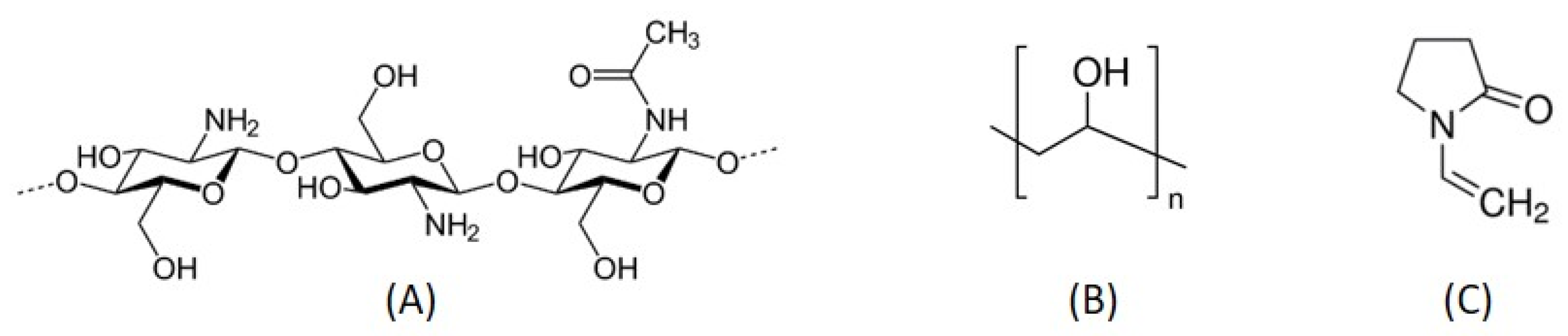
Abstract
:1. Introduction
2. Materials and Methods
2.1. Materials
2.2. Preparation of Chitosan-Based Membranes
2.3. Membranes’ Characterization
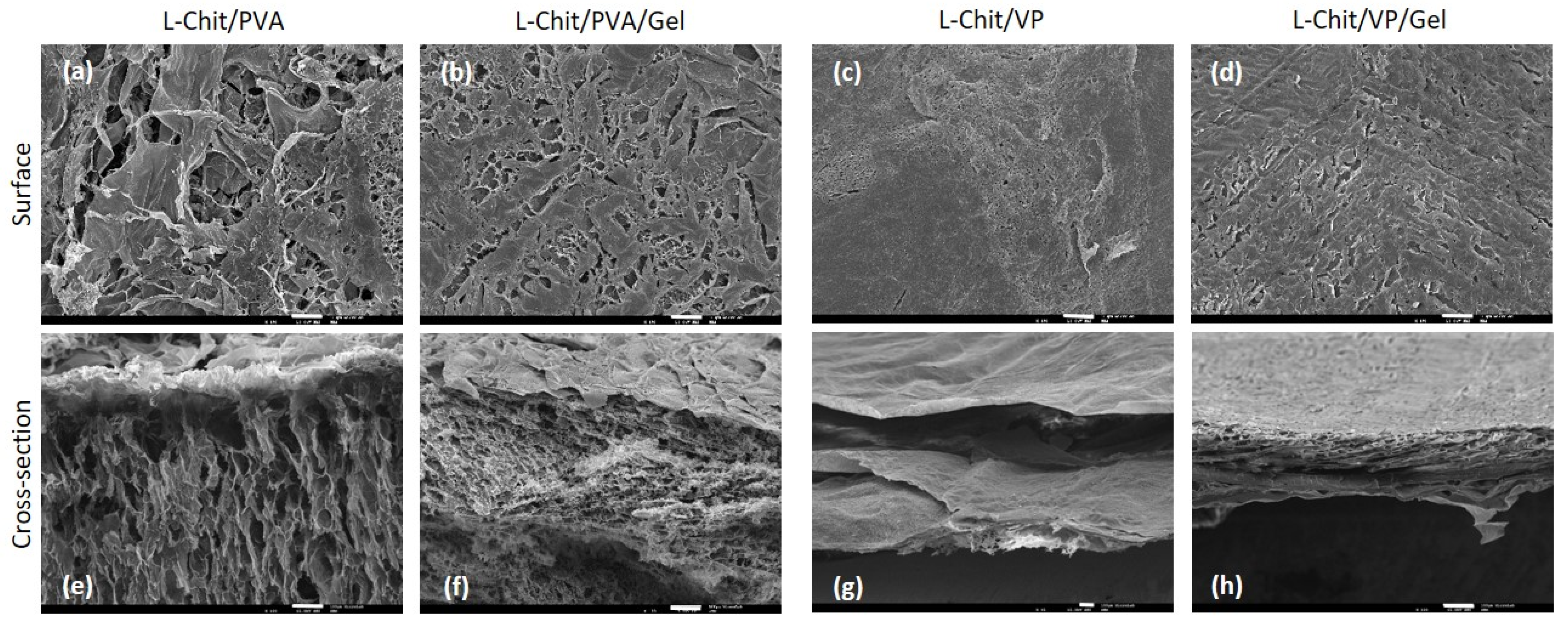
2.3.1. Scanning Electron Microscopy (SEM)
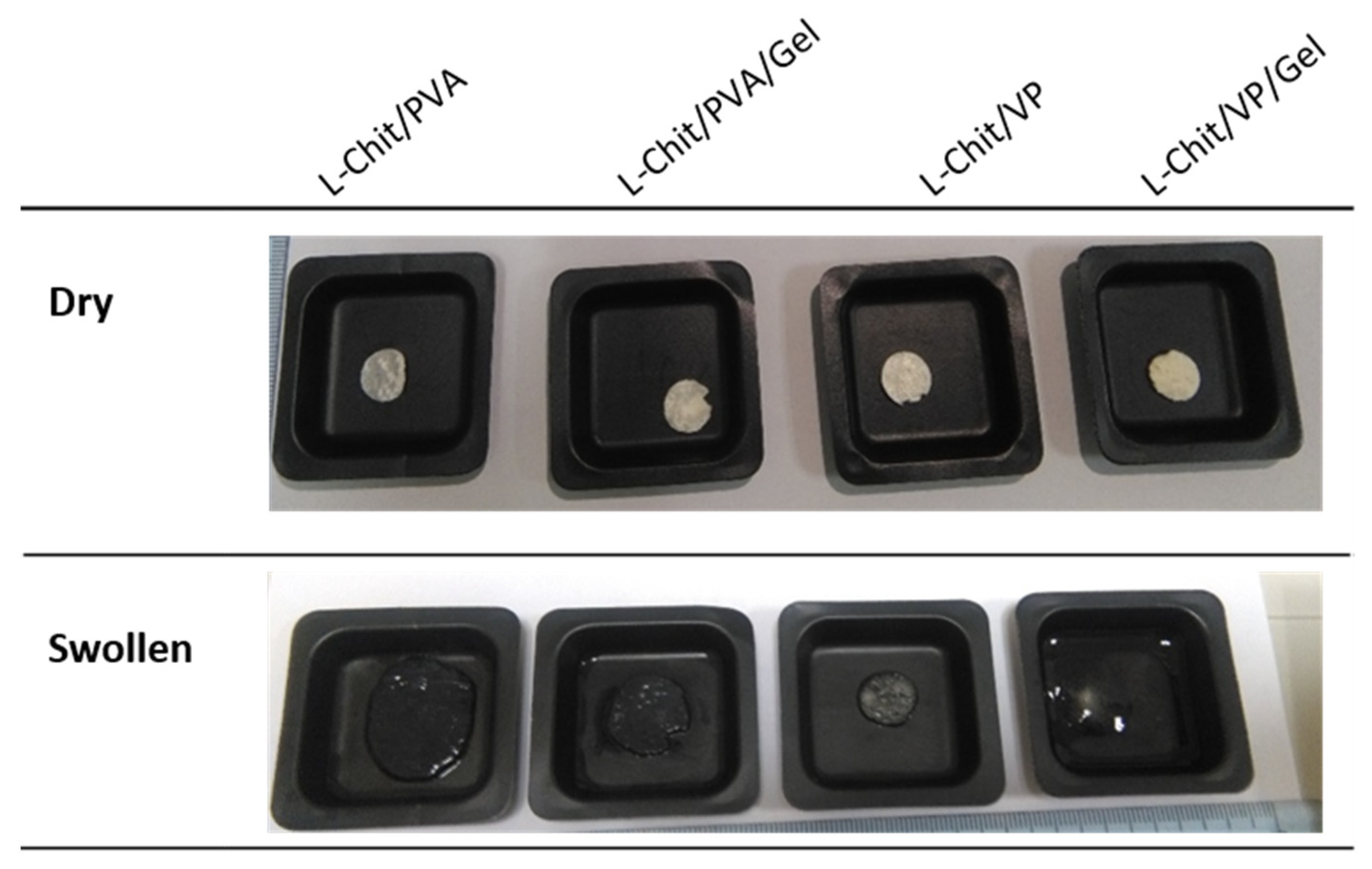
2.3.2. Swelling
2.3.3. In Vitro Degradation
2.4. In Vitro Evaluation
Cell Viability Assay (almarBlue®)
- Indirect Method
- Direct Method
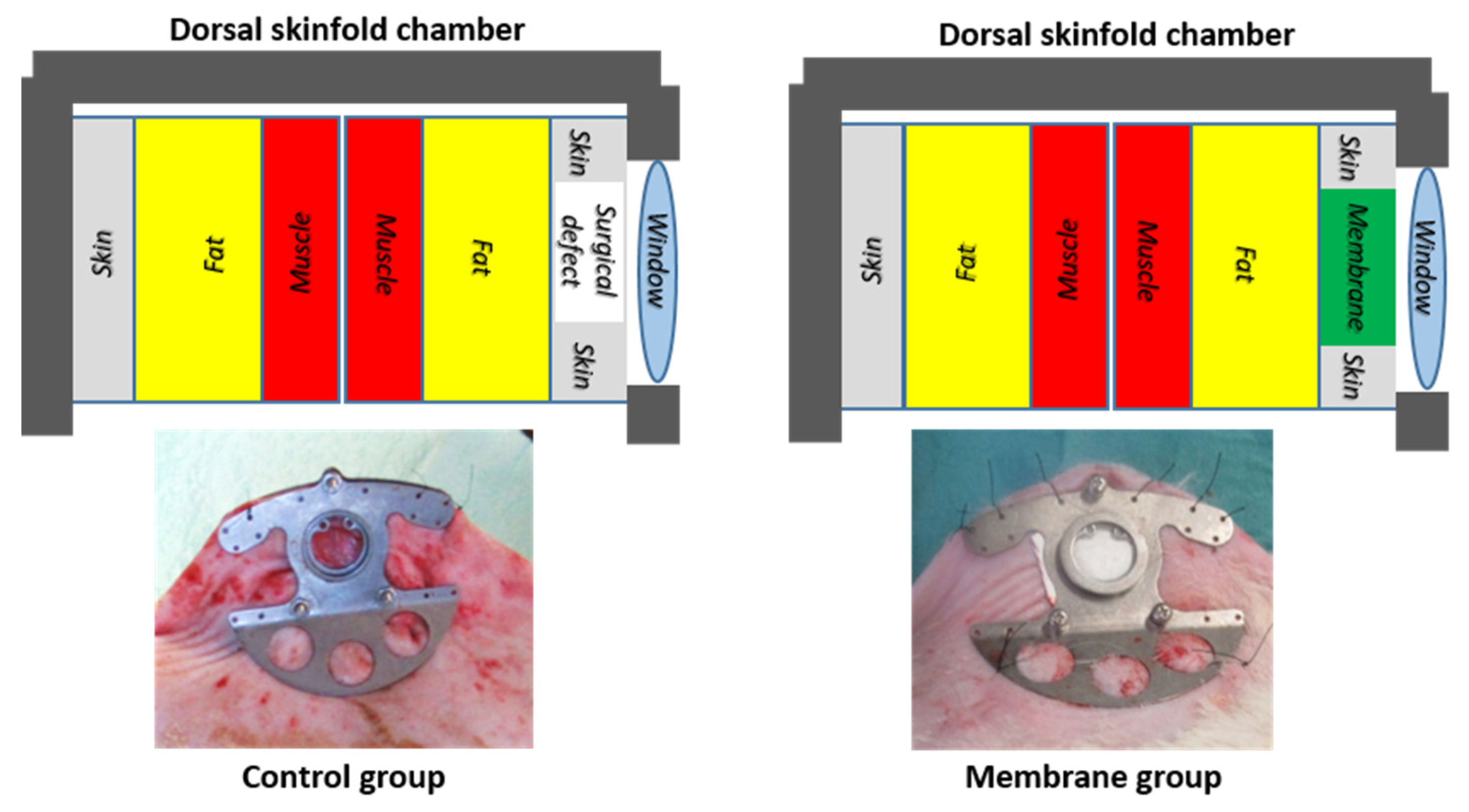
2.5. In Vivo Evaluation
- Daily clinical assessment of rats;
- Wound epithelization area was determined weekly by non-invasive intravital microscopy using a transillumination technique;
- Histological assessment of the ulcer region 3 weeks post-operatively concerning:
- (i)
- General architecture (hematoxylin-eosin, and Masson’s Trichrome staining—these are common methods for assessing cellular and extracellular composition and spatial distribution, particularly of collagen fibers in human skin [30]);
- (ii)
- Epithelial growth (immunohistochemical staining for basal undifferentiated keratinocytes (CK5—Rabbit Anti-Human Keratin 5 Monoclonal Antibody (Clone SP178 ®Roche Diagnostic);
- (iii)
- Connective tissue growth (immunohistochemical staining for fibroblasts (Vimentin-Rabbit Anti-Human Vimentin Monoclonal Antibody (Clone SP20) ®Roche Diagnostic);
- (iv)
3. Results and Discussion
3.1. Membranes’ Characterization
3.1.1. Scanning Electron Microscopy (SEM)
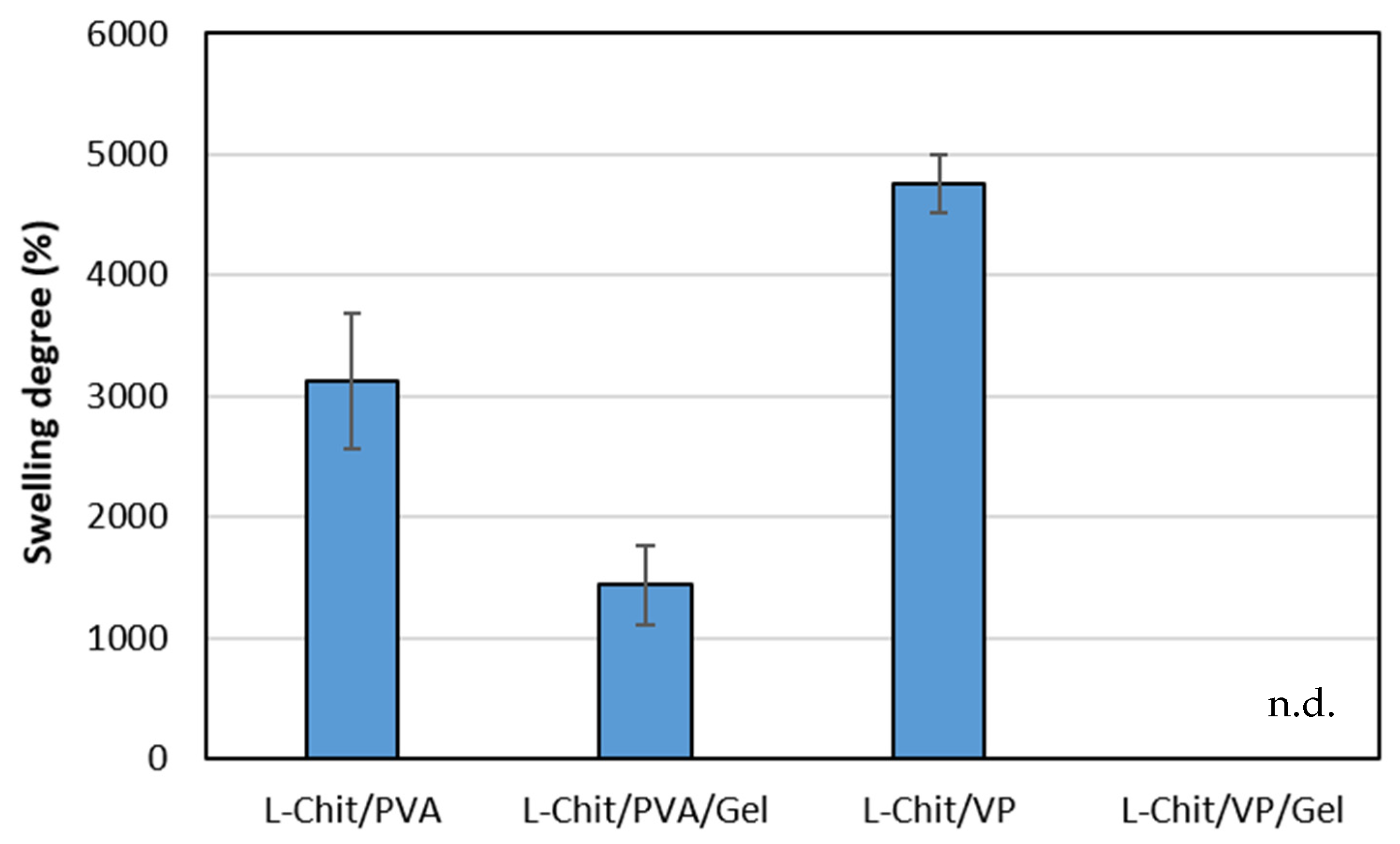
3.1.2. Swelling
3.1.3. In Vitro Degradation
3.2. In Vitro Evaluation
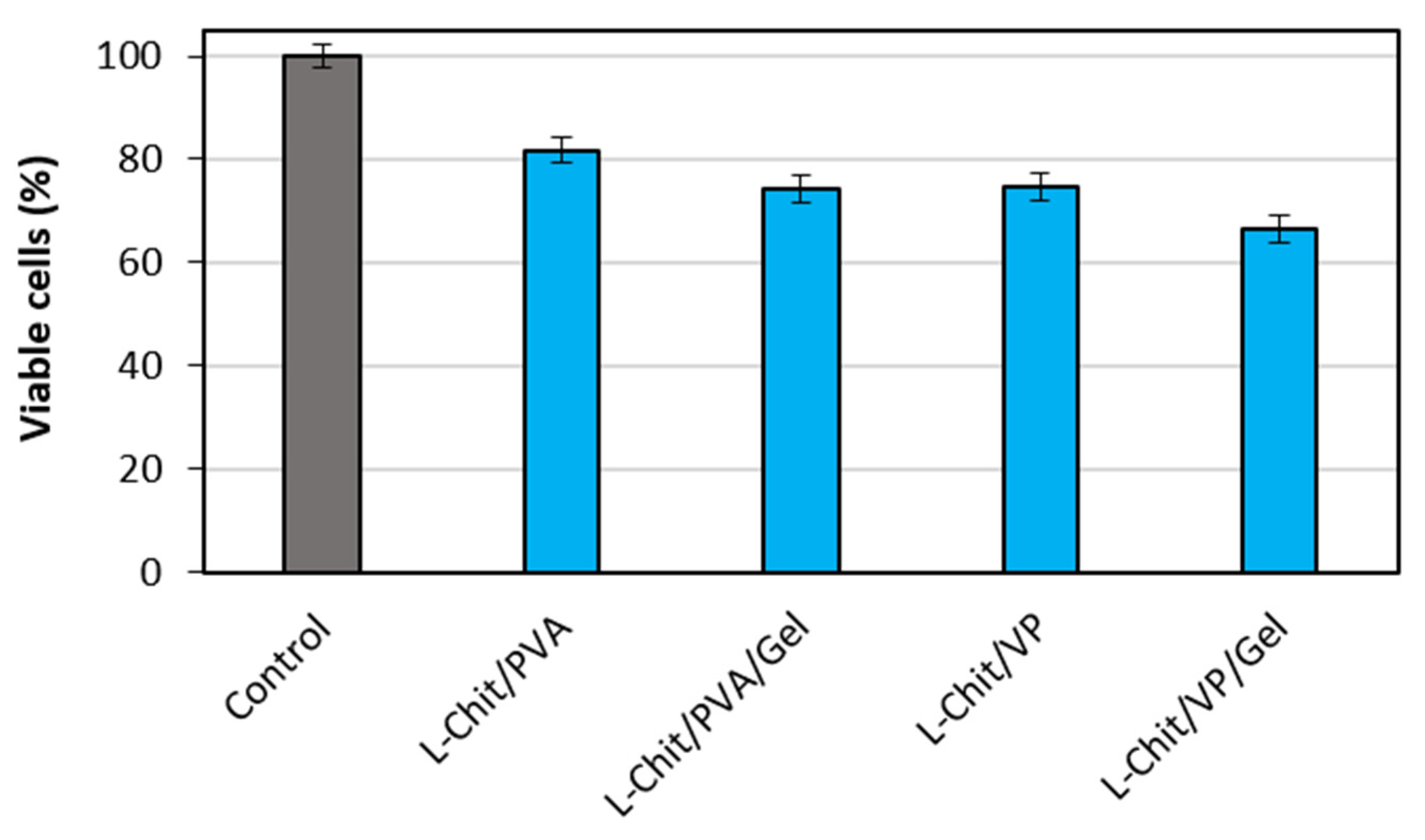
3.2.1. Indirect Method
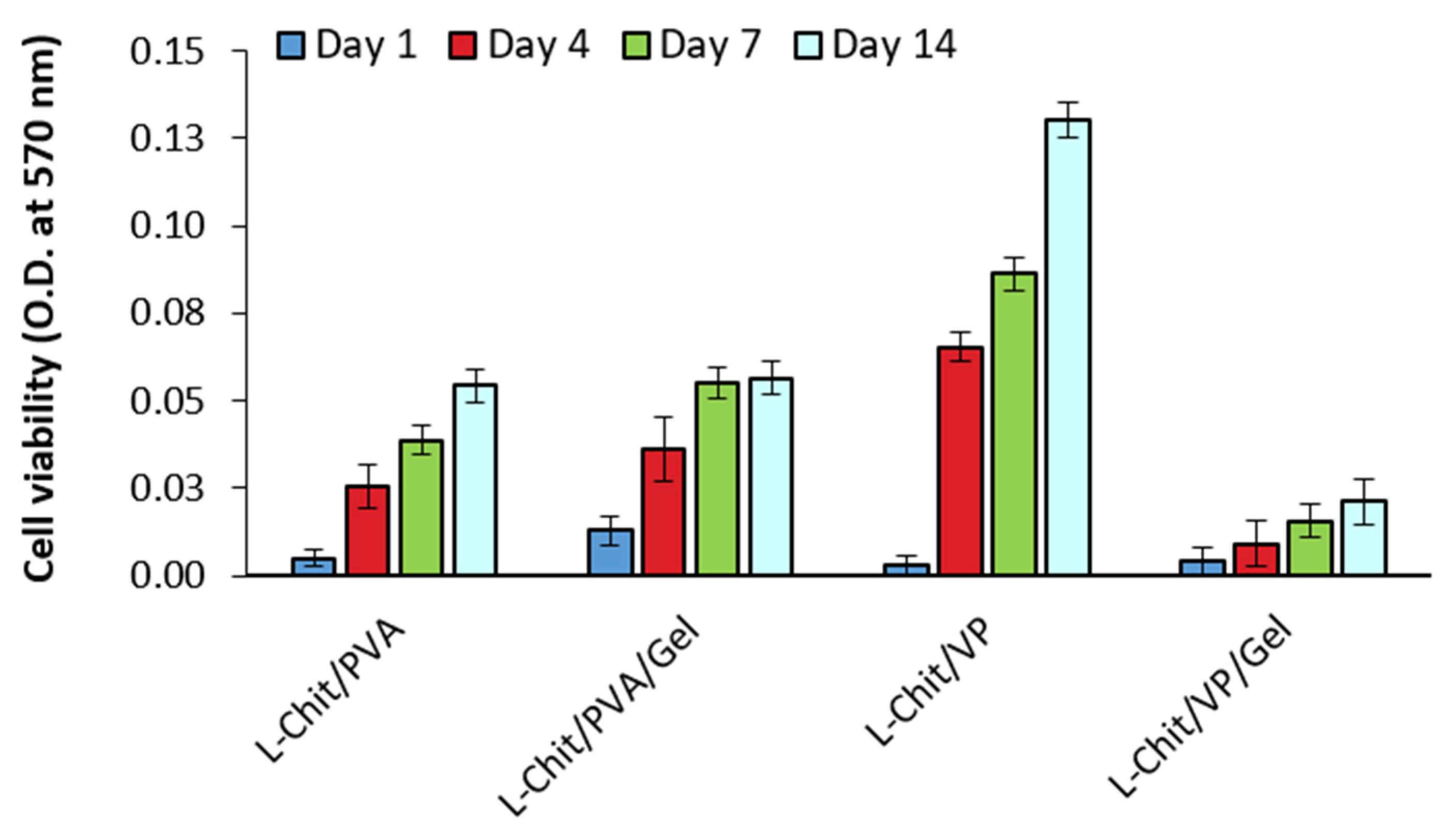
3.2.2. Direct Method
3.3. In Vivo Evaluation
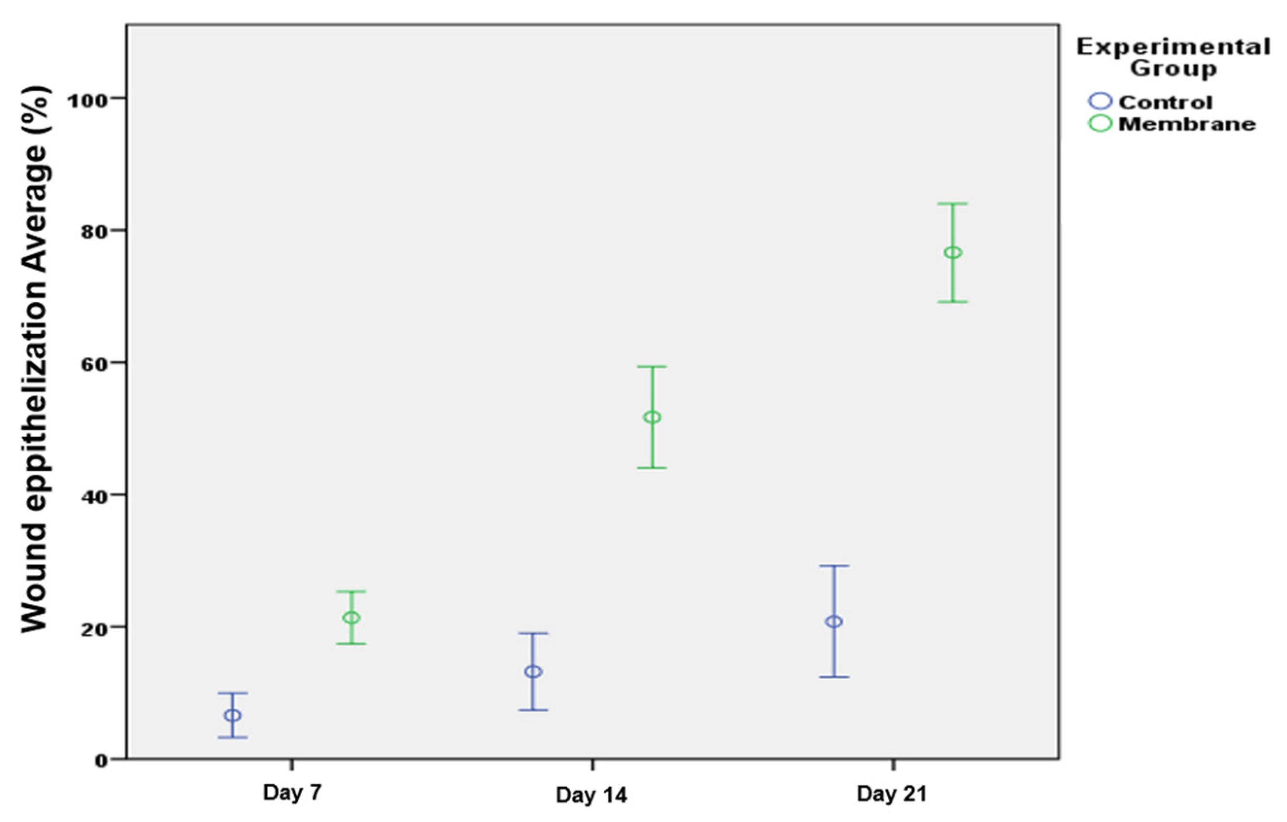
3.3.1. Wound Epithelization
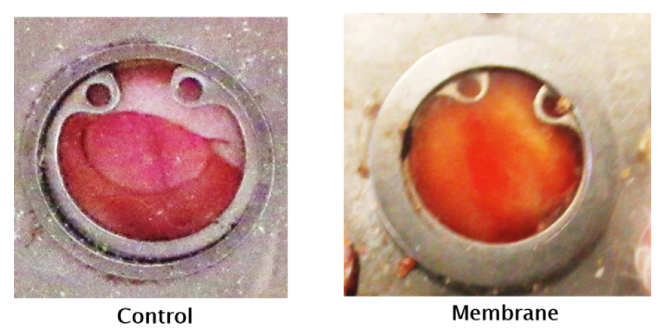
3.3.2. Typical Aspect on the 21st Postoperative Day
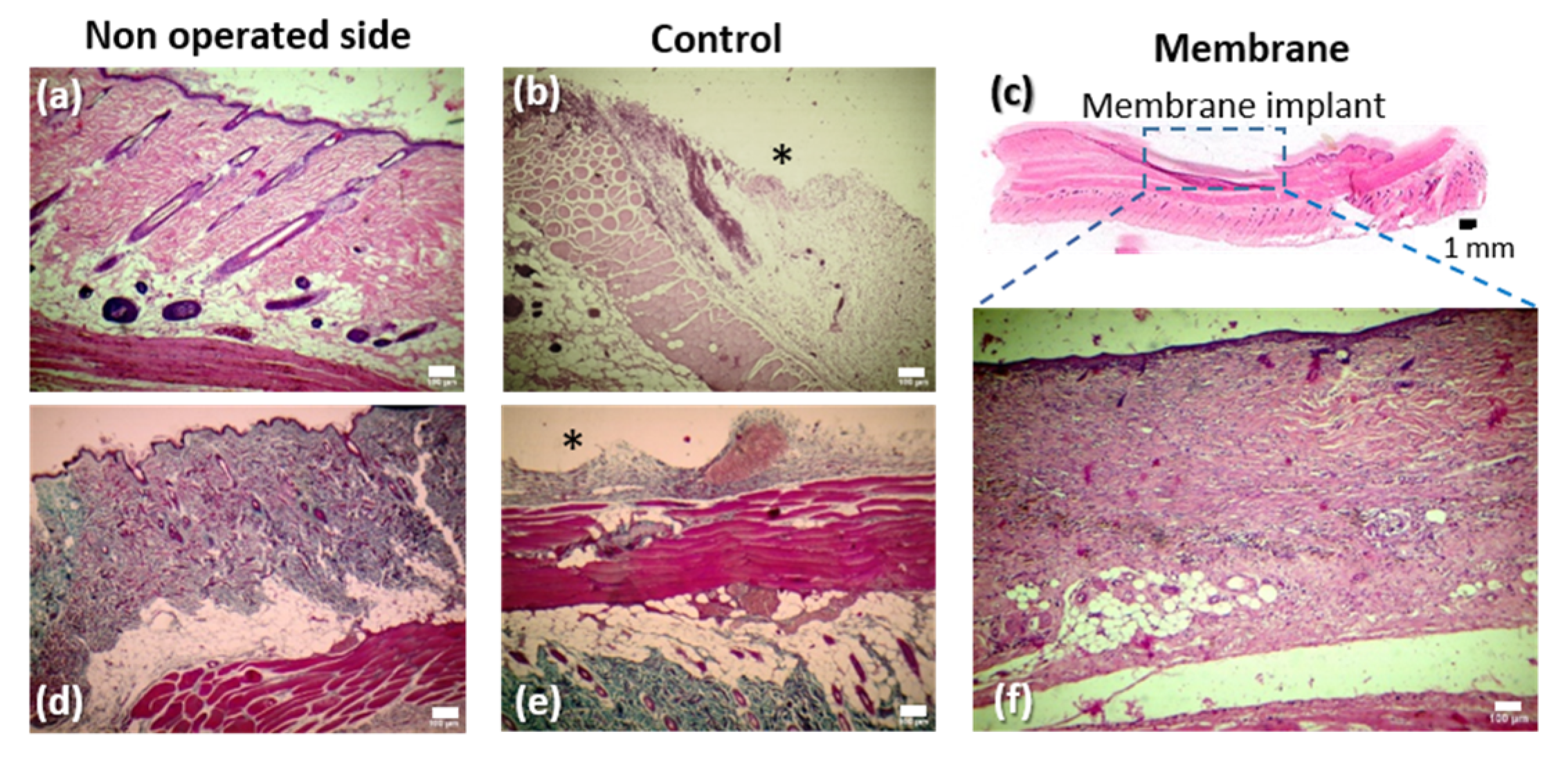
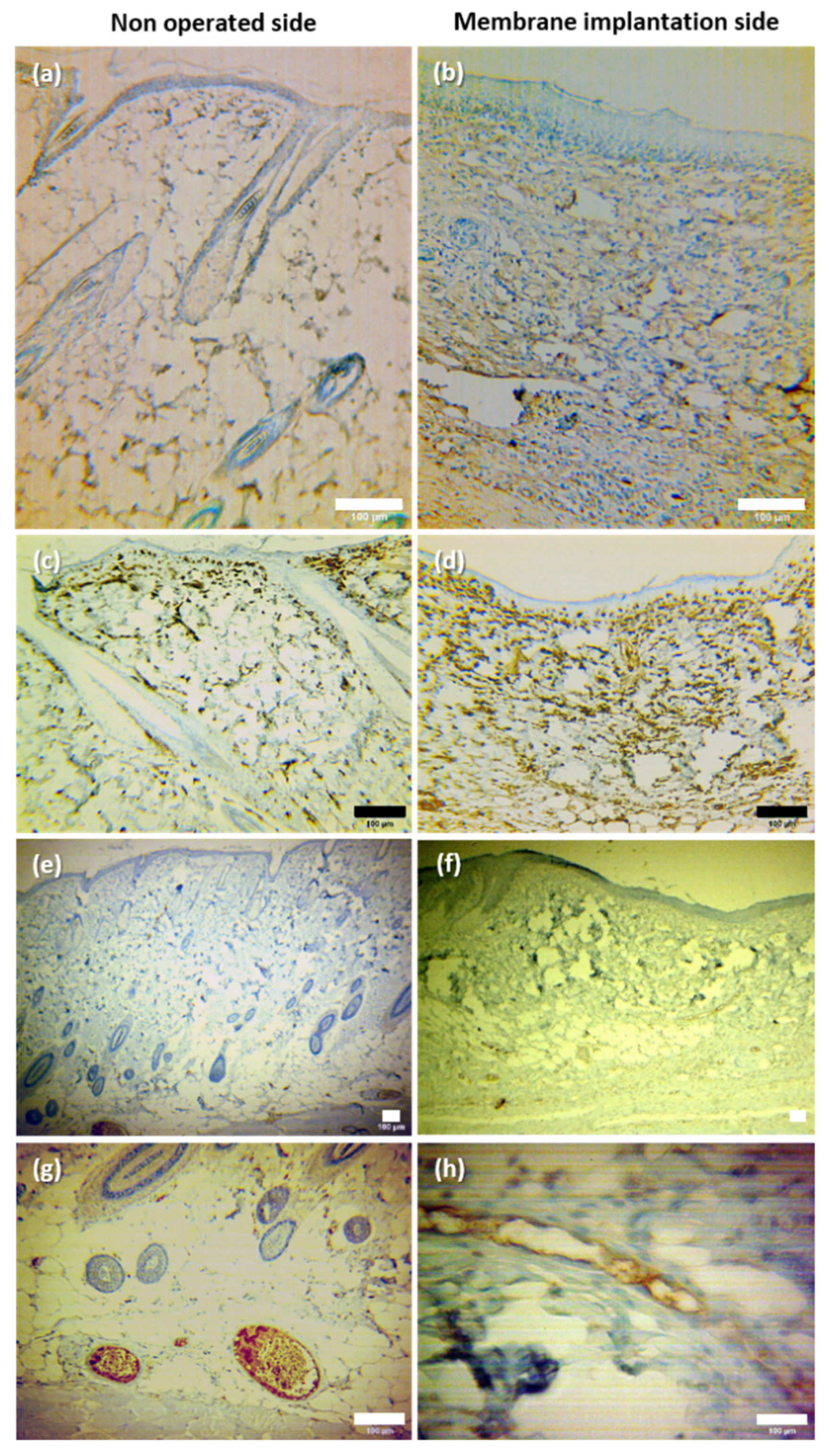
3.3.3. Histological Evaluation
- (i)
- General architecture
- -
- The control group presented a variable extension where skin was absent in the central part of the wound, being replaced by granulation tissue (*).
- -
- The membrane group showed reconstitution of the two layers of the skin. However, the skin in this group presented some differences relative to the normal skin, namely absence of dermal papillae, ridges and skin appendages.
- (ii)
- Epithelial, connective and vascular tissue growth
4. Conclusions
Supplementary Materials
Author Contributions
Funding
Institutional Review Board Statement
Informed Consent Statement
Data Availability Statement
Acknowledgments
Conflicts of Interest
References
- Yu, J.R.; Navarro, J.; Coburn, J.C.; Mahadik, B.; Molnar, J.; Iv, J.H.H.; Nam, A.J.; Fisher, J.P. Current and Future Perspectives on Skin Tissue Engineering: Key Features of Biomedical Research, Translational Assessment, and Clinical Application. Adv. Healthc. Mater. 2019, 8, e1801471. [Google Scholar] [CrossRef] [PubMed]
- Dhasmana, A.; Singh, S.; Kadian, S.; Singh, L. Skin Tissue Engineering: Principles and Advances. J. Dermatol. Ski. Care 2018, 1, 1–11. [Google Scholar]
- Savoji, H.; Godau, B.; Hassani, M.S.; Akbari, M. Skin Tissue Substitutes and Biomaterial Risk Assessment and Testing. Front. Bioeng. Biotechnol. 2018, 6, 86. [Google Scholar] [CrossRef] [PubMed]
- Folli, S.; Curcio, A.; Melandri, D.; Bondioli, E.; Rocco, N.; Catanuto, G.; Falcini, F.; Purpura, V.; Mingozzi, M.; Buggi, F.; et al. A New Human-Derived Acellular Dermal Matrix for Breast Reconstruction Available for the European Market: Preliminary Results. Aesthetic Plast. Surg. 2018, 42, 434–441. [Google Scholar] [CrossRef] [PubMed]
- Groth, A.K.; Ono, M.C.C.; Weihermann, V.; Bastos, L.Z.B.; Rezende, T.M.D.S.; Dalke, D.B.D.Z.; Ferreira, C.I.B. A Picture of Breast Reconstruction in a Public Oncology Hospital in Latin America: A Ten-Year Experience. Eur. J. Breast Health 2020, 16, 244–249. [Google Scholar] [CrossRef]
- Wessels, Q.; Pretorius, E. Development and ultra-structure of an ultra-thin silicone epidermis of bioengineered alternative tissue. Int. Wound J. 2013, 12, 428–431. [Google Scholar] [CrossRef] [Green Version]
- Kamolz, L.-P.; Kotzbeck, P.; Schintler, M.; Spendel, S. Skin regeneration, repair, and reconstruction: Present and future. Eur. Surg. 2022, 54, 163–169. [Google Scholar] [CrossRef]
- Keyhan, S.O.; Ramezanzade, S.; Yazdi, R.G.; Valipour, M.A.; Fallahi, H.R.; Shakiba, M.; Aeinehvand, M. Prevalence of complications associated with polymer-based alloplastic materials in nasal dorsal augmentation: A systematic review and meta-analysis. Maxillofac. Plast. Reconstr. Surg. 2022, 44, 1–21. [Google Scholar] [CrossRef]
- Johnston, D.T.; Lohmeier, S.J.; Langdell, H.C.; Pyfer, B.J.; Komisarow, J.; Powers, D.B.; Erdmann, D. Current Concepts in Cranial Reconstruction: Review of Alloplastic Materials. Plast. Reconstr. Surg.-Glob. Open 2022, 10, e4466. [Google Scholar] [CrossRef]
- Cohen, M.; Morales, R.; Fildes, J.; Barrett, J. Staged Reconstruction after Gunshot Wounds to the Abdomen. Plast. Reconstr. Surg. 2001, 108, 83–92. [Google Scholar] [CrossRef]
- Alrubaiy, L. Skin Substitutes: A Brief Review of Types and Clinical Applications. Oman. Med. J. 2009, 24, 4–6. [Google Scholar] [CrossRef]
- Kamel, R.A.; Ong, J.F.; Eriksson, E.; Junker, J.P.; Caterson, E.J. Tissue Engineering of Skin. J. Am. Coll. Surg. 2013, 217, 533–555. [Google Scholar] [CrossRef]
- Ahmed, S.; Ikram, S. Chitosan Based Scaffolds and Their Applications in Wound Healing. Achiev. Life Sci. 2016, 10, 27–37. [Google Scholar] [CrossRef] [Green Version]
- Kamoun, E.A.; Kenawy, E.-R.S.; Chen, X. A review on polymeric hydrogel membranes for wound dressing applications: PVA-based hydrogel dressings. J. Adv. Res. 2017, 8, 217–233. [Google Scholar] [CrossRef]
- Liao, S.; Wang, W.; Uo, M.; Ohkawa, S.; Akasaka, T.; Tamura, K.; Cui, F.; Watari, F. A three-layered nano-carbonated hydroxyapatite/collagen/PLGA composite membrane for guided tissue regeneration. Biomaterials 2005, 26, 7564–7571. [Google Scholar] [CrossRef]
- Tenchurin, T.K.; Pavlovsky, M.M.; Shepelev, A.D.; Mamagulashvilli, V.G.; I Gomzyak, V.; Sedush, N.G.; Krasheninnikov, S.V.; A Puchkov, A.; Malakhov, S.N.; Sharikov, R.V.; et al. Modification of non-woven materials based on sodium alginate for tissue-engineering. J. Physics Conf. Ser. 2019, 1347, 012072. [Google Scholar] [CrossRef]
- O'Brien, F.J. Biomaterials & scaffolds for tissue engineering. Mater. Today 2011, 14, 88–95. [Google Scholar] [CrossRef]
- Deville, S.; Saiz, E.; Tomsia, A.P. Freeze casting of hydroxyapatite scaffolds for bone tissue engineering. Biomaterials 2006, 27, 5480–5489. [Google Scholar] [CrossRef] [Green Version]
- Gomes, S.R.; Rodrigues, G.; Martins, G.G.; Roberto, M.A.; Mafra, M.; Henriques, C.M.R.; Silva, J.C. In vitro and in vivo evaluation of electrospun nanofibers of PCL, chitosan and gelatin: A comparative study. Mater. Sci. Eng. C 2015, 46, 348–358. [Google Scholar] [CrossRef]
- Ayres, C.E.; Jha, B.S.; Sell, S.A.; Bowlin, G.L.; Simpson, D.G. Nanotechnology in the design of soft tissue scaffolds: Innovations in structure and function. Wiley Interdiscip. Rev. Nanomed. Nanobiotechnol. 2009, 2, 20–34. [Google Scholar] [CrossRef]
- Safinia, L.; Datan, N.; Höhse, M.; Mantalaris, A.; Bismarck, A. Towards a methodology for the effective surface modification of porous polymer scaffolds. Biomaterials 2005, 26, 7537–7547. [Google Scholar] [CrossRef] [PubMed]
- Casimiro, M.H.; Pereira, A.; Leal, J.P.; Rodrigues, G.; Ferreira, L.M. Chitosan/PVA Based Membranes Processed by Gamma Radiation as Scaffolding Materials for Skin Regeneration. Membranes 2021, 11, 561. [Google Scholar] [CrossRef] [PubMed]
- Casimiro, M.H.; Gomes, S.R.; Rodrigues, G.; Leal, J.P.; Ferreira, L.M. Chitosan/Poly(vinylpyrrolidone) Matrices Obtained by Gamma-Irradiation for Skin Scaffolds: Characterization and Preliminary Cell Response Studies. Materials 2018, 11, 2535. [Google Scholar] [CrossRef] [PubMed] [Green Version]
- Casimiro, M.H.; Lancastre, J.J.H.; Rodrigues, A.P.; Gomes, S.R.; Rodrigues, G.; Ferreira, L.M. Chitosan-Based Matrices Prepared by Gamma Irradiation for Tissue Regeneration: Structural Properties vs. Preparation Method. Top. Curr. Chem. 2016, 375, 5. [Google Scholar] [CrossRef] [PubMed]
- Cabo Verde, S.; Nunes, I.; Dores, V.; Melo, R.; Oliveira, S.; Botelho, M.L. Establishment of Sterilization Doses for Biomaterial Products. In Isotope Technologies and Applications–New Horizons, Proceedings of the NAARRI International Conference, Mumbai, India, 13–15 December 2010; NAARRI: Navi Mumbai, India, 2010. [Google Scholar]
- Singh, R.; Singh, D.; Singh, A. Radiation sterilization of tissue allografts: A review. World J. Radiol. 2016, 8, 355–369. [Google Scholar] [CrossRef]
- Casimiro, M.H.; Gil, M.H.; Leal, J.P. Suitability of gamma irradiated chitosan based membranes as matrix in drug release system. Int. J. Pharm. 2010, 395, 142–146. [Google Scholar] [CrossRef]
- Chmielewski, A.G.; Haji-Saeid, M. Radiation technologies: Past, present and future. Radiat. Phys. Chem. 2004, 71, 17–21. [Google Scholar] [CrossRef]
- Huang, Y.-C.; Xiao, J.; Leung, V.Y.L.; Lu, W.W.; Hu, Y.; Luk, K.D.K. Lumbar intervertebral disc allograft transplantation: Healing and remodelling of the bony structure. Eur. Cell. Mater. 2016, 32, 216–227. [Google Scholar] [CrossRef]
- Hong, J.H.; Kim, D.H.; Rhyu, I.J.; Kye, Y.C.; Ahn, H.H. A simple morphometric analysis method for dermal microstructure using color thresholding and moments. Ski. Res. Technol. 2019, 26, 132–136. [Google Scholar] [CrossRef]
- Schreiter, J.; Meyer, S.; Schmidt, C.; Schulz, R.M.; Langer, S. Dorsal skinfold chamber models in mice. GMS Interdiscip. Plast. Reconstr. Surg. DGPW 2017, 6, 1–8. [Google Scholar] [CrossRef]
- Bružauskaitė, I.; Bironaitė, D.; Bagdonas, E.; Bernotienė, E. Scaffolds and cells for tissue regeneration: Different scaffold pore sizes—Different cell effects. Cytotechnology 2016, 68, 355–369. [Google Scholar] [CrossRef]
- Job, K.; Składzień, J. The suitability of scanning electron microscopy in the evaluation of bone structure surfaces and selection of alloplastic materials for facial skeletal reconstruction. Otolaryngol. Polska 2020, 75, 14–19. [Google Scholar] [CrossRef]
- Liu, J.; Wang, W.; Wang, A. Synthesis, characterization, and swelling behaviors of chitosan-g-poly(acrylic acid)/poly(vinyl alcohol) semi-IPN superabsorbent hydrogels. Polym. Adv. Technol. 2011, 22, 627–634. [Google Scholar] [CrossRef]
- Silvestro, I.; Francolini, I.; Di Lisio, V.; Martinelli, A.; Pietrelli, L.; D’Abusco, A.S.; Scoppio, A.; Piozzi, A. Preparation and Characterization of TPP-Chitosan Crosslinked Scaffolds for Tissue Engineering. Materials 2020, 13, 3577. [Google Scholar] [CrossRef]
- Nho, Y.C.; Park, K.R. Preparation and properties of PVA/PVP hydrogels containing chitosan by radiation. J. Appl. Polym. Sci. 2002, 85, 1787–1794. [Google Scholar] [CrossRef]
- Laschke, M.W.; Menger, M.D. The dorsal skinfold chamber: A versatile tool for preclinical research in tissue engineering and regenerative medicine. Eur. Cell. Mater. 2016, 32, 202–215. [Google Scholar] [CrossRef]


Publisher’s Note: MDPI stays neutral with regard to jurisdictional claims in published maps and institutional affiliations. |
© 2022 by the authors. Licensee MDPI, Basel, Switzerland. This article is an open access article distributed under the terms and conditions of the Creative Commons Attribution (CC BY) license (https://creativecommons.org/licenses/by/4.0/).
Share and Cite
Casimiro, M.H.; Ferreira, L.M.; Santos, P.M.P.; Leal, J.P.; Rodrigues, G.; Iria, I.; Alves, S.; Pais, D.; Casal, D. Chitosan-Based Membranes for Skin Wound Repair in a Dorsal Fold Chamber Rat Model. Pharmaceutics 2022, 14, 2736. https://doi.org/10.3390/pharmaceutics14122736
Casimiro MH, Ferreira LM, Santos PMP, Leal JP, Rodrigues G, Iria I, Alves S, Pais D, Casal D. Chitosan-Based Membranes for Skin Wound Repair in a Dorsal Fold Chamber Rat Model. Pharmaceutics. 2022; 14(12):2736. https://doi.org/10.3390/pharmaceutics14122736
Chicago/Turabian StyleCasimiro, Maria Helena, Luís M. Ferreira, Pedro M. P. Santos, João P. Leal, Gabriela Rodrigues, Inês Iria, Sara Alves, Diogo Pais, and Diogo Casal. 2022. "Chitosan-Based Membranes for Skin Wound Repair in a Dorsal Fold Chamber Rat Model" Pharmaceutics 14, no. 12: 2736. https://doi.org/10.3390/pharmaceutics14122736