
Insights into the Small Molecule Targeting of Biologically Relevant G-Quadruplexes: An Overview of NMR and Crystal Structures
 ,
,  , , and
Abstract
:
1. Introduction

2. Small-Molecule Ligands Targeting Telomeric, Oncogenic and Viral G-Quadruplexes
2.1. Telomeric G-Quadruplexes
2.1.1. Human Telomeric Unimolecular Antiparallel G-Quadruplexes

2.1.2. Human Telomeric Unimolecular Hybrid G-Quadruplexes
2.1.3. Human Telomeric Unimolecular Parallel G-Quadruplexes

2.1.4. Human Telomeric Bimolecular Parallel G-Quadruplexes

2.1.5. Human Telomeric Tetramolecular Parallel G-Quadruplexes

2.1.6. Oxytricha Telomeric Unimolecular Parallel G-Quadruplexes
2.1.7. Oxytricha Telomeric Bimolecular Antiparallel G-Quadruplexes

2.1.8. Oxytricha Telomeric Tetramolecular Parallel G-Quadruplexes
2.1.9. RNA Telomeric G-Quadruplexes

2.2. Oncogenic G-Quadruplexes
2.2.1. C-MYC Oncogene Promoter G-Quadruplexes

2.2.2. RET Oncogene Promoter G-Quadruplexes
2.2.3. PDGFR-β Oncogene Promoter G-Quadruplexes
2.2.4. VEGF Oncogene Promoter G-Quadruplexes
2.3. Viral G-Quadruplexes
3. Summary and Outlook

| PDB ID | Technique | Ligand Class | Ligand Family | Ligand Name | Sequence | Sequence Name | Prevalent Cation | Molecularity | Topology | Binding Mode (G-Quadruplex/ Ligand Stoichiometry) | Binding Interactions | Refs. |
|---|---|---|---|---|---|---|---|---|---|---|---|---|
| Telomeric G-quadruplexes | ||||||||||||
| Human telomeric unimolecular antiparallel G-quadruplexes | ||||||||||||
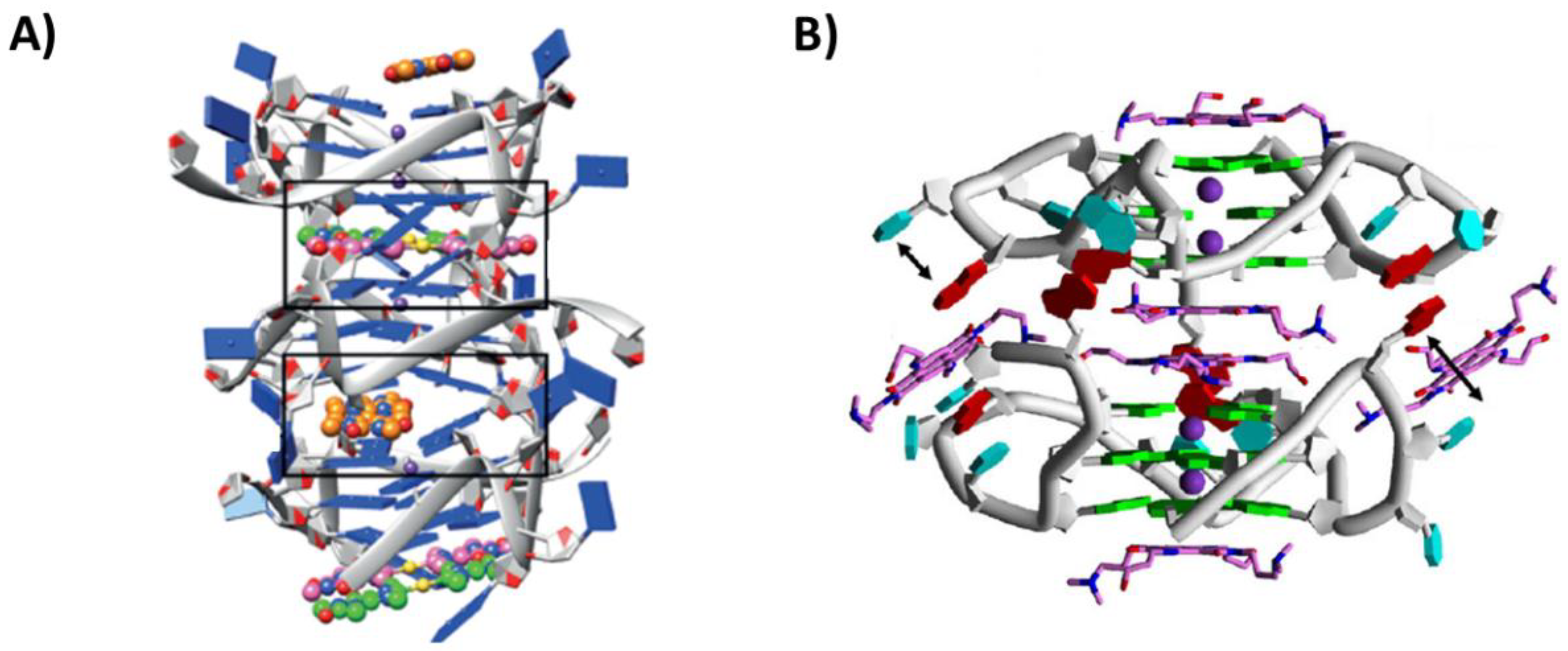
| 2MCO | NMR | Metal-organic | Dinuclear Ru(II) complex | ΛΛ-[{Ru(phen)2}2tpphz]4+ | d[AGGG(TTAGGG)3] | h-tel22 | Na+ | Unimolecular | Antiparallel basket | Intercalation between 5′-end G-tetrad and diagonal loop (1:1) | Stacking and electrostatic interactions | [31] |
| 2MCC | NMR | Metal-organic | Dinuclear Ru(II) complex | ΔΔ-[{Ru(phen)2}2tpphz]4+ | d[AGGG(TTAGGG)3] | h-tel22 | Na+ | Unimolecular | Antiparallel basket | 3′-end G-tetrad stacking (1:1) | Stacking | [31] |
| 7OTB | X-ray crystallography | Metal-organic | Ru(II) complex | Λ-[Ru(phen)2(qdppz)]2+ | d[GGG(TTAGGG)2TTTGGG] | h-tel21 | Na+ | Unimolecular | Antiparallel chair | 5′-end G-tetrad stacking (1:1) | Stacking | [33] |
| Human telomeric unimolecular hybrid G-quadruplexes | ||||||||||||
| 6CCW | NMR | Organic | Berberine | Epiberberine | d[(TTAGGG)4TT] | h-tel26 | K+ | Unimolecular | Hybrid-2 | Intercalation between 5′-end G-tetrad and T2:T13:A15 triad layer and T1:T14 pair (1:1) | Stacking and hydrogen bonds | [37] |
| 5MVB | NMR | Metal-organic | Dinuclear Au(III) complex | Auoxo6 | d[(TTAGGG)4TT] | h-tel26 | K+ | Unimolecular | Hybrid-2 | Intercalation between 5′-end G-tetrad and flanking A3 (1:1) | Stacking | [38] |
| 6KFJ | NMR | Organic | Tripod | NBTE | d[(TTAGGG)4TT] | h-tel26 | K+ | Unimolecular | Hybrid-2 | Intercalation between 5′-end G-tetrad and capping triad A3, T14, A21 (1:1) | Stacking, π-cation and electrostatic interactions | [39] |
| 6KFI | NMR | Organic | Tripod | NBTE | d[AAAGGG(TTAGGG)3AA] | h-tel26A | K+ | Unimolecular | Hybrid-1 | Intercalation between 5′-end G-tetrad and capping triad A3, A9, T20 (1:1) | Stacking, π-cation and electrostatic interactions | [39] |
| 5Z80 | NMR | Metal-organic | Pt(II) complex | Pt(II)-based tripod | d[AAAGGG(TTAGGG)3AA] | h-tel26A | K+ | Unimolecular | Hybrid-1 | Intercalation between 5′-end G-tetrad and capping triad A3, A9, T20 (1:1) | Stacking, hydrogen bonds and electrostatic interactions | [40] |
| 5Z8F | NMR | Metal-organic | Pt(II) complex | Pt(II)-based tripod | d[AAAGGG(TTAGGG)3AA] | h-tel26A | K+ | Unimolecular | Hybrid-1 | 5′- and 3′-end G-tetrad stacking within a 3′-3′ dimer (2:4) | Stacking, hydrogen bonds and electrostatic interactions | [40] |
| 2MB3 | NMR | Organic | Telomestatin | L2H2-6M(2)OTD | d[TTGGG(TTAGGG)3A] | h-tel24 | K+ | Unimolecular | Hybrid-1 | 5′-end G-tetrad stacking (1:1) | Stacking and electrostatic interactions | [41] |
| 7Z9L | NMR | Organic | Phenanthroline-quinoline | Phen-DC3 | d[TAGGG(TTAGGG)3] | h-tel23 | K+ | Unimolecular | From Hybrid-1 to Antiparallel chair | Intercalation between a two-tetrad unit and a 5′-end “pseudo-tetrad” (1:1) | Stacking | [42] |
| Human telomeric unimolecular parallel G-quadruplexes | ||||||||||||
| 3R6R | X-ray crystallography | Organic | Berberine | Berberine | d[TAGGG(TTAGGG)3] | h-tel23 | K+ | Unimolecular | Parallel | 5′- and 3′-end G-tetrad stacking within a 5′-5′ dimer (1:3) | Stacking and hydrogen bonds | [43] |
| 5CCW | X-ray crystallography | Metal-organic | Au(I) complex | [Au(9-methylcaffein-8-ylidene)2]+ | d[TAGGG(TTAGGG)3] | h-tel23 | K+ | Unimolecular | Parallel | 5′- and 3′-end G-tetrad stacking (1:3) | Stacking | [44] |
| 6H5R | X-ray crystallography | Metal-organic | Au(I) complex | [Au(1-butyl-3-methyl-2-ylidene)2]+ | d[TAGGG(TTAGGG)3T] | h-tel24 | K+ | Unimolecular | Parallel | 5′-end G-tetrad stacking (1:1) | Stacking | [45] |
| 3CDM | X-ray crystallography | Organic | Naphthalene diimide | NDI-1 | d[TAGGG(TTAGGG)3] | h-tel23 | K+ | Unimolecular | Parallel | 5′- and 3′-end G-tetrad stacking within a 5′-5′ dimer and loop binding (1:6) | Stacking | [47] |
| 3SC8 | X-ray crystallography | Organic | Naphthalene diimide | BMSG-SH3 | d[AGGG(TTAGGG)3] | h-tel22 | K+ | Unimolecular | Parallel | 3′-end G-tetrad stacking within a 5′-5′ dimer (1:1) | Stacking and electrostatic interactions | [48] |
| 3T5E | X-ray crystallography | Organic | Naphthalene diimide | BMSG-SH4 | d[AGGG(TTAGGG)3] | h-tel22 | K+ | Unimolecular | Parallel | 3′-end G-tetrad stacking within a 5′-5′ dimer (1:1) | Stacking and electrostatic interactions | [48] |
| 3UYH | X-ray crystallography | Organic | Naphthalene diimide | 3d | d[AGGG(TTAGGG)3] | h-tel22 | K+ | Unimolecular | Parallel | 3′-end G-tetrad stacking within a 5′-5′ dimer (1:1) | Stacking and electrostatic interactions | [49] |
| 4DA3 | X-ray crystallography | Organic | Naphthalene diimide | 3d | d[GGG(TTAGGG)3] | h-tel21 | K+ | Unimolecular | Parallel | 3′-end G-tetrad stacking within a 5′-5′ dimer (1:1) | Stacking and electrostatic interactions | [49] |
| 4DAQ | X-ray crystallography | Organic | Naphthalene diimide | BMSG-SH3 | d[GGG(TTAGGG)3] | h-tel21 | K+ | Unimolecular | Parallel | 3′-end G-tetrad stacking within a 5′-5′ dimer (1:1) | Stacking and electrostatic interactions | [49] |
| 4FXM | X-ray crystallography | Organic | Porphyrin | N-methyl mesoporphyrin IX | d[AGGG(TTAGGG)3] | h-tel22 | K+ | Unimolecular | Parallel | 3′-end G-tetrad stacking within a 5′-5′ dimer (1:1) | Stacking | [50] |
| 4G0F | X-ray crystallography | Organic | Porphyrin | N-methyl mesoporphyrin IX | d[AGGG(TTAGGG)3] | h-tel22 | K+ | Unimolecular | Parallel | 3′-end G-tetrad stacking within a 5′-5′ dimer (1:1) | Stacking | [50] |
| 6XCL | X-ray crystallography | Metal-organic | Pt(II) complex | Pt(II)-based ligand | d[AGGG(TTAGGG)3] | h-tel22 | K+ | Unimolecular | Parallel | 5′- and 3′-end G-tetrad stacking within a 5′-5′ dimer (2:3) | Stacking | [53] |
| Human telomeric bimolecular parallel G-quadruplexes | ||||||||||||
| 5CDB | X-ray crystallography | Organic | Berberine | NAX053 | d(TAGGGTTAGGGT) | h-tel12 | K+ | Bimolecular | Parallel | Intercalation between 3′-end G-tetrad and 5′-end T:A:T:A tetrad (2:3) | Stacking | [54] |
| 6S15 | X-ray crystallography | Organic | Berberine | 13-alkylpyridine berberine | d(TAGGGTTAGGGT) | h-tel12 | K+ | Bimolecular | Parallel | Intercalation between 3′-end G-tetrad and 5′-end T:A:T:A tetrad (2:3) | Stacking | [55] |
| 3CCO | X-ray crystallography | Organic | Naphthalene diimide | NDI-1 | d(TAGGGTTAGGGT) | h-tel12 | K+ | Bimolecular | Parallel | 3′-end G-tetrad stacking and loop binding (1:3) | Stacking | [47] |
| 3CE5 | X-ray crystallography | Organic | Acridine | BRACO-19 | d(TAGGGTTAGGGT) | h-tel12 | K+ | Bimolecular | Parallel | Intercalation between 3′-end G-tetrad and 5′-end T:A:T:A tetrad (1:2) | Stacking and hydrogen bonds | [58] |
| 2HRI | X-ray crystallography | Organic | Porphyrin | TMPyP4 | d(TAGGGTTAGGG) | h-tel11 | K+ | Bimolecular | Parallel | Flanking residues and loop binding (1:2) | Stacking | [59] |
| 3QSC | X-ray crystallography | Metal-organic | Cu(II) complex | Cu(II) salphen metal complex | d(AGGGTBrUAGGTT) | h-tel11Br | K+ | Bimolecular | Parallel | 3′-end G-tetrad stacking within a 5′-5′ dimer (1:1) | Stacking | [60] |
| 3QSF | X-ray crystallography | Metal-organic | Ni(II) complex | Ni(II) salphen metal complex | d(AGGGTBrUAGGTT) | h-tel11Br | K+ | Bimolecular | Parallel | 3′-end G-tetrad stacking within a 5′-5′ dimer (1:1) | Stacking | [60] |
| Human telomeric tetramolecular parallel G-quadruplexes | ||||||||||||
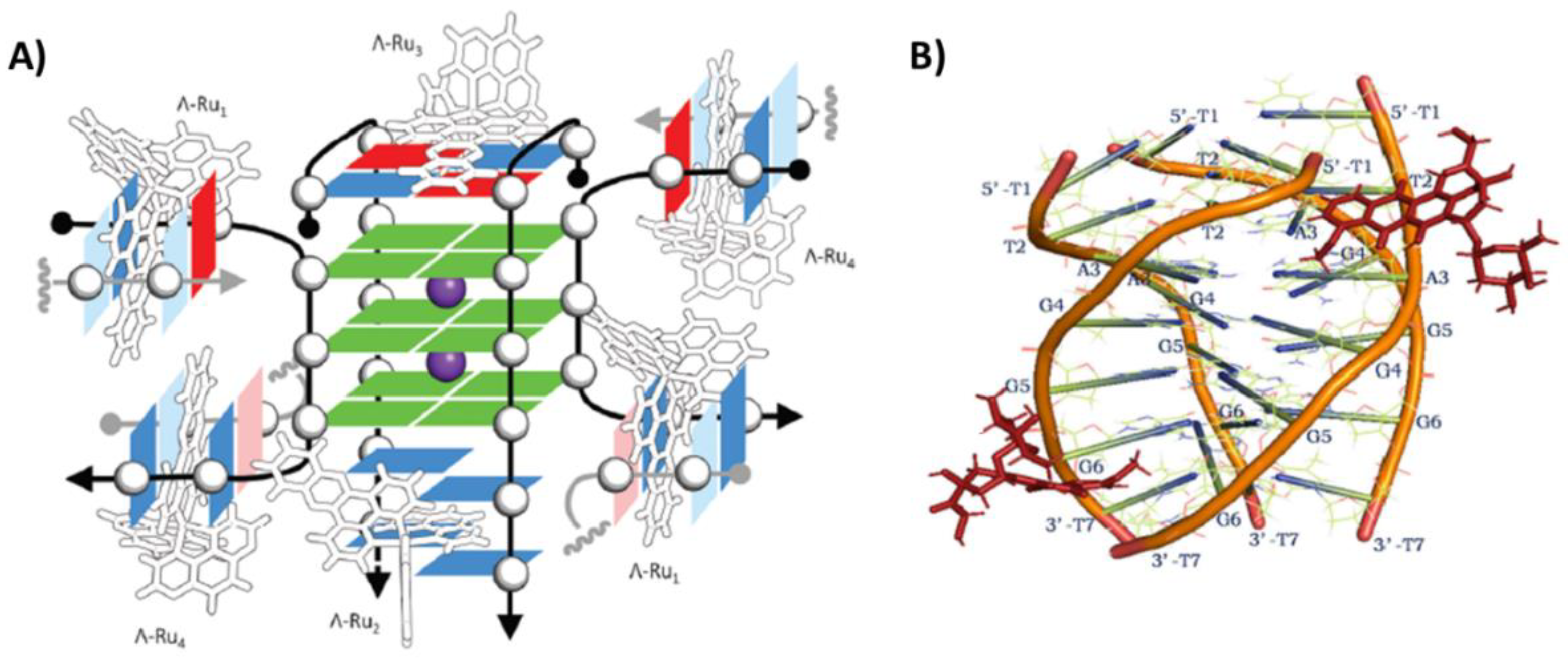
| 5LS8 | X-ray crystallography | Metal-organic | Ru(II) complex | Λ/Δ-[Ru(TAP)2(11-CN-dppz)]2+ | d(TAGGGTTA) | h-tel8 | K+ | Tetramolecular | From Parallel to Antiparallel | 5′- and 3′-end binding (1:4 and 1:2) | Stacking | [61] |
| 6RNL | X-ray crystallography | Metal-organic | Ru(II) complex | Λ-[Ru(TAP)2(dppz)]2+ | d(TAGGGTT) | h-tel7 | K+ | Tetramolecular | Parallel | Flanking residues binding (1:4) | Stacking | [62] |
| 6KXZ | NMR | Organic | Anthracyclin | Epirubicin | d(TTAGGGT) | h-tel7 | K+ | Tetramolecular | Parallel | Groove binding (1:2) | Hydrogen bonds | [63] |
| 6KN4 | NMR | Organic | Anthracyclin | Adriamycin | d(TTAGGGT) | h-tel7 | K+ | Tetramolecular | Parallel | Groove binding (1:2) | Stacking and hydrogen bonds | [64] |
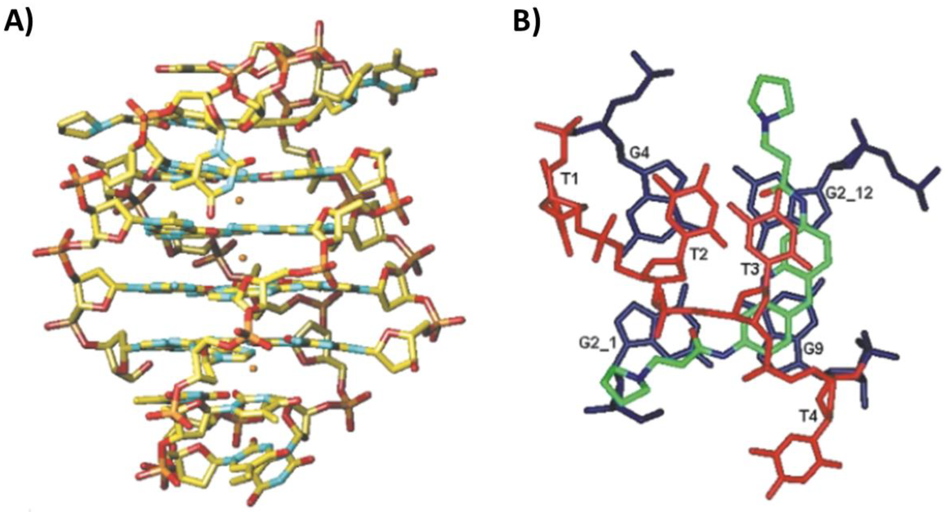
| 2MS6 | NMR | Organic | Flavonoid | Quercetin | d(TTAGGGT) | h-tel7 | K+ | Tetramolecular | Parallel | Stacking between T1 and T2 tetrads and between 3′-end G-tetrad and T7 tetrad (1:2) | Stacking | [67] |
| 2JWQ | NMR | Organic | Quinacridine | MMQ1 | d(TTAGGGT) | h-tel7 | K+ | Tetramolecular | Parallel | 5′- and 3′-end G-tetrad stacking (1:2) | Stacking and electrostatic interactions | [68] |
| 1NZM | NMR | Organic | Acridine | RHPS4 | d(TTAGGGT) | h-tel7 | K+ | Tetramolecular | Parallel | 5′- and 3′-end G-tetrad stacking (1:2) | Stacking and electrostatic interactions | [69] |
| Oxytricha telomeric unimolecular parallel G-quadruplexes | ||||||||||||
| 6P45 | X-ray crystallography | Organic | Porphyrin | N-methyl mesoporphyrin IX | d[(TGGGT)4] | o-tel20 | K+ | Unimolecular | Parallel | 3′-end G-tetrad stacking within a 5′-5′ dimer (1:1) | Stacking | [70] |
| 6PNK | X-ray crystallography | Organic | Porphyrin | N-methyl mesoporphyrin IX | d[(GGGTT)3GGG] | o-tel18 | K+ | Unimolecular | Parallel | 3′-end G-tetrad stacking within a 5′-5′ dimer (1:1) | Stacking | [70] |
| 6K3X | NMR | Organic | Dinucleotide | d(AG) | d[TTGGT(GGGT)3] | o-tel17 | K+ | Unimolecular | Parallel | G-vacancy filling (1:1) | Stacking and hydrogen bonds | [71] |
| 6K3Y | NMR | Organic | Dinucleotide | cGAMP | d[TTGGT(GGGT)3] | o-tel17 | K+ | Unimolecular | Parallel | G-vacancy filling (1:1) | Stacking and hydrogen bonds | [71] |
| Oxytricha telomeric bimolecular antiparallel G-quadruplexes | ||||||||||||
| 1L1H | X-ray crystallography | Organic | Acridine | BSU6039 | d(GGGGTTTTGGGG) | o-tel12 | K+ | Bimolecular | Antiparallel | Intercalation between 5′-end G-tetrad and T3 (1:1) | Stacking and hydrogen bonds | [72] |
| 3EM2 | X-ray crystallography | Organic | Acridine | 3-morpholinopropionamido 3,6-disubstituted acridine derivative | d(GGGGTTTTGGGG) | o-tel12 | K+ | Bimolecular | Antiparallel | Intercalation between 5′-end G-tetrad and T3 (1:1) | Stacking and hydrogen bonds | [73] |
| 3EQW | X-ray crystallography | Organic | Acridine | 3-(2-ethylpiperidino)propionamido 3,6-disubstituted acridine derivative | d(GGGGTTTTGGGG) | o-tel12 | K+ | Bimolecular | Antiparallel | Intercalation between 5′-end G-tetrad and T3 (1:1) | Stacking and hydrogen bonds | [73] |
| 3EUI | X-ray crystallography | Organic | Acridine | 3-(2-ethylpiperidino)propionamido 3,6-disubstituted acridine derivative | d(GGGGTTTTGGGG) | o-tel12 | K+ | Bimolecular | Antiparallel | Intercalation between 5′-end G-tetrad and T3 (1:1) | Stacking and hydrogen bonds | [73] |
| 3ERU | X-ray crystallography | Organic | Acridine | 3-(2-methylpiperidino)propionamido 3,6-disubstituted acridine derivative | d(GGGGTTTTGGGG) | o-tel12 | K+ | Bimolecular | Antiparallel | Intercalation between 5′-end G-tetrad and T3 (1:1) | Stacking and hydrogen bonds | [73] |
| 3ES0 | X-ray crystallography | Organic | Acridine | 3-(4-methylpiperidino)propionamido 3,6-disubstituted acridine derivative | d(GGGGTTTTGGGG) | o-tel12 | K+ | Bimolecular | Antiparallel | Intercalation between 5′-end G-tetrad and T3 (1:1) | Stacking and hydrogen bonds | [73] |
| 3ET8 | X-ray crystallography | Organic | Acridine | 3-(3-methylpiperidino)propionamido groups 3,6-disubstituted acridine derivative | d(GGGGTTTTGGGG) | o-tel12 | K+ | Bimolecular | Antiparallel | Intercalation between 5′-end G-tetrad and T3 (1:1) | Stacking and hydrogen bonds | [73] |
| 3EUM | X-ray crystallography | Organic | Acridine | 3-(azepan-1-yl)propionamido 3,6-disubstituted acridine derivative | d(GGGGTTTTGGGG) | o-tel12 | K+ | Bimolecular | Antiparallel | Intercalation between 5′-end G-tetrad and T3 (1:1) | Stacking and hydrogen bonds | [73] |
| 3NYP | X-ray crystallography | Organic | Acridine | bis-3-fluoropyrrolidine enantiomer (R,R) | d(GGGGTTTTGGGG) | o-tel12 | K+ | Bimolecular | Antiparallel | Intercalation between 5′-end G-tetrad and T3 (1:1) | Stacking and electrostatic interactions | [74] |
| 3NZ7 | X-ray crystallography | Organic | Acridine | bis-3-fluoropyrrolidine enantiomer (S,S) | d(GGGGTTTTGGGG) | o-tel12 | K+ | Bimolecular | Antiparallel | Intercalation between 5′-end G-tetrad and T3 (1:1) | Stacking and electrostatic interactions | [74] |
| 5HIX | X-ray crystallography | Organic | Quinoline | Oligoamide foldamer | d(GGGGTTTTGGGG) | o-tel12 | K+ | Bimolecular | Antiparallel | Loop binding (1:1) | Electrostatic interactions | [75] |
| Oxytricha telomeric tetramolecular parallel G-quadruplexes | ||||||||||||
| 1O0K | X-ray crystallography | Organic | Anthracyclin | Daunomycin | d(TGGGGT) | o-tel6 | Na+ | Tetramolecular | Parallel | 5′-end G-tetrad stacking within a 5′-5′ dimer (1:3) | Stacking and hydrogen bonds | [76] |
| 3TVB | X-ray crystallography | Organic | Anthracyclin | Daunomycin | d(GGGG) | o-tel4 | Na+ | Tetramolecular | Parallel | 5′-end G-tetrad stacking within a 5′-5′ dimer (1:4) | Stacking | [77] |
| 2JT7 | NMR | Organic | Distamycin | Distamycin | d(TGGGGT) | o-tel6 | K+ | Tetramolecular | Parallel | Groove binding (1:4) | Electrostatic interactions | [78] |
| 2KVY | NMR | Organic | Distamycin | N-methyl amide distamycin derivative | d(TGGGGT) | o-tel6 | K+ | Tetramolecular | Parallel | Groove binding (1:4) | Hydrogen bonds | [79] |
| RNA telomeric G-quadruplexes | ||||||||||||
| 3MIJ | X-ray crystallography | Organic | Acridine | triazole-phenyl-diethylamine 3,6-disubstituted acridine | r[(UAGGGU)2] | hr-tel12 | K+ | Bimolecular | Parallel | 5′-end G-tetrad stacking within a 5′-5′ dimer (1:2) | Stacking | [84] |
| Oncogenic G-quadruplexes | ||||||||||||
| C-MYConcogene promoter G-quadruplexes | ||||||||||||
| 2MGN | NMR | Organic | Phenanthroline-quinoline | Phen-DC3 | d(TGAGGGTGGTGAGGGTGGGGAAGG) | Pu24T | K+ | Unimolecular | Parallel | 5′-end G-tetrad stacking (1:1) | Stacking | [86] |
| 2A5R | NMR | Organic | Porphyrin | TMPyP4 | d(TGAGGGTGGIGAGGGTGGGGAAGG) | Pu24I | K+ | Unimolecular | Parallel | Intercalation between 5′-end G-tetrad and flanking T1 and G2 (1:1) | Stacking and electrostatic interactions | [87] |
| 7N7D | NMR | Organic | Berberine | Berberine | d(TGAGGGTGGGTAGGGTGGGGAA) | Pu22 | K+ | Unimolecular | Parallel | 5′- and 3′-end G-tetrad stacking (1:2) | Stacking | [88] |
| 7N7E | NMR | Organic | Berberine | Berberine | d(TGAGGGTGGGTAGGGTGGGGAA) | Pu22 | K+ | Unimolecular | Parallel | 5′- and 3′-end G-tetrad stacking (1:2) | Stacking | [88] |
| 2L7V | NMR | Organic | Quindoline | Quindoline | d(TGAGGGTGGGTAGGGTGGGTAA) | Pu22T | K+ | Unimolecular | Parallel | 5′-end G-tetrad stacking and intercalation between 3′-end G-tetrad and flanking T23 (1:2) | Stacking | [89] |
| 7KBW | NMR | Organic | Quinoline | PEQ | d(TGAGGGTGGGTAGGGTGGGGAA) | Pu22 | K+ | Unimolecular | Parallel | Intercalation between 5′-end G-tetrad and flanking A6 and between 3′-end G-tetrad and flanking G20 (1:2) | Stacking | [90] |
| 7KBX | NMR | Organic | Quinoline | PEQ | d(TGAGGGTGGGTAGGGTGGGTAA) | Pu22T | K+ | Unimolecular | Parallel | Intercalation between 5′-end G-tetrad and flanking A6 and between 3′-end G-tetrad and flanking T20 (1:2) | Stacking | [90] |
| 5W77 | NMR | Organic | Benzofuran | DC-34 | d(TGAGGGTGGGTAGGGTGGGTAA) | Pu22T | K+ | Unimolecular | Parallel | 5′- and 3′-end G-tetrad stacking (1:2) | Stacking and hydrogen bonds | [91] |
| 6JJ0 | NMR | Organic | Carbazole | BMVC | d(TGAGGGTGGGTAGGGTGGGTAA) | Pu22T | K+ | Unimolecular | Parallel | 5′-end G-tetrad stacking (1:1) | Stacking | [92] |
| 6O2L | NMR | Organic | Carbazole | BMVC | d(TGAGGGTGGGTAGGGTGGGTAA) | Pu22T | K+ | Unimolecular | Parallel | 5′- and 3′-end G-tetrad stacking (1:2) | Stacking | [92] |
| 5LIG | NMR | Organic | Triangulenium | DAOTA-M2 | d(TAGGGAGGGTAGGGAGGGT) | Pu19 | K+ | Unimolecular | Parallel | Intercalation between 5′-end G-tetrad and flanking T1 and between 3′-end G-tetrad and flanking T19 (1:2) | Stacking | [93] |
| REToncogene promoter G-quadruplexes | ||||||||||||
| 6JWE | NMR | Organic | Colchicine | Colchicine | d(GGGGCGGGGCGGGGCGGGGT) | RET | K+ | Unimolecular | Parallel | 3′-end G-tetrad stacking (1:1) | Stacking | [94] |
| 6JWD | NMR | Organic | Berberine | Berberine | d(GGGGCGGGGCGGGGCGGGGT) | RET | K+ | Unimolecular | Parallel | 3′-end G-tetrad stacking (1:1) | Stacking | [94] |
| PDGFR-βoncogene promoter G-quadruplexes | ||||||||||||
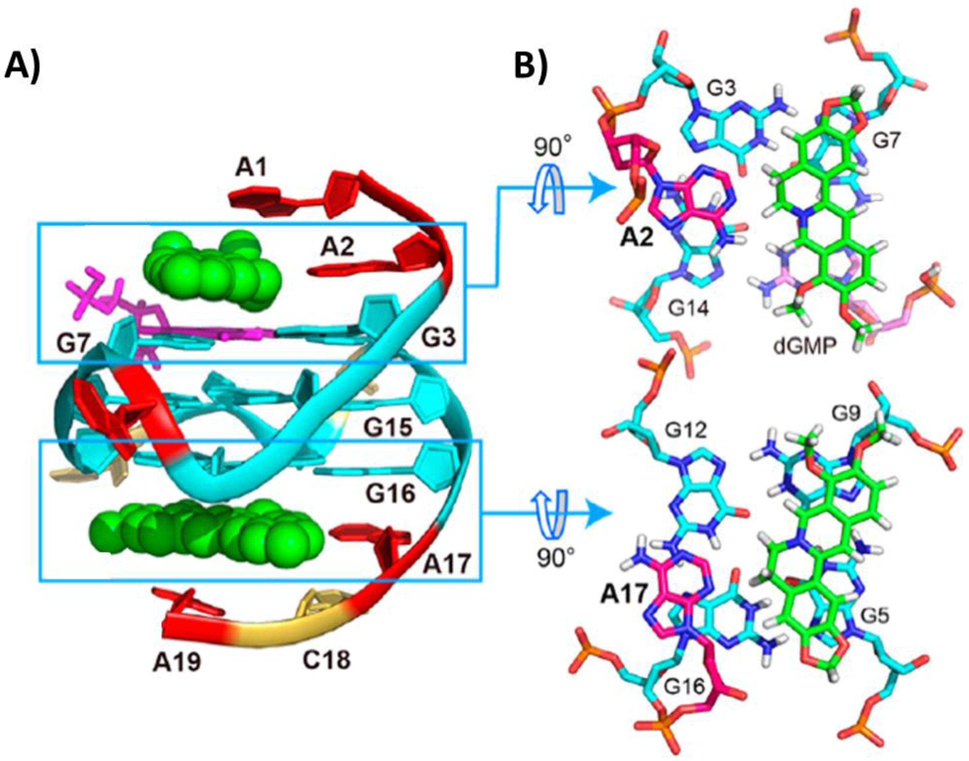
| 6V0L | NMR | Organic | Nucleotide | dGMP | d(AAGGGAGGGCGGCGGGACA) | PDGFR-β | K+ | Unimolecular | Parallel | G-vacancy filling (1:1) | Stacking and hydrogen bonds | [95] |
| 7MSV | NMR | Organic | Nucleotide, berberine | dGMP and berberine | d(AAGGGAGGGCGGCGGGACA) | PDGFR-β | K+ | Unimolecular | Parallel | G-vacancy filling, and 5′- and 3′-end G-tetrad stacking (1:1:2) | Stacking, hydrogen bonds and electrostatic interactions | [96] |
| VEGFoncogene promoter G-quadruplexes | ||||||||||||
| 6LNZ | NMR | Metal-organic | Pt(II) complex | Pt2 | d(CGGGGCGGGCCTTGGGCGGGGT) | VEGF | K+ | Unimolecular | Parallel | Intercalation between 3′-end G-tetrad and loop/flanking C10/G21 (1:1) | Stacking | [97] |
| Viral G-quadruplexes | ||||||||||||
| 6JJI | X-ray crystallography | Organic | Porphyrin | TMPyP4 | r(GGCUCGGCGGCGGA) | IE180 | K+ | Bimolecular | Parallel | Intercalation between 3′-end G-tetrad and AA-coupling plane (1:1) | Stacking | [98] |
| 6JJH | X-ray crystallography | Organic | Porphyrin | TMPyP4 | r(GGCUCGGCGGCGGA) | IE180 | K+ | Bimolecular | Parallel | Intercalation between 3′-end G-tetrad and AA-coupling plane, and stacking between a pair of cytocines and a pair of uracils of two G-quadruplex units (1:2) | Stacking | [98] |
Supplementary Materials
Author Contributions
Funding
Institutional Review Board Statement
Informed Consent Statement
Data Availability Statement
Conflicts of Interest
References
- Bochman, M.L.; Paeschke, K.; Zakian, V.A. DNA secondary structures: Stability and function of G-quadruplex structures. Nat. Rev. Genet. 2012, 13, 770–780. [Google Scholar] [CrossRef] [PubMed] [Green Version]
- Asamitsu, S.; Obata, S.; Yu, Z.; Bando, T.; Sugiyama, H. Recent progress of targeted G-quadruplex-preferred ligands toward cancer therapy. Molecules 2019, 24, 429. [Google Scholar] [CrossRef] [PubMed] [Green Version]
- Hui, W.W.I.; Simeone, A.; Zyner, K.G.; Tannahill, D.; Balasubramanian, S. Single-cell mapping of DNA G-quadruplex structures in human cancer cells. Sci. Rep. 2021, 11, 23641. [Google Scholar] [CrossRef] [PubMed]
- Platella, C.; Napolitano, E.; Riccardi, C.; Musumeci, D.; Montesarchio, D. Disentangling the structure-activity relationships of naphthalene diimides as anticancer G-quadruplex-targeting drugs. J. Med. Chem. 2021, 64, 3578–3603. [Google Scholar] [CrossRef]
- Burge, S.; Parkinson, G.N.; Hazel, P.; Todd, A.K.; Neidle, S. Quadruplex DNA: Sequence, topology and structure. Nucleic Acids Res. 2006, 34, 5402–5415. [Google Scholar] [CrossRef] [Green Version]
- Platella, C.; Riccardi, C.; Montesarchio, D.; Roviello, G.N.; Musumeci, D. G-quadruplex-based aptamers against protein targets in therapy and diagnostics. Biochim. Biophys. Acta-Gen. Subj. 2017, 1861, 1429–1447. [Google Scholar] [CrossRef]
- Spiegel, J.; Adhikari, S.; Balasubramanian, S. The structure and function of DNA G-quadruplexes. Trends Chem. 2020, 2, 123–136. [Google Scholar] [CrossRef] [Green Version]
- Hazel, P.; Parkinson, G.N.; Neidle, S. Predictive modelling of topology and loop variations in dimeric DNA quadruplex structures. Nucleic Acids Res. 2006, 34, 2117–2127. [Google Scholar] [CrossRef]
- Platella, C.; Napolitano, E.; Riccardi, C.; Musumeci, D.; Montesarchio, D. Affinity chromatography-based assays for the screening of potential ligands selective for G-quadruplex structures. ChemistryOpen 2022, 11, e202200090. [Google Scholar] [CrossRef]
- Shay, J.W.; Wright, W.E. Telomeres and telomerase: Three decades of progress. Nat. Rev. Genet. 2019, 20, 299–309. [Google Scholar] [CrossRef]
- Shay, J.W.; Wright, W.E. Telomerase therapeutics for cancer: Challenges and new directions. Nat. Rev. Drug Discov. 2006, 5, 577–584. [Google Scholar] [CrossRef]
- Huppert, J.L.; Balasubramanian, S. G-quadruplexes in promoters throughout the human genome. Nucleic Acids Res. 2007, 35, 406–413. [Google Scholar] [CrossRef] [PubMed] [Green Version]
- Huppert, J.L.; Bugaut, A.; Kumari, S.; Balasubramanian, S. G-quadruplexes: The beginning and end of UTRs. Nucleic Acids Res. 2008, 36, 6260–6268. [Google Scholar] [CrossRef] [PubMed] [Green Version]
- Eddy, J.; Maizels, N. Conserved elements with potential to form polymorphic G-quadruplex structures in the first intron of human genes. Nucleic Acids Res. 2008, 36, 1321–1333. [Google Scholar] [CrossRef] [PubMed] [Green Version]
- Ruggiero, E.; Zanin, I.; Terreri, M.; Richter, S.N. G-quadruplex targeting in the fight against viruses: An update. Int. J. Mol. Sci. 2021, 22, 10984. [Google Scholar] [CrossRef]
- Abiri, A.; Lavigne, M.; Rezaei, M.; Nikzad, S.; Zare, P.; Mergny, J.L.; Rahimi, H.R. Unlocking G-quadruplexes as antiviral targets. Pharmacol. Rev. 2021, 73, 897–923. [Google Scholar] [CrossRef] [PubMed]
- Maiti, A.K. Identification of G-quadruplex DNA sequences in SARS-CoV2. Immunogenetics 2022, 74, 455–463. [Google Scholar] [CrossRef]
- Nakanishi, C.; Seimiya, H. G-quadruplex in cancer biology and drug discovery. Biochem. Biophys. Res. Commun. 2020, 531, 45–50. [Google Scholar] [CrossRef]
- Li, Q.; Xiang, J.F.; Yang, Q.F.; Sun, H.X.; Guan, A.J.; Tang, Y.L. G4LDB: A database for discovering and studying G-quadruplex ligands. Nucleic Acids Res. 2013, 41, 1115–1123. [Google Scholar] [CrossRef] [Green Version]
- Santos, T.; Salgado, G.F.; Cabrita, E.J.; Cruz, C. G-quadruplexes and their ligands: Biophysical methods to unravel G-quadruplex/ligand interactions. Pharmaceuticals 2021, 14, 769. [Google Scholar] [CrossRef]
- Zok, T.; Kraszewska, N.; Miskiewicz, J.; Pielacinska, P.; Zurkowski, M.; Szachniuk, M. ONQUADRO: A database of experimentally determined quadruplex structures. Nucleic Acids Res. 2022, 50, D253–D258. [Google Scholar] [CrossRef] [PubMed]
- Wang, Y.H.; Yang, Q.F.; Lin, X.; Chen, D.; Wang, Z.Y.; Chen, B.; Han, H.Y.; Chen, H.D.; Cai, K.C.; Li, Q.; et al. G4LDB 2.2: A database for discovering and studying G-quadruplex and i-Motif ligands. Nucleic Acids Res. 2022, 50, D150–D160. [Google Scholar] [CrossRef]
- Mendes, E.; Aljnadi, I.M.; Bahls, B.; Victor, B.L.; Paulo, A. Major achievements in the design of quadruplex-interactive small molecules. Pharmaceuticals 2022, 15, 300. [Google Scholar] [CrossRef] [PubMed]
- Platella, C.; Trajkovski, M.; Doria, F.; Freccero, M.; Plavec, J.; Montesarchio, D. On the interaction of an anticancer trisubstituted naphthalene diimide with G-quadruplexes of different topologies: A structural insight. Nucleic Acids Res. 2020, 48, 12380–12393. [Google Scholar] [CrossRef]
- Platella, C.; Pirota, V.; Musumeci, D.; Rizzi, F.; Iachettini, S.; Zizza, P.; Biroccio, A.; Freccero, M.; Montesarchio, D.; Doria, F. Trifunctionalized naphthalene diimides and dimeric analogues as G-quadruplex-targeting anticancer agents selected by affinity chromatography. Int. J. Mol. Sci. 2020, 21, 1964. [Google Scholar] [CrossRef] [Green Version]
- Pirota, V.; Platella, C.; Musumeci, D.; Benassi, A.; Amato, J.; Pagano, B.; Colombo, G.; Freccero, M.; Doria, F.; Montesarchio, D. On the binding of naphthalene diimides to a human telomeric G-quadruplex multimer model. Int. J. Biol. Macromol. 2021, 166, 1320–1334. [Google Scholar] [CrossRef] [PubMed]
- Murat, P.; Singh, Y.; Defrancq, E. Methods for investigating G-quadruplex DNA/ligand interactions. Chem. Soc. Rev. 2011, 40, 5293–5307. [Google Scholar] [CrossRef]
- Campbell, N.H.; Parkinson, G.N. Crystallographic studies of quadruplex nucleic acids. Methods 2007, 43, 252–263. [Google Scholar] [CrossRef]
- Lin, C.; Dickerhoff, J.; Yang, D. NMR studies of G-quadruplex structures and G-quadruplex-interactive compounds. Methods Mol. Biol. 2019, 2035, 157–176. [Google Scholar]
- Parkinson, G.N.; Collie, G.W. X-ray crystallographic studies of G-quadruplex structures. Methods Mol. Biol. 2019, 2035, 131–155. [Google Scholar]
- Wilson, T.; Costa, P.J.; Félix, V.; Williamson, M.P.; Thomas, J.A. Structural studies on dinuclear ruthenium(II) complexes that bind diastereoselectively to an antiparallel folded human telomere sequence. J. Med. Chem. 2013, 56, 8674–8683. [Google Scholar] [CrossRef] [PubMed]
- Wilson, T.; Williamson, M.P.; Thomas, J.A. Differentiating quadruplexes: Binding preferences of a luminescent dinuclear ruthenium(ii) complex with four-stranded DNA structures. Org. Biomol. Chem. 2010, 8, 2617–2621. [Google Scholar] [CrossRef]
- McQuaid, K.T.; Takahashi, S.; Baumgaertner, L.; Cardin, D.J.; Paterson, N.G.; Hall, J.P.; Sugimoto, N.; Cardin, C.J. Ruthenium polypyridyl complex bound to a unimolecular chair-form G-quadruplex. J. Am. Chem. Soc. 2022, 144, 5956–5964. [Google Scholar] [CrossRef] [PubMed]
- Franceschin, M.; Rossetti, L.; D’Ambrosio, A.; Schirripa, S.; Bianco, A.; Ortaggi, G.; Savino, M.; Schultes, C.; Neidle, S. Natural and synthetic G-quadruplex interactive berberine derivatives. Bioorg. Med. Chem. Lett. 2006, 16, 1707–1711. [Google Scholar] [CrossRef]
- Noureini, S.K.; Esmaeili, H.; Abachi, F.; Khiali, S.; Islam, B.; Kuta, M.; Saboury, A.A.; Hoffmann, M.; Sponer, J.; Parkinson, G.; et al. Selectivity of major isoquinoline alkaloids from Chelidonium majus towards telomeric G-quadruplex: A study using a transition-FRET (t-FRET) assay. Biochim. Biophys. Acta-Gen. Subj. 2017, 1861, 2020–2030. [Google Scholar] [CrossRef] [PubMed] [Green Version]
- Zhang, L.; Liu, H.; Shao, Y.; Lin, C.; Jia, H.; Chen, G.; Yang, D.; Wang, Y. Selective lighting up of epiberberine alkaloid fluorescence by fluorophore-switching aptamer and stoichiometric targeting of human telomeric DNA G-quadruplex multimer. Anal. Chem. 2015, 87, 730–737. [Google Scholar] [CrossRef] [PubMed] [Green Version]
- Lin, C.; Wu, G.; Wang, K.; Onel, B.; Sakai, S.; Shao, Y.; Yang, D. Molecular recognition of the hybrid-2 human telomeric G-quadruplex by epiberberine: Insights into conversion of telomeric G-quadruplex structures. Angew. Chem.-Int. Ed. 2018, 57, 10888–10893. [Google Scholar] [CrossRef]
- Wirmer-Bartoschek, J.; Bendel, L.E.; Jonker, H.R.A.; Grün, J.T.; Papi, F.; Bazzicalupi, C.; Messori, L.; Gratteri, P.; Schwalbe, H. Solution NMR structure of a ligand/hybrid-2-G-quadruplex complex reveals rearrangements that affect ligand binding. Angew. Chem.-Int. Ed. 2017, 56, 7102–7106. [Google Scholar] [CrossRef] [Green Version]
- Liu, L.Y.; Liu, W.; Wang, K.N.; Zhu, B.C.; Xia, X.Y.; Ji, L.N.; Mao, Z.W. Quantitative detection of G-quadruplex DNA in live cells based on photon counts and complex structure discrimination. Angew. Chem.-Int. Ed. 2020, 59, 9719–9726. [Google Scholar] [CrossRef]
- Liu, W.; Zhong, Y.-F.; Liu, L.-Y.; Shen, C.-T.; Zeng, W.; Wang, F.; Yang, D.; Mao, Z.-W. Solution structures of multiple G-quadruplex complexes induced by a platinum(II)-based tripod reveal dynamic binding. Nat. Commun. 2018, 9, 3496–3506. [Google Scholar] [CrossRef]
- Chung, W.J.; Heddi, B.; Tera, M.; Iida, K.; Nagasawa, K.; Phan, A.T. Solution structure of an intramolecular (3 + 1) human telomeric G-quadruplex bound to a telomestatin derivative. J. Am. Chem. Soc. 2013, 135, 13495–13501. [Google Scholar] [CrossRef] [PubMed]
- Ghosh, A.; Trajkovski, M.; Teulade-Fichou, M.; Gabelica, V.; Plavec, J. Phen-DC 3 induces refolding of human telomeric DNA into a chair-type antiparallel G-quadruplex through ligand intercalation. Angew. Chem.-Int. Ed. 2022, 61, e202207384. [Google Scholar] [CrossRef] [PubMed]
- Bazzicalupi, C.; Ferraroni, M.; Bilia, A.R.; Scheggi, F.; Gratteri, P. The crystal structure of human telomeric DNA complexed with berberine: An interesting case of stacked ligand to G-tetrad ratio higher than 1:1. Nucleic Acids Res. 2013, 41, 632–638. [Google Scholar] [CrossRef] [PubMed]
- Bazzicalupi, C.; Ferraroni, M.; Papi, F.; Massai, L.; Bertrand, B.; Messori, L.; Gratteri, P.; Casini, A. Determinants for tight and selective binding of a medicinal dicarbene gold(I) complex to a telomeric DNA G-quadruplex: A joint ESI MS and XRD investigation. Angew. Chem.-Int. Ed. 2016, 55, 4256–4259. [Google Scholar] [CrossRef] [PubMed]
- Guarra, F.; Marzo, T.; Ferraroni, M.; Papi, F.; Bazzicalupi, C.; Gratteri, P.; Pescitelli, G.; Messori, L.; Biver, T.; Gabbiani, C. Interaction of a gold(i) dicarbene anticancer drug with human telomeric DNA G-quadruplex: Solution and computationally aided X-ray diffraction analysis. Dalton Trans. 2018, 47, 16132–16138. [Google Scholar] [CrossRef] [PubMed]
- Cuenca, F.; Greciano, O.; Gunaratnam, M.; Haider, S.; Munnur, D.; Nanjunda, R.; Wilson, W.D.; Neidle, S. Tri- and tetra-substituted naphthalene diimides as potent G-quadruplex ligands. Bioorg. Med. Chem. Lett. 2008, 18, 1668–1673. [Google Scholar] [CrossRef]
- Parkinson, G.N.; Cuenca, F.; Neidle, S. Topology conservation and loop flexibility in quadruplex-drug recognition: Crystal structures of inter- and intramolecular telomeric DNA quadruplex-drug complexes. J. Mol. Biol. 2008, 381, 1145–1156. [Google Scholar] [CrossRef]
- Collie, G.W.; Promontorio, R.; Hampel, S.M.; Micco, M.; Neidle, S.; Parkinson, G.N. Structural basis for telomeric G-quadruplex targeting by naphthalene diimide ligands. J. Am. Chem. Soc. 2012, 134, 2723–2731. [Google Scholar] [CrossRef]
- Micco, M.; Collie, G.W.; Dale, A.G.; Ohnmacht, S.A.; Pazitna, I.; Gunaratnam, M.; Reszka, A.P.; Neidle, S. Structure-based design and evaluation of naphthalene diimide G-quadruplex ligands as telomere targeting agents in pancreatic cancer cells. J. Med. Chem. 2013, 56, 2959–2974. [Google Scholar] [CrossRef]
- Nicoludis, J.M.; Miller, S.T.; Jeffrey, P.D.; Barrett, S.P.; Rablen, P.R.; Lawton, T.J.; Yatsunyk, L.A. Optimized end-stacking provides specificity of N-methyl mesoporphyrin IX for human telomeric G-quadruplex DNA. J. Am. Chem. Soc. 2012, 134, 20446–20456. [Google Scholar] [CrossRef]
- Arthanari, H.; Basu, S.; Kawano, T.L.; Bolton, P.H. Fluorescent dyes specific for quadruplex DNA. Nucleic Acids Res. 1998, 26, 3724–3728. [Google Scholar] [CrossRef] [PubMed] [Green Version]
- Ren, J.; Chaires, J.B. Sequence and structural selectivity of nucleic acid binding ligands. Biochemistry 1999, 38, 16067–16075. [Google Scholar] [CrossRef] [PubMed]
- Miron, C.E.; van Staalduinen, L.; Rangaswamy, A.M.; Chen, M.; Liang, Y.; Jia, Z.; Mergny, J.L.; Petitjean, A. Going platinum to the tune of a remarkable guanine quadruplex binder: Solution- and solid-state investigations. Angew. Chem.-Int. Ed. 2021, 60, 2500–2507. [Google Scholar] [CrossRef] [PubMed]
- Ferraroni, M.; Bazzicalupi, C.; Papi, F.; Fiorillo, G.; Guamán-Ortiz, L.M.; Nocentini, A.; Scovassi, A.I.; Lombardi, P.; Gratteri, P. Solution and solid-state analysis of binding of 13-substituted berberine analogues to human telomeric G-quadruplexes. Chem.-Asian J. 2016, 11, 1107–1115. [Google Scholar] [CrossRef] [PubMed]
- Papi, F.; Bazzicalupi, C.; Ferraroni, M.; Ciolli, G.; Lombardi, P.; Khan, A.Y.; Kumar, G.S.; Gratteri, P. Pyridine derivative of the natural alkaloid berberine as human telomeric G4-DNA binder: A solution and solid-state study. ACS Med. Chem. Lett. 2020, 11, 645–650. [Google Scholar] [CrossRef]
- Incles, C.M.; Schultes, C.M.; Kempski, H.; Koehler, H.; Kelland, L.R.; Neidle, S. A G-quadruplex telomere targeting agent produces p16-associated senescence and chromosomal fusions in human prostate cancer cells. Mol. Cancer Ther. 2004, 3, 1201–1206. [Google Scholar] [CrossRef]
- Burger, A.M.; Dai, F.; Schultes, C.M.; Reszka, A.P.; Moore, M.J.; Double, J.A.; Neidle, S. The G-quadruplex-interactive molecule BRACO-19 inhibits tumor growth, consistent with telomere targeting and interference with telomerase function. Cancer Res. 2005, 65, 1489–1496. [Google Scholar] [CrossRef] [Green Version]
- Campbell, N.H.; Parkinson, G.N.; Reszka, A.P.; Neidle, S. Structural basis of DNA quadruplex recognition by an acridine drug. J. Am. Chem. Soc. 2008, 130, 6722–6724. [Google Scholar] [CrossRef]
- Parkinson, G.N.; Ghosh, R.; Neidle, S. Structural basis for binding of porphyrin to human telomeres. Biochemistry 2007, 46, 2390–2397. [Google Scholar] [CrossRef]
- Campbell, N.H.; Karim, N.H.A.; Parkinson, G.N.; Gunaratnam, M.; Petrucci, V.; Todd, A.K.; Vilar, R.; Neidle, S. Molecular basis of structure-activity relationships between salphen metal complexes and human telomeric DNA quadruplexes. J. Med. Chem. 2012, 55, 209–222. [Google Scholar] [CrossRef]
- McQuaid, K.; Abell, H.; Gurung, S.P.; Allan, D.R.; Winter, G.; Sorensen, T.; Cardin, D.J.; Brazier, J.A.; Cardin, C.J.; Hall, J.P. Structural studies reveal enantiospecific recognition of a DNA G-quadruplex by a ruthenium polypyridyl complex. Angew. Chem.-Int. Ed. 2019, 58, 9881–9885. [Google Scholar] [CrossRef] [PubMed]
- McQuaid, K.; Hall, J.P.; Baumgaertner, L.; Cardin, D.J.; Cardin, C.J. Three thymine/adenine binding modes of the ruthenium complex Λ-[Ru(TAP)2(dppz)]2+ to the G-quadruplex forming sequence d(TAGGGTT) shown by X-ray crystallography. Chem. Commun. 2019, 55, 9116–9119. [Google Scholar] [CrossRef] [PubMed]
- Barthwal, R.; Raje, S.; Pandav, K. Structural basis for stabilization of human telomeric G-quadruplex [d-(TTAGGGT)]4 by anticancer drug epirubicin. Bioorg. Med. Chem. 2020, 28, 115761. [Google Scholar] [CrossRef] [PubMed]
- Barthwal, R.; Raje, S.; Pandav, K. Structural basis for stabilization of human telomeric G-quadruplex [d-(TTAGGGT)]4 by anticancer drug adriamycin. J. Biomol. Struct. Dyn. 2021, 39, 795–815. [Google Scholar] [CrossRef]
- Kessler, M.; Ubeaud, G.; Jung, L. Anti- and pro-oxidant activity of rutin and quercetin derivatives. J. Pharm. Pharmacol. 2010, 55, 131–142. [Google Scholar] [CrossRef]
- Carini, J.P.; Klamt, F.; Bassani, V.L. Flavonoids from achyrocline satureioides: Promising biomolecules for anticancer therapy. RSC Adv. 2014, 4, 3131–3144. [Google Scholar] [CrossRef]
- Tawani, A.; Kumar, A. Structural insight into the interaction of flavonoids with human telomeric sequence. Sci. Rep. 2015, 5, 17574. [Google Scholar] [CrossRef] [Green Version]
- Hounsou, C.; Guittat, L.; Monchaud, D.; Jourdan, M.; Saettel, N.; Mergny, J.L.; Teulade-Fichou, M.P. G-quadruplex recognition by quinacridines: A SAR, NMR, and biological study. ChemMedChem 2007, 2, 655–666. [Google Scholar] [CrossRef]
- Gavathiotis, E.; Heald, R.A.; Stevens, M.F.G.; Searle, M.S. Drug recognition and stabilisation of the parallel-stranded DNA quadruplex d(TTAGGGT)4 containing the human telomeric repeat. J. Mol. Biol. 2003, 334, 25–36. [Google Scholar] [CrossRef]
- Lin, L.Y.; McCarthy, S.; Powell, B.M.; Manurung, Y.; Xiang, I.M.; Dean, W.L.; Chaires, B.; Yatsunyk, L.A. Biophysical and X-ray structural studies of the (GGGTT)3GGG G-quadruplex in complex with N-methyl mesoporphyrin IX. PLoS ONE 2020, 15, e0241513. [Google Scholar] [CrossRef]
- Winnerdy, F.R.; Das, P.; Heddi, B.; Phan, A.T. Solution structures of a G-quadruplex bound to linear- and cyclic-dinucleotides. J. Am. Chem. Soc. 2019, 141, 18038–18047. [Google Scholar] [CrossRef] [PubMed]
- Haider, S.M.; Parkinson, G.N.; Neidle, S. Structure of a G-quadruplex-ligand complex. J. Mol. Biol. 2003, 326, 117–125. [Google Scholar] [CrossRef]
- Campbell, N.H.; Patel, M.; Tofa, A.B.; Ghosh, R.; Parkinson, G.N.; Neidle, S. Selectivity in ligand recognition of G-quadruplex loops. Biochemistry 2009, 48, 1675–1680. [Google Scholar] [CrossRef] [PubMed]
- Campbell, N.H.; Smith, D.L.; Reszka, A.P.; Neidle, S.; O’Hagan, D. Fluorine in medicinal chemistry: β-fluorination of peripheral pyrrolidines attached to acridine ligands affects their interactions with G-quadruplex DNA. Org. Biomol. Chem. 2011, 9, 1328–1331. [Google Scholar] [CrossRef] [PubMed]
- Mandal, P.K.; Baptiste, B.; Langlois d’Estaintot, B.; Kauffmann, B.; Huc, I. Multivalent interactions between an aromatic helical foldamer and a DNA G-quadruplex in the solid state. ChemBioChem 2016, 17, 1911–1914. [Google Scholar] [CrossRef] [PubMed]
- Clark, G.R.; Pytel, P.D.; Squire, C.J.; Neidle, S. Structure of the first parallel DNA quadruplex-drug complex. J. Am. Chem. Soc. 2003, 125, 4066–4067. [Google Scholar] [CrossRef]
- Clark, G.R.; Pytel, P.D.; Squire, C.J. The high-resolution crystal structure of a parallel intermolecular DNA G-4 quadruplex/drug complex employing syn glycosyl linkages. Nucleic Acids Res. 2012, 40, 5731–5738. [Google Scholar] [CrossRef] [Green Version]
- Martino, L.; Virno, A.; Pagano, B.; Virgilio, A.; Di Micco, S.; Galeone, A.; Giancola, C.; Bifulco, G.; Mayol, L.; Randazzo, A. Structural and thermodynamic studies of the interaction of distamycin A with the parallel quadruplex structure [d(TGGGGT)]4. J. Am. Chem. Soc. 2007, 129, 16048–16056. [Google Scholar] [CrossRef]
- Cosconati, S.; Marinelli, L.; Trotta, R.; Virno, A.; De Tito, S.; Romagnoli, R.; Pagano, B.; Limongelli, V.; Giancola, C.; Baraldi, P.G.; et al. Structural and conformational requisites in DNA quadruplex groove binding: Another piece to the puzzle. J. Am. Chem. Soc. 2010, 132, 6425–6433. [Google Scholar] [CrossRef]
- Azzalin, C.M.; Reichenbach, P.; Khoriauli, L.; Giulotto, E.; Lingner, J. Telomeric repeat–containing RNA and RNA surveillance factors at mammalian chromosome ends. Science 2007, 318, 798–801. [Google Scholar] [CrossRef]
- Schoeftner, S.; Blasco, M.A. Developmentally regulated transcription of mammalian telomeres by DNA-dependent RNA polymerase II. Nat. Cell Biol. 2008, 10, 228–236. [Google Scholar] [CrossRef] [PubMed]
- Martadinata, H.; Phan, A.T. Structure of propeller-type parallel-stranded RNA G-quadruplexes, formed by human telomeric RNA sequences in K+ solution. J. Am. Chem. Soc. 2009, 131, 2570–2579. [Google Scholar] [CrossRef]
- Phan, A.T. Human telomeric G-quadruplex: Structures of DNA and RNA sequences. FEBS J. 2010, 277, 1107–1117. [Google Scholar] [CrossRef]
- Collie, G.W.; Sparapani, S.; Parkinson, G.N.; Neidle, S. Structural basis of telomeric RNA quadruplex-acridine ligand recognition. J. Am. Chem. Soc. 2011, 133, 2721–2728. [Google Scholar] [CrossRef] [PubMed]
- Yang, D.; Okamoto, K. Structural insights into G-quadruplexes: Towards new anticancer drugs. Future Med. Chem. 2010, 2, 619–646. [Google Scholar] [CrossRef] [PubMed] [Green Version]
- Chung, W.J.; Heddi, B.; Hamon, F.; Teulade-Fichou, M.P.; Phan, A.T. Solution structure of a G-quadruplex bound to the bisquinolinium compound Phen-DC3. Angew. Chem.-Int. Ed. 2014, 53, 999–1002. [Google Scholar] [CrossRef] [PubMed]
- Phan, A.T.; Kuryavyi, V.; Gaw, H.Y.; Patel, D.J. Small-molecule interaction with a five-guanine-tract G-quadruplex structure from the human myc promoter. Nat. Chem. Biol. 2005, 1, 167–173. [Google Scholar] [CrossRef] [Green Version]
- Dickerhoff, J.; Brundridge, N.; McLuckey, S.A.; Yang, D. Berberine molecular recognition of the parallel MYC G-quadruplex in solution. J. Med. Chem. 2021, 64, 16205–16212. [Google Scholar] [CrossRef]
- Dai, J.; Carver, M.; Hurley, L.H.; Yang, D. Solution structure of a 2:1 quindoline-c-MYC G-quadruplex: Insights into G-quadruplex-interactive small molecule drug design. J. Am. Chem. Soc. 2011, 133, 17673–17680. [Google Scholar] [CrossRef] [Green Version]
- Dickerhoff, J.; Dai, J.; Yang, D. Structural recognition of the MYC promoter G-quadruplex by a quinoline derivative: Insights into molecular targeting of parallel G-quadruplexes. Nucleic Acids Res. 2021, 49, 5905–5915. [Google Scholar] [CrossRef]
- Calabrese, D.R.; Chen, X.; Leon, E.C.; Gaikwad, S.M.; Phyo, Z.; Hewitt, W.M.; Alden, S.; Hilimire, T.A.; He, F.; Michalowski, A.M.; et al. Chemical and structural studies provide a mechanistic basis for recognition of the MYC G-quadruplex. Nat. Commun. 2018, 9, 4229. [Google Scholar] [CrossRef] [PubMed] [Green Version]
- Liu, W.; Lin, C.; Wu, G.; Dai, J.; Chang, T.C.; Yang, D. Structures of 1:1 and 2:1 complexes of BMVC and MYC promoter G-quadruplex reveal a mechanism of ligand conformation adjustment for G4-recognition. Nucleic Acids Res. 2019, 47, 11931–11942. [Google Scholar] [CrossRef] [PubMed]
- Kotar, A.; Wang, B.; Shivalingam, A.; Gonzalez-Garcia, J.; Vilar, R.; Plavec, J. NMR structure of a triangulenium-based long-lived fluorescence probe bound to a G-quadruplex. Angew. Chem.-Int. Ed. 2016, 55, 12508–12511. [Google Scholar] [CrossRef] [PubMed]
- Wang, F.; Wang, C.; Liu, Y.; Lan, W.; Han, H.; Wang, R.; Huang, S.; Cao, C. Colchicine selective interaction with oncogene: RET G-quadruplex revealed by NMR. Chem. Commun. 2020, 56, 2099–2102. [Google Scholar] [CrossRef]
- Wang, K.B.; Dickerhoff, J.; Wu, G.; Yang, D. PDGFR-β promoter forms a vacancy G-quadruplex that can be filled in by dGMP: Solution structure and molecular recognition of guanine metabolites and drugs. J. Am. Chem. Soc. 2020, 142, 5204–5211. [Google Scholar] [CrossRef]
- Wang, K.B.; Dickerhoff, J.; Yang, D. Solution structure of ternary complex of berberine bound to a dGMP-fill-in vacancy G-quadruplex formed in the PDGFR-β promoter. J. Am. Chem. Soc. 2021, 143, 16549–16555. [Google Scholar] [CrossRef] [PubMed]
- Zhu, B.C.; He, J.; Liu, W.; Xia, X.Y.; Liu, L.Y.; Liang, B.B.; Yao, H.G.; Liu, B.; Ji, L.N.; Mao, Z.W. Selectivity and targeting of G-quadruplex binders activated by adaptive binding and controlled by chemical kinetics. Angew. Chem.-Int. Ed. 2021, 60, 15340–15343. [Google Scholar] [CrossRef]
- Zhang, Y.; El Omari, K.; Duman, R.; Liu, S.; Haider, S.; Wagner, A.; Parkinson, G.N.; Wei, D. Native de novo structural determinations of non-canonical nucleic acid motifs by X-ray crystallography at long wavelengths. Nucleic Acids Res. 2020, 48, 9886–9898. [Google Scholar] [CrossRef]
- Krafcikova, M.; Hänsel-Hertsch, R.; Trantirek, L.; Foldynova-Trantirkova, S. In cell NMR spectroscopy: Investigation of G-quadruplex structures inside living xenopus laevis oocytes. In Methods in Molecular Biology; Oxford University Press: Oxford, UK, 2019; Volume 2035, pp. 397–405. ISBN 9781493996667. [Google Scholar]
- Krafčík, D.; Ištvánková, E.; Džatko, Š.; Víšková, P.; Foldynová-Trantírková, S.; Trantírek, L. Towards profiling of the G-quadruplex targeting drugs in the living human cells using NMR spectroscopy. Int. J. Mol. Sci. 2021, 22, 6042. [Google Scholar] [CrossRef]
- Krzyżak, A.T.; Habina-Skrzyniarz, I.; Mazur, W.; Sułkowski, M.; Kot, M.; Majka, M. Nuclear magnetic resonance footprint of Wharton Jelly mesenchymal stem cells death mechanisms and distinctive in-cell biophysical properties in vitro. J. Cell. Mol. Med. 2022, 26, 1501–1514. [Google Scholar] [CrossRef]


Publisher’s Note: MDPI stays neutral with regard to jurisdictional claims in published maps and institutional affiliations. |
© 2022 by the authors. Licensee MDPI, Basel, Switzerland. This article is an open access article distributed under the terms and conditions of the Creative Commons Attribution (CC BY) license (https://creativecommons.org/licenses/by/4.0/).
Share and Cite
Criscuolo, A.; Napolitano, E.; Riccardi, C.; Musumeci, D.; Platella, C.; Montesarchio, D. Insights into the Small Molecule Targeting of Biologically Relevant G-Quadruplexes: An Overview of NMR and Crystal Structures. Pharmaceutics 2022, 14, 2361. https://doi.org/10.3390/pharmaceutics14112361
Criscuolo A, Napolitano E, Riccardi C, Musumeci D, Platella C, Montesarchio D. Insights into the Small Molecule Targeting of Biologically Relevant G-Quadruplexes: An Overview of NMR and Crystal Structures. Pharmaceutics. 2022; 14(11):2361. https://doi.org/10.3390/pharmaceutics14112361
Chicago/Turabian StyleCriscuolo, Andrea, Ettore Napolitano, Claudia Riccardi, Domenica Musumeci, Chiara Platella, and Daniela Montesarchio. 2022. "Insights into the Small Molecule Targeting of Biologically Relevant G-Quadruplexes: An Overview of NMR and Crystal Structures" Pharmaceutics 14, no. 11: 2361. https://doi.org/10.3390/pharmaceutics14112361