
Evaluation of Ribavirin–Poloxamer Microparticles for Improved Intranasal Absorption

 and
and 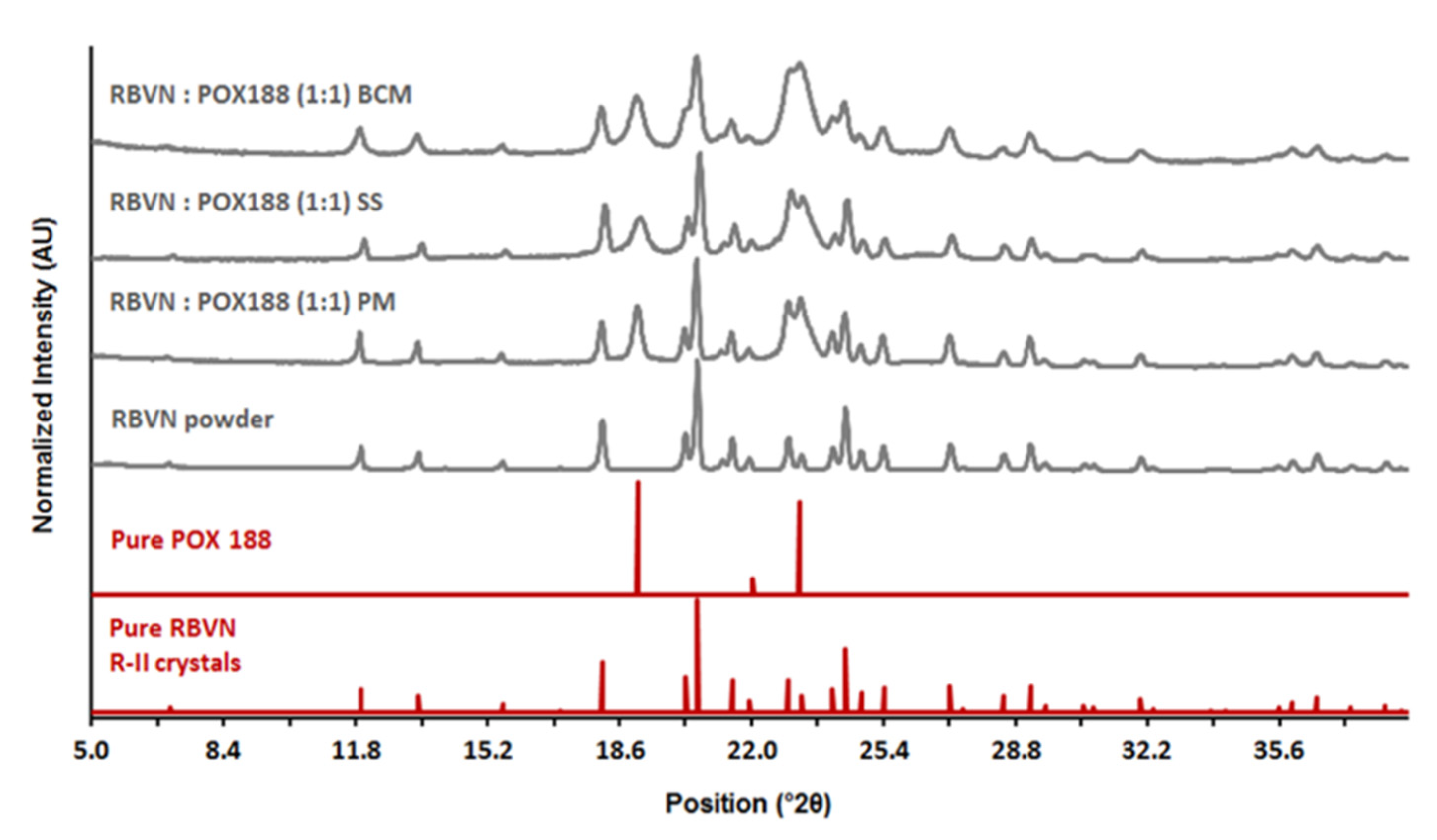
Abstract
:1. Introduction
2. Materials and Methods
2.1. Determination of Ribavirin Solubility in Solid Poloxamer 188
2.2. Preparation of Binary Composite Microparticles
2.3. Ribavirin Physical Stability in Poloxamer Microparticles
2.4. Assessment of Particle Properties
2.5. High-Performance Liquid Chromatography (HPLC)
2.6. Microparticle Drug Content and Content Uniformity
2.7. In Vitro Drug Release
2.8. In Vitro Ribavirin Permeation
2.9. In Vitro Nasal Cell Toxicity
3. Results and Discussion
3.1. Preparation and Characterization of Binary Composite Microparticles
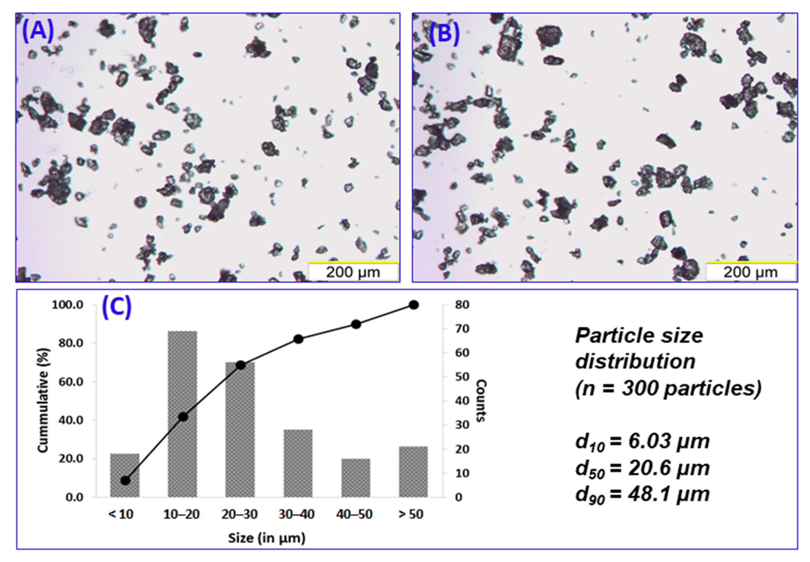
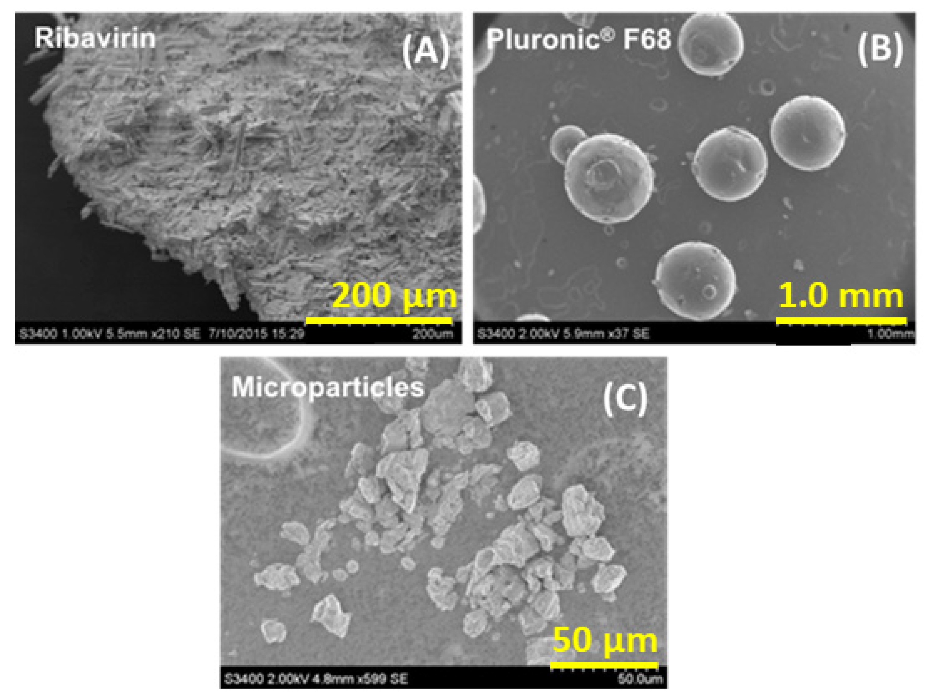
3.2. Particle Properties
3.3. Binary Composite Microparticle Performance
3.3.1. In Vitro Drug Release
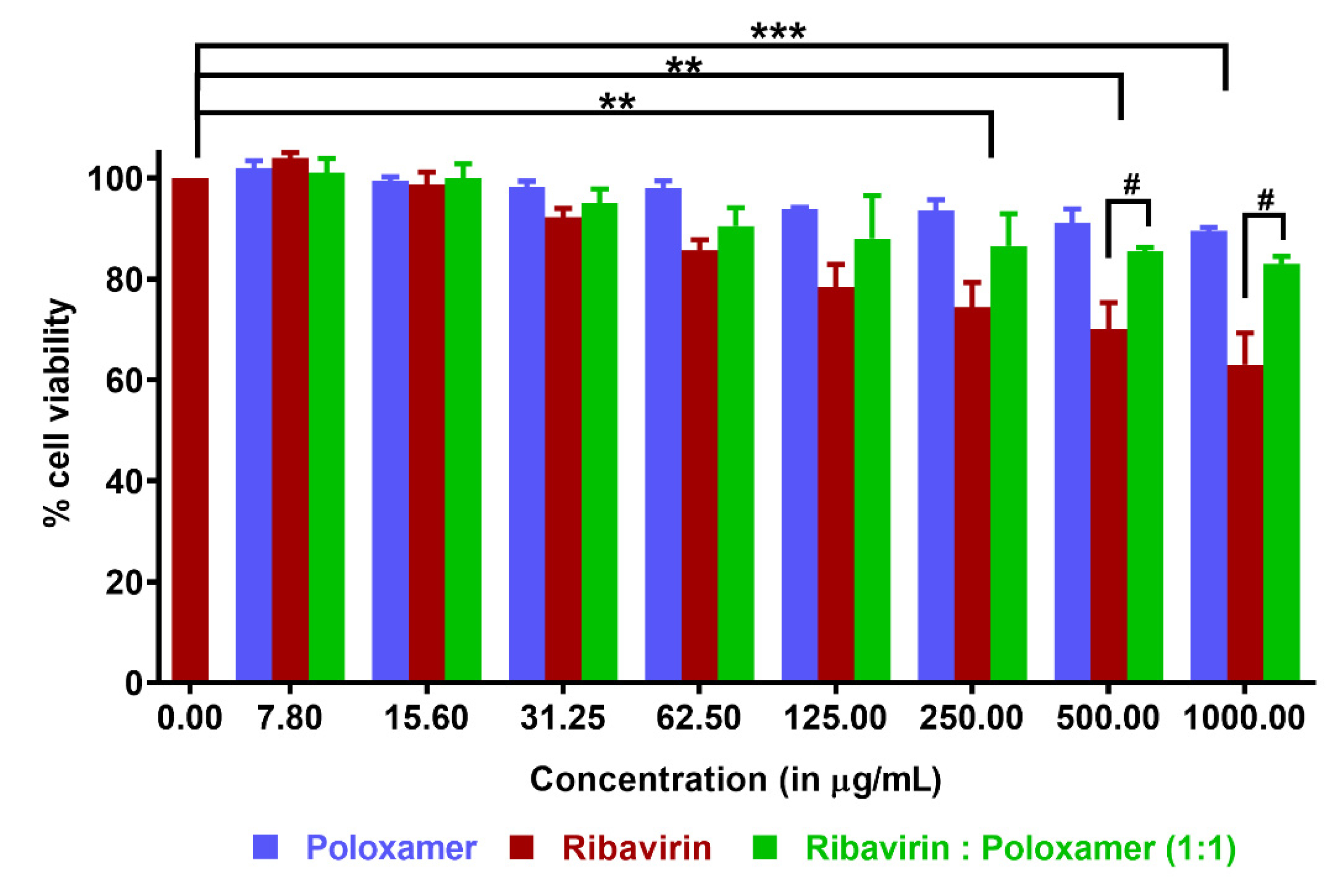
3.3.2. In Vitro Cytotoxicity
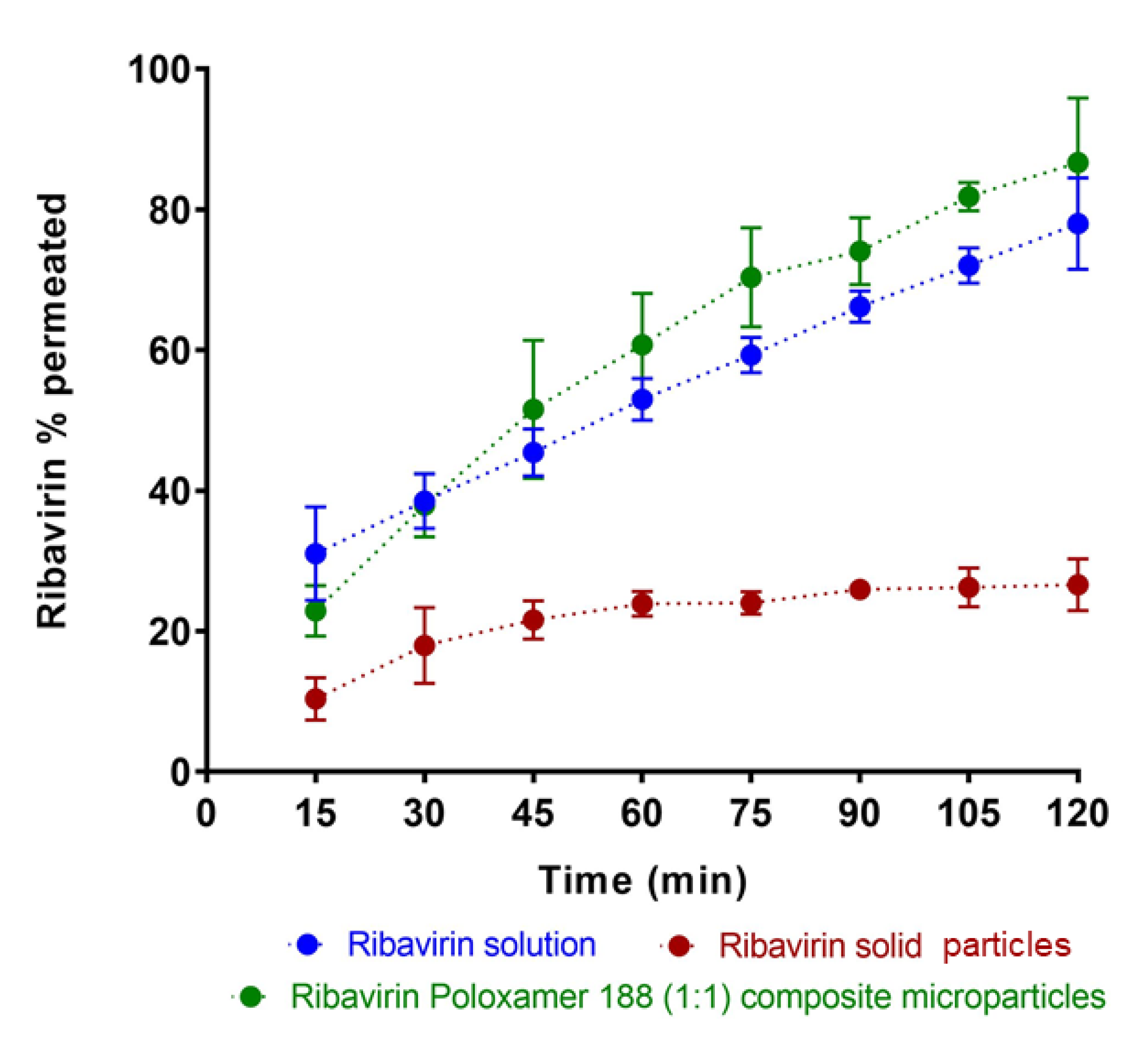
3.3.3. Ribavirin Permeation through Olfactory Mucosa
4. Conclusions
Author Contributions
Funding
Institutional Review Board Statement
Informed Consent Statement
Data Availability Statement
Acknowledgments
Conflicts of Interest
References
- Huffman, J.H.; Sidwell, R.W.; Khare, G.P.; Witkowski, J.T.; Allen, L.B.; Robins, R.K. In Vitro Effect of 1-β-d-Ribofuranosyl-1,2,4-Triazole-3-Carboxamide (Virazole, ICN 1229) on Deoxyribonucleic Acid and Ribonucleic Acid Viruses. Antimicrob. Agents Chemother. 1973, 3, 235–241. [Google Scholar] [CrossRef] [Green Version]
- Cassidy, L.F.; Patterson, J.L. Mechanism of La Crosse virus inhibition by ribavirin. Antimicrob. Agents Chemother. 1989, 33, 2009–2011. [Google Scholar] [CrossRef] [PubMed] [Green Version]
- Hosoya, M.; Shigeta, S.; Nakamura, K.; De Clercq, E. Inhibitory effect of selected antiviral compounds on measles (SSPE) virus replication in vitro. Antivir. Res. 1989, 12, 87–97. [Google Scholar] [CrossRef]
- Jordan, I.; Briese, T.; Fischer, N.; Lau, J.Y.-N.; Lipkin, W.I. Ribavirin Inhibits West Nile Virus Replication and Cytopathic Effect in Neural Cells. J. Infect. Dis. 2000, 182, 1214–1217. [Google Scholar] [CrossRef] [PubMed] [Green Version]
- Bussereau, F.; Picard, M.; Blancou, J.; Sureau, P. Treatment of rabies in mice and foxes with antiviral compounds. Acta Virol. 1988, 32, 33–49. [Google Scholar]
- Honda, Y.; Hosoya, M.; Ishii, T.; Shigeta, S.; Suzuki, H. Effect of ribavirin on subacute sclerosing panencephalitis virus infections in hamsters. Antimicrob. Agents Chemother. 1994, 38, 653–655. [Google Scholar] [CrossRef] [Green Version]
- Dhuria, S.V.; Hanson, L.R.; Frey, W.H. Intranasal delivery to the central nervous system: Mechanisms and experimental considerations. J. Pharm. Sci. 2010, 99, 1654–1673. [Google Scholar] [CrossRef]
- Graff, C.L.; Pollack, G.M. Nasal Drug Administration: Potential for Targeted Central Nervous System Delivery. J. Pharm. Sci. 2005, 94, 1187–1195. [Google Scholar] [CrossRef] [PubMed]
- Illum, L. Is nose-to-brain transport of drugs in man a reality? J. Pharm. Pharmacol. 2014, 56, 3–17. [Google Scholar] [CrossRef]
- Lochhead, J.J.; Thorne, R.G. Intranasal delivery of biologics to the central nervous system. Adv. Drug Deliv. Rev. 2012, 64, 614–628. [Google Scholar] [CrossRef]
- Merkus, F.W.; van den Berg, M.P. Can Nasal Drug Delivery Bypass the??Blood-Brain Barrier? Drugs R D 2007, 8, 133–144. [Google Scholar] [CrossRef]
- Mittal, D.; Ali, A.; MD, S.; Baboota, S.; Sahni, J.K.; Ali, J. Insights into direct nose to brain delivery: Current status and future perspective. Drug Deliv. 2014, 21, 75–86. [Google Scholar] [CrossRef] [PubMed]
- Vyas, T.K.; Shahiwala, A.; Marathe, S.; Misra, A. Intranasal Drug Delivery for Brain Targeting. Curr. Drug Deliv. 2005, 2, 165–175. [Google Scholar] [CrossRef] [PubMed]
- Landis, M.S.; Boyden, T.; Pegg, S. Nasal-to-CNS drug delivery: Where are we now and where are we heading? An industrial perspective. Ther. Deliv. 2012, 3, 195–208. [Google Scholar] [CrossRef]
- Vasa, D.M.; O’Donnell, L.; Wildfong, P.L.D. Influence of dosage form, formulation, and delivery device on olfactory deposi-tion and clearance: Enhancement of nose-to-CNS uptake. J. Pharm. Innov. 2015, 10, 200–210. [Google Scholar] [CrossRef]
- Bacon, R.; Newman, S.; Rankin, L.; Pitcairn, G.; Whiting, R. Pulmonary and nasal deposition of ketorolac tromethamine solution (SPRIX) following intranasal administration. Int. J. Pharm. 2012, 431, 39–44. [Google Scholar] [CrossRef]
- Harris, A.S.; Hedner, P.; Vilhardt, H. Nasal administration of desmopressin by spray and drops. J. Pharm. Pharmacol. 1987, 39, 932–934. [Google Scholar] [CrossRef]
- Marttin, E.; Romeijn, S.G.; Verhoef, J.C.; Merkus, F.W.H.M. Nasal Absorption of Dihydroergotamine from Liquid and Powder Formulations in Rabbits. J. Pharm. Sci. 1997, 86, 802–807. [Google Scholar] [CrossRef]
- Djupesland, P.; Dočekal, P.; The Czech Migraine Investigators Group. Intranasal sumatriptan powder delivered by a novel breath-actuated bi-directional device for the acute treatment of migraine: A randomised, placebocontrolled study. Cephalalgia 2010, 30, 933–942. [Google Scholar] [CrossRef]
- Djupesland, P.G.; Skretting, A. Nasal Deposition and Clearance in Man: Comparison of a Bidirectional Powder Device and a Traditional Liquid Spray Pump. J. Aerosol Med. Pulm. Drug Deliv. 2012, 25, 280–289. [Google Scholar] [CrossRef] [Green Version]
- Illum, L.; Jorgensen, H.; Bisgaard, H.; Krogsgaard, O.; Rossing, N. Bioadhesive microspheres as a potential nasal drug delivery system. Int. J. Pharm. 1987, 39, 189–199. [Google Scholar] [CrossRef]
- Ugwoke, M.I.; Agu, R.U.; Verbeke, N.; Kinget, R. Nasal mucoadhesive drug delivery: Background, applications, trends and future perspectives. Adv. Drug Deliv. Rev. 2005, 57, 1640–1665. [Google Scholar] [CrossRef] [PubMed]
- Giuliani, A.; Balducci, A.G.; Zironi, E.; Colombo, G.; Bortolotti, F.; Lorenzini, L.; Galligioni, V.; Pagliuca, G.; Scagliarini, A.; Calza, L.; et al. In vivo nose-to-brain delivery of the hydrophilic antiviral ribavirin by microparticle agglomerates. Drug Deliv. 2018, 25, 376–387. [Google Scholar] [CrossRef] [PubMed] [Green Version]
- Miller, D.W.; Batrakova, E.V.; Kabanov, A.V. Inhibition of Multidrug Resistance-Associated Protein (MRP) Functional Activity with Pluronic Block Copolymers. Pharm. Res. 1999, 16, 396–401. [Google Scholar] [CrossRef] [PubMed]
- Miller, N.W.; Batrakova, E.V.; Waltner, T.O.; Alakhov, V.Y.; Kabanov, A.V. Interactions of Pluronic Block Copolymers with Brain Microvessel Endothelial Cells: Evidence of Two Potential Pathways for Drug Absorption. Bioconjugate Chem. 1997, 8, 649–657. [Google Scholar] [CrossRef] [PubMed]
- Batrakova, E.V.; Han, H.-Y.; Alakhov, V.Y.; Miller, D.W.; Kabanov, A.V. Effects of Pluronic Block Copolymers on Drug Absorption in Caco-2 Cell Monolayers. Pharm. Res. 1998, 15, 850–855. [Google Scholar] [CrossRef]
- Godwin, D.A.; Michniak, B.B. Influence of Drug Lipophilicity on Terpenes as Transdermal Penetration Enhancers. Drug Dev. Ind. Pharm. 1999, 25, 905–915. [Google Scholar] [CrossRef] [PubMed]
- Liaw, J.; Chang, S.-F.; Hsiao, F.-C. In vivo gene delivery into ocular tissues by eye drops of poly(ethylene oxide)-poly(propylene oxide)-poly(ethylene oxide) (PEO-PPO-PEO) polymeric micelles. Gene Ther. 2001, 8, 999–1004. [Google Scholar] [CrossRef] [PubMed] [Green Version]
- Ved, P.M.; Kim, K. Poly(ethylene oxide/propylene oxide) copolymer thermo-reversible gelling system for the enhancement of intranasal zidovudine delivery to the brain. Int. J. Pharm. 2011, 411, 1–9. [Google Scholar] [CrossRef]
- Cheng, Y.H.; Watts, P.; Hinchcliffe, M.; Hotchkiss, R.; Nankervis, R.; Faraj, N.F.; Smith, A.; Davis, S.S.; Illum, L. Development of a novel nasal nicotine formulation comprising an optimal pulsatile and sustained plasma nicotine profile for smoking cessation. J. Control. Release 2002, 79, 243–254. [Google Scholar] [CrossRef]
- Crowley, K.J.; Zografi, G. Cryogenic grinding of indomethacin polymorphs and solvates: Assessment of amorphous phase formation and amorphous phase physical stability. J. Pharm. Sci. 2002, 91, 492–507. [Google Scholar] [CrossRef] [PubMed]
- Vasa, D.M.; Wildfong, P.L. Solid-state transformations of ribavirin as a result of high-shear mechanical processing. Int. J. Pharm. 2017, 524, 339–350. [Google Scholar] [CrossRef]
- Tong, H.; Shekunov, B.Y.; Chan, J.P.; Mok, C.K.; Hung, H.C.; Chow, A.H. An improved thermoanalytical approach to quantifying trace levels of polymorphic impurity in drug powders. Int. J. Pharm. 2005, 295, 191–199. [Google Scholar] [CrossRef] [PubMed]
- Schneider, C.A.; Rasband, W.S.; Eliceiri, K.W. NIH Image to ImageJ: 25 years of image analysis. Nat. Methods 2012, 9, 671–675. [Google Scholar] [CrossRef] [PubMed]
- Shah, Y.; Joshi, S.; Jindal, K.C.; Khanna, S. Stability Indicating Hplc Method for Ribavirin and its Pharmaceutical Dosage Forms. Drug Dev. Ind. Pharm. 1994, 20, 85–91. [Google Scholar] [CrossRef]
- Chemuturi, N.V.; Donovan, M.D. Role of Organic Cation Transporters in Dopamine Uptake across Olfactory and Nasal Respiratory Tissues. Mol. Pharm. 2007, 4, 936–942. [Google Scholar] [CrossRef]
- Newa, M.; Bhandari, K.H.; Li, D.X.; Kwon, T.-H.; Kim, J.A.; Yoo, B.K.; Woo, J.S.; Lyoo, W.S.; Yong, C.S.; Choi, H.G. Preparation, characterization and in vivo evaluation of ibuprofen binary solid dispersions with poloxamer 188. Int. J. Pharm. 2007, 343, 228–237. [Google Scholar] [CrossRef]
- Xi, C.; Zhi, F.; Jia, X.; Zhang, X.; Ambardekar, R.; Meng, Z.; Paradkar, A.; Hu, Y.; Yang, Y. Enhanced brain targeting of cur-cumin by intranasal administration of a thermosensitive poloxamer hydrogel. J. Pharm. Pharmacol. 2013, 65, 807–816. [Google Scholar]
- Wengst, A.; Reichl, S. RPMI 2650 epithelial model and three-dimensional reconstructed human nasal mucosa as in vitro models for nasal permeation studies. Eur. J. Pharm. Biopharm. 2010, 74, 290–297. [Google Scholar] [CrossRef] [PubMed]
- Bai, S.; Yang, T.; Abbruscato, T.J.; Ahsan, F. Evaluation of human nasal RPMI 2650 cells grown at an air–liquid interface as a model for nasal drug transport studies. J. Pharm. Sci. 2008, 97, 1165–1178. [Google Scholar] [CrossRef]
- Kreft, M.; Jerman, U.; Lasic, E.; Rizner, T.; Hevir-Kene, N.; Peternel, L.; Kristan, K. The characterization of the human nasal epithelial cell line RPMI 2650 under different culture conditions and their optimization for an appropriate in vitro nasal mod-el. Pharm. Res. 2015, 32, 665–679. [Google Scholar] [CrossRef]
- Werner, U.; Kissel, T. In-vitro Cell Culture Models of the Nasal Epithelium: A Comparative Histochemical Investigation of Their Suitability for Drug Transport Studies. Pharm. Res. 1996, 13, 978–988. [Google Scholar] [CrossRef]
- Goswami, B.B.; Borek, E.; Sharma, O.K.; Fujitaki, J.; Smith, R.A. The broad spectrum antiviral agent ribavirin inhibits capping of mRNA. Biochem. Biophys. Res. Commun. 1979, 89, 830–836. [Google Scholar] [CrossRef]
- Kochhar, D.; Penner, J.D.; Knudsen, T.B. Embryotoxic, teratogenic, and metabolic effects of ribavirin in mice. Toxicol. Appl. Pharmacol. 1980, 52, 99–112. [Google Scholar] [CrossRef]
- Kerleta, V.; Andrlik, I.; Braunmuller, S.; Franke, T.; Wirth, M.; Gabor, F. Poloxamer 188 supplemented culture medium in-creases the vitality of Caco-2 cells after subcultivation and freeze/thaw cycles. Altern. Anim. Exp. 2010, 27, 191. [Google Scholar]
- Müller, R.H.; Maaben, S.; Weyhers, H.; Mehnert, W. Phagocytic Uptake and Cytotoxicity of Solid Lipid Nanoparticles (SLN) Sterically Stabilized with Poloxamine 908 and Poloxamer 407. J. Drug Target. 1996, 4, 161–170. [Google Scholar] [CrossRef]
- Lee, R.C. Cytoprotection by Stabilization of Cell Membranes. Ann. N. Y. Acad. Sci. 2002, 961, 271–275. [Google Scholar] [CrossRef]
- Natoli, R.M.; Athanasiou, K.A. P188 Reduces Cell Death and IGF-I Reduces GAG Release Following Single-Impact Loading of Articular Cartilage. J. Biomech. Eng. 2008, 130, 041012. [Google Scholar] [CrossRef] [Green Version]
- Maskarinec, S.; Hannig, J.; Lee, R.C.; Lee, K.Y.C. Direct Observation of Poloxamer 188 Insertion into Lipid Monolayers. Biophys. J. 2002, 82, 1453–1459. [Google Scholar] [CrossRef] [Green Version]
- Colombo, G.; Lorenzini, L.; Zironi, E.; Galligioni, V.; Sonvico, F.; Balducci, A.; Pagliuca, G.; Giuliani, A.; Calza, L.; Scagliarini, A. Brian distribution of ribavirin after intranasal administration. Antivir. Res. 2011, 92, 408–414. [Google Scholar] [CrossRef]
- Al-Ghabeish, M. Drug Transporters in the Nasal Epithelia and Their Contribution in Drug Delivery. Ph.D. Thesis, University of Iowa May, Iowa City, IA, USA, 2014. [Google Scholar]
- Batrakova, E.V.; Li, S.; Alakhov, V.Y.; Elmquist, W.F.; Miller, N.W.; Kabanov, A.V. Sensitization of cells overexpressing multidrug-resistant proteins by pluronic P85. Pharm. Res. 2003, 20, 1581–1590. [Google Scholar] [CrossRef]
- Cho, H.-J.; Balakrishnan, P.; Park, E.-K.; Song, K.-W.; Hong, S.-S.; Jang, T.-Y.; Kim, K.-S.; Chung, S.-J.; Shim, C.-K.; Kim, D.-D. Poloxamer/Cyclodextrin/Chitosan-Based Thermoreversible Gel for Intranasal Delivery of Fexofenadine Hydrochloride. J. Pharm. Sci. 2011, 100, 681–691. [Google Scholar] [CrossRef] [PubMed]
- Endres, C.; Moss, A.; Govindarajan, R.; Choi, D.; Unadkat, J. The role of nucleoside transporters in the erythrocyte disposi-tion oand oral absorption of ribavirin in the wild-type and equilibrative nucleoside transporter 1 (−/−) mice. J. Pharm. Exp. Ther. 2009, 331, 287–296. [Google Scholar] [CrossRef] [Green Version]
- Moss, A.; Endres, C.; Ruiz-Garcia, A.; Choi, D.; Unadkat, J. Role of the equilibrative and concentrative nucleoside transport-ers in the intestinal absorption of the nucleoside drug, ribavirin, in wild-type and Ent1 (−/−) mice. Mol. Pharm. 2012, 9, 2442–2449. [Google Scholar] [CrossRef] [Green Version]
- Patil, S.; Ngo, L.; Glue, P.; Unadkat, J. Intestinal absorption of ribavirin is perferentially mediated by the Na+-nuclleoside pu-rine (N1) transporter. Pharm. Res. 1998, 15, 950–952. [Google Scholar] [CrossRef] [PubMed]
- Doan, K.M.M.; Humphreys, J.E.; Webster, L.O.; Wring, S.A.; Shampine, L.J.; Serabjit-Singh, C.J.; Adkison, K.K.; Polli, J. Passive Permeability and P-Glycoprotein-Mediated Efflux Differentiate Central Nervous System (CNS) and Non-CNS Marketed Drugs. J. Pharmacol. Exp. Ther. 2002, 303, 1029–1037. [Google Scholar] [CrossRef] [Green Version]
- Soriano, V.; Labarga, P.; Barreiro, P.; Fernandez-Montero, J.V.; De Mendoza, C.; Esposito, I.; Benítez-Gutiérrez, L.; Peña, J.M. Drug interactions with new hepatitis C oral drugs. Expert Opin. Drug Metab. Toxicol. 2015, 11, 333–341. [Google Scholar] [CrossRef]
- Bakri, Z. The Effect of Nucleoside Transporters and P-GP on the Nasal Uptake of Ribavirin. Master’s Thesis, University of Iowa, Iowa City, IA, USA, 2018. [Google Scholar]


Publisher’s Note: MDPI stays neutral with regard to jurisdictional claims in published maps and institutional affiliations. |
© 2021 by the authors. Licensee MDPI, Basel, Switzerland. This article is an open access article distributed under the terms and conditions of the Creative Commons Attribution (CC BY) license (https://creativecommons.org/licenses/by/4.0/).
Share and Cite
Vasa, D.M.; Bakri, Z.; Donovan, M.D.; O’Donnell, L.A.; Wildfong, P.L.D. Evaluation of Ribavirin–Poloxamer Microparticles for Improved Intranasal Absorption. Pharmaceutics 2021, 13, 1126. https://doi.org/10.3390/pharmaceutics13081126
Vasa DM, Bakri Z, Donovan MD, O’Donnell LA, Wildfong PLD. Evaluation of Ribavirin–Poloxamer Microparticles for Improved Intranasal Absorption. Pharmaceutics. 2021; 13(8):1126. https://doi.org/10.3390/pharmaceutics13081126
Chicago/Turabian StyleVasa, Dipy M., Zainab Bakri, Maureen D. Donovan, Lauren A. O’Donnell, and Peter L. D. Wildfong. 2021. "Evaluation of Ribavirin–Poloxamer Microparticles for Improved Intranasal Absorption" Pharmaceutics 13, no. 8: 1126. https://doi.org/10.3390/pharmaceutics13081126