
Ion Pairs for Transdermal and Dermal Drug Delivery: A Review

 ,
,
Abstract
:1. Introduction
2. Ion Pairs
2.1. Background
- zj = charge number of the ion,
- zm = charge number of the central ion, and
- Q0 = elementary electric charge 1.602 × 10⁻¹⁹ C (C refers to the differential double layer capacity with the units µF·cm−2).
2.2. Ion Pairs in Topical and Transdermal Drug Delivery
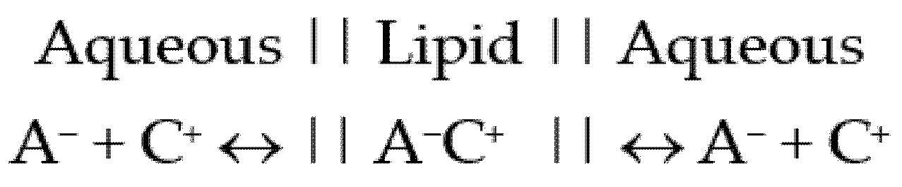
Partition Coefficient
- = unionised compound,
- = ionised version of the compound (anion),
- = counter ion (cation), and
- = ion pair.
2.3. Factors Influencing the Formation and Partition of Counter Ions
2.3.1. Size and Type of Counter Ion
2.3.2. Dielectric Constant (ε)
2.3.3. Temperature
2.3.4. pH
2.3.5. Counter Ion Concentration
2.3.6. Ion Pair and Penetration Enhancers
Penetration Enhancers Used as Ion Pairs
Ion Pairs and the Inclusion of Penetration Enhancers
2.4. Ion Pairs and the Customisation of Drug Permeating Amounts
2.5. Kinetics
2.6. Ion Pairs in Marketed Formulations
3. Conclusions
Funding
Institutional Review Board Statement
Informed Consent Statement
Data Availability Statement
Conflicts of Interest
References
- Kligman, A.M. Skin permeability: Dermatologic aspects of transdermal drug delivery. Am. Heart J. 1984, 108, 200–206. [Google Scholar] [CrossRef]
- Kulkarni, V.S. Handbook of Non-Invasive Drug Delivery Systems: Science and Technology, 1st ed.; Elsevier Science: Oxford, UK, 2010. [Google Scholar]
- Song, T.; Quan, P.; Xiang, R.; Fang, L. Regulating the skin permeation rate of escitalopram by ion-pair formation with organic acids. AAPS PharmSciTech 2016, 17, 1267–1273. [Google Scholar] [CrossRef] [PubMed] [Green Version]
- Singh, P.; Roberts, M.S. Skin permeability and local tissue concentrations of nonsteroidal antiinflammatory drugs after topical application. J. Pharmacol. Exp. Ther. 1994, 268, 144–151. [Google Scholar] [PubMed]
- Hadgraft, J.; Lane, M.E. Advanced topical formulations (atf). Int. J. Pharm. 2016, 514, 52–57. [Google Scholar] [CrossRef] [Green Version]
- Mitragotri, S. Devices for overcoming biological barriers: The use of physical forces to disrupt the barriers. Adv. Drug Deliver. Rev. 2013, 65, 100–103. [Google Scholar] [CrossRef] [PubMed]
- Hadgraft, J. Passive enhancement strategies in topical and transdermal drug delivery. Int. J. Pharm. 1999, 184, 1–6. [Google Scholar] [CrossRef]
- Lane, M.E. Skin penetration enhancers. Int. J. Pharm. 2013, 447, 12–21. [Google Scholar] [CrossRef]
- Lane, M.E.; Santos, P.; Watkinson, A.C.; Hadgraft, J. Passive skin permeation enhancement. Topical and Transdermal Drug Delivery: Principles and Practice, 1st ed.; Benson, H.A.E., Watkinson, A.C., Eds.; John Wiley & Sons: Hoboken, NJ, USA, 2011; pp. 23–42. [Google Scholar]
- Rigg, P.C.; Barry, B.W. Shed snake skin and hairless mouse skin as model membranes for human skin during permeation studies. J. Investig. Dermatol. 1990, 94, 235–240. [Google Scholar] [CrossRef]
- Bond, J.R.; Barry, B.W. Limitations of hairless mouse skin as a model for in vitro permeation studies through human skin: Hydration damage. J. Investig. Dermatol. 1988, 90, 486. [Google Scholar] [CrossRef]
- Steiling, W.; Kreutz, J.; Hofer, H. Percutaneous penetration/dermal absorption of hair dyes in vitro. Toxicol. In Vitro 2001, 15, 565–570. [Google Scholar] [CrossRef]
- McNaught, A.D.; Wilkinson, A. IUPAC. Compendium of Chemical Terminology, 2nd ed.; Blackwell Scientific Publications: Oxford, UK, 1997; Available online: https://goldbook.iupac.org (accessed on 3 April 2021).
- Levine, I.N. Physical Chemistry, 6th ed.; McGraw-Hill Higher Education: New York, NY, USA, 2009; pp. 315–318. [Google Scholar]
- Fini, A.; Fazio, G.; Gonzalez-Rodriguez, M.; Cavallari, C.; Passerini, N.; Rodriguez, L. Formation of ion-pairs in aqueous solutions of diclofenac salts. Int. J. Pharm. 1999, 187, 163–173. [Google Scholar] [CrossRef]
- Cavallari, C.; Passerini, N.; Fini, A.; Fazio, G.; Cini, M.; Rodriguez, L. Partition of diclofenac salts as ion-pairs. Dip Scienze Farmaceutiche, Università di Bologna, Via S. Donato 19/2, 40127 Bologna, Italy. Istituto di Scienze Chimiche, Università di Bologna, Italy. Eur. J. Pharm. Sci. 1998, 6, S63. [Google Scholar]
- Bagotsky, V.S. Fundamentals of Electrochemistry, 2nd ed.; Wiley-Interscience: Hoboken, NJ, USA, 2006; pp. 122–125. [Google Scholar]
- Bjerrum, N. Untersuchungen Über Ionenassoziation, 1st ed.; AF Host: Copenhagen, Denmark, 1926. [Google Scholar]
- Kraus, C.A. The ion-pair concept, its evolution and some applications. J. Phys. Chem. 1956, 60, 129–141. [Google Scholar] [CrossRef]
- Lee, S.J.; Kurihara-Bergstrom, T.; Kim, S.W. Ion-paired drug diffusion through polymer membranes. Int. J. Pharm. 1987, 39, 59–73. [Google Scholar]
- D’Aprano, A.; Goffredi, M.; Trilio, R. Ion-pair formation in water methanol and water-ethylene carbonate mixtures at 25°C. Electrochim. Acta 1976, 21, 139–141. [Google Scholar] [CrossRef]
- Byberg, J.; Jensen, S.J.K.; Kläning, U.K. Extension of the Bjerrum theory of ion association. Trans. Faraday Soc. 1969, 65, 3023–3031. [Google Scholar] [CrossRef]
- Potts, R.O.; Guy, R.H. Predicting skin permeability. Pharm. Res. 1992, 9, 663–669. [Google Scholar] [CrossRef]
- Cronin, M.T.; Dearden, J.C.; Moss, G.P.; Murray-Dickson, G. Investigation of the mechanism of flux across human skin in vitro by quantitative structure-permeability relationships. Eur. J. Pharm. Sci. 1999, 7, 325–330. [Google Scholar] [CrossRef]
- Patel, H.; Berge, W.T.; Cronin, M.T.D. Quantitative structure–activity relationships (QSARS) for the prediction of skin permeation of exogenous chemicals. Chemosphere 2002, 48, 603–613. [Google Scholar] [CrossRef]
- Inagi, T.; Muramatsu, T.; Nagai, H.; Terada, H. Mechanism of indomethacin partition between n-octanol and water. Chem. Pharm. Bull. 1981, 29, 2330–2337. [Google Scholar] [CrossRef] [Green Version]
- Moffat, A.C.; Osselton, M.D.; Widdop, B. Clarke’s Analysis of Drugs and Poisons; Pharmaceutical Press: London, UK, 2010; Available online: https://www-new-medicinescomplete-com (accessed on 3 April 2021).
- Green, P.G.; Guy, R.H.; Hadgraft, J. In vitro and in vivo enhancement of skin permeation with oleic and lauric acids. Int. J. Pharm. 1988, 48, 103–111. [Google Scholar] [CrossRef]
- Valenta, C.; Siman, U.; Kratzel, M.; Hadgraft, J. The dermal delivery of lignocaine: Influence of ion pairing. Int. J. Pharm. 2000, 197, 77–85. [Google Scholar] [CrossRef]
- Sarveiya, V.; Templeton, J.F.; Benson, H.A.E. Effect of lipophilic counter-ions on membrane diffusion of benzydamine. Eur. J. Pharm. Sci. 2005, 26, 39–46. [Google Scholar] [CrossRef] [PubMed]
- Megwa, S.A.; Cross, S.E.; Benson, H.A.E.; Roberts, M.S. Ion-pair formation as a strategy to enhance topical delivery of salicylic acid. J. Pharm. Pharmacol. 2000, 52, 919–928. [Google Scholar] [CrossRef]
- Fini, A.; Bassini, G.; Monastero, A.; Cavallari, C. Diclofenac salts, VIII. Effect of the counterions on the permeation through porcine membrane from aqueous saturated solutions. Pharmaceutics 2012, 4, 413–429. [Google Scholar] [CrossRef]
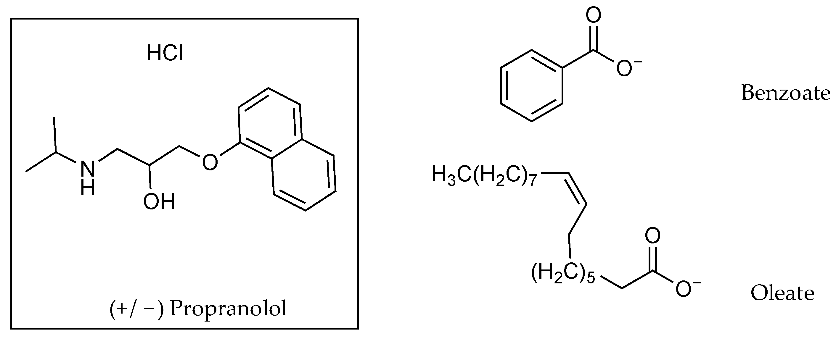
- Cilurzo, F.; Minghetti, P.; Alberti, E.; Gennari, C.G.M.; Pallavicini, M.; Valoti, E.; Montanari, L. An investigation into the influence of counterion on the rs-propranolol and s-propranolol skin permeability. J. Pharm. Sci. 2010, 99, 1217–1224. [Google Scholar] [CrossRef]
- Megwa, S.A.; Cross, S.E.; Whitehouse, M.W.; Benson, H.A.E.; Roberts, M.S. Effect of ion pairing with alkylamines on the in-vitro dermal penetration and local tissue disposition of salicylates. J. Pharm. Pharmacol. 2000, 52, 929–940. [Google Scholar] [CrossRef]
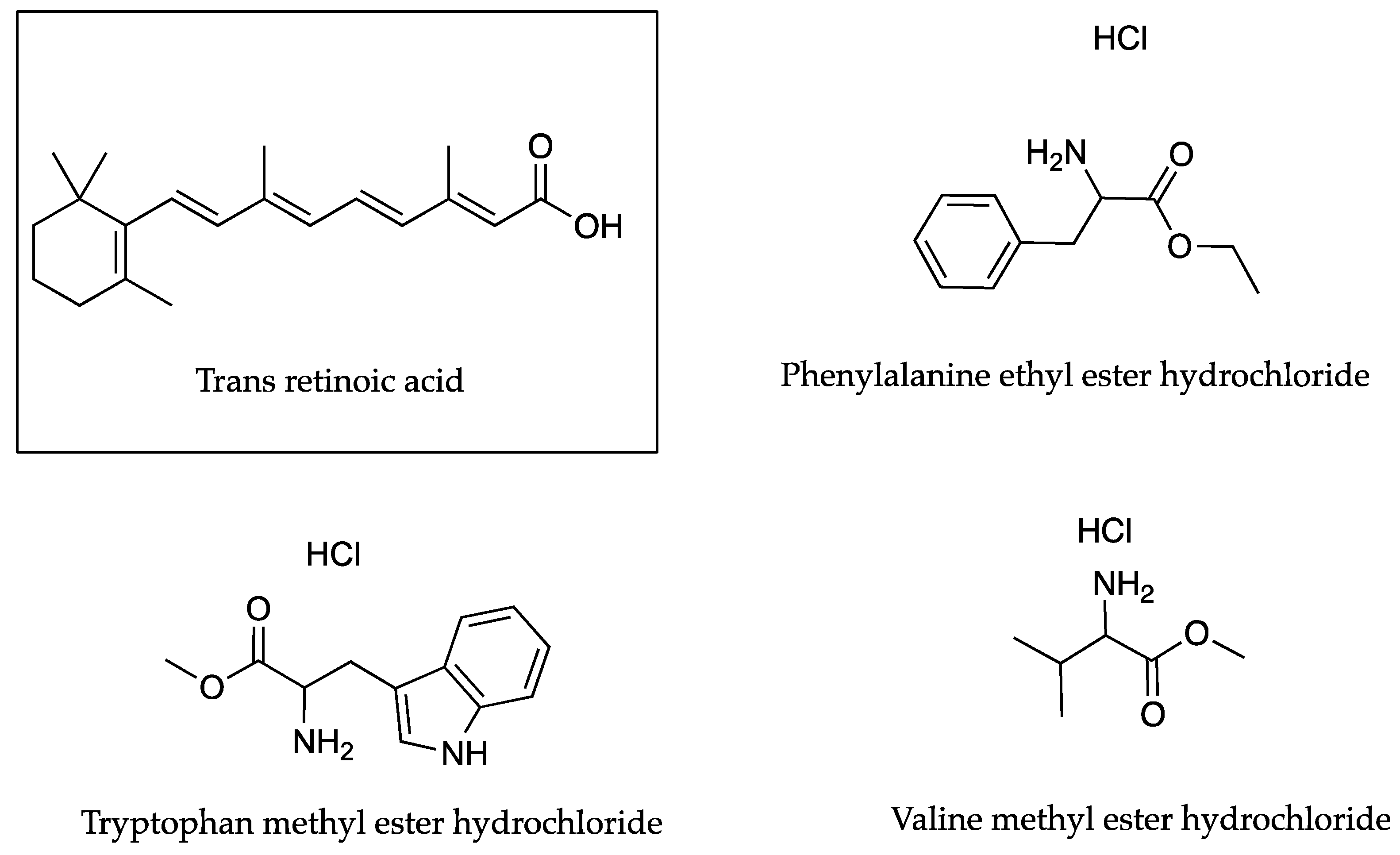
- Trotta, M.; Ugazio, E.; Peira, E.; Pulitano, C. Influence of ion pairing on topical delivery of retinoic acid from microemulsions. J. Control. Release 2003, 86, 315–321. [Google Scholar] [CrossRef]
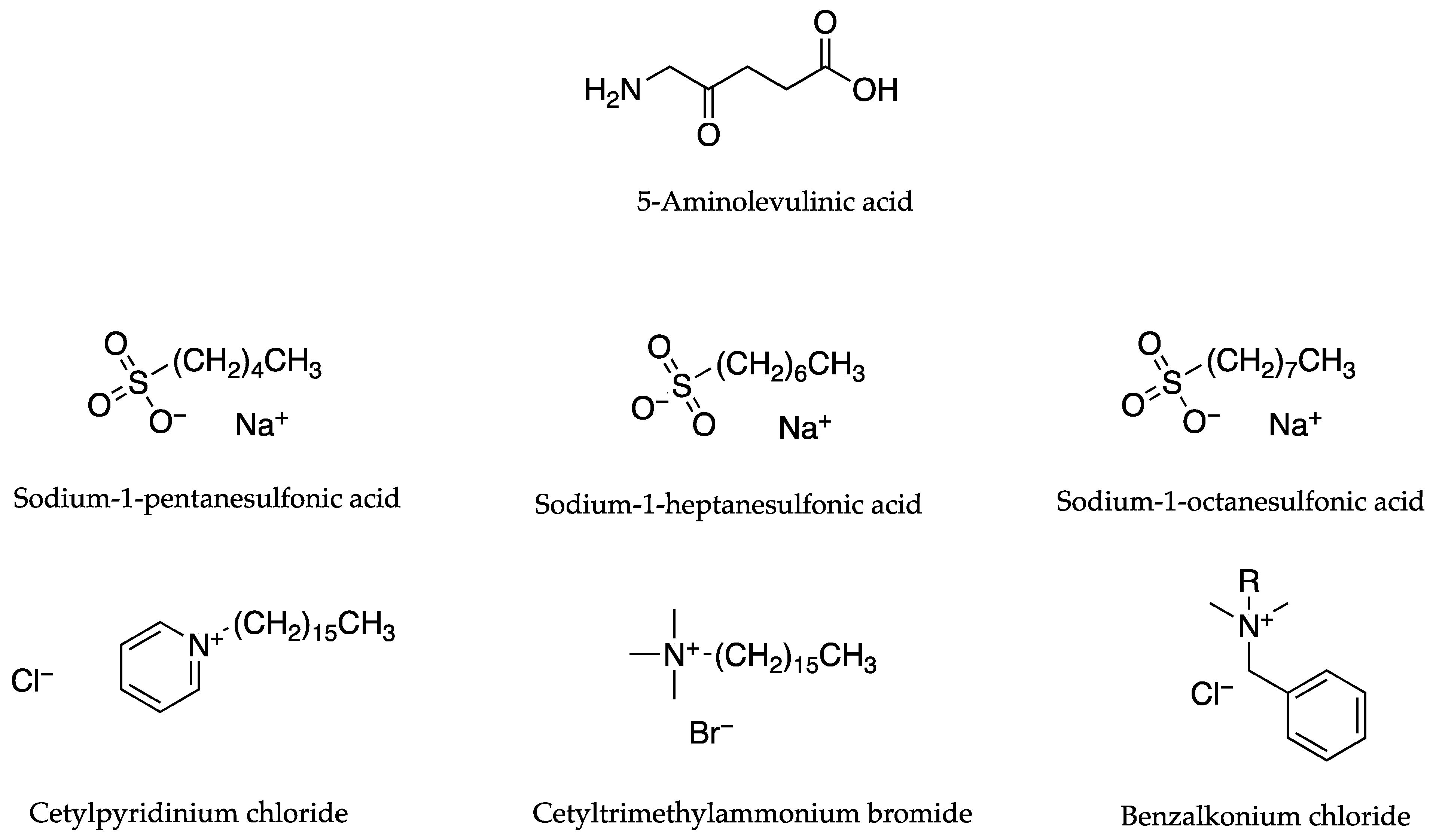
- Auner, B.; Valenta, C.; Hadgraft, J. Influence of lipophilic counter-ions in combination with phloretin and 6-ketocholestanol on the skin permeation of 5-aminolevulinic acid. Int. J. Pharm. 2003, 255, 109–116. [Google Scholar] [CrossRef]
- Takács-Novák, K.; Szász, G. Ion-pair partition of quaternary ammonium drugs: The influence of counter ions of different lipophilicity, size, and flexibility. Pharm. Res. 1999, 16, 1633–1638. [Google Scholar] [CrossRef] [PubMed]
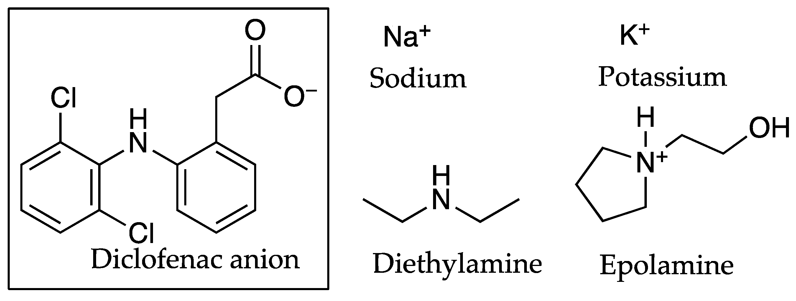
- Minghetti, P.; Cilurzo, F.; Casiraghi, A.; Montanari, L.; Fini, A. Ex Vivo study of transdermal permeation of four diclofenac salts from different vehicles. J. Pharm. Sci. 2007, 96, 814–823. [Google Scholar] [CrossRef]
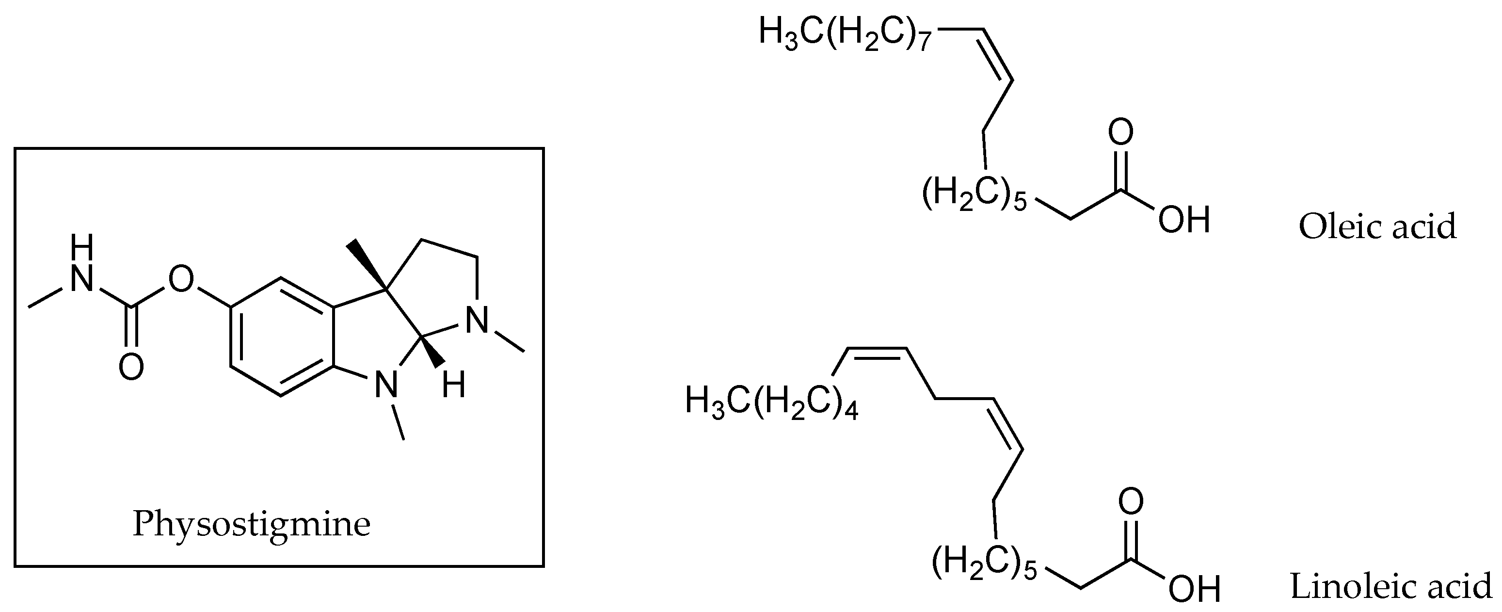
- Wang, M.Y.; Yang, Y.Y.; Heng, P.W.S. Skin permeation of physostigmine from fatty acids-based formulations: Evaluating the choice of solvent. Int. J. Pharm. 2005, 290, 25–36. [Google Scholar] [CrossRef]
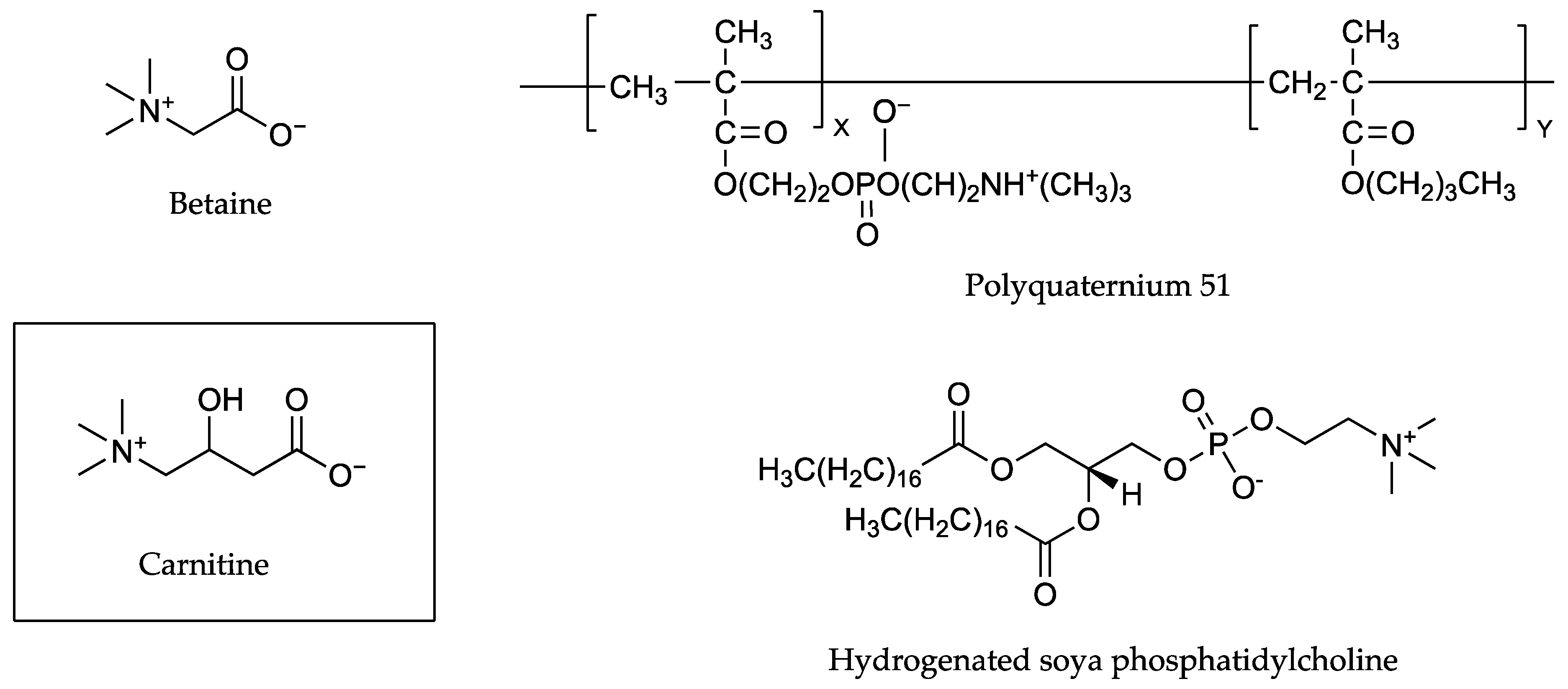
- In, S.; Yook, N.; Kim, J.-H.; Shin, M.; Tak, S.; Jeon, J.H.; Ahn, B.; Park, S.-G.; Lee, C.-K.; Kang, N.-G. Enhancement of exfoliating efficacy of l-carnitine with ion-pair method monitored by nuclear magnetic resonance spectroscopy. Sci. Rep. 2019, 9, 13507–13509. [Google Scholar] [CrossRef]
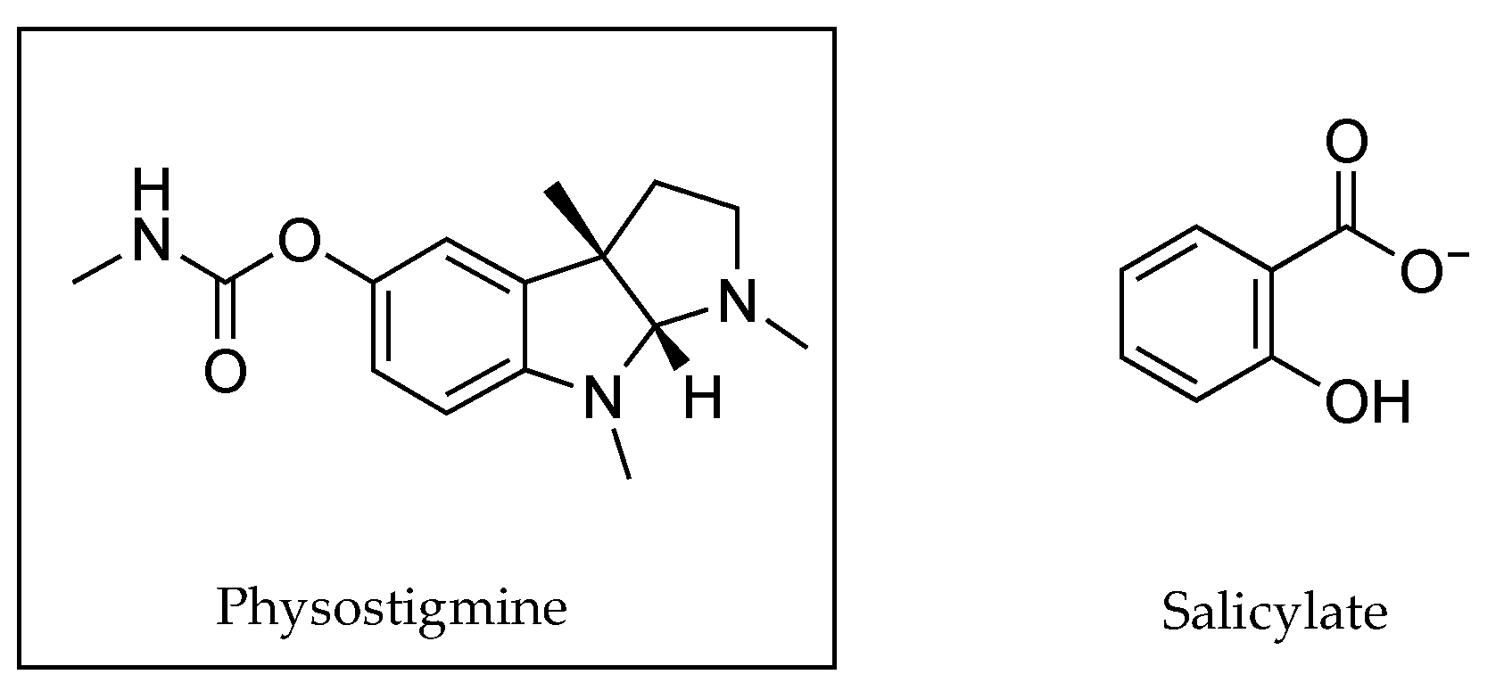
- Pardo, A.; Shiri, Y.; Cohen, S. Kinetics of transdermal penetration of an organic ion pair: Physostigmine salicylate. J. Pharm. Sci. 1992, 81, 990–995. [Google Scholar] [CrossRef] [PubMed]
- Rowe, R.C.; Shesky, P.J.; Cook, W.G.; Fenton, M.E. Handbook of Pharmaceutical Excipients, 7th ed.; Pharmaceutical Press: London, UK, 2012; Available online: https://www-new-medicinescomplete-com (accessed on 3 April 2021).
- Akerlof, G. Dielectric constants of some organic solvent-water mixtures at various temperatures. J. Am. Chem. Soc. 1932, 54, 4125–4139. [Google Scholar] [CrossRef]
- Harada, S.; Takahashi, Y.; Nakagawa, H.; Yamashita, F.; Hashida, M. Effect of vehicle properties on skin penetration of emedastine. Biol. Pharm. Bull. 2000, 23, 1224–1228. [Google Scholar] [CrossRef] [PubMed] [Green Version]
- Katz, M.; Poulsen, B.J. Absorption of drugs through the skin. Concepts Biochem. Pharm. 1971, 3, 103–104. [Google Scholar]
- Hadgraft, J.; Walters, K.A.; Wotton, P.K. Facilitated percutaneous absorption: A comparison and evaluation of two in vitro models. Int. J. Pharm. 1986, 32, 257–263. [Google Scholar] [CrossRef]
- Smith, J.C.; Irwin, W.J. Ionisation and the effect of absorption enhancers on transport of salicylic acid through silastic rubber and human skin. Int. J. Pharm. 2000, 210, 69–82. [Google Scholar] [CrossRef]
- Cázares-Delgadillo, J.; Naik, A.; Kalia, Y.N.; Quintanar-Guerrero, D.; Ganem-Quintanar, A. Skin permeation enhancement by sucrose esters: A pH-dependent phenomenon. Int. J. Pharm. 2005, 297, 204–212. [Google Scholar] [CrossRef] [PubMed]
- Vávrová, K.; Lorencová, K.; Novotný, J.; Holý, A.; Hrabálek, A. Permeation enhancer dodecyl 6-(dimethylamino)hexanoate increases transdermal and topical delivery of adefovir: Influence of pH, ion-pairing and skin species. Eur. J. Pharm. Biopharm. 2008, 70, 901–907. [Google Scholar] [CrossRef]
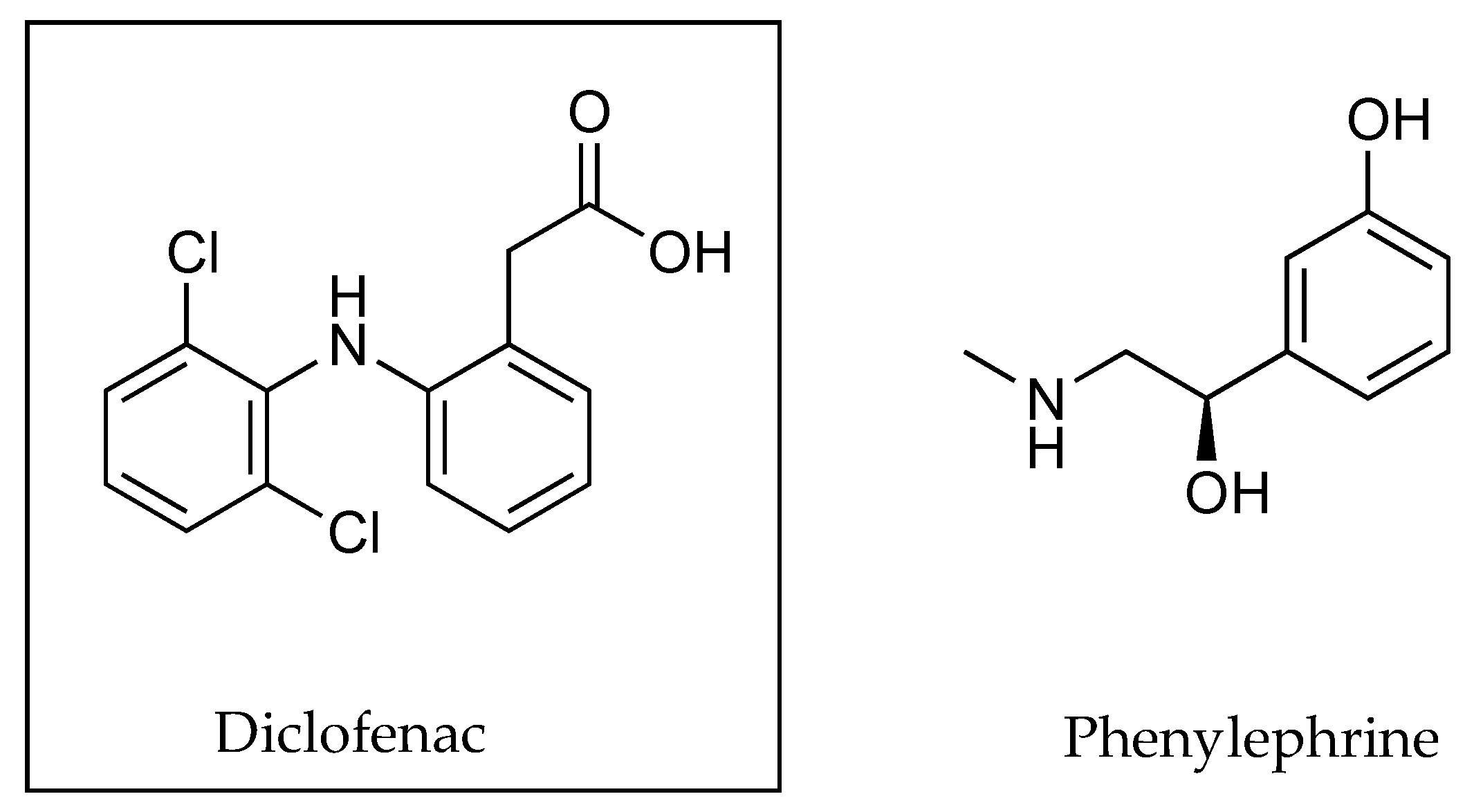
- Uchino, T.; Miyazaki, Y.; Ishikawa, A.; Kagawa, Y. Development of a novel simple gel formulation containing an ion-pair complex of diclofenac and phenylephrine. Skin Pharmacol. Physi. 2019, 32, 318–327. [Google Scholar] [CrossRef]
- Cross, S.E.; Thompson, M.J.; Roberts, M.S. Transdermal penetration of vasoconstrictors-present understanding and assessment of the human epidermal flux and retention of free bases and ion-pairs. Pharm. Res. 2003, 20, 270–274. [Google Scholar] [CrossRef]
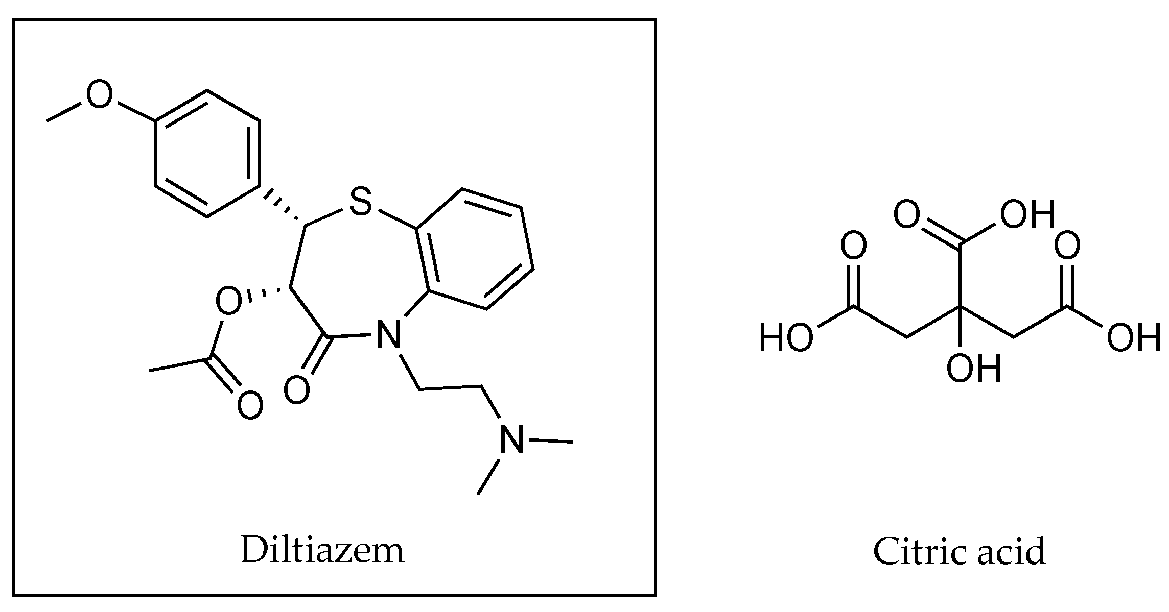
- Chirio, D.; Trotta, M.; Gallarate, M.; Peira, E.; Carlotti, M.E. Thermosensitive gels for the topical administration of diltiazem. J. Disper. Sci. Technol. 2011, 32, 320–325. [Google Scholar] [CrossRef]
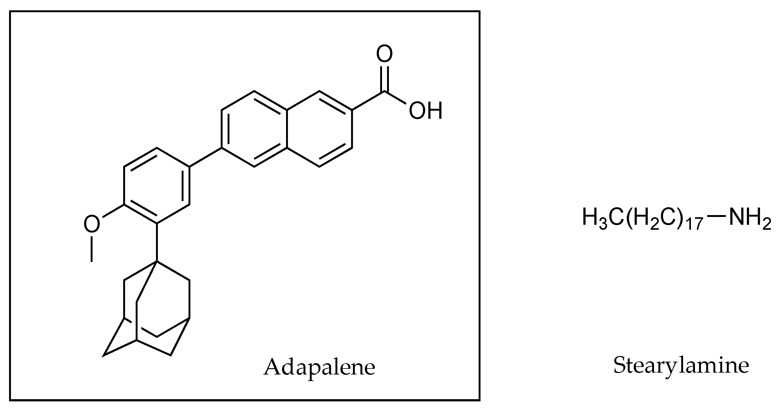
- Rodrigues, L.B.O.; Lima, F.A.; Alves, C.P.B.; Martins-Santos, E.; Aguiar, M.M.G.; Oliveira, C.A.; Oréfice, R.L.; Ferreira, L.A.M.; Goulart, G.A.C. Ion pair strategy in solid lipid nanoparticles: A targeted approach to improve epidermal targeting with controlled adapalene release, resulting reduced skin irritation. Pharm. Res. 2020, 37, 1–14. [Google Scholar] [CrossRef] [PubMed]
- Lanes, S.F.; Rodrigeuz, L.A.; Hwangg, E. Baseline risk of gastrointestinal disorders among new users of meloxicam, ibuprofen, diclofenac, naproxen and indomethacin. Pharmacoepidemiol. Drug Saf. 2000, 9, 113–117. [Google Scholar] [CrossRef]
- Basketter, D.A.; York, M.; McFadden, J.P.; Robinson, M.K. Determination of skin irritation potential in the human 4-h patch test. Contact Dermat. 2004, 51, 1–4. [Google Scholar] [CrossRef]


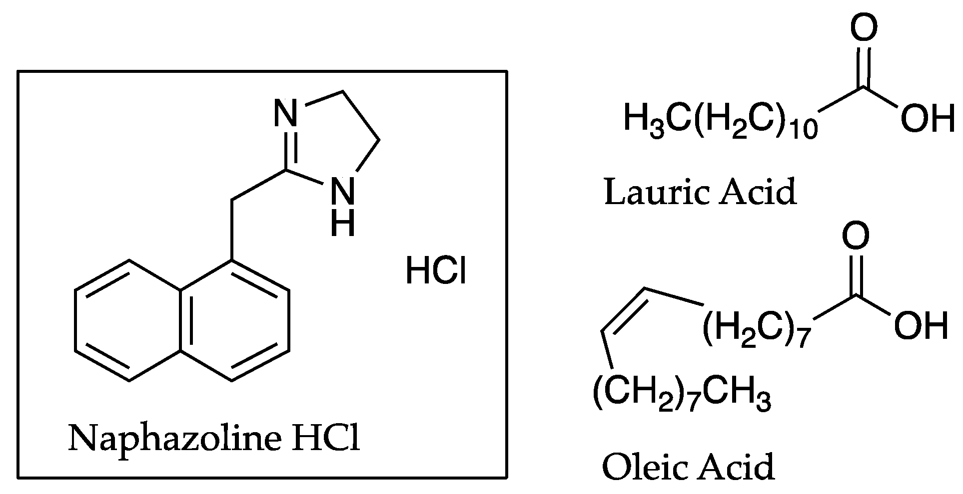
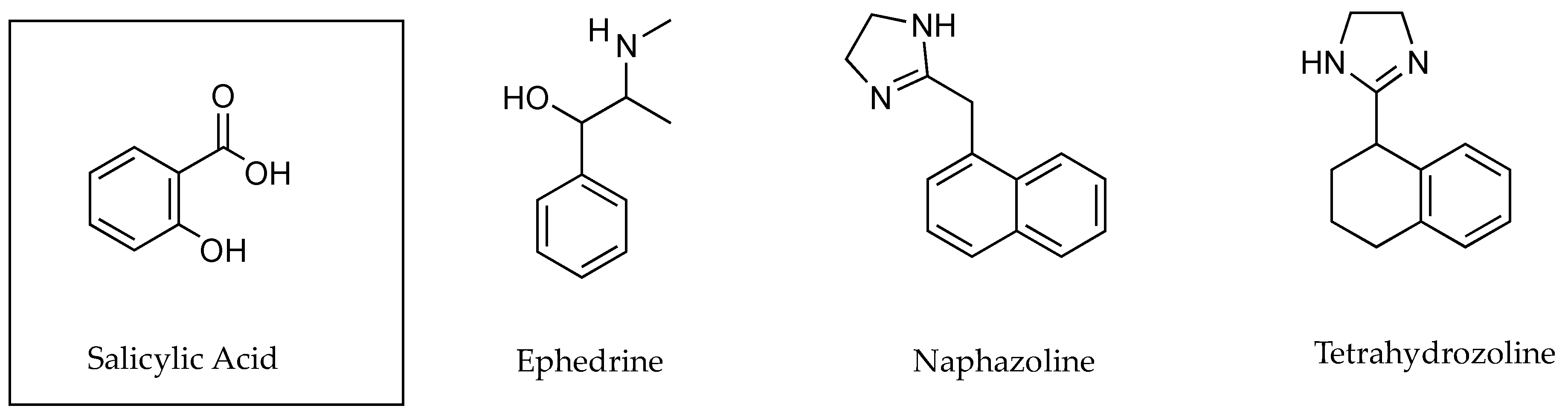
| Partition Coefficient of naphazoline | Isopropyl myristate (IPM)/buffer (pH 7.4) | 0.02 ± 0.01 |
| OA in IPM/buffer (pH 7.4) | 0.36 ± 0.04 | |
| LA in IPM/buffer (pH 7.4) | 0.45 ± 0.06 | |
| IPM/buffer (pH 8.0) | 0.03 ± 0.01 | |
| Permeability coefficient of naphazoline using human skin (cm h−1 × 10⁻³) | Naphazoline HCl | ~0.33 |
| Naphazoline plus OA | ~2.17 | |
| Naphazoline plus LA | ~2.58 |
| pH | PC (Calculated) | Flux (µg cm−2 h−1) |
|---|---|---|
| 4.0 | ~0.19 | 1.2 ± 1.2 |
| 5.5 | ~0.40 | 13.0 ± 2.0 |
| 7.0 | ~6.76 | 118.0 ± 30.0 |
| pH | PC ~ | Fraction Unionised | Permeability Coefficient (cm h−1) | Flux (µg cm−2 h−1) |
|---|---|---|---|---|
| 5.0 | 1.62 | 6.3 × 10⁻³ | 2.6 × 10⁻⁴ | 5.13 ± 2.42 |
| 6.0 | 5.75 | 0.0631 | 3.8 × 10⁻³ | 39.07 ± 10.5 |
| 7.0 | 28.18 | 0.627 | 6.1 × 10⁻² | 269.09 ± 58.5 |
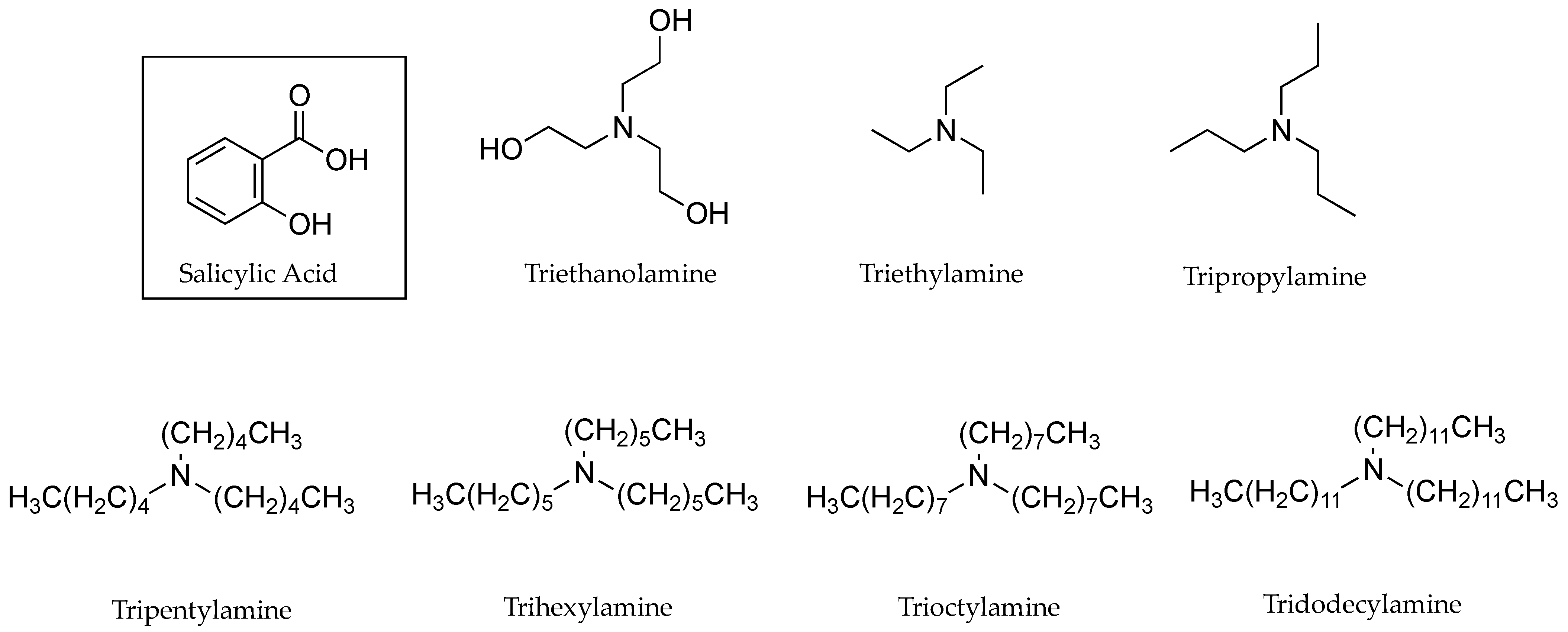
| SA with Counter Ion: | PC in Octanol-Phosphate Buffer (pH 5.0) | Flux (mg cm −2 h −1 × 10 −2) |
|---|---|---|
| Triethanolamine | 0.007 ± 0.00 | 11.90 ± 1.23 |
| Triethylamine | 0.360 ± 0.00 | 15.40 ± 3.85 |
| Tripropylamine | 3.180 ± 0.04 | 18.50 ± 2.26 |
| Tripentylamine | 109.77 ± 11.37 | 19.50 ± 3.63 |
| Trihexylamine | 152.17 ± 26.81 | 22.60 ± 1.14 |
| Trioctylamine | 140.58 ± 16.33 | 27.90 ± 3.98 |
| Tridodecylamine | 140.66 ± 17.23 | 42.70 ± 2.04 |
| DIC Plus Counter Ion: | PC | Solubility (µg cm−3 × 10³) | Permeation Coefficient (cm h−1 × 10³) |
|---|---|---|---|
| Monoethanolamine | 1.20 | 9.9 | 0.70 |
| Monoethylamine | 1.02 | 6.1 | 2.00 |
| Diethanolamine | 1.20 | 18.0 | 2.80 |
| Diethylamine | 1.48 | 13.7 | 3.70 |
| Triethanolamine | 4.37 | 3.4 | 3.00 |
| Triethylamine | 7.08 | 6.7 | 3.40 |
| N-2-hydroxyethyl pyrrolidine | 1.48 | 20.2 | 9.60 |
| Pyrrolidine | 1.62 | 2.0 | 21.00 |
| N-2-hydroxyethyl piperidine | 1.95 | 10.7 | 7.70 |
| Piperidine | 9.33 | 4.3 | 20.00 |
| N-2-hydroxyethyl morpholine | 10.96 | 4.4 | 4.80 |
| Morpholine | 2.24 | 6.9 | 3.80 |
| N-2-hydroxyethyl piperazine | 1.74 | 12.5 | 13.00 |
| Piperazine | 4.68 | 0.4 | 45.00 |
| ALA Plus Counter Ion | pH | PC ~(×10−1) | Cumulative Amount of ALA ~(µg cm−2) after 4 h Using Porcine Skin |
|---|---|---|---|
| None | 4.0 | 1.20 | 5.11 |
| Sodium-1-octanesulfonic acid | 4.0 | 2.14 | 10.7 |
| Sodium-1-heptanesulfonic acid | 4.0 | 4.68 | 10.0 |
| Sodium-1-pentanesulfonic acid monohydrate | 4.0 | 2.69 | 10.0 |
| None | 7.0 | 1.51 | 6.5 |
| Cetylpyridinium chloride | 7.0 | 9.12 | 11.0 |
| Cetyltrimethylammonium bromide | 7.0 | 8.13 | 5.0 |
| Benzalkonium chloride | 7.0 | 6.03 | 7.0 |
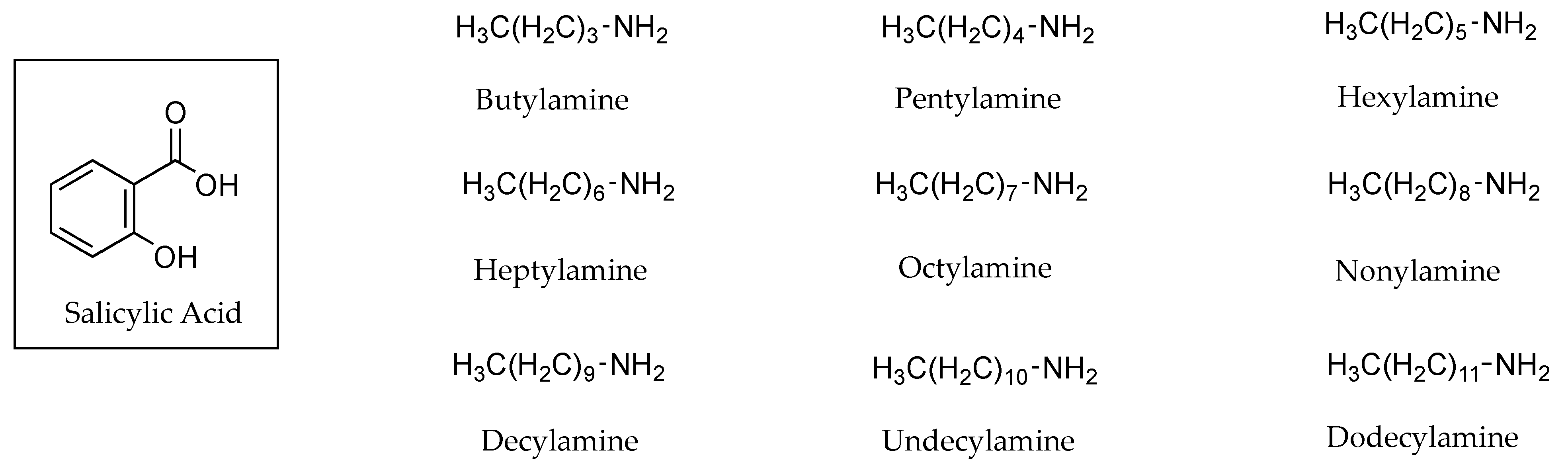
| Salicylic Acid Plus Counter Ion | Molecular Weight of Counter Ion (Da) | Molal Volume of Counter Ion (cm3 mol−1) | Flux (mg cm−2 h−1) × 10−2 | Conductivity (mS cm−1) |
|---|---|---|---|---|
| None | - | - | 8.90 ± 1.20 | 2.03 ± 0.06 |
| Triethylamine | 101.20 | 108.00 | 15.40 ± 3.85 | 1.77 ± 0.06 |
| Tripropylamine | 143.27 | 156.60 | 18.50 ± 2.26 | 1.35 ± 0.06 |
| Tripentylamine | 227.44 | 253.80 | 19.50 ± 3.63 | 0.90 ± 0.00 |
| Trihexylamine | 269.52 | 302.40 | 22.60 ± 1.14 | 0.80 ± 0.00 |
| Trioctylamine | 353.68 | 399.60 | 27.90 ± 3.98 | 0.60 ± 0.00 |
| Tridodecylamine | 522.00 | 592.50 | 42.70 ± 2.04 | 0.30 ± 0.00 |
| Counter Ions | PC Value ~ | Solubility Saline (mg mL−1) | Flux Saline (µg cm−2h−1) | Solubility MO (mg mL−1) | Flux MO (µg cm−2 h−1) |
|---|---|---|---|---|---|
| (RS)-PR | 10.00 | 0.189 | 18.0 ± 5.1 | 1.321 | 24.2 ± 4.7 |
| (S)-PR | 9.33 | 0.432 | 44.7 ± 5.1 | 3.111 | 41.9 ± 1.5 |
| (RS)-PR-Bz | 11.74 | 4.430 | 1.7 ± 0.2 | 0.065 | 1.6 ± 0.3 |
| (S)-PR Bz | 11.48 | 9.560 | 3.0 ± 0.4 | 0.118 | 2.9 ± 0.7 |
| (RS)-PR-Ol | 17.78 | 0.020 | 7.0 ± 1.4 | 3.930 | 7.3 ± 1.4 |
| (S)-PR-Ol | 19.05 | 0.020 | 6.2 ± 0.9 | 5.874 | 11.1 ± 1.7 |
| Volume Fraction of IPA in IPA-IPM Solvent Mixture | Flux × 104 µmol cm−2 m−1 | |
|---|---|---|
| PHY | Salicylate | |
| 0 | 1.28 ± 0.35 | 1.47 ± 0.11 |
| 0.1 | 5.75 ± 0.58 | 6.3 ± 0.50 |
| 0.3 | 14.55 ± 0.71 | 18.2 ± 0.56 |
| 0.5 | 31.70 ± 3.10 | 47.8 ± 11.50 |
| 0.7 | 44.27 ± 9.16 | 61.66 ± 2.54 |
| 0.9 | 5.52 ± 0.28 | 17.70 ± 1.90 |
| 1 | 0.56 ± 0.08 | 1.87 ± 0.10 |
| DIC Plus Counter Ion | Parameter | Solvents | |||
|---|---|---|---|---|---|
| Water | PG | TC® | OA | ||
| Sodium | Solubility (µg mL−1) | 37 ± 10 | 567 ± 31 | 660 ± 70 | 25 ± 10 |
| Flux (µg cm−2 h−1) | 2.29 ± 0.37 | 1.21 ± 0.06 | 0.06 ± 0.01 | 1.84 ± 0.18 | |
| Potassium | Solubility (µg mL−1) | 218 ± 80 | 898 ± 79 | 709 ± 52 | 60 ± 50 |
| Flux (µg cm−2 h−1) | 1.35 ± 0.72 | 0.04 ± 0.02 | 0.84 ± 0.06 | 1.17 ± 0.17 | |
| Diethylamine | Solubility (µg mL−1) | 19 ± 10 | 384 ± 14 | 279 ± 10 | 63 ± 60 |
| Flux (µg cm−2 h−1) | 5.60 ± 2.14 | 0.35 ± 0.04 | 0.96 ± 0.59 | 2.74 ± 0.94 | |
| Epolamine | Solubility (µg mL−1) | 557 ±15 | 637 ± 60 | 430 ± 0.00 | 94 ± 70 |
| Flux (µg cm−2 h−1) | 2.90 ± 0.91 | 0.46 ± 0.21 | 0.03 ± 0.00 | 3.11 ± 0.18 | |
| PHY Plus Fatty Acid Counter Ion: | Flux (PG) (µg cm−2 h−1) | Con ~ (µS cm−1) PG | Sol (mg mL−1) | Flux (MO) (µg cm−2 h−1) | Con ~ (µS cm−1) MO ×10−2 | Sol (mg mL−1) |
|---|---|---|---|---|---|---|
| None | - | 0.25 | 71.0 | 2.5 ± 0.7 | 9.25 | 1.7 |
| 2 | - | 16 | - | - | 9.45 | - |
| 3 | - | 14 | - | - | 8.65 | - |
| 8 | - | 10 | 116.0 | 3.2 ± 0.6 | 9.05 | 5.1 |
| 10 | - | 9.5 | 80.7 | 6.6 ± 2.3 | 9.40 | 10.8 |
| 12 | 0.2 ± 0.0 | 8 | 88.7 | 14.0 ± 3.0 | 8.95 | 15.3 |
| 18:1 | 19.7 ± 8.2 | 3 | 83.9 | 13.9 ± 7.1 | 8.80 | 15.9 |
| 18:2 | 2.4 ± 0.9 | 4 | 85.3 | 3.9 ± 1.1 | 9.15 | 15.7 |
| ~Temperature °C | ~Conductance |
|---|---|
| 80 | 500 × 106 |
| 100 | 576 × 106 |
| 120 | 638 × 106 |
| 150 | 700 × 106 |
| 180 | 654 × 106 |
| 200 | 577 × 106 |
| 220 | 400 × 106 |
| 240 | 14.2 × 10⁻² |
| Solvent | ε | Conductance Maximum Reached at °C |
|---|---|---|
| Methylamine | 9.4 at 25 °C | 15 |
| Ammonia | 22.4 at ~33.3 °C | 25 |
| Methanol | 32.8 at 25 °C | 150 |
| pH | Flux (µmol cm−2 h−1) |
|---|---|
| 2.10 | 0.72 ± 0.057 |
| 2.27 | 0.59 ± 0.016 |
| 2.72 | 0.54 ± 0.009 |
| 3.13 | 0.25 ± 0.006 |
| 3.50 | 0.15 ± 0.005 |
| 3.90 | 0.07 ± 0.001 |
| 4.30 | 0.05 ± 0.001 |
| 4.71 | 0.04 ± 0.000 |
| 5.13 | 0.01 ± 0.000 |
| pH | Lidocaine HCl | Lidocaine Plus 2% L-TC | Lidocaine Plus 2% O-TC |
|---|---|---|---|
| Permeability Coefficient ×104 cm h−1 | Permeability Coefficient ×104 cm h−1 | Permeability Coefficient ×104 cm h−1 | |
| 5.0 | 1.55 ± 0.31 | 18.46 ± 5.60 | 5.83 ± 0.65 |
| 7.0 | 1.81 ± 1.1 | 19.58 ± 3.82 | 6.24 ± 0.31 |
| 9.0 | 5.65 ± 1.52 | 3.34 ± 0.22 | 15.08 ± 2.30 |
| pH Values | 3.4 | 3.8 | 4.8 | 5.8 | 6.8 | 7.8 | |
|---|---|---|---|---|---|---|---|
| Flux of Adefovir ~ (µg cm−2 h−1) | 2% Adefovir | 0.2 | 1 | 2 | 1 | 3 | 4 |
| 2% Adefovir + 1% DDAK | 9.5 | 10 | 18 | 27 | 26.5 | 10 | |
| Adefovir Skin Concentration ~(µg g−1) | 2% Adefovir | 103 | 221 | 235 | 191 | 191 | 221 |
| 2% Adefovir + 1% DDAK | 412 | 538 | 708 | 771 | 693 | 412 | |
| PHY: SA Ratio | Flux × 104 (µmol cm−2 min−1) | Permeability Coefficient × 104 (cm min−1) | Cv µmol cm−3 | |||
|---|---|---|---|---|---|---|
| PHY | salicylate | PHY | salicylate | PHY | salicylate | |
| 1:1 | 44.27 ± 9.16 | 61.66 ± 2.54 | 1.86 ± 0.39 | 2.60 ± 0.10 | 23.70 ± 0.55 | 23.70 ± 0.55 |
| 1:8 | 48.40 ± 9.50 | 247.30 ± 36.50 | 3.25 ± 0.64 | 2.10 ± 0.30 | 14.90 ± 0.40 | 120.00 ± 15.20 |
| 6.5:1 | 67.20 ± 7.70 | 63.90 ± 7.00 | 0.44 ± 0.05 | 2.68 ± 0.30 | 153.40 ± 7.70 | 23.70 ± 3.40 |
| Membrane | Application | Flux ~(µg cm−2 h−1) | Skin Concentration (Epidermins Incl SC for Human Skin)~ |
|---|---|---|---|
| Porcine skin | Co-application of 2% adefovir, 1% DDAK | 25.6 | 31.3 µg g−1 |
| 2% adefovir pre-treatment of skin with DDAK | 16.5 | 17.1 µg g−1 | |
| Human skin | Co-application of 2% adefovir, 1% DDAK | 8.93 | 21.30 µg cm−2 |
| 2% adefovir no DDAK | 0.04 | 5.72 µg cm−2 |
| ALA with: | pH | Cumulative amount of ALA ~(µg/cm−2) after 4 h Using Porcine Skin |
|---|---|---|
| counter ion CP plus phosphatidylcholine liposomes loaded with KC | 7.0 | 23.00 |
| counter ion CP plus unloaded phosphatidylcholine liposomes | 7.0 | 11.25 |
| unloaded phosphatidylcholine liposomes | 7.0 | 6.25 |
| VC | Flux (µg cm−2 h−1) | Skin Retention (µg mg−1) | ||
|---|---|---|---|---|
| VC | SA | VC | SA | |
| Ephedrine | 11.5 ± 2.3 | 18.6 ± 0.6 | 10.0 ± 0.4 | 4.2 ± 0.7 |
| Naphazoline | 12.0 ± 1.6 | 7.8 ± 0.8 | 20.7 ± 6.0 | 3.5 ± 1.1 |
| Tetrahydrozoline | 2.9 ± 0.5 | 1.1 ± 0.1 | 3.7 ± 0.6 | 2.8 ± 1.1 |
| RA with: | Skin Accumulation after 24 h (µg cm−2) | Flux (µg cm−2 h−1) |
|---|---|---|
| alone | 1.0 ± 0.2 | 0.13 ± 0.02 |
| tryptophan methyl ester hydrochloride | 2.3 ± 0.6 | 0.19 ± 0.02 |
| phenylalanine ethyl ester hydrochloride | 3.4 ± 0.6 | 0.23 ± 0.03 |
| valine methyl ester hydrochloride | 3.7 ± 0.8 | 0.21 ± 0.03 |
| Formulated as microemulsions oil in water microemulsions (a) and (b): | ||
| (a) alone as microemulsion | 3.3 ± 0.50 | 0.05 ± 0.01 |
| (a) phenylalanine ethyl ester hydrochloride | 13.3 ± 2.10 | <0.01 |
| (a) valine methyl ester hydrochloride | 8.7 ± 1.6 | <0.01 |
| (b) alone as microemulsion | 2.3 ± 0.50 | 0.04 ± 0.01 |
| (b) phenylalanine ethyl ester hydrochloride | 12.6 ± 1.8 | <0.01 |
| (b) valine methyl ester hydrochloride | 10.8 ± 1.5 | <0.01 |
| Commercial Name | Active | Ion Pair | Owner |
|---|---|---|---|
| Diclofenac 1% gel | DFA | Sodium | A A H Pharmaceuticals Ltd. |
| Diclofenac 1% gel | DFA | Sodium | Actavis UK Ltd. |
| Diclofenac 1% gel | DFA | Sodium | Alliance Healthcare (Distribution) Ltd. |
| Diclofenac 1% gel | DFA | Sodium | Typharm Ltd. |
| Diclofenac sodium topical gel 1% | DFA | Sodium | AvKARE |
| Diclofenac sodium topical gel 3% | DFA | Sodium | Taro |
| Diclofenac sodium topical solution (1.5%) | DFA | Sodium | Sola Pharmaceuticals |
| Flector gel 1% | DFA | Epolamine, sodium | Laboratoires Genevrier |
| Flector EP 10 mg/g | DFA | Epolamine | Medichemie |
| Flector tissugel 140 mg (medicated plaster) | DFA | Sodium | Windzor Pharma |
| Flector (patch) | DFA | Epolamine | Pfizer |
| Pennsaid ®® (2% solution) | DFA | Sodium | Horizon Medicines LLC |
| Solaraze 3% gel | DFA | Sodium | Almirall Ltd. |
| Solacutan 3% gel | DFA | Sodium | Mibe Pharma UK Ltd. |
| Voltarol®® joint pain relief 2.32% gel | DFA | diethylammonium | GSK |
| Voltarol®® joint 12 h joint pain relief 2.32% gel | DFA | diethylammonium | GSK |
| Voltarol®® osteoarthritis joint pain relief 1.16% gel | DFA | diethylammonium | GSK |
| Voltarol®® back and muscle pain relief 1.16% gel | DFA | diethylammonium | GSK |
| Voltarol®® 140 mg medicated plaster | DFA | Sodium | GSK |
Publisher’s Note: MDPI stays neutral with regard to jurisdictional claims in published maps and institutional affiliations. |
© 2021 by the authors. Licensee MDPI, Basel, Switzerland. This article is an open access article distributed under the terms and conditions of the Creative Commons Attribution (CC BY) license (https://creativecommons.org/licenses/by/4.0/).
Share and Cite
Cristofoli, M.; Kung, C.-P.; Hadgraft, J.; Lane, M.E.; Sil, B.C. Ion Pairs for Transdermal and Dermal Drug Delivery: A Review. Pharmaceutics 2021, 13, 909. https://doi.org/10.3390/pharmaceutics13060909
Cristofoli M, Kung C-P, Hadgraft J, Lane ME, Sil BC. Ion Pairs for Transdermal and Dermal Drug Delivery: A Review. Pharmaceutics. 2021; 13(6):909. https://doi.org/10.3390/pharmaceutics13060909
Chicago/Turabian StyleCristofoli, Mignon, Chin-Ping Kung, Jonathan Hadgraft, Majella E. Lane, and Bruno C. Sil. 2021. "Ion Pairs for Transdermal and Dermal Drug Delivery: A Review" Pharmaceutics 13, no. 6: 909. https://doi.org/10.3390/pharmaceutics13060909