Article

Safety and Tolerability of Topical Ophthalmic Triamcinolone Acetonide-Loaded Liposomes Formulation and Evaluation of Its Biologic Activity in Patients with Diabetic Macular Edema

by
Jose Navarro-Partida
1,2,
Juan Carlos Altamirano-Vallejo
1,2,
Alejandro Gonzalez-De la Rosa
1,2,
Juan Armendariz-Borunda
1,3,
Carlos Rodrigo Castro-Castaneda
1 and
Arturo Santos
1,2,*
1
Tecnologico de Monterrey, Escuela de Medicina y Ciencias de la Salud, Campus Guadalajara, P.C. 45138 Zapopan, Mexico
2
Centro de Retina Medica y Quirurgica, S.C., Centro Medico Puerta de Hierro, P.C. 45116 Zapopan, Mexico
3
Instituto de Biología Molecular y Terapia Genica, Centro Universitario de Ciencias de la Salud, Universidad de Guadalajara, P.C. 44340 Guadalajara, Mexico
*
Author to whom correspondence should be addressed.
Pharmaceutics 2021, 13(3), 322; https://doi.org/10.3390/pharmaceutics13030322
Submission received: 23 January 2021 / Revised: 22 February 2021 / Accepted: 24 February 2021 / Published: 2 March 2021
(This article belongs to the Special Issue Overcoming Physiological Barriers Using Lipid Nanosystems)

Abstract

:
Intravitreal injections (IVTs) of corticosteroids as triamcinolone acetonide (TA) are frequently used for the treatment of many vitreous and retinal disorders. However, IVTs are related to severe ocular complications. Lately, a topical ophthalmic TA-loaded liposomes formulation (TALF) was designed to transport TA into the posterior segment of the eye when instilled on the ocular surface. To evaluate the safety, tolerability, and biological activity of TALF, an animal study and a phase I clinical assay were performed. Moreover, four patients with diabetic macular edema (DME) were treated with TALF in order to explore the biological activity of the formulation. No inflammation, lens opacity, swelling, or increase in intraocular pressure were recorded after the instillation of TALF in any of the animal or clinical studies. Mainly, mild and transient adverse events such as dry eye and burning were reported. TALF significantly improves visual acuity and diminishes central foveal thickness in patients with DME. The current data demonstrate the safety, tolerability, and biological activity of TALF. It seems that TALF can be used topically to treat vitreous and retinal diseases that respond to TA such as DME, avoiding the use of corticosteroid IVTs and their associated hazards.

1. Introduction

Intravitreal injections of corticosteroids are commonly used for the treatment of many vitreous and retinal disorders such as retinal vein occlusions [1], uveitis [2], and diabetic macular edema (DME) [3]. Among the different synthetic corticosteroids, triamcinolone acetonide (TA) is extensively used in intravitreal injections (IVTs). In addition its advantages such as its low cost, TA is not exempt from severe complications, including a detached retina, endophthalmitis, and vitreous hemorrhage [4,5,6,7], as well as the risk of severe and intractable intraocular pressure (IOP) elevation [8,9,10], which raises constant concerns about intraocular TA administration. In addition, the discomfort caused to the patient by IVT itself could cause poor compliance with intraocular steroid treatment [11,12,13].
Although oral or topical corticosteroid routes of administration could be safer than IVTs, these have limited effectiveness for vitreoretinal disorders. Due to the blood–retinal barriers, these routes scarcely reach the posterior segment of the eye [14]. Therefore, to diminish the ocular risks associated with intravitreal injections, different topical approaches have been developed to deliver TA into the vitreous cavity [15,16,17,18]. Among these, topical liposomes (LPs) are one of the most promising strategies [19]. In fact, LPs constitute the only topical system to deliver TA into the vitreous area with clinical evidence of its effectiveness [20,21,22]. The vesicle composition of LPs share almost an indistinguishable outer structure with the cell membrane, conferring its ability to transport either hydrophilic or lipophilic drugs. LPs represent a potential alternative for ocular drug delivery based on their various advantages—increased residence time for drug absorption, protection of the encapsulated drug from the external environment, prolonged half-lives in vitreous bodies with low toxicity, increased efficacy and therapeutic drug index, and the potential to improve penetration to ocular tissues [19].
In recent years, different topical liposomes have been developed to deliver drugs into the posterior segment of the eye [23,24,25,26,27,28,29,30,31]. For example, fluorescent molecules like 5(6)-carboxyfluorescein and coumarin-6 [23,25,29,30], drugs such as edaravone and diclofenac [26,27], and biomolecules such as nucleic acids [28,31] and bevacizumab (a monoclonal antibody against vascular endothelial growth factor A) [24] have been released into the vitreous body and retina by liposomes.
Recently, a topical ophthalmic triamcinolone acetonide-loaded liposome formulation (TALF) was designed for our group to transport TA into the posterior segment of the eye. The efficiency of this drug delivery nanosystem was successfully tested using in vitro and in vivo models [18]. The in vitro diffusion assay of TALF was performed using diffusion chambers with rabbit corneas as membranes, whereas the in vivo assay was carried out in New Zealand white rabbits. As exposed in the previous report [18], TALF was able to cross the cornea and to deliver TA to the vitreous body and retina, reaching the highest peak at 12 h (32.6 ± 10.2 ng/g and 252.1 ± 90.0 ng/g respectively). Additionally, in the same report, TALF underwent a physicochemical characterization, as well as a cell toxicity assay using primary human corneal fibroblast cultures. We found that TALF has a pH of 5.8, viscosity of 70 cP and osmolarity of 334 mOsm/L. Therefore, TALF was found to be highly viscous, near to the physiologic pH of tears (6.5 to 7.6) [32] and non-irritating to the eye according to the criteria of the Pharmacopoeia of Estados Unidos Mexicanos, in which a formulation is considered suitable for ophthalmic use when its osmolarity is between 205 and 684 mOsmol/L. Moreover, cell viability was unaffected by TALF. Finally, the microscopic characterization of TALF using scanning electron microscopy (SEM) and transmission electron microscopy (TEM) revealed that TALF is capable of solubilizing large TA crystals into nanoparticles and encapsulating them at the same time [22].
After an extensive physicochemical, microscopic, and pharmacokinetic characterization of TALF by our group, the purpose of the present study was to effectively evaluate its ocular safety and tolerability in animals, as well as its safety and biological activity in patients with diabetic macular edema, in order to establish the feasibility of the potential clinical use of TALF.

2. Materials and Methods

2.1. Preparation of TALF

A self-forming and thermodynamically stable liposome platform (QuSomes®, OPKO Health Inc., Miami, FL, USA) was used as a carrier for TA. OPKO Health Inc., Guadalajara, Mexico, provided the TALF. The preparation of TALF was performed in good manufacturing practice (GMP) facilities and was carried out as previously described [18]. Briefly, triamcinolone acetonide is first added to a lipid mixture containing polyethyleneglycol (PEG-12) glyceryl dimyristate and ethyl alcohol. An aqueous mixture containing grade 2 purified water, polyethylene glycol (15)-hydroxystearate (Kolliphor HS 15), citric acid anhydrous, sodium citrate dehydrate, and benzalkonium chloride is commingled in a flask and set aside for compounding. The water mixture is gently added to the lipid mixture to obtain the final formulation. Nitrogen pressure (≤200 psi) was implemented for the extraction of TALF for a 10-cycle duration using a 0.22-µm pore size polycarbonate membrane in order to ensure the least size variability between lipid vesicles. A pH of 5.8 was achieved in the final formulation, with a 70 cP viscosity and an osmolarity of 334 mOsm/L. The size of the loaded liposomes was 187.8 nm (Zetasizer Nano ZS; Malvern Instruments, Malvern, UK). The composition of TALF is provided in Table 1. The final TA concentration was 2 mg/mL (0.2%) in the resultant aqueous suspension [18].

2.2. Evaluation of Safety and Tolerability of TALF in Animals

Thirty-two male New Zealand white rabbits, weighing 2–2.5 kg each and free of any sign of ocular inflammation or gross abnormality, were used. The animals were housed individually in standard cages with controlled conditions, exposed to a 12 h dark/12 h light cycle, with continuously ventilated rooms at constant, identical, and defined room temperature (18 ± 3 °C) and humidity (45–75% relative humidity). The rabbits received a standard dry pellet diet and water ad libitum.
Animals received 1 drop (50 μL) of TALF every two hours 6 times daily in the right eyes, also called study eyes (only in the 12-h of light period), and an eye clinical evaluation was carried out under anesthesia at 10, 30, and 60 min; 6, 12, and 24 h; and 7 and 14 days after treatment. Anesthesia was achieved using an intramuscular injection of ketamine hydrochloride 30 mg/kg and chlorpromazine hydrochloride 15 mg/kg. The control eyes (left eyes) received one drop (50 μL) of placebo solution (saline balanced solution) in the same frequency as TALF was applied. TALF and placebo solution were stored at controlled room temperature (18 °C ± 3 °C) in the interim. Four rabbits were euthanized posterior to each clinical evaluation to obtain ocular tissues and fluids, which were placed under freezing conditions at −70 °C, until their storage and subsequent shipment to the Research and Pharmaceutical Research and Development Laboratory facilities (PRDL) to determinate the TA concentration in them. TA levels were analyzed by means of high-performance liquid chromatography (HPLC) at 30 °C. The pharmacokinetic findings were described in a previous report [18].
The potential ocular irritancy and/or damaging effects of the formulation (TALF) were evaluated at each established time according to a modified Draize test [33]. The Draize test is an evaluation for the harmfulness of chemicals to the human eye that invalves dropping the test substance into one eye of a rabbit without anesthesia, using the other eye as a control. A slit lamp (CSO Elite, Stagnacci, Firenze, Italy) was used for the ophthalmic evaluation of rabbits. Congestion, swelling, and discharge of the conjunctiva were graded on scales from 0 to 3, 0 to 4, and 0 to 3, respectively (0 was considered normal). Iris hyperemia and lens opacity were also graded on a scale from 0 to 4 (0 was considered normal). Ophthalmic evaluation of the rabbits also included fluorescein (AK-Fluor® Akorn, Lake Forest, IL, USA) and lissamine green (Rose Stone Enterprises, Alta Loma, CA, US) stains, for evaluation of the cornea and conjunctiva, respectively. The ocular surface stains were performed under topical anesthesia with tetracaine hydroclholide 0.5% (Ponti®, Laboratorios SOPHIA, Jal, Mexico). For lissamine green (LG) staining, 1.5-mg impregnated strips of 1% of the reagent were used. These were moistened with about 10–20 uL of saline and applied to the lower fornix. The time for evaluating conjunctival staining was between two and four minutes after the instillation to avoid instant viewing of the staining pattern, which could result in misinterpretation due to any pooled dye which had not dissipated. The number of conjunctival spots (if any) stained with LG were reported. For fluorescein staining of the cornea, fluorescein solution at 2% was applied six minutes following the instillation (and clinical examination) of LG. The number of corneal spots (if any) stained with fluorescein was reported.
Based on the Pharmacopoeia of Estados Unidos Mexicanos document, the presence of ocular irritation was evaluated. The existence of characteristic findings of irritation, such as inflammation of the iris or conjunctiva, conjunctival vessel dilation, corneal ulceration, and/or opacity in more than one animal, was considered a positive test result.
Furthermore, intraocular pressure measurement (IOP) (Icare® TA01i tonometer, Belleville, MI, USA) and a fundus evaluation with binocular indirect ophthalmoscope (Killer Vantage Plus LED, Malvern, PA, USA) were performed.

2.3. Evaluation of Safety and Tolerability in Healthy Volunteers

A phase I clinical assay was designed to evaluate the safety and local tolerability of TALF upon repeated-dose topical application to one eye in healthy male and female volunteers between 18 and 60 years old. Healthy subjects were defined as having the absence of medical and surgical history (except cataract surgery) in their medical records. Healthy eyes were defined as best corrected visual acuity (BCVA) >80 letters in the Early Treatment Diabetic Retinopathy Study (ETDRS) chart, tear film rupture time >9 s, unanesthetized Schirmer test ≥10 mm, and central foveal thickness (CFT) <300 μm (measured using optical coherence tomography (OCT; Cirrus OCT Carl Zeiss, Meditech, Dublin, CA, USA)).
Abnormal slit lamp ophthalmologic examination result, defined as the presence of dry eye syndrome, previous trauma, eyelid pathologies (blepharitis, glandular dysfunctions) or Stevens–Johnson syndrome, were part of the exclusion criteria. Subjects were also excluded if they had a history of any intraocular surgery (except cataract surgery at least 3 months before baseline visit) or ocular laser surgery. Subjects were not included if current allergic reactions, active infection, either ocular or systemic, or a 3-month previous screening history of contact lens usage were present. Eye suitability was dependent on the fulfilment of the inclusion criteria and the rejection of the exclusion criteria. Finally, subjects were also excluded if an abnormal tear break-up time or Schirmer test on each eye, as well as a dry eye syndrome defined by the ocular surface disease index (OSDI), was present at the screening.
Demographic and baseline clinical exams were collected for every volunteer 1–5 days before the use of TALF. The study eye was randomly selected. Subjects who met all eligibility requirements began the study with a TALF instillation regimen. All the healthy volunteers were instructed to apply one drop of TALF 6 times a day for 2 weeks. Follow up was extended for another week to monitor local and systemic tolerability (week 3), for a total of 21 days of follow up. Compliance of the patient to TALF instillation was analyzed through a patient care journal as follows:
AD   =   RA ( 100 ) IA
where AD is adherence, RA corresponds to the registered applications, and IA represents the indicated number of applications. Compliance failure was considered with a value of adherence lower than 80%. In the case of compliance failure, the patient was excluded from the statistical analysis.
COFEPRIS guidelines were used for general safety assessments in vital sign monitoring. Information gathered from patients, personnel, or elsewhere about the ocular, non-ocular, or serious adverse events (SAEs), as well as vital signs and ocular examinations, determined the standards regarding tolerability. If evident adverse events (AEs), especially involving the anterior segment or poor tolerability, were recorded in relation to the application of TALF, subjects were removed from the study. Verification of drug severity and its relationship was verified by the examiner. The MedDRA coding dictionary, version 18.1, was used for the assignment of AE standard codes. In case any AEs were detected, the subject was instructed to stop the research drug and was excluded from the study.
The safety and ocular tolerability assessments of the Farmacopea de los Estados Unidos Mexicanos 2018 (FEUM) were also included. Ocular AEs were reported in obedience to NOM-220-SSA1-2016, which establishes the Mexican regulatory guidelines for instillation and handling of research and commercial drugs and its adverse events. In accordance with this guideline, ocular AEs must be reported using establishing degrees—mild (no treatment is needed), moderate (specific treatment may be needed, study drug is not suspended), and severe (specific treatment is needed, study drug is suspended).
Other ocular examinations included BCVA, assessed at 4 m using standardized procedures based on the ETDRS protocol; contrast sensitivity (CS), evaluated using the Pelli–Robson contrast sensitivity test; intraocular pressure (IOP), measured using a Goldmann Applanation Tonometer; corneal endothelial cell density (cECD), determined by specular microscopy (Perseus endothelial microscope, Costruzione Strumenti Oftalmici, Firenze, Italy); and retinal thickness and structural changes of the retina, evaluated by means of OCT before and after treatment.

2.4. Evaluation of the Biologic Activity of TALF in Patients with Diabetic Macular Edema

A single center, single arm, open-label, prospective, nonrandomized study was performed to evaluate biological activity in patients with DME. The inclusion criteria were as follows—glycated hemoglobin (HbA1c) less than 6.5%, diagnosis of DME as defined by ETDRS [34], BCVA equal or worse than 20/40 Snellen equivalent, and CFT equal to or more than 250 μm measured by OCT (Cirrus OCT Carl Zeiss, Meditech, Dublin, CA, USA). The exclusion criteria included proliferative diabetic retinopathy, ocular hypertension or glaucoma, previous vitreo-retinal surgery, laser photocoagulation, and/or IVT in the study eye within 3 months prior to enrollment.
Two weeks prior to initiating the intervention, data from selected participants regarding demographics and initial clinical exams were gathered. Instructions regarding TALF application and preservation of drops were delivered to the patients. The indication was to apply the formulation 6 times a day and to keep the dropper bottle containing TALF at room temperature. TALF therapy was applied for 6 months. Follow-up visits were scheduled monthly. An ophthalmic clinical evaluation was performed during each visit. This evaluation included BCVA estimation using the ETDRS chart at 4 m, IOP measurement, anterior segment and retina observation with a slit-lamp, and CFT measurement by OCT. In addition, the AEs were recorded as previously stated. It is important to emphasize that a new dropper containing TALF was provided during each follow-up visit.
Rescue therapy with 0.5 mg of intravitreal ranibizumab (Lucentis®, Novartis Farmacéutica, S.A. de C.V., Ciudad de Mexico, Mexico) was considered if a diminution in visual acuity was registered (five letters or more in the ETDRS chart) compared to a previous visit, or if an increase was recorded in the CFT of 50 μm or more, measured by OCT, compared to a previous visit.

2.5. Ethical Considerations

The animal assay met the guidelines of the Association for Research in Vision and Ophthalmology (ARVO) and the guidelines from the 2010/63/UE European Convention for the Protection of Vertebrate Animals used for Experimental and Other Scientific Purposes. The clinical studies were conducted at a private-based retina specialty center in Guadalajara, Mexico (Centro de Retina Medica y Quirurgica S.C.). Institutional Review Board (IRB)/Ethics Committee approval was obtained before the enrollment of patients (IJICSA Committee; ID: CRMQ-2015-05-T-02; approved on 13 December 2016; COFEPRIS 173300410A0035/2017). The studies were implemented in accordance with the tenets of the Declaration of Helsinki. Written informed consent was obtained from all patients.

2.6. Statistical Analysis

Quantitative variables are presented as means ± standard deviations of the mean. Qualitative variables are described using frequencies and percentages. For the analysis of differences, a Fisher exact test or a Friedman test of repeated measures was performed. Significance was defined as a p-value less than 0.05. To analyze the data, we used the SPSS 22.0 software (IBM SPSS Statistics for Macintosh, Version 22.0 (IBM Corp, Armonk, NY, USA).

3. Results

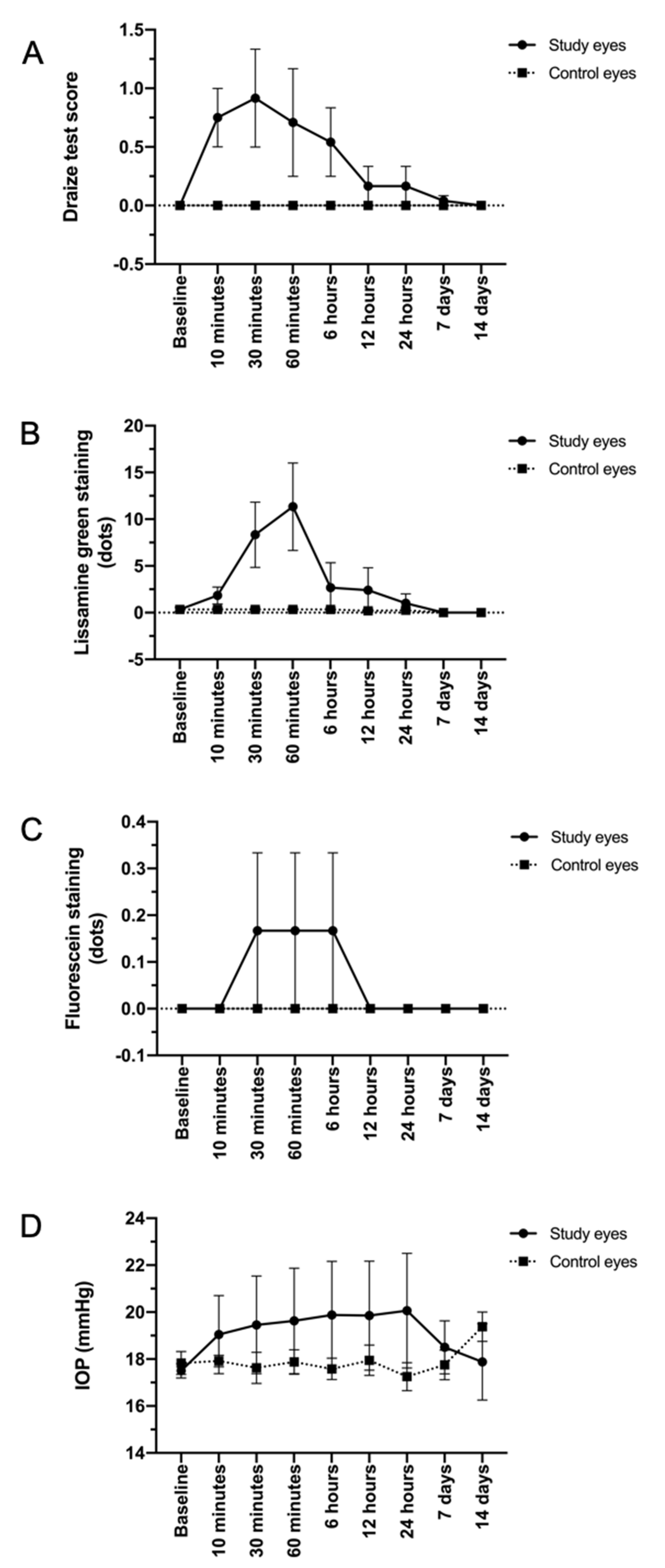
3.1. TALF Is Well Tolerated in the Preclinical Model

Examination of the ocular surface (cornea and conjunctiva), as well as the inner parts of the eye (iris, lens, and retina) of rabbits was performed in order to characterize the potential side effects that may result from a high number of daily instillations of TALF. Ocular examination did not show any major findings or adverse events through the follow-up. Figure 1 shows the mean Draize test score for the study and control eyes at different points of the follow-up.
To quantify the ocular irritation potential of TALF, the parameters of congestion, swelling, and discharge of conjunctiva, iris, hyperemia, and corneal opacity were evaluated in the rabbits. Since the first instillation of TALF and until the end of the study, there was no evidence of inflammation, tissue alteration, and/or discomfort in rabbit eyes. A zero score (0) was achieved in all eyes for inflammation. For swelling and conjunctival congestion, a zero score was also registered at the end of the follow-up in all groups. Iris hyperemia and corneal opacity scores were also equal to zero at all observations. We did not observe changes in the lens during the follow-up of the animal groups. Representative images of study and control eyes that show no inflammation, swelling, or discharge after the instillation of TALF or placebo are presented in Figure 2.
Staining with fluorescein and lissamine green showed superficial epithelium punctate keratitis in the first 6 h after instillation of the formulation. This condition was resolved in all cases in the examination at 12 h after the administration of the formulation. Staining scores of ocular surfaces are presented in Figure 1.
Finally, no increase in intraocular pressure was observed in any of the study animals (normal intraocular pressure in this species is 12–28 mmHg). The behavior of IOP is presented in Figure 1.

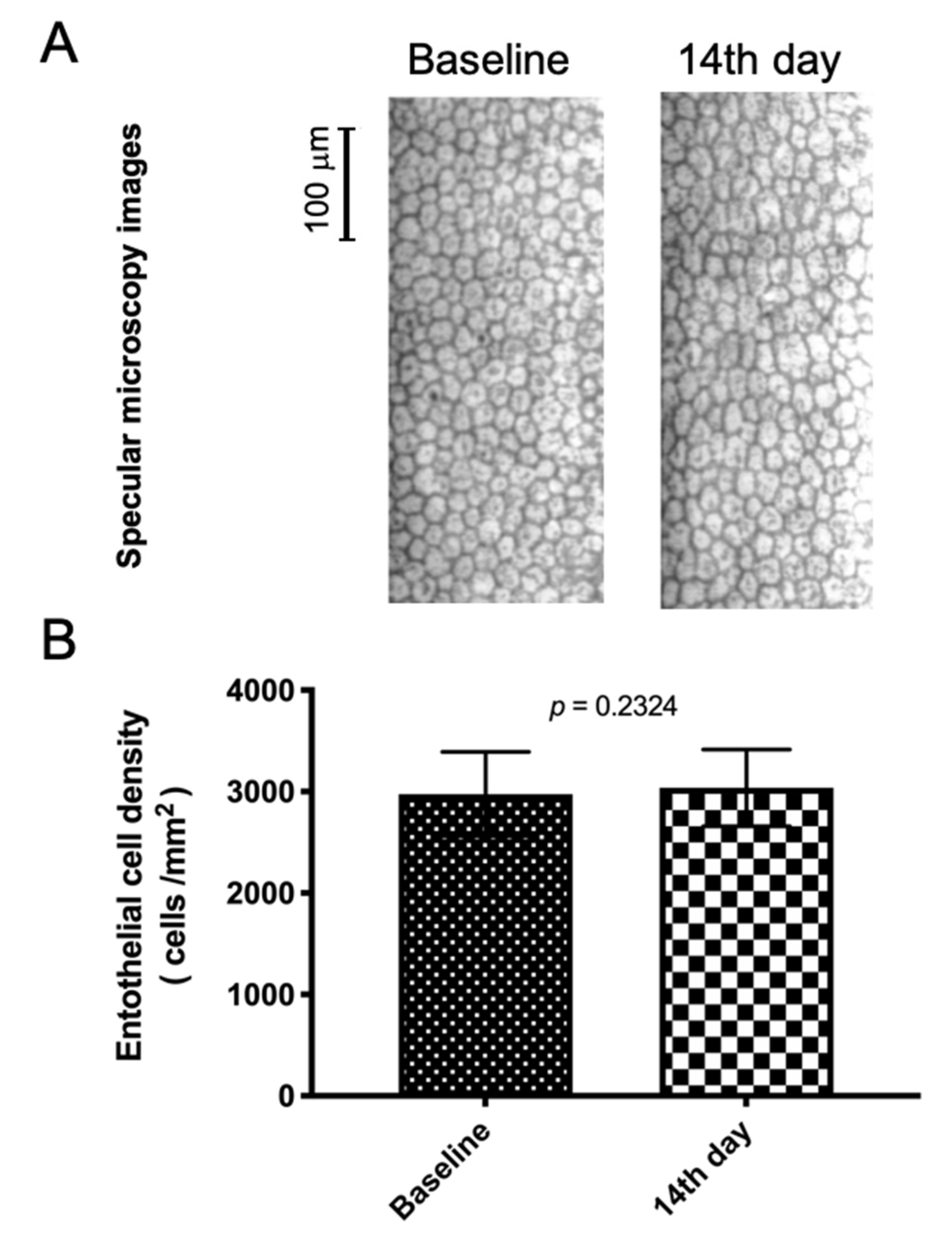
3.2. TALF Is Safe for Healthy Volunteers

Twenty healthy volunteers were enrolled in the study. The mean age was 37 ± 14.8 years. Thirteen (65%) subjects were women and seven (35%) were men. Fifteen of the 20 study eyes were right and five were left. All subject demographics and characteristics are summarized in Table 2.
In relation to safety and tolerability outcomes, we observed that the TALF was well tolerated during the study period. No systemic AEs were reported. None of the 20 patients showed significant changes in IOP, BCVA, contrast sensitivity, or CFT (Table 2). After using the study formulation, none of the patients required treatment with IOP-lowering drugs. No serious AEs were reported (Table 3) after the end of study drug application (day 14) and at the end of the study follow-up (day 21).
Ocular AEs were reported according to the NOM-220-SSA1-2016 as a COFEPRIS request. Most AEs were mild and two of them were moderate in severity. Six subjects (30%) reported occasional mild dryness on one occasion and six others (30%) reported a mild burning sensation (one or two times) during the instillation. Two volunteers (10%) reported moderate secretions that disappeared after the first week of the study and three (15%) reported tearing at the moment of application. A relationship with the drug could not be excluded. There was no pain or discomfort reported. Neither eyelid redness, conjunctival hyperemia, nor edema were observed across the follow-up. No subconjunctival hemorrhages were reported in this study. The examiner acknowledged the nonsignificant results ranked from 0 (no changes) to 1 (mild changes) in relation to the ocular surface staining. The anterior eye chamber and lens showed no abnormal findings. Vitreous cells and flares were absent. Retinal normal appearance remained unchanged before and after intervention. Endothelial cell concentration and retinal condensation were unaltered (2976 ± 414 cells/mm2 vs. 3037 ± 377 cells/mm2) (Figure 3). No clinically relevant changes in vital sign parameters were observed. TALF eye drops did not affect blood pressure or pulse rate. No local or systemic findings required TALF to be stopped. A summary of reported ocular AEs are presented in Table 3.

3.3. TALF Improves BCVA and Reduces CFT in Patients with DME

Four patients with DME were enrolled in the study. The mean age was 62 ± 7.87 years. Two patients (50%) were women. All study eyes were left eyes.
Concerning safety and tolerability outcomes, we observed that the TALF was well tolerated during the study period. No systemic or severe AEs were reported. None of the patients showed significant changes in IOP. After using TALF for 6 months, none of the patients showed intraocular hypertension.
On the other hand, BCVA and CFT improved significantly over time (Figure 4). ETDRS letter scores increased from 41.5 ± 23.5 at baseline to 58 ± 17.79 letters after 6 months of TALF therapy (p = 0.001), whereas CFT measured by OCT decreased from 483 ± 55.39 at baseline to 268 ± 27.26 μm at the same point (p = 0.025). OCT images from all cases are provided in Figure 5. Additionally, no patients required rescue therapy.

4. Discussion

Over the last decade of research, dramatic changes have been observed in the field of drug delivery. Developing a novel drug delivery system to target a particular human tissue has become a major goal for researchers in the field [35,36]. Although there has been great interest in the development of new topical ocular delivery systems, ocular drug delivery has been a major challenge to scientists due to its unique anatomy and physiology. Topical ophthalmic drug molecules must cross several layers of eye tissue and trespass several dynamic and metabolic barriers before reaching the vitreous body and retina, and very low concentrations with almost no clinical effect are usually obtained [36,37]. This is why conventional topical administration of drugs has not been as effective as intravitreal delivery in the treatment of retinal diseases [36].
Nowadays, IVTs are the most used pathway to deliver drugs for the posterior segment of the eye and it has become the most common intraocular procedure worldwide, with increasing numbers every year [38]. IVTs are the standard drug delivery method for the treatment of retinal diseases that cause non-reversible vision impairment [37,39]. The advantage of the intravitreal method is to circumvent the ocular barriers which keep most drugs out of the eye, and the ability to minimize the loss of vision and to improve it over a period of years is an untold clinical benefit [40]. Due to the widespread use of IVT, new therapeutic opportunities for previously untreated ophthalmic patients have improved their vision using drugs already known to the market, such as anti-vascular endothelial growth factor (VEGF), VEGF trap-eye, and triamcinolone [13].
However, IVTs are not exempt from potential problems. The need for multiple consecutive injections increases the incidence of adverse events (AEs) such as endophthalmitis, lens injury, and retinal detachment over the months and years of use [11,12,13,41,42]. In addition, it may be a burden for patients, physicians, and health systems, with poor compliance in many cases [11,12,13]. In addition, IVT requires highly specialized human resources and special infrastructure for its application, making it a costly option in developing countries [13]. Scientists, organizations, and pharmaceutical companies worldwide are continuously looking for a safer, more accessible, and effective ocular drug delivery method for ocular use.
Different implantable and non-implantable [43,44,45,46,47,48,49,50,51] delivery systems, as well as novel ocular drug delivery systems such as microemulsions, nanosuspensions, nanoparticles, and liposome formulations [52,53,54], have been analyzed in order to reduce side effects and reach better patient compliance, to bring about better outcomes. In a previous work, we showed that TALF, a novel system used to deliver TA to the posterior segment of the eye, reaches significant concentrations in the posterior segment of the intraocular tissues and is non-toxic for human keratocyte cultures [18], but the in vivo safety and tolerability of this formulation was still a concern, and the biological and therapeutic effects on a chronic retinal disorder such as diabetic macular edema had not been explored previously. Thus, this report summarizes the findings about the biological activity, safety, and tolerability of TALF.
With respect to the biological activity, we found that TALF was able to reduce the thickness of the fovea (measured by OCT) in patients with adequate glycemic control but the presence of DME. This finding is not surprising because the benefits of synthetic corticosteroids for the therapy of DME are well-known due its numerous targets, but the remarkable finding is the administration route. TALF is the first liposome formulation carrying TA that has shown therapeutic activity clinically. Although a complete understanding of the mechanism of action of corticosteroids has not been fully elucidated, it has been demonstrated that corticosteroids, like the TA contained in TALF, interfere with regulatory components of gene expression, inhibiting the synthesis of vascular endothelial growth factor (VEGF) and multiple pro-inflammatory genes such as tumor necrosis factor α and interleukin 6, while inducing anti-inflammatory factors such as pigment epithelium-derived growth factor (PEDF) [55,56,57]. Furthermore, steroids also inhibit the phospholipase A2 pathway, diminishing the production and release of inflammatory cell mediators and chemokines. Moreover, TA specifically seems to reduce the expression of matrix metalloproteinases (MMPs) and downregulates intercellular adhesion molecule 1 in choroidal endothelial cells [58].
On the other hand, ocular tolerability is a major goal for all topical drugs. Many eye conditions are treated with ophthalmic topical formulations with good results, but tolerability is still a concern for many patients. In our animal study, we did not observe superficial irritation signs such as conjunctival hyperemia in any of the study eyes. We observed superficial epithelium punctate keratitis in rabbits during the first hours with fluorescein sodium and lissamine green staining; however, it was not visible after 6 h of the first instillation of TALF. According to the Pharmacopeia of Estados Unidos Mexicanos, an ocular irritability test was satisfactory, and TALF is considered nonirritant. Additionally, in the phase I clinical assay, healthy volunteers reported predominantly minor and transient adverse events with TALF instillation, which supports the tolerability of the liposomal formulation.
Although further evaluation is needed to completely define the tolerability profile of this formulation, the preliminary results of our experimental study suggest that TALF is well tolerated and is safe in the eyes of New Zealand white rabbits, and it may have the potential to be used as a novel delivery system to reach the intraocular tissues of the posterior segment of the eyeball. These results can be considered useful for other topical liposome formulations comprising different steroids or other molecules. It is worth mentioning that the use of liposomal formulations through IVTs have demonstrated their effectiveness and safety in previous reports [59,60,61,62]. However, topical liposomes are a new approach for posterior ocular segment TA delivery when thinking about substituting IVT. Recently, Li, J and colleagues [15] have developed triamcinolone acetonide chitosan-coated liposomes as eye drops to deliver this steroid to the posterior segment of the eye. They have shown that this system has high entrapment efficiency (90.66%  ±  3.21%) and exhibits a sustained release profile, excellent physical stability, and no significant toxicity on the cornea, conjunctiva, and retina. Furthermore, Li, J and colleagues argued that their liposomal delivery platform is potentially more effective than our system since they showed higher concentrations of TA in posterior segment tissues. However, increased intraocular TA concentrations have shown major limitations, despite their increased effectiveness, concerning plausible unfavorable effects, including an elevation in and the formation of a posterior subcapsular opacity [4,5,6,63,64]. In our study we did not observe changes or opacities in the lenses of any eyes in the study groups and IOP also did not rise significantly in study eyes, but the therapeutic effect persisted in the retina (reduction of CFT). This background guided us to consider that TALF was safe and that it may have the potential to be a new drug delivery system for the posterior segment.

5. Conclusions

In conclusion, TALF has proven to be safe and well tolerated when used topically, and possesses therapeutic activity in humans. This represents a potential steroid therapy that could replace the use of IVTs of steroids, or could be used as an adjuvant to reduce the frequency of IVTs. In any case, it seems that complications related to intravitreal injections, as well as the adverse events related to the use of intraocular steroids, may be avoided by TALF. However, future larger experimental and clinical TALF studies to evaluate longer-term safety and the therapeutic profile are necessary.

Author Contributions

Formal analysis, J.C.A.-V., A.G.-D.l.R., and J.A.-B.; investigation, J.C.A.-V. and A.G.-D.l.R.; methodology, J.N.-P.; project administration, A.S.; supervision, A.S.; validation, J.A.-B. and A.S.; writing—review and editing, C.R.C.-C. All authors have read and agreed to the published version of the manuscript.

Funding

This research received no external funding.

Institutional Review Board Statement

The study was conducted according to the guidelines of the Declaration of Helsinki, and approved by the Institutional Review Board (or Ethics Committee) of IJICSA Committee (ID: CRMQ-2015-05-T-02; approved on 13 December 2016; COFEPRIS 173300410A0035/2017).

Informed Consent Statement

Informed consent was obtained from all subjects involved in the study.

Data Availability Statement

The data presented in this study are available on request from the corresponding author. The data are not publicly available due to privacy and ethical restrictions.

Conflicts of Interest

The authors declare no conflict of interest.

Abbreviations

AEs, adverse events; ARVO, Association for Research in Vision and Ophthalmology; BCVA, best corrected visual acuity; cECD, corneal endothelial cell density; CFT, central foveal thickness; CS, contrast sensitivity; DME, diabetic macular edema; ETDRS, Early Treatment Diabetic Retinopathy Study; F, female; FEUM, Farmacopea de los Estados Unidos Mexicanos; GMP, good manufacturing practice; HPLC, high-performance liquid chromatography; IBM, International Business Machines; IOP, intraocular pressure; IRB, Institutional Review Board; IVTs, intravitreal injections; LG, lissamine green; LPs, liposomes; M, male; MMPs, matrix metalloproteinases; OCT, optical coherence tomography; OD, right eye; OS, left eye; OSDI, ocular surface disease index; PEDF, pigment epithelium-derived growth factor; PRDL, Pharmaceutical Research and Development Laboratory; SAEs, serious adverse events; SEM, scanning electron microscopy; SPSS, Statistical Package for the Social Sciences; TA, triamcinolone acetonide; TALF, TA-loaded liposome formulation; TEM, transmission electron microscopy; VEGF, vascular endothelial growth factor.

References

  1. Kwon, S.I.; Kim, Y.W.; Bang, Y.W.; Lee, J.Y.; Park, I.W. Comparison of natural course, intravitreal triamcinolone, and intravitreal bevacizumab for treatment of macular edema secondary to branch retinal vein occlusion. J. Ocul. Pharmacol. Ther. 2013, 29, 5–9. [Google Scholar] [CrossRef] [PubMed]
  2. Habot-Wilner, Z.; Sallam, A.; Pacheco, P.A.; Do, H.H.; McCluskey, P.; Lightman, S. Intravitreal triamcinolone acetonide as adjunctive treatment with systemic therapy for uveitic macular edema. Eur. J. Ophthalmol. 2011, 21 (Suppl. 6), S56–S61. [Google Scholar] [CrossRef]
  3. Yalcinbayir, O.; Gelisken, O.; Kaderli, B.; Avci, R. Intravitreal versus sub-tenon posterior triamcinolone injection in bilateral diffuse diabetic macular edema. Ophthalmologica 2011, 225, 222–227. [Google Scholar] [CrossRef] [PubMed]
  4. Arikan, G.; Osman Saatci, A.; Hakan Oner, F. Immediate intraocular pressure rise after intravitreal injection of ranibizumab and two doses of triamcinolone acetonide. Int. J. Ophthalmol. 2011, 4, 402–405. [Google Scholar]
  5. Chan, C.K.; Fan, D.S.; Chan, W.M.; Lai, W.W.; Lee, V.Y.; Lam, D.S. Ocular-hypertensive response and corneal endothelial changes after intravitreal triamcinolone injections in Chinese subjects: A 6-month follow-up study. Eye 2005, 19, 625–630. [Google Scholar] [CrossRef] [PubMed] [Green Version]
  6. Veritti, D.; Di Giulio, A.; Sarao, V.; Lanzetta, P. Drug safety evaluation of intravitreal triamcinolone acetonide. Expert Opin. Drug Saf. 2012, 11, 331–340. [Google Scholar] [CrossRef] [PubMed]
  7. Chu, Y.K.; Chung, E.J.; Kwon, O.W.; Lee, J.H.; Koh, H.J. Objective evaluation of cataract progression associated with a high dose intravitreal triamcinolone injection. Eye 2008, 22, 895–899. [Google Scholar] [CrossRef] [PubMed]
  8. Quiram, P.A.; Gonzales, C.R.; Schwartz, S.D. Severe steroid-induced glaucoma following intravitreal injection of triamcinolone acetonide. Am. J. Ophthalmol. 2006, 141, 580–582. [Google Scholar] [CrossRef]
  9. Viola, F.; Morescalchi, F.; Staurenghi, G. Argon laser trabeculoplasty for intractable glaucoma following intravitreal triamcinolone. Arch. Ophthalmol. 2006, 124, 133–134. [Google Scholar] [CrossRef] [Green Version]
  10. Agrawal, S.; Agrawal, J.; Agrawal, T.P. Management of intractable glaucoma following intravitreal triamcinolone acetonide. Am. J. Ophthalmol. 2005, 139, 575–576. [Google Scholar] [CrossRef] [PubMed]
  11. Lyall, D.A.; Tey, A.; Foot, B.; Roxburgh, S.T.; Virdi, M.; Robertson, C.; MacEwen, C.J. Post-intravitreal anti-VEGF endophthalmitis in the United Kingdom: Incidence, features, risk factors, and outcomes. Eye 2012, 26, 1517–1526. [Google Scholar] [CrossRef] [PubMed]
  12. Poku, E.; Rathbone, J.; Wong, R.; Everson-Hock, E.; Essat, M.; Pandor, A.; Wailoo, A. The safety of intravitreal bevacizumab monotherapy in adult ophthalmic conditions: Systematic review. BMJ Open 2014, 4, e005244. [Google Scholar] [CrossRef]
  13. Azad, R.; Chandra, P.; Gupta, R. The economic implications of the use of anti-vascular endothelial growth factor drugs in age-related macular degeneration. Indian J. Ophthalmol. 2007, 55, 441–443. [Google Scholar] [CrossRef]
  14. Patel, A.; Cholkar, K.; Agrahari, V.; Mitra, A.K. Ocular drug delivery systems: An overview. World J. Pharmacol. 2013, 2, 47–64. [Google Scholar] [CrossRef] [PubMed]
  15. Li, J.; Cheng, T.; Tian, Q.; Cheng, Y.; Zhao, L.; Zhang, X.; Qu, Y. A more efficient ocular delivery system of triamcinolone acetonide as eye drop to the posterior segment of the eye. Drug Deliv. 2019, 26, 188–198. [Google Scholar] [CrossRef] [PubMed] [Green Version]
  16. Raval, N.; Khunt, D.; Misra, M. Microemulsion-based delivery of triamcinolone acetonide to posterior segment of eye using chitosan and butter oil as permeation enhancer: An in vitro and in vivo investigation. J. Microencapsul. 2018, 35, 62–77. [Google Scholar] [CrossRef] [PubMed]
  17. Santer, V.; Chen, Y.; Kalia, Y.N. Controlled non-invasive iontophoretic delivery of triamcinolone acetonide amino acid ester prodrugs into the posterior segment of the eye. Eur. J. Pharm. Biopharm. 2018, 132, 157–167. [Google Scholar] [CrossRef] [PubMed]
  18. Altamirano-Vallejo, J.C.; Navarro-Partida, J.; Gonzalez-De la Rosa, A.; Hsiao, J.H.; Olguin-Gutierrez, J.S.; Gonzalez-Villegas, A.C.; Keller, B.C.; Bouzo-Lopez, L.; Santos, A. Characterization and Pharmacokinetics of Triamcinolone Acetonide-Loaded Liposomes Topical Formulations for Vitreoretinal Drug Delivery. J. Ocul. Pharmacol. Ther. 2018, 34, 416–425. [Google Scholar] [CrossRef]
  19. Meza-Rios, A.; Navarro-Partida, J.; Armendariz-Borunda, J.; Santos, A. Therapies Based on Nanoparticles for Eye Drug Delivery. Ophthalmol. Ther. 2020, 9, 1–14. [Google Scholar] [CrossRef]
  20. Gonzalez-De la Rosa, A.; Navarro-Partida, J.; Altamirano-Vallejo, J.C.; Jauregui-Garcia, G.D.; Acosta-Gonzalez, R.; Ibanez-Hernandez, M.A.; Mora-Gonzalez, G.F.; Armendáriz-Borunda, J.; Santos, A. Novel Triamcinolone Acetonide-Loaded Liposomal Topical Formulation Improves Contrast Sensitivity Outcome After Femtosecond Laser-Assisted Cataract Surgery. J. Ocul. Pharmacol. Ther. 2019, 35, 512–521. [Google Scholar] [CrossRef] [PubMed] [Green Version]
  21. Gonzalez-De la Rosa, A.; Navarro-Partida, J.; Altamirano-Vallejo, J.C.; Hernandez-Gamez, A.G.; Garcia-Banuelos, J.J.; Armendariz-Borunda, J.; Santos, A. Novel triamcinolone acetonide-loaded liposomes topical formulation for the treatment of cystoid macular edema after cataract surgery: A Pilot Study. J. Ocul. Pharmacol. Ther. 2019, 35, 106–115. [Google Scholar] [CrossRef] [PubMed] [Green Version]
  22. Navarro-Partida, J.; Altamirano-Vallejo, J.C.; Lopez-Naranjo, E.J.; Gonzalez-De la Rosa, A.; Manzano-Ramírez, A.; Apatiga-Castro, L.M.; Armendáriz-Borunda, J.; Santos, A. Topical Triamcinolone Acetonide-Loaded Liposomes as Primary Therapy for Macular Edema Secondary to Branch Retinal Vein Occlusion: A Pilot Study. J. Ocul. Pharmacol. Ther. 2020, 36, 393–403. [Google Scholar] [CrossRef] [PubMed]
  23. Sasaki, H.; Karasawa, K.; Hironaka, K.; Tahara, K.; Tozuka, Y.; Takeuchi, H. Retinal drug delivery using eyedrop preparations of poly-L-lysine-modified liposomes. Eur. J. Pharm. Biopharm. 2013, 83, 364–369. [Google Scholar] [CrossRef] [PubMed]
  24. Davis, B.M.; Normando, E.M.; Guo, L.; Turner, L.A.; Nizari, S.; O’Shea, P.; Moss, S.E.; Somavarapu, S.; Cordeiro, M.F. Topical delivery of Avastin to the posterior segment of the eye in vivo using annexin A5-associated liposomes. Small 2014, 10, 1575–1584. [Google Scholar] [CrossRef]
  25. Inokuchi, Y.; Hironaka, K.; Fujisawa, T.; Tozuka, Y.; Tsuruma, K.; Shimazawa, M.; Takeuchi, H.; Hara, H. Physicochemical properties affecting retinal drug/coumarin-6 delivery from nanocarrier systems via eyedrop administration. Investig. Ophthalmol. Vis. Sci. 2010, 51, 3162–3170. [Google Scholar] [CrossRef] [Green Version]
  26. Fujisawa, T.; Miyai, H.; Hironaka, K.; Tsukamoto, T.; Tahara, K.; Tozuka, Y.; Ito, M.; Takeuchi, H. Liposomal diclofenac eye drop formulations targeting the retina: Formulation stability improvement using surface modification of liposomes. Int. J. Pharm. 2012, 436, 564–567. [Google Scholar] [CrossRef] [PubMed]
  27. Shimazaki, H.; Hironaka, K.; Fujisawa, T.; Tsuruma, K.; Tozuka, Y.; Shimazawa, M.; Takeuchi, H.; Hara, H. Edaravone-loaded liposome eyedrops protect against light-induced retinal damage in mice. Investig. Ophthalmol. Vis. Sci. 2011, 52, 7289–7297. [Google Scholar] [CrossRef] [PubMed] [Green Version]
  28. Takashima, Y.; Tsuchiya, T.; Igarashi, Y.; Kanazawa, T.; Okada, H.; Urtti, A. Non-invasive ophthalmic liposomes for nucleic acid delivery to posterior segment of eye. Yakugaku Zasshi 2012, 132, 1365–1370. [Google Scholar] [CrossRef] [PubMed] [Green Version]
  29. Hironaka, K.; Fujisawa, T.; Sasaki, H.; Tozuka, Y.; Tsuruma, K.; Shimazawa, M.; Hara, H.; Takeuchi, H. Fluorescence investigation of the retinal delivery of hydrophilic compounds via liposomal eyedrops. Biol. Pharm. Bull. 2011, 34, 894–897. [Google Scholar] [CrossRef] [Green Version]
  30. Hironaka, K.; Inokuchi, Y.; Tozuka, Y.; Shimazawa, M.; Hara, H.; Takeuchi, H. Design and evaluation of a liposomal delivery system targeting the posterior segment of the eye. J. Control. Release 2009, 136, 247–253. [Google Scholar] [CrossRef]
  31. Masuda, I.; Matsuo, T.; Yasuda, T.; Matsuo, N. Gene transfer with liposomes to the intraocular tissues by different routes of administration. Investig. Ophthalmol. Vis. Sci. 1996, 37, 1914–1920. [Google Scholar]
  32. Abelson, M.B.; Udell, I.J.; Weston, J.H. Normal human tear pH by direct measurement. Arch. Ophthalmol. 1981, 99, 301. [Google Scholar] [CrossRef] [PubMed]
  33. McDonald, T.O.; Shadduck, J.A. Eye Irritation; Wiley & Sons: New York, NY, USA, 1977; Volume 4. [Google Scholar]
  34. Kinyoun, J.; Barton, F.; Fisher, M.; Hubbard, L.; Aiello, L.; Ferris, F., III; Group, E.R. Detection of diabetic macular edema: Ophthalmoscopy versus photography—Early Treatment Diabetic Retinopathy Study report number 5. Ophthalmology 1989, 96, 746–751. [Google Scholar] [CrossRef]
  35. Gaudana, R.; Ananthula, H.K.; Parenky, A.; Mitra, A.K. Ocular drug delivery. AAPS J. 2010, 12, 348–360. [Google Scholar] [CrossRef] [PubMed]
  36. Fangueiro, J.F.; Veiga, F.; Silva, A.M.; Souto, E.B. Ocular Drug Delivery—New Strategies for Targeting Anterior and Posterior Segments of the Eye. Curr. Pharm. Des. 2016, 22, 1135–1146. [Google Scholar] [CrossRef]
  37. Kim, S.H.; Lutz, R.J.; Wang, N.S.; Robinson, M.R. Transport barriers in transscleral drug delivery for retinal diseases. Ophthalmic Res. 2007, 39, 244–254. [Google Scholar] [CrossRef] [PubMed]
  38. Grzybowski, A.; Told, R.; Sacu, S.; Bandello, F.; Moisseiev, E.; Loewenstein, A.; Schmidt-Erfurth, U.; Euretina, B. 2018 Update on Intravitreal Injections: Euretina Expert Consensus Recommendations. Ophthalmologica 2018, 239, 181–193. [Google Scholar] [CrossRef]
  39. Raghava, S.; Hammond, M.; Kompella, U.B. Periocular routes for retinal drug delivery. Expert Opin. Drug Deliv. 2004, 1, 99–114. [Google Scholar] [CrossRef]
  40. Martin, D.F.; Maguire, M.G.; Fine, S.L.; Ying, G.S.; Jaffe, G.J.; Grunwald, J.E.; Toth, C.; Redford, M.; Ferris, F.L., 3rd; Comparison of Age-related Macular Degeneration Treatments Trials (CATT) Research Group. Ranibizumab and bevacizumab for treatment of neovascular age-related macular degeneration: Two-year results. Ophthalmology 2012, 119, 1388–1398. [Google Scholar] [CrossRef] [PubMed] [Green Version]
  41. Fung, A.E.; Rosenfeld, P.J.; Reichel, E. The International Intravitreal Bevacizumab Safety Survey: Using the internet to assess drug safety worldwide. Br. J. Ophthalmol. 2006, 90, 1344–1349. [Google Scholar] [CrossRef] [PubMed] [Green Version]
  42. Droege, K.M.; Muether, P.S.; Hermann, M.M.; Caramoy, A.; Viebahn, U.; Kirchhof, B.; Fauser, S. Adherence to ranibizumab treatment for neovascular age-related macular degeneration in real life. Graefe’s Arch. Clin. Exp. Ophthalmol. 2013, 251, 1281–1284. [Google Scholar] [CrossRef] [PubMed]
  43. Heier, J.S.; Brown, D.M.; Chong, V.; Korobelnik, J.F.; Kaiser, P.K.; Nguyen, Q.D.; Kirchhof, B.; Ho, A.; Ogura, Y.; Yancopoulos, G.D.; et al. Intravitreal aflibercept (VEGF trap-eye) in wet age-related macular degeneration. Ophthalmology 2012, 119, 2537–2548. [Google Scholar] [CrossRef] [PubMed]
  44. Thomas, M.; Mousa, S.S.; Mousa, S.A. Comparative effectiveness of aflibercept for the treatment of patients with neovascular age-related macular degeneration. Clin. Ophthalmol. 2013, 7, 495–501. [Google Scholar]
  45. Pacella, E.; Vestri, A.R.; Muscella, R.; Carbotti, M.R.; Castellucci, M.; Coi, L.; Turchetti, P.; Pacella, F. Preliminary results of an intravitreal dexamethasone implant (Ozurdex(R)) in patients with persistent diabetic macular edema. Clin. Ophthalmol. 2013, 7, 1423–1428. [Google Scholar] [CrossRef] [Green Version]
  46. Pearson, P.A.; Comstock, T.L.; Ip, M.; Callanan, D.; Morse, L.S.; Ashton, P.; Levy, B.; Mann, E.S.; Eliott, D. Fluocinolone acetonide intravitreal implant for diabetic macular edema: A 3-year multicenter, randomized, controlled clinical trial. Ophthalmology 2011, 118, 1580–1587. [Google Scholar] [CrossRef] [PubMed]
  47. Patel, S.R.; Berezovsky, D.E.; McCarey, B.E.; Zarnitsyn, V.; Edelhauser, H.F.; Prausnitz, M.R. Targeted administration into the suprachoroidal space using a microneedle for drug delivery to the posterior segment of the eye. Investig. Ophthalmol. Vis. Sci. 2012, 53, 4433–4441. [Google Scholar] [CrossRef]
  48. Lo, R.; Li, P.Y.; Saati, S.; Agrawal, R.; Humayun, M.S.; Meng, E. A refillable microfabricated drug delivery device for treatment of ocular diseases. Lab Chip 2008, 8, 1027–1030. [Google Scholar] [CrossRef] [PubMed]
  49. Saati, S.; Lo, R.; Li, P.Y.; Meng, E.; Varma, R.; Humayun, M.S. Mini drug pump for ophthalmic use. Trans. Am. Ophthalmol. Soc. 2009, 107, 60–70. [Google Scholar] [CrossRef] [Green Version]
  50. Gutierrez-Hernandez, J.C.; Caffey, S.; Abdallah, W.; Calvillo, P.; Gonzalez, R.; Shih, J.; Brennan, J.; Zimmerman, J.; Martinez-Camarillo, J.C.; Rodriguez, A.R.; et al. One-Year Feasibility Study of Replenish MicroPump for Intravitreal Drug Delivery: A Pilot Study. Transl. Vis. Sci. Technol. 2014, 3, 8. [Google Scholar] [CrossRef]
  51. Humayun, M.; Santos, A.; Altamirano, J.C.; Ribeiro, R.; Gonzalez, R.; de la Rosa, A.; Shih, J.; Pang, C.; Jiang, F.; Calvillo, P.; et al. Implantable MicroPump for Drug Delivery in Patients with Diabetic Macular Edema. Transl. Vis. Sci. Technol. 2014, 3, 5. [Google Scholar] [CrossRef] [Green Version]
  52. Hans, M.L.; Lowman, A.M. Biodegradable Nanoparticles for Drug Delivery and Targeting. Curr. Opin. Solid State Mater. Sci. 2002, 6, 319–327. [Google Scholar] [CrossRef]
  53. Moya-Ortega, M.D.; Alves, T.F.; Alvarez-Lorenzo, C.; Concheiro, A.; Stefansson, E.; Thorsteinsdottir, M.; Loftsson, T. Dexamethasone eye drops containing gamma-cyclodextrin-based nanogels. Int. J. Pharm. 2013, 441, 507–515. [Google Scholar] [CrossRef] [PubMed]
  54. Kaur, I.P.; Garg, A.; Singla, A.K.; Aggarwal, D. Vesicular systems in ocular drug delivery: An overview. Int. J. Pharm. 2004, 269, 1–14. [Google Scholar] [CrossRef] [PubMed]
  55. Kim, Y.; Choi, M.; Kim, Y.; Park, C.; Lee, J.; Chung, I.; Yoo, J.; Choi, W.; Cho, G.; Kang, S. Triamcinolone acetonide protects the rat retina from STZ-induced acute inflammation and early vascular leakage. Life Sci. 2007, 81, 1167–1173. [Google Scholar] [CrossRef]
  56. Zhang, S.X.; Wang, J.J.; Gao, G.; Shao, C.; Mott, R.; Ma, J.X. Pigment epithelium-derived factor (PEDF) is an endogenous antiinflammatory factor. FASEB J. 2006, 20, 323–325. [Google Scholar] [CrossRef]
  57. Tsaprouni, L.G.; Ito, K.; Punchard, N.; Adcock, I.M. Triamcinolone Acetonide and Dexamethasome Suppress TNF-α-Induced Histone H4 Acetylation on Lysine Residues 8 and 12 in Mononuclear Cells. Ann. N. Y. Acad. Sci. 2002, 973, 481–483. [Google Scholar] [CrossRef] [PubMed]
  58. Bandello, F.; Parodi, M.B.; Lanzetta, P.; Loewenstein, A.; Massin, P.; Menchini, F.; Veritti, D. Diabetic macular edema. In Macular Edema; Karger Publishers: Basel, Switzerland, 2017; Volume 58, pp. 102–138. [Google Scholar]
  59. Umscheid, C.A.; Margolis, D.J.; Grossman, C.E. Key concepts of clinical trials: A narrative review. Postgrad. Med. 2011, 123, 194–204. [Google Scholar] [CrossRef] [Green Version]
  60. Akula, S.K.; Ma, P.E.; Peyman, G.A.; Rahimy, M.H.; Hyslop, N.E., Jr.; Janney, A.; Ashton, P. Treatment of cytomegalovirus retinitis with intravitreal injection of liposome encapsulated ganciclovir in a patient with AIDS. Br. J. Ophthalmol. 1994, 78, 677–680. [Google Scholar] [CrossRef] [PubMed] [Green Version]
  61. Zeng, S.; Hu, C.; Wei, H.; Lu, Y.; Zhang, Y.; Yang, J.; Yun, G.; Zou, W.; Song, B. Intravitreal pharmacokinetics of liposome-encapsulated amikacin in a rabbit model. Ophthalmology 1993, 100, 1640–1644. [Google Scholar] [CrossRef]
  62. Cannon, J.P.; Fiscella, R.; Pattharachayakul, S.; Garey, K.W.; De Alba, F.; Piscitelli, S.; Edward, D.P.; Danziger, L.H. Comparative toxicity and concentrations of intravitreal amphotericin B formulations in a rabbit model. Investig. Ophthalmol. Vis. Sci. 2003, 44, 2112–2117. [Google Scholar] [CrossRef] [PubMed] [Green Version]
  63. Shabir, G.A. Validation of high-performance liquid chromatography methods for pharmaceutical analysis. Understanding the differences and similarities between validation requirements of the US Food and Drug Administration, the US Pharmacopeia and the International Conference on Harmonization. J. Chromatogr. A 2003, 987, 57–66. [Google Scholar] [PubMed]
  64. Purslow, C.; Wolffsohn, J.S. Ocular surface temperature: A review. Eye Contact Lens 2005, 31, 117–123. [Google Scholar] [CrossRef] [PubMed]
Figure 1. Mean Draize test scores (A), staining scores (B,C), and intraocular pressure (D) in study and control eyes. Non-significant differences were observed between eyes treated with triamcinolone acetonide-loaded liposomes formulation and control eyes receiving an instillation of saline-balanced solution during the follow-up.
Pharmaceutics 13 00322 g001
Figure 2. Representative images of study and control eyes. (A) Study eyes and (B) control eyes photographs show no inflammation, swelling, or discharge after the instillation of triamcinolone acetonide-loaded liposomes formulation (TALF) or placebo solution, respectively.
Pharmaceutics 13 00322 g002
Figure 3. Corneal endothelial cell density analysis in healthy subjects treated with triamcinolone acetonide-loaded liposomes formulation. (A) Images of specular microscopy of a representative case at baseline and after 14 days of TALF instillation. (B) Column bar graph from corneal endothelial cell density (cECD) analysis. Non-significant differences in cECD values were established between baseline and after 14 days of TALF instillation. TALF; triamcinolone acetonide-loaded liposomes formulation.
Pharmaceutics 13 00322 g003
Figure 4. Best corrected visual acuity, central foveal thickness, and intraocular pressure follow-up in patients with diabetic macular edema treated with triamcinolone acetonide-loaded liposomes formulation. (A) Best corrected visual acuity, measured by ETDRS test, improved through the follow-up, reaching statistical significance at month 5. (B) CFT, measured by OCT, contracted through the follow-up, accomplishing statistical significance at month 4. (C) Non-significant variation in IOP was recorded during the study. ETDRS; Early Treatment Diabetic Retinopathy Study, CFT; central foveal thickness, OCT; optical coherence tomography.
Pharmaceutics 13 00322 g004
Figure 5. Optical coherence tomography images from patients with diabetic macular edema treated with triamcinolone acetonide-loaded liposomes formulation. A significant reduction in CFT was revealed through OCT analysis in patients receiving TALF. The CFT reduction was maintained through the follow-up in almost all cases. CFT; central foveal thickness, OCT; optical coherence tomography, TALF; triamcinolone acetonide-loaded liposomes formulation.
Pharmaceutics 13 00322 g005
Table 1. Composition of triamcinolone acetonide-loaded liposome formulation (1 mL).
(w or v)(%)
Triamcinolone acetonide2.0 mg0.2w/v
Kolliphor HS 1550 mg5w/v
PEG-12 glyceryl dimyristate100 mg10w/v
Ethyl alcohol14 mL1.40v/v
Citric acid anhydrous0.8 mg0.08w/v
Sodium citrate dihydrate4.675 mg0.4675w/v
Benzalkonium chloride0.1 mg0.01w/v
Grade 2 purified waterQ.S.1.0 mlNANA
NA; not applicable, v; volume, w, weight.
Table 2. Demographics and clinical characteristics of healthy subjects treated with triamcinolone acetonide-loaded liposomes formulation. BCVA, best corrected visual acuity; CFT, central foveal thickness; ETDRS, Early Treatment Diabetic Retinopathy Study; IOP, intraocular pressure; OD, right eye; OS, left eye.
Baseline14th Day
PatientGenderStudy EyeAgeBCVAContrastIOP CFTBCVAContrastIOP CFT
(Years)(ETDRS Letters)(L/Contrast)(mmHg)(μm)(ETDRS Letters)(L/Contrast)(mmHg)(μm)
1FOD25851.6510250851.6512248
2FOS25851.6511264851.513269
3FOD26851.3510256851.3516260
4FOS26851.3510259851.3516261
5MOD25831.3513242841.514243
6MOS25841.3512252851.512254
7FOD24841.3511243851.58247
8FOS24851.3511247851.57241
9FOD24851.516255851.511258
10FOS24851.3517257851.659256
11MOD56851.516257851.6515256
12MOD35851.6517263851.6514265
13FOD47851.6513246851.6515245
14FOD35851.6518245851.6516247
15FOD63831.3512258851.6514260
16MOD56851.6515260851.6514262
17FOD40851.6516262841.6514259
18FOD38841.512252851.511255
19MOD63841.516249841.513248
20MOD59851.3515263851.6516260
X ± s 37 ± 14.884.6 ± 0.61.5 ± 0.113.5 ± 2.7254 ± 784.9 ± 0.3 1.6 ± 0.1 13 ± 2.7 254.7 ± 7.8
inF = 13 M = 7OD = 15 OS = 5
p 0.0960.0140.48530.2825
in; frequency, F; female, M; male, OD; right eye, BCVA; best corrected visual acuity, CFT; central foveal thickness, ETDRS; Early Treatment Diabetic Retinopathy Study, IOP; intraocular pressure, TALF; triamcinolone acetonide-loaded liposomes formulation, ; statistically significant differences from baseline values (p < 0.05), ; no statistically significant differences from baseline values (p > 0.05).
Table 3. Adverse events reported in healthy subjects treated with triamcinolone acetonide-loaded liposomes formulation.
Adverse EventsCategoriesDry EyesBurning DischargeTearingBlurred VisionNone
Safety Variablesn (%)n (%)n (%)n (%)n (%)n (%)
FrequencyNot presented14 (70)14 (70)18 (90)17 (85)18 (90)8 (40)
Rare6 (30)4 (20)
Occasionally 2 (10) 3 (15)
Most of the time 2 (10) 2 (10)
All the time
SeverityNot presented14 (70)14 (70)18 (90)17 (85)18 (90)8 (40)
Mild6 (30)6 (30) 3 (15)2 (10)
Moderate 2 (10)
Severe
TALF; triamcinolone acetonide-loaded liposomes formulation.
Publisher’s Note: MDPI stays neutral with regard to jurisdictional claims in published maps and institutional affiliations.

