
Advances in Use of Nanomaterials for Musculoskeletal Regeneration


Abstract
:1. Introduction
2. Applied Nanomaterials
3. Nanomaterials for Cartilage Healing and Regeneration
4. Nanomaterials for Bone Healing and Regeneration
4.1. Nano-Hydroxyapatite
Metal Doped Hydroxyapatite
4.2. Carbon-Based Nanomaterials
4.2.1. Carbon Nanotubes
4.2.2. Graphene-Based Materials
4.2.3. Other Carbon-Based Nanomaterials
4.3. Silicates and Clays
4.4. Metal-Based Nanomaterials
4.4.1. Magnesium-Based Nanomaterials
4.4.2. Titanium-Based Nanomaterials
4.4.3. Other Metal-Based Nanomaterials
4.5. Polymers and Other Organic Materials
5. Critical View and Perspectives
6. Conclusions
Author Contributions
Funding
Institutional Review Board Statement
Informed Consent Statement
Data Availability Statement
Conflicts of Interest
Abbreviations
References
- WHO. Musculoskeletal Conditions. 2021. Available online: https://www.who.int/news-room/fact-sheets/detail/musculoskeletal-conditions (accessed on 27 June 2021).
- Sozen, T.; Ozisik, L.; Basaran, N.C. An overview and management of osteoporosis. Eur. J. Rheumatol. 2017, 4, 46–56. [Google Scholar] [CrossRef]
- Nakamura, K.; Ogata, T. Locomotive syndrome: Definition and management. Clin. Rev. Bone Miner. Metab. 2016, 14, 56–67. [Google Scholar] [CrossRef] [PubMed] [Green Version]
- Föger-Samwald, U.; Dovjak, P.; Azizi-Semrad, U.; Kerschan-Schindl, K.; Pietschmann, P. Osteoporosis: Pathophysiology and therapeutic options. EXCLI J. 2020, 19, 1017–1037. [Google Scholar]
- Gibofsky, A. Epidemiology, pathophysiology, and diagnosis of rheumatoid arthritis: A synopsis. Am. J. Manag. Care 2014, 20, 128–135. [Google Scholar]
- Song, Y.; Zhang, J.; Xu, H.; Lin, Z.; Chang, H.; Liu, W.; Kong, L. Mesenchymal stem cells in knee osteoarthritis treatment: A systematic review and meta-analysis. J. Orthop. Translat. 2020, 24, 121–130. [Google Scholar] [CrossRef] [PubMed]
- Van den Bergh, J.P.; van Geel, T.A.; Geusens, P.P. Osteoporosis, frailty and fracture: Implications for case finding and therapy. Nat. Rev. Rheumatol. 2012, 8, 163–172. [Google Scholar] [CrossRef] [PubMed]
- Hendrickx, G.; Boudin, E.; Van Hul, W. A look behind the scenes: The risk and pathogenesis of primary osteoporosis. Nat. Rev. Rheumatol. 2015, 11, 462–474. [Google Scholar] [CrossRef]
- Zhang, Y.; Jordan, J.M. Epidemiology of osteoarthritis. Clin. Geriatr. Med. 2010, 26, 355–369. [Google Scholar] [CrossRef] [Green Version]
- Melrose, J.; Fuller, E.S.; Little, C.B. The biology of meniscal pathology in osteoarthritis and its contribution to joint disease: Beyond simple mechanics. Connect. Tissue Res. 2017, 58, 282–294. [Google Scholar] [CrossRef] [PubMed]
- Melrose, J. The importance of the knee joint meniscal fibrocartilages as stabilizing weight bearing structures providing global protection to human knee-joint tissues. Cells 2019, 8, 324. [Google Scholar] [CrossRef] [Green Version]
- He, Y.; Li, Z.; Alexander, P.G.; Ocasio-Nieves, B.D.; Yocum, L.; Lin, H.; Tuan, R.S. Pathogenesis of osteoarthritis: Risk factors, regulatory pathways in chondrocytes, and experimental models. Biology 2020, 9, 194. [Google Scholar] [CrossRef] [PubMed]
- Grassel, S.; Zaucke, F.; Madry, H. Osteoarthritis: Novel molecular mechanisms increase our understanding of the disease pathology. J. Clin. Med. 2021, 10, 1938. [Google Scholar] [CrossRef] [PubMed]
- Almutairi, K.; Nossent, J.; Preen, D.; Keen, H.; Inderjeeth, C. The global prevalence of rheumatoid arthritis: A meta-analysis based on a systematic review. Rheumatol. Int. 2021, 41, 863–877. [Google Scholar] [CrossRef] [PubMed]
- Smolen, J.S.; Aletaha, D.; McInnes, I.B. Rheumatoid arthritis. Lancet 2016, 388, 2023–2038. [Google Scholar] [CrossRef]
- Guo, Q.; Wang, Y.; Xu, D.; Nossent, J.; Pavlos, N.J.; Xu, J. Rheumatoid arthritis: Pathological mechanisms and modern pharmacologic therapies. Bone Res. 2018, 6, 15. [Google Scholar] [CrossRef] [PubMed]
- Chen, J.; Zheng, J.; Chen, M.; Lin, S.; Lin, Z. The efficacy and safety of Chinese herbal medicine xianling gubao capsule combined with alendronate in the treatment of primary osteoporosis: A systematic review and meta-analysis of 20 randomized controlled trials. Front. Pharmacol. 2021, 12, 695832. [Google Scholar] [CrossRef] [PubMed]
- Tanaka, Y. Managing osteoporosis and joint damage in patients with rheumatoid arthritis: An overview. J. Clin. Med. 2021, 10, 1241. [Google Scholar] [CrossRef] [PubMed]
- Tu, K.N.; Lie, J.D.; Wan, C.K.V.; Cameron, M.; Austel, A.G.; Nguyen, J.K.; Van, K.; Hyun, D. Osteoporosis: A Review of treatment options. Pharm. Ther. 2018, 43, 92–104. [Google Scholar]
- Zeng, L.; Yu, G.; Yang, K.; Hao, W.; Chen, H. The improving effect and safety of probiotic supplements on patients with osteoporosis and osteopenia: A systematic review and meta-analysis of 10 randomized controlled trials. Evid. Based Complement. Altern. Med. 2021, 2021, 9924410. [Google Scholar] [CrossRef]
- Abbasi, M.; Mousavi, M.J.; Jamalzehi, S.; Alimohammadi, R.; Bezvan, M.H.; Mohammadi, H.; Aslani, S. Strategies toward rheumatoid arthritis therapy; the old and the new. J. Cell. Physiol. 2019, 234, 10018–10031. [Google Scholar] [CrossRef]
- Smolen, J.S.; Landewe, R.B.M.; Bijlsma, J.W.J.; Burmester, G.R.; Dougados, M.; Kerschbaumer, A.; McInnes, I.B.; Sepriano, A.; van Vollenhoven, R.F.; de Wit, M.; et al. EULAR recommendations for the management of rheumatoid arthritis with synthetic and biological disease-modifying antirheumatic drugs: 2019 update. Ann. Rheum. Dis. 2020, 79, 685–699. [Google Scholar] [CrossRef] [Green Version]
- Kohler, B.M.; Gunther, J.; Kaudewitz, D.; Lorenz, H.M. Current therapeutic options in the treatment of rheumatoid arthritis. J. Clin. Med. 2019, 8, 938. [Google Scholar] [CrossRef] [Green Version]
- Grassel, S.; Muschter, D. Recent advances in the treatment of osteoarthritis. F1000Research 2020, 9, 325. [Google Scholar] [CrossRef] [PubMed]
- Oo, W.M.; Little, C.; Duong, V.; Hunter, D.J. The development of disease-modifying therapies for osteoarthritis (DMOADs): The evidence to date. Drug Des. Dev. Ther. 2021, 15, 2921–2945. [Google Scholar] [CrossRef] [PubMed]
- Makarczyk, M.J.; Gao, Q.; He, Y.; Li, Z.; Gold, M.S.; Hochberg, M.C.; Bunnell, B.A.; Tuan, R.S.; Goodman, S.B.; Lin, H. Current models for development of disease-modifying osteoarthritis drugs. Tissue Eng. C Meth. 2021, 27, 124–138. [Google Scholar] [CrossRef]
- Ferro, M.; Charneca, S.; Dourado, E.; Guerreiro, C.S.; Fonseca, J.E. Probiotic supplementation for rheumatoid arthritis: A promising adjuvant therapy in the gut microbiome era. Front. Pharmacol. 2021, 12, 711788. [Google Scholar] [CrossRef] [PubMed]
- Cao, J.H.; Feng, D.G.; Wang, Y.Z.; Zhang, H.Y.; Zhao, Y.D.; Sun, Z.H.; Feng, S.G.; Chen, Y.; Zhu, M.S. Chinese herbal medicine Du-Huo-Ji-Sheng-decoction for knee osteoarthritis: A protocol for systematic review and meta-analysis. Medicine 2021, 100, e24413. [Google Scholar] [CrossRef]
- Fernandez-Martin, S.; Gonzalez-Cantalapiedra, A.; Munoz, F.; Garcia-Gonzalez, M.; Permuy, M.; Lopez-Pena, M. Glucosamine and chondroitin sulfate: Is there any scientific evidence for their effectiveness as disease-modifying drugs in knee osteoarthritis preclinical studies? A systematic review from 2000 to 2021. Animals 2021, 11, 1608. [Google Scholar] [CrossRef] [PubMed]
- Dennis, J.E.; Splawn, T.; Kean, T.J. High-throughput, temporal and dose dependent, effect of vitamins and minerals on chondrogenesis. Front. Cell Dev. Biol. 2020, 8, 92. [Google Scholar] [CrossRef] [PubMed] [Green Version]
- Liu, G.; Tao, W.; Mei, L. Emerging Advances in Bio-Nano Engineered Approaches toward Intelligent Nanomedicine; Frontiers Media: Lausanne, Swizerland, 2021. [Google Scholar]
- Pisarcik, M.; Lukac, M.; Jampilek, J.; Bilka, F.; Bilkova, A.; Paskova, L.; Devinsky, F.; Horakova, R.; Brezina, M.; Opravil, T. Silver nanoparticles stabilised with phosphorus-containing heterocyclic surfactants: Synthesis, physico-chemical properties, and biological activity determination. Nanomaterials 2021, 11, 1883. [Google Scholar] [CrossRef]
- Placha, D.; Jampilek, J. Graphenic materials for biomedical applications. Nanomaterials 2019, 9, 1758. [Google Scholar] [CrossRef] [PubMed] [Green Version]
- Jampilek, J.; Kralova, K. Advances in drug delivery nanosystems using graphene-based materials and carbon nanotubes. Materials 2021, 14, 1059. [Google Scholar] [CrossRef] [PubMed]
- Skrlova, K.; Malachova, K.; Munoz-Bonilla, A.; Merinska, D.; Rybkova, Z.; Fernandez-Garcia, M.; Placha, D. Biocompatible polymer materials with antimicrobial properties for preparation of stents. Nanomaterials 2019, 9, 1548. [Google Scholar] [CrossRef] [Green Version]
- Siafaka, P.I.; Okur, N.U.; Karantas, I.D.; Okur, M.E.; Gundogdu, E.A. Current update on nanoplatforms as therapeutic and diagnostic tools: A review for the materials used as nanotheranostics and imaging modalities. Asian J. Pharm. Sci. 2021, 16, 24–46. [Google Scholar] [CrossRef] [PubMed]
- Mohammadinejad, R.; Ashrafizadeh, M.; Pardakhty, A.; Uzieliene, I.; Denkovskij, J.; Bernotiene, E.; Janssen, L.; Lorite, G.S.; Saarakkala, S.; Mobasheri, A. Nanotechnological strategies for osteoarthritis diagnosis, monitoring, clinical management, and regenerative medicine: Recent advances and future opportunities. Curr. Rheumatol. Rep. 2020, 22, 12. [Google Scholar] [CrossRef] [PubMed] [Green Version]
- Oliveira, I.M.; Fernandes, D.C.; Cengiz, I.F.; Reis, R.L.; Oliveira, J.M. Hydrogels in the treatment of rheumatoid arthritis: Drug delivery systems and artificial matrices for dynamic in vitro models. J. Mater. Sci. Mater. Med. 2021, 32, 74. [Google Scholar] [CrossRef] [PubMed]
- Jampilek, J.; Kos, J.; Kralova, K. Potential of nanomaterial applications in dietary supplements and foods for special medical purposes. Nanomaterials 2019, 9, 296. [Google Scholar] [CrossRef] [PubMed] [Green Version]
- Jampilek, J.; Kralova, K. Potential of nanonutraceuticals in increasing immunity. Nanomaterials 2020, 10, 2224. [Google Scholar] [CrossRef] [PubMed]
- Placha, D.; Jampilek, J. Chronic inflammatory diseases, anti-inflammatory agents and their delivery nanosystems. Pharmaceutics 2021, 13, 64. [Google Scholar] [CrossRef]
- Pham, T.T.; Nguyen, H.T.; Phung, C.D.; Pathak, S.; Regmi, S.; Ha, D.H.; Kim, J.O.; Yong, C.S.; Kim, S.K.; Choi, J.E.; et al. Targeted delivery of doxorubicin for the treatment of bone metastasis from breast cancer using alendronate-functionalized graphene oxide nanosheets. J. Ind. Eng. Chem. 2019, 76, 310–317. [Google Scholar] [CrossRef]
- Li, S.; Su, J.; Cai, W.; Liu, J.X. Nanomaterials manipulate macrophages for rheumatoid arthritis treatment. Front. Pharmacol. 2021, 12, 699245. [Google Scholar] [CrossRef] [PubMed]
- Lawson, T.B.; Makela, J.T.A.; Klein, T.; Snyder, B.D.; Grinstaff, M.W. Nanotechnology and osteoarthritis; part 1: Clinical landscape and opportunities for advanced diagnostics. J. Orthop. Res. 2021, 39, 465–472. [Google Scholar] [CrossRef]
- Lawson, T.B.; Makela, J.T.A.; Klein, T.; Snyder, B.D.; Grinstaff, M.W. Nanotechnology and osteoarthritis. Part 2: Opportunities for advanced devices and therapeutics. J. Orthop. Res. 2021, 39, 473–484. [Google Scholar] [CrossRef] [PubMed]
- Wu, T.; Sun, J.; Tan, L.; Yan, Q.; Li, L.; Chen, L.; Liu, X.; Bin, S. Enhanced osteogenesis and therapy of osteoporosis using simvastatin loaded hybrid system. Bioact. Mater. 2020, 5, 348–357. [Google Scholar] [CrossRef]
- Zhou, K.; Yu, P.; Shi, X.; Ling, T.; Zeng, W.; Chen, A.; Yang, W.; Zhou, Z. Hierarchically porous hydroxyapatite hybrid scaffold incorporated with reduced graphene oxide for rapid bone ingrowth and repair. ACS Nano 2019, 13, 9595–9606. [Google Scholar] [CrossRef] [PubMed]
- National Science Foundation—Tissue Engineering. 2000. Available online: https://www.nsf.gov/about/history/nifty50/tissueengineering.jsp (accessed on 10 November 2021).
- Zheng, X.; Zhang, P.; Fu, Z.; Meng, S.; Dai, L.; Yang, H. Applications of nanomaterials in tissue engineering. RSC Adv. 2021, 11, 19041–19058. [Google Scholar] [CrossRef]
- Hasan, A.; Morshed, M.; Memic, A.; Hassan, S.; Webster, T.J.; Marei, H.E. Nanoparticles in tissue engineering: Applications, challenges and prospects. Int. J. Nanomed. 2018, 13, 5637–5655. [Google Scholar] [CrossRef] [PubMed] [Green Version]
- Kwon, S.G.; Kwon, Y.W.; Lee, T.W.; Park, G.T.; Kim, J.H. Recent advances in stem cell therapeutics and tissue engineering strategies. Biomater. Res. 2018, 22, 36. [Google Scholar] [CrossRef] [PubMed] [Green Version]
- Hayes, A.J.; Melrose, J. Glycosaminoglycan and proteoglycan biotherapeutics in articular cartilage protection and repair strategies: Novel approaches to visco-supplementation in orthobiologics. Adv. Ther. 2019, 2, 1900034. [Google Scholar] [CrossRef]
- Farrugia, B.L.; Lord, M.S.; Whitelock, J.M.; Melrose, J. Harnessing chondroitin sulphate in composite scaffolds to direct progenitor and stem cell function for tissue repair. Biomater. Sci. 2018, 6, 947–957. [Google Scholar] [CrossRef]
- Bhagyaraj, S.M.; Oluwafemi, O.S.; Kalarikkal, N.; Thomas, S. Applications of Nanomaterials: Advances and Key Technologies (Micro and Nano Technologies); Woodhead Publishing: Sawston, UK; Elsevier: Amsterdam, The Netherlands, 2018. [Google Scholar]
- Khan, I.; Saeed, K.; Khan, I. Nanoparticles: Properties, applications and toxicities. Arab. J. Chem. 2019, 12, 908–931. [Google Scholar] [CrossRef]
- Gupta, R.; Xie, H. Nanoparticles in daily life: Applications, toxicity and regulations. J. Environ. Pathol. Toxicol. Oncol. 2018, 37, 209–230. [Google Scholar] [CrossRef] [PubMed]
- Beshchasna, N.; Saqib, M.; Kraskiewicz, H.; Wasyluk, L.; Kuzmin, O.; Duta, O.C.; Ficai, D.; Ghizdavet, Z.; Marin, A.; Ficai, A.; et al. Recent advances in manufacturing innovative stents. Pharmaceutics 2020, 12, 349. [Google Scholar] [CrossRef] [PubMed] [Green Version]
- Cherian, A.M.; Nair, S.V.; Maniyal, V.; Menon, D. Surface engineering at the nanoscale: A way forward to improve coronary stent efficacy. APL Bioeng. 2021, 5, 021508. [Google Scholar] [CrossRef] [PubMed]
- Domsta, V.; Seidlitz, A. 3D-Printing of drug-eluting implants: An overview of the current developments described in the literature. Molecules 2021, 26, 4066. [Google Scholar] [CrossRef]
- Jampilek, J.; Kralova, K. Application of nanotechnology in agriculture and food industry, its prospects and risks. Ecol. Chem. Eng. S 2015, 22, 321–361. [Google Scholar] [CrossRef] [Green Version]
- Pala, R.; Pattnaik, S.; Busi, S.; Nauli, S.M. Nanomaterials as novel cardiovascular theranostics. Pharmaceutics 2021, 13, 348. [Google Scholar] [CrossRef] [PubMed]
- Sosna, T.; Mikeska, M.; Dutko, O.; Simha Martynkova, G.; Skrlova, K.; Dedkova, K.; Peikertova, P.; Placha, D. Micronization of ibuprofen particles using supercritical fluid technology. J. Nanosci. Nanotechnol. 2019, 19, 2814–2820. [Google Scholar] [CrossRef] [PubMed]
- Vaculikova, E.; Grunwaldova, V.; Kral, V.; Dohnal, J.; Jampilek, J. Preparation of candesartan and atorvastatin nanoparticles by solvent evaporation. Molecules 2012, 17, 13221–13234. [Google Scholar] [CrossRef] [PubMed]
- Vaculikova, E.; Placha, D.; Pisarcik, M.; Peikertova, P.; Dedkova, K.; Devinsky, F.; Jampilek, J. Preparation of risedronate nanoparticles by solvent evaporation technique. Molecules 2014, 19, 17848–17861. [Google Scholar] [CrossRef] [Green Version]
- Vaculikova, E.; Cernikova, A.; Placha, D.; Pisarcik, M.; Dedkova, K.; Peikertova, P.; Devinsky, F.; Jampilek, J. Cimetidine nanoparticles for permeability enhancement. J. Nanosci. Nanotechnol. 2016, 16, 7840–7843. [Google Scholar] [CrossRef]
- Vaculikova, E.; Cernikova, A.; Placha, D.; Pisarcik, M.; Peikertova, P.; Dedkova, K.; Devinsky, F.; Jampilek, J. Preparation of hydrochlorothiazide nanoparticles for solubility enhancement. Molecules 2016, 21, 1005. [Google Scholar] [CrossRef] [Green Version]
- Vaculikova, E.; Pokorna, A.; Placha, D.; Pisarcik, M.; Dedkova, K.; Peikertova, P.; Devinský, F.; Jampilek, J. Improvement of glibenclamide water solubility by nanoparticle preparation. J. Nanosci. Nanotechnol. 2019, 19, 3031–3034. [Google Scholar] [CrossRef] [PubMed]
- Albalawi, F.; Hussein, M.Z.; Fakurazi, S.; Masarudin, M.J. Engineered Nanomaterials: The challenges and opportunities for nanomedicines. Int. J. Nanomed. 2021, 16, 161–184. [Google Scholar] [CrossRef]
- Velu, R.; Calais, T.; Jayakumar, A.; Raspall, F. A Comprehensive review on bio-nanomaterials for medical implants and feasibility studies on fabrication of such implants by additive manufacturing technique. Materials 2020, 13, 92. [Google Scholar] [CrossRef] [PubMed] [Green Version]
- Choi, A.H.; Karacan, I.; Ben-Nissan, B. Surface modifications of titanium alloy using nanobioceramic-based coatings to improve osseointegration: A review. Mater. Technol. 2020, 35, 742–751. [Google Scholar] [CrossRef]
- Bramhill, J.; Ross, S.; Ross, G. Bioactive nanocomposites for tissue repair and regeneration: A review. Int. J. Environ. Res. Public Health 2017, 14, 66. [Google Scholar] [CrossRef] [PubMed] [Green Version]
- Eivazzadeh-Keihan, R.; Maleki, A.; de la Guardia, M.; Salimi Bani, M.; Chenab, K.K.; Pashazadeh-Panahi, P.; Baradaran, B.; Mokhtarzadeh, A.; Hamblin, M.R. Carbon based nanomaterials for tissue engineering of bone: Building new bone on small black scaffolds: A review. J. Adv. Res. 2019, 18, 185–201. [Google Scholar] [CrossRef]
- Rahmanian, M.; Seyfoori, A.; Dehghan, M.M.; Eini, L.; Naghib, S.M.; Gholami, H.; Mohajeri, S.F.; Mamaghani, K.R.; Majidzadeh, A.K. Multifunctional gelatin-tricalcium phosphate porous nanocomposite scaffolds for tissue engineering and local drug delivery: In vitro and in vivo studies. J. Taiwan Inst. Chem. Eng. 2019, 101, 214–220. [Google Scholar] [CrossRef]
- Kunrath, M.F.; Diz, F.M.; Magini, R.; Galarraga-Vinueza, M.E. Nanointeraction: The profound influence of nanostructured and nano-drug delivery biomedical implant surfaces on cell behavior. Adv. Colloid Interface Sci. 2020, 284, 102265. [Google Scholar] [CrossRef]
- Russell, U.; Deepanjan, G.; Inam, R.; Kaushal, R. Inorganic nanomaterials for soft tissue repair and regeneration. Annu. Rev. Biomed. Eng. 2018, 20, 353–374. [Google Scholar]
- Jackson, R.J.; Patrick, P.S.; Page, K.; Powell, M.J.; Lythgoe, M.F.; Miodownik, M.A.; Parkin, I.P.; Carmalt, C.J.; Kalber, T.L.; Bear, J.C. Chemically treated 3D printed polymer scaffolds for biomineral formation. ACS Omega 2018, 3, 4342–4351. [Google Scholar] [CrossRef]
- Shafiei, S.S.; Shavandi, M.; Ahangari, G.; Shokrolahi, F. Electrospun layered double hydroxide/poly(epsilon-caprolactone) nanocomposite scaffolds for adipogenic differentiation of adipose-derived mesenchymal stem cells. Appl. Clay Sci. 2016, 127, 52–63. [Google Scholar] [CrossRef]
- Haw-Ming, H. Medical Application of Polymer-Based Composites. Polymers 2020, 12, 2560. [Google Scholar]
- Holmes, B.; Fang, X.Q.; Zarate, A.; Keidar, M.; Zhang, L.G. Enhanced human bone marrow mesenchymal stem cell chondrogenic differentiation in electrospun constructs with carbon nanomaterials. Carbon 2016, 97, 1–13. [Google Scholar] [CrossRef] [Green Version]
- Hu, X.; Man, Y.; Li, W.; Li, L.; Xu, J.; Parungao, R.; Wang, Y.; Zheng, S.; Nie, Y.; Liu, T.; et al. 3D Bio-Printing of CS/Gel/HA/Gr Hybrid Osteochondral Scaffolds. Polymers 2019, 11, 1601. [Google Scholar] [CrossRef] [PubMed] [Green Version]
- Deepthi, S.; Jayakumar, R. Prolonged release of TGF-beta from polyelectrolyte nanoparticle loaded macroporous chitin-poly(caprolactone) scaffold for chondrogenesis. Int. J. Biol. Macromol. 2017, 93, 1402–1409. [Google Scholar] [CrossRef] [PubMed]
- Chen, Y.T.; Lee, H.S.; Hsieh, D.J.; Periasamy, S.; Yeh, Y.C.; Lai, Y.P.; Tarng, Y.W. 3D composite engineered using supercritical CO2 decellularized porcine cartilage scaffold, chondrocytes, and PRP: Role in articular cartilage regeneration. J. Tissue Eng. Regen. Med. 2021, 15, 163–175. [Google Scholar] [CrossRef]
- Zhong, C.; Li, X.; Diao, W.; Hu, J.; Wang, S.; Lin, X.; Wu, J. Potential use of 3D-printed graphene oxide scaffold for construction of the cartilage layer. J. Nanobiotechnol. 2020, 18, 97. [Google Scholar]
- Rajzer, I.; Kurowska, A.; Jabłoński, A.; Kwiatkowski, R.; Piekarczyk, W.; Hajduga, M.B.; Kopeć, J.; Sidzina, M.; Menaszek, A. Scaffolds modified with graphene as future implants for nasal cartilage. J. Mater. Sci. 2020, 55, 4030–4042. [Google Scholar] [CrossRef]
- Gong, M.; Sun, J.; Guoming, L.; Li, L.; Wu, S.; Xiang, Z. Graphene oxide-modified 3D acellular cartilage extracellular matrix scaffold for cartilage regeneration. Mater. Sci. Eng. C Mater. Biol. Appl. 2021, 119, 111603. [Google Scholar] [CrossRef] [PubMed]
- Yuan, Y.R.; Liu, H.Z.; Zheng, N.; Gao, L.G.; Liu, F.Y.; Guan, G.F.; Zhang, G.L. Simple fabrication of sericin/graphene nanocomposites for application in articular cartilage repair in knee joints in nursing care. Appl. Nanosci. 2020, 10, 695–702. [Google Scholar] [CrossRef]
- Zhou, X.; Nowicki, M.; Cui, H.T.; Zhu, W.; Fang, X.Q.; Miao, S.D.; Lee, S.J.; Keidar, M.; Zhang, L.J.G. 3D bioprinted graphene oxide-incorporated matrix for promoting chondrogenic differentiation of human bone marrow mesenchymal stem cells. Carbon 2017, 116, 615–624. [Google Scholar] [CrossRef]
- Deliormanli, A.M. Direct write assembly of graphene/poly(epsilon-caprolactone) composite scaffolds and evaluation of their biological performance using mouse bone marrow mesenchymal stem cells. Appl. Biochem. Biotechnol. 2019, 188, 1117–1133. [Google Scholar] [CrossRef]
- Wang, X.D.; Wan, X.C.; Liu, A.F.; Li, R.; Wei, Q. Effects of umbilical cord mesenchymal stem cells loaded with graphene oxide granular lubrication on cytokine levels in animal models of knee osteoarthritis. Int. Orthop. 2021, 45, 381–390. [Google Scholar] [CrossRef] [PubMed]
- Shamekhi, A.M.; Mirzadeh, H.; Mahdavi, H.; Rabiee, A.; Mohebbi-Kalhori, D.; Baghaban Eslaminejad, M. Graphene oxide containing chitosan scaffolds for cartilage tissue engine. Int. J. Biol. Macromol. 2019, 127, 396–405. [Google Scholar] [CrossRef] [PubMed]
- Su, W.; Wang, Z.Y.; Jiang, J.; Liu, X.Y.; Zhao, J.Z.; Zhang, Z.J. Promoting tendon to bone integration using graphene oxide-doped electrospun poly(lactic-co-glycolic acid) nanofibrous membrane. Int. J. Nanomed. 2019, 14, 1835–1847. [Google Scholar] [CrossRef] [PubMed] [Green Version]
- Lu, Z.; Liu, S.; Le, Y.; Qin, Z.; He, M.; Xu, F.; Zhu, Y.; Zhao, J.; Mao, C.; Zheng, L. An injectable collagen-genipin-carbon dot hydrogel combined with photodynamic therapy to enhance chondrogenesis. Biomaterials 2019, 218, 119190. [Google Scholar] [CrossRef] [PubMed]
- Su, J.Y.; Chen, S.H.; Chen, Y.P.; Chen, W.C. Evaluation of magnetic nanoparticle-labeled chondrocytes cultivated on a type II collagen-chitosan/poly(lactic-co-glycolic) acid biphasic scaffold. Int. J. Mol. Sci. 2017, 18, 87. [Google Scholar] [CrossRef] [Green Version]
- Moreira, C.D.F.; Carvalho, S.M.; Mansur, H.S.; Pereira, M.M. Thermogelling chitosan-collagen-bioactive glass nanoparticle hybrids as potential injectable systems for tissue engineering. Mater. Sci. Eng. C Mater. Biol. Appl. 2016, 58, 1207–1216. [Google Scholar] [CrossRef]
- Gan, S.; Lin, W.; Zou, Y.; Xu, B.; Zhang, X.; Zhao, J.; Rong, J. Nano-hydroxyapatite enhanced double network hydrogels with excellent mechanical properties for potential application in cartilage repair. Carbohydr. Polym. 2020, 229, 115523. [Google Scholar] [CrossRef] [PubMed]
- Cao, L.; Wu, X.F.; Wang, Q.G.; Wang, J.D. Biocompatible nanocomposite of TiO2 incorporated bi-polymer for articular cartilage tissue regeneration: A facile material. J. Photochem. Photobiol. B Biol. 2017, 178, 440–446. [Google Scholar] [CrossRef] [PubMed]
- Dumont, V.C.; Mansur, H.S.; Mansur, A.A.P.; Carvalho, S.M.; Capanema, N.S.V.; Barrioni, B.R. Glycol chitosan/nanohydroxyapatite biocomposites for potential bone tissue engineering and regenerative medicine. Int. J. Biol. Macromol. 2016, 93, 1465–1478. [Google Scholar] [CrossRef] [PubMed]
- Moreira, C.D.F.; Carvalho, S.M.; Sousa, R.G.; Mansur, H.S.; Pereira, M.M. Nanostructured chitosan/gelatin/bioactive glass in situ forming hydrogel composites as a potential injectable matrix for bone tissue engineering. Mater. Chem. Phys. 2018, 218, 304–316. [Google Scholar] [CrossRef]
- Radhakrishnan, J.; Subramanian, A.; Sethuraman, S. Injectable glycosaminoglycan–protein nano-complex in semi-interpenetrating networks: A biphasic hydrogel for hyaline cartilage regeneration. Carbohydr. Polym. 2017, 175, 63–74. [Google Scholar] [CrossRef]
- Yang, W.; Zhu, P.; Huang, H.L.; Zheng, Y.Y.; Liu, J.; Feng, L.B.; Guo, H.M.; Tang, S.; Guo, R. Functionalization of novel theranostic hydrogels with kartogenin-grafted USPIO nanoparticles to enhance cartilage regeneration. ACS Appl. Mater. Interface 2019, 11, 34744–34754. [Google Scholar] [CrossRef]
- Zhao, X.F.; Li, L.F.; Chen, M.K.; Xu, Y.F.; Zhang, S.O.; Chen, W.Z.; Liang, W.Q. Nanotechnology assisted targeted drug delivery for bone disorders: Potentials and clinical perspectives. Curr. Top. Med. Chem. 2020, 20, 2801–2819. [Google Scholar] [CrossRef]
- Ye, G.; Bao, F.Y.; Zhang, X.Z.; Song, Z.; Liao, Y.G.; Fei, Y.; Bunpetch, V.; Heng, B.C.; Shen, W.L.; Liu, H. Nanomaterial-based scaffolds for bone tissue engineering and regeneration. Nanomedicine 2020, 15, 1995–2017. [Google Scholar] [CrossRef]
- Khan, M.U.A.; Raza, M.A.; Mehboob, H.; Kadir, M.R.A.; Abd Razak, S.I.; Shah, S.A.; Iqbal, M.Z.; Amin, R. Development and in vitro evaluation of kappa-carrageenan based polymeric hybrid nanocomposite scaffolds for bone tissue engineering. RSC Adv. 2020, 10, 40529–40542. [Google Scholar] [CrossRef]
- De Armentia, S.L.; del Real, J.C.; Paz, E.; Dunne, N. Advances in biodegradable 3D printed scaffolds with carbon-based nanomaterials for bone regeneration. Materials 2020, 13, 5083. [Google Scholar] [CrossRef]
- Bordea, I.R.; Candrea, S.; Alexescu, G.T.; Bran, S.; Baciut, M.; Baciut, G.; Lucaciu, O.; Dinu, C.M.; Todea, D.A. Nano-hydroxyapatite use in dentistry: A systematic review. Drug Metabol. Rev. 2020, 52, 319–332. [Google Scholar] [CrossRef] [PubMed]
- Lowe, B.; Hardy, J.G.; Walsh, L.J. Optimizing nanohydroxyapatite nanocomposites for bone tissue engineering. ACS Omega 2020, 5, 1–9. [Google Scholar] [CrossRef] [PubMed]
- Sharifianjazi, F.; Esmaeilkhanian, A.; Moradi, M.; Pakseresht, A.; Shahedi Asl, M.; Karimi-Maleh, H.; Jang, H.W.; Shokouhimehr, M.; Varma, R.S. Biocompatibility and mechanical properties of pigeon bone waste extracted natural nano-hydroxyapatite for bone tissue engineering. Mater. Sci. Eng. B 2021, 264, 114950. [Google Scholar] [CrossRef]
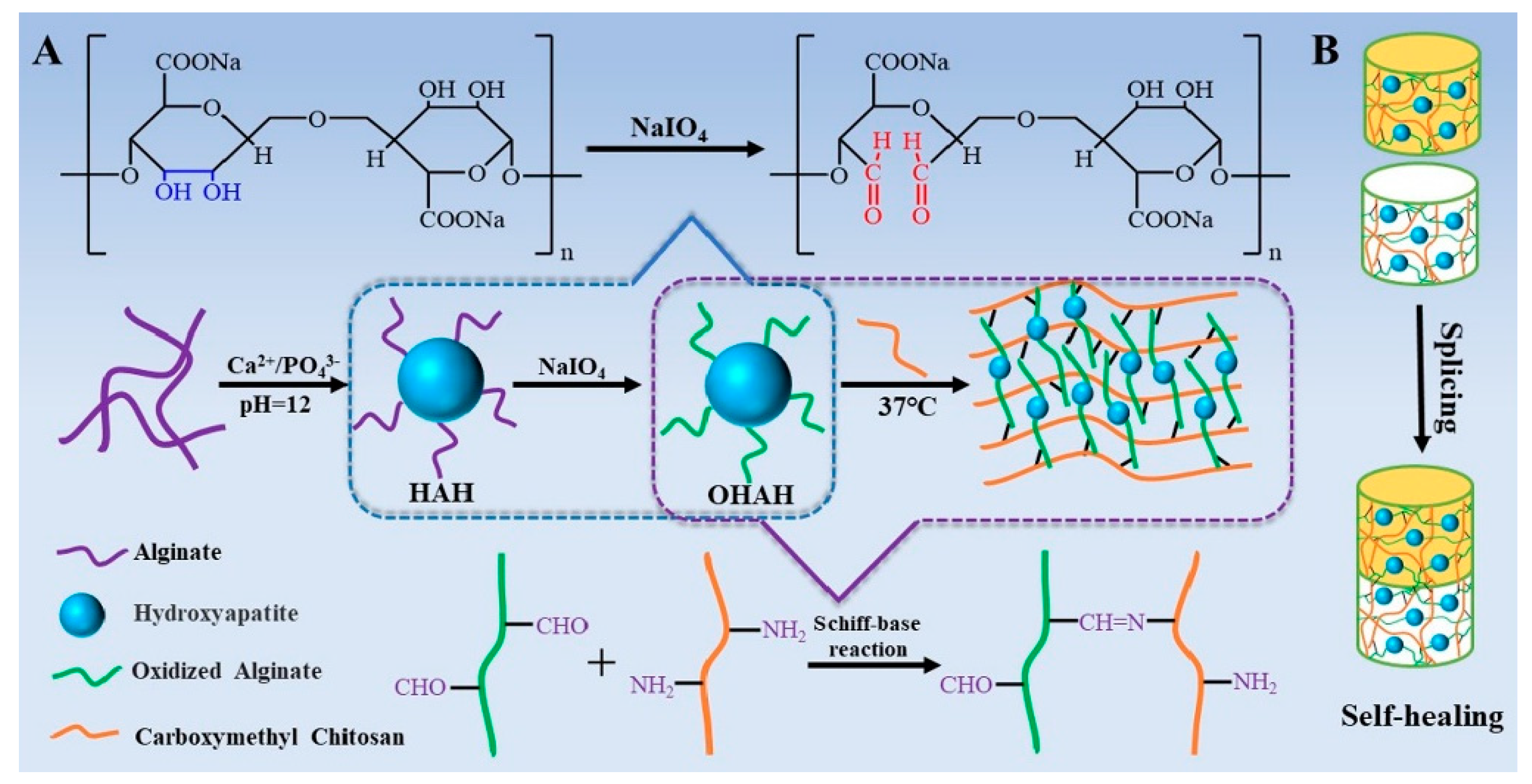
- Ma, L.; Su, W.; Ran, Y.Q.; Ma, X.M.; Yi, Z.; Chen, G.C.; Chen, X.Y.; Deng, Z.W.; Tong, Q.L.; Wang, X.L. Synthesis and characterization of injectable self-healing hydrogels based on oxidized alginate-hybrid-hydroxyapatite nanoparticles and carboxymethyl chitosan. Int. J. Biol. Macromol. 2020, 165, 1164–1174. [Google Scholar] [CrossRef]
- Chuan, D.; Fan, R.R.; Wang, Y.L.; Ren, Y.M.; Wang, C.; Du, Y.; Zhou, L.X.; Yu, J.; Gu, Y.C.; Chen, H.F. Stereocomplex poly(lactic acid)-based composite nanofiber membranes with highly dispersed hydroxyapatite for potential bone tissue engineering. Compos. Sci. Technol. 2020, 192, 108107. [Google Scholar] [CrossRef]
- Shuai, C.J.; Xu, Y.; Feng, P.; Xu, L.; Peng, S.P.; Deng, Y.W. Co-enhance bioactive of polymer scaffold with mesoporous silica and nano-hydroxyapatite. J. Biomater. Sci. Pol. Ed. 2019, 30, 1097–1113. [Google Scholar] [CrossRef] [PubMed]
- Yang, Y.H.; Zhang, Q.; Xu, T.P.; Zhang, H.Y.; Zhang, M.; Lu, L.; Hao, Y.F.; Fuh, J.Y.H.; Zhao, X. Photocrosslinkable nanocomposite ink for printing strong, biodegradable and bioactive bone graft. Biomaterials 2020, 263, 120378. [Google Scholar] [CrossRef] [PubMed]
- Li, L.; Xincui, S.; Wang, Z.; Guo, M.; Wang, Y.; Jiao, Z.; Zhang, P. Porous Scaffolds of Poly(lactic-co-glycolic acid) and mesoporous hydroxyapatite surface modified by poly(gamma-benzyl-L-glutamate) (PBLG) for in vivo bone repair. ACS Biomater. Sci. Eng. 2019, 5, 2466–2481. [Google Scholar] [CrossRef]
- Mondal, D.; Willett, T.L. Mechanical properties of nanocomposite biomaterials improved by extrusion during direct ink writing. J. Mech. Behav. Biomed. Mater. 2020, 104, 103653. [Google Scholar] [CrossRef] [PubMed]
- Ren, P.F.; Wang, F.M.; Zhan, T.T.; Hu, W.J.; Zhou, N.Z.; Zhang, T.Z.; Ye, J.H. A biomimetic nano-ydroxyapatite/chitosan/ poly(methyl vinyl ether-alt-maleic anhydride) composite with excellent biocompatibility. Mater. Let. 2020, 261, 127102. [Google Scholar] [CrossRef]
- Shi, X.C.; Wu, H.T.; Yan, H.H.; Wang, Y.; Wang, Z.L.; Zhang, P.B. Electroactive nanocomposite porous scaffolds of PAP(n)/op-HA/PLGA enhance osteogenesis in vivo. ACS Appl. Biomater. 2019, 2, 1464–1476. [Google Scholar] [CrossRef]
- McGough, M.A.P.; Boller, L.A.; Groff, D.M.; Schoenecker, J.G.; Nyman, J.S.; Wenke, J.C.; Rhodes, C.; Shimko, D.; Duvall, C.L.; Guelcher, S.A. Nanocrystalline hydroxyapatite-poly(thioketal urethane) nanocomposites stimulate a combined intramembranous and endochondral ossification response in rabbits. ACS Biomater. Sci. Eng. 2020, 6, 564–574. [Google Scholar] [CrossRef]
- Silva, A.D.; Rodrigues, B.V.M.; Oliveira, F.C.; Carvalho, J.O.; de Vasconcellos, L.M.R.; de Araujo, J.C.R.; Marciano, F.R.; Lobo, A.O. Characterization and In Vitro and In Vivo assessment of poly(butylene adipate-co-terephthalate)/nano-hydroxyapatite composites as scaffolds for bone tissue engineering. J. Polym. Res. 2019, 26, 53. [Google Scholar] [CrossRef]
- Liang, H.; Xu, X.M.; Feng, X.B.; Ma, L.; Deng, X.Y.; Wu, S.L.; Liu, X.M.; Yang, C. Gold nanoparticles-loaded hydroxyapatite composites guide osteogenic differentiation of human mesenchymal stem cells through Wnt/beta-catenin signaling pathway. Int. J. Nanomed. 2019, 14, 6151–6163. [Google Scholar] [CrossRef] [PubMed] [Green Version]
- Dalavi, P.A.; Prabhu, A.; Shastry, R.P.; Venkatesan, J. Microspheres containing biosynthesized silver nanoparticles with alginate-nano hydroxyapatite for biomedical applications. J. Biomater. Sci. Polym. Ed. 2020, 31, 2025–2043. [Google Scholar] [CrossRef] [PubMed]
- Martinez-Zelaya, V.R.; Zarranz, L.; Herrera, E.Z.; Alves, A.T.; Uzeda, M.J.; Mavropoulos, E.; Rossi, A.L.; Mello, A.; Granjeiro, J.M.; Calasans-Maia, M.D. In vitro and in vivo evaluations of nanocrystalline Zn-doped carbonated hydroxyapatite/alginate microspheres: Zinc and calcium bioavailability and bone regeneration. Int. J. Nanomed. 2019, 14, 3471–3490. [Google Scholar] [CrossRef] [PubMed] [Green Version]
- Cao, Y.; Shi, T.S.; Jiao, C.; Liang, X.; Chen, R.Y.; Tian, Z.J.; Zou, A.C.; Yang, Y.W.; Wei, Z.; Wang, C.J. Fabrication and properties of zirconia/hydroxyapatite composite scaffold based on digital light processing. Ceram. Int. 2020, 46, 2300–2308. [Google Scholar] [CrossRef]
- Zhu, Y.; Jiang, P.P.; Luo, B.; Lan, F.; He, J.; Wu, Y. Dynamic protein corona influences immune-modulating osteogenesis in magnetic nanoparticle (MNP)-infiltrated bone regeneration scaffolds in vivo. Nanoscale 2019, 11, 6817–6827. [Google Scholar] [CrossRef]
- Torgbo, S.; Sukyai, P. Fabrication of microporous bacterial cellulose embedded with magnetite and hydroxyapatite nanocomposite scaffold for bone tissue engineering. Mater. Chem. Phys. 2019, 237, 121868. [Google Scholar] [CrossRef]
- Mushtaq, A.; Zhao, R.B.; Luo, D.D.; Dempsey, E.; Wang, X.M.; Iqbal, M.Z.; Kong, X.D. Magnetic hydroxyapatite nanocomposites: The advances from synthesis to biomedical applications. Mater. Des. 2021, 197, 109269. [Google Scholar] [CrossRef]
- Scialla, S.; Palazzo, B.; Sannino, A.; Verri, T.; Gervaso, F.; Barca, A. Evidence of modular responsiveness of osteoblast-like cells exposed to hydroxyapatite-containing magnetic nanostructures. Biology 2020, 9, 357. [Google Scholar] [CrossRef] [PubMed]
- Munir, K.S.; Wen, C.; Li, Y. Carbon nanotubes and graphene as nanoreinforcements in metallic biomaterials: A review. Adv. Biosyst. 2019, 3, e1800212. [Google Scholar] [CrossRef] [PubMed]
- Wang, G.; Qi, F.; Yang, W.; Yang, Y.; He, C.; Peng, S.; Shuai, C. Crystallinity and reinforcement in poly-l-lactic acid scaffold induced by carbon nanotubes. Adv. Polym. Technol. 2019, 2019, 8625325. [Google Scholar] [CrossRef] [Green Version]
- Oliveira, F.C.; Oliveira, C.J.; Magalhaes, L.S.S.M.; Marques da Silva, J.; Pereira, S.R.; Gomes Júnior, A.L.; Soares, L.M.; Cruz Cariman, L.I.; da Silva, R.I.; Viana, B.C.; et al. Biomineralization inspired engineering of nanobiomaterials promoting bone repair. Mater. Sci. Eng. C Mater. Biol. Appl. 2021, 120, 111776. [Google Scholar] [CrossRef] [PubMed]
- Liu, L.; Yang, B.; Wang, L.Q.; Huang, J.P.; Chen, W.Y.; Ban, Q.; Zhang, Y.; You, R.; Yin, L.; Guan, Y.Q. Biomimetic bone tissue engineering hydrogel scaffolds constructed using ordered CNTs and HA induce the proliferation and differentiation of BMSCs. J. Mater. Chem. B 2020, 8, 558–567. [Google Scholar] [CrossRef] [PubMed]
- Wang, C.; Cao, G.; Zhao, T.; Wang, X.; Niu, X.; Fan, Y.; Li, X. Terminal Group Modification of Carbon Nanotubes Determines Covalently Bound Osteogenic Peptide Performance. ACS Biomater. Sci. Eng. 2020, 6, 865–878. [Google Scholar] [CrossRef]
- Liu, X.; George, M.N.; Li, L.; Gamble, D.; Millerli, A.L.; Gaihre, B.; Waletzki, B.E.; Lu, L. Injectable electrical conductive and phosphate releasing gel with two-dimensional black phosphorus and carbon nanotubes for bone tissue engineering. ACS Biomater. Sci. Eng. 2020, 6, 4653–4665. [Google Scholar] [CrossRef] [PubMed]
- Du, Z.; Feng, X.; Cao, G.; She, Z.; Tan, R.; Aifantis, K.E.; Zhang, R.; Li, X. The effect of carbon nanotubes on osteogenic functions of adipose-derived mesenchymal stem cells in vitro and bone formation in vivo compared with that of nano-hydroxyapatite and the possible mechanism. Bioact. Mater. 2021, 6, 333–345. [Google Scholar] [CrossRef] [PubMed]
- E Silva, E.P.; Huang, B.; Helaehil, J.V.; Nalesso, P.R.L.; Bagne, L.; de Oliveira, M.A.; Albiazetti, G.C.C.; Aldalbahi, A.; El-Newehy, M.; Santamaria, M., Jr.; et al. In vivo study of conductive 3D printed PCL/MWCNTs scaffolds with electrical stimulation for bone tissue engineering. Bio-Des. Manuf. 2021, 4, 190–202. [Google Scholar] [CrossRef]
- Huang, B.; Vyas, C.; Byun, J.J.; El-Newehy, M.; Huang, Z.; Bártolo, P. Aligned multi-walled carbon nanotubes with nanohydroxyapatite in a 3D printed polycaprolactone scaffold stimulates osteogenic differentiation. Mater. Sci. Eng. C Mater. Biol. Appl. 2020, 108, 110374. [Google Scholar] [CrossRef]
- Zhang, P.; Xin, Y.; Ai, F.; Cao, C. Preparation and properties of multi-walled carbon nanotubes and eggshell dual-modified polycaprolactone composite scaffold. J. Polym. Eng. 2019, 39, 343–350. [Google Scholar] [CrossRef]
- Cui, H.; Yu, Y.; Li, X.; Sun, Z.; Ruan, J.; Wu, Z.; Qian, J.; Yin, J. Direct 3D printing of a tough hydrogel incorporated with carbon nanotubes for bone regeneration. J. Mater. Chem. B 2019, 7, 7207–7217. [Google Scholar] [CrossRef] [PubMed]
- Nur, I.S.M.; Nur, S.M.; Nor, H.A.N.; Yusof, N.H.; Idris, A. Review on nanocrystalline cellulose in bone tissue engineering applications. Polymers 2020, 12, 2818. [Google Scholar]
- Zhang, X.; Yin, X.; Luo, J.; Zheng, X.; Wang, H.; Wang, J.; Xi, Z.; Liao, X.; Machuki, J.O.; Guo, K.; et al. Novel hierarchical nitrogen-doped multiwalled carbon nanotubes/cellulose/nanohydroxyapatite nanocomposite as an osteoinductive scaffold for enhancing bone regeneration. ACS Biomater. Sci. Eng. 2019, 5, 294–307. [Google Scholar] [CrossRef] [PubMed]
- Dinescu, S.; Ionita, M.; Ignat, S.R.; Costache, M.; Hermenean, A. Graphene oxide enhances chitosan-based 3D scaffold properties for bone tissue engineering. Int. J. Mol. Sci. 2019, 20, 5077. [Google Scholar] [CrossRef] [PubMed] [Green Version]
- Wang, P.J.; Yu, T.B.; Lv, Q.L.; Li, S.K.; Ma, X.X.; Yang, G.P.; Xu, D.X.; Liu, X.; Wang, G.T.; Chen, Z.Q. Fabrication of hydroxyapatite/hydrophilic graphene composites and their modulation to cell behavior toward bone reconstruction engineering. Colloids Surf. B Biointerfaces 2019, 173, 512–520. [Google Scholar] [CrossRef]
- Huang, H.Y.; Fan, F.Y.; Shen, Y.K.; Wang, C.H.; Huang, Y.T.; Chern, M.J.; Wang, Y.H.; Wang, L. 3D poly-epsilon-caprolactone/graphene porous scaffolds for bone tissue engineering. Colloids Surf. A Physicochem. Eng. Asp. 2020, 606, 125393. [Google Scholar] [CrossRef]
- He, M.M.; Zhu, C.; Xu, H.; Sun, D.; Chen, C.; Feng, G.J.; Liu, L.M.; Li, Y.B.; Zhang, L. Conducting polyetheretherketone nanocomposites with an electrophoretically deposited bioactive coating for bone tissue regeneration and multimodal therapeutic applications. ACS Appl. Mater. Interfaces 2020, 12, 56924–56934. [Google Scholar] [CrossRef] [PubMed]
- He, M.M.; Chen, X.C.; Guo, Z.J.; Qiu, X.T.; Yang, Y.T.; Su, C.L.; Jiang, N.; Li, Y.B.; Sun, D.; Zhang, L. Super tough graphene oxide reinforced polyetheretherketone for potential hard tissue repair applications. Compos. Sci. Technol. 2019, 174, 194–201. [Google Scholar] [CrossRef] [Green Version]
- Huang, Z.; Wan, Y.; Zhu, X.; Zhang, P.; Yang, Z.; Yao, F.; Lu, H. Simultaneous engineering of nanofillers and patterned surface macropores of graphene/hydroxyapatite/polyetheretherketone ternary composites for potential bone implants. Mater. Sci. Eng. C 2021, 123, 111967. [Google Scholar] [CrossRef]
- Lopes, C.C.; Pinheiro, W.A.; da Rocha, D.N.; Neves, J.G.; Correr, A.B.; Ferreira, J.R.M.; Barbosa, R.M.; Soares, J.R.F.; Santos, J.L.; Prado da Silva, M.H. Nanocomposite powders of hydroxyapatite-graphene oxide for biological applications. Ceram. Int. 2021, 47, 7653–7665. [Google Scholar] [CrossRef]
- Zhao, Y.; Chen, J.; Zou, L.; Xu, G.; Geng, Y. Facile one-step bioinspired mineralization by chitosan functionalized with graphene oxide to activate bone endogenous regeneration. Chem. Eng. J. 2019, 378, 122174. [Google Scholar] [CrossRef]
- Ghorai, S.K.; Maji, S.; Subramanian, B.; Maiti, T.K.; Chattopadhyay, S. Coining attributes of ultra-low concentration graphene oxide and spermine: An approach for high strength, anti-microbial and osteoconductive nanohybrid scaffold for bone tissue regeneration. Carbon 2019, 141, 370–389. [Google Scholar] [CrossRef]
- Zhang, Y.; Hu, J. Isocyanate modified go shape-memory polyurethane composite. Polymers 2020, 12, 118. [Google Scholar] [CrossRef] [Green Version]
- Oliveira, F.C.; Carvalho, J.O.; Gusmao, S.B.S.; Goncalves, L.D.; Mendes, L.M.S.; Freitas, S.A.P.; Gusmao, G.O.D.; Bartolomeu Cruz, V.; Marciano, F.R.; Anderson Oliveira, L. High loads of nano-hydroxyapatite/graphene nanoribbon composites guided bone regeneration using an osteoporotic animal model. Int. J. Nanomed. 2019, 14, 865–874. [Google Scholar] [CrossRef] [PubMed] [Green Version]
- Wu, T.; Li, B.; Wang, W.; Chen, L.; Li, Z.; Wang, M.; Zha, Z.; Lin, Z.; Xia, H.; Zhang, T. Strontium-substituted hydroxyapatite grown on graphene oxide nanosheet-reinforced chitosan scaffold to promote bone regeneration. Biomater. Sci. 2020, 8, 4603–4615. [Google Scholar] [CrossRef] [PubMed]
- Chen, Y.H.; Zheng, Z.W.; Zhou, R.P.; Zhang, H.Z.; Chen, C.S.; Xiong, Z.Z.; Liu, K.; Wang, X.S. Developing a strontium-releasing graphene oxide-/collagen-based organic inorganic nanobiocomposite for large bone defect regeneration via MAPK signaling pathway. ACS Appl. Mater. Interfaces 2019, 11, 15986–15997. [Google Scholar] [CrossRef] [PubMed]
- Liu, S.; Zhou, C.; Mou, S.; Li, J.; Zhou, M.; Zeng, Y.; Luo, C.; Sun, J.; Wang, Z.; Xu, W. Biocompatible graphene oxide-collagen composite aerogel for enhanced stiffness and in situ bone regeneration. Mater. Sci. Eng. C Mater. Biol. Appl. 2019, 105, 110137. [Google Scholar] [CrossRef] [PubMed]
- Zhang, Y.D.; Wang, C.; Fu, L.; Ye, S.; Wang, M.; Zhou, Y.M. Fabrication and application of novel porous scaffold in situ-loaded graphene oxide and osteogenic peptide by cryogenic 3D printing for repairing critical-sized bone defect. Molecules 2019, 24, 1669. [Google Scholar] [CrossRef] [Green Version]
- Wu, J.N.; Zheng, A.; Liu, Y.; Jiao, D.L.; Zeng, D.L.; Wang, X.; Cao, L.Y.; Jiang, X.Q. Enhanced bone regeneration of the silk fibroin electrospun scaffolds through the modification of the graphene oxide functionalized by BMP-2 peptide. Int. J. Nanomed. 2019, 14, 733–750. [Google Scholar] [CrossRef] [PubMed] [Green Version]
- Ou, L.L.; Lan, Y.; Feng, Z.A.; Feng, L.B.; Yang, J.J.; Liu, Y.; Bian, L.M.; Tan, J.L.; Lai, R.F.; Guo, R. Functionalization of SF/HAP scaffold with GO-PEI-miRNA inhibitor complexes to enhance bone regeneration through activating transcription factor 4. Theranostics 2019, 9, 4525–4541. [Google Scholar] [CrossRef]
- Zhang, J.; Eyisoylu, H.; Qin, X.H.; Rubert, M.; Müller, R. 3D bioprinting of graphene oxide-incorporated cell-laden bone mimicking scaffolds for promoting scaffold fidelity, osteogenic differentiation and mineralization. Acta Biomater. 2021, 121, 637–652. [Google Scholar] [CrossRef] [PubMed]
- Wang, W.; Liu, Y.; Yang, C.; Qi, X.; Li, S.W.; Liu, C.S.; Li, X.L. Mesoporous bioactive glass combined with graphene oxide scaffolds for bone repair. Int. J. Biol. Sci. 2019, 15, 2156–2169. [Google Scholar] [CrossRef] [PubMed] [Green Version]
- Yan, F.F.; Liu, Z.B.; Zhang, T.; Zhang, Q.; Chen, Y.; Xie, Y.L.; Lei, J.; Cai, L. Biphasic injectable bone cement with Fe3O4/GO nanocomposites for the minimally invasive treatment of tumor-induced bone destruction. ACS Biomater. Sci. Eng. 2019, 5, 5833–5843. [Google Scholar] [CrossRef] [PubMed]
- Chopra, V.; Thomas, J.; Sharma, A.; Panwar, V.; Kaushik, S.; Sharma, S.; Porwal, K.; Kulkarni, C.; Rajput, S.; Singh, H.; et al. Synthesis and evaluation of a zinc eluting rGO/hydroxyapatite nanocomposite optimized for bone augmentation. ACS Biomater. Sci. Eng. 2020, 6, 6710–6725. [Google Scholar] [CrossRef] [PubMed]
- Zhang, Y.C.; Hu, J.L.; Zhao, X.; Xie, R.Q.; Qin, T.W.; Ji, F.L. Mechanically robust shape memory polyurethane nanocomposites for minimally invasive bone repair. ACS Appl. Biomater. 2019, 2, 1056–1065. [Google Scholar] [CrossRef]
- Jiao, D.L.; Cao, L.Y.; Liu, Y.; Wu, J.N.; Zheng, A.; Jiang, X.Q. Synergistic osteogenesis of biocompatible reduced graphene oxide with methyl vanillate in BMSCs. ACS Biomater. Sci. Eng. 2019, 5, 1920–1936. [Google Scholar] [CrossRef]
- Senthil, R.; Basaran, B.; Vijayan, S.; Mert, A.; Bayraktar, O.; Wilson, A.A. Electrospun nano-bio membrane for bone tissue engineering application- a new approach. Mater. Chem. Phys. 2020, 249, 123010. [Google Scholar]
- Bu, W.; Xu, X.; Wang, Z.; Jin, N.; Liu, L.; Liu, J.; Zhu, S.; Zhang, K.; Jelinek, R.; Zhou, D.; et al. Ascorbic acid-PEI carbon dots with osteogenic effects as miR-2861 carriers to effectively enhance bone regeneration. ACS Appl. Mater. Interfaces 2020, 12, 50287–50302. [Google Scholar] [CrossRef]
- Wang, B.; Yang, M.; Liu, L.; Yan, G.; Yan, H.; Feng, J.; Li, Z.; Li, D.; Sun, H.; Yang, B. Osteogenic potential of Zn2+-passivated carbon dots for bone regeneration In Vivo. Biomater. Sci. 2019, 7, 5414–5423. [Google Scholar] [CrossRef]
- Lai, L.; Song, H.; Zhen, J.; Qiu, Y.; Liu, X.; Xu, W.; Zhang, S. Study on the bone morphogenetic protein 2 loaded synergistic hierarchical porous silk/carbon nanocage scaffold for the repair of bone defect. Mater. Des. 2020, 196, 109105. [Google Scholar] [CrossRef]
- Nekounam, H.; Allahyari, Z.; Gholizadeh, S.; Mirzaei, E.; Shokrgozar, M.A.; Faridi-Majidi, R. Simple and robust fabrication and characterization of conductive carbonized nanofibers loaded with gold nanoparticles for bone tissue engineering applications. Mater. Sci. Eng. C 2020, 117, 111226. [Google Scholar] [CrossRef]
- Swaminathan, P.D.; Uddin, M.N.; Wooley, P.; Asmatulu, R. Fabrication and biological analysis of highly porous PEEK bionanocomposites incorporated with carbon and hydroxyapatite nanoparticles for biological applications. Molecules 2020, 25, 3572. [Google Scholar] [CrossRef] [PubMed]
- Wang, Y.; Cui, W.G.; Zhao, X.; Wen, S.Z.; Sun, Y.L.; Han, J.M.; Zhang, H.Y. Bone remodeling-inspired dual delivery electrospun nanofibers for promoting bone regeneration. Nanoscale 2019, 11, 60–71. [Google Scholar] [CrossRef] [PubMed]
- Kao, C.T.; Chen, Y.J.; Huang, T.H.; Lin, Y.H.; Hsu, T.T.; Ho, C.C. Assessment of the release profile of fibroblast growth factor-2-load mesoporous calcium silicate/poly-epsilon-caprolactone 3D scaffold for regulate bone regeneration. Processes 2020, 8, 1249. [Google Scholar] [CrossRef]
- Liang, H.; Jin, C.; Ma, L.; Feng, X.B.; Deng, X.Y.; Wu, S.L.; Liu, X.M.; Yang, C. Accelerated bone regeneration by gold-nanoparticle-loaded mesoporous silica through stimulating immunomodulation. ACS Appl. Mater. Interfaces 2019, 11, 41758–41769. [Google Scholar] [CrossRef]
- Chen, M.; Zhang, Y.; Xie, Q.; Zhang, W.; Pan, X.; Gu, P.; Zhou, H.; Gao, Y.; Walther, A.; Fan, X. Long-term bone regeneration enabled by a polyhedral oligomeric silsesquioxane (POSS)-enhanced biodegradable hydrogel. ACS Biomater. Sci. Eng. 2019, 5, 4612–4623. [Google Scholar] [CrossRef]
- Carrow, J.K.; Di Luca, A.; Dolatshahi-Pirouz, A.; Moroni, L.; Gaharwar, A.K. 3D-printed bioactive scaffolds from nanosilicates and PEOT/PBT for bone tissue engineering. Regen. Biomater. 2019, 6, 29–37. [Google Scholar] [CrossRef] [PubMed]
- Gao, C.D.; Yao, M.; Shuai, C.J.; Peng, S.P.; Deng, Y.W. Nano-SiC reinforced Zn biocomposites prepared via laser melting: Microstructure, mechanical properties and biodegradability. J. Mater. Sci. Technol. 2019, 35, 2608–2617. [Google Scholar] [CrossRef]
- Pang, L.B.; Shen, Y.F.; Hu, H.R.; Zeng, X.Q.; Huang, W.H.; Gao, H.; Wang, H.; Wang, D.P. Chemically and physically cross-linked polyvinyl alcohol-borosilicate gel hybrid scaffolds for bone regeneration. Mater. Sci. Eng. C 2019, 105, 110076. [Google Scholar] [CrossRef]
- Zhao, C.C.; Shen, A.F.; Zhang, L.Z.; Lin, K.L.; Wang, X.D. Borocarbonitrides nanosheets engineered 3D-printed scaffolds for integrated strategy of osteosarcoma therapy and bone regeneration. Chem. Eng. J. 2020, 401, 125989. [Google Scholar] [CrossRef]
- Liu, Y.H.; Zhu, Z.; Pei, X.B.; Zhang, X.; Cheng, X.T.; Hu, S.S.; Gao, X.M.; Wang, J.; Chen, J.Y.; Wan, Q.B. ZIF-8-modified multifunctional bone-adhesive hydrogels promoting angiogenesis and osteogenesis for bone regeneration. ACS Appl. Mater. Interfaces 2020, 12, 36978–36995. [Google Scholar] [CrossRef]
- Cidonio, G.; Glinka, M.; Kim, Y.H.; Kanczler, J.M.; Lanham, S.A.; Ahlfeld, T.; Lode, A.; Dawson, J.I.; Gelinsky, M.; Oreffo, R.O.C. Nanoclay-based 3D printed scaffolds promote vascular ingrowth ex vivo and generate bone mineral tissue In Vitro and In Vivo. Biofabrication 2020, 12, 035010. [Google Scholar] [CrossRef] [PubMed] [Green Version]
- Zhang, Y.; Chen, M.; Dai, Z.; Cao, H.; Li, J.; Zhang, W. Sustained protein therapeutics enabled by self-healing nanocomposite hydrogels for non-invasive bone regeneration. Biomater. Sci. 2020, 8, 682–693. [Google Scholar] [CrossRef] [PubMed]
- Ibrahim, D.M.; Sani, E.S.; Soliman, A.M.; Zandi, N.; Mostafavi, E.; Youssef, A.M.; Allam, N.K.; Annabi, N. Bioactive and elastic nanocomposites with antimicrobial properties for bone tissue regeneration. ACS Appl. Biomater. 2020, 3, 3313–3325. [Google Scholar] [CrossRef]
- Zhao, H.B.; Zhang, X.M.; Zhou, D.; Weng, Y.P.; Qin, W.; Pan, F.; Lv, S.W.; Zhao, X.B. Collagen, polycaprolactone and attapulgite composite scaffolds forin vivobone repair in rabbit models. Biomed. Mater. 2020, 15, 045022. [Google Scholar] [CrossRef] [PubMed] [Green Version]
- Kundu, K.; Afshar, A.; Katti, D.R.; Edirisinghe, M.; Katti, K.S. Composite nanoclay-hydroxyapatite-polymer fiber scaffolds for bone tissue engineering manufactured using pressurized gyration. Compos. Sci. Technol. 2021, 202, 108598. [Google Scholar] [CrossRef]
- Doostmohammadi, A.; Esfahani, Z.K.; Ardeshirylajimi, A.; Dehkordi, Z.R. Zirconium modified calcium-silicate-based nanoceramics: An in vivo evaluation in a rabbit tibial defect model. Int. J. Appl. Ceram. Technol. 2019, 16, 431–437. [Google Scholar] [CrossRef]
- Liu, K.; Li, W.Y.; Chen, S.T.; Wen, W.; Lu, L.; Liu, M.X.; Zhou, C.R.; Luo, B.H. The design, fabrication and evaluation of 3D printed gHNTs/gMgO whiskers/PLLA composite scaffold with honeycomb microstructure for bone tissue engineering. Compos. Part B Eng. 2020, 192, 108001. [Google Scholar] [CrossRef]
- Tian, Q.; Lin, J.; Rivera-Castaneda, L.; Tsanhani, A.; Dunn, Z.S.; Rodrriguez, A.; Aslani, A.; Liu, H. Nano-to-submicron hydroxyapatite coatings for magnesium-based bioresorbable implants—Deposition, characterization, degradation, mechanical properties, and cytocompatibility. Sci. Rep. 2019, 9, 810. [Google Scholar] [CrossRef]
- Safari, N.; Golafshan, N.; Kharaziha, M.; Toroghinejad, M.R.; Utomo, L.; Malda, J.; Castilho, M. Stable and antibacterial magnesium-graphene nanocomposite-based implants for bone repair. ACS Biomater. Sci. Eng. 2020, 6, 6253–6262. [Google Scholar] [CrossRef] [PubMed]
- Parande, G.; Manakari, V.; Prasadh, S.; Chauhan, D.; Rahate, S.; Wong, R.; Gupta, M. Strength retention, corrosion control and biocompatibility of Mg-Zn-Si/HA nanocomposites. J. Mech. Behav. Biomed. Mater. 2020, 103, 103584. [Google Scholar] [CrossRef]
- Khalili, V.; Frenzel, J.; Eggeler, G. Degradation behavior of the MgO/HA surface ceramic nano-composites in the simulated body fluid and its use as a potential bone implant. Mater. Chem. Phys. 2021, 258, 123965. [Google Scholar] [CrossRef]
- Kumar, S.; Gautam, C.; Chauhan, B.S.; Srikrishna, S.; Yadav, R.S.; Rai, S.B. Enhanced mechanical properties and hydrophilic behavior of magnesium oxide added hydroxyapatite nanocomposite: A bone substitute material for load bearing applications. Ceram. Int. 2020, 46, 16235–16248. [Google Scholar] [CrossRef]
- Shuai, C.; Zan, J.; Yang, Y.; Peng, S.; Yang, W.; Qi, F.; Shen, L.; Tian, Z. Surface modification enhances interfacial bonding in PLLA/MgO bone scaffold. Mater. Sci. Eng. C 2020, 108, 110486. [Google Scholar] [CrossRef]
- Zhao, Y.; Liang, H.; Zhang, S.Q.; Qu, S.W.; Jiang, Y.; Chen, M.F. Effects of magnesium oxide (MgO) shapes on in vitro and in vivo degradation behaviors of PLA/MgO composites in long term. Polymers 2020, 12, 1074. [Google Scholar] [CrossRef]
- Go, E.J.; Kang, E.Y.; Lee, S.K.; Park, S.; Kim, J.H.; Park, W.; Kim, I.H.; Choi, B.; Han, D.K. An osteoconductive PLGA scaffold with bioactive beta-TCP and anti-inflammatory Mg(OH)(2) to improve in vivo bone regeneration. Biomater. Sci. 2020, 8, 937–948. [Google Scholar] [CrossRef]
- Zhao, Q.H.; Tang, H.M.; Ren, L.S.; Wei, J. In vitro apatite mineralization, degradability, cytocompatibility and in vivo new bone formation and vascularization of bioactive scaffold of polybutylene succinate/magnesium phosphate/wheat protein ternary composite. Int. J. Nanomed. 2020, 15, 7279–7295. [Google Scholar] [CrossRef] [PubMed]
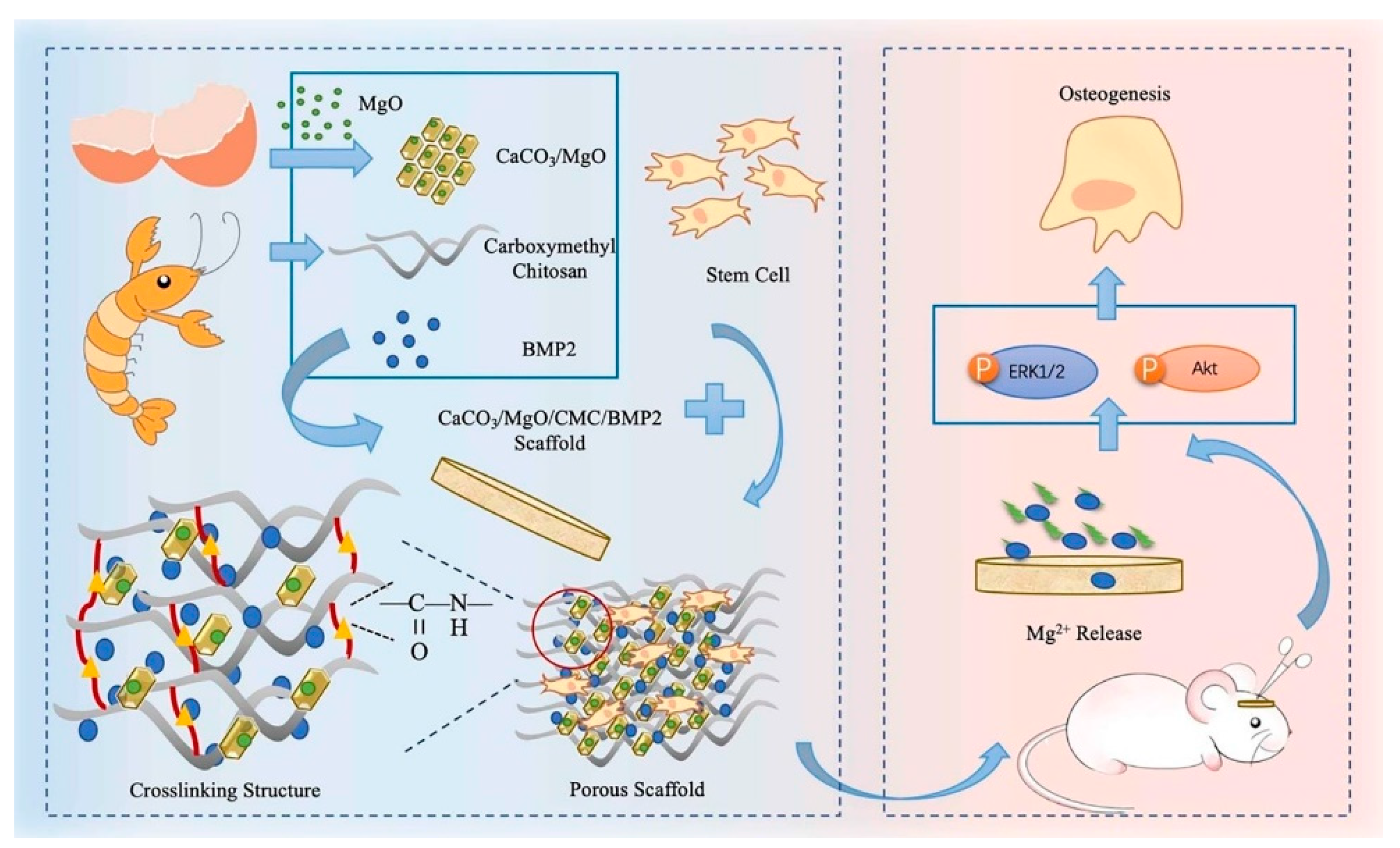
- Huang, Y.Z.; Ji, Y.R.; Kang, Z.W.; Li, F.; Ge, S.F.; Yang, D.P.; Ruan, J.; Fan, X.Q. Integrating eggshell-derived CaCO3/MgO nanocomposites and chitosan into a biomimetic scaffold for bone regeneration. Chem. Eng. J. 2020, 395, 125098. [Google Scholar] [CrossRef]
- Hussain, A.; Gautam, C.; Jafri, A.; Mishra, V.K.; Madheshiya, A.; Gautam, A.; Singh, M.K.; Gautam, R.K.; Gupta, M.; Arshad, M. Formation of multifunctional ZrO2-MgO-hBN nanocomposite for enhanced bone regeneration and E. coli bacteria filtration applications. Ceram. Int. 2020, 46, 23006–23020. [Google Scholar] [CrossRef]
- Zheng, Z.; Chen, Y.; Hong, H.; Shen, Y.; Wang, Y.; Sun, J.; Wang, X. The “Yin and Yang” of immunomodulatory magnesium-enriched graphene oxide nanoscrolls decorated biomimetic scaffolds in promoting bone regeneration. Adv. Healthc. Mater. 2021, 10, 2000631. [Google Scholar] [CrossRef] [PubMed]
- Li, B.; Xia, X.; Guo, M.; Jiang, Y.; Li, Y.; Zhang, Z.; Liu, S.; Li, H.; Liang, C.; Wang, H. Biological and antibacterial properties of the micro-nanostructured hydroxyapatite/chitosan coating on titanium. Sci. Rep. 2019, 9, 14052. [Google Scholar] [CrossRef] [PubMed] [Green Version]
- Ren, B.; Wan, Y.; Liu, C.; Wang, H.; Yu, M.; Zhang, X.; Huang, Y. Improved osseointegration of 3D printed Ti-6Al-4V implant with a hierarchical micro/nano surface topography: An In Vitro and In Vivo study. Mater. Sci. Eng. C 2021, 118, 111505. [Google Scholar] [CrossRef]
- Wang, R.; Shi, M.; Xu, F.; Qiu, Y.; Zhang, P.; Shen, K.; Zhao, Q.; Yu, J.; Zhang, Y. Graphdiyne-modified TiO2 nanofibers with osteoinductive and enhanced photocatalytic antibacterial activities to prevent implant infection. Nat. Commun. 2020, 11, 4465. [Google Scholar] [CrossRef] [PubMed]
- Yigit, O.; Dikici, B.; Cagri Senocak, T.; Ozdemir, N. One-step synthesis of nano-hydroxyapatite/graphene nanosheet hybrid coatings on Ti6Al4V alloys by hydrothermal method and their in-vitro corrosion responses. Surf. Coat. Technol. 2020, 394, 125858. [Google Scholar] [CrossRef]
- Tümer, D.; Güngörürler, M.; Havıtçıoğlu, H.; Arman, Y. Investigation of effective coating of the Ti–6Al–4V alloy and 316L stainless steel with graphene or carbon nanotubes with finite element methods. J. Mater. Res. Technol. 2020, 9, 15880–15893. [Google Scholar] [CrossRef]
- Zalnezhad, E.; Musharavati, F.; Chen, T.; Jaber, F.; Uzun, K.; Chowdury, M.E.H.; Khandakar, A.; Liu, J.; Bae, S. Tribo-mechanical properties evaluation of HA/TiO2/CNT nanocomposite. Sci. Rep. 2021, 11, 1867. [Google Scholar] [CrossRef] [PubMed]
- Rafieerad, A.R.; Bushroa, A.R.; Nasiri-Tabrizi, B.; Baradaran, S.; Amiri, A.; Saber-Samandari, S.; Khanahmadi, S.; Zeimaran, E.; Basirun, W.J.; Kalaiselvam, K.; et al. Simultaneous enhanced antibacterial and osteoblast cytocompatibility performance of Ti6Al7Nb implant by nano-silver/graphene oxide decorated mixed oxide nanotube composite. Surf. Coat. Technol. 2019, 360, 181–195. [Google Scholar] [CrossRef]
- Kawaguchi, M.; Segawa, A.; Shintani, K.; Nakamura, Y.; Ishigaki, Y.; Yonezawa, K.; Sasamoto, T.; Kaneuji, A.; Kawahara, N. Bone formation at Ti-6Al-7Nb scaffolds consisting of 3D honeycomb frame and diamond-like carbon coating implanted into the femur of beagles. J. Biomed. Mater. Res. Part B Appl. Biomater. 2021, 109, 1283–1291. [Google Scholar] [CrossRef]
- Yılmaz, E.; Çakıroğlu, B.; Gökçe, A.; Findik, F.; Gulsoy, H.O.; Gulsoy, N.; Mutlu, Ö.; Özacar, M. Novel hydroxyapatite/graphene oxide/collagen bioactive composite coating on Ti16Nb alloys by electrodeposition. Mater. Sci. Eng. C Mater. Biol. Appl. 2019, 101, 292–305. [Google Scholar] [CrossRef] [PubMed]
- Cao, H.; Qin, H.; Zhao, Y.; Jin, G.; Lu, T.; Meng, F.; Zhang, X.; Liu, X. Nano-thick calcium oxide armed titanium: Boosts bone cells against MRSA. Sci. Rep. 2016, 6, 21761. [Google Scholar] [CrossRef] [PubMed]
- Lu, M.; Liao, J.; Dong, J.; Wu, J.; Qiu, H.; Zhou, X.; Li, J.; Jiang, D.; He, T.C.; Quan, Z. An effective treatment of experimental osteomyelitis using the antimicrobial titanium/silver-containing nHP66 (nano-hydroxyapatite/polyamide-66) nanoscaffold biomaterials. Sci. Rep. 2016, 6, 39174. [Google Scholar] [CrossRef] [PubMed] [Green Version]
- Wang, P.; Hao, L.L.; Wang, Z.L.; Wang, Y.; Guo, M.; Zhang, P.B. Gadolinium-doped BTO-functionalized nanocomposites with enhanced MRI and X-ray dual imaging to simulate the electrical properties of bone. ACS Appl. Mater. Interfaces 2020, 12, 49464–49479. [Google Scholar] [CrossRef] [PubMed]
- Garino, N.; Sanvitale, P.; Dumontel, B.; Laurenti, M.; Colilla, M.; Izquierdo-Barba, I.; Cauda, V.; Vallet-Regi, M. Zinc oxide nanocrystals as a nanoantibiotic and osteoinductive agent. RSC Adv. 2019, 9, 11312–11321. [Google Scholar] [CrossRef] [PubMed]
- Bejarano, J.; Boccaccini, A.R.; Covarrubias, C.; Palza, H. Effect of Cu- and Zn-doped bioactive glasses on the in vitro bioactivity, mechanical and degradation behavior of biodegradable PDLLA scaffolds. Materials 2020, 13, 2908. [Google Scholar] [CrossRef] [PubMed]
- He, J.; Ye, H.X.; Li, Y.L.; Fang, J.; Mei, Q.S.; Lu, X.; Ren, F.Z. Cancellous-bone-like porous iron scaffold coated with strontium incorporated octacalcium phosphate nanowhiskers for bone regeneration. ACS Biomater. Sci. Eng. 2019, 5, 509–518. [Google Scholar] [CrossRef] [PubMed]
- Govindan, R.; Karthi, S.; Kumar, G.S.; Girija, E.K. Development of Fe3O4 integrated polymer/phosphate glass composite scaffolds for bone tissue engineering. Mater. Adv. 2020, 1, 3466–3475. [Google Scholar] [CrossRef]
- Purohit, S.D.; Singh, H.; Bhaskar, R.; Yadav, I.; Chou, C.F.; Gupta, M.K.; Mishra, N.C. Gelatin-alginate-cerium oxide nanocomposite scaffold for bone regeneration. Mater. Sci. Eng. C Mater. Biol. Appl. 2020, 116, 111111. [Google Scholar] [CrossRef]
- Xue, X.; Hu, Y.; Deng, Y.H.; Su, J.C. Recent advances in design of functional biocompatible hydrogels for bone tissue engineering. Adv. Funct. Mater. 2021, 31, 2009432. [Google Scholar] [CrossRef]
- Garcia-Garcia, P.; Ruiz, M.; Reyes, R.; Delgado, A.; Evora, C.; Riancho, J.A.; Rodriguez-Rey, J.C.; Perez-Campo, F.M. Smurf1 silencing using a LNA-ASOs/lipid nanoparticle system to promote bone regeneration. Stem Cells Ttransl. Med. 2019, 8, 1306–1317. [Google Scholar] [CrossRef] [Green Version]
- Jin, H.; Liu, Z.S.; Li, W.; Jiang, Z.L.; Li, Y.; Zhang, B. Polyethylenimine-alginate nanocomposites based bone morphogenetic protein 2 gene-activated matrix for alveolar bone regeneration. RSC Adv. 2019, 9, 26598–26608. [Google Scholar] [CrossRef] [Green Version]
- Zeng, Y.; Zhou, M.; Mou, S.; Yang, J.; Yuan, Q.; Guo, L.; Zhong, A.; Wang, J.; Sun, J.; Wang, Z. Sustained delivery of alendronate by engineered collagen scaffold for the repair of osteoporotic bone defects and resistance to bone loss. J. Biomed. Mater. Res. Part A 2020, 108, 2460–2472. [Google Scholar] [CrossRef]
- Wu, J.J.; Zheng, K.; Huang, X.T.; Liu, J.Y.; Liu, H.M.; Boccaccini, A.R.; Wan, Y.; Guo, X.D.; Shao, Z.W. Thermally triggered injectable chitosan/silk fibroin/bioactive glass nanoparticle hydrogels for in-situ bone formation in rat calvarial bone defects. Acta Biomater. 2019, 91, 60–71. [Google Scholar] [CrossRef] [PubMed]
- Xu, J.X.; Feng, Y.H.; Wu, Y.X.; Li, Y.J.; Ouyang, M.; Zhang, X.P.; Wang, Y.; Wang, Y.Y.; Xu, L.J. Noninvasive monitoring of bone regeneration using NaYF4: Yb3+, Er3+ upconversion hollow microtubes supporting PLGA-PEG-PLGA hydrogel. React. Funct. Polym. 2019, 143, 104333. [Google Scholar] [CrossRef]
- Mani, M.P.; Jaganathan, S.K. Engineered multicomponent electrospun nanocomposite scaffolds comprising polyurethane loaded with ghee and propolis for bone tissue repair. J. Ind. Text. 2020. [Google Scholar] [CrossRef]
- Li, Y.; Liao, C.; Tjong, S.C. Electrospun polyvinylidene fluoride-based fibrous scaffolds with piezoelectric characteristics for bone and neural tissue engineering. Nanomaterials 2019, 9, 952. [Google Scholar] [CrossRef] [PubMed] [Green Version]
- Fernandes, M.M.; Correia, D.M.; Ribeiro, C.; Castro, N.; Correia, V.; Lanceros-Mendez, S. Bioinspired three-dimensional magnetoactive scaffolds for bone tissue engineering. ACS Appl. Mater. Interfaces 2019, 11, 45265–45275. [Google Scholar] [CrossRef]
- He, Y.; Li, Q.Y.; Ma, C.Y.; Xie, D.H.; Li, L.M.; Zhao, Y.T.; Shan, D.Y.; Chomos, S.K.; Dong, C.; Tierney, J.W. Development of osteopromotive poly(octamethylene citrate glycerophosphate) for enhanced bone regeneration. Acta Biomater. 2019, 93, 180–191. [Google Scholar] [CrossRef] [PubMed]
- Cai, W.; Gu, Y.; Cui, H.; Cao, Y.; Wang, X.; Yao, Y.; Wang, M. The efficacy and safety of mainstream medications for patients with cDMARD-naive rheumatoid arthritis: A network meta-analysis. Front. Pharmacol. 2018, 9, 138. [Google Scholar] [CrossRef] [Green Version]
- Umemura, M. Challenging the problem of ‘fit’: Advancing the regenerative medicine industries in the United States, Britain and Japan. Bus. Hist. 2019, 61, 456–480. [Google Scholar] [CrossRef]
- Umemura, M.; Morrison, M. Comparative lessons in regenerative medicine readiness: Learning from the UK and Japanese experience. Regen. Med. 2021, 16, 269–282. [Google Scholar] [CrossRef] [PubMed]
- Faulkner, A.; Kent, J.; Geesink, I.; Fitzpatrick, D. Purity and the dangers of regenerative medicine: Regulatory innovation of human tissue-engineered technology. Soc. Sci. Med. 2006, 63, 2277–2288. [Google Scholar] [CrossRef] [PubMed]
- Europe Tissue Engineering Market 2020; Research and Markets: Dublin, Ireland, 2020.


| Scaffolds | Fillers | Tested Cell Cultures | In Vitro Tests | In Vivo Tests | Preparation | Effect | Ref. |
|---|---|---|---|---|---|---|---|
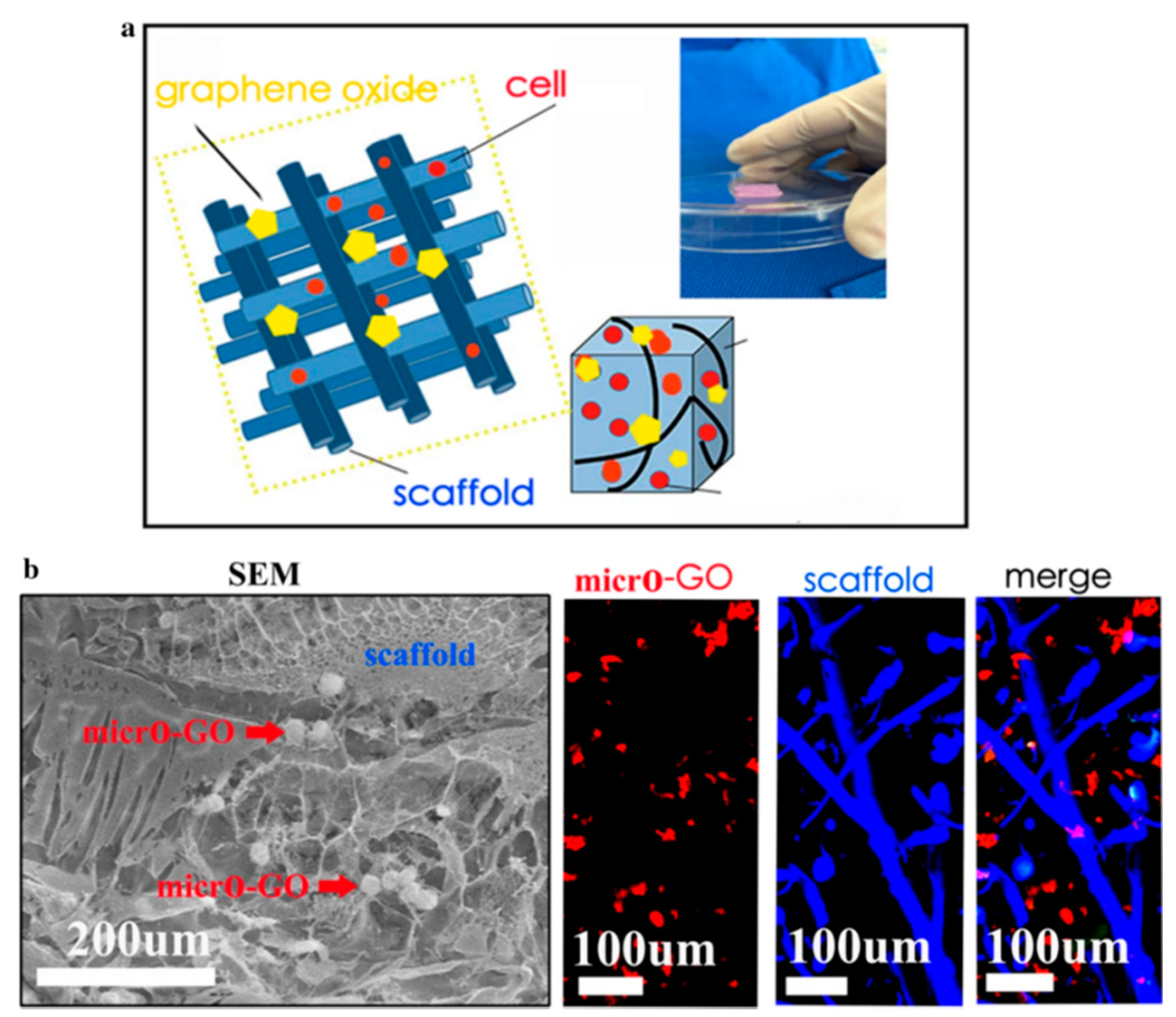
| Collagen, chitosan | Micro graphene oxide, BMP-2 (1:1) | Chondrocytes | SEM, immunofluorescence | Rats, knee, femur cartilages | 3D printed | Enhanced chondrocyte proliferation | [83] |
| Polycaprolactone | Graphene nanoplatelets, polycarboxylate modified graphene nanoplatelets 0.5, 5, 10 wt% | Human chondrocytes knee, hip | Cytotoxicity, cell proliferation | - | Injection molding process to make filaments in form of sticks, 3D printing | Improved mechanical properties, support of proliferation of chondrocytes | [84] |
| Acellular cartilage extracellular matrix, distal femoral condyle of market-weight pigs | Graphene oxide 0, 1, 2, 4, 6 mg/mL | Chondrocytes | Cell viability, adhesion, and proliferation, chondrogenesis | Implantation in rats, cartilage defect model in rabbits and histological evaluation | - | Improvement of internal structure and mechanical properties | [85] |
| Sericin | Reduced graphene oxide in ratio 10:1, 50:1, 100:1 | Mesenchymal stem cells derived from bone marrow of humans | Mesenchymal stem cells differentiation, growth, adhesion | - | - | Increased levels of collagen and glycosaminoglycan, chondrogenic differentiation stimulation | [86] |
| Gelatin, methacrylate polyethylene (glycol) diacrylate | Graphene oxide | Primary human bone marrow mesenchymal stem cells | Mesenchymal stem cells proliferation, chondrogenic differentiation, collagen II secretion, glycosaminoglycan synthesis, total collagen levels, RT-PCR | - | 3D printed scaffolds | Favorable mechanical properties, biocompatibility, increased collagen, glycosaminoglycan, protein levels; chondrogenic differentiation of mesenchymal stem cells | [87] |
| Chitosan, gelatin, anionic non-sulfated glycosaminoglycan | Graphene 0, 0.024, 0.06, 1% | Bone marrow mesenchymal stem cells | - | - | Bioink, 3D-printing | Enhanced water absorption, porosity, compression modulus, cytocompatibility, cell growth, higher cells proliferation survival | [80] |
| Poly(ε-caprolactone) | Graphene nanopowders 1, 3, 5, 10 wt% | Mouse bone marrow mesenchymal stem cells | Cell culture studies, MTT Assay, Live/Dead® assays, glycosaminoglycan formation, cell attachment and morphology | - | Printing ink, 3D-printing, robocasting method | Highest cell viability rates of cells seeded onto composite scaffolds, cells proliferated well, attached to scaffold surfaces | [88] |
| Polycaprolactone | Graphene and single-wall carbon nanotubes, 0.5% and 1.0% poly-l-lysine coated | Mesenchymal stem cells | Mesenchymal stem cells cell adhesion, proliferation, and chondrogenic differentiation | - | Electrospinning, microfibrous scaffolds | Improved mechanical properties, more homogenous fiber morphology, surface properties, good cytocompatibility | [79] |
| α-Chitin, poly(caprolactone) | Chondroitin sulfate, transforming growth factor-β encapsulation | Adipose derived stem cell from inguinal fat pads of female New Zealand albino rabbit | Cell viability, attachment, and proliferation study, chondrogenic differentiation and analysis of a murine rheumatoid arthritis model | - | Lyophilization technique | Prolonged release of TGF-β achieved, macroporous, extremely porous structure, enhanced cell attachment, proliferation, differentiation | [81] |
| - | Graphene oxide granules | Umbilical cord mesenchymal stem cells | - | Male New Zealand white rabbits: expression levels of nitric oxide, interleukin-6, tumor necrosis factor-α, glycosaminoglycan, collagen-II in serum and articular fluid | Mixing | Reduction in inflammatory level, improve of level of biochemical environment in articular cavity, promotion of cartilage repair | [89] |
| 2% chitosan | 0, 0.1, 0.2, 0.3 (w/v) % suspensions of graphene oxide in deionized water | Human articular chondrocytes | MTT assay | - | Ultra-sonication process | Improvement of physical, mechanical properties, increased proliferation of human articular chondrocytes | [90] |
| Poly(lactide-co-glycolide acid) | Graphene oxide | Bone marrow mesenchymal stem cells | Rabbit bone marrow mesenchymal stem cells | Rabbit supraspinatus tendon repair model | Electrospining | Accelerated proliferation and osteogenic differentiation, promoted healing, increased bone and cartilage generation, improved collagen arrangement | [91] |
| Collagen-I, genipin | Carbon dots | Bone marrow derived stem cells | Chondrocyte differentiation medium, intracellular ROS production, Cell Counting Kit (CCK)-8 assay, cell viability | Articular cartilage intracellular ROS production | Mixing | ROS production by photodynamic therapy, enhanced cartilage regeneration, chondrogenic differentiation, increased stiffness, reduced degradation | [92] |
| Collagen-II, chitosan, poly(lactic-co-glycolic acid) | - | Rabbit chondrocytes labelled with magnetic Iron oxide nanoparticles, TANBead® USPIO-101 (Amine group, Taiwan Advanced Nanotech Inc., Taipei, Taiwan) | Cell proliferation assay reagent WST-1, cell viability, cytotoxicity, relative proliferation activity | New Zealand White rabbits: levels of chondrogenetic marker genes including Sox-9, aggrecan, collagen-II | Mixing | Incorporation of chondrocytes into cartilage by magnetic force | [93] |
| Chitosan, collagen-I | Bioactive glass nanoparticles | Human osteosarcoma cell culture (SAOS) and kidney cells line of human embryo (HEK 293T) | The cytotoxicity and cell viability of hydrogels, MTT, Live/Dead® assays | - | Mixing | Improvement of physicochemical, morphological and rheological properties | [94] |
| 2-Hydroxypropyltrimethyl ammonium chloride chitosan, polyvinyl alcohol | Nano-hydroxyapatite, sodium citrate dihydrate | Mouse preosteoblast cells MC3T3-E1 | Tests of cell viability and proliferation | - | Freezing/thawing technique and immersing process | Improvement of mechanical and tribological properties, biological compatibility | [95] |
| Polyvinyl alcohol, polyvinyl pyrrolidone | Stick-like TiO2 nanostructures | Human osteosarcoma (HOS; MG-63) cell line | Osteoblast adhesion and proliferation | - | Sol–gel method | Excellent antibacterial efficiency, well cell adhesion and proliferations, bone formation improved | [96] |
| Glycol, chitosan | Nano-hydroxyapatite | Human sarcoma cell line culture, kidney cell line of a human embryo culture (HEK293T cells) and human bone marrow mesenchymal stem cells (HBMS) | MTT assay, Live/Dead® assays | - | solvent cast and evaporation | Potential bone-related biomedical applications | [97] |
| Chitosan, β-glycerophosphate disodium salt, gelatin | Bioactive glass nanoparticles | Rat bone marrow mesenchymal stem cells | Cytocompatibility of the hydrogels | Injecting hydrogels into dorsum of Swiss rats | Sol gel method | 27% increase in cell viability | [98] |
| Alginate, polyvinyl alcohol | Chondroitin sulfate loaded zein nanoparticles | Chondrocytes | Degradation studies, chondrocyte culture, Live/Dead® assays, MTS assay, RT-PCR, western blotting | - | Constant stirring and ultrasonication | Chondrocyte improvement, biomimetic matrices upregulating early chondrogenic marker gene (Sox-9) and differentiated genes specific for hyaline cartilage | [99] |
| Cellulose nanocrystal/dextran hydrogels | Kartogenin and ultrasmall superparamagnetic iron-oxide | Bone marrow-derived mesenchymal stem cells | CCK-8 assay, Live/Dead® assays, gene expression levels | Rabbit articular cartilage | - | Mechanical strength, kartogenin long-term release, support of hyaline cartilage regeneration | [100] |
| Matrix | Filler | Tested Cells | In Vitro | In Vivo | Ref. |
|---|---|---|---|---|---|
| Carboxymethyl chitosan | Sodium alginate | - | MTT assay, life/dead assays | - | [108] |
| Poly(d-lactic acid) | Bone mesenchymal stem cells | Proliferation assay, live/dead assays, osteogenic differentiation | - | [109] | |
| Poly(l-lactic acid) | Mesoporous silica Santa Barbara Amorphous-15 | MG63 osteoblast cells | MTT assay, cell proliferation, osteogenic differentiation, | - | [110] |
| Poly (lactide-co-propylene glycol-co-lactide) dimethacrylate | Hydroxyethyl methacrylate | Long-term release BMP-2 | Biocompatibility in rat mesenchymal stem cells, live/dead assays, proliferation cell, osteogenesis, gene expressions of osteogenesis-related markers | Rabbit femoral condyle defect animal model, micro-CT, histological observations | [111] |
| Poly(lactic-co-glycolic acid) | Poly(γ-benzyl-l-glutamate) | Mouse preosteoblast cells MC3T3-E1 | Cell culture, viability and morphology assay using mouse preosteoblast cells, MTT assay, ALP assay | Repair of rabbit radius defect, X-ray, micro-CT tests | [112] |
| Acrylated epoxidized soybean oil, polyethylene glycol diacrylate, phenylbis(2,4,6-trimethylbenzoyl)phosphine oxide | - | - | - | [113] | |
| Chitosan, poly(methyl vinyl ether-alt-maleic anhydride | SD rat bone marrow mesenchymal stem cells | Biocompatibility, viability of cells | - | [114] | |
| hydroxyl-capped poly(lactide), carboxyl-capped aniline pentamer, poly(lactide-co-glycolide) | l-Lactic acid oligomer | Mouse preosteoblast cells MC3T3-E1 | MC3T3-E1 cell proliferation activity with and without electrical stimulation, MTT assays | Intramuscular implantation into rabbits dorsal muscles, implantation for repair of radius defects in rabbits and of tibia defects in sheep | [115] |
| Poly(thioketal urethane) | - | - | Femoral defects in New Zealand White rabbits | [116] | |
| Poly(butylene-adipate-co-terephthalate) | Graphene nanoribbons | - | - | Implantation into critical tibia defects in rats, radiography analysis, tomography, bone remodeling, biomechanical properties | [117] |
| - | Gold nanoparticles | Human bone marrow-derived mesenchymal stem cells | Cell viability, proliferation by CCK-8 assay, Alizarin red S staining, RT-PCR, western blotting | - | [118] |
| Alginate | Chitooligosaccharide coated silver nanoparticles | MG-63 cells | Antimicrobial testing, MTT assay, cell viability and proliferation, Hoechst 33342 staining assay | - | [119] |
| Alginate | Zinc | Murine osteoblastic mycoplasm-free cell line | Cytocompatibility assay, cell viability, cytotoxicity assays | Wistar rats: implantation into critical-sized calvarial defects, histological preparation, histomorphometric evaluation, degradation, bioavailability | [120] |
| - | Zirconia | Mouse osteoblast precursor cell line | Cell cytocompatibility, adhesion, proliferation and differentiation | - | [121] |
| - | Magnetite | Mouse preosteoblast cells MC3T3-E1 | Cell proliferation and morphology | Female SD rats: protein corona formation and determination | [122] |
| Bacterial cellulose | Magnetite | Mouse fibroblast L929 cells | Cell cytocompatibility on MC3T3-E1, proliferation | - | [123] |
| Dextran-grafted iron oxide | Human-derived osteoblast-like cells | Cell cytocompatibility, gene expression, RNA isolation and reverse transcription, RT-PCR | - | [125] |
| Matrix | Filler | Tested Cells | In Vitro | In Vivo | Ref. |
|---|---|---|---|---|---|
| Poly-(l-lactic acid) | Carbon nanotubes | Human osteosarcoma MG63 osteoblast cells | Cell morphology, viability, proliferation | - | [127] |
| Poly(l-lactic acid) | Graphene nanoribbons, nano-hydroxyapatite | - | Allium cepa test, hemolysis | Female Wistar Rats: surgical defects in tibias, comet assay, bone regeneration | [128] |
| Collagen | Carbon nanotubes, hydroxyapatite | Rat bone mesenchymal stem cells | Cell morphology, viability, proliferation, BMP-2 level, insulin-like growth factor 1 receptor | Female SD rats: X-ray, mason staining and toxicology experiments | [129] |
| Arginine-glycine-aspartic acid/BMP-2 peptides, poly-(l-lactic acid) | Carbon nanotubes with carboxyl and amino groups | Mouse preosteoblast cells MC3T3-E1 | Cell adhesion, proliferation, differentiation, mineralization | - | [130] |
| Oligo(poly(ethylene glycol)fumarate), poly(ethylene glycol)acrylate | Carbon nanotubes, black phosphorus | MC3T3 preosteoblast cells | Cell adhesion, proliferation, osteogenic differentiation, electric stimulation enhanced osteogenesis | - | [131] |
| - | Multi-walled carbon nanotubes, nano- hydroxyapatite | Human adipose-derived mesenchymal stem cells | Cell adhesion, attachment, strength, proliferation, osteogenic differentiation, RT-PCR, DNA, ALP assay, total protein analyses | New Zealand white rabbits: dorsal musculature, histological examinations, collagen-I immunostaining analysis, bone-mineral content, macrophage infiltration | [132] |
| Polycaprolactone | Multi-walled carbon nanotubes | UMR-106 cells | Mitochondrial activity of osteoblasts, MTT assay | Male Wistar rats: immunohistochemistry, extraction of bone proteins | [133] |
| Polycaprolactone | Multi-walled carbon nanotubes, nano-hydroxyapatite | Human adipose-derived stromal/stem cells | Cell proliferation, osteogenic differentiation, mineralization, Alamar blue assay, ALP assay, amount of collagen and osteocalcin, Alizarin red S staining | - | [134] |
| Polycaprolactone | Multi-walled carbon nanotubes, eggshell | Bone marrow mesenchymal stem cells | Adhesion and proliferation of osteoblasts | - | [135] |
| Polyion complex (sodium p-styrenesulfonate, 3-(methacryloylamino)propyl-trimethylammonium chloride) | Multi-walled carbon nanotubes | Rat bone marrow-derived mesenchymal stem cells | Biocompatibility, osteogenic differentiation, viability and morphology, Alizarin red S staining, gene expression analysis | SD rats: calvarial defect, micro-CT, histological and immunological staining | [136] |
| - | Nitrogen-doped multiwalled carbon nanotubes, cellulose, nano-hydroxyapatite | SPF Spragu–Dawley rats mesenchymal stem cells | Cellular attachment, proliferation, viability, mineralization, ALP assay, osteogenic gene expressions | Distal femoral condyle critical size defect in rabbit, bone regeneration and micro-CT analysis, histological analysis | [138] |
| Chitosan | Graphene oxide | Human adipose derived stromal/stem cells | Cell viability, proliferation, MTT, cytotoxicity, Live/Dead® assays, distribution and morphology, osteogenic differentiation | Mouse models with a calvaria bone defect, Osx osteogenic marker evaluation | [139] |
| - | Hydroxyapatite, hydrophilic graphene | Mouse preosteoblast cells MC3T3-E1 | MTS assay, cell adhesion, proliferation | - | [140] |
| Carrageenan, acrylic acid | Graphene, hydroxyapatite | Mouse preosteoblast cells MC3T3-E1 | Cell viability and proliferation using neutral red dye assay | - | [103] |
| Poly-ε-caprolactone | Graphene | Mouse preosteoblast cells MC3T3-E1 | Cell adhesion and growth behaviors, MTT assay, ALP assay | - | [141] |
| Polyether ether ketone | Graphene nanosheets | Bone marrow mesenchymal stem cells | Antibacterial screening, cytocompatibility, bone regeneration, Live/Dead® assays, MTT assay, tumor inhibition | Laser treatment on a nude mouse, P/G10 and P/G10-HA tumor growth inhibition | [142] |
| Polyether ether ketone | Graphene oxide | Bone marrow stromal stem cells | Cell adhesion, cytotoxicity | - | [143] |
| Polyether ether ketone | Graphene oxide, nano-hydroxyapatite | Mouse preosteoblast cells MC3T3-E1 | Cell morphology, proliferation | - | [144] |
| - | Graphene oxide, nano-hydroxyapatite | Mouse preosteoblast cells MC3T3-E1, stem cells derived from human dental pulp | Bioactivity assay, cytotoxicity analysis, MTT assay | - | [145] |
| Chitosan | Graphene oxide, nano-hydroxyapatite | Mouse preosteoblast cells MC3T3-E1 | Degradation behavior, biomineralization study, Live/Dead® assays | SD rats: histological assessment, bone specific proteins, osteogenesis gene expression | [146] |
| Spermine based polyurethane-urea | Graphene oxide, 2D rod-like nano-hydroxyapatite, | MG63 osteoblast cells | Cell viability and proliferation, RT-PCR, cell osteogenesis induction, antibacterial study | Mature male SD rats: tibial model, histological assessment | [147] |
| Shape-memory polyurethane | Isocyanate modified graphene oxide | - | - | - | [148] |
| - | Graphene nanoribbons, nano-hydroxyapatite | - | - | Wistar rats: histological, biochemical, and radiographic analyses | [149] |
| Chitosan, quaternized chitosan | Graphene oxide, strontium-substituted hydroxyapatite | Bone marrow stromal cells | Antibacterial test, cytocompatibility using CCK-8 assay, ALP activity | Male SD rats | [150] |
| Collagen | Strontium-graphene oxide | Human adipose-derived stem cells, human umbilical vein endothelial cell | Cytotoxicity assay, viability, morphology, adhesion, osteogenic differentiation, RT-PCR, western blot, angiogenic effects | Male rats: micro-CT analysis, micro-CT angiography, calvarial undecalcified sections, calvarial decalcified sections | [151] |
| Collagen-I | Graphene oxide | Rat bone marrow mesenchymal stem cells | Bioactivity, biodegradation, cytocompatibility | Male Wistar rats: biodegradation; male SD rats: craniofacial bone defect study | [152] |
| Poly(lactic-co-glycolic acid), β-tricalcium phosphate | Graphene oxide, BMP-2 | Rat bone marrow-derived mesenchymal stem cell | Peptide release and scaffold degradation, cell viability, adhesion, morphology, osteogenic differentiation | Male Wistar rats: micro-CT and histological evaluation | [153] |
| Silk fibroin | Graphene oxide, BMP-2 | Bone marrow mesenchymal stem cells | Degradation, cell proliferation, adhesion, Live/Dead® assays, osteogenic differentiation, RT-PCR | Male SD rats: calvarial bone defect implantation, micro-CT measurement, histological evaluation | [154] |
| Silk fibroin, hydroxyapatite | Polyethylenimine functionalized graphene oxide, miR-214 inhibitor | Mouse preosteoblast cells MC3T3-E1, bone marrow mesenchymal stem cells | Cytotoxicity, CCK-8 assay, osteoblast cell proliferation and differentiation, degradation | Calvarial bone-defect model in rats, micro-CT, implantation subcutaneously on back of nude mice | [155] |
| Alginate, gelatin | Graphene oxide, human mesenchymal stem cells-laden | Human mesenchymal stem cells | Live/Dead® assays, cell morphology, DNA content, ALP activity, osteogenic-related gene expression, micro-CT, histology staining | - | [156] |
| - | Graphene oxide, mesoporous bioactive glasses | Rat bone marrow mesenchymal stem cells | Cells proliferation, adhesion, ALP assay, immunofluorescence evaluation, Alizarin red S staining, OCN immunofluorescence assay, osteogenic-related gene expression | Male SD rats: cranial bone defect model, micro-CT, microfil perfusion in bone defect, sequential fluorescent labeling in bone defect, newly bone formation and mineralization analysis, immunohistochemical analysis | [157] |
| - | Fe3O4/graphene oxide, α-tricalcium, phosphate/calcium sulfate, hydroxypropyl methylcellulose | Rat bone marrow derived mesenchymal stem cells | Antitumor effect of hyperthermia, cell viability via CCK-8 assay, Live/Dead® assays proliferation and osteogenic activity | Nude mice, antitumor efficiency, osteogenesis, | [158] |
| - | Reduced graphene oxide, nano-hydroxyapatite | Rat bone marrow mesenchymal stem cells | Cell morphology, adhesion, proliferation, Alizarin red S staining, ALP assay, osteopontin expression | Male SD rats: segmental diaphyseal bone defect on, micro-CT, histological changes, bone volume fraction, trabecular thickness and separation | [47] |
| - | Reduced graphene oxide, zinc-doped hydroxyapatite | Mesenchymal stem cell | Osteogenic differentiation, Alizarin red S staining, osteogenic marker expression, runt-related transcription factor 2, ALP assay, levels of collagen-1, BMP-2, osteocalcin, osteopontin, antimicrobial activity | Rats and mice, a rat femur osteotomy model, micro-CT, calcein evaluation, angiogenesis | [159] |
| Shape memory polyurethane | Reduced graphene oxide, hydroxyapatite, arginyl-glycyl-aspartic acid | Rabbit bone mesenchymal stem cells | Adhesion | - | [160] |
| Gelatin | Reduced graphene oxide, methyl vanillate | Bone marrow stromal cells | ALP assay, Alizarin red S staining, immunofluorescence staining, gene expression, endocytosis of bone marrow stromal cells, osteogenic effect | - | [161] |
| Polyvinyl alcohol | Carbon nanoparticles nano-demineralized bone | MG63 osteoblast cells | Biomineralization, antimicrobial screening, cell viability via MTT assay | - | [162] |
| Ascorbic acid, polyethylenimine | Carbon dots, miR-2861, | Bone marrow stromal cells | ALP assay, Alizarin red S staining, RT-PCR, fluorescent images, cytotoxicity | Male Wistar rats: intra-tibial injection for bone formation and calvarial bone defect for bone repair on fluorescent images and cytotoxicity, osteogenic effects, bone regeneration, biocompatibility | [163] |
| Gelatin, hydroxyapatite | Zinc-passivated carbon dots | Rat bone marrow stromal cells | Cytotoxicity and proliferation via MTS assay, intracellular ROS detection, ALP assay, Alizarin red S staining | Male SD rats: calvarial bone defect, radiographic and histological analysis | [164] |
| Silk fibroin | Carbon nanocages, BMP-2 | Rat bone marrow mesenchymal stem cells | ALP assay, cell adhesion, cytotoxicity, RT-PCR | Rats, micro-CT, volume percentage of new bone formation | [165] |
| - | Carbon nanofiber, gold nanoparticles | MG63 osteoblast cells | MTT and lactate dehydrogenase toxicity assays, cell attachment, morphology | - | [166] |
| Polyether ether ketone | Carbon nanotubes and fibers hydroxyapatite | Rat bone marrow and raw cell culture | Cytotoxicity and cell viability, MTT assay, ALP assay, telomerase repeated amplification protocol assay | - | [167] |
Publisher’s Note: MDPI stays neutral with regard to jurisdictional claims in published maps and institutional affiliations. |
© 2021 by the authors. Licensee MDPI, Basel, Switzerland. This article is an open access article distributed under the terms and conditions of the Creative Commons Attribution (CC BY) license (https://creativecommons.org/licenses/by/4.0/).
Share and Cite
Jampilek, J.; Placha, D. Advances in Use of Nanomaterials for Musculoskeletal Regeneration. Pharmaceutics 2021, 13, 1994. https://doi.org/10.3390/pharmaceutics13121994
Jampilek J, Placha D. Advances in Use of Nanomaterials for Musculoskeletal Regeneration. Pharmaceutics. 2021; 13(12):1994. https://doi.org/10.3390/pharmaceutics13121994
Chicago/Turabian StyleJampilek, Josef, and Daniela Placha. 2021. "Advances in Use of Nanomaterials for Musculoskeletal Regeneration" Pharmaceutics 13, no. 12: 1994. https://doi.org/10.3390/pharmaceutics13121994