
Assessment of the Substance Antioxidative Profile by Hyaluronan, Cu(II) and Ascorbate


Abstract
:1. Introductory Remarks
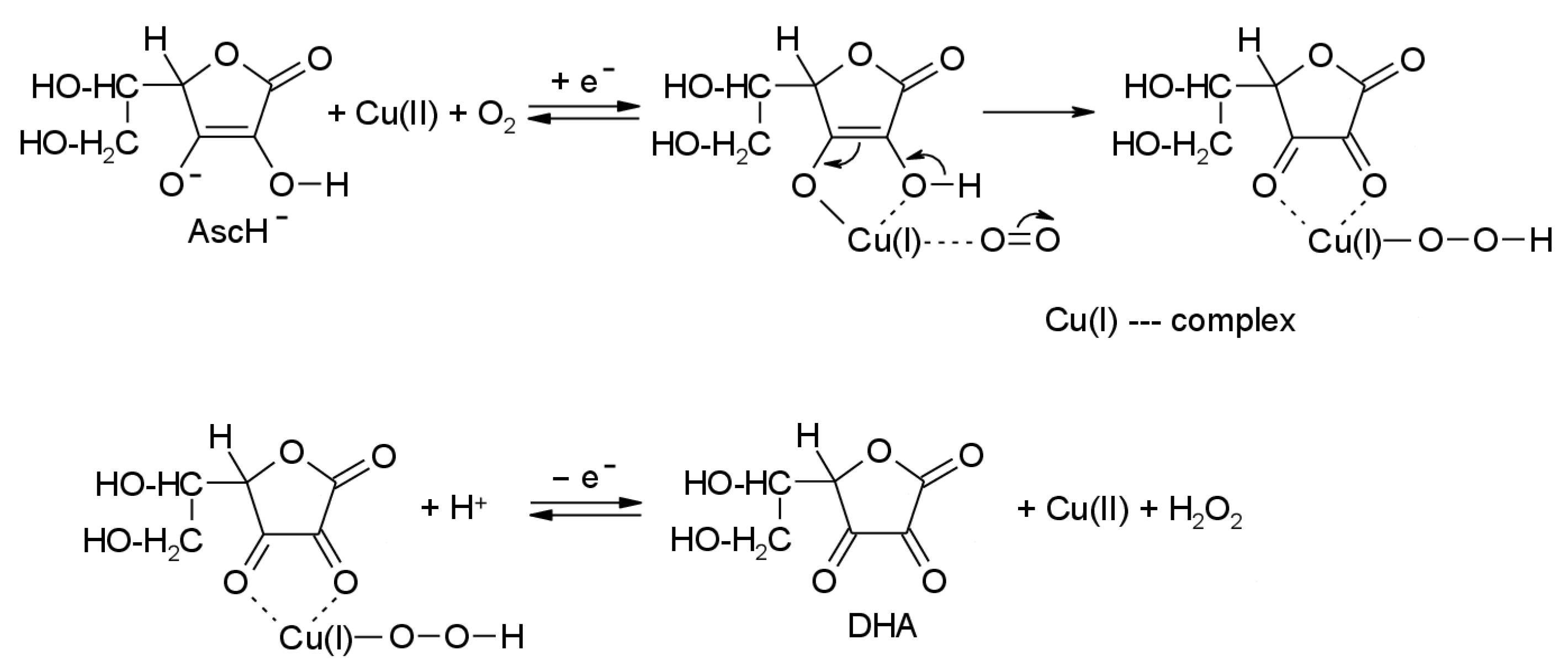
2. Sequestration of Copper Cations with Ascorbate
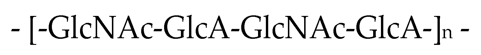
3. Hyaluronan—Oxidizable Biological Substrate
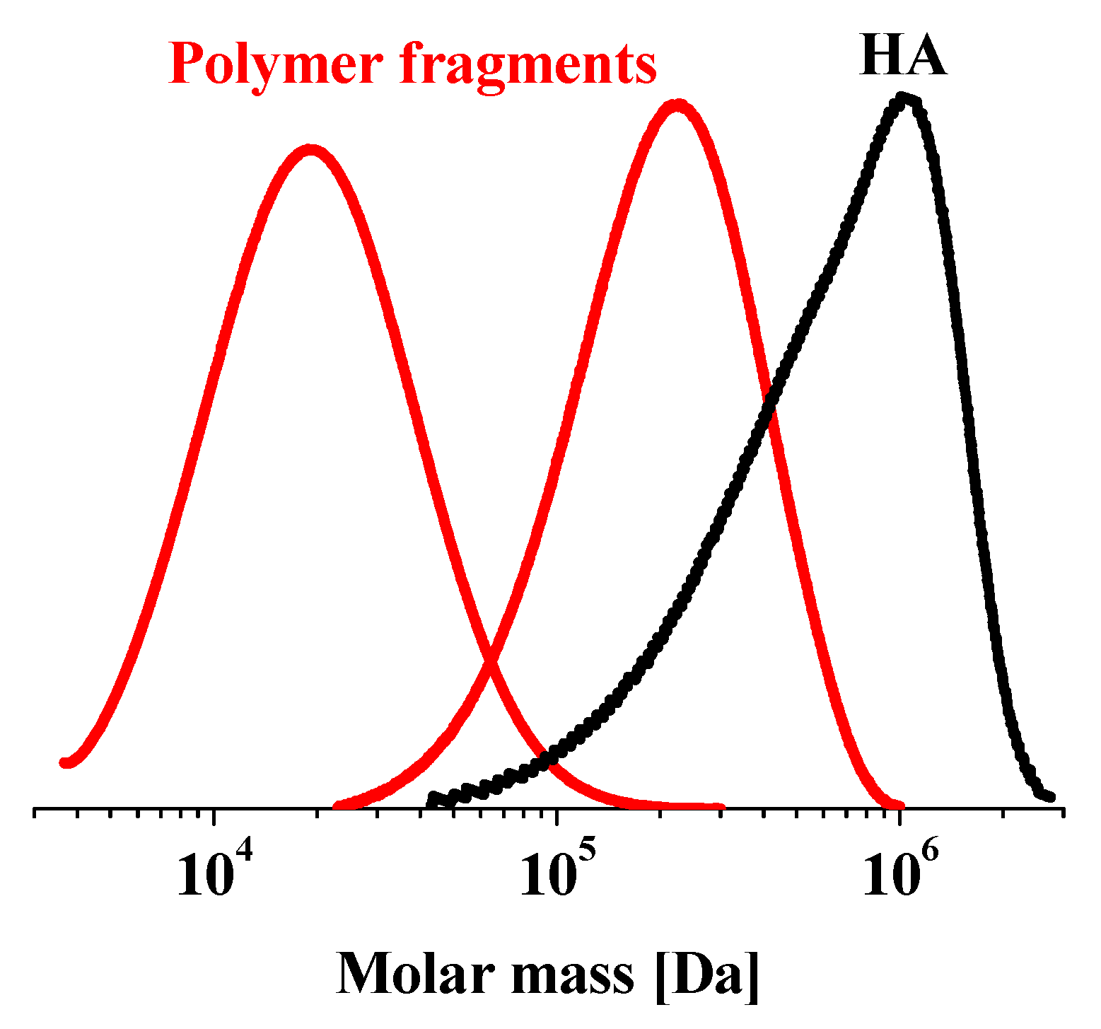
3.1. Hyaluronan Oxidative Degradation by Free-Radical-Chain Reaction
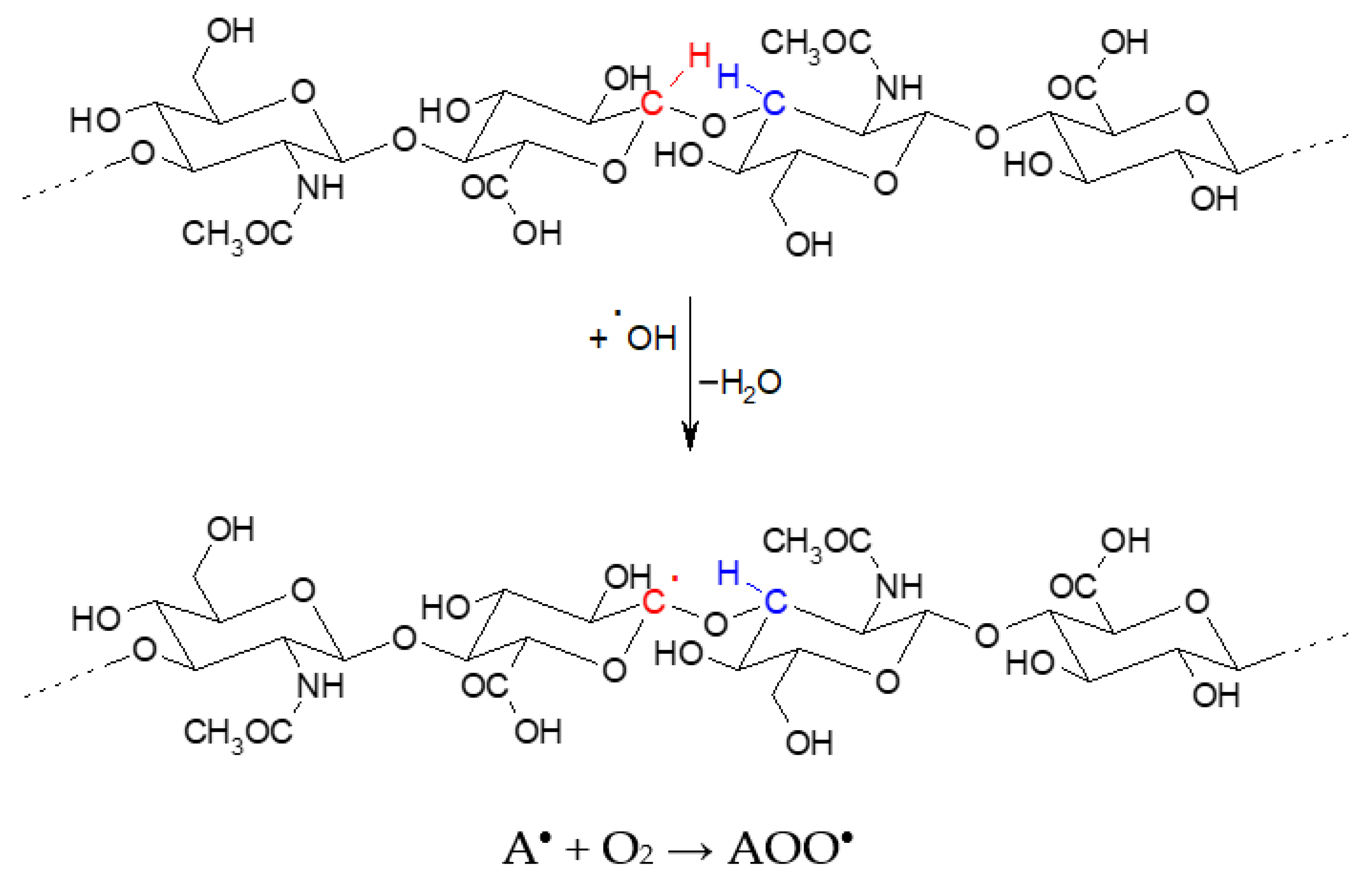
3.1.1. Initiation Reaction(s)
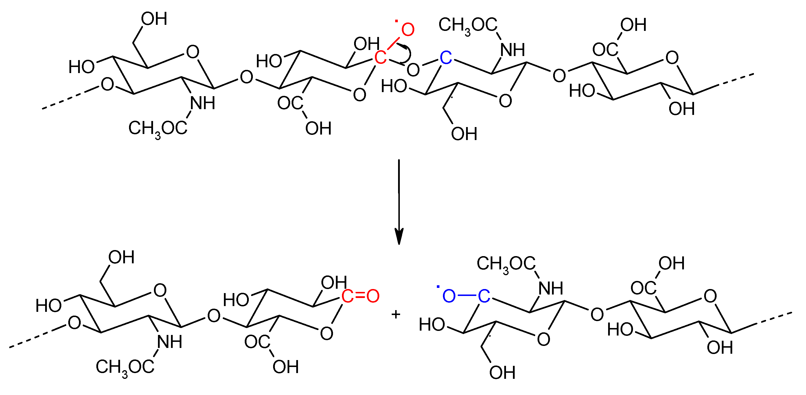
3.1.2. Transfer of the Free-Radical Centre and Fragmentation Reaction(s)
3.1.3. Termination Reaction(s)
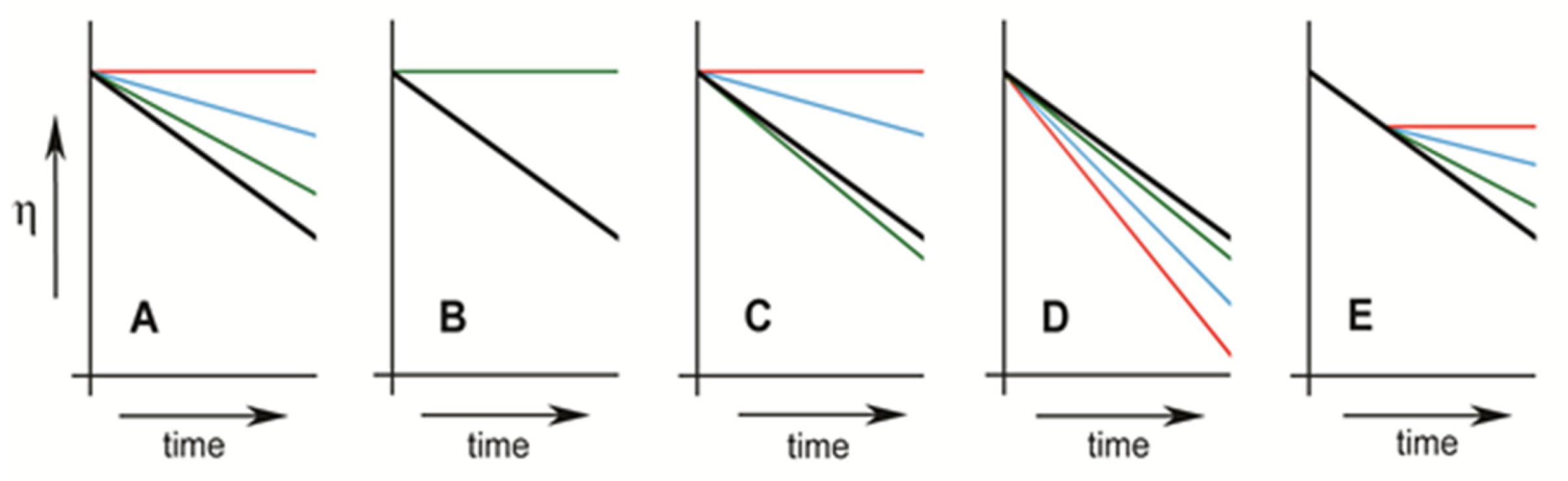
3.2. Assessment the Substance Antioxidative Profile by Hyaluronan Plus the Weissberger System
4. Concluding Remarks
Author Contributions
Funding
Institutional Review Board Statement
Informed Consent Statement
Data Availability Statement
Acknowledgments
Conflicts of Interest
References
- Free Radical School Presentations. Available online: https://sfrbm.org/education/frs-presentations/ (accessed on 9 August 2021).
- Shukla, N.; Maher, J.; Masters, J.; Angelini, G.D.; Jeremy, J.Y. Does oxidative stress change ceruloplasmin from a protective to a vasculopathic factor? Atherosclerosis 2006, 187, 238–250. [Google Scholar] [CrossRef]
- Aaseth, J.; Munthe, E.; Førre, Ø.; Steinnes, E. Trace Elements in Serum and Urine of Patients with Rheumatoid Arthritis. Scand. J. Rheumatol. 1978, 7, 237–240. [Google Scholar] [CrossRef]
- Rafter, G. Plasma thiols, copper and rheumatoid arthritis. Med. Hypotheses 1994, 43, 59–61. [Google Scholar] [CrossRef]
- Valko, M.; Morris, H.; Cronin, M.T.D. Metals, toxicity and oxidative stress. Curr. Med. Chem. 2005, 12, 1161–1208. [Google Scholar] [CrossRef] [Green Version]
- Udenfriend, S.; Clark, C.T.; Axelrod, J.; Brodie, B.B. Ascorbic acid in aromatic hydroxylation. I. A model system for aromatic hydroxylation. J. Biol. Chem. 1954, 208, 731–739. [Google Scholar] [CrossRef]
- Weissberger, A.; LuValle, J.E.; Thomas, D.S., Jr. Oxidation processes. XVI. The autoxidation of ascorbic acid. J. Am. Chem. Soc. 1943, 65, 1934–1939. [Google Scholar] [CrossRef]
- Khan, M.M.T.; Martell, A.E. Metal ion and metal chelate catalyzed oxidation of ascorbic acid by molecular oxygen. I. Cupric and ferric ion catalyzed oxidation. J. Am. Chem. Soc. 1967, 89, 4176–4185. [Google Scholar] [CrossRef]
- Wong, F.S.; Halliwell, B.; Richmond, R.; Skowroneck, W.R. The role of superoxide and hydroxyl radicals in the degradation of hyaluronic acid induced by metal ions and by ascorbic acid. J. Inorg. Biochem. 1981, 14, 127–134. [Google Scholar] [CrossRef]
- Fisher, A.E.O.; Naughton, D.P. Iron supplements: The quick fix with long-term consequences. Nutr. J. 2004, 3, 2. [Google Scholar] [CrossRef] [Green Version]
- Fisher, A.; Naughton, D. Vitamin C contributes to inflammation via radical generating mechanisms: A cautionary note. Med. Hypotheses 2003, 61, 657–660. [Google Scholar] [CrossRef]
- Fisher, A.E.O.; Naughton, D.P. Therapeutic chelators for the twenty first century: New treatments for iron and copper mediated inflammatory and neurological disorders. Curr. Drug Deliv. 2005, 2, 261–268. [Google Scholar] [CrossRef]
- Fisher, A.E.O.; Naughton, D.P. Why nutraceuticals do not prevent or treat Alzheimer’s disease. Nutr. J. 2005, 4, 14. [Google Scholar] [CrossRef] [Green Version]
- Šoltés, L.; Mendichi, R.; Kogan, G.; Schiller, J.; Stankovská, A.M.; Arnhold, J. Degradative action of reactive oxygen species on hyaluronan. Biomacromolecules 2006, 7, 659–668. [Google Scholar] [CrossRef]
- Valachová, K.; Tamer, T.M.; Eldin, M.M.; Šoltés, L. Radical-scavenging activity of glutathione, chitin derivatives and their combination. Chem. Pap. 2016, 70. [Google Scholar] [CrossRef]
- Valachová, K.; Topoľská, D.; Mendichi, R.; Collins, M.N.; Sasinková, V.; Šoltés, L. Hydrogen peroxide generation by the Weissberger biogenic oxidative system during hyaluronan degradation. Carbohydr. Polym. 2016, 148, 189–193. [Google Scholar] [CrossRef]
- Buettner, G.; Jurkiewicz, B.A. Ascorbate free radical as a marker of oxidative stress: An EPR study. Free Radic. Biol. Med. 1993, 14, 49–55. [Google Scholar] [CrossRef]
- Šoltés, L.; Kogan, G.; Stankovská, M.; Mendichi, R.; Rychlý, J.; Schiller, A.J.; Gemeiner, P. Degradation of high-molar-mass hyaluronan and characterization of fragments. Biomacromolecules 2007, 8, 2697–2705. [Google Scholar] [CrossRef]
- Fornaro, A.; Coichev, N. Ácido L-ascórbico: Reações de complexação e de óxido-redução com alguns íons metálicos de transição. Divulgação 1998, 21, 642–650. [Google Scholar] [CrossRef]
- Lepperdinger, G.; Fehrer, C.; Reitinger, S. Chemistry and biology of hyaluronan. In Chemistry and Biology of Hyaluronan; Garg, H.G., Hales, C.A., Eds.; Elsevier Press: Amsterdam, NY, USA, 2004; pp. 71–82. [Google Scholar]
- Stern, R.; Asari, A.A.; Sugahara, K.N. Hyaluronan fragments: An information-rich system. Eur. J. Cell. Biol. 2006, 85, 699–715. [Google Scholar] [CrossRef]
- Mendichi, R.; Šoltés, L. Hyaluronan molecular weight and polydispersity in some commercial intra-articular injectable preparations and in synovial fluid. Inflamm. Res. 2002, 51, 115–116. [Google Scholar] [CrossRef]
- Šoltés, L.; Valachová, K. Biopolymer hyaluronan messenging the status of synovial joints. In Advances in Chemistry Research; Taylor, J.C., Ed.; Nova Science Publishers, Inc.: Hauppauge, NY, USA, 2018; pp. 157–180. [Google Scholar]
- Valachová, K.; Šoltés, L. Versatile use of chitosan and hyaluronan in medicine. Molecules 2021, 26, 1195. [Google Scholar] [CrossRef]
- Nitzan, D.W.; Nitzan, U.; Dan, P.; Yedgar, S. The role of hyaluronic acid in protecting surface-active phospholipids from lysis by exogenous phospholipasae A2. Rheumatology 2001, 40, 336–340. [Google Scholar] [CrossRef] [Green Version]
- Kogan, G.; Šoltés, L.; Stern, R.; Gemeiner, P. Hyaluronic acid: A natural biopolymer with a broad range of biomedical and industrial applications. Biotechnol. Lett. 2007, 29, 17–25. [Google Scholar] [CrossRef]
- Furth, G.; Knierim, R.; Buss, V.; Mayer, C. Binding of bivalent cations by hyaluronate in aqueous solution. Int. J. Biol. Macromol. 2008, 42, 33–40. [Google Scholar] [CrossRef]
- Rinaudo, M.; Rozand, Y.; Mathieu, P.; Conrozier, T. Role of different pre-treatments on composition and rheology of synovial fluids. Polymers 2009, 1, 16–34. [Google Scholar] [CrossRef]
- Volpi, N.; Schiller, J.; Stern, R.; Šoltés, L. Role, metabolism, chemical modifications and applications of hyaluronan. Curr. Med. Chem. 2009, 16, 1718–1745. [Google Scholar] [CrossRef]
- Schiller, J.; Volpi, N.; Hrabarová, E.; Šoltés, L. Hyaluronic acid: A natural biopolymer. In Handbook of Biopolymers and Their Applications; Kalia, S., Averous, L., Eds.; Wiley & Scrivener Publishing: Beverly, MA, USA, 2011; pp. 3–34. [Google Scholar]
- Valachová, K.; Volpi, N.; Stern, R.; Šoltés, L. Hyaluronan in medical practice. Curr. Med. Chem. 2016, 23, 3607–3617. [Google Scholar] [CrossRef]
- Bishop, P.N. Structural macromolecules and supramolecular organisation of the vitreous gel. Prog. Retin. Eye Res. 2000, 19, 323–344. [Google Scholar] [CrossRef]
- Nickerson, C.S.; Kornfield, J.A.A. “Cleat” geometry for suppressing wall slip. J. Rheol. 2005, 49, 865–874. [Google Scholar] [CrossRef]
- Rychlý, J.; Šoltés, L.; Stankovská, M.; Janigová, I.; Csomorová, K.; Sasinková, V.; Kogan, G.; Gemeiner, P. Unexplored capabilities of chemiluminescence and thermoanalytical methods in characterization of intact and degraded hyaluronans. Polym. Degrad. Stab. 2006, 91, 3174–3184. [Google Scholar] [CrossRef]
- Hrabárová, E.; Valachová, K.; Rychlý, J.; Rapta, P.; Sasinková, V.; Malíková, M.; Šoltés, L. High-molar-mass hyaluronan degradation by Weissberger’s system: Pro- and anti-oxidative effects of some thiol compounds. Polym. Degrad. Stab. 2009, 94, 1867–1875. [Google Scholar] [CrossRef]
- Collins, M.N.; Birkinshaw, C. Hyaluronic acid based scaffolds for tissue engineering−A review. Carbohydr. Polym. 2013, 92, 1262–1279. [Google Scholar] [CrossRef]
- Chabreček, P.; Šoltés, L.; Kállay, Z.; Fügedi, A. Isolation and characterisation of high moleculr weight (3H) hyaluronic acid. J. Label. Comp. Radiopharm. 1990, 28, 1121–1125. [Google Scholar] [CrossRef]
- Chabreček, P.; Šoltés, L.; Orviský, E. Comparative depolymerization of sodium hyaluronate by ultrasonic and enzymatic treatments. J. App. Polymer Sci. App. Polym. Symp. 1991, 48, 233–241. [Google Scholar] [CrossRef]
- Chabreček, P.; Šoltés, L.; Hradec, H.; Filip, J.; Orviský, E. Preparation and characterization of the high molecular weight (3H) hyaluronic acid. Collect. Czechoslov. Chem. Commun. 1992, 57, 2151–2156. [Google Scholar] [CrossRef]
- Orviský, E.; Šoltés, L.; Chabreček, P.; Novák, I.; Kéry, V.; Stančíková, M.; Vinš, I. The determination of hyaluronan molecular weight distribution by means of high-performance size exclusion chromatography. J. Liq. Chromatogr. 1992, 15, 3203–3218. [Google Scholar] [CrossRef]
- Orviský, E.; Šoltés, L.; Chabreček, P.; Novák, I.; Stančíková, M. Size exclusion chromatographic characterization of sodium hyaluronate fractions prepared by high energetic sonication. Chromatographia 1993, 37, 20–22. [Google Scholar] [CrossRef]
- Laurent, T.C.; Laurent, U.B.; Fraser, J.R. Serum hyaluronan as disease marker. Ann. Med. 1996, 28, 241–253. [Google Scholar] [CrossRef]
- Fraser, J.R.; Laurent, T.C.; Laurent, U.B. Hyaluronan: Its nature, distribution, functions and turnover. J. Int. Med. 1997, 242, 27–33. [Google Scholar] [CrossRef]
- Grootveld, M.; Henderson, E.B.; Farrell, A.; Blake, D.R.; Parkes, H.G.; Haycock, P. Oxidative damage to hyaluronate and glucose in synovial fluid during exercise of the inflamed rheumatoid joint: Detection of abnormal low-molecular-mass metabolites by proton-n.m.r. spectroscopy. Biochem. J. 1991, 273, 459–467. [Google Scholar] [CrossRef] [Green Version]
- Šoltés, L.; Stankovská, M.; Brezová, V.; Schiller, J.; Arnhold, J.; Kogan, G.; Gemeiner, P. Hyaluronan degradation by copper(II) chloride and ascorbate: Rotational viscometric, EPR spin-trapping, and MALDI-TOF mass spectrometric investigations. Carbohydr. Res. 2006, 341, 2826–2834. [Google Scholar] [CrossRef]
- Juránek, I.; Šoltés, L. Reactive oxygen species in joint physiology: Possible mechanism of maintaining hypoxia to protect chondrocytes from oxygen excess via synovial fluid hyaluronan peroxidation. In Kinetics, Catalysis and Mechanism of Chemical Reactions: From Pure to Applied Science; Islamova, R.M., Kolesov, S.V., Zaikov, G.E., Eds.; Nova Science Publishers: New York, NY, USA, 2012; pp. 1–10. [Google Scholar]
- Juránek, I.; Stern, R.; Šoltés, L. Hyaluronan peroxidation is required for normal synovial function: An hypothesis. Med. Hypotheses 2014, 82, 662–666. [Google Scholar] [CrossRef]
- Stern, R.; Kogan, G.; Jedrzejas, M.J.; Šoltés, L. The many ways to cleave hyaluronan. Biotechnol. Adv. 2007, 25, 537–557. [Google Scholar] [CrossRef]
- Koch, P.; Sidloi, M.; Tonks, D.B. Estimation of serum ascorbic acid in patients and the effect of ascorbic acid and its oxidation products on SMA 12/60 parameters. Clin. Biochem. 1980, 13, 73–77. [Google Scholar] [CrossRef]
- Halliwell, B.; Wasil, M.; Grootveld, M. Biologically significant scavenging of the myeloperoxidase-derived oxidant hypochlorous acid by ascorbic acid. Implications for antioxidant protection in the inflamed rheumatoid joint. FEBS Lett. 1987, 213, 15–17. [Google Scholar] [CrossRef] [Green Version]
- Šoltés, L.; Valachová, K.; Mendichi, R.; Kogan, G.; Arnhold, J.; Gemeiner, P. Solution properties of high-molar-mass hyaluronans: The biopolymer degradation by ascorbate. Carbohydr. Res. 2007, 342, 1071–1077. [Google Scholar] [CrossRef] [PubMed]
- Hawkins, C.L.; Davies, M.J. Detection of intermediates formed on reaction of hyaluronic acid and related materials with the hydroxyl radical. Biochem. Soc. Trans. 1995, 23, S248. [Google Scholar] [CrossRef] [Green Version]
- Hawkins, C.L.; Davies, M.J. Direct detection and identification of radicals generated during the hydroxyl radical-induced degradation of hyaluronic acid and related materials. Free Radic. Biol. Med. 1996, 21, 275–290. [Google Scholar] [CrossRef]
- Kogan, G.; Šoltés, L.; Stern, R.; Mendichi, R. Hyaluronic acid: A biopolymer with versatile physico-chemical and biological properties. In Handbook of Polymer Research: Monomers, Oligomers, Polymers and Composites; Pethrick, R.A., Ballada, A., Zaikov, G.E., Eds.; Nova Science Publishers: New York, NY, USA, 2007; pp. 393–439. [Google Scholar]
- Kogan, G.; Šoltés, L.; Stern, R.; Schiller, J.; Mendichi, R. Studies in Natural Products Chemistry. Hyaluronic Acid: Its Function and Degradation in In Vivo Systems; Elsevier: Amsterdam, The Netherland, 2008; pp. 789–882. [Google Scholar]
- Valachova, K.; Svik, K.; Biro, C.; Soltes, L. Skin wound healing with composite biomembranes loaded by tiopronin or captopril. J. Biotechnol. 2020, 310, 49–53. [Google Scholar] [CrossRef] [PubMed]
- Buettner, G.R. The pecking order of free radicals and antioxidants: Lipid peroxidation, alpha-tocopherol, and ascorbate. Arch. Biochem. Biophys. 1993, 300, 535–543. [Google Scholar] [CrossRef]
- Buettner, G.R.; Schafer, F.Q. Ascorbate (vitamin C) as an antioxidant. In Vitamin C: Its Functions and Biochemistry in Animals and Plants; May, J.M., Asard, H., Smirnoff, N., Eds.; BIOS Scientific Publishers: Oxford, UK, 2004; pp. 173–188. [Google Scholar]
- Valachová, K.; Mach, M.; Šoltés, L. Oxidative degradation of high-molar-mass hyaluronan: Effects of some indole derivatives to hyaluronan decay. Int. J. Mol. Sci. 2020, 20, 5609. [Google Scholar] [CrossRef]
- Orviský, E.; Šoltés, L.; Stanèíková, M.; Vyletelová, Z.; Juránek, I. Assessment of Antioxidative Properties of Hydrophilic Xenobiotics on the Basis of Inhibition of the Radical Degradation of Hyaluronan by Reactive Oxygen Species. Slovak Patent No. 2764, 6 June 1994. [Google Scholar]
- Orviský, E.; Šoltés, L.; Stanèíková, M. High-molecular-weight hyaluronan—A valuable tool in testing the antioxidative activity of amphiphilic drugs stobadine and vinpocetine. J. Pharm. Biomed. Anal. 1997, 16, 419–424. [Google Scholar] [CrossRef]
- Stankovská, M.; Šoltés, L.; Vikartovská, A.; Mendichi, R.; Láth, D.; Molnárová, M.; Gemeiner, P. Study of hyaluronan degradation by means of rotational viscometry: Contribution of the material of viscometer. Chem. Pap. 2004, 58, 348–352. [Google Scholar]
- Baòasová, M.; Valachová, K.; Rychlý, J.; Janigová, I.; Csomorová, K.; Mendichi, R.; Mislovièová, D.; Juránek, I.; Šoltés, L. Free-radical-mediated degradation of high-molar-mass hyaluronan. Action of an antiinflammatory drug. Polymers 2014, 6, 2625–2644. [Google Scholar] [CrossRef] [Green Version]
- Topoľská, D.; Valachová, K.; Rapta, P.; Šilhár, S.; Panghyová, E.; Horváth, A.; Šoltés, L. Antioxidative properties of Sambucus Nigra extracts. Chem. Pap. 2015, 69, 1202–1210. [Google Scholar] [CrossRef]
- Mendichi, R.; Šoltés, L.; Schieroni, A.G. Evaluation of radius of gyration and intrinsic viscosity molar mass dependence and stiffness of hyaluronan. Biomacromolecules 2003, 4, 1805–1810. [Google Scholar] [CrossRef]
- Valachova, K.; Topolska, D.; Mendichi, R.; Rychly, J.; Soltes, L. Protective effects of catalase indicate hydrogen peroxide involvement in hyaluronan degradation initiated by Cu(II) ions plus ascorbate in situ. In Applied Chemistry and Chemical Engineering, Principles, Methodology, and Evaluation Methods; Haghi, A.K., Pogliani, L., Balkose, D., Mukbaniani., O.V., Mercader, A.G., Eds.; Apple Academic Press: Waretown, NJ, USA; Taylor & Francis Group: Palm Bay, FL, USA, 2018; Volume 2, pp. 157–173. [Google Scholar]
- Valachová, K.; Šušaníková, I.; Topoľská, D.; Bögi, E.; Šoltés, L. Effects of N-acetylcysteine, glutathione, and glutathione disulfide on NIH 3T3, VH10 and MCF-7 cells exposed to ascorbate and Cu(II) ions. In Green Chemistry and Sustainable Technology. Biological, Pharmaceutical, And Macromolecular Systems; Dake, S.A., Shinde, R.S., Ameta, S.C., Haghi, A.K., Eds.; Apple Academic Press: Waretown, NJ, USA; Taylor & Francis Group: Palm Bay, FL, USA, 2021; pp. 77–93. [Google Scholar]
- Hrabárová, E.; Rychlý, J.; Sasinková, V.; Valachová, K.; Janigová, I.; Csomorová, K.; Juránek, I.; Šoltés, L. Structural characterisation of thiol-modified hyaluronans. Cellulose 2012, 19, 2093–2104. [Google Scholar] [CrossRef]
- Hrabarova, E.; Valachova, K.; Juranek, I.; Soltes, L. Free-radical degradation of high-molar-mass hyaluronan induced by ascorbate plus cupric ions: Evaluation of antioxidative effect of cysteine derived compounds. Chem. Biodivers. 2012, 9, 309–317. [Google Scholar] [CrossRef]
- Šoltés, L.; Stankovská, M.; Kogan, G.; Mendichi, R.; Volpi, N.; Sasinková, V.; Gemeiner, P. Degradation of high-molar-mass hyaluronan by an oxidative system comprising ascorbate, Cu(II), and hydrogen peroxide: Inhibitory action of antinflammatory drugs—Naproxen and acetylsalicylic acid. J. Pharm. Biomed. Anal. 2007, 44, 1056–1063. [Google Scholar] [CrossRef]
- Valachová, K.; Rapta, P.; Slováková, M.; Priesolová, E.; Nagy, M.; Mislovičová, D.; Dráfi, F.; Bauerová, K.; Šoltés, L. Radical degradation of high molar mass hyaluronan induced by ascorbate plus cupric ions. Testing of arbutin in the function of antioxidant. In Advances in Kinetics and Mechanism of Chemical Reactions; Zaikov, G.E., Valente, A.J.M., Iordanskii, A.L., Eds.; Apple Academic Press: Waretown, NJ, USA, 2013; pp. 1–19. [Google Scholar]
- Valachová, K.; Vargová, A.; Rapta, P.; Hrabárová, E.; Dráfi, F.; Bauerová, K.; Juránek, I.; Šoltés, L. Aurothiomalate as preventive and chain-breaking antioxidant in radical degradation of high-molar-mass hyaluronan. Chem. Biodivers. 2011, 8, 1274–1283. [Google Scholar] [CrossRef]
- Valachová, K.; Hrabárová, E.; Priesolová, E.; Nagy, M.; Baňasová, M.; Juránek, I.; Šoltés, L. Free-radical degradation of high-molecular-weight hyaluronan induced by ascorbate plus cupric ions. Testing of bucillamine and its SA981-metabolite as antioxidants. J. Pharm. Biomed. Anal. 2011, 56, 664–670. [Google Scholar] [CrossRef] [PubMed]
- Baňasová, M.; Valachová, K.; Hrabárová, E.; Priesolová, E.; Nagy, M.; Juránek, I.; Šoltés, L. Early stage of the acute phase of joint inflammation in vitro testing of bucillamine and its oxidized metabolite SA981 in function of antioxidants. Interdis. Toxicol. 2011, 4, A22. [Google Scholar]
- Baňasová, M.; Sasinková, V.; Mendichi, R.; Perečko, T.; Valachová, K.; Juránek, I.; Šoltés, L. Free-radical degradation of high-molar-mass hyaluronan induced by Weissberger’s oxidative system: Potential antioxidative effect of bucillamine. Neuroendocrinol. Lett. 2012, 3 (Suppl. S33), 151–154. [Google Scholar]
- Baňasová, M.; Kerner, L.; Juránek, I.; Putala, M.; Valachová, K.; Šoltés, L. Radical scavenging capacity of N-(2-mercapto-2-methylpropionyl)-l-cysteine. Design and synthesis of its derivative with enhanced potential to scavenge hypochlorite. J. Inf. Intell. Knowl. 2014, 6, 453–470. [Google Scholar]
- Baňasová, M.; Valachová, K.; Juránek, I.; Šoltés, L. Dithiols as more effective than monothiols in protecting biomacromolecules from free-radical-mediated damage: In vitro oxidative degradation of high-molar-mass hyaluronan. Chem. Pap. 2014, 68, 1428–1434. [Google Scholar] [CrossRef]
- Valachová, K.; Baňasová, M.; Topoľská, D.; Sasinková, V.; Juránek, I.; Collins, M.N.; Šoltés, L. Influence of tiopronin, captopril and levamisole therapeutics on the oxidative degradation of hyaluronan. Carbohydr. Polym. 2015, 134, 516–523. [Google Scholar] [CrossRef]
- Dráfi, F.; Bauerová, K.; Valachová, K.; Poništ, S.; Mihálová, D.; Juránek, I.; Boldyrev, A.; Hrabárová, E.; Šoltés, L. Carnosine inhibits degradation of hyaluronan induced by free radical processes in vitro and improves the redox imbalance in adjuvant arthritis in vivo. Neuroendocrinol. Lett. 2010, 31 (Suppl. S2), 96–100. [Google Scholar] [PubMed]
- Valachová, K.; Hrabárová, E.; Dráfi, F.; Juránek, I.; Bauerová, K.; Priesolová, E.; Nagy, M.; Šoltés, L. Ascorbate and Cu(II) induced oxidative degradation of high-molar-mass hyaluronan. Pro- and antioxidative effects of some thiols. Neuroendocrinol. Lett. 2010, 31 (Suppl. S2), 101–104. [Google Scholar]
- Valent, I.; Topoľská, D.; Valachová, K.; Bujdák, J.; Šoltés, L. Kinetics of ABTS derived radical cation scavenging by bucillamine, cysteine, and glutathione. Catalytic effect of Cu2+ ions. Biophys. Chem. 2016, 212, 9–16. [Google Scholar] [CrossRef]
- Tamer, T.M.; Valachová, K.; Hassan, M.A.; Omer, A.M.; El-Shafeey, M.; Eldin, M.M.; Šoltés, L. Chitosan/hyaluronan/edaravone membranes for anti-inflammatory wound dressing: In vitro and in vivo evaluation studies. Mater. Sci. Eng. C 2018, 90, 227–235. [Google Scholar] [CrossRef]
- Valachová, K.; Švík, K.; Biró, C.; Collins, M.N.; Jurčík, R.; Ondruška, L.; Šoltés, L. Impact of ergothioneine, hercynine and histidine on oxidative degradation of hyaluronan and wound healing. Polymers 2021, 13, 95. [Google Scholar] [CrossRef]
- Valachová, K.; Mendichi, R.; Šoltés, L. Effect of L-Glutathione on High-Molar-Mass Hyaluronan Degradation by Oxidative System Cu(II) Plus Ascorbate. Monomers, Oligomers, Polymers, Composites, and Nanocomposites; Pethrick, R.A., Petkov, P., Zlatarov, A., Zaikov, G.E., Rakovsky, S.K., Eds.; Nova Science Publishers: New York, NY, USA, 2010; pp. 101–111. [Google Scholar]
- Tamer, M.T.; Valachová, K.; Omer, A.M.; Sabet, M.M.; Šoltés, L. Effects of glutathione, phosphonate, or sulfonated chitosans and their combination on scavenging free radicals. In High-Performance Materials and Engineered Chemistry; Torrens, F., Balkose, D., Thomas, S., Eds.; Apple Academic Press: New Jersey, NJ, USA, 2018; pp. 371–389. [Google Scholar]
- Tamer, M.T.; Hassan, M.A.; Valachová, K.; Omer, A.M.; El-Shafeey, M.E.A.; Mohy Eldin, M.S.; Šoltés, L. Enhancement of wound healing by chitosan/hyaluronan polyelectrolyte membrane loaded with glutathione: In vitro and in vivo evaluations. J. Biotechnol. 2020, 310, 103–113. [Google Scholar] [CrossRef]
- Šoltés, L.; Láth, D.; Mendichi, R.; Bystrický, P. Radical degradation of high molecular weight hyaluronan: Inhibition of the reaction by ibuprofen enantiomers. Method and Find. Exp. Clin. Pharmacol. 2001, 23, 65–71. [Google Scholar]
- Baňasová, M.; Valachová, K.; Juránek, I.; Šoltés, L. Aloe vera and methylsulfonylmethane as dietary supplements: Their potential benefits for arthritic patients with diabetic complications. J. Inf. Intell. Knowl. 2013, 5, 51–68. [Google Scholar]
- Valachová, K.; Rapta, P.; Moura, N.M.M.; Batinic-Haberle, I.; Šoltés, L. Ortho isomeric Mn(III) N-alkyl- and alkoxyalkylpyridylporphyrins−enhancers of hyaluronan degradation induced by ascorbate and cupric ions. Int. J. Mol. Sci. 2021, 22, 8608. [Google Scholar] [CrossRef]
- Valachová, K.; Hrabárová, E.; Gemeiner, P.; Šoltés, L. Study of pro- and anti-oxidative properties of D-penicillamine in a system comprising high-molar-mass hyaluronan, ascorbate, and cupric ions. Neuroendocrinol. Lett. 2008, 29, 697–701. [Google Scholar] [PubMed]
- Valachová, K.; Rapta, P.; Kogan, G.; Hrabárová, E.; Gemeiner, P.; Šoltés, L. Degradation of high-molar-mass hyaluronan by ascorbate plus cupric ions: Effects of D-penicillamine addition. Chem. Biodivers. 2009, 6, 389–395. [Google Scholar] [CrossRef] [PubMed]
- Hassan, M.A.; Tamer, M.T.; Valachová, K.; Omer, A.M.; El-Shafeey, M.; Mohy Eldin, M.S.; Šoltés, L. Antioxidant and antibacterial polyelectrolyte wound dressing based on chitosan/hyaluronan/phosphatidylcholine dihydroquercetin. Int. J. Biol. Macromol. 2021, 166, 18–31. [Google Scholar] [CrossRef]
- Rapta, P.; Valachová, K.; Zalibera, M.; Šnirc, V.; Šoltés, L. Hyaluronan degradation by reactive oxygen species: Scavenging effect of the hexahydropyridoindole stobadine and two of its derivatives. In Monomers, Oligomers, Polymers, Composites, and Nanocomposites; Pethrick, A.R., Petkov, P., Zlatarov, A., Zaikov, G.E., Rakovsky, S.K., Eds.; Nova Science Publishers: New York, NY, USA, 2010; pp. 113–126. [Google Scholar]
- Surovčíková, L.; Valachová, K.; Baňasová, M.; Šnirc, V.; Priesolová, E.; Nagy, M.; Juránek, I.; Šoltés, L. Free-radical degradation of high-molar-mass hyaluronan induced by ascorbate plus cupric ions: Testing of stobadine and its two derivatives in function as antioxidants. Gen. Physiol. Biophys. 2012, 31, 57–64. [Google Scholar] [CrossRef] [PubMed] [Green Version]
- Oikawa, S.; Yamada, K.; Yamashita, N.; Tada-Oikawa, S.; Kawanishi, S. N-Acetylcysteine, a cancer chemopreventive agent, causes oxidative damage to cellular and isolated DNA. Carcinogenesis 1999, 20, 1485–1490. [Google Scholar] [CrossRef] [Green Version]
- Koppenol, W.H. Chemistry of iron and copper in radical reactions. In Free Radical Damage and Its Control; Rice-Evans, C.A., Burdon, R.H., Eds.; Elsevier: Amsterdam, The Netherlands, 1994; pp. 3–24. [Google Scholar]


| Substance | Preventive Action | Chain-Breaking Action | B | C | D | Reference/Note |
|---|---|---|---|---|---|---|
| N-Acetylcysteine | Effective | Effective | Effective a | [67,68,69] | ||
| Acetylsalicylic acid | Effective | Effective | [70] | |||
| Arbutin | Effective | Effective | [71] | |||
| Aurothiomalate | Effective | Effective | [72] | |||
| Bucillamine | Effective | Effective | [63,73,74,75,76,77]/It is a dithiol. | |||
| Captopril | Effective | Effective | Effective | [56,78] | ||
| Carnosine | Effective | Effective | [79] | |||
| Cemtirestat | Effective | Effective | Effective b | [59] | ||
| Cysteamine | Effective | Effective | [80] | |||
| l-Cysteine | Effective | Effective | Effective a | [73,81] | ||
| Dithiothreitol | Effective | Effective | [77]/It is a dithiol. | |||
| Dithioerythritol | Effective | Effective | [77]/It is a dithiol; SH groups’ pKa values = 9.2 and 10.1. | |||
| d-Mannitol | Effective | Effective | [61] | |||
| Edaravone | Effective | Effective | [82] | |||
| Ergothioneine | Effective | Effective | [83] | |||
| Isatin | Effective | Effective | Effective b | [59] | ||
| l-Glutathione | Effective | Effective | Effective a | [15,35,67,84,85,86] | ||
| Glutathione disulfide | Effective | Effective | [67]/It is the oxidized l-glutathione. | |||
| Hercynine | Ineffective | Ineffective | [83] | |||
| Histidine | Effective | Ineffective | [83] | |||
| Homocysteine | Effective | Effective | At neutral pH it is a zwitterion. | |||
| Ibuprofen | Effective | Not investigated | [87]/RS-(±)-, R- and S-enantiomers were evaluated. | |||
| Levamisole | Ineffective | Ineffective | [78] | |||
| Methotrexate | Effective | Effective | [79] | |||
| Methylsulfonylmethane | Ineffective | Effective | [88]/Chain-breaker in extremely high dose. | |||
| Mn(III)-porphyrins c | Effective c | [89] | ||||
| Naproxen | Effective | Effective | [70] | |||
| d-Penicillamine | Ineffective | Effective | [90,91]/SH group’s pKa value = 10.5 | |||
| Phosphatidylcholine dihydroquercetin | Ineffective | Ineffective | [92] | |||
| Stobadine | Effective | [59,93,94] | ||||
| Taxifolin | Ineffective | Ineffective | Dihydroquercetin, belongs to a subgroup of flavanols | |||
| Tiopronin | Effective | Effective | [56,78] | |||
| Vanillin | Effective | Effective | Phenol-type compound | |||
| Vinpocetine | Effective | Effective | [61] |
Publisher’s Note: MDPI stays neutral with regard to jurisdictional claims in published maps and institutional affiliations. |
© 2021 by the authors. Licensee MDPI, Basel, Switzerland. This article is an open access article distributed under the terms and conditions of the Creative Commons Attribution (CC BY) license (https://creativecommons.org/licenses/by/4.0/).
Share and Cite
Valachová, K.; Šoltés, L. Assessment of the Substance Antioxidative Profile by Hyaluronan, Cu(II) and Ascorbate. Pharmaceutics 2021, 13, 1815. https://doi.org/10.3390/pharmaceutics13111815
Valachová K, Šoltés L. Assessment of the Substance Antioxidative Profile by Hyaluronan, Cu(II) and Ascorbate. Pharmaceutics. 2021; 13(11):1815. https://doi.org/10.3390/pharmaceutics13111815
Chicago/Turabian StyleValachová, Katarína, and Ladislav Šoltés. 2021. "Assessment of the Substance Antioxidative Profile by Hyaluronan, Cu(II) and Ascorbate" Pharmaceutics 13, no. 11: 1815. https://doi.org/10.3390/pharmaceutics13111815