Next Article in Journal
Design, Optimization, and Correlation of In Vitro/In Vivo Disintegration of Novel Fast Orally Disintegrating Tablet of High Dose Metformin Hydrochloride Using Moisture Activated Dry Granulation Process and Quality by Design Approach
Next Article in Special Issue
Protective Role of IRBIT on Sodium Bicarbonate Cotransporter-n1 for Migratory Cancer Cells
Previous Article in Journal
Formulation and Optimization of Avanafil Biodegradable Polymeric Nanoparticles: A Single-Dose Clinical Pharmacokinetic Evaluation
Previous Article in Special Issue
PLGA Nanoparticles for the Intraperitoneal Administration of CBD in the Treatment of Ovarian Cancer: In Vitro and In Ovo Assessment
 
 
Retraction published on 29 January 2024, see Pharmaceutics 2024, 16(2), 191.
Article

RETRACTED: The Enhanced Cytotoxic and Pro-Apoptotic Effects of Optimized Simvastatin-Loaded Emulsomes on MCF-7 Breast Cancer Cells

1
Department of Clinical Biochemistry, Faculty of Medicine, King Abdulaziz University, Jeddah 21589, Saudi Arabia
2
Department of Pharmaceutics, Faculty of Pharmacy, King Abdulaziz University, Jeddah 21589, Saudi Arabia
3
Advanced Drug Delivery Research Group, Faculty of Pharmacy, King Abdulaziz University, Jeddah 21589, Saudi Arabia
4
Department of Pharmaceutics and Industrial Pharmacy, Faculty of Pharmacy, Cairo University, Cairo 11562, Egypt
5
Department of Pharmaceutical Chemistry, Faculty of Pharmacy, King Abdulaziz University, Jeddah 21589, Saudi Arabia
6
Department of Medical Microbiology and Parasitology, Faculty of Medicine, King Abdulaziz University, Jeddah 21589, Saudi Arabia
7
Center of Excellence for Drug Research and Pharmaceutical Industries, King Abdulaziz University, Jeddah 21589, Saudi Arabia
8
King Fahd Medical Research Center, King Abdulaziz University, Jeddah 21589, Saudi Arabia
9
Department of Biochemistry, Cancer Metabolism and Epigenetic Unit, Faculty of Science, King Abdulaziz University, Jeddah 21589, Saudi Arabia
10
Department of Medical Laboratory Technology, Faculty of Applied Medical Sciences, King Abdulaziz University, Jeddah 21589, Saudi Arabia
11
Department of Medical Laboratory Technology, College of Applied Medical Sciences, University of Jeddah, Jeddah 21959, Saudi Arabia
12
Department of Biochemistry, College of Sciences, University of Jeddah, Jeddah 21959, Saudi Arabia
13
Pharmacy Practice Department, Faculty of Pharmacy, King Abdulaziz University, Jeddah 21589, Saudi Arabia
14
Oasi Research Institute—IRCCS, Via Conte Ruggero, 73, 94018 Troina, EN, Italy
15
Department of Drug Sciences, University of Catania, 95125 Catania, Italy
*
Author to whom correspondence should be addressed.
Pharmaceutics 2020, 12(7), 597; https://doi.org/10.3390/pharmaceutics12070597
Submission received: 10 May 2020 / Revised: 15 June 2020 / Accepted: 24 June 2020 / Published: 27 June 2020 / Retracted: 29 January 2024
(This article belongs to the Special Issue New Formulations for Cancer Therapy)

Abstract

:
Statins, including simvastatin (SMV), are commonly used for the control of hyperlipidaemia and have also proven therapeutic and preventative effects in cardiovascular diseases. Besides that, there is an emerging interest in their use as antineoplastic drugs as demonstrated by different studies showing their cytotoxic activity against different cancer cells. In this study, SMV-loaded emulsomes (SMV-EMLs) were formulated and evaluated for their cytotoxic activity in MCF-7 breast cancer cells. The emulsomes were prepared using a modified thin-film hydration technique. A Box–Behnken model was used to investigate the impact of formulation conditions on vesicle size and drug entrapment. The optimized formulation showed a spherical shape with a vesicle size of 112.42 ± 2.1 nm and an entrapment efficiency of 94.34 ± 1.11%. Assessment of cytotoxic activities indicated that the optimized SMV-EMLs formula exhibited significantly lower half maximal inhibitory concentration (IC50) against MCF-7 cells. Cell cycle analysis indicated the accumulation of cells in the G2-M phase as well as increased cell fraction in the pre-G1 phase, suggesting an enhancement of anti-apoptotic activity of SMV. The staining of cells with Annex V revealed an increase in early and late apoptosis, in line with the increased cellular content of caspase-3 and Bax. In addition, the mitochondrial membrane potential (MMP) was significantly decreased. In conclusion, SMV-EMLs demonstrated superior cell death-inducing activity against MCF-7 cells compared to pure SMV. This is mediated, at least in part, by enhanced pro-apoptotic activity and MMP modulation of SMV.

1. Introduction

Breast cancer is the most common type of cancer in women, which is frequently detected as an abnormal lump in the breast tissue [1,2]. Breast cancer treatment may require one or a combination of treatments considering the stage of cancer and the patient’s medical history. Treatment modalities of breast cancer are immunotherapy, chemotherapy, radiotherapy, hormonal therapy, targeted therapy, and surgery [3,4,5]. However, many of these treatments could lead to undesirable side effects due to high toxicity and long treatment process. Since the Food and Drug Administration (FDA) approval of statin drugs, such as simvastatin (SMV), they were mainly used to treat and protect against cardiovascular diseases and control the level of low-density lipoprotein (LDL) cholesterol in the blood [6,7].
SMV is on the list of essential medicines published by the World Health Organization (WHO) as the most effective and safest medicines required for a health system [8]. Lately, various scientific research and clinical trials demonstrated the potential of statins in the treatment of breast cancer [9]. These studies demonstrated that statins have the ability to enhance apoptosis and radiosensitivity of breast cancer cells [10,11,12]. Statins represent a new potential therapy to improve local control and provide a benefit for patients with breast cancer. Statins, especially SMV, have shown encouraging results in breast cancer treatment given their low cost, ease of administration, and reduced toxicity [13,14,15]. With regard to cell transport, in general, the transport of the statins, to which SMV belongs, is a competitive process between passive facilitated diffusion and active transport (e.g., pinocytosis) [16]. Lipophilic statins, such as SMV, are considered to enter into the cells by passive diffusion more efficiently than hydrophilic statins, such as pravastatin and rosuvastatin [17]. The more lipid-soluble the statin, the more easily it crosses cell membranes, and gains access to hepatic and non-hepatic cells through passive diffusion [18].
Liposomes are carrier systems that were first described more than 50 years ago [19]. These artificial vesicles, characterized by a spherical shape with a size ranging from 0.025 to 2.5 μm, are composed of one or more phospholipidic double layers (lamellae) surrounding aqueous units. Liposomes, due to their biocompatibility and biodegradability, are often used for drug delivery purposes, being able to deliver hydrophilic and hydrophobic drugs [20]. Differently from liposomes, niosomes are formed by a non-aqueous bilayer composed of non-ionic surfactants (e.g., alkyl ethers or alkyl esters), a hydration medium, and lipids such as cholesterol [21]. With regard to drug delivery, hydrophobic drugs can be transported into the bilayer in close contact with hydrophobic tails, while the hydrophilic drugs are stored into the aqueous core. Emulsomes (EMLs) are nanocarriers made with a lipid core enclosed/covered by a phospholipid bilayer. These nanocarriers formulations are made by sonication to form nanovesicles and consist of a lipid polar core and hydrophilic liposomes surfaces [22,23]. The use of EML nanocarriers can significantly increase the bioavailability, solubility, and effectiveness of poorly aqueous soluble medicines such as SMV [24], that due to their lipophilic nature are able to cross cell membranes and gain access to hepatic and non-hepatic cells by passive diffusion [18]. The higher stability of EMLs compared to other vesicular systems such as liposomes [25] along with the safe cytotoxicity profile, slow drug release profile, and prolonged drug efficacy [26] could be responsible for enhanced cytotoxic and pro-apoptotic effects of optimized simvastatin-loaded emulsomes (SMV-EMLs) on MCF-7 breast cancer cells or for the reduction of the dosage of the drug required compared to the classical formulation [23,26].
The aim of this work was to investigate the ability of EMLs to improve the cytotoxic and apoptotic efficacy of SMV in human breast cancer (MCF-7) cells. This specific cell line was selected because it represents an established in vitro experimental model in drug discovery processes in the field of oncology to study the toxic potential of SMV alone or in combination with other antineoplastic drugs [21,27,28,29,30,31]. A Box–Behnken experimental design was applied to obtain the optimized formula with minimum vesicle size and maximum entrapment efficiency. The optimized SMV-EMLs were subjected to in vitro drug release. The SMV-EMLs were then examined in MCF-7 cells for the determination of half maximal inhibitory concentration (IC50) values, cell cycle, apoptosis and necrosis analysis, mitochondrial membrane potential (MMP), caspase-3, and Bax determination.

2. Materials and Methods

2.1. Materials and Reagents

All chemicals were of analytical grade and purchased from Sigma-Aldrich Corporate (St. Louis, MO, USA) or Thermo Fisher Scientific Inc. (Pittsburgh, PA, USA) unless specified otherwise. SMV was supplied as a kind gift from Jamjoom Pharma (Jeddah, Saudi Arabia), while Phospholipon® 90 H (Hydrogenated soybean lecithin, containing at least 90% phosphatidylcholine) was a gift received from Lipoid GmbH (Frigenstr, Ludwigshafen, Germany).

2.2. Experimental Design of SMV-EMLs

A Box–Behnken experimental design was utilized for SMV-EMLs formulation using Design-Expert software (Stat-Ease Inc., Minneapolis, MN, USA). The independent variables were SMV concentration (% w/w, X1), Phospholipon® 90 H concentration (% w/w, X2), and ultrasonication time (min, X3), while vesicle size (nm, Y1) and entrapment efficiency (%, Y2) were studied as responses. The coded levels nominated as (−1, 0, +1) for each factor and their corresponding actual values are shown in Table 1.
The software generated 17 experimental runs according to the combination of different variables’ levels. The levels of variables for each experimental run, and its measured responses are demonstrated in Table 2.
Statistical analysis was performed to select the best-fitting model for each response parameter based on the predicted and adjusted correlation coefficient (R2). Analysis of variance (ANOVA) was used to statistically analyze the responses, and the statistical significance level was set at 95%. To explore the interaction between the studied variables, three-dimensional surface plots were produced.

2.3. Preparation of SMV-EMLs

To prepare SMV-EMLs, a modified thin-film hydration technique was employed [23]. EMLs were prepared by dissolving specified quantities of SMV, Phospholipon® 90 H, cholesterol (4% w/v), and tripalmitin (2% w/v) in 20 mL of chloroform/methanol mixture (2:1, v/v) in a round-bottom flask. A rotavapor at 40 °C was used for the evaporation of the solvent under reduced pressure. To ensure the removal of organic solvent, the deposited films were kept in a vacuum oven overnight for complete drying [32]. The dried film was hydrated with 10 mL phosphate buffer saline (PBS) (pH 5.5) and then ultra-sonicated (Sonics & Materials Inc., Newtown, CT, USA). SMV concentration, lipid concentration, and ultrasonication time were specified for each run according to the experimental design. The prepared SMV-EMLs were kept at 4 °C until further investigation.

2.4. Measurement of Vesicle Size

For determining the vesicle size of the prepared SMV-EMLs, dynamic light scattering technique was applied using a vesicle size analyzer (Zetasizer Nano ZSP, Malvern Panalytical Ltd., Malvern, UK). The results were reported as the mean of five determinations.

2.5. Entrapment Efficiency Determination

The efficiency of SMV entrapment in the EMLs was determined by analyzing the amount of SMV content of EMLs in comparison to the total amount added. For this purpose, a pre-weighed quantity of the EMLs was subjected to disruption using n-propanol (50% v/v) in PBS (pH 7.4), and the amount of SMV released was measured by a previously reported high performance liquid chromatography (HPLC) method [33]. The entrapment efficiency of SMV was calculated using Equation (1):
Entrapment efficiency (%) = (Determined SMV amount/Total SMV added) × 100

2.6. Optimization of SMV-EMLs

The investigated formulation and process parameters were optimized using numerical methods following the desirability approach. The optimization process was set with the goal of minimizing vesicle size and maximize drug entrapment. The optimized formulation was prepared according to the predicted levels of the investigated variables and further characterized.

2.7. Characterization of Optimized SMV-EMLs

For the investigation of vesicle size, polydispersity index (PDI), and zeta potential of the optimized SMV-EMLs, the same method mentioned in Section 2.4 using a Malvern size analyzer was followed. Three freeze-thaw cycles (12 h, −20 °C then 12 h, +25 °C) were performed to study the stability of the optimum formula. The entrapment efficiency percentage of the optimized SMV-EML was measured. In addition, optimized SMV-EMLs were subjected to transmission electron microscopy (TEM). The sample was placed on a copper grid and stained using phosphotungstic acid. After removing the excess stain, the stained sample was dried and studied using a TEM, JEOL-JEM-1011 (JEOL-Tokyo, Japan).

2.8. In Vitro Drug Release

In vitro release of SMV from the EMLs was studied following a previously reported method [33]. PBS (pH 7.4) containing Tween 80 (0.1%) was used to perform the release study. Accurately pre-weighed quantity of 2 mg SMV-raw and SMV-EMLs containing 2 mg SMV was introduced into a previously activated dialysis bag (MWCO = 12,000 Da). The sample contained in the dialysis bag was maintained at 37 °C in a shaker water bath. The samples were removed at time points of 0.5, 1, 2, 4, 6, 8, 12, 18, and 24 h and analyzed for SMV content by the same reported HPLC method that was previously used for entrapment determination [33]. The experiment was carried out in triplicate.

2.9. Determination of IC50 Values

The IC50 values of MCF-7 breast cancer cells plated in 96-well plates (1 × 10⁵ cells/well) under our different experimental conditions were measured by the MTT [3-(4,5-dimethylthiazol-2-yl)-2,5-diphenyltetrazolium bromide] assay as previously described [34], with slight modifications. Briefly, after seeding, cells were incubated in a humidified environment at 37 °C and 5% CO2. On the day of the experiment, MCF-7 cells were left untreated (control) or treated with Placebo-EMLs, pure SMV (SMV-raw), or SMV-loaded EMLs (SMV-EMLs) for 24 h. Next, the medium was removed from each well, and the MTT solution (1 mg/mL in medium) was added to the cell cultures and incubated for two h at 37 °C. The formed crystals were melted by using 200 μL of dimethyl sulfoxide (DMSO), and the absorbance at 569 nm was read with a Spark® multimode microplate reader (Tecan Group Ltd., Seestrasse, Maennedorf, Switzerland). The IC50 for Placebo-EMLs, pure SMV (SMV-raw), or SMV-loaded EMLs (SMV-EMLs) was calculated based on the curves obtained measuring the variation of cell viability (%) as a function of increasing concentrations (0.1, 1, 10, 100, 1000 μM) of SMV-raw or SMV-EMLs, the IC50 of SMV-raw. Data are the mean of 3 independent experiments.

2.10. Cell Cycle, Apoptosis, and Necrosis Analysis

The study of the cell cycle as well as apoptosis and necrosis in untreated MCF-7 cells or treated with Placebo-EMLs, SMV-raw, or SMV-EMLs for 24 h was performed by flow cytometry (FACS Calibur, BD Bioscience, San Jose, CA, USA). SMV was used at a concentration of 0.1 µM. The procedure followed by studying the cell cycle is the same method previously described elsewhere [35]. MCF-7 cells, previously seeded in 6-well plates (3 × 10⁵ cells/well), were left untreated or treated and incubated for 24 h. CycleTEST™ PLUS DNA Reagent Kit (Becton Dickinson Immunocytometry Systems, San Jose, CA, USA) was used to analyze the cell cycle according to the manufacturer’s instructions. Control cells (peripheral blood mononuclear cells) with known DNA content provided a reference point for determining the DNA index for a test sample. Data analysis was carried out by using MultiCycle AV software (Phoenix Flow Systems, San Diego, CA, USA).
The Annexin V-FITC Apoptosis Kit (BD Bioscience, San Jose, CA, USA), allowing for the detection of different stages of apoptosis and differentiates apoptosis from necrosis (performing both Annexin V-FITC and propidium iodide (PI) staining) was used to study cell death following the instructions provided by the manufacturer. Since PI is able to enter into damaged and dead cells only, its use allows to distinguish between apoptosis and necrosis.

2.11. Mitochondrial Membrane Potential (MMP)

Mitochondrial function, one of the major determinant and indicator of cell fate, was assessed in untreated or treated (Placebo-EMLs, SMV-raw or SMV-EMLs) MCF-7 cells plated in 96-well plates (1 × 10⁵ cells/well) by monitoring the changes in MMP using a MitoProbe™ TMRM Assay Kit for Flow Cytometry (Thermo Fisher Scientific Inc., Pittsburgh, PA, USA) according to the manufacturer’s instructions. SMV was used at a concentration of 0.1 µM. This assay is based on the use of a cell-permeant, cationic, tetramethylrhodamine methyl ester (TMRM) probe that is readily sequestered by active mitochondria, thereby emitting a red-orange fluorescent signal. When apoptosis is induced, the MMP is depolarized, and the TMRM signal diminishes. The cells were kept in the dark with a TMRM reagent solution for the suggested time, washed with PBS to remove any residual probe, added of live-cell imaging buffer, and analyzed by using a flow cytometer (FACS Calibur).

2.12. Caspase-3 Assay

Caspase-3 Assay Kit (BD Biosciences, San Jose, CA, USA) was employed to measure caspase-3 activity in untreated or treated (Placebo-EMLs, SMV-raw, or SMV-EMLs) MCF-7 cells plated in 96-well plates (5 × 10⁴ cells/well) according to the protocol supplied by the manufacturer. SMV was used at a concentration of 0.1 μM. Following treatment for 24 h, the MCF-7 cells were washed, lysed by using a cell extraction buffer, and the obtained lysates were used for assaying caspase-3 activity by reading the absorbance at 405 nm with a Spark® multimode microplate reader (Tecan Group Ltd., Männedorf, Switzerland).

2.13. Bax Protein Determination

The quantitative determination of Bax protein in the MCF-7 lysates obtained by MCF-7 cells subjected to the different treatments (untreated, Placebo-EMLs, SMV-raw, or SMV-EMLs) was carried out by using Human Bax ELISA kit (DRG Instruments GmbH, Marburg, Germany) according to the manufacturer’s instructions. SMV was used at a concentration of 0.1 µM. Briefly, the different cell lysates were added to the microtiter plate containing a monoclonal antibody for Bax protein. After a short incubation, the excess sample or standard (a recombinant human Bax-α standard) was washed out, and a biotinylated monoclonal antibody for Bax, able to bind to the Bax captured on the plate, was added. After a short incubation followed by a washing step, streptavidin conjugated to horseradish peroxidase was added. The enzyme reaction was blocked by the addition of a stop solution and the optical density read at 450 nm was measured by using a Spark® multimode microplate reader (Tecan Group Ltd.). A correction between 570 and 590 nm was applied.

2.14. Statistical Analysis

The software selected to perform the statistical analysis was Graphpad Prism (San Diego, CA, USA). One-way or two-way analysis of variance (ANOVA), followed by Tukey’s post hoc test, was used for multiple comparisons. Only values of p < 0.05 were considered statistically significant. Each set of experiments was performed at least in triplicate and is reported as means ± SD.

3. Results

3.1. Experimental Design of SMV-EMLs

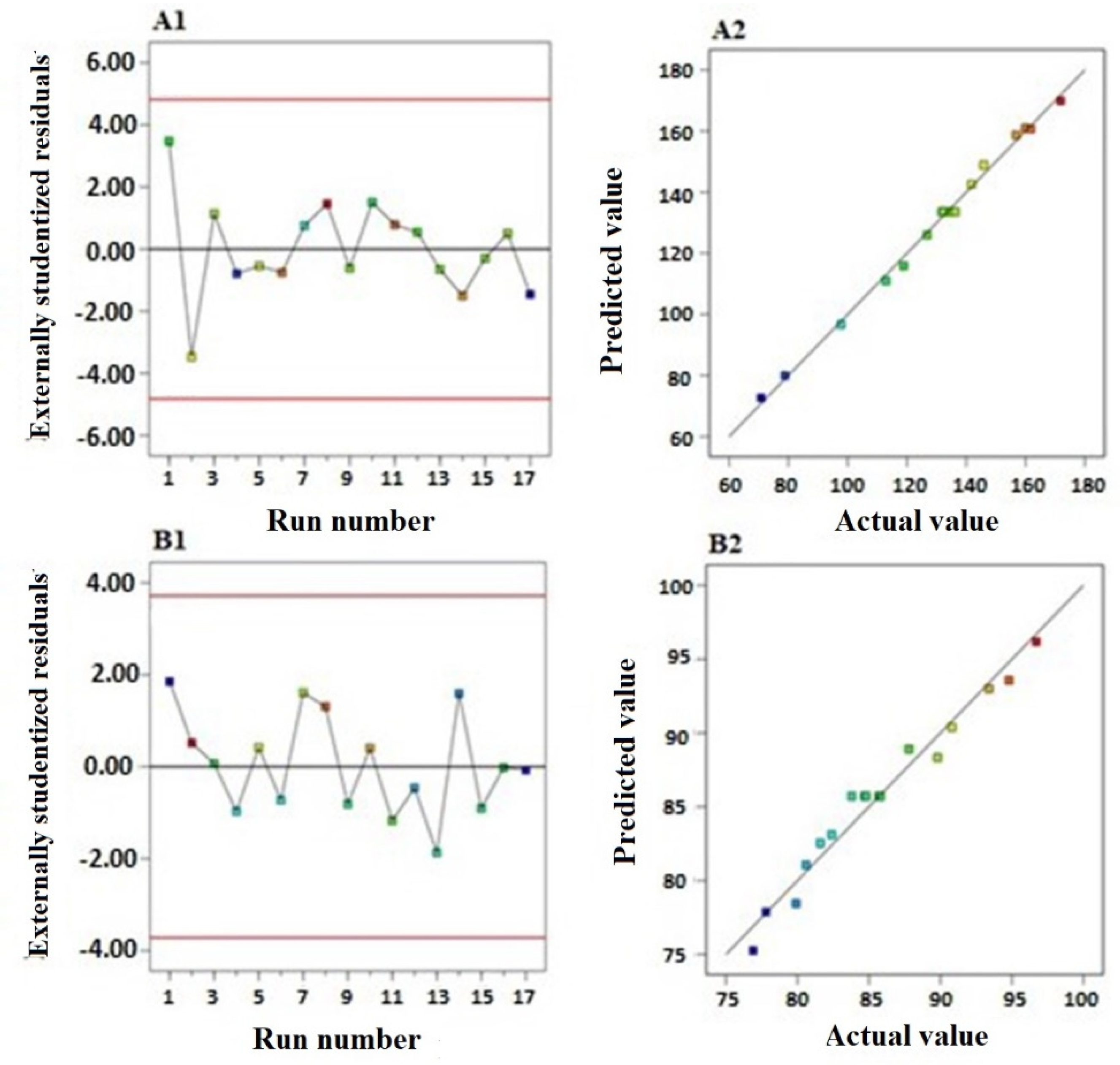
3.1.1. Sequential Model Selection and Validation

Data of vesicle size and drug entrapment were best fitted to the quadratic and linear models, respectively. Fitting to the selected models was based on the highest R2 and the lowest predicted residual error sum of squares (PRESS) (Table 3).
Moreover, the predicted and adjusted R2 were in reasonable agreement, indicating the validity of the model. Adequate signal to noise ratio is depicted by adequate precision greater than 4 for both responses; this ratio confirms the suitability of the selected models to navigate the experimental design space.
Diagnostic plots for both responses were produced to evaluate the goodness of fit of the selected models and confirm their validity (Figure 1). Figure 1A1,B1, illustrating the residual vs. run plots, showed randomly and uniformly scattered points, indicating that the response is not affected by any lurking variable.
Also, the predicted vs. actual values of both responses’ plots (Figure 1A2,B2) demonstrated a highly linear pattern revealing that the observed responses were in good correlation with the predicted ones.

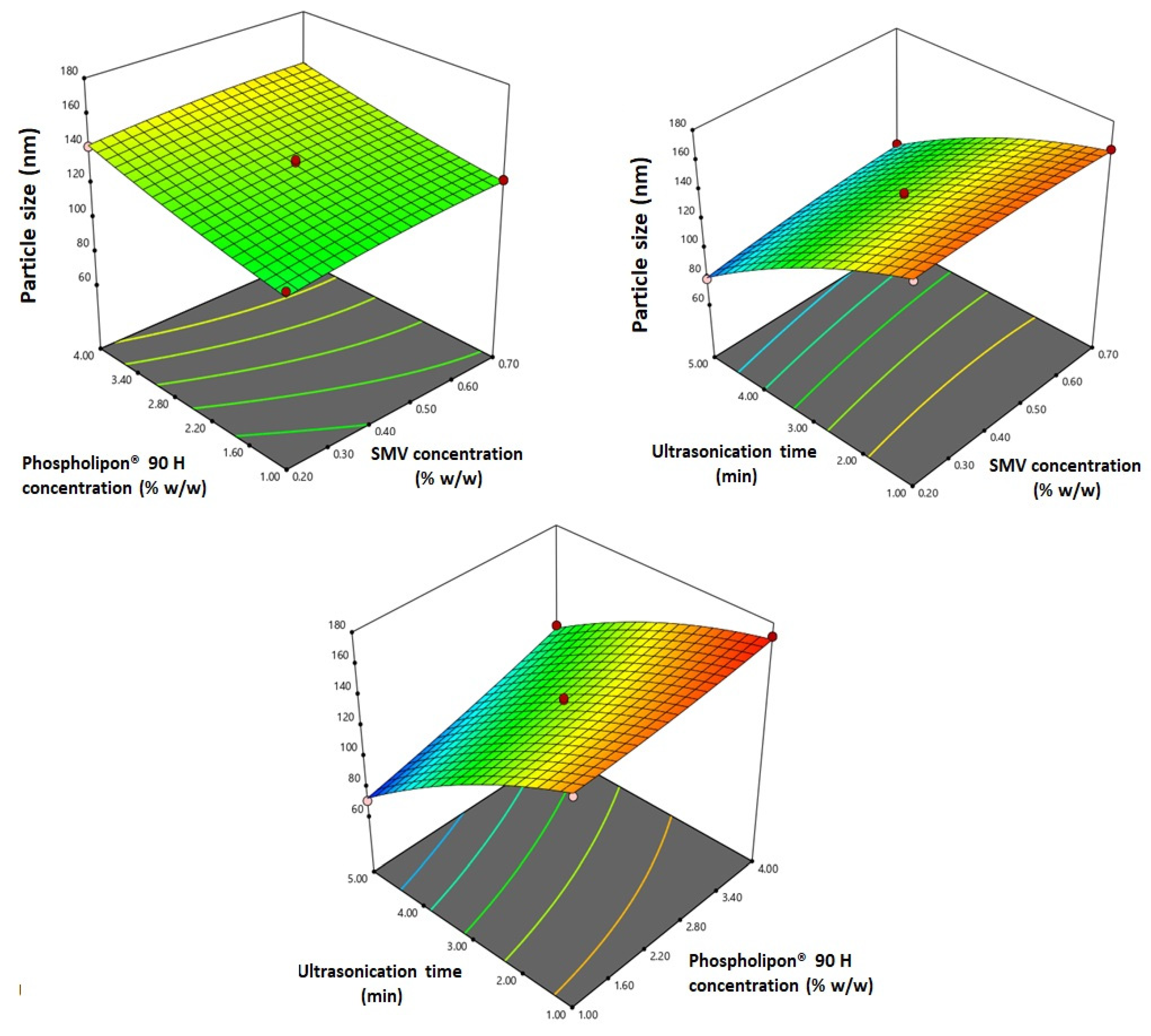
3.1.2. Statistical Analysis for the Effect of Variables on Vesicle Size (Y1)

SMV-EMLs exhibited nano-sized vesicles with an average size ranging from 71 ± 0.46 to 172 ± 1.45 (Table 2). The relatively low SD indicates homogeneity and uniformity of the EMLs dispersion. ANOVA for the vesicle size affirmed the significance of the quadratic model as evidenced by its F-value of 181.03 (p = 0.0054). Non-significant lack of fit, depicted by the lack of fit F-value of 3.34 (p = 0.1374), ensured data fitting to the selected model. Equation (2), representing the quadratic model, was generated in terms of coded factor:
Y1 = 133.58 + 4.10 X1 + 12.39 X2 − 36.25 X3 − 0.9575 X1X2 + 4.25 X1X3 + 6.68 X2X3 − 1.87 X12 + 1.63 X22 − 7.13 X32
ANOVA results revealed that all the linear terms (X1, X2, and X3), corresponding to the investigated variables, have a significant effect on vesicle size at a 95% level of significance. The quadratic term corresponding to the ultrasonication time (X32) in addition to the interaction terms X1X3 and X2X3 corresponding to the interaction between ultrasonication time and either SMV concentration or Phospholipon® 90 H concentration were also found to be significant at the same level. Figure 2 illustrates the response surface plots for the effects of the investigated variables on vesicle size.
It was evident that the vesicle size significantly increases with increasing both SMV and Phospholipon® 90 H concentrations (p = 0.0004 and p < 0.0001, respectively). The observed trend is confirmed by the positive coefficients of both X1 and X2 in the coded equation. The effect of phospholipid concentration was more pronounced than that of the drug, as evidenced by the higher coefficient of its corresponding term. On the other hand, the vesicle size significantly decreased with increasing ultrasonication time (p < 0.0001). This reduction could be ascribed to the main principle underlying the sonication process.

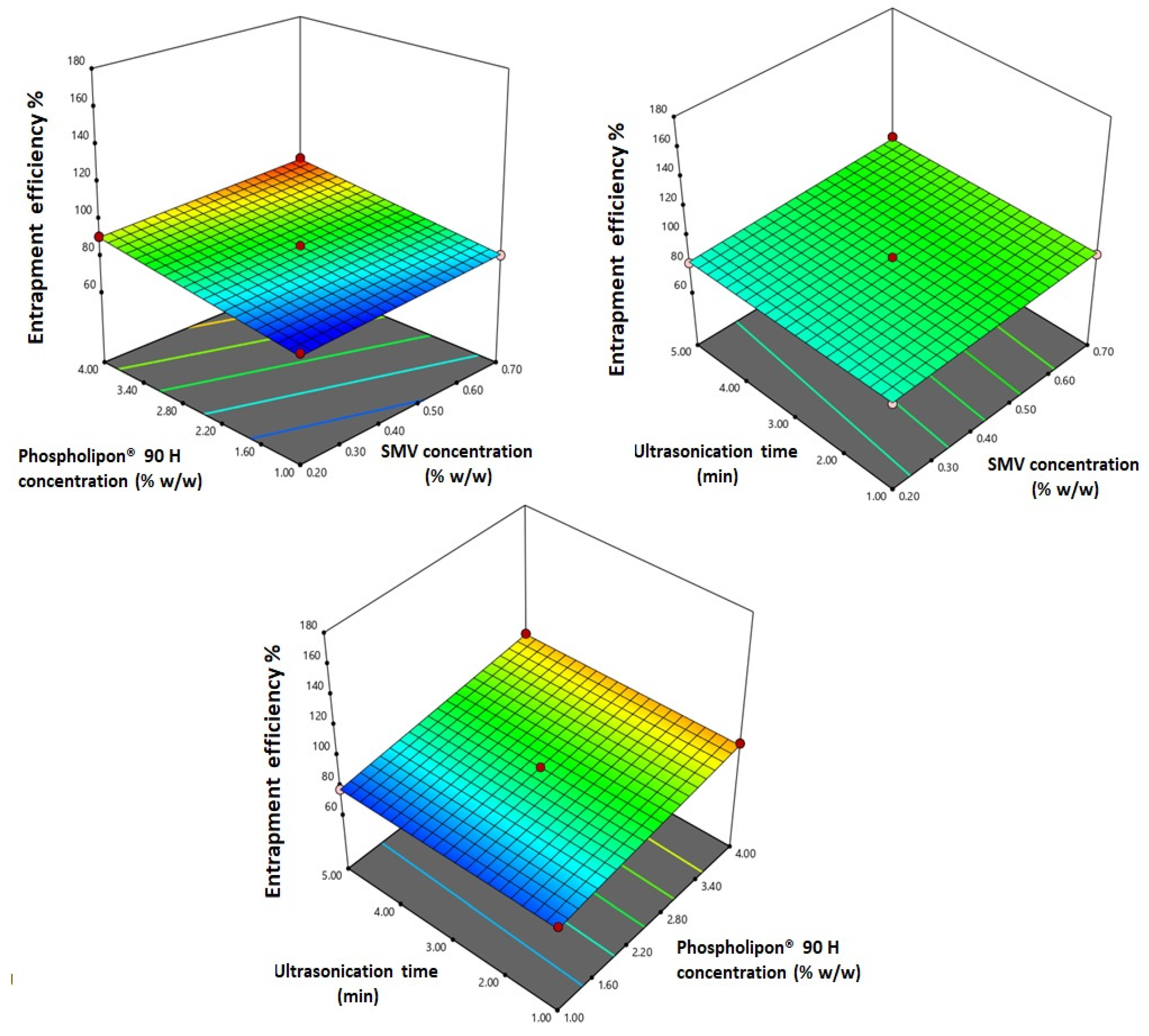
3.1.3. Statistical Analysis for the Effect of Variables on Entrapment Efficiency (Y2)

The prepared SMV-EMLs showed marked variation in drug entrapment with entrapment efficiency ranging from 76.9 ± 0.65 to 96.7 ± 0.83. ANOVA for the entrapment efficiency confirmed fitting of the data to the linear model, as evidenced by its F-value of 130.28 (p < 0.0001). Non-significant lack of fit, depicted by the lack of fit F-value of 2.44 (p = 0.2024), ensured data fitting to the selected model. Equation (3), representing the linear model, was generated in terms of coded factor:
Y1 = 68.33 + 11.60 X1 + 5.04 X2 − 0.14 X3
ANOVA results revealed that both terms X1 and X2 corresponding to both SMV and Phospholipon® 90 H concentrations displayed a significant positive impact on the entrapment efficiency as proved by their positive coefficients (p < 0.0001). Figure 3 illustrates the response surface plots for the effects of the investigated variables on entrapment efficiency.

3.2. Optimization of SMV-EMLs

The vesicle size and entrapment efficiency constraints were used for the prediction of the optimized levels of the variables utilizing a numerical optimization approach. The desirability function was computed as 0.732. The optimized formulation was prepared and assessed. The percentage error between the predicted and observed responses was relatively low, indicating that the optimization technique used was valid. The PDI of the optimized formulation was found to be 0.221 ± 0.012, while the zeta potential was equal to 10.17 ± 1.23 mV. The stability test revealed a statistically non-significant difference in the entrapment efficiency %, zeta potential, or particle size. All formulations revealed good dispersion stability. The optimized formulation was subjected to further investigations. The variables levels predicted, and observed responses for the optimized formulation are depicted in Table 4.

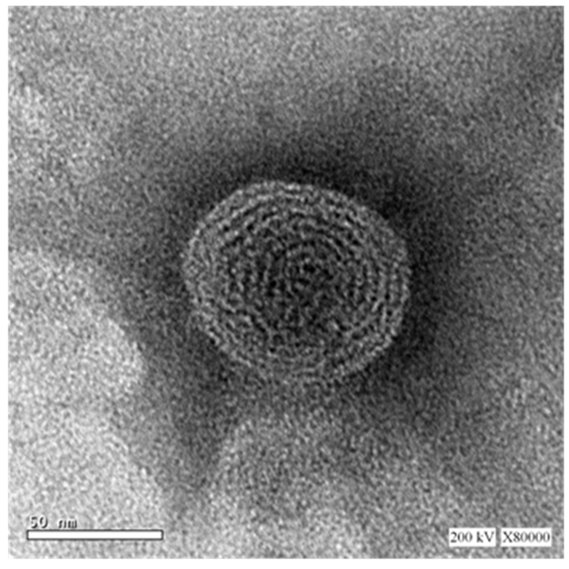
3.3. Microscopic Examination of the Optimized SMV-EMLs

Representative TEM micrograph of the optimized SMV-EMLs is shown in Figure 4. The microscopic examination of the optimized formulation showed the almost spherical shape and the presence of multiple bilayers (multilamellar vesicle).

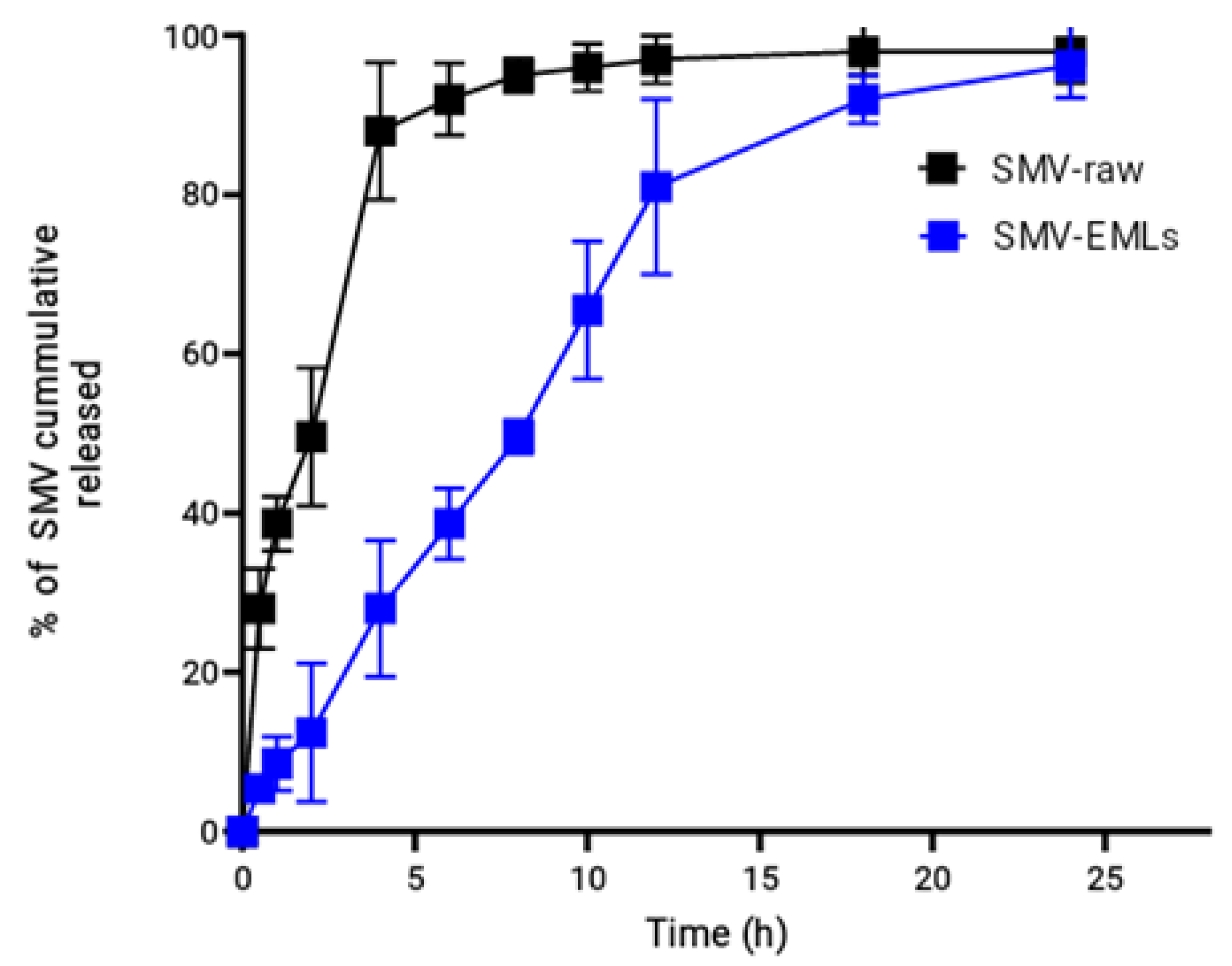
3.4. In Vitro Release of the Optimized SMV-EMLs

The in vitro dissolution of SMV from the optimized EMLs was studied. The drug release profile is shown in Figure 5.
A controlled and gradual release profile was observed for the prepared SMV-EMLs. Around 80% of drug release was achieved within 12 h. SMV released from EMLs revealed slower release when compared to SMV-raw. The drug release was almost complete by 24 h. The cumulative amount released could be regarded as satisfactory, considering the poor solubility of SMV.

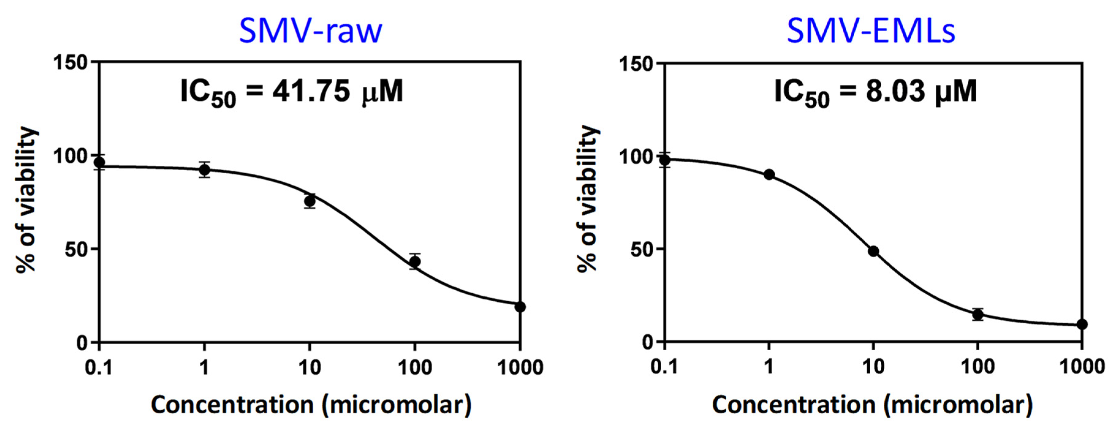
3.5. Optimized SMV-EMLs Treatment Shows the Lowest IC50 Value

In order to evaluate the pharmacological activity and the toxic potential, expressed as IC50, of the different treatments (Placebo-EMLs, SMV-raw, and SMV-EMLs) on MCF-7 cells, we carried out the MTT assay. The lowest IC50 value, thus the highest toxic potential, was observed in cells treated for 24 h with SMV-EMLs (IC50 = 8.03 μM; p < 0.05 vs. all) (Figure 6).
SMV-raw exhibited a weaker activity compared to SMV-EMLs, with an IC50 value of about 5-fold higher (IC50 = 41.75 μM) than that observed for the drug encapsulated into emulsomes. As expected, the highest IC50 was observed for the treatment with Placebo-EMLs, then in the absence of drug (IC50 = 78.29 μM). From this set of data, we were able to select the SMV-raw concentration (0.1 µM) that produced a negligible decrease in cell viability in MCF-7 cells and compare it with the same concentration of optimized SMV-EMLs, which exhibits nano-size and enhanced drug entrapment with gradual and complete in vitro release, inquiring into a possible enhancement for the antineoplastic effects of the drug [36]. The selection of this concentration was also based on a preliminary experiment and proved to be optimum under our experimental conditions and complied with the sensitivity of the analytical methods used.

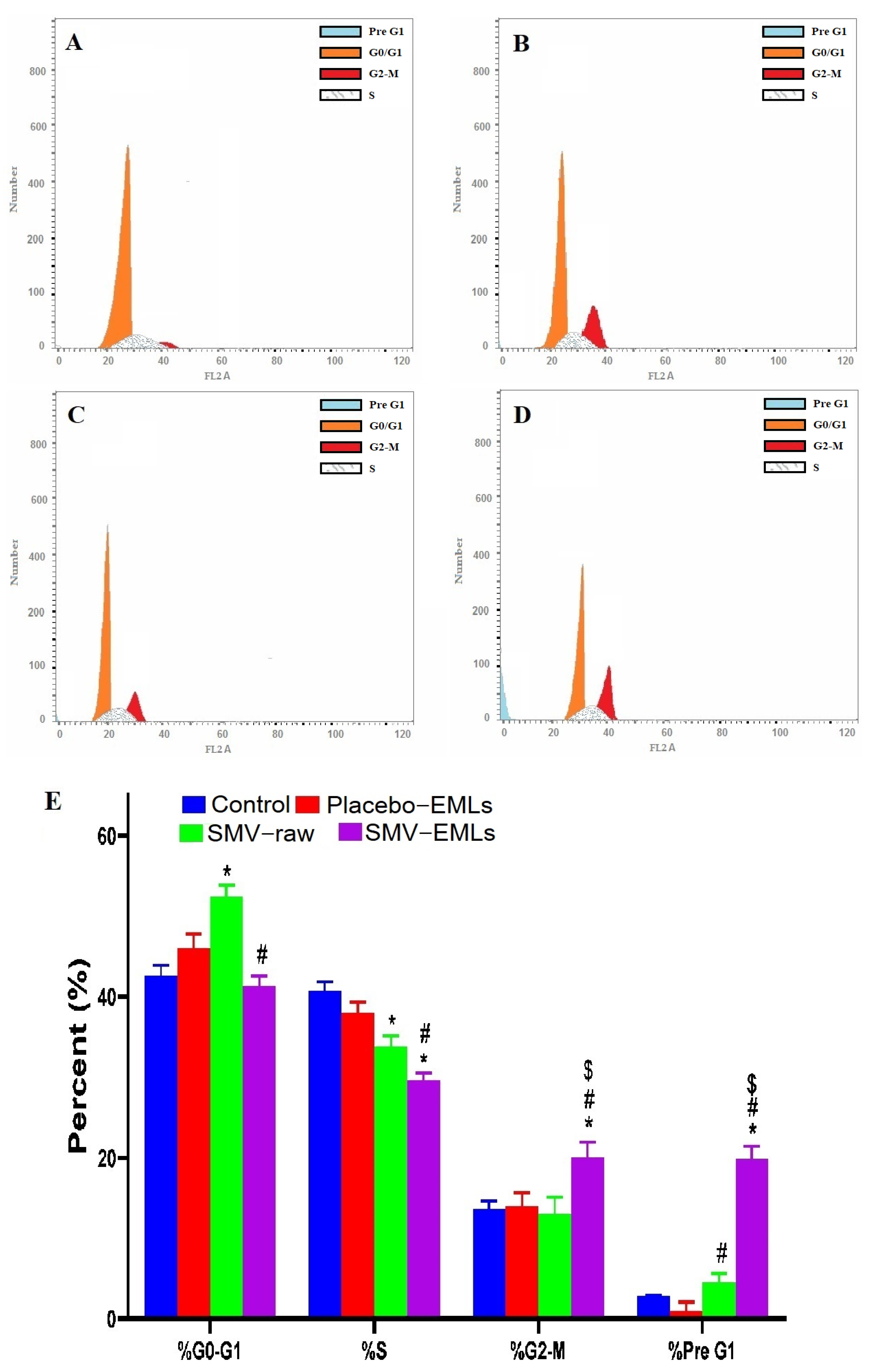
3.6. Optimized SMV-EMLs Treatment Significantly Inhibits MCF-7 Cell Proliferation

Figure 7 depicts the impact of Placebo-EMLs, SMV-raw, or optimized SMV-EMLs treatments on MCF-7 cell cycle phases.
In the case of untreated cells, quick proliferative properties were observed, with percent values for G0/G1, S, G2-M, and pre-G1 phases equal to 42.49 ± 2.3%, 40.99 ± 2.1%, 16.52 ± 2.12% and 3.34 ± 0.3% respectively (Figure 7A,E). No significant difference for each phase of the cell cycle was found concerning the treatment with Placebo-EMLs compared to control cells (Figure 7B,E). SMV-raw treatment led to significant cell cycle changes compared to control cells (p < 0.05) only in the case of S and pre-G1 phases with a significant reduction of the percent of cells in the S phase (Figure 7C,E). Interestingly, the treatment of MCF-7 with optimized SMV-EMLs was able to significantly inhibit cell proliferation with a greater extent compared to all the other treatment conditions (SMV-raw included) (p < 0.05), with a significant and biologically-relevant accumulation of cells in the G2/M and pre-G1 phases combined with a significant reduction of cells in the S phase (Figure 7D,E).

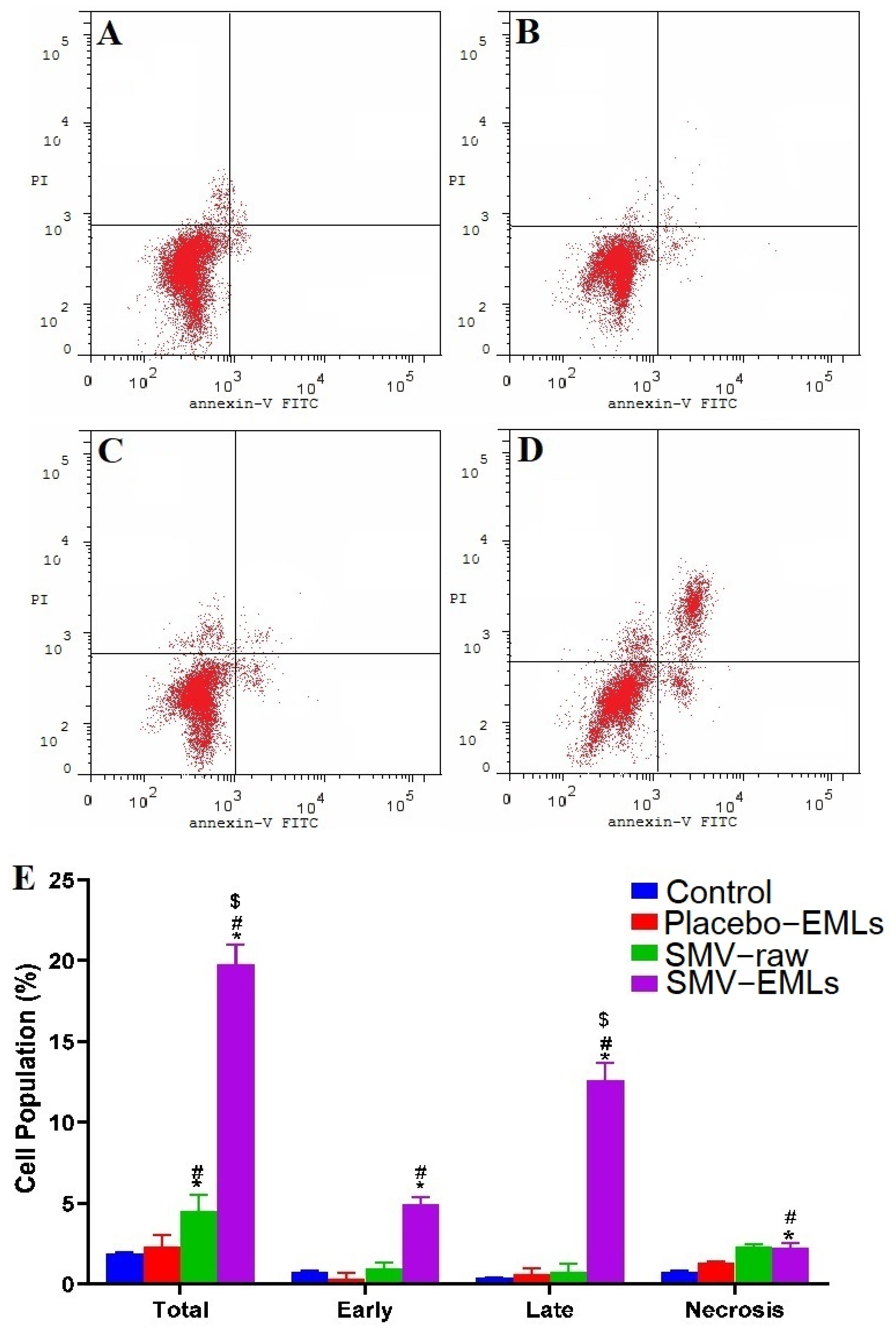
3.7. Optimized SMV-EMLs Treatment Increases Apoptotic and Necrotic Cell Populations

In order to investigate whether the observed antiproliferative activity of the optimized SMV-EMLs was also related to cell death phenomena, the impact of the different treatments on the rate of apoptosis/necrosis in MCF-7 cells was assessed. Figure 8 shows the effects of the different treatments on MCF-7 cell status.
It is clear that Placebo-EMLs and SMV-raw treatments did not lead to any significant changes in the apoptotic stages and necrosis compared to untreated cells (Figure 8A–C,E). The only significant difference between untreated and SMV-raw-treated MCF-7 cells (p < 0.05) was observed when considering the sum of apoptotic + necrotic cell populations. As expected, the treatment with optimized SMV-EMLs led to a significant increase in the percentage of cells in apoptosis + necrosis (total), early and late stages of apoptosis, and necrosis compared to untreated cells as well as to Placebo-EMLs and SMV-raw-treated MCF-7 cells (p < 0.05), underlining the highly relevant pro-apoptotic activity of SMV-EMLs.

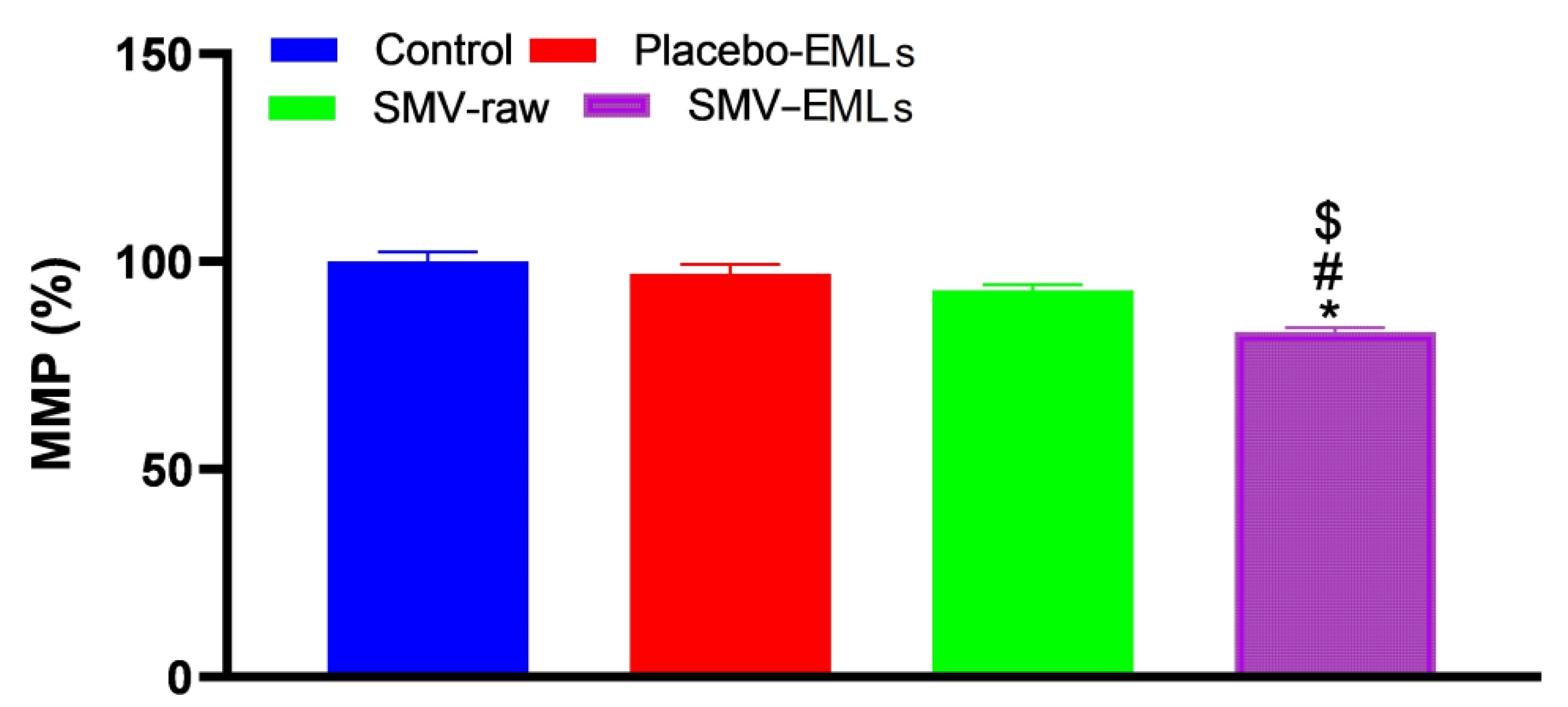
3.8. MMP Was Decreased Only in the Presence of SMV-EMLs

No significant difference in MMP was observed when considering untreated, Placebo-EMLs, and SMV-raw-treated MCF-7 cells (Figure 9).
The only treatment able to give a significant (p < 0.05) reduction of MMP compared to all the other treatments is represented by optimized SMV-EMLs.

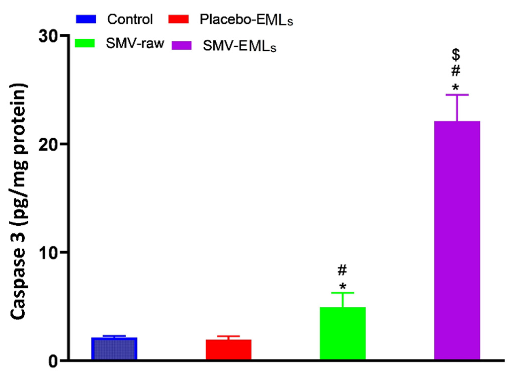
3.9. Caspase-3 Activity Is Enhanced by SMV-Raw and Optimized SMV-EMLs Treatments

In line with the results shown in Figure 10, the treatment of MCF-7 cells with Placebo-EMLs (1.95 ± 0.32 pg/mg protein) did not lead to any significant changes in the caspase-3 content compared to untreated cells (2.17 ± 0.12 pg/mg protein).
Exposure of the cells to SMV-raw gave a content of caspase-3 about 2.5-fold higher than that of control cells (5.23 ± 1.32 pg/mg protein) (p < 0.05), while the highest content of caspase-3 was detected in cells stimulated with optimized SMV-EMLs (22.5 ± 2.4 pg/mg protein), that was significantly higher compared to untreated cells as well as to Placebo-EMLs and SMV-raw treatments (p < 0.05 vs. all the other experimental conditions).

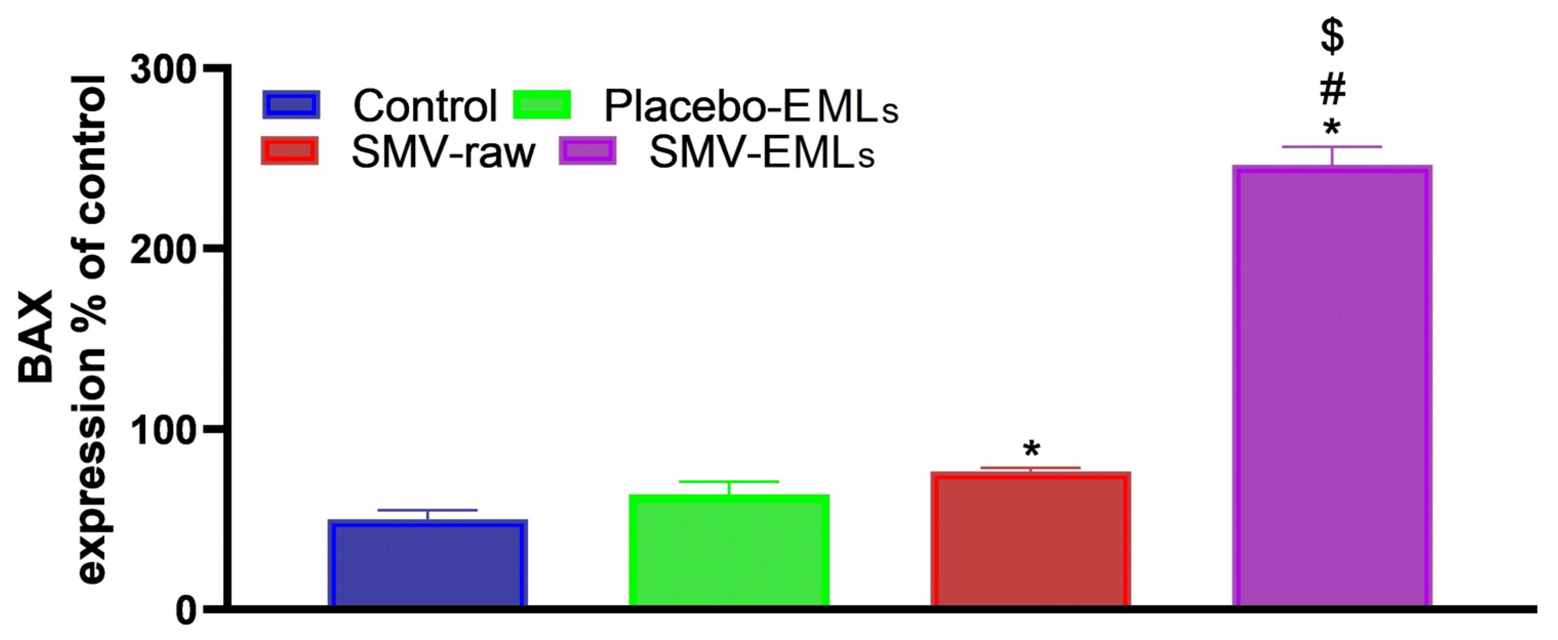
3.10. Bax Protein Levels Are Enhanced in SMV-Raw- and Optimized SMV-EMLs Treated Cells

Figure 11 shows the Bax protein levels in MCF-7 cells determined by the different treatments. As expected, a slight, even though significant, change in Bax protein content was observed for cells treated with SMV-raw compared to untreated cells (p < 0.05).
Once again, the treatment giving the more evident changes was represented by optimized SMV-EMLs, where an increase of about ~5-fold higher than that of control cells (p < 0.05) was observed.

4. Discussion

Statins, including SMV, are poorly soluble drugs and drug delivery approaches such as emulsomes [37] represent one of the most promising and efficient tools to enhance their solubility, improve the dissolution rate, reach a specific target more efficiently also allowing the control of drug release finally improving clinical efficacy [38]. Vesicle size of nanocarrier delivery systems is a crucial criterion that demonstrate a significant impact on the release pattern and consequently drugs’ penetration through the biological membranes [39,40]. SMV-EMLs exhibited nano-sized vesicles with an average size ranging from 71 ± 0.46 to 172 ± 1.45 (Table 2). TEM microscopy revealed a multilamellar vesicle of the optimized formula (Figure 4). SMV, as a hydrophobic drug, is entrapped within the bilayers (hydrophobic area) of the vesicles. According to the outcome of experimental design data, the increase of the size of the vesicles due to the increase in SMV concentrations could be associated with the observed enhancement of drug entrapment, while the increased vesicle size at higher phospholipid concentrations could be due to the increase in the number of formed multiple bilayers [23]. On the other hand, the vesicle size significantly decreased with increasing ultrasonication time (Figure 2; p < 0.0001). This reduction could be ascribed to the main principle underlying the sonication process, generating cavitation bubbles in the EMLs dispersion by the ultrasound mechanical waves. Bubbles having a size close to the resonant size for the employed frequency starts to oscillate non-linearly and collapse, resulting in bubble implosion that generates extremely high temperatures, high pressures, and shock waves. Thus, the ultrasonic generated high energy result in size reduction [41,42]. Accordingly, as the sonication time increases, the generated energy increases, leading to smaller vesicle size. The enhanced entrapment due to increasing SMV and phospholipid concentrations could be explained on the basis of the presence of an abundant phospholipid amount for the formation of lamellar structure, in addition to the hydrophobic nature of the SMV drug [43]. SMV, being a lipophilic drug, will have a higher chance of being entrapped at higher phospholipid concentrations due to its increased solubility in the available lipids. One of the most flexible and famous methods for the evaluation of drug release from nano-sized formulations is the dialysis bag method, in which the physical separation of the dosage forms is achieved through the use of a dialysis membrane that facilitates sampling at regular intervals [44,45]. The observed complete SMV release in our study could be explained by the solubilizing ability of EMLs towards SMV, by means of the lipophilic nature of its components. The EMLs capability to enhance the solubility of other poorly water-soluble drugs has been previously reported [46]. The zeta potential of the colloidal EML determines the dispersed particle surface charge. This particulate matter surface charge attracts the counterions, forming a layer around the surface of the particles. This double layer diffuses every time the nanoparticle travels within the solution. The zeta potential at the double-layer boundary is thus called the electrical potential. It means the stability of the particles formed in the continuous media, where the higher value of the zeta potential exhibits higher repulsive force between them, thus minimizing the possibility of size enlargement due to particles aggregation.
SMV is currently used and approved to lower cholesterol production and reduce dyslipidemia associated complications, because of its ability to reduce LDL by inhibiting the hydroxymethylglutaryl coenzyme A reductase, and then the hepatic synthesis of cholesterol [47]. This drug, belonging to the well-known statins family, has also proven to significantly increase the levels of reactive oxygen species (ROS) and malondialdehyde, to increase cell death, and to reduce proliferation in different cancer cell lines [48,49,50], according to the ability of statins to reduce the risk of multiple cancers, including breast cancer [7,51,52]. With specific regard to MCF-7 breast cancer cells, SMV has been shown able to induce dose-dependent cytotoxic effects along with a reduction of DNA synthesis and an induction of the cell cycle arrest in the G1 phase [30]. Additionally, SMV can exert a synergic effect in combination with other antineoplastic drugs, such as doxorubicin [28,31]. In particular, it has been shown that SMV decreases DNA synthesis and cell proliferation, induces intracellular ROS production, also increasing cytochrome c protein release and caspase-3 activity [28].
Based on the above evidence, we performed in vitro experiments in which the ability of EMLs to enhance the toxic potential of SMV towards MCF-7 cells, representing a well-known experimental model in cancer research [53], was tested through the use of an optimized SMV loaded EMLs formula (SMV-EMLs). The first evidence of a relevant improvement in the therapeutic potential of SMV-EMLs is clearly demonstrated in Figure 6, showing that the IC50 of SMV-EMLs (8.07 µM), a parameter often used to compare the antiproliferative activity and the toxic potential of different antineoplastic drugs [54], is significantly lower than that for SMV alone (SMV-raw) (41.75 µM). Despite the widespread use of statins to lower cholesterol levels [6,7] and reduce cardiovascular morbidity and mortality [55] as well as for cancer treatments [10,11,12,13,14,15], clinically-relevant adverse effects (such as muscle pain) have also been observed at therapeutic doses [55,56,57]. The development of new formulations able to maximize the clinical efficacy, then reducing the doses, of a statin drug is then of the utmost importance. When MCF-7 cells were treated with optimized SMV-EMLs, the enhanced inhibition of cell proliferation (Figure 7) and apoptotic and necrotic death (Figure 7, Figure 8, Figure 10, and Figure 11) were also observed. Our data suggest that SMV-EMLs exert their antiproliferative activity by reducing the transition from G1 to the S phase as well as inhibiting the transition from G2 to M phase also inducing apoptotic cell death. These toxic effects could depend on the ability of EMLs to increase the solubility and the intracellular availability of poorly water-soluble bioactive agents (e.g., SMV) [26,46], also ensuring sustained drug release over a prolonged period of time that can be validated in future in vivo studies [23,46]. Of note, the only treatment able to decrease the MMP, an index of cell suffering and death, in MCF-7 cells was that with optimized SMV-EMLs (Figure 9). This is relevant because the decrease in MMP has been associated with an increased release of apoptotic factors and an improved rate of cancer cell death [58] as well as with a minor resistance to conventional anticancer therapy [59]. Differently from the evident pro-apoptotic effects showed by SMV-EMLs, the slight but significant effect on decreasing the MMP was in part expected; in fact, it has been demonstrated that the hyperpolarisation of MMP can be greater (more than 50%) in cancer cells than in healthy cells [60,61], making them more resistant to MMP-induced changes. Furthermore, as demonstrated by the strong induction in caspase-3 and Bax (Figure 10 and Figure 11), among the different mechanisms regulating MCF-7 cell status, SMV could exert its effects especially by promoting apoptotic phenomena rather than regulating MMP.
The present study clearly shows the enhanced cytotoxic activity exerted by the optimized SMV-EMLs formula on the well-characterized MCF-7 cancer cell model [21,27,28,29,30,31,53] compared with SMV alone. Despite that, future studies, employing the same experimental conditions, will be carried out in other validated breast cancer cells (e.g., MDA-MB-231) to further validate the relevance of our findings. Furthermore, in order to improve the above-mentioned activities of our formulation, alternative and innovative methods to determine the kinetic release of the content from the EMLs, such as USP 4 [62], will be investigated. Our results also make the base for the investigation of our optimized formula in 3D/organoid systems [63] as well as in vivo.

5. Conclusions

In the present study, a Box–Behnken design was successfully applied for the formulation and optimization of SMV-EMLs. The prepared EMLs exhibited nano-size and enhanced drug entrapment exceeding 80%. The optimized EMLs formulation with minimized vesicle size and maximized drug entrapment exhibited spherical shape with gradual and complete in vitro release. The in vitro drug release indicated the ability of the EMLs to enhance the dissolution of the poorly soluble drug SMV. By performing in vitro experiments with MCF-7 cancer cells, it was possible to show that drug loading of EMLs (SMV-EMLs) significantly improved the anti-proliferative activity and enhanced the toxic and pro-apoptotic effects of SMV towards cancer cells as demonstrated by the lower IC50. Additionally, SMV-EMLs containing a sub-toxic concentration of drug led to the inhibition of cell proliferation, the increase of apoptotic and necrotic cell populations, the decrease of the mitochondrial membrane potential, the increase of the intracellular concentration of caspase-3, and the increased intracellular levels of Bax protein compared to the treatment with SMV in the absence of EMLs. Our data obtained by using an optimized SMV-EMLs formula suggest that SMV-EMLs might represent a novel path for the development of a specific and more effective treatment of breast cancer.

Author Contributions

Conceptualization, U.A.F., F.C., and G.C.; methodology, N.A.A., A.O.N., and D.M.A.; software, H.Z.A. and Z.A.A.; validation, A.A., S.M.B.-E., and N.A.A.; formal analysis, Z.A.A., N.H. and H.A.S.; investigation, M.W.A.-R.; resources, W.H.A.; data curation, Z.A.A., U.A.F., T.S.I., M.W.A.-R., F.C., and G.C.; writing—original draft preparation, U.A.F., S.M.B.-E., F.C., and G.C.; writing—review and editing, U.A.F., F.C., and G.C.; visualization, T.S.I. and H.Z.A.; supervision, N.A.A.; project administration, H.A.S.; funding acquisition, N.A.A. All authors have read and agreed to the published version of the manuscript.

Funding

This project was funded by the Deanship of Scientific Research (DSR) at King Abdulaziz University, Jeddah, under grant no. (RG-5–166–38). The authors, therefore, acknowledge with thanks DSR for technical and financial support.

Conflicts of Interest

The authors declare no conflict of interest.

References

  1. Saslow, D.; Hannan, J.; Osuch, J.; Alciati, M.H.; Baines, C.; Barton, M.; Bobo, J.K.; Coleman, C.; Dolan, M.; Gaumer, G.; et al. Clinical breast examination: Practical recommendations for optimizing performance and reporting. CA Cancer J. Clin. 2004, 54, 327–344. [Google Scholar] [CrossRef] [PubMed]
  2. Gajjar, A.; Reaman, G.H.; Racadio, J.M.; Smith, F.O. Brain Tumors in Children; Springer: Berlin/Heidelberg, Germany, 2018. [Google Scholar]
  3. Waks, A.G.; Winer, E.P. Breast cancer treatment: A review. JAMA 2019, 321, 288–300. [Google Scholar] [CrossRef] [PubMed]
  4. Ohri, N.; Rapkin, B.D.; Guha, C.; Kalnicki, S.; Garg, M. Radiation therapy noncompliance and clinical outcomes in an urban academic cancer center. Int. J. Radiat. Oncol. Biol. Phys. 2016, 95, 563–570. [Google Scholar] [CrossRef] [PubMed]
  5. Hershman, D.L.; Kushi, L.H.; Shao, T.; Buono, D.; Kershenbaum, A.; Tsai, W.Y.; Fehrenbacher, L.; Gomez, S.L.; Miles, S.; Neugut, A.I. Early discontinuation and nonadherence to adjuvant hormonal therapy in a cohort of 8,769 early-stage breast cancer patients. J. Clin. Oncol. 2010, 28, 4120–4128. [Google Scholar] [CrossRef] [PubMed]
  6. Zhou, Q.; Liao, J.K. Statins and cardiovascular diseases: From cholesterol lowering to pleiotropy. Curr. Pharm. Des. 2009, 15, 467–478. [Google Scholar] [CrossRef]
  7. Islam, M.M.; Yang, H.C.; Nguyen, P.A.; Poly, T.N.; Huang, C.W.; Kekade, S.; Khalfan, A.M.; Debnath, T.; Li, Y.J.; Abdul, S.S. Exploring association between statin use and breast cancer risk: An updated meta-analysis. Arch. Gynecol. Obstet. 2017, 296, 1043–1053. [Google Scholar] [CrossRef]
  8. World Health Organization. World Health Organization Model List of Essential Medicines: 21st List 2019; World Health Organization: Geneva, Switzerland, 2019. [Google Scholar]
  9. Cardwell, C.R.; Hicks, B.M.; Hughes, C.; Murray, L.J. Statin use after diagnosis of breast cancer and survival: A population-based cohort study. Epidemiology 2015, 26, 68–78. [Google Scholar] [CrossRef]
  10. Stancu, C.; Sima, A. Statins: Mechanism of action and effects. J. Cell Mol. Med. 2001, 5, 378–387. [Google Scholar] [CrossRef]
  11. Koyuturk, M.; Ersoz, M.; Altiok, N. Simvastatin induces apoptosis in human breast cancer cells: P53 and estrogen receptor independent pathway requiring signalling through jnk. Cancer Lett. 2007, 250, 220–228. [Google Scholar] [CrossRef]
  12. Van Wyhe, R.D.; Rahal, O.M.; Woodward, W.A. Effect of statins on breast cancer recurrence and mortality: A review. Breast Cancer (Dove Med. Press) 2017, 9, 559–565. [Google Scholar] [CrossRef]
  13. Kwan, M.L.; Habel, L.A.; Flick, E.D.; Quesenberry, C.P.; Caan, B. Post-diagnosis statin use and breast cancer recurrence in a prospective cohort study of early stage breast cancer survivors. Breast Cancer Res. Treat 2008, 109, 573–579. [Google Scholar] [CrossRef] [PubMed]
  14. Ahern, T.P.; Pedersen, L.; Tarp, M.; Cronin-Fenton, D.P.; Garne, J.P.; Silliman, R.A.; Sorensen, H.T.; Lash, T.L. Statin prescriptions and breast cancer recurrence risk: A danish nationwide prospective cohort study. J. Natl. Cancer Inst. 2011, 103, 1461–1468. [Google Scholar] [CrossRef] [PubMed]
  15. Manthravadi, S.; Shrestha, A.; Madhusudhana, S. Impact of statin use on cancer recurrence and mortality in breast cancer: A systematic review and meta-analysis. Int. J. Cancer 2016, 139, 1281–1288. [Google Scholar] [CrossRef] [PubMed]
  16. Fong, C. Statins in Therapy: Cellular Transport, Side Effects, Drug-Drug Interactions and Cytotoxicity—The Unrecognized Role of Lactones. 2016. Available online: https://hal.archives-ouvertes.fr/hal-01185910/ (accessed on 14 June 2020).
  17. Dulak, J.; Józkowicz, A. Anti-angiogenic and anti-inflammatory effects of statins: Relevance to anti-cancer therapy. Curr. Cancer Drug Targets 2005, 5, 579–594. [Google Scholar] [CrossRef] [PubMed]
  18. McKenney, J.M.; Ganz, P.; Wiggins, B.S.; Saseen, J.S. Statins. In Clinical Lipidology; Elsevier: Amsterdam, The Netherlands, 2009; pp. 253–280. [Google Scholar]
  19. Akbarzadeh, A.; Rezaei-Sadabady, R.; Davaran, S.; Joo, S.W.; Zarghami, N.; Hanifehpour, Y.; Samiei, M.; Kouhi, M.; Nejati-Koshki, K. Liposome: Classification, preparation, and applications. Nanoscale Res. Lett. 2013, 8, 102. [Google Scholar] [CrossRef]
  20. Alavi, M.; Karimi, N.; Safaei, M. Application of various types of liposomes in drug delivery systems. Adv. Pharm. Bull. 2017, 7, 3–9. [Google Scholar] [CrossRef]
  21. Ag Seleci, D.; Seleci, M.; Walter, J.-G.; Stahl, F.; Scheper, T. Niosomes as nanoparticular drug carriers: Fundamentals and recent applications. J. Nanomater. 2016, 2016. [Google Scholar] [CrossRef]
  22. Gill, B.; Singh, J.; Sharma, V.; Kumar, S.H. Emulsomes: An emerging vesicular drug delivery system. Asian J. Pharm. (AJP) Free Full Text Articles Asian J. Pharm. 2014, 6, 133–142. [Google Scholar] [CrossRef]
  23. Vyas, S.P.; Subhedar, R.; Jain, S. Development and characterization of emulsomes for sustained and targeted delivery of an antiviral agent to liver. J. Pharm. Pharmacol. 2006, 58, 321–326. [Google Scholar] [CrossRef]
  24. Schwarz, C.; Mehnert, W.; Lucks, J.; Müller, R. Solid lipid nanoparticles (sln) for controlled drug delivery. I. Production, characterization and sterilization. J. Control. Release 1994, 30, 83–96. [Google Scholar] [CrossRef]
  25. Paliwal, R.; Paliwal, S.R.; Mishra, N.; Mehta, A.; Vyas, S.P. Engineered chylomicron mimicking carrier emulsome for lymph targeted oral delivery of methotrexate. Int. J. Pharm. 2009, 380, 181–188. [Google Scholar] [CrossRef] [PubMed]
  26. Ucisik, M.H.; Sleytr, U.B.; Schuster, B. Emulsomes meet s-layer proteins: An emerging targeted drug delivery system. Curr. Pharm. Biotechnol. 2015, 16, 392–405. [Google Scholar] [CrossRef] [PubMed]
  27. Sánchez, C.A.; Rodríguez, E.; Varela, E.; Zapata, E.; Páez, A.; Massó, F.A.; Montaño, L.F.; Lóopez-Marure, R. Statin-induced inhibition of mcf-7 breast cancer cell proliferation is related to cell cycle arrest and apoptotic and necrotic cell death mediated by an enhanced oxidative stress. Cancer Investig. 2008, 26, 698–707. [Google Scholar] [CrossRef] [PubMed]
  28. Buranrat, B.; Suwannaloet, W.; Naowaboot, J. Simvastatin potentiates doxorubicin activity against mcf-7 breast cancer cells. Oncol. Lett. 2017, 14, 6243–6250. [Google Scholar] [CrossRef] [PubMed]
  29. Ibrahim, A.B.; Zaki, H.F.; Wadie, W.; Omran, M.M.; Shouman, S.A. Simvastatin evokes an unpredicted antagonism for tamoxifen in mcf-7 breast cancer cells. Cancer Manag. Res. 2019, 11, 10011–10028. [Google Scholar] [CrossRef]
  30. Afzali, M.; Vatankhah, M.; Ostad, S.N. Investigation of simvastatin-induced apoptosis and cell cycle arrest in cancer stem cells of mcf-7. J. Cancer Res. Ther. 2016, 12, 725–730. [Google Scholar]
  31. Friday, E.; Welbourne, T.; Olliver, R.; Turturro, F. Simvastatin combined with troglitazone decreases proliferation by increasing cellular acidosis in mcf-7 breast cancer cell line. Cancer Res. 2006, 66, 1098. [Google Scholar]
  32. Zhou, X.; Chen, Z. Preparation and performance evaluation of emulsomes as a drug delivery system for silybin. Arch. Pharm. Res. 2015, 38, 2193–2200. [Google Scholar] [CrossRef]
  33. Fahmy, U.A. Quantification of simvastatin in mice plasma by near-infrared and chemometric analysis of spectral data. Drug Des. Dev. Ther. 2016, 10, 2507–2513. [Google Scholar] [CrossRef]
  34. Caruso, G.; Distefano, D.A.; Parlascino, P.; Fresta, C.G.; Lazzarino, G.; Lunte, S.M.; Nicoletti, V.G. Receptor-mediated toxicity of human amylin fragment aggregated by short- and long-term incubations with copper ions. Mol. Cell Biochem. 2017, 425, 85–93. [Google Scholar] [CrossRef]
  35. Alhakamy, N.A.; Ahmed, O.A.A.; Aldawsari, H.M.; Alfaifi, M.Y.; Eid, B.G.; Abdel-Naim, A.B.; Fahmy, U.A. Encapsulation of lovastatin in zein nanoparticles exhibits enhanced apoptotic activity in hepg2 cells. Int. J. Mol. Sci. 2019, 20, 5788. [Google Scholar] [CrossRef] [PubMed]
  36. Alhakamy, N.A.; Fahmy, U.A.; Ahmed, O.A.A.; Caruso, G.; Caraci, F.; Asfour, H.Z.; Bakhrebah, M.A.; Alomary, M.N.; Abdulaal, W.H.; Okbazghi, S.Z.; et al. Chitosan coated microparticles enhance simvastatin colon targeting and pro-apoptotic activity. Mar. Drugs 2020, 18, 226. [Google Scholar] [CrossRef] [PubMed]
  37. Kumar, R.; Seth, N. Emulsomes: An emerging vesicular drug delivery system. J. Drug Deliv. Ther. 2013, 3, 133–142. [Google Scholar] [CrossRef]
  38. Tiwari, R.; Pathak, K. Statins therapy: A review on conventional and novel formulation approaches. J. Pharm. Pharmacol. 2011, 63, 983–998. [Google Scholar] [CrossRef]
  39. Persson, D.; Thoren, P.E.; Esbjorner, E.K.; Goksor, M.; Lincoln, P.; Norden, B. Vesicle size-dependent translocation of penetratin analogs across lipid membranes. Biochim. Biophys. Acta 2004, 1665, 142–155. [Google Scholar] [CrossRef] [PubMed]
  40. Lin, H.; Xie, Q.; Huang, X.; Ban, J.; Wang, B.; Wei, X.; Chen, Y.; Lu, Z. Increased skin permeation efficiency of imperatorin via charged ultradeformable lipid vesicles for transdermal delivery. Int. J. Nanomed. 2018, 13, 831–842. [Google Scholar] [CrossRef]
  41. Harbi, I.; Aljaeid, B.; El-Say, K.M.; Zidan, A.S. Glycosylated sertraline-loaded liposomes for brain targeting: Qbd study of formulation variabilities and brain transport. AAPS PharmSciTech 2016, 17, 1404–1420. [Google Scholar] [CrossRef]
  42. Essa, E.A. Effect of formulation and processing variables on the particle size of sorbitan monopalmitate niosomes. Asian J. Pharm. (AJP) Free Full Text Artic. Asian J. Pharm. 2014, 4. [Google Scholar] [CrossRef]
  43. Ahmed, T.A.; Badr-Eldin, S.M.; Ahmed, O.A.; Aldawsari, H. Intranasal optimized solid lipid nanoparticles loaded in situ gel for enhancing trans-mucosal delivery of simvastatin. J. Drug Deliv. Sci. Technol. 2018, 48, 499–508. [Google Scholar] [CrossRef]
  44. Yadav, S.; Gupta, S. Development and in vitro characterization of docetaxel-loaded ligand appended solid fat nanoemulsions for potential use in breast cancer therapy. Artif. Cells Nanomed. Biotechnol. 2015, 43, 93–102. [Google Scholar] [CrossRef]
  45. Janas, C.; Mast, M.P.; Kirsamer, L.; Angioni, C.; Gao, F.; Mäntele, W.; Dressman, J.; Wacker, M.G. The dispersion releaser technology is an effective method for testing drug release from nanosized drug carriers. Eur. J. Pharm. Biopharm. 2017, 115, 73–83. [Google Scholar] [CrossRef] [PubMed]
  46. Ucisik, M.H.; Kupcu, S.; Schuster, B.; Sleytr, U.B. Characterization of curcuemulsomes: Nanoformulation for enhanced solubility and delivery of curcumin. J. Nanobiotechnol. 2013, 11, 37. [Google Scholar] [CrossRef] [PubMed]
  47. Mauro, V.F.; MacDonald, J.L. Simvastatin: A review of its pharmacology and clinical use. Ann. Pharmacother. 1991, 25, 257–264. [Google Scholar] [CrossRef] [PubMed]
  48. Ahn, K.S.; Sethi, G.; Aggarwal, B.B. Simvastatin potentiates tnf-alpha-induced apoptosis through the down-regulation of nf-kappab-dependent antiapoptotic gene products: Role of ikappabalpha kinase and tgf-beta-activated kinase-1. J. Immunol. 2007, 178, 2507–2516. [Google Scholar] [CrossRef] [PubMed]
  49. Li, Y.; Fu, J.; Yuan, X.; Hu, C. Simvastatin inhibits the proliferation of a549 lung cancer cells through oxidative stress and up-regulation of sod2. Pharmazie 2014, 69, 610–614. [Google Scholar]
  50. Hwang, K.E.; Kim, Y.S.; Hwang, Y.R.; Kwon, S.J.; Park, D.S.; Cha, B.K.; Kim, B.R.; Yoon, K.H.; Jeong, E.T.; Kim, H.R. Enhanced apoptosis by pemetrexed and simvastatin in malignant mesothelioma and lung cancer cells by reactive oxygen species-dependent mitochondrial dysfunction and bim induction. Int. J. Oncol. 2014, 45, 1769–1777. [Google Scholar] [CrossRef]
  51. Wu, Q.J.; Tu, C.; Li, Y.Y.; Zhu, J.; Qian, K.Q.; Li, W.J.; Wu, L. Statin use and breast cancer survival and risk: A systematic review and meta-analysis. Oncotarget 2015, 6, 42988–43004. [Google Scholar] [CrossRef]
  52. Shai, A.; Rennert, H.S.; Lavie, O.; Ballan-Haj, M.; Bitterman, A.; Steiner, M.; Keren, S.; Rennert, G. Statins, aspirin and risk of venous thromboembolic events in breast cancer patients. J. Thromb. Thrombolysis 2014, 38, 32–38. [Google Scholar] [CrossRef]
  53. Comsa, S.; Cimpean, A.M.; Raica, M. The story of mcf-7 breast cancer cell line: 40 years of experience in research. Anticancer Res. 2015, 35, 3147–3154. [Google Scholar]
  54. Schaffrath, J.; Schmoll, H.J.; Voigt, W.; Muller, L.P.; Muller-Tidow, C.; Mueller, T. Efficacy of targeted drugs in germ cell cancer cell lines with differential cisplatin sensitivity. PLoS ONE 2017, 12, e0178930. [Google Scholar] [CrossRef]
  55. Ward, N.C.; Watts, G.F.; Eckel, R.H. Statin toxicity. Circ. Res. 2019, 124, 328–350. [Google Scholar] [CrossRef] [PubMed]
  56. Newman, C.B.; Preiss, D.; Tobert, J.A.; Jacobson, T.A.; Page, R.L.; Goldstein, L.B.; Chin, C.; Tannock, L.R.; Miller, M.; Raghuveer, G. Statin safety and associated adverse events: A scientific statement from the american heart association. Arterioscler. Thromb. Vasc. Biol. 2019, 39, e38–e81. [Google Scholar] [CrossRef] [PubMed]
  57. Mach, F.; Ray, K.K.; Wiklund, O.; Corsini, A.; Catapano, A.L.; Bruckert, E.; De Backer, G.; Hegele, R.A.; Hovingh, G.K.; Jacobson, T.A.; et al. Adverse effects of statin therapy: Perception vs. The evidence—Focus on glucose homeostasis, cognitive, renal and hepatic function, haemorrhagic stroke and cataract. Eur. Heart J. 2018, 39, 2526–2539. [Google Scholar] [CrossRef] [PubMed]
  58. Capozzi, A.; Mantuano, E.; Matarrese, P.; Saccomanni, G.; Manera, C.; Mattei, V.; Gambardella, L.; Malorni, W.; Sorice, M.; Misasi, R. A new 4-phenyl-1,8-naphthyridine derivative affects carcinoma cell proliferation by impairing cell cycle progression and inducing apoptosis. Anticancer Agents Med. Chem. 2012, 12, 653–662. [Google Scholar] [CrossRef]
  59. Song, I.S.; Kim, H.K.; Lee, S.R.; Jeong, S.H.; Kim, N.; Ko, K.S.; Rhee, B.D.; Han, J. Mitochondrial modulation decreases the bortezomib-resistance in multiple myeloma cells. Int. J. Cancer 2013, 133, 1357–1367. [Google Scholar] [CrossRef]
  60. Forrest, M.D. Why cancer cells have a more hyperpolarised mitochondrial membrane potential and emergent prospects for therapy. BioRxiv 2015, 025197. [Google Scholar] [CrossRef]
  61. Houston, M.A.; Augenlicht, L.H.; Heerdt, B.G. Stable differences in intrinsic mitochondrial membrane potential of tumor cell subpopulations reflect phenotypic heterogeneity. Int. J. Cell Biol. 2011, 2011, 978583. [Google Scholar] [CrossRef]
  62. Shen, J.; Burgess, D.J. In Vitro dissolution testing strategies for nanoparticulate drug delivery systems: Recent developments and challenges. Drug Deliv. Transl. Res. 2013, 3, 409–415. [Google Scholar] [CrossRef]
  63. Caruso, G.; Musso, N.; Grasso, M.; Costantino, A.; Lazzarino, G.; Tascedda, F.; Gulisano, M.; Lunte, S.M.; Caraci, F. Microfluidics as a Novel Tool for Biological and Toxicological Assays in Drug Discovery Processes: Focus on Microchip Electrophoresis. Micromachines 2020, 11, 593. [Google Scholar] [CrossRef]
Figure 1. The diagnostic plots for vesicle size: (A1) externally studentized residuals vs. run number plot and (A2) predicted vs. actual values plot. The diagnostic plots for the entrapment efficiency of SMV-EMLs: (B1) externally studentized residuals vs. run number plot and (B2) predicted vs. actual values plot.
Pharmaceutics 12 00597 g001
Figure 2. Response 3D-plots for the effect of SMV concentration (X1), Phospholipon® 90 H (X2), and ultrasonication time (X3) on the vesicle size of SMV-EMLs.
Pharmaceutics 12 00597 g002
Figure 3. Response 3D-plots for the effect of SMV concentration (X1), Phospholipon® 90 H (X2), and ultrasonication time (X3) on the entrapment efficiency of SMV-EMLs.
Pharmaceutics 12 00597 g003
Figure 4. TEM of optimized SMV-EMLs (80,000× magnification).
Pharmaceutics 12 00597 g004
Figure 5. In vitro release profile of optimized SMV-EMLs in PBS (pH 7.4) containing Tween 80 (0.1%) at 37 ± 0.5 °C.
Pharmaceutics 12 00597 g005
Figure 6. IC50 of the SMV-raw and SMV-EMLs in the MCF-7 cells. Data are the mean of 3 independent experiments.
Pharmaceutics 12 00597 g006
Figure 7. Impact of Placebo-EMLs, SMV-raw, or optimized SMV-EMLs treatments on MCF-7 cell cycle phases. (A) Control, (B) Placebo-EMLs, (C) SMV-raw, (D) SMV-EMLs, and (E) graphical presentation of each phase. * Significantly different vs. Control (p < 0.05); # Significantly different vs. Placebo-EMLs (p < 0.05); $ Significantly different vs. SMV-raw (p < 0.05).
Pharmaceutics 12 00597 g007
Figure 8. Impact of Placebo-EMLs, SMV-raw, or optimized SMV-EMLs treatments on the annexin-V FITC positive-staining of MCF-7 cells. (A) Control, (B) Placebo-EMLs, (C) SMV-raw, (D) SMV-EMLs, and (E) graphical presentation of apoptosis + necrosis (total), early and late stages of apoptosis, and necrosis. * Significantly different vs. Control (p < 0.05); # Significantly different vs. Placebo-EMLs (p < 0.05); $ Significantly different vs. SMV-raw (p < 0.05).
Pharmaceutics 12 00597 g008
Figure 9. Impact of Placebo-EMLs, SMV-raw, or optimized SMV-EMLs treatments on MMP of MCF-7 cells. Values were normalized with respect to control untreated MCF-7 cells and are expressed as the percent variation of MMP. * Significantly different vs. Control (p < 0.05); # Significantly different vs. Placebo-EMLs (p < 0.05); $ Significantly different vs. SMV-raw (p < 0.05).
Pharmaceutics 12 00597 g009
Figure 10. Modulation of Placebo-EMLs, SMV-raw, or optimized SMV-EMLs treatments on caspase-3 enzyme concentrations in MCF-7 cells. Values were normalized with respect to control untreated MCF-7 cells and are expressed as the percent variation of caspase 3. * Significantly different vs. Control (p < 0.05); # Significantly different vs. Placebo-EMLs (p < 0.05); $ Significantly different vs. SMV-raw (p < 0.05).
Pharmaceutics 12 00597 g010
Figure 11. Modulation of Placebo-EMLs, SMV-raw, or SMV-EMLs treatments on Bax protein concentrations in MCF-7 cells. * Significantly different vs. Control (p < 0.05); # Significantly different vs. Placebo-EMLs (p < 0.05); $ Significantly different vs. SMV-raw (p < 0.05).
Pharmaceutics 12 00597 g011
Table 1. Independent variables and responses used in the Box–Behnken design for the formulation and optimization SMV-EMLs.
Independent VariablesLevels
(−1)(0)(+1)
X1: SMV concentration (% w/w)2.04.57.0
X2: Phospholipon® 90 H concentration (% w/w)1.02.54.0
X3: Ultrasonication time (min)1.03.05.0
ResponsesDesirability constraints
Y1: Vesicle size (nm)Minimize
Y2: Entrapment efficiency (%)Maximize
Table 2. Experimental runs, variables levels, and observed responses of SMV-EMLs prepared according to Box–Behnken design.
Experimental Run NumberIndependent VariablesVesicle Size (nm) ± SDEntrapment Efficiency (%) ± SD
SMV Concentration (% w/w)Phospholipon® 90 H Concentration (% w/w)Ultrasonication Time (min)
F10.201.003.00119 ± 1.1276.9 ± 0.65
F20.704.003.00146 ± 0.9896.7 ± 0.83
F30.452.503.00136 ± 1.1185.8 ± 0.33
F40.202.505.0079 ± 0.4981.6 ± 0.47
F50.204.003.00142 ± 0.6790.8 ± 0.89
F60.202.501.00160 ± 0.9382.4 ± 0.71
F70.702.505.0098 ± 0.8789.8 ± 1.08
F80.454.001.00172 ± 1.4594.8 ± 0.76
F90.452.503.00132 ± 1.2984.8 ± 0.73
F100.454.005.00113 ± 0.7393.4 ± 0.82
F110.702.501.00162 ± 0.5487.8 ± 0.69
F120.701.003.00127 ± 1.5680.6 ± 0.77
F130.452.503.00132 ± 0.9683.8 ± 0.59
F140.451.001.00157 ± 0.8979.9 ± 0.47
F150.452.503.00133 ± 0.7284.7 ± 0.53
F160.452.503.00135 ± 1.2685.7 ± 0.69
F170.451.005.0071 ± 0.4677.8 ± 0.67
Abbreviations: SD, standard deviation.
Table 3. Fit model statistics of SMV-EMLs responses prepared according to Box–Behnken design.
ResponsesModelSequential p-ValueLack of Fit p-ValueR2Adjusted R2Predicted R2Adequate PrecisionPRESSSignificant Terms
Y1: Vesicle size (nm)Quadratic0.00540.13740.99570.99020.949246.0233631.46X1, X2, X3, X1X3, X2X3, X32
Y2: Entrapment Efficiency (%)Linear<0.00010.20240.96780.96040.942637.023631.18X1, X2
Table 4. Optimized variables levels of the optimized SMV-EMLs and its predicted and observed responses.
VariablesX1: SMV Concentration (% w/w)X2: Phospholipon® 90 H Concentration (% w/w)X3: Ultrasonication Time (min)
Optimum values0.74.05.0
Predicted valueObserved valueError %
Vesicle size (nm)116.52112.423.52
Entrapment efficiency (%)95.8994.341.62

