Article

Genome Characterization and Infectivity Potential of Vibriophage-ϕLV6 with Lytic Activity against Luminescent Vibrios of Penaeus vannamei Shrimp Aquaculture

by
Manikantha Benala
1,2,
Murugadas Vaiyapuri
3,
Visnuvinayagam Sivam
3,
Karthika Raveendran
3,
Mukteswar Prasad Mothadaka
1,3 and
Madhusudana Rao Badireddy
1,*
1
Visakhapatnam Research Centre of ICAR-Central Institute of Fisheries Technology (ICAR-CIFT), Visakhapatnam 530003, India
2
Department of Microbiology and FST, School of Science, GITAM, Visakhapatnam 530045, India
3
ICAR-Central Institute of Fisheries Technology (ICAR-CIFT), Willingdon Island, Cochin 682029, India
*
Author to whom correspondence should be addressed.
Viruses 2023, 15(4), 868; https://doi.org/10.3390/v15040868
Submission received: 31 January 2023 / Revised: 4 March 2023 / Accepted: 26 March 2023 / Published: 28 March 2023

Abstract

:
Shrimp aquaculture, especially during the hatchery phase, is prone to economic losses due to infections caused by luminescent vibrios. In the wake of antimicrobial resistance (AMR) in bacteria and the food safety requirements of farmed shrimp, aqua culturists are seeking alternatives to antibiotics for shrimp health management, and bacteriophages are fast emerging as natural and bacteria-specific antimicrobial agents. This study analyzed the whole genome of vibriophage-ϕLV6 that showed lytic activity against six luminescent vibrios isolated from the larval tanks of P. vannamei shrimp hatcheries. The Vibriophage-ϕLV6 genome was 79,862 bp long with 48% G+C content and 107 ORFs that coded for 31 predicted protein functions, 75 hypothetical proteins, and a tRNA. Pertinently, the vibriophage-ϕLV6 genome harbored neither AMR determinants nor virulence genes, indicating its suitability for phage therapy. There is a paucity of whole genome-based information on vibriophages that lyse luminescent vibrios, and this study adds pertinent data to the database of V. harveyi infecting phage genomes and, to our knowledge, is the first vibriophage genome report from India. Transmission electron microscopy (TEM) of vibriophage-ϕLV6 revealed an icosahedral head (~73 nm) and a long, flexible tail (~191 nm) suggesting siphovirus morphology. The vibriophage-ϕLV6 phage at a multiplicity of infection (MOI) of 80 inhibited the growth of luminescent V. harveyi at 0.25%, 0.5%, 1%, 1.5%, 2%, 2.5%, and 3% salt gradients. In vivo experiments conducted with post-larvae of shrimp showed that vibriophage-ϕLV6 reduced luminescent vibrio counts and post-larval mortalities in the phage-treated tank compared to the bacteria-challenged tank, suggesting the potentiality of vibriophage-ϕLV6 as a promising candidate in treating luminescent vibriosis in shrimp aquaculture. The vibriophage-ϕLV6 survived for 30 days in salt (NaCl) concentrations ranging from 5 ppt to 50 ppt and was stable at 4 °C for 12 months.

1. Introduction

Aquaculture, the farming of aquatic animals, significantly contributes to meeting the global demand for animal protein and generates large-scale employment opportunities for millions of people worldwide [1]. Shrimp farming is a major aquaculture activity in several countries, and farmed shrimp have a significant trade value across the globe. However, bacterial infections caused by vibrio species are extremely hazardous for sustainable shrimp aquaculture. Vibrio species such as V. parahaemolyticus, V. harveyi, V. alginolyticus, V. campbelli, V. penaeicida, V. splendidus, V. fluvialis, and V. tubiashii cause infections in aquatic animals. V. harveyi is the most important bacterial pathogen of penaeid shrimp that causes mortalities, especially in larval shrimp, and causes huge losses to shrimp aquaculture [2,3,4,5], which have been reported in several countries [6,7,8]. V. harveyi is the etiological agent of luminescent vibriosis that causes the affected aquatic animals to glow in the dark, and the pathogenicity mechanisms involve bacterial LPS and proteases [9]. Luminescent V. harveyi was detected in shrimp hatcheries [8,10] but was more frequently recovered from hatcheries affected by luminescent bacterial disease [11]. Use of antibiotics [12,13] for the control of vibrio bacteria has led to the emergence of antimicrobial resistance in vibrio species [14], and AMR vibrios were reported to cause mortalities in Penaeus monodon shrimp larvae [8] and P. vannamei shrimp [15]. Moreover, the use of antibiotics in food-producing animals has implications for food safety.
Alternatives to antibiotics for aquatic animal-health management are being increasingly pursued, and in this regard, bacteriophages, the viruses that kill bacteria, are being actively revisited as biocontrol agents in aquaculture [16]. Lytic vibriophages are bacteriophages that have the ability to lyse Vibrio bacteria, are being investigated as potential biocontrol agents for use in aquaculture [17,18]. Phage therapy employing vibriophages has been researched and found to be effective in controlling vibrio infections in aquaculture [18,19,20], and phages were reported to be successful in inhibiting the growth of Vibrio parahaemolyticus [21,22,23], Vibrio coralliilyticus, Vibrotubiashii [24], Vibrio anguillarum [25], Vibrio alginolyticus [26,27], Vibrio campbelli [28], and Vibrio harveyi [17,29,30,31,32,33] in aquatic animals. Phages with lytic activity against V. harveyi were isolated from P. monodon shrimp farms and hatchery waters [11,29,30,34,35]. Further, bacteriophages active against V. harveyi were reported to be ineffective against microorganisms beneficial for aquaculture, such as probiotic bacteria and nitrifying bacteria [36]. However, complete genomic characterization of the bacteriophages vis-à-vis antibiotic-resistance genes (ARGs) and virulence genes is a pre-requisite prior to their selection for application as biocontrol agents [37,38,39,40]. The present study reports the isolation, genomic characterization, and infectivity assessment of vibriophage-ϕLV6 with lytic activity against luminescent V. harveyi for potential application in shrimp aquaculture.

2. Material and Methods

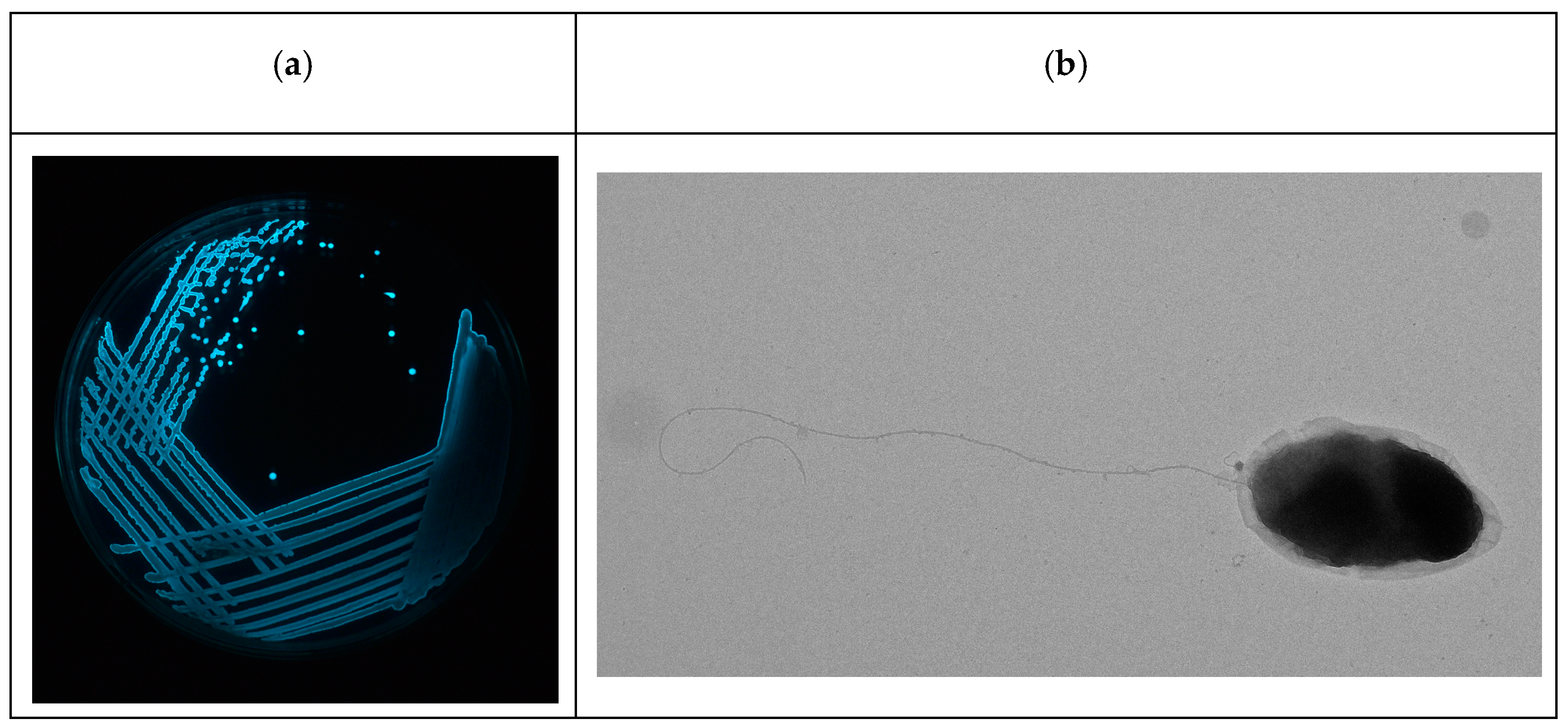
2.1. Isolation of Luminescent V. harveyi Hosts

Water samples collected from P. vannamei shrimp hatcheries (n = 20) and aquaculture farms (n = 12) were screened for the occurrence of luminescent vibrios by spread plating on nutrient agar supplemented with 3% salt [41] and thiosulfate citrate bile sucrose agar (TCBS agar). The plates were incubated at 28 °C for 18 h and observed in the dark to view the luminescence. The luminescent colonies were isolated and purified by streaking on nutrient agar supplemented with 3% salt. The well-isolated colony was re-streaked and observed for luminescence. The luminescent bacteria were assigned to the Vibrio genus based on the results of growth on TCBS agar, Gram’s staining, nitrate reduction, oxidase production, and the Hugh and Leifson glucose oxidation/fermentation test [42] and used as bacterial hosts. The luminescent vibrios were further tested for V. harveyi based on biochemical tests, viz., sugar fermentation (arabinose, cellobiose, dulcitol, galactose, glucose, m-inositol, maltose, mannose, raffinose, rhamnose, salicin, sorbitol, and sucrose), amino-acid decarboxylase/dihydrolase (arginine, ornithine, and lysine), salt tolerance (0%, 0.5%, 1%, 3%, 6%, 8%, and 10% NaCl), amylolytic, proteolytic, and lipolytic DNAase activities, and luminescence production [43]

2.2. Isolation of Vibriophage

Water samples from P. vannamei shrimp hatcheries (n = 20) and aquaculture farms (n = 12) of Andhra Pradesh, India, and the sewage treatment plant of Visakhapatnam, India, were collected and screened for lytic vibriophages against luminescent vibrios by employing the single-host enrichment method. Briefly, 47.5 mL of water sample was mixed with 12.5 mL of overnight culture of luminescent V. harveyi-LV6 as the host strain, which was added to 15 mL of 5×nutrient broth with 3% salt (peptone 5 g L−1, beef extract 3 g L−1, NaCl 30 g L−1) and incubated for 6 h at 28 ± 2 °C. Post-enrichment, the phage-enriched culture was centrifuged (10,000 rpm for 20 min at 4 °C), filtered through a 0.22 µm sterile syringe filter to remove residual bacterial host cells, and the filtrate was tested for vibriophages by spotting 10 µL of the filtrate on NA + 3% salt plates seeded separately with overnight cultures of each of the luminescent vibriohosts. The appearance of clearance at the spotted area indicated the presence of lytic vibriophages.

2.3. Purification and Precipitation of Vibriophage

Vibriophage was purified by employing a single-agar method [44], in which 1 mL of phage (filtrate) and 1 mL of luminescent vibrio host (V. harveyi-LV6) were mixed and added to 8 mL of molten and cooled soft nutrient agar supplemented with 3% salt (peptone 5 g L−1, sodium chloride 30 g L−1, and agar-agar 8 L−1) and finally poured onto a sterile petri plate. The plates were incubated at 28 °C ± 2 °C for 8 h to obtain plaques. The phage filtrate was serially diluted in SM buffer and analyzed separately. The phage titer is expressed as pfu mL−1 and calculated using the following formula:
Number of phages (pfu mL−1) = Total number of plaques × Dilution factor
The isolated plaques were picked, and the process was repeated three times to obtain purified vibriophage with consistent plaque morphology. Phage precipitation was performed by treating purified vibriophage (25 mL) with polyethylene glycol (10% w/v PEG 8000 and 1 M NaCl) at 4 °C for 1 h, followed by overnight incubation at −20 °C for 24 h and centrifugation at 10,000 rpm for 20 min at 4 °C [45,46]. The pellet was resuspended in SM buffer (100 mM NaCl, 8 mM MgSO4.7H2O, 50 mM Tris-Cl, pH 7.5), which constituted the purified and enriched vibriophage, and stored at 4 °C for downstream analysis.

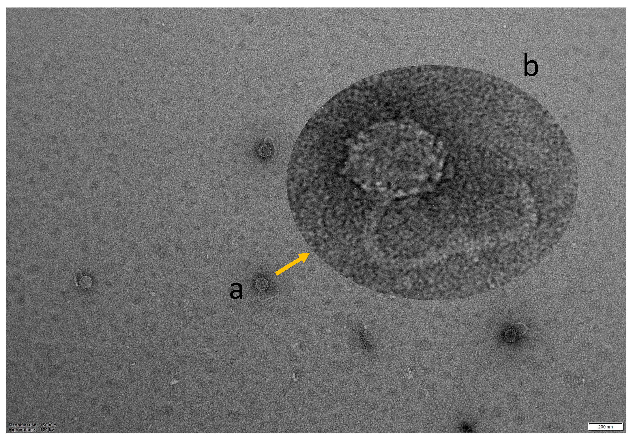
2.4. TEM Morphology of Vibriophage-ϕLV6

For morphological analysis, 10 µL of purified and enriched vibriophage (~108 pfu mL−1) was loaded on a 200-mesh copper grid, stained with Uranyless 22409, and examined under 15,000× nm magnification at an accelerated voltage of 120 kV using a transmission electron microscope (JEOL Japan) at the National Institute of Animal Biotechnology, Hyderabad, India.

2.5. Host Range Determination

The host range of vibriophage-ϕLV6 was determined by performing a spotting assay on 27 luminescent Vibrio spp. isolates from P. vannamei shrimp hatcheries, viz., LV6, LV20, LV21, LV22, LV23, LV24, LV25, LV26, LV27, LV28, LV29, LV30, LV31, LV32, LV33, LV34, LV35, LV36, LV37, LV38, LV39, LV40, LV41, LV42, LV43, LV44, and LV45.

2.6. DNA Extraction and Whole Genome Sequencing of Vibriophage-ϕLV6

Vibriophage-ϕLV6 DNA was extracted and purified with Qiagen’s DNeasy Blood & Tissue Kit [47]. Initially, PEG-precipitated vibriophage (500 µL) was treated with 1.25 µL DNase and RNase (20 mg mL−1) and incubated at 37 °C for 1 h; then treated with 1.25 µL proteinase (20 mg mL−1) and 25 µL of 10% SDS and incubated at 60 °C for 1 h. The vibriophage-ϕLV6 DNA was extracted as per the kit manufacturer’s instructions and finally suspended in TE buffer. The quality and concentration of the extracted DNA were assessed using the Qubit® dsDNA HS Assay Kit, and the integrity of the DNA was determined by electrophoresis on 1% agarose gel. The whole genome sequencing libraries were prepared using the NEBNext®UltraTM II FS DNA Library Prep Kit for Illumina at ClevergeneBiocorp Private Limited, Bangalore, India. The QC-passed library was diluted to 2 nM and sequenced on the Illumina HiSeq 4000. The high-quality reads were used to assemble the genomes using the HGA genome assembler [48].

2.7. Bioinformatic Analysis of Vibriophage-ϕLV6 Genome

The genes in the vibriophage-ϕLV6 genome were predicted and annotated using GeneMarkS, Glimmer, and Prokka [49,50,51]. The functional characteristics of the predicted genes were determined on the NCBI BLASTp platform with the non-redundant protein sequences (nr) database. The presence of transfer RNA (tRNA) was predicted employing Prokka 1.14.6 [50]. The presence of antibiotic-resistance genes was screened in Resfinder 4.1 (https://cge.food.dtu.dk/services/ResFinder/ (accessed on 26 November 2022)) and the presence of bacterial virulent genes was screened in Virulence finder 2.0 (https://cge.food.dtu.dk/services/VirulenceFinder/ (accessed on 26 November 2022)).
Multiple phage genomes belonging to siphoviruses were compared and visualized using BRIG (Blast Ring Image Generator) with default settings. Comparative genome analysis was performed using ViPTree [52]. The phage genome was categorized into structural modules, DNA metabolism modules, packaging modules, lysis modules, hypothetical modules, and additional functional modules [31,53,54]. Data pertaining to the major capsid protein (n = 16) terminase large subunit (n = 18) of vibriophages and other phages related to vibriophage-ϕLV6 were downloaded from the NCBI database and aligned using the MUSCLE algorithm. Phylogenetic analysis was performed using MEGA 10.0.5 software [55] based on the major capsid protein and terminase large subunit using the neighbor-joining method with robust 1000 bootstrap replicates. The genome map of vibriophage-ϕLV6 was drawn using Proksee (https://proksee.ca/ (accessed on 2 December 2022)).

2.8. In Vitro Determination of Optimum Multiplicity of Infection (MOI) for Determining Phage Infectivity Potential

The multiplicity of infection, i.e., the ratio of the number of vibrophage-ϕLV6 required to lyse luminescent V. harveyi, was determined employing the 2-step microtiter plate assay [56]. Briefly, in the 2-step microtiter assay, a broad range of MOIs (ranging from MOI-0.0001 to MOI-10000) were initially tested for their ability to inhibit the growth of target bacteria. A narrow range of effective MOIs from the first step was selected to determine the optimum MOI in the second step. The optimum MOI, i.e., the lowest number of phages required to inhibit the growth of the target bacteria, was determined in the second step. The optimum MOI of vibriophage-ϕLV6 against the luminescent V. harveyi-LV6 host was 79 and was previously determined [56]. On similar lines, the optimum MOIs of vibriophage-ϕLV6 against five other luminescent vibrio hosts, viz., LV36, LV38, LV40, LV44, and LV45, isolated from shrimp hatcheries, were determined.

2.9. In Vivo Challenge Studies to Assess the Ability of Vibriophage-ϕLV6 to Inhibit the Growth of Luminescent V. harveyi in P. vannamei Shrimp Post-Larvae Tanks

Challenge studies to check the in vivo efficacy of the phage to treat induced luminescent vibriosis were performed in glass tanks containing P. vannamei post-larvae. The tank experiments were designed in two variations,
  • Application of vibriophage-ϕLV6 at optimized MOI against a single luminescent V. harveyi host, i.e., LV6;
  • Application of vibriophage-ϕLV6 at optimized MOIs against multiple (n = 6) luminescent Vibrio Hosts, i.e., LV6, LV36, LV38, LV40, LV44, and LV45.

2.9.1. Effectiveness of Vibriophage-ϕLV6 Application at Optimized MOI against a Single Luminescent V. harveyi Host

Glass tanks filled with 27 ppt seawater (10 L) and P. vannamei post-larvae of PL-11 size (n = 250) per tank were used for the in vivo study. Tank-1 (control) contained only shrimp post-larvae; Tank-2 (bacteria control) was spiked with luminescent V. harveyi-LV6 at an 8.6 × 10⁶ cfu mL−1 concentration. Tank-3 (phage control) was inoculated with vibriophage-ϕLV6 (8.0 × 10⁶ pfu mL−1). Tank-4 (treatment tank) was spiked with luminescent V. harveyi-LV6 and simultaneously treated with vibriophage-ϕLV6 at an MOI of 80 (80 pfu phage to 1 cfu bacteria). All the tanks were kept at ambient temperature (28–30 °C) under illuminated conditions with continuous aeration. Feed was not provided to the post-larvae during the experiment period. Water samples were taken at hourly intervals for 6 h and checked for OD600 values; the total vibrio counts and phage activity were checked in the water after 4 h, and shrimp post-larval survivability was determined after 24 h of exposure.

2.9.2. Effect of Vibriophage-ϕLV6 on the Growth of Multiple Luminescent Vibrio Hosts (n = 6)

An in vivo experiment was conducted in glass tanks containing 25 ppt salinity sea water (1 L) and P. vannamei post-larvae of PL-3 size (n = 100) per tank. The bacteria control tanks were spiked with six luminescent Vibrio spp. isolates (LV36, LV38, LV40, LV44, LV45, and LV6; 109 cfu mL−1). The vibriophage-treatment tanks were spiked with six luminescent vibrio bacteria at a concentration of 109 cfu mL−1 (each bacteria) and simultaneously treated with vibriophage-ϕLV6 at pre-determined MOIs (MOI-79 for the LV6 host, MOI-41.5 for the LV40 host, MOI-33.6 for the LV36 host, MOI-29.3 for the LV38 host, MOI-1.5 for the LV45 host, and MOI-0.7 for the LV44 host). The control tanks were spiked with neither bacteria nor bacteriophage. All the tanks were kept at ambient temperature (28–30 °C) under illuminated conditions with continuous aeration. Feed was not provided to the post-larvae during the experiment period. The total vibrio counts and post-larvae survivability were checked for 48 h.

2.10. Lytic Ability of Vibriophage-ϕLV6 under Different Salinity Conditions

In the shrimp aquaculture system of India, the salinity of the water in the shrimp hatcheries is maintained between 25 and 35 ppt (2.5 and 3.5%) but the farming of P. vannamei shrimp in aquaculture farms is carried out at different salinities ranging from 4 ppt to 45 ppt [57]. In order to check the applicability of the vibriophage-ϕLV6, both in hatcheries and different farming conditions of P. vannamei, a salt gradient experiment on determining the lytic activity of vibriophage was taken up. Tubes containing nutrient broth with different salt concentrations, viz., 0.5%, 1%, 2%, 3%, 4%, and 5%, were inoculated with vibriophage-ϕLV6 at a 10% level and incubated at 28 ± 2 °C. The tubes were taken out every 3 days for 30 days and checked for the lytic activity of vibriophage-ϕLV6 by the spotting method.

2.11. Vibriophage-ϕLV6 Activity against Luminescent V. harveyi Host LV6 at Different Salt Gradients

Nutrient broth with different salt concentrations, viz., 0%, 0.25%, 0.5%, 1%, 1.5%, 2%, 2.5%, and 3%, was prepared, and 240 µL of NB with a specific salt concentration was loaded in triplicate wells of a 96-well microtiter plate reader (BioTek, Winooski, VT, USA). Vibriophage-ϕLV6 (30 µL) and V. harveyi host (30 µL) were inoculated at a pre-determined MOI of 80 into each well of a 96-well microtiter plate.
Three controls, viz., bacterial control (without phage), phage control (without bacteria), and media control (without bacteria and phage), were also introduced in triplicate wells at the same salt gradients. The OD600 readings were taken at 30 min intervals for 4 h.

2.12. Storage Stability of Vibriophage

The vibriophage-ϕLV6 suspended in SM buffer was stored at 4 °C for one year. Samples were drawn intermittently, and the counts (pfu mL−1) of vibriophage-ϕLV6 were determined by performing the single-agar method [44].

2.13. Statistical Analysis

The results of post-larval survivability and vibrio counts in phage-treatment tanks and bacterial-challenged tanks were analyzed statistically employing an unpaired t-test and a chi-square test to test the difference at a 5% level of significance using InVivoStat, Version 4.7 [58].

3. Results and Discussion

3.1. Isolation of Luminescent Vibrios

A total of 27 luminescent vibrios were isolated from P. vannamei shrimp hatchery water samples that showed characteristic luminescence activity. Luminescent vibrios were not detected in water samples from shrimp aquaculture farms. One luminescent vibrio (LV6) that was isolated from a luminescent vibriosis-infected P.s vannamei post-larvae tank was identified as V. harveyi based on the results of biochemical and morphintorial analysis. The remaining luminescent vibrios were identified to the genus level and categorized as luminescent Vibrio spp. (Figure 1a,b).

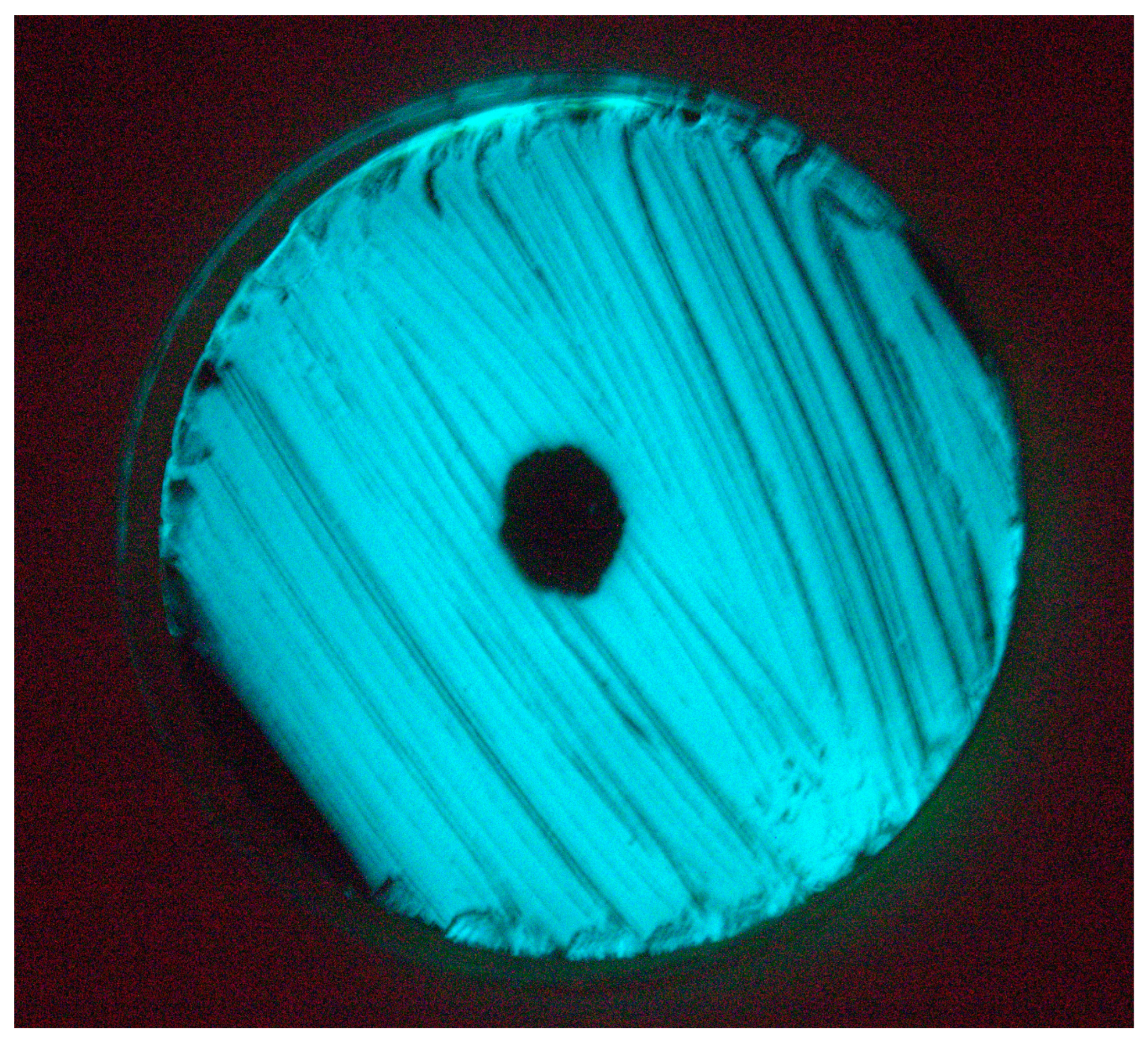
3.2. Isolation and Lytic Spectrum of Vibriophage

A lytic phage named vibriophage-ϕLV6 was isolated from the water of the post-larvae tank of a luminescent vibriosis-infected P. vannamei shrimp hatchery. Vibriophage-ϕLV6 showed a clear lytic zone against luminescent V. harveyi-LV6 (Figure 2) after single-host enrichment and yielded pinpointed plaques with a diameter of <1 mm on the single-agar method. The vibriophage count after purification, PEG precipitation, and suspension in SM buffer was 3.42 × 10¹⁰ pfu mL−1. PEG precipitation increased the concentration of the vibriophage 100 times. None of the remaining 19 water samples collected from different shrimp hatchery sites, 20 water samples from shrimp aquaculture farms, and one water sample from a sewage treatment plant revealed the presence of vibriophage against luminescent V. harveyi. The vibriophage-ϕLV6 showed lytic activity against one luminescent V. harveyi (LV6) and five isolates of luminescent Vibrio spp. from shrimp hatcheries, viz., LV36, LV38, LV40, LV44, and LV45.

3.3. TEM Morphology of Vibriophage-ϕLV6

The morphology of vibriophage-ϕLV6 observed under transmission electron microscopy (Figure 3) revealed that the phage had an icosahedral head (~73 nm) and a long filamentous non-contractile tail (~191 nm), respectively, suggesting that the vibriophage-ϕLV6 might belong to siphoviruses.

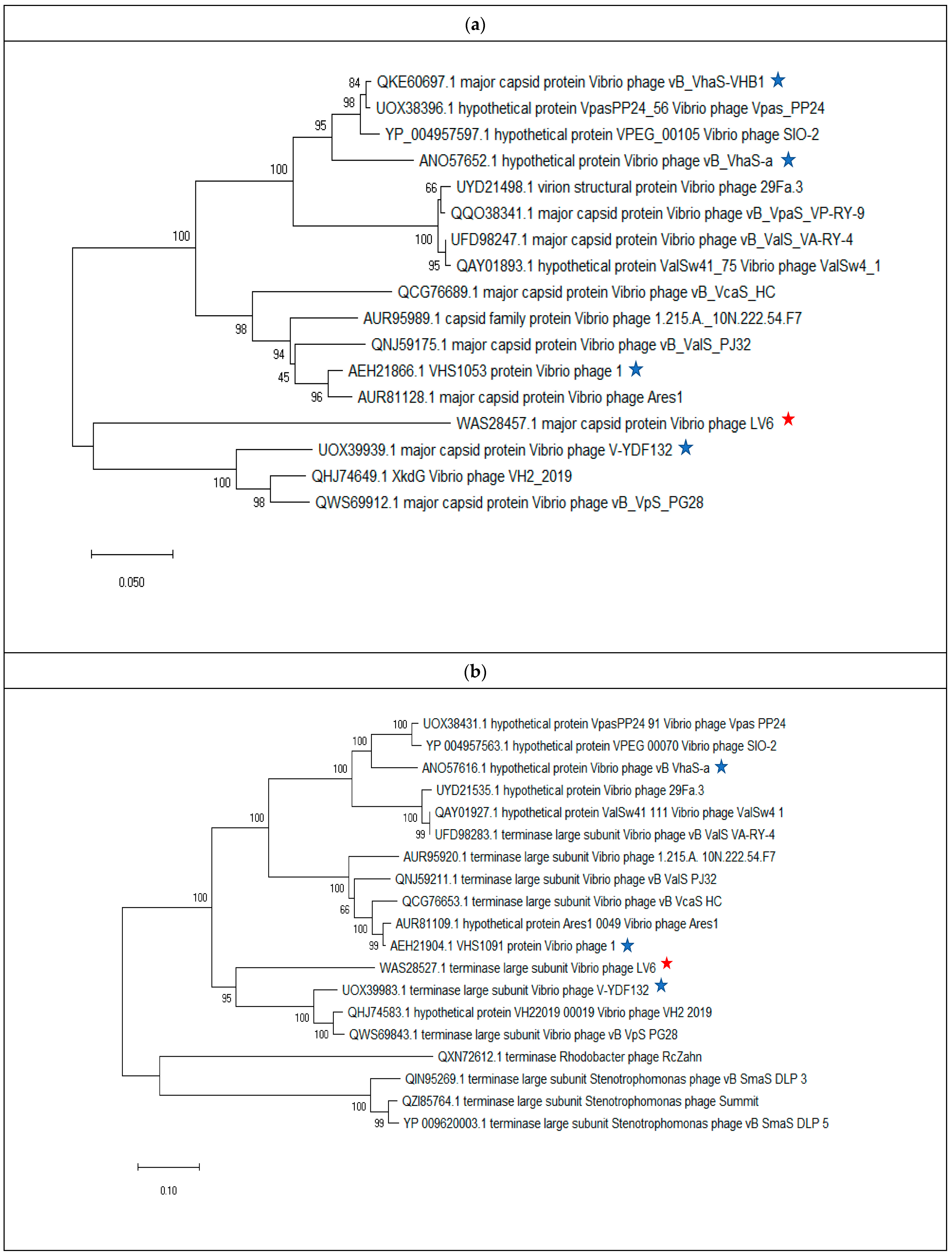
3.4. Vibriophage-ϕLV6 Genome Characterization and Phylogenetic Analysis

The genome size of the vibriophage-ϕLV6 was 79,862 bp with a G+C content of 48%. The coding sequences (CDS) accounted for 93% of the vibriophage genome (74,178 bp) and coded for proteins with known functions (41%), and hypothetical proteins (52%). A total of 107 putative ORFs were predicted in the genome of vibriophage-ϕLV6 with gene lengths ranging between 129 and 4146 bases. The details of the ORFs and the predicted proteins are presented in Figure 4 and Table 1. Out of 107 ORFs, 31 of the ORFs in the vibriophage-ϕLV6 genome had predicted functions, and 75 ORFs had currently unknown functions, i.e., hypothetical proteins. One tRNA coding for lysine was found in the genome of vibriophage-ϕLV6. No antibiotic resistance genes or bacterial virulence genes were found in the genome of vibriophage-ϕLV6.

3.5. Intergenomic Comparison of Vibriophage-ϕLV6

3.5.1. Phylogenetic Analysis

Phylogenetic trees were constructed targeting single gene analysis of major capsid protein (Figure 5a) and terminase large subunit protein (Figure 5b). Results from the two phylogenetic trees indicated that vibriophage-ϕLV6 is closely related to vibriophage-V-YDF132, vibriophage-VH2_2019, and vibriophage-vB_VpS_PG28.

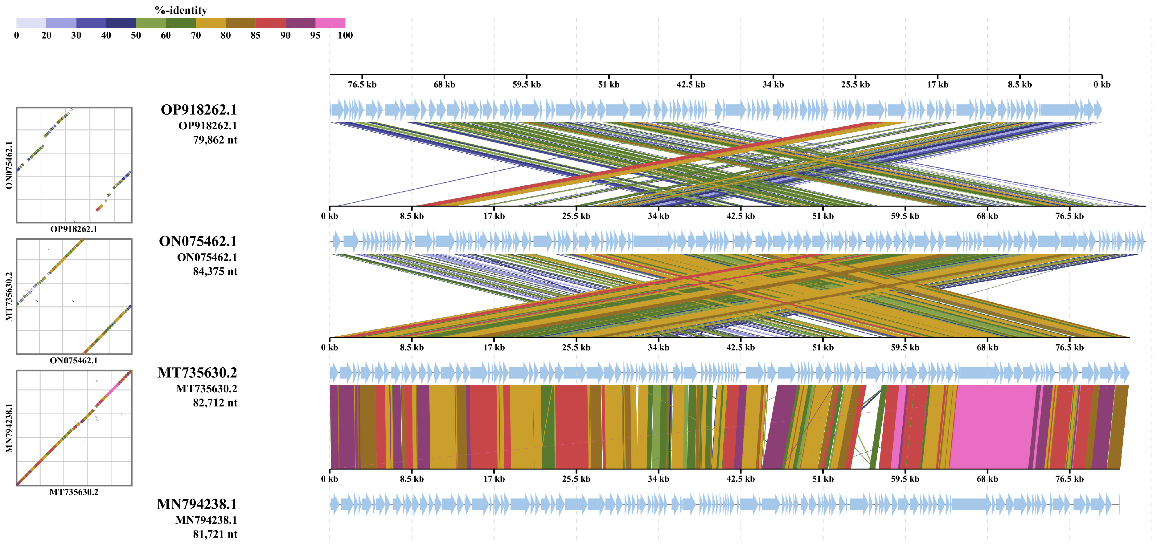
3.5.2. Multiple Genome Alignments

Vibriophage-ϕLv6 (NCBI Accession ID: OP918262.1) had genome sequence similarity of 79.89%, 74.97%, and 74.44% to vibriophage-vB_VpS_PG28 (NCBI Accession ID: MT735630.2), vibriophage-V-YDF132 (NCBI Accession ID: 0N075462.1) and vibriophage-VH2_2019 (NCBI Accession ID: MN79428.1). The Bacterial and Archaeal Subcommittee (BAVS) considers that any genus having similarity greater than 50% can be considered a coalescent group [59]. Multiple sequence alignment was performed with Vibriophage-ϕLV6 and its closely related vibriophages, i.e., vibriophage-V-YDF132, vibriophage-VH2_2019, and vibriophage-vB_VpS_PG28 (Figure 6).

3.5.3. Whole Genome Comparison

The whole genome sequences of the vibriophages used in phylogenetic tree construction of terminase subunit and major capsid protein were obtained from NCBI and compared with the genome of vibriophage-ϕLV6. The results were similar to the phylogenetic analysis, i.e., vibriophage-V-YDF132 (isolated from a group fish farm in China and active against V. harveyi), vibriophage-VH2_2019 (isolated from Hatches Creek, USA, and active against V. natriegens), and vibriophage-vB_VpS_PG28 (isolated from sewage at a seafood market in China and active against V. parahaemolyticus) shared more similarity with vibriophage-ϕLV6 compared to other phages (Figure 7).
A whole genome homologs similarity search revealed that vibriophage-vB_VpS_PG28 infecting V. parahaemolyticus shared the highest similarity to vibriophage-ϕLV6, but with a similarity coverage of only 79.89%, followed by two other vibriophages, i.e., vibriophage-V-YDF132 and vibriophage-VH2_2019, with sequence similarities of 74.97% and 74.44%, respectively. Vibriophage-ϕLV6 harbored replication, regulation, structural, and packaging modules similar to those of vibriophage-vB_VpS_PG28 and vibriophage-VH2_2019 but lacked a lysis module. In contrast, vibriophage-ϕLV6 and vibriophage-V-YDF132 shared all the modules (Figure 8). Vibriophage-ϕLV6 had one tRNA encoding for lysine.

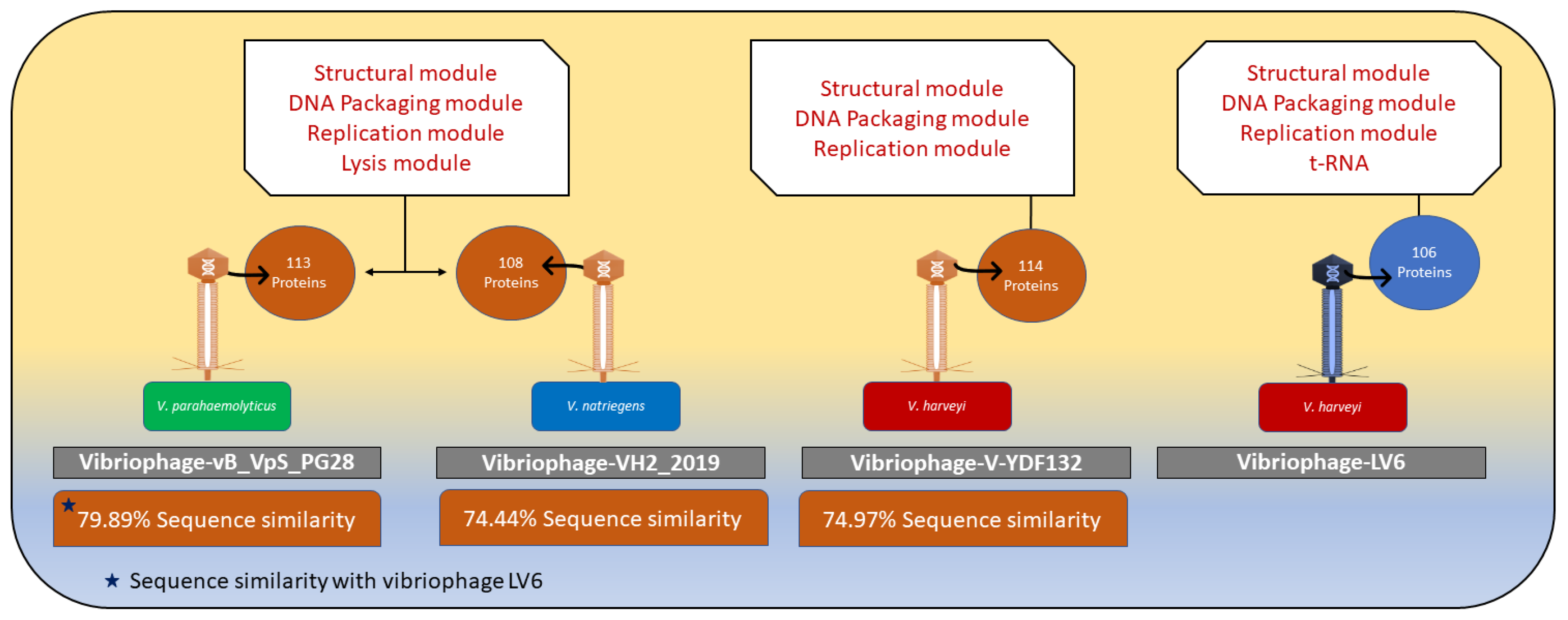
3.5.4. Comparison of the Genome of Vibriophage-ϕLV6 with the Genomes of Other Vibriophages Active against V. harveyi Clade

The whole genome features of vibriophage-ϕLV6 were compared to the features of other vibriophages that were previously reported to be active on V. harveyi clade bacteria, viz., siphophage-VHS1, vibriophage-vB_VhaM_pir03, vibriophage-VB_VhaS_PcB-1G, vibriophage-VB_VcaS_HC, and vibriophage-Virtus. The genome size of vibriophage-ϕLV6 (79.86 kb) was similar to that of siphophage-VHS1 (81.5 kb), Vibriophage-VB_VcaS_HC (81.6 kb), and vibriophage-virtus (82.96 kb), but was smaller than the jumbo vibriophage-vB_VhaM_pir03 (286.3 kb) (Table 2). The G+C content of vibriophage-ϕLV6 (48%) was comparable to that of the previously reported vibriophages (43.6% to 47.6%). The ORFs with predicted function were relatively higher in vibriophage-ϕLV6 (138) compared to other vibriophages with similar genome sizes (121–127 ORFs). The jumbo-sized vibriophage-vB_VhaM_pir03, with a genome size larger than 200 kb, had 137 ORFs. Moreover, tRNA was reported only in vibriophage-ϕLV6 that encoded for lysine.
The vibriophage-ϕLV6 genome was negative for antimicrobial-resistance determinants (ARGs), integrase, and bacterial virulence genes, making it a suitable candidate for in vivo phage applications to control luminescent vibriosis in shrimp aquaculture.

3.6. Vibriophage-ϕLV6 Proteome

Vibriophage-ϕLV6 has six ORFs, i.e., ORF 3, ORF 6, ORF 7, ORF 8, ORF 9, and ORF 11, predicted for functions related to the structural composition of the phage. Vibriophage-ϕLV6 has dedicated machinery for 7 ORFs, i.e., ORF 62, ORF 68, ORF 72, ORF 86, ORF 92, ORF 94, and ORF 96, involved in phage DNA metabolism. ORF 82, which encodes for the terminase large subunit, plays a vital role in viral genome packaging. The terminase large subunit has two subunits: a smaller subunit involved in viral DNA packaging and a larger subunit involved in ATPase and endonuclease activities.

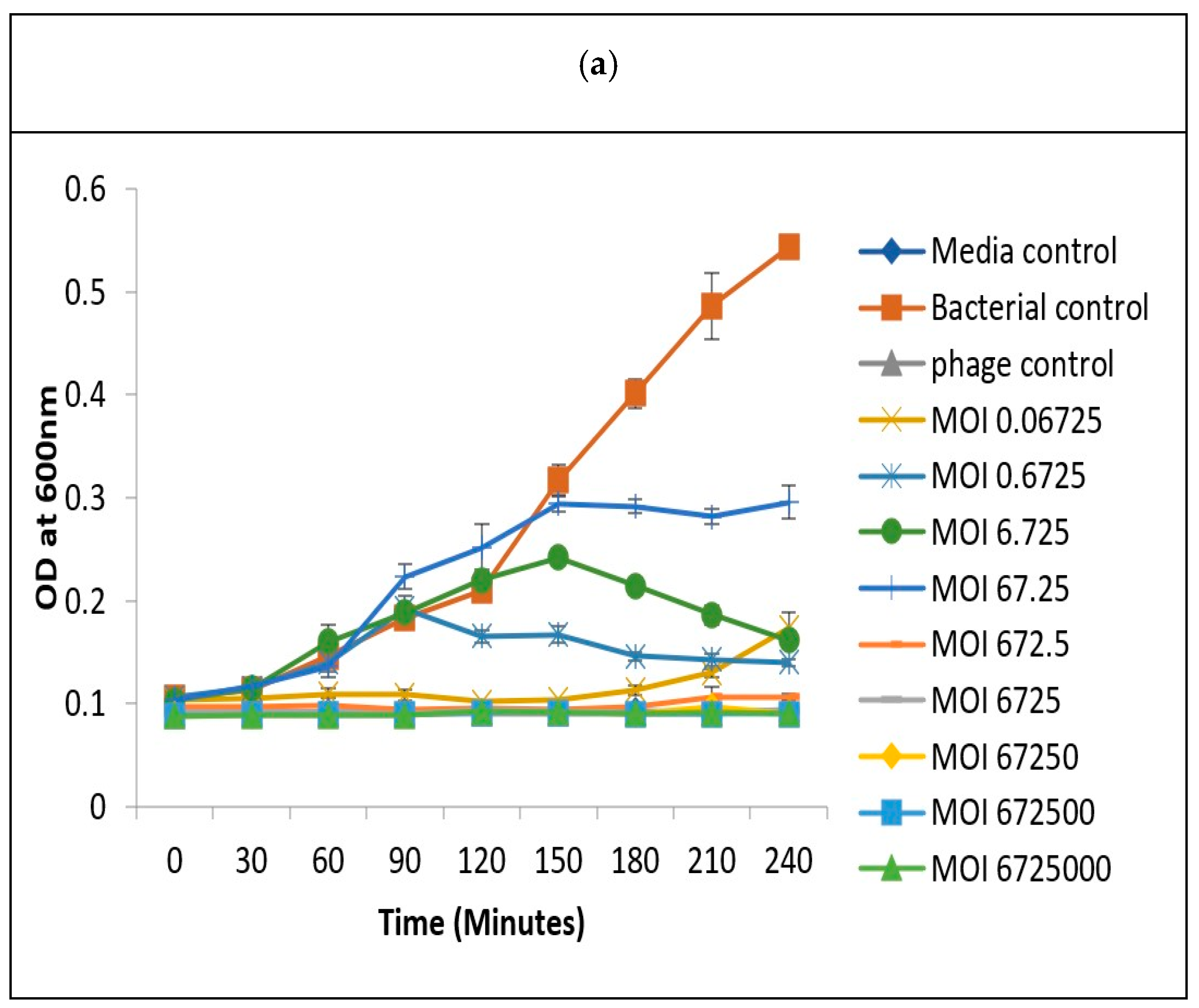
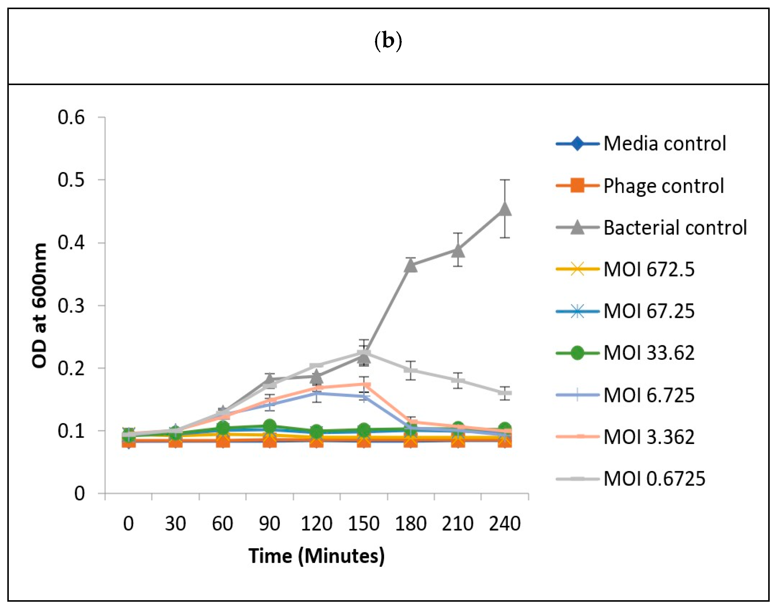
3.7. Determination of Optimum Multiplicity of Infection

The optimization of MOI is important to determine the lowest number of phages required to inhibit the growth of a specific bacteria. The luminescent V. harveyi isolates that were susceptible to vibriophage-ϕLV6 in the spot assay were selected for MOI determination. The optimum MOI for the isolate LV6 was previously reported by us at MOI-79 [56]. Similarly, the optimum MOI of vibriophage-ϕLV6 to inhibit the growth of the remaining five susceptible luminescent Vibrio spp. isolates, viz., LV36, LV38, LV40, LV44, and LV45, was determined using the two-step microtiter plate assay. In the two-step microtiter assay, a narrow range of MOIs were selected in the first step, and the optimum MOI was determined in the second step (Figure 9a,b). The narrow range of MOIs out of the nine MOIs (0.0001 to 10,000) that were selected in the first step ranged between 6.725 and 672.5 for LV36; 5.854 and 585.4 for LV38; 8.3 and 83 for LV40; 0.01 to 1.42 for LV44; and 0.03 to 3 for LV45. The optimum MOIs of vibriophage-ϕLV6 obtained in the second step were 41.5 for LV40, 33.6 for LV36, 29.3 for LV38, 1.5 for LV45, and 0.7 for LV44. These optimized MOIs were applied for challenge studies in glass tanks.

3.8. Challenge Studies to Test the In Vivo Lytic Ability of Vibriophage-ϕLV6

Vibriophage-ϕLV6 was employed at an optimized MOI to control the growth of luminescent V. harveyi and luminescent Vibrio spp. in tanks containing post-larvae of P. vannamei shrimp that were spiked with either single or multiple isolates of luminescent Vibrio spp.

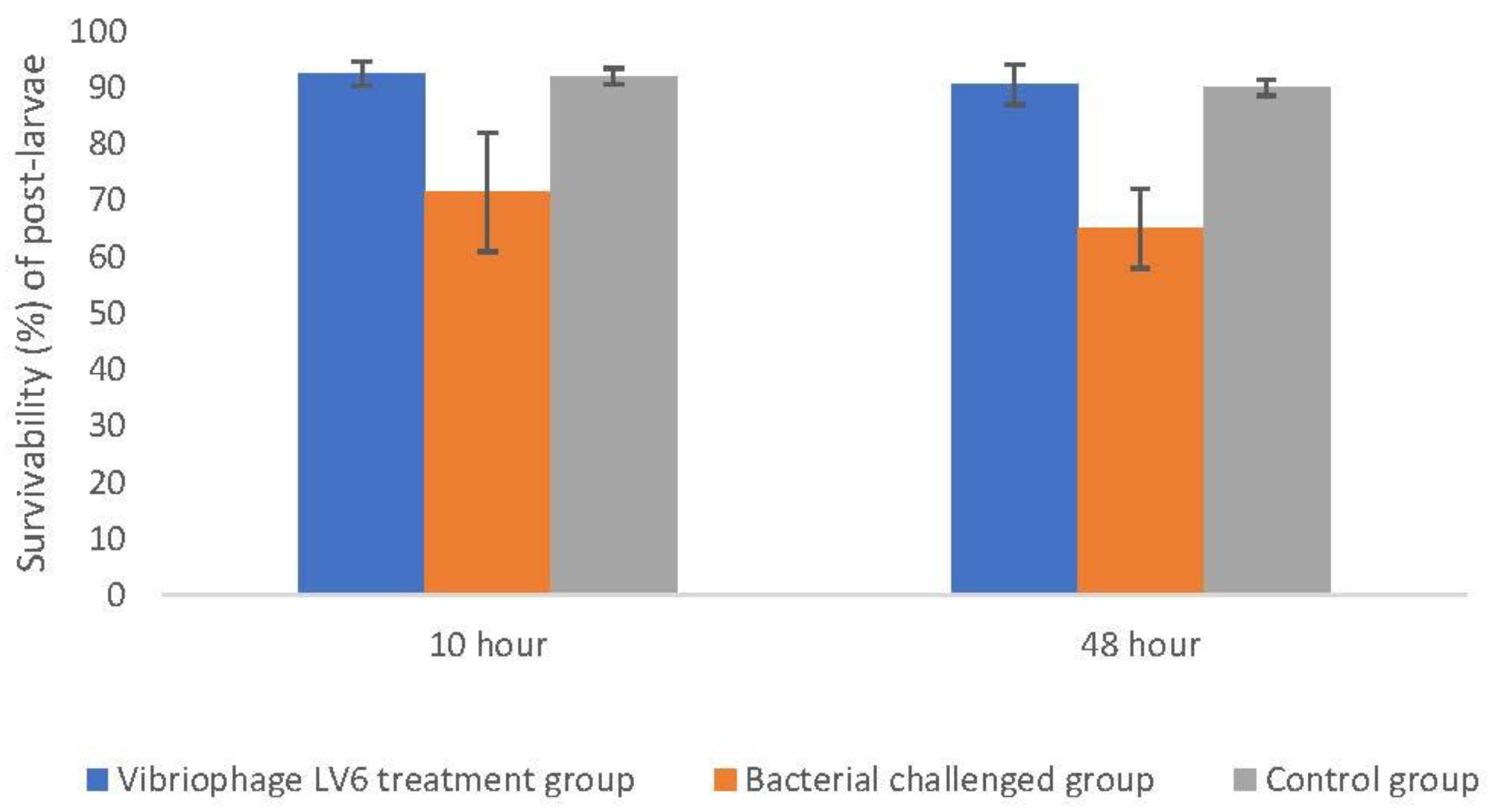
3.8.1. Effectiveness of Vibriophage-ϕLV6 Treatment at an Optimized MOI of 80 against a Single Luminescent V. harveyi-LV6

There was a continuous increase in OD600 in the bacteria-control tank, indicating uncontrolled proliferation of bacteria, whereas the vibriophage-treated tanks showed a negligible increase in OD600 until 6 h of exposure. The luminescent bacteria count was very high in the bacterial-control tank (1.02 × 10⁸ cfu mL−1) after 4 h of exposure; however, the luminescent bacteria count was less than 300 cfu mL−1 in vibriophage-ϕLV6 treated tanks (Figure 10). The vibriophage count ranged between 3.6 × 10⁹ and 3.7 × 10⁹ pfu mL−1 in phage-treated tanks, whereas no vibriophage was detected in bacteria-control tanks. There was a continuous increase in the OD600 values of the water in the bacteria-control tank, indicating uncontrolled proliferation of bacteria. However, the vibriophage-ϕLV6 treated tank showed a negligible increase in OD600 values, lower luminescent bacterial counts, but very high counts of vibriophage and higher post-larvae survival. The results of the tank (10 L) indicated the effectiveness of employing the vibriophage-ϕLV6 in controlling the growth of luminescent V. harveyi, and the optimum MOI determined by the two-step microtiter plate method was sufficient to control the growth for 6 h.

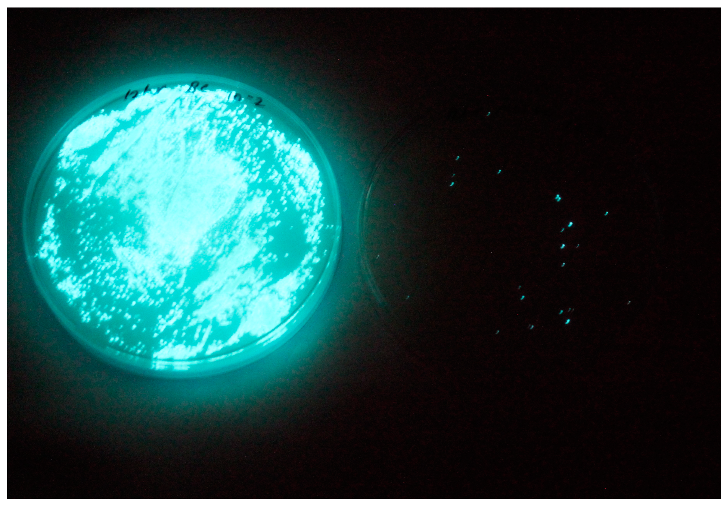
3.8.2. Effect of Vibriophage-ϕLV6 on Inhibiting the Growth of Multiple Luminescent Vibrio Hosts (n = 6)

The shrimp post-larvae mortality was higher in bacteria-challenged tanks (37.5% ± 3%) compared to phage-treated tanks (9.5% ± 3%) and control tanks (8% ± 1%). In other words, significantly higher survivability of the shrimp post-larvae was observed in the phage-treated tank (Figure 11) compared to the bacteria-challenged group (unpaired t-test and chi-square test, p < 0.05). The sucrose non-fermenting vibrio loads were distinctly higher in bacteria-spiked tanks (357,100 cfu mL−1) compared to vibriophage-treated tanks (1000 cfu mL−1). Phage activity was detected only in vibriophage-treated tanks but not in control or bacteria-spiked tanks. The results indicate that Vibriophage-ϕLV6 effectively reduced the numbers of multiple luminescent vibrios and reduced the mortality of shrimp post-larvae.

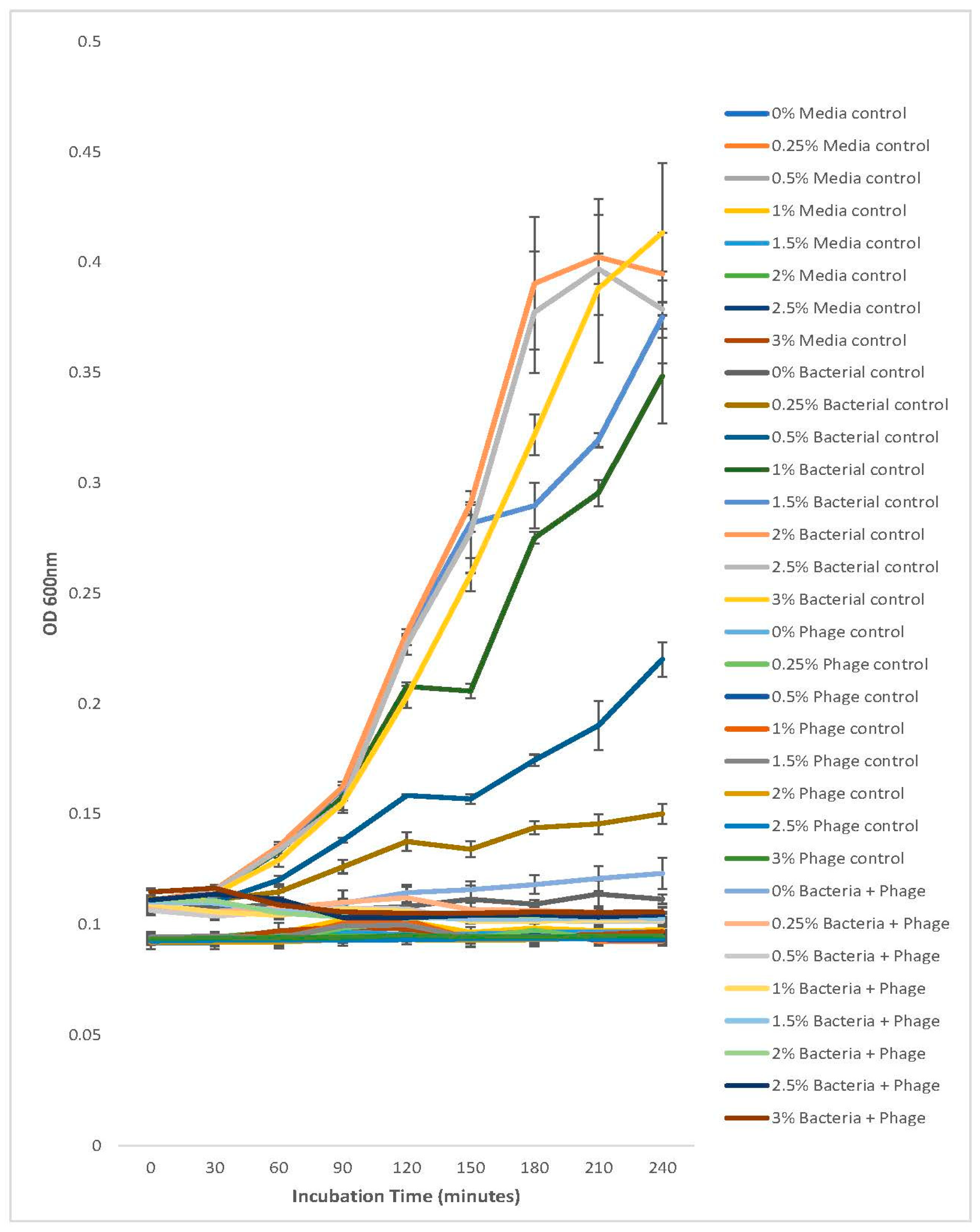
3.9. Survivability of Vibriophage under Different Salinity Conditions

The vibriophage-ϕLV6 survived under different salt conditions of 5 ppt, 10 ppt, 20 ppt, 30 ppt, 40 ppt, and 50 ppt, indicating their applicability in brackish water and marine waters. Vibriophage-ϕLV6 survived and exhibited its lytic activity for 30 days (maximum period tested) at both 28 °C and 35 °C.

3.10. Stability Testing of Vibriophage-ϕLV6 Activity against Luminescent V. harveyi Host LV6 at Different Salt Gradients

V. harveyi-LV6 (bacteria controls) did not show any growth at 0%, very weak growth at 0.25% salt concentration, and relatively weak growth at 0.5% salt concentration. The growth of V. harveyi-LV6 was optimal at salt concentrations between 1% and 3%, yielding an OD600 value of ~0.3 to 0.4 (Figure 12). This growth pattern vis-à-vis salt concentration was on expected lines, as V. harveyi is a halophilic organism and salt is integral to its growth [30]. It was pertinent to note that the lytic activity of vibriophage-ϕLV6 when applied at an MOI of 80 against V. harveyi-LV6 was not affected by the different salt concentrations (0% to 3%) and efficiently halted the growth of V. harveyi-LV6 as evidenced by lower (~0.1) OD600 that were similar to the media controls and phage controls at the end of 240 min of incubation (Figure 12).

3.11. Storage Stability of Vibriophage-ϕLV6

Concentrated suspension of vibriophage-ϕLV6 stored at 4 °C did not show any reduction in phage numbers for 9 months of storage as the plaque counts obtained on single agar remained almost similar (1010 pfu mL−1), but a slight reduction in the phage numbers (less than one log) was observed at the end of 12 months of storage at 4 °C (Table 3).

4. Discussion

The use of bacteriophages as therapeutic agents in aquatic animal-health management has gained renewed interest due to the emergence of resistance in pathogenic bacteria towards antibiotics and safety issues related to antibiotic residues in food products. Globally, shrimp farming is increasingly contributing to animal protein requirements, but farm productivity is adversely affected by diseases caused by bacteria of the genus Vibrio. V. harveyi is the major causative agent of luminescent vibriosis in shrimp hatcheries and aquaculture farms. In the present study, a vibriophage, named vibriophage-ϕLV6, was isolated from the water of a P. vannamei shrimp hatchery. Vibriophage-ϕLV6 showed in vitro lytic activity against luminescent V. harveyi (LV6) that was isolated from a shrimp hatchery affected with luminescent vibriosis. Vibriophage-ϕLV6 produced pinpoint plaques on soft-agar plates seeded with the bacterial host. Misol et al. (2020) also reported that vibriophage-vB-VhaM-pir03 produced pin-hole plaques with a diameter of 0.27 ± 0.05 mm on V. harveyi-seeded plates. A 100-fold increase in the concentration of vibriophage-ϕLV6 was achieved by PEG precipitation, and a similar increase in the concentration of coliphages by PEG precipitation was reported [46]. The host spectrum of vibriophage-ϕLV6 (six luminescent Vibrios) was relatively lower compared to recently reported V. harveyi bacteriophages. Vibriophage-Virtus, isolated from the water of a fish brood stock section in Crete, Greece, could infect 8 of the 16 strains of V. harveyi [39], and vibriophage-B_VhaM_pir03, isolated from the water of the Port of Piraeus, Greece, showed lytic activity against 31 AMR strains of V. harveyi, V. alginolyticus, V. campbellii, and V. owensii [38]. Most of the phages that were isolated against V. harveyi are reported to be lytic, but two bacteriophages, viz., VHML [4] and VHS1 [60], were found to be temperate.
Transmission electron microscopy images indicate that vibriophage-ϕLV6 belongs to the morphological group of siphoviruses. The bacterial viruses subcommittee of the International Committee on Taxonomy of Viruses (ICTV) has recently abolished the morphology-based families of bacteriophages, viz., Myoviridae, Podoviridae, and Siphoviridae, due to their polyphyletic nature and non-reflection of shared evolutionary histories. All the tailed phages with icosahedral capsids and dsDNA genomes are now grouped under the class Caudoviricetes [61,62,63,64,65]. The terms myovirus, podovirus, and siphovirus can be used to represent distinctive morphological features and retain their historical reference. The process of assigning myoviruses, podoviruses, and siphoviruses into genomically coherent families has been initiated [65]. Though vibriophage-ϕLV6 reveals a siphovirus morphology, it is grouped under Caudoviricetes as per the new ICTV classification. Most of the phages that showed lytic activity against V. harveyi were reported to be siphoviruses [29,32,34,39,66,67,68], followed by myoviruses [34,38,68,69,70].
The genome analysis of vibriophage-ϕLV6 indicated a genome size of 79.8 kb with a G+C content of 48% that was comparable to the previously reported vibriophages (43.6% to 47.6%). The genome of vibriophage-ϕLV6 was highly functional. The ORFs with predicted function were relatively higher in vibriophage-ϕLV6 (138) compared to other vibriophages with similar genome sizes (121–127 ORFs). Even the jumbo-sized vibriophage-vB_VhaM_pir03, with a genome size larger than 200 kb, had only 137 ORFs. Vibriophage-ϕLV6 harbored replication, regulation, structural, and packaging modules. Though the vibriophage-ϕLV6 genome did not reveal a lysis module, it carried a sufficient number of genes that encode for early DNA metabolism, which play an essential role in early viral infection similar to vibriophage-V-YDF132 [39]. Vibriophage-ϕLV6 possesses an ORF7 that encodes for a structural protein (tail tube measure protein). Wu et al. (2020) stated that tail tubular protein, encoded by vibriophage-PcB-1G, plays a critical role in bacterial-cell lysis [32], suggesting that Vibriophage-ϕLV6 can mediate bacterial-cell lysis through the tail tubular system. Vibriophage-ϕLV6 possessed a single tRNA that encoded lysine. Many vibriophages, such as Vibrio parahaemolyticus phage-seahorse and KVP-40 carried high numbers of tRNAs, which may provide the phage with a small degree of autonomy when it comes to the translation of its own genes [71]. It is pertinent to note that more than half of the 107 ORFs of vibriophage-ϕLV6 code for hypothetical proteins whose function is currently unknown. Research efforts are needed to decipher the true function of these hypothetical proteins, as the viral genome machinery is relatively small and has no reason to burden itself with unwanted proteins.
Phylogenetic trees constructed with conserved proteins in the bacteriophage genomes, viz., the major capsid protein and the terminase large subunit protein [72], indicated that vibriophage-ϕLV6 is closely related to vibriophage-V-YDF132, vibriophage-VH2_2019, and vibriophage-vB_VpS_PG28, asserting that their origin is from a common ancestor. Multiple genome alignments showed that vibriophage-ϕLv6 had genome sequence similarity of ~75% to vibriophage-vB_VpS_PG28, vibriophage-V-YDF132, and vibriophage-VH2_2019. The proteome of vibriophage-ϕLV6 had a dedicated machinery of ORFs involved in phage DNA metabolism and viral genome packaging. The proteome of vibriophage-ϕLV6 is similar to the proteome of vibriophage-V-YDF132 [31]. Vibriophage-ϕLV6 possessed an auxiliary metabolic gene that encodes for pyruvate phosphate dikinase (PPDK), an essential component of the Embden–Meyerhof–Parnas (EMP) glycolytic pathway that was also reported in the vibriophages belonging to siphovirus [69,73]. Genomes of the marine vibriophages isolated from nutrient-deficient environments were abundant in auxiliary metabolic genes compared to those isolated from nutrient-rich environments [59].
Horizontal gene transfer (HGT) occurs between phages and bacterial populations through either generalized or specialized transductions [70,74]. Prior to their therapeutic application, the profiling of vibriophages for genomic traits is an essential pre-requisite to ward off an unwanted increase in the virulence of their hosts [16,59,70]. Vibriophage-ϕLV6 appears to be a suitable candidate phage for in vivo phage applications to control luminescent vibriosis as it does not carry antimicrobial resistance determinants or bacterial virulence genes. Moreover, it does not have an integrase gene associated with phage lysogeny. The vibriophage-ϕLV6 produces clear plaques against the host bacteria and reduced the counts of host bacteria in microtiter plate assays, and the reduction in growth was proportional to the number of phages used (i.e., lower bacterial growth at higher MOIs). These results indicate the vibriophage-LV6 was lytic and not lysogenic. Siphophage-VHS was reported to carry a shrimp haemocyte agglutination gene [55]. Siphophage-VHS1 and vibriophage-ϕLV6 were tested on V. harveyi in Pacific white shrimp (P. vannamei) tanks, while vibriophage-vB_VhaM_pir03 was tested in brine shrimp (Artemia) culture. On the other hand, vibriophage-VB_VhaS_PcB-1G and vibriophage-virtus were tested on V. harveyi in finfish tanks.
Several studies have demonstrated the effectiveness of vibriophages in treating vibriosis in a variety of animal models [17,29,38,39,75,76,77,78,79]. Karunasagar et al. (2007) reported that vibriophages resulted in higher survival rates (80%) of black tiger shrimp (P. monodon) in hatcheries compared to survival achieved by conventional antibiotic treatment (40%). vB_VhaM_pir03 when applied to Artemia naupli, increased the survival rates of larvae in the phage-treated group to 15–20% in 48 h than the V. harveyi bacterial control group [38]. Droubogiannis, and Katharios, reported 35% survival of gilthead seabream larvae in a single dose of phage application, while Vinod et al. (2006) reported higher survival of P. monodon in double dose (80%) phage treated groups compared to the V. harveyi challenged control group. Misol et al. (2020) observed that during vibriophage treatment, the bacterial population infected at MOI-10 showed the lowest growth. Droubogiannis and Raveearios (2022) reported that the growth of bacteria was inhibited within 2 h of post-infection with vibriophage at an MOI of 100 but took longer time at lower MOIs of 0.1, 1, and 10. Here we report that the application of vibriophage-ϕLV6 to post-larvae of P. vannamei challenged with V. harveyi at an optimized MOI of 80 resulted in a steep decrease in the luminescent V. harveyi counts in phage-treated tanks compared to counts in bacterial-challenged tanks. Shrimp post-larvae survivability was higher in phage-treated tanks compared to bacteria-spiked tanks. As vibriophage-ϕLV6 phage suspension resulted in higher post-larval survival, decreased luminescent vibrio loads, and decreased sucrose non-fermenting vibrio counts, it can be considered therapeutic to control luminescent vibriosis in hatcheries and aquaculture systems. However, employing a cocktail of phages can overcome possible phage resistance and simultaneously inhibit several strains of luminescent vibrios. The survivability of vibriophage-ϕLV6 under different salt conditions (0.5% to 5%) indicates its applicability in shrimp hatcheries and growth ponds.

5. Conclusions

The present study demonstrates the isolation and genomic characterization of vibriophage-ϕLV6 and assesses its in vitro and in vivo lytic ability against luminescent Vibrio harveyi. There is a paucity of complete genome data on vibriophages against V. harveyi available in the NCBI database, and in this context, the genomic information of vibriophage-ϕLV6 adds new information from India. The vibriophage-ϕLV6 genome codes for many hypothetical proteins, and research efforts are needed to elucidate their function for a complete understanding of the vibriophage. In vitro and in vivo inhibition trials with vibriophage-ϕLV6, indicated a decrease in luminescent vibrio loads and higher shrimp post-larval survival in phage-treated tanks compared to bacteria-control tanks, suggesting that vibriophage-ϕLV6 can be a potential alternative to antibiotics in reducing luminescent vibriosis in shrimp aquaculture. Prior to phage therapy becoming a common practice for aquatic animal health management in aquaculture, issues such as mass production of bacteriophages, the designing of phage cocktails for warding off phage resistance, the creation of phage repositories, etc., must be addressed.

Author Contributions

Conceptualization, V.S., M.P.M. and M.R.B.; Data curation, M.V., K.R. and M.R.B.; Formal analysis, M.V., K.R. and M.R.B.; Investigation, M.B.; Methodology, M.B., M.V., K.R. and M.R.B.; Resources, M.B.; Writing—original draft, M.B. and K.R.; Writing—review and editing, M.R.B. All authors have read and agreed to the published version of the manuscript.

Funding

The project is funded by the Department of Biotechnology, Government of India (Grant no: BT/PR26949/AAQ/3/888/2017).

Institutional Review Board Statement

Not applicable.

Informed Consent Statement

Not applicable.

Data Availability Statement

The data generated from this study is available from the corresponding author. The whole genome sequence data of vibriophage-Lv6 was submitted to NCBI (GenBank) and is available under accession number OP918262.1. The accession details of the associated SRA, Bioproject, and Biosample are SRX11714574, PRJNA753649, and SAMN20703012, respectively.

Acknowledgments

The authors are thankful to the Director, ICAR-CIFT, for providing the institutional support for carrying out the work. The authors are grateful to the Department of Biotechnology (DBT), Government of India, for providing financial support for the project. The authors thank the National Institute of Animal Biotechnology, Hyderabad, India, for TEM analyses, and Clevergene Biocorp Pvt. Ltd., India, for supporting next-generation sequencing.

Conflicts of Interest

The authors declare that there is no conflict of interest.

References

  1. FAO. FAO Aquaculture News; December 2022, No. 66; FAO: Rome, Italy, 2022. [Google Scholar]
  2. Harris, L.J.; Owens, L. Production of exotoxins by two luminous Vibrio harveyi strains known to be primary pathogens of Penaeus monodon larvae. Dis. Aquat. Organ. 1999, 38, 11–22. [Google Scholar] [CrossRef]
  3. Ruangpan, L.; Danayadol, Y.; Direkbusarakom, S.; Siurairatana, S.; Flegel, T.W. Lethal toxicity of Vibrio harveyi to cultivated Penaeus monodon induced by a bacteriophage. Dis. Aquat. Organ. 1999, 35, 195–201. [Google Scholar] [CrossRef]
  4. Oakey, H.J.; Owens, L. A new bacteriophage, VHML, isolated from a toxin-producing strain of Vibrio harveyi in tropical Australia. J. Appl. Microbiol. 2000, 89, 702–709. [Google Scholar] [CrossRef] [PubMed]
  5. Selvin, J.; Huxley, A.J.; Lipton, A.P. Pathogenicity, antibiogram and biochemical characteristics of luminescent Vibrio harveyi associated with ’Black Shell Disease’ of Penaeus monodon. Fish Technol. 2005, 42, 191–196. [Google Scholar]
  6. Lavilla-Pitogo, C.R.; Baticados, M.C.L.; Cruz-Lacierda, E.R.; de la Pena, L.D. Occurrence of luminous bacterial disease of Penaeus monodon larvae in the Philippines. Aquaculture 1990, 91, 1–13. [Google Scholar] [CrossRef] [Green Version]
  7. Jiravanichpaisal, P.; Miyazaki, T.; Limsuwan, C. Histopathology, biochemistry, and pathogenicity of Vibrio harveyi Infecting black tiger prawn Penaeus monodon. J. Aquat. Anim. Health 1994, 6, 27–35. [Google Scholar] [CrossRef]
  8. Karunasagar, I.; Pai, R.; Malathi, G.R.; Karunasagar, I. Mass mortality of Penaeus monodon larvae due to antibiotic-resistant Vibrio harveyi infection. Aquaculture 1994, 128, 203–209. [Google Scholar] [CrossRef]
  9. Zhang, X.H.; He, X.; Austin, B. Vibrio harveyi: A serious pathogen of fish and invertebrates in mariculture. Mar. Life Sci. Technol. 2020, 2, 231–245. [Google Scholar] [CrossRef] [Green Version]
  10. Abraham, T.J.; Palaniappan, R. Distribution of luminous bacteria in semi-intensive penaeid shrimp hatcheries of Tamil Nadu, India. Aquaculture 2004, 232, 81–90. [Google Scholar] [CrossRef]
  11. Chrisolite, B.; Thiyagarajan, S.; Alavandi, S.V.; Abhilash, E.C.; Kalaimani, N.; Vijayan, K.K.; Santiago, T.C. Distribution of luminescent Vibrio harveyi and their bacteriophages in a commercial shrimp hatchery in South India. Aquaculture 2008, 275, 13–19. [Google Scholar] [CrossRef] [Green Version]
  12. Lulijwa, R.; Rupia, E.J.; Alfaro, A.C. Antibiotic use in aquaculture, policies and regulation, health and environmental risks: A review of the top 15 major producers. Rev. Aquac. 2020, 12, 640–663. [Google Scholar] [CrossRef]
  13. Rigos, G.; Kogiannou, D.; Padrós, F.; Cristòfol, C.; Florio, D.; Fioravanti, M.; Zarza, C. Best Therapeutic Practices for the Use of Antibacterial Agents in Finfish Aquaculture: A Particular View on European Seabass (Dicentrarchus labrax) and Gilthead Seabream (Sparus aurata) in Mediterranean Aquaculture. Rev. Aquac. 2021, 13, 1285–1323. [Google Scholar] [CrossRef]
  14. Vaiyapuri, M.; Pailla, S.; Badireddy, M.R.; Pillai, D.; Nagarajarao, R.C.; Mothadaka, M.P. Antimicrobial resistance in Vibrios of shrimp aquaculture: Incidence, identification schemes, drivers and mitigation measures. Aquac. Res. 2021, 52, 2923–2941. [Google Scholar] [CrossRef]
  15. Nurhafizah, W.W.I.; Lee, K.L.; Laith, A.A.R.; Nadirah, M.; Danish-Daniel, M.; Zainathan, S.C.; Najiah, M. Virulence properties and pathogenicity of multidrug-resistant Vibrio harveyi associated with luminescent vibriosis in Pacific white shrimp, Penaeus vannamei. J. Invetebr. Pathol. 2021, 186, 107594. [Google Scholar] [CrossRef]
  16. Rao, B.M.; Lalitha, K.V. Bacteriophages for aquaculture: Are they beneficial or inimical. Aquaculture 2015, 437, 146–154. [Google Scholar]
  17. Karunasagar, I.; Shivu, M.M.; Girisha, S.K.; Krohne, G.; Karunasagar, I. Biocontrol of pathogens in shrimp hatcheries using bacteriophages. Aquaculture 2007, 268, 288–292. [Google Scholar] [CrossRef]
  18. Letchumanan, V.; Chan, K.G.; Pusparajah, P.; Saokaew, S.; Duangjai, A.; Goh, B.H.; Mutalib, N.S.; Lee, L.H. Insights into Bacteriophage Application in Controlling Vibrio Species. Front. Microbiol. 2016, 7, 1114. [Google Scholar] [CrossRef] [PubMed] [Green Version]
  19. Schulz, P.; Pajdak-Czaus, J.; Siwicki, A.K. In Vivo Bacteriophages’ Application for the Prevention and Therapy of Aquaculture Animals–Chosen Aspects. Animals 2022, 12, 1233. [Google Scholar] [CrossRef]
  20. Ramos-Vivas, J.; Superio, J.; Galindo-Villegas, J.; Acosta, F. Phage Therapy as a Focused Management Strategy in Aquaculture. Int. J. Mol. Sci. 2021, 22, 10436. [Google Scholar] [CrossRef]
  21. Liang, X.; Wang, Y.; Hong, B.; Li, Y.; Ma, Y.; Wang, J. Isolation and Characterization of a Lytic Vibrio parahaemolyticus Phage vB_VpaP_GHSM17 from Sewage Samples. Viruses 2022, 14, 1601. [Google Scholar] [CrossRef]
  22. Ding, T.; Sun, H.; Pan, Q.; Zhao, F.; Zhang, Z.; Ren, H. Isolation and characterization of Vibrio parahaemolyticus bacteriophage vB_VpaS_PG07. Virus Res. 2020, 286, 198080. [Google Scholar] [CrossRef]
  23. Yang, M.; Liang, Y.; Huang, S.; Zhang, J.; Wang, J.; Chen, H.; Ye, Y.; Gao, X.; Wu, Q.; Tan, Z. Isolation and Characterization of the Novel Phages vB_VpS_BA3 and vB_VpS_CA8 for Lysing Vibrio parahaemolyticus. Front. Microbiol. 2020, 21, 259. [Google Scholar] [CrossRef] [PubMed]
  24. Richards, G.P.; Watson, M.A.; Madison, D.; Soffer, N.; Needleman, D.S.; Soroka, D.S.; Uknalis, J.; Baranzoni, G.M.; Church, K.M.; Polson, S.W.; et al. Bacteriophages against Vibrio coralliilyticus and Vibrio tubiashii: Isolation, Characterization, and Remediation of Larval Oyster Mortalities. Appl. Environ. Microbiol. 2021, 27, 8–21. [Google Scholar] [CrossRef] [PubMed]
  25. Silva, Y.J.; Costa, L.; Pereira, C.; Mateus, C.; Cunha, A.; Calado, R.; Gomes, N.C.; Pardo, M.A.; Hernandez, I.; Almeida, A. Phage therapy as an approach to prevent Vibrio anguillarum infections in fish larvae production. PLoS ONE 2014, 2, 114–197. [Google Scholar] [CrossRef]
  26. Le, T.S.; Southgate, P.C.; O’Connor, W.; Vu, S.V.; Kurtböke, D.İ. Application of bacteriophages to control Vibrio alginolyticus contamination in oyster (Saccostrea glomerata) larvae. Antibiotics 2020, 9, 415. [Google Scholar] [CrossRef]
  27. Kalatzis, P.G.; Bastías, R.; Kokkari, C.; Katharios, P. Isolation and characterization of two lytic bacteriophages, φSt2 and φGrn1; phage therapy application for biological control of Vibrio alginolyticus in aquaculture live feeds. PLoS ONE 2016, 7, e0151101. [Google Scholar] [CrossRef] [Green Version]
  28. Srisangthong, I.; Sangseedum, C.; Chaichanit, N.; Surachat, K.; Suanyuk, N.; Mittraparp-Arthorn, P. Characterization and Genome Analysis of Vibrio campbellii Lytic Bacteriophage OPA17. Microbiol. Spectr. 2023, 31, 0162322. [Google Scholar] [CrossRef] [PubMed]
  29. Vinod, M.G.; Shivu, M.M.; Umesha, K.R.; Rajeeva, B.C.; Krohne, G.; Karunasagar, I.; Karunasagar, I. Isolation of Vibrio harveyi bacteriophage with a potential for biocontrol of luminous vibriosis in hatchery environments. Aquaculture 2006, 225, 117–124. [Google Scholar] [CrossRef]
  30. Stalin, N.; Srinivasan, P. Efficacy of potential phage cocktails against Vibrio harveyi and closely related Vibrio species isolated from shrimp aquaculture environment in the south east coast of India. Vet. Microbiol. 2017, 207, 83–96. [Google Scholar] [CrossRef]
  31. Kang, S.; Zhang, L.; Liao, J.; Zhang, D.; Wu, S.; Zhang, X.; Qin, Q.; Wei, J. Isolation and Characterization of a Newly Discovered Phage, V-YDF132, for Lysing Vibrio harveyi. Viruses 2022, 14, 1802. [Google Scholar] [CrossRef]
  32. Wu, L.; Tian, Y.; Pang, M.; Yang, Z.; Bao, H.; Zhou, Y.; Sun, L.; Wang, R.; Zhang, H. A Novel Vibriophage VB_VhaS_PcB-1G Capable of Inhibiting Virulent Vibrio harveyi Pathogen. Aquaculture 2021, 542, 736854. [Google Scholar] [CrossRef]
  33. Quiroz-Guzmán, E.; Peña-Rodriguez, A.; Vázquez-Juárez, R.; Barajas-Sandoval, D.R.; Balcázar, J.L.; Martínez-Díaz, S.F. Bacteriophage cocktails as an environmentally-friendly approach to prevent Vibrio parahaemolyticus and Vibrio harveyi infections in brine shrimp (Artemia franciscana) production. Aquaculture 2018, 492, 273–279. [Google Scholar] [CrossRef]
  34. Pasharawipas, T.; Thaikua, S.; Sriurairatana, S.; Ruangpan, L.; Direkbusarakum, S.; Manopvisetcharean, J.; Flegel, T.W. Partial characterization of a novel bacteriophage of Vibrio harveyi isolated from shrimp culture ponds in Thailand. Virus Res. 2005, 114, 63–69. [Google Scholar] [CrossRef]
  35. Crothers-Stomps, C.; Høj, L.; Bourne, D.G.; Hall, M.R.; Owens, L. Isolation of lytic bacteriophage against Vibrio harveyi. J. Appl. Microbiol. 2010, 108, 1744–1750. [Google Scholar] [CrossRef]
  36. Surekhamol, I.S.; Deepa, G.D.; Pai, S.; Sreelakshmi, B.; Varghese, S.; Bright, I.S. Isolation and characterization of broad spectrum bacteriophages lytic to Vibrio harveyi from shrimp farms of Kerala, India. Lett. Appl. Microbiol. 2013, 58, 197–204. [Google Scholar] [CrossRef]
  37. Hyman, P. Phages for phage therapy: Isolation, characterization, and host range breadth. Pharmaceuticals 2019, 12, 35. [Google Scholar] [CrossRef] [PubMed] [Green Version]
  38. Misol, G.N., Jr.; Kokkari, C.; Katharios, P. Biological and genomic characterization of a novel jumbo bacteriophage, vB_VhaM_pir03 with broad host lytic activity against Vibrio harveyi. Pathogens 2020, 9, 1051. [Google Scholar] [CrossRef] [PubMed]
  39. Droubogiannis, S.; Raveearios, P. Genomic and Biological Profile of a Novel Bacteriophage, Vibrio phage Virtus, Which Improves Survival of Sparus aurata Larvae Challenged with Vibrio harveyi. Pathogens 2022, 11, 630. [Google Scholar] [CrossRef]
  40. Raveendran, K.; Vaiyapuri, M.; Benala, M.; Sivam, V.; Badireddy, M.R. Diverse infective and lytic machineries identified in genome analysis of tailed coliphages against broad spectrum multidrug-resistant Escherichia coli. Int. Microbiol. 2022. [Google Scholar] [CrossRef]
  41. Czyz, A.; Jasiecki, J.; Bogdan, A.; Szpilewska, H.; Wegrzyn, G. Genetically modified Vibrio harveyi strains as potential bioindicators of mutagenic pollution of marine environments. Appl. Environ. Microbiol. 2000, 66, 599–605. [Google Scholar] [CrossRef] [Green Version]
  42. Rao, B.M.; Surendran, P.K. Pathogenic vibrios in Penaeus monodon shrimp hatcheries and aquaculture farms. Fish Technol. 2013, 50, 161–167. [Google Scholar]
  43. Brenner, D.J.; Krieg, N.R.; Staley, J.T. Bergey’s Manual of Systematic Bacteriology, 2nd ed.; Part B, The Gamma Proteobacteria Order XI Vibrionales; Springer: New York, NY, USA, 2005; p. 491. [Google Scholar]
  44. Rao, B.M.; Surendran, P.K. Coliphage test: A quick and easy method to detect faecal pollution in water and fish. In Proceedings of the Symposium on Seafood Safety-Status and Strategies, Society of Fisheries Technologists, Cochin, India, 28–30 May 2003; pp. 556–561. [Google Scholar]
  45. Vaiyapuri, V.; Raveendran, K.; George, I.; Gundubilli, D.; Sivam, V.; Krishnan, S.G.; George, J.C.; Mothadaka, M.P.; Nagarajarao, R.C.; Badireddy, M.R. Comparison of single and multi-host enrichment approach for harnessing lytic phages against antimicrobial-resistant E. coli: Repurposing the enrichment step. Biologia 2021, 76, 1041–1052. [Google Scholar] [CrossRef]
  46. Karthika, R.; Iris, G.; Murugadas, V.; Jeswin, J.; Sherin, P.S.; Devi, S.; Visnuvinayagam, S.; Manikanta, B.; Prasad, M.M.; Rao, B.M. Characterization of tailed phages against E. coli using DNA restriction digestion analysis. Fish Technol. 2021, 58, 53–58. [Google Scholar]
  47. Jakočiūnė, D.; Moodley, A. A rapid bacteriophage DNA extraction method. Methods Protoc. 2018, 1, 27. [Google Scholar] [CrossRef] [PubMed] [Green Version]
  48. Al-Okaily, A.A. HGA: De novo genome assembly method for bacterial genomes using high coverage short sequencing reads. BMC Genom. 2016, 17, 193. [Google Scholar] [CrossRef] [PubMed] [Green Version]
  49. Besemer, J.; Borodovsky, M. GeneMark: Web software for gene finding in prokaryotes, eukaryotes and viruses. Nucleic Acids Res. 2005, 33, W451–W454. [Google Scholar] [CrossRef] [Green Version]
  50. Seemann, T. Prokka: Rapid prokaryotic genome annotation. Bioinformatics 2014, 30, 2068–2069. [Google Scholar] [CrossRef] [Green Version]
  51. Delcher, A.L.; Bratke, K.A.; Powers, E.C.; Salzberg, S.L. Identifying bacterial genes and endosymbiont DNA with Glimmer. Bioinformatics 2007, 23, 673–679. [Google Scholar] [CrossRef] [Green Version]
  52. Nishimura, Y.; Yoshida, T.; Kuronishi, M.; Uehara, H.; Ogata, H.; Goto, S. ViPTree: The viral proteomic tree server. Bioinformatics 2017, 33, 2379–2380. [Google Scholar] [CrossRef]
  53. Neve, H.; Zenz, K.I.; Desiere, F.; Koch, A.; Heller, K.J.; Brüssow, H. Comparison of the Lysogeny Modules from the Temperate Streptococcus thermophilus Bacteriophages TP-J34 and Sfi21: Implications for the Modular Theory of Phage Evolution. Virology 1998, 241, 61–72. [Google Scholar] [CrossRef] [Green Version]
  54. Iandolo, J.J.; Worrell, V.; Groicher, K.H.; Qian, Y.; Tian, R.; Kenton, S.; Dorman, A.; Ji, H.; Lin, S.; Loh, P.; et al. Comparative analysis of the genomes of the temperate bacteriophages φ11, φ12 and φ13 of Staphylococcus aureus 8325. Gene 2002, 289, 109–118. [Google Scholar] [CrossRef] [PubMed]
  55. Kumar, S.; Stecher, G.; Li, M.; Knyaz, C.; Tamura, K. MEGA X: Molecular evolutionary genetics analysis across computing platforms. Mol. Biol. Evol. 2018, 35, 1547. [Google Scholar] [CrossRef] [PubMed]
  56. Benala, M.; Vaiyapuri, M.; Visnuvinayagam, S.; George, J.C.; Raveendran, K.; George, I.; Mothadaka, M.P.; Badireddy, M.R. A revisited two-step microtiter plate assay: Optimization of in vitro multiplicity of infection (MOI) for Coliphage and Vibriophage. J. Virol. Methods 2021, 294, 114–177. [Google Scholar] [CrossRef] [PubMed]
  57. Joseph, T.C.; Rao, B.M.; Chandrarao, P.; Bibindas, K.S.; Lakshmi, T.R.; Joshy, C.G.; El Tholth, M.; Murray, F.C. Prophylactic health products use in Penaeus vannamei farms in Andhra Pradesh: Perception of shrimp farmers of north, central and south coastal regions. Indian J. Fish 2021, 68, 143–147. [Google Scholar] [CrossRef]
  58. Bate, S.T.; Clark, R.A. Stanford SC Using InVivoStat to perform the statistical analysis of experiments. J. Psychopharmacol. 2017, 31, 644–652. [Google Scholar] [CrossRef] [PubMed] [Green Version]
  59. Khemayan, K.; Prachumwat, A.; Sonthayanon, B.; Intaraprasong, A.; Sriurairatana, S.; Flegel, T.W. Complete Genome Sequence of Virulence-Enhancing Siphophage VHS1 from Vibrio harveyi. Appl. Environ. Microbiol. 2012, 78, 2790–2796. [Google Scholar] [CrossRef] [Green Version]
  60. Li, C.; Wang, Z.; Zhao, J.; Wang, L.; Xie, G.; Huang, J.; Zhang, Y. A Novel Vibriophage VB_VcaS_HC Containing Lysogeny-Related Gene Has Strong Lytic Ability against Pathogenic Bacteria. Virol. Sin. 2021, 36, 281–290. [Google Scholar] [CrossRef]
  61. Lawrence, J.G.; Hatfull, G.F.; Hendrix, R.W. Imbroglios of viral taxonomy: Genetic exchange and failings of phenetic approaches. J. Bacteriol. 2002, 184, 4891–4905. [Google Scholar] [CrossRef] [Green Version]
  62. Lima-Mendez, G.; Van Helden, J.; Toussaint, A.; Leplae, R. Reticulate representation of evolutionary and functional relationships between phage genomes. Mol. Biol. Evol. 2008, 25, 762–777. [Google Scholar] [CrossRef] [PubMed] [Green Version]
  63. Iranzo, J.; Krupovic, M.; Koonin, E.V. The double-stranded DNA virosphere as a modular hierarchical network of gene sharing. mBio 2016, 7, e00978-16. [Google Scholar] [CrossRef] [Green Version]
  64. Aiewsakun, P.; Adriaenssens, E.M.; Lavigne, R.; Kropinski, A.M.; Simmonds, P. Evaluation of the genomic diversity of viruses infecting bacteria, archaea and eukaryotes using a common bioinformatic platform: Steps towards a unified taxnomy. J. Gen. Virol. 2018, 99, 1331–1343. [Google Scholar] [CrossRef] [PubMed]
  65. Turner, D.; Shkoporov, A.N.; Lood, C.; Millard, A.D.; Dutilh, B.E.; Alfenas-Zerbini, P.; van Zyl, L.J.; Aziz, R.K.; Oksanen, H.M.; Poranen, M.M.; et al. Abolishment of morphology-based taxa and change to binomial species names: 2022 taxonomy update of the ICTV bacterial viruses subcommittee. Arch. Virol. 2023, 168, 74. [Google Scholar] [CrossRef] [PubMed]
  66. Baudoux, A.C.; Hendrix, R.W.; Lander, G.C.; Bailly, X.; Podell, S.; Paillard, C.; Johnson, J.E.; Potter, C.S.; Carragher, B.; Azam, F. Genomic and Functional Analysis of Vibrio Phage SIO-2 Reveals Novel Insights into Ecology and Evolution of Marine Siphoviruses. Environ. Microbiol. 2012, 14, 2071–2086. [Google Scholar] [CrossRef] [PubMed] [Green Version]
  67. Nurhafizah, W.W.I.; Alia, S.A.; Azna, N.S.; Lee, K.L.; Nadirah, M.; Laith, A.R.; Danish-Daniel, M.; Zainathan, S.C.; Shariff, M.M.D.; Mazlan, A.G.; et al. In-Vitro Characterization of Lytic Bacteriophage PhVh6 as Potential Biocontrol Agent against Pathogenic Vibrio harveyi. AACL Bioflux 2017, 10, 64–76. [Google Scholar]
  68. Lal, T.M.; Sano, M.; Ransangan, J. Isolation and Characterization of Large Marine Bacteriophage (Myoviridae), VhKM4 Infecting Vibrio harveyi. J. Aquat. Anim. Health 2017, 29, 26–30. [Google Scholar] [CrossRef]
  69. Shivu, M.M.; Rajeeva, B.C.; Girisha, S.K.; Karunasagar, I.; Krohne, G.; Karunasagar, I. Molecular Characterization of Vibrio harveyi Bacteriophages Isolated from Aquaculture Environments along the Coast of India. Environ. Microbiol. 2007, 9, 322–331. [Google Scholar] [CrossRef]
  70. Thiyagarajan, S.; Chrisolite, B.; Alavandi, S.V.; Poornima, M.; Kalaimani, N.; Santiago, T.C. Characterization of Four Lytic Transducing Bacteriophages of Luminescent Vibrio harveyi Isolated from Shrimp (Penaeus Monodon) Hatcheries. FEMS Microbiol. Lett. 2011, 325, 85–91. [Google Scholar] [CrossRef] [Green Version]
  71. Bailly-Bechet, M.; Vergassola, M.; Rocha, E. Causes for the intriguing presence of tRNAs in phages. Genome Res. 2007, 17, 1486–1495. [Google Scholar] [CrossRef] [Green Version]
  72. Tétart, F.; Desplats, C.; Kutateladze, M.; Monod, C.; Ackermann, H.W.; Krisch, H.M. Phylogeny of the major head and tail genes of the wide-ranging T4-type bacteriophages. J. Bacteriol. 2001, 183, 358–366. [Google Scholar] [CrossRef] [Green Version]
  73. Kalatzis, P.G.; Castillo, D.; Katharios, P.; Middelboe, M. Bacteriophage interactions with marine pathogenic vibrios: Implications for phage therapy. Antibiotics 2018, 7, 15. [Google Scholar] [CrossRef] [Green Version]
  74. Canchaya, C.; Fournous, G.; Chibani-Chennoufi, S.; Dillmann, M.L.; Brüssow, H. Phage as Agents of Lateral Gene Transfer. Curr. Opin. Microbiol. 2003, 6, 417–424. [Google Scholar] [CrossRef] [PubMed]
  75. Higuera, G.; Bastías, R.; Tsertsvadze, G.; Romero, J.; Espejo, R.T. Recently discovered Vibrio anguillarum phages can protect against experimentally induced vibriosis in Atlantic salmon, Salmo salar. Aquaculture 2013, 392, 128–133. [Google Scholar] [CrossRef]
  76. Jun, J.W.; Shin, T.H.; Kim, J.H.; Shin, S.P.; Han, J.E.; Heo, G.J.; De Zoysa, M.; Shin, G.W.; Chai, J.Y.; Park, S.C. Bacteriophage therapy of a Vibrio parahaemolyticus infection caused by a multiple-antibiotic–resistant O3: K6 pandemic clinical strain. J. Infect. Dis. 2014, 210, 72–78. [Google Scholar] [CrossRef] [PubMed]
  77. Zhang, L.; Song, X.; Hamel, J.F.; Mercier, A. Aquaculture, stock enhancement, and restocking. In Developments in Aquaculture and Fisheries Science; Elsevier: Amsterdam, The Netherlands, 2015; pp. 289–322. [Google Scholar]
  78. Li, Z.; Zhang, J.; Li, X.; Wang, X.; Cao, Z.; Wang, L.; Xu, Y. Efficiency of a bacteriophage in controlling vibrio infection in the juvenile sea cucumber Apostichopus japonicus. Aquaculture 2016, 451, 345–352. [Google Scholar] [CrossRef]
  79. Stalin, N.; Srinivasan, P. Characterization of Vibrio parahaemolyticus and its specific phage from shrimp pond in Palk Strait, South East coast of India. Biologicals 2016, 44, 526–533. [Google Scholar] [CrossRef]
Figure 1. Luminescent V. harveyi-LV6 isolated from a luminescent vibriosis infected P. vannamei shrimp hatchery. (a) Luminescent colonies on nutrient agar with 3% salt, observed under complete darkness. (b) Transmission electron microscopic (TEM) image of V. harveyi-LV6 (6000× magnification).
Viruses 15 00868 g001
Figure 2. Lytic activity of vibriophage-ϕLV6. Clear lytic zone on the lawn of luminescent V. harveyi in spotting assay observed under darkness.
Viruses 15 00868 g002
Figure 3. Transmission electron microscopy image of vibriophage-ϕLV6. The phage shows an icosahedral head and a long flexible tail. (a) TEM image of vibriophage at 15,000× magnification (scale 200 nm). (b) Enlarged image of vibriophage-ϕLV6.
Viruses 15 00868 g003
Figure 4. Genome map of Vibriophage-ϕLV6.
Viruses 15 00868 g004
Figure 5. Phylogenetic trees of relatedness of vibriophage-ϕLV6 with other similar Vibriophages belonging to Siphovirus and Caudoviricetes. (a) Analysis based on major capsid protein. (b) Analysis based on terminase large subunit. The red pentagram indicates Vibriophage-ϕLV6. The blue pentagram indicates other V. harveyi phages.
Viruses 15 00868 g005
Figure 6. Multiple genome alignment of Vibriophages. The whole genomes of vibriophage-ϕLV6 (OP918262.1), vibriophage-V-YDF132(ON075462.1), vibriophage-VH2_2019 (MT735630.2) and vibriophage-vB_VpS_PG28 (MN794238.1) compared using ViP tree.
Viruses 15 00868 g006
Figure 7. Circular genome comparison of Vibriophage-ϕLV6 against 16 other related vibriophages. The inner first ring in purple color represents the genome of Vibriophage-ϕLV6 and the fragments surrounding the purple ring in different colors represents the similarity sequences shared with vibriophage-ϕLV6 and other 16 vibriophage genomes data obtained from NCBI.
Viruses 15 00868 g007
Figure 8. Comparison of lytic machinery among the related Vibriophages.
Viruses 15 00868 g008
Figure 9. Optimum MOI determination of Vibriophage-ϕLV6 against luminescent Vibrio spp.-LV36 in the two-step microtiter plate assay. (a) broad range MOIs (step 1). (b) narrow range MOIs (step 2).
Viruses 15 00868 g009aViruses 15 00868 g009b
Figure 10. Vibriophage-ϕLV6 treatment at an optimized MOI of 80 against a single luminescent V. harveyi-LV6. Very high loads of luminescent bacteria in the water of bacteria-control tank (Petri plate 1) compared to water in vibriophage-treated tanks (Petri plate 2).
Viruses 15 00868 g010
Figure 11. Survival of P. vannamei post-larvae in vibriophage-ϕLV6 treated tanks.
Viruses 15 00868 g011
Figure 12. Stability testing of vibriophage-ϕLV6 activity against luminescent V. harveyi host-LV6 at different salt gradients.
Viruses 15 00868 g012
Table 1. ORFs of Vibriophage-ϕLV6 and its predicted proteins including gene length.
Sl.NoORF/tRNAStart BaseEnd BaseNucleotide
Length
Predicted ProteinCategory
1ORF 184718511005Hypothetical ProteinCurrently unknown
2ORF 218642250387Hypothetical ProteinCurrently unknown
3ORF 3224763924146Tail tape measure proteinStructural
4ORF 464056554150Hypothetical ProteinCurrently unknown
5ORF 566057009405Hypothetical ProteinCurrently unknown
6ORF 670867883798Major tail proteinStructural
7ORF 779418390450Tail completion proteinStructural
8ORF 883878857471Neck proteinStructural
9ORF 988579462606Head completion adapterStructural
10ORF 1094809794315Hypothetical ProteinCurrently unknown
11ORF 11986810,818951Major capsid proteinStructural
12ORF 1210,88612,0401155Transport and Binding ProteinAdditional function
13ORF 1312,04112,667627Hypothetical ProteinCurrently unknown
14ORF 1412,78913,115327Hypothetical ProteinCurrently unknown
15ORF 1513,12915,0571929Hypothetical ProteinCurrently unknown
16ORF 1615,05015,289240Hypothetical ProteinCurrently unknown
17ORF 1715,28215,470189Hypothetical ProteinCurrently unknown
18ORF 1815,51016,280771Coil containing proteinAdditional function
19ORF 1916,35416,893540Hypothetical ProteinCurrently unknown
20ORF 2016,90817,279372Hypothetical ProteinCurrently unknown
21ORF 2117,26918,030762Polyamine aminopropyl transferaseAdditional function
22ORF 2218,03218,226195Hypothetical ProteinCurrently unknown
23ORF 2318,33318,881549Hypothetical ProteinCurrently unknown
24ORF 2418,88119,198318Hypothetical ProteinCurrently unknown
25ORF 2519,27419,651378Hypothetical ProteinCurrently unknown
26ORF 2619,64520,028384Hypothetical ProteinCurrently unknown
27ORF 2720,21822,1551938Hypothetical ProteinCurrently unknown
28ORF 2822,25022,486237Hypothetical ProteinCurrently unknown
29ORF 2922,49924,3761878Hypothetical ProteinCurrently unknown
30ORF 3024,47124,782312Hypothetical ProteinCurrently unknown
31ORF 3124,80225,536735Hypothetical ProteinCurrently unknown
32ORF 3225,58826,301714Hypothetical ProteinCurrently unknown
33ORF 3326,30626,560255Hypothetical ProteinCurrently unknown
34ORF 3426,56326,922360Hypothetical ProteinCurrently unknown
35ORF 3526,93527,432498Hypothetical ProteinCurrently unknown
36ORF 3627,43527,779345Hypothetical ProteinCurrently unknown
37ORF 3727,76328,176414Hypothetical ProteinCurrently unknown
38ORF 3828,32328,727405Hypothetical ProteinCurrently unknown
39ORF 3928,72429,125402Hypothetical ProteinCurrently unknown
40ORF 4029,11829,561444Hypothetical ProteinCurrently unknown
41ORF 4129,56929,982414Hypothetical ProteinCurrently unknown
42ORF 4230,43531,250816Hypothetical ProteinCurrently unknown
43ORF 4331,41131,752342Hypothetical ProteinCurrently unknown
44ORF 4431,79832,142345Hypothetical ProteinCurrently unknown
45ORF 4532,30032,740441Hypothetical ProteinCurrently unknown
46ORF 4632,74032,985246Hypothetical ProteinCurrently unknown
47t RNA33,83333,91684LysineAdditional function
48ORF 4834,32734,752426Hypothetical ProteinCurrently unknown
49ORF 4934,75235,234483Hypothetical ProteinCurrently unknown
50ORF 5035,28035,669390Hypothetical ProteinCurrently unknown
51ORF 5135,80136,199399Hypothetical ProteinCurrently unknown
52ORF 5236,30336,671369Hypothetical ProteinCurrently unknown
53ORF 5336,81938,9032085Hypothetical ProteinCurrently unknown
54ORF 5438,90639,238333Hypothetical ProteinCurrently unknown
55ORF 5539,22840,010783Hypothetical ProteinCurrently unknown
56ORF 5640,11040,238129Hypothetical ProteinCurrently unknown
57ORF 5740,81841,078261Hypothetical ProteinCurrently unknown
58ORF 5841,16141,376216Hypothetical ProteinCurrently unknown
59ORF 5941,36641,824459Hypothetical ProteinCurrently unknown
60ORF 6041,82142,075255Hypothetical ProteinCurrently unknown
61ORF 6142,07542,263189Hypothetical ProteinCurrently unknown
62ORF 6242,24442,627384Putative DNA PolymeraseDNA metabolism
63ORF 6342,63142,771141Hypothetical ProteinCurrently unknown
64ORF 6442,76843,400633Hypothetical ProteinCurrently unknown
65ORF 6543,51143,933423Hypothetical ProteinCurrently unknown
66ORF 6643,94544,355411TM Helix proteinAdditional function
67ORF 6744,43944,753315Hypothetical ProteinCurrently unknown
68ORF 6844,87045,565696DNA MethyltransferaseDNA metabolism
69ORF 6945,58046,6861107Par B Nuclease domain proteinAdditional function
70ORF 7046,67947,014336MazG-like family proteinAdditional function
71ORF 7147,03048,8741845Gene transfer agent portal proteinAdditional function
72ORF 7248,93851,3072370DNA PolymeraseDNA metabolism
73ORF 7351,31252,214903Transporter proteinAdditional function
74ORF 7452,20553,2631059Hypothetical ProteinCurrently unknown
75ORF 7553,36953,932564Putative protein—Tyrosine phosphataseAdditional function
76ORF 7653,93654,403468Hypothetical ProteinCurrently unknown
77ORF 7754,40656,4812076Pyruvate/phosphate dikinaseAdditional function
78ORF 7856,54756,936390Coil containing proteinAdditional function
79ORF 7956,94657,509564Hypothetical ProteinCurrently unknown
80ORF 8057,49957,738240Hypothetical ProteinCurrently unknown
81ORF 8157,87358,118246Hypothetical ProteinCurrently unknown
82ORF 8258,13260,0061875Terminase large subunitPackaging
83ORF 8360,00360,830828Coil containing proteinAdditional function
84ORF 8460,84561,204360Hypothetical ProteinCurrently unknown
85ORF 8561,26862,251984Hypothetical ProteinCurrently unknown
86ORF 8662,46063,014555Cross over junction endodeoxyribonucleaseDNA metabolism
87ORF 8763,01664,0621047Rubredoxin-type fold proteinAdditional function
88ORF 8864,05264,462411Hypothetical ProteinCurrently unknown
89ORF 8964,41665,5101095RecA ProteinAdditional function
90ORF 9065,54365,902360Hypothetical ProteinCurrently unknown
91ORF 9165,90466,305402Putative DNA binding domain proteinAdditional function
92ORF 9266,38167,7331353Putative DNA helicaseDNA metabolism
93ORF 9367,84668,808963Hypothetical ProteinCurrently unknown
94ORF 9468,85269,847996DNA PrimaseDNA metabolism
95ORF 9569,84070,445606Hypothetical ProteinCurrently unknown
96ORF 9670,50071,9601461DNA B-like HelicaseDNA metabolism
97ORF 9771,96972,550582Hypothetical ProteinCurrently unknown
98ORF 9872,56074,2151656Hypothetical ProteinCurrently unknown
99ORF 9974,37174,922552Hypothetical ProteinCurrently unknown
100ORF 10074,91976,1271209ATP-dependent zinc metalloproteaseAdditional function
101ORF 10176,33676,872537TM Helix proteinAdditional function
102ORF 10276,87777,323447Hypothetical ProteinCurrently unknown
103ORF 10377,32077,550231Hypothetical ProteinCurrently unknown
104ORF 10477,54777,849303Hypothetical ProteinCurrently unknown
105ORF 10577,84278,384543Hypothetical ProteinCurrently unknown
106ORF 10678,38179,6791299Hypothetical ProteinCurrently unknown
107ORF 10779,68979,862174Hypothetical ProteinCurrently unknown
Table 2. Comparison of genome of Vibriophage-ϕLV6 with genomes of other vibriophages against V. harveyi clade (reported in the literature).
Vibriophage-ϕLV6Siphophage-VHS1Vibriophage-vB_VhaM_pir03Vibriophage-VB_VhaS_PcB-1GVibriophage-VB_VcaS_HCVibriophage-Virtus
Genome Size (bp)79,86281,509286,28448,71981,56682,960
G+C (%)4846.8743.643.0647.647.42
AnnotatedORFs13812513780121127
tRNAs100000
ARGsAbsentAbsentAbsentAbsentAbsentAbsent
Virulence genesAbsent1—Shrimp haemocyte agglutinationAbsentAbsentAbsentAbsent
IntegraseAbsentAbsentAbsentAbsentAbsentAbsent
Aquatic Animal SpeciesShrimp
(P. vannamei)
Shrimp
(P. vannamei)
Brine shrimp (Artemia)Fish
(Zebrafish)
-Fish
(Gilt Head Sea Bream)
CountryIndiaThailandGreeceChinaChinaGreece
Ref.(This study)[59][38][32][60][39]
Table 3. Storage stability of vibriophage-ϕLV6 at 4 °C.
Period of Storage at 4 °C (Months)Vibriophage Count (Log Pfu mL−1)
010.5
110.5
410.7
910.4
129.7
Disclaimer/Publisher’s Note: The statements, opinions and data contained in all publications are solely those of the individual author(s) and contributor(s) and not of MDPI and/or the editor(s). MDPI and/or the editor(s) disclaim responsibility for any injury to people or property resulting from any ideas, methods, instructions or products referred to in the content.

