Review

Phosphorylation of Human Polyomavirus Large and Small T Antigens: An Ignored Research Field

by
Ugo Moens
1,*,
Sara Passerini
2,
Mar Falquet
1,
Baldur Sveinbjørnsson
1 and
Valeria Pietropaolo
2,*
1
Department of Medical Biology, Faculty of Health Sciences, University of Tromsø—The Arctic University of Norway, 9037 Tromsø, Norway
2
Department of Public Health and Infectious Diseases, “Sapienza” University of Rome, 00185 Rome, Italy
*
Authors to whom correspondence should be addressed.
Viruses 2023, 15(11), 2235; https://doi.org/10.3390/v15112235
Submission received: 17 October 2023 / Revised: 2 November 2023 / Accepted: 7 November 2023 / Published: 9 November 2023
(This article belongs to the Special Issue Merkel Cell Polyomavirus and Merkel Cell Carcinoma)

Abstract

:
Protein phosphorylation and dephosphorylation are the most common post-translational modifications mediated by protein kinases and protein phosphatases, respectively. These reversible processes can modulate the function of the target protein, such as its activity, subcellular localization, stability, and interaction with other proteins. Phosphorylation of viral proteins plays an important role in the life cycle of a virus. In this review, we highlight biological implications of the phosphorylation of the monkey polyomavirus SV40 large T and small t antigens, summarize our current knowledge of the phosphorylation of these proteins of human polyomaviruses, and conclude with gaps in the knowledge and a proposal for future research directions.

1. Introduction

Posttranslational modification is a pivotal mechanism regulating the function of a protein. One such posttranslational modification is the reversible phosphorylation/dephosphorylation of target proteins controlled by the opposite action of protein kinases and protein phosphatases, respectively [1,2]. The human genome encodes 518 protein kinases, while the human protein phosphatome is composed of 189 known and predicted protein phosphatase genes [3,4]. Phosphorylation occurs at serine (Ser, S), threonine (Thr, T), and tyrosine (Tyr, Y) residues, and it is estimated that about 30% of cellular proteins are phosphorylated at least at one residue [5]. Of these three residues, Ser is the most often phosphorylated, followed by Thr and Tyr [6]. The phosphorylation status of a protein provides a way to control its activity, stability, subcellular localization, and interaction with itself or other proteins [7,8]. Therefore, protein phosphorylation plays a crucial role in cellular processes such as signal transduction, transcription regulation, cell cycle progression, metabolic regulation, autophagy, and apoptosis [9,10,11,12]. Aberrant expression or activity of many protein kinases and protein phosphatases are associated with malignant and non-malignant diseases, underscoring the pivotal role of these enzymes [13,14].
Historically, phosphorylated residues were identified by using in vitro or in vivo 32P-labeled proteins. 32P-labeled peptides were separated through polyacrylamide gel electrophoresis or high-performance liquid chromatography and detected using autoradiography or scintillation counting. Edman degradation was then used to sequence the radiolabeled peptides to determine the phosphorylated residue(s). More recently, mass spectrometry is the method of choice for characterizing phosphorylated proteins. The protein of interest is enzymatically digested into peptides and analyzed using a mass spectrometer, in which the instrument records the mass-to-charge ratios of the various peptides. To identify a phosphorylated peptide, its mass-to-charge values are compared to the expected mass-to-charge values of peptides. Phosphorylated peptides have an increased mass of n × 79.9663 Da (i.e., mass of phosphate group; n = number of phosphates in the peptide). For reviews, see [15,16].
Among substrates for protein kinases and protein phosphatases are proteins from both RNA and DNA viruses, and their phosphorylation forms an essential step in the viral life cycle [17,18,19,20,21,22]. Phosphorylation/dephosphorylation is mediated by cellular protein kinases/phosphatases, but some viruses encode their own protein kinases and protein phosphatases [23,24,25,26,27,28]. Proteins of the DNA virus family Polyomaviridae are a target for protein kinases. Polyomaviruses are non-enveloped viruses with an icosahedral capsid surrounding a circular double-stranded genome of approximately 5000 base pairs. Polyomavirus infections have been described in many vertebrates, and polyomavirus DNA has also been reported in invertebrates [29,30]. According to the International Committee on Taxonomy of Viruses, this family is divided into eight genera: Alphapolyomavirus, Betapolyomavirus, Gammapolyomavirus, Deltapolyomavirus, Epsilonpolyomavirus, Zetapolyomavirus, Etapolyomavirus, and Thetapolyomavirus [ICTV; email ratification March 2023 (MSL #38)]. Sixteen different PyVs have been described in humans: SV40 (or Betapolyomavirus macacae), BKPyV (Betapolyomavirus hominis), JCPyV (Betapolyomavirus secuhominis), Karolinska Institute polyomavirus (KIPyV or Betapolyomavirus tertihominis), Washington University polyomavirus (WUPyV or Betapolyomavirus quartihominis), Merkel cell polyomavirus (MCyPyV or Alphapolyomavirus quintihominis), HPyV6 (Deltapolyomavirus sextihominis), HPyV7 (Deltapolyomavirus septihominis), Trichodysplasia spinulosa-associated polyomavirus (TSPyV or Alphapolyomavirus octihominis), HPyV9 (or Alphapolyomavirus nonihominis), HPyV10/Malawi polyomavirus (MWPyV or Deltapolyomavirus decihominis), STLPyV (Saint Louis polyomavirus or undecihominis), HPyV12, New Jersey polyomavirus (NJPyV or Alphapolyomavirus terdecihominis), Lyon IARC polyomavirus (LiPyV), and QPyV (unclassified Deltapolyomavirus) [31,32,33,34,35,36,37,38,39,40,41,42,43,44]. However, not all are genuine human polyomaviruses (HPyVs). SV40 has Rhesus macaques as its natural host [45,46,47,48], whereas HPyV12 seems to infect shrews [49] and LiPyV is more likely a feline PyV because seropositivity in cats is 92.5%, compared to only 2.3% in humans [50]. QPyV DNA has only been detected in a few human specimens, and serological studies are lacking. Because of its high sequence identity with HPyV6 and HPyV7 (67% and 80%, respectively), QPyV is classified as a Deltapolyomavirus [44,51]. In this review, HPyV12 and LIPyV will not be considered as true HPyVs, whilst QPyV is included, although only two reports have described the presence of viral DNA in human samples [44,51].
The HPyV genome encodes regulatory and structural proteins. The structural proteins are the capsid proteins VP1, VP2, and in most cases VP3, whereas the regulatory proteins include LTAg and sTag [52,53]. Some HPyVs encode additional regulatory proteins such as JCPyV T’135, T’136, and T’165 [54] and ALTO protein for MCPyV and TSPyV [55,56]. In contrast to LTAg and sTAg, the functions of these additional regulatory proteins are poorly understood. Capsid proteins of several members of the Polyomaviridae family have been shown to be phosphorylated, which is essential for viral attachment to the host cell and propagation, for example [57,58]. The biological implications of LTAg have been mostly studied for SV40 and will be summarized in this review. Potential and proven phosphorylation sites in human polyomavirus LTAg and sTAg and their impact on their functions will also be discussed.

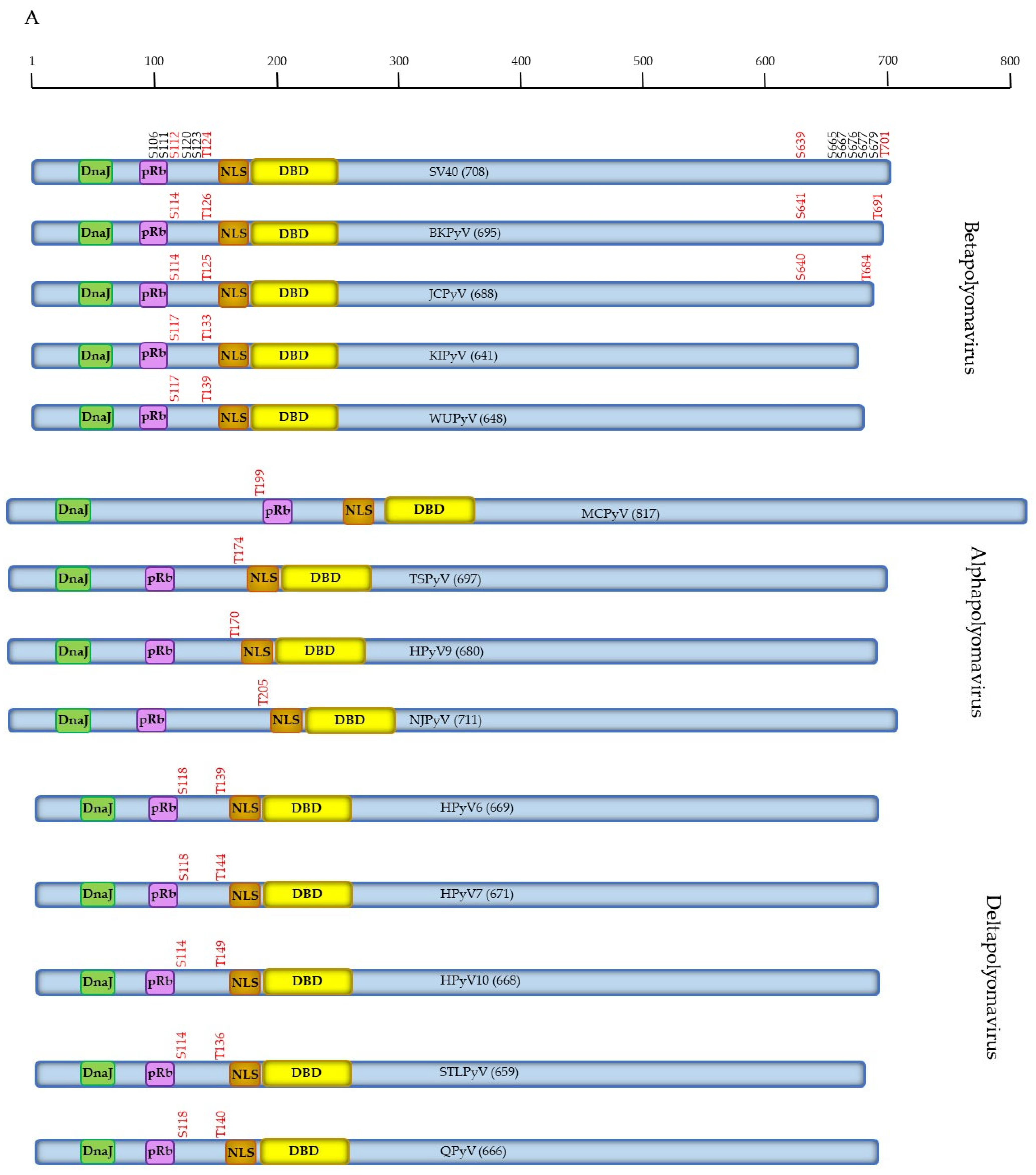
2. Phosphorylation of Large T Antigen

2.1. Functions of SV40 LTAg

SV40 LTAg consists of 708 amino acids and is a multifunctional protein. The structure and different functions have been extensively and excellently reviewed before and will therefore only be briefly mentioned in this review [59,60,61,62,63,64,65,66]. Because of the sequence homology with LTAg from HPyVs, it is assumed that HPyV LTAg exhibits similar functions. The LTAg of SV40 is absolutely required for replication of viral DNA. LTAg is the only viral protein involved in replication of the viral genome. LTAg binds to repeated 5′-GAGGC-3′ motifs in the non-coding control region, which consists of the origin of replication (ORI) and the transcriptional control region. The core ORI consists of LTAg binding site I with tandem repeats of the GAGGC motif, the early palindrome, which also contains two GAGGC motifs in the opposite direction, a central palindrome encompassing binding site II with double tandem GAGGC repeats in opposite direction, and an AT-rich region [66,67,68]. DNA replication is initiated when two hexamers of LTAg attach in a head-to-head orientation at site II in the ORI. LTAg recruits cellular enzymes involved in DNA replication, and the intrinsic helicase activity of LTAg helps unwind double-stranded DNA. Following initiation, the double hexamers separate from each other and move in opposite directions to replicate viral DNA in a bidirectional manner [59,66,68]. As will be outlined in Section 2.3.2 phosphorylation of LTAg regulates the role of LTAg in viral DNA replication. In addition, LTAg is involved in regulating transcription of the early and late genes. LTAg can also transform cells in vitro and induce tumors in animal models [59,63,65,66]. SV40 DNA, RNA and the LTAg protein have been detected in human cancers, such as mesothelioma and brain cancer tissue, but SV40’s role in human malignancy remains to be discussed [69,70,71].

2.2. Putative and Proven Phosphorylation Sites in SV40 LTAg

The phosphorylation prediction algorithm program Netphos 3.1 was used to identify putative phosphoacceptor sites and protein kinases that mediate their phosphorylation [72,73]. The results are summarized in Supplementary Table S1 and predicted more than 50 phosphoacceptor sites spread throughout the LTAg sequence. However, studies have shown that SV40 LTAg is only phosphorylated in the N- and C-terminal region of the protein at residues S106, S111, S112, S120, S123, T124, S639, S665, S667, S676, S677, S679 and T701, whilst the central part appears to be unphosphorylated (see Table 1; [74,75,76]), although not all studies confirmed phosphorylation of S676 [74]. Phosphorylation could be mediated in vitro and/or in vivo by casein kinases 1 and 2 (CKI and CKII), DNA-dependent protein kinase (DNAPK), glycogen synthase kinase-3 (GSK3), cyclin-dependent kinase 1 (CDK1 or previously known as cell division cycle protein 2 homolog cdc2), and ataxia-telangiectasia-mutated (ATM). The protein kinases cAMP-dependent protein kinase (or protein kinase A, PKA), cGMP-dependent protein kinase (or PKG), PKC, protein kinase N1 (PKN1 or protein kinase C-related kinase 1, PRK1), and PKN2 failed to mediate in vitro phosphorylation of SV40 LTAg [75,77]. The biological effects of mutation in the phosphoacceptor sites of SV40 are summarized in Table 1 and will be discussed in detail in the following section.

2.3. Biological Implications of SV40 LTAg Phosphorylation

2.3.1. SV40 LTAg Phosphorylation and Subcellular Distribution

SV40 LTAg, like all eukaryotic proteins, is synthesized in the cytoplasm, but it is mainly relocalized to the nucleus. A minor fraction associates with the plasma membrane and is exposed to the cell surface [88]. Nuclear import is regulated by a nuclear localization report (NLS) with the sequence PKKKRKV (residues 126–131) and the nuclear transport factor importin [89]. Early studies showed differences in the phosphorylation of cytoplasmic and nuclear LTAg, suggesting that in addition to the NLS, phosphorylation may affect the subcellular localization of LTAg [90]. Indeed, it was found that phosphorylation either inhibited or stimulated nuclear import depending on the residues phosphorylated. Phosphorylation of S106 by CKI or GSK3 inhibited nuclear import by approximately 50% compared to non-phosphorylated LTAg [75]. The group of Jans showed that the binding of the retinoblastoma family member p110 (RB1) to SV40 LTAg inhibited the nuclear import of LTAg, and phosphorylation of S106 within LTAg enhanced the interaction with RB1 and further reduced the nuclear import. However, RB1 binding did not impair the NLS of LTAg interacting with importin [91]. It should be mentioned that these studies were performed with GFP fusion proteins containing aa 87 (respectively 102 and 110) −135 of LTAg, so that the effect of phosphorylation in the C-terminal part on RB1-mediated cytoplasmic retention was not examined. Phosphorylation of T124 by CDK1 also had a negative effect on the nuclear import [81,92,93,94]. Nuclear accumulation of the LTAg T124A mutant increased compared to wild-type LTAg, and the T124D mutant reduced the nuclear import of LTAg [95], as reviewed in [17]. Fulcher et al. showed that the BRCA1 binding protein 2 (BRAP2) binds LTAg and blocks nuclear import. Phosphorylation of T124 and an intact NLS are necessary for the interaction between LTAg and BRAP2, and this association prevents LTAg from entering the nucleus. The T124A mutation abrogates binding of BRAP2 to LTAg, whereas T124D increases the affinity of BRAP2 for LTAg, explaining the increased or reduced nuclear import of LTAg, respectively [95]. Phosphorylation of S111/S112 by CKII and S120 by DNAPK enhanced the nuclear import, and mutation/deletion of S111/S112 to prevent phosphorylation decreased the nuclear import rate [81,92,93,94,96]. The LTAg S112D mutant displayed enhanced nuclear import compared to wild-type LTAg [92,97], as reviewed in [17]. CKII-mediated phosphorylation of S111/S112 increased the affinity of importin for LTAg approximately 50-fold, whilst phosphorylation of S120 by DNAPK enhanced importin binding by 40% and 100-fold in synergy with CKII-catalyzed phosphorylation of S111/S112 [93], as reviewed in [17]. S112 seems to be the main site of phosphorylation by CKII [98]. The role of S120 in nuclear import was elucidated by generating the LTAg mutants S120A and S120D. Both mutations reduced nuclear accumulation by 70% and 40%, respectively, compared to wild-type LTAg, but the S120D mutant showed a faster import rate than wild-type LTAg [94]. The participation of S123 in nuclear localization was also examined but revealed that mutation of S123 did not influence the nuclear transport rate of SV40 LTAg [81]. Taken together, the phosphorylation of LTAg allows finetuning of its subcellular localization and may therefore play an import role in the viral life cycle because this protein is required for viral DNA replication and transcription, which occur in the nucleus [61,65,66].

2.3.2. SV40 LTAg Phosphorylation and Replication

A major role of LTAg involves viral DNA replication and requires that LTAg binds to the ORI. Upon binding to the GAGGC motifs in site II of the ORI, LTAg monomers assemble into hexamers and subsequently into double hexamers [99,100]. It was shown that the phosphorylation pattern of unbound and DNA-associated LTAg is different [90]. This suggested that phosphorylation may affect the binding of LTAg to DNA. Indeed, treatment of LTAg with potato acid phosphatase, which removes Ser- and Thr-bound phosphate groups, reduced LTAg binding to SV40 DNA [101], whereas treatment with alkaline phosphatase, which removes Ser-bound phosphate groups, increased DNA binding [102,103]. Simmons and colleagues found that alkaline phosphatase treatment of immunoprecipitated LTAg increased its in vitro DNA binding activity, whereas phosphorylation of the N-terminal region (residues 106–124) reduced in vitro DNA binding [104]. Other groups reported that treatment with calf intestinal alkaline phosphatase, which removes Ser-bound phosphates, enhanced LTAg’s ability to stimulate DNA replication but had no effect on DNA binding nor on the ATPase activity of LTAg [105,106,107]. McVey and co-workers demonstrated, using bacterial-expressed LTAg, that T124 was a phosphoacceptor site for CDK1. This in vitro T124 phosphorylated purified LTAg, bound better to SV40 ORI, and stimulated SV40 DNA replication compared to unphosphorylated purified LTAg under in vitro conditions [86]. In vivo phosphorylation of LTAg at T124 by CDK1 also stimulated viral DNA replication. Phosphorylation of T124 by CDK1, a master regulator of the cell cycle [108], stimulated LTAg binding to the ORI and DNA unwinding, but had no effect on DNA polymerase α binding or the and helicase and transcriptional activity of LTAg [109]. Later studies showed that phosphorylation of T124 had no effect on the formation of the first hexamer but promoted double hexamer formation [110]. This was confirmed by the fact that the LTAg T124A mutant, although able to bind the SV40 ORI, was defective in the initial opening of the duplex at the ORI, possibly because of distorted double hexamer formation [111]. The ability of LTAg phosphorylated at T124 to enhance SV40 DNA replication seems contradictory to the finding that phosphorylation of this residue inhibits the nuclear import of LTAg, as described in Section 2.3.2. Phosphorylation of T124 may occur in a cell-cycle-dependent manner because CDK1, which mediates phosphorylation of this site, is regulated in a cell-cycle-dependent manner [108]. The observation that phosphorylation of LTAg in the cytoplasm was relatively stable, whereas phosphorylation of LTAg in the nucleus exhibited a higher turnover rate, may restrict the time during which LTAg contributes to viral DNA replication and regulate the switch to the late phase in the viral life cycle, which includes expression of the capsid proteins and their assembly with viral DNA into new virus particles [112].
Other phosphorylation events are involved in viral DNA replication by LTAg. Vishrup et al. demonstrated that PP2A removed phosphates from phosphoSer 120, 123, 677, and perhaps 679, but not phosphoThr residues in purified 32P-labeled LTAg, and purified PP2A preferentially stimulated SV40 DNA replication in extracts from early G1 phase cells [83,113]. Dephosphorylation of LTAg by PP2A promoted binding of the second LTAg hexamer to the SV40 ORI, thereby stimulating DNA unwinding [114]. CKI phosphorylated residues S120, S123, S676, and S679 (Table 1). CKI-catalyzed phosphorylation of S120 and S123 inhibited viral DNA replication, whereas phosphorylation of S676 and S679 were not required for the inhibition of replication [77]. Dephosphorylation of phosphoS120 and phosphoS123 by PP2A were required for full activation of LTAg replication potential in vitro [77,83,86,105]. Phosphorylation occurred on full-length LTAg (FL-LTAg) but not on an N-terminal fragment containing the first 259 amino acids. The authors suggested that CKI-mediated phosphorylation required a three-dimensional structure positioning the N-terminal and C-terminal domains in proximity [77]. This assumption is underscored by the finding that mutations in the phosphoacceptor sites in the N-terminal part reduced the phosphorylation of residues in the C-terminal domain and vice versa. For example, mutation of S106 prevented the phosphorylation of S639, and mutation of S677 reduced the phosphorylation of S120 and S123 [74]. In agreement with this mechanism, Scheidtman et al. proposed that phosphorylation of Ser-677 is required for the subsequent phosphorylation of S120 and S123 [74]. S120 is also the substrate for ataxia-telangiectasia-mutated (ATM). However, in contrast to CKI-mediated phosphorylation, phosphorylation of S120 by ATM activated SV40 replication in CV1 African green monkey cells [85]. Moreover, an SV40 mutant encoding LTAg S120A displayed reduced replication in monkey cells compared to wild-type SV40 [78,85]. The opposite effect of S120 phosphorylation on SV40 replication may be explained by the experimental conditions (in vitro versus in vivo). Alternatively, the phosphorylation of S120 by CKI and ATM may be timeously regulated and result in the stimulation or inhibition of viral DNA replication in a time-dependent manner. Hence, the timing of S120 phosphorylation may be crucial to prevent premature SV40 DNA replication [85]. Interestingly, SV40 sTAg prevented dephosphorylation of LTAg by PP2A, preferentially of S120 and S123 [115]. Hence, sTAg may regulate the replication activities of LTAg.

2.3.3. SV40 LTAg Phosphorylation and Protein Interaction

SV40 LTAg can interact with several proteins (see Supplementary Table S2 in [53]). One of the interaction partners is F-Box and WD Repeat Domain Containing 7, E3 Ubiquitin Protein Ligase (Fbxw7), which is part of the phosphorylation-dependent ubiquitination process in proteasomal degradation of substrates [116]. Fbxw7 was shown to bind to SV40 LTAg in a phosphoT701-dependent manner [117]. As mentioned above, phosphorylation of SV40 LTAg at T124 is required for the interaction with BRAP2, and this association inhibits nuclear import of LTAg [95]. Whether binding of SV40 LTAg to other proteins depends on phosphorylation was not examined.

2.3.4. SV40 LTAg Phosphorylation and Transformation

Phosphorylation of specific residues in the C-terminal part had an effect on the transforming activities of LTAg (Table 1). The mutations S639A and S67A enhanced the transforming activity in Rat-1 cells, whereas the substitutions S667A and S679A had the opposite effect [74,78]. In the same cell line, LTAg S106F and S189D displayed reduced transforming activity [74,118]. The latter site is not a known phosphorylation site, although it is part of a weak PKA and RSK consensus motif (Supplementary Table S1), suggesting that phosphorylation is not the only mechanism to control the transforming activity of LTAg. Another study, performed on Rat-2 cells, reported that the LTAg S106A mutant displayed comparable transforming activity to wild-type LTAg (Table 1; [78]).
In conclusion, the phosphorylation state of SV40 LTAg has an impact on its subcellular localization and on viral DNA replication. The phosphorylation profile of residues S106, S111/S112, and T124 controls the nuclear import of LTAg, whereas phosphorylation of T124 is crucial for viral DNA replication. LTAg can bind to p53 and pRb, and this will induce cell progression, resulting in activation of CDK1 and PP2A. CDK1 mediates the phosphorylation of T124, whereas PP2A dephosphorylates Ser residues. The role of phosphorylation of the C-terminal Ser residues could be to repress early transcription and stimulate late transcription, but this remains to be determined (and is reviewed in [119]). Despite its role in Fbxw7 interaction (Section 2.3.3), the biological implication of T701 phosphorylation remains elusive because the LTAg T701A showed no differences to wild-type LTAg concerning virus viability, replication, transforming ability, nuclear localization, site I and site II binding, expression levels, and ATPase activity (Table 1; [74,76,78,79]).

2.4. Phosphorylation of HPyV LTAg

2.4.1. Phosphorylation of Human Betapolyomavirus LTAg

Very few studies have investigated the phosphorylation of LTAg of HPyVs. Tryptic phosphopeptide mapping revealed that JCPyV LTAg is phosphorylated at Ser and Thr but not Tyr residues [120]. Similar to SV40 LTAg, phosphorylation regions were mapped in the N-terminal and C-terminal domains, with the majority of phosphorylation in the N-terminal part. The exact residues were not determined, but by using the LTAg T664A mutant, Swenson and Frisque found that T664 was not phosphorylated. The authors also found that the phosphorylation pattern is slightly different in JCPyV LTAg isolated from human and rat cells. The protein kinases that mediated JCPyV LTAg phosphorylation have not been identified, but CKI can phosphorylate LTAg in vitro [120]. Residues S248 and S640 are predicted as weak CKI phosphoacceptor sites by the Netphos 3.1 algorithm, but CKI-mediated phosphorylation of these amino acids has not been experimentally proven. Based upon sequence homology with SV40 LTAg, one or several of the JCPyV residues S114, S121, S124, and T125 were predicted to be phosphoacceptor sites [121]. Later studies examined the possible phosphorylation and biological role of T125 in JCPyV LTAg, which corresponds to T124 in SV40 LTAg, by generating non-phosphorylable and phosphomimicking mutants [122]. The T125A mutant protein was less stable than wt LTAg, whereas the stability of the T125A variants of T’135, T’136, and T’165 was comparable to wild-type T’ variants. The stability of the T125D LTAg proteins was similar to the wild-type proteins. T125A substitution slightly reduced the ability of mutant LTAg proteins to bind to the retinoblastoma proteins p107 (RBL1) and p110 (RBL2) and failed to release transcription factor E2F from the E2F:RB protein complex. T125A mutants, on the other hand, bound to RBL1 and RBL2 more efficiently compared to wild-type LTAg and released E2F. T125A LTAg mutants had no transforming activity, whereas the T125D protein could transform Rat-2 cells. JCPyV expressing LTAg T125A or T125D failed to replicate in primary human fetal glial cells [121,122]. These results indicate that phosphorylation of T125 has an effect on the biological properties of JCPyV LTAg proteins. Although prediction algorithms can be useful tools to predict phosphosites, of the putative sites S114, S121, S124, and T125 [121], only S121 was suggested as a possible phosphoacceptor site by Netphos 3.1 (Supplementary Table S1). T664 is a predicted weak phosphoacceptor site for ATM and CKII (Supplementary Table S1). However, substitution of T664 into nonphosphorylable alanine had no effect on JCPyV replication in primary human fetal glial cells, and this residue was shown not to be phosphorylated [121].
Only one study reports the functional implication of JCPyV LTAg phosphorylation. Beta-transducin-repeat-containing proteins (BTRCPs or β-TrCPs) are components of the Skp1-Cul-F-box (SCF) protein E3 ubiquitin ligase complex, which plays a role in proteasomal degradation. They interact with the consensus motif DpSGX2–4pS on their substrates [123]. BTRCP1 and BTRCP2 (or FBXW1B) were shown to bind to LTAg of JCPyV in a phosphorylation-dependent manner and involved residues 639–645 (DSGHGSS) [124]. BTRCP1-LTAg interaction required phosphorylated S640 and to a lesser extent phosphorylated S644 of JCPyV LTAg, whereas only phosphorylated S640 seemed to be necessary for interaction with BTRCP2. The JCPyV LTAg-BTRCP interaction may therefore affect the proteasomal pathway and contribute to the pathogenicity of JCPyV. The sequence DSGHGSS is completely conserved in BKPyV LTAg (amino acids 640–646), but not in SV40 (DSGHETG; residues 656–662). SV40 LTAg was unable to bind BTRCPs, but the interaction with BKPyV LTAg was not investigated [124]. The BTRCP binding consensus motif DSGX2–4S is present in the LTAg of the non-Betapolyomaviruses MCPyV (DSGTFSQ; residues 811–817) and HPyV6 (DSTQESG; residues 656–662), but not in the LTAg of other HPyVs. Dysfunction of the proteasomal pathway is associated with cancer, and a role for BKPyV, MCPyV, and HPyV6 in malignancy is known or emerging [125,126,127], suggesting that disturbance of the proteasomal pathway by LTAg of these HPyVs may be a contributing factor in HPyV-induced oncogenesis.
BKPyV is another well-studied Betapolyomavirus. Although phosphorylation of its capsid proteins has been examined and phosphorylation sites have been identified [58,128], little is known about the phosphorylation of its LTAg. Immunoprecipitation of 32P-labelled cells demonstrated that BKPyV is a phosphoprotein, but the phosphoacceptor sites and protein kinases that mediate phosphorylation remain unknown [129].
The phosphorylation of LTAg of the other members of Betapolyomavirus, KIPyV and WUPyV, has not been studied. Of the proven phosphorylation sites in SV40 LTAg (i.e., S106, S111, S112, S120, S123, T124, S639, S665, S667, S676, S677, S679, and T701), S112 (S114 in BKPyV and JCPyV, S117 in KIPyV, and WUPyV) and T124 (T126, T125, T133, and T139 in BKPyV, JCPyV, KIPyV, and WUPyV, respectively) are conserved among LTAg of Betapolyomavirus (Figure 1A and Figure S1). The SV40 S112 corresponding residue in these HPyVs is a putative target for CKII (BKPyV and JCPyV), CDK5 and GSK3 (KIPyV), and CKI, GSK3, and p38MAPK (WUPyV). T124 is part of a conserved TPPKKK motif and is a predicted phosphoacceptor for p38MAPK, CDK5, GSK3, and PKC (Supplementary Table S1). SV40 S123 is only conserved in LTAg of BKPyV (S125) and JCPyV (S124). The C-terminal S639 and T701 residues are also only present in BKPyV (S641 and T691) and JCPyV (S640 and T684) LTAg. Further studies are required to confirm whether these are genuine phosphoacceptor sites and whether their phosphorylation affects the functions of LTAg.

2.4.2. Phosphorylation of Human Alphapolyomavirus LTAg

Phosphorylation of Merkel Cell Polyomavirus Full-Length LTAg

Merkel cell polyomavirus (MCPyV), a causative agent of Merkel cell carcinoma (MCC), was first isolated in 2008 [35]. The virus is present in about 80% of all MCC samples examined and is always integrated in the host genome. MCPyV encodes an 817-amino-acid-long LTAg (hereafter referred to as full-length LTAg or FL-LTAg). Another hallmark of MCPyV-positive MCCs is the presence of a nonsense mutation in the LTAg gene resulting in expression of a C-terminal truncated LTAg [130]. The size of the truncated LTAg (hereafter referred to as tLTAg) described so far ranges from 163 residues [131] to 538 amino acids [unpublished; accession number KJ022619]. Because phosphorylation of both FL- and tLTAg has been studied and sometimes opposite effects have been reported, they are discussed in first two separate subsections in Section 2.4.2, respectively. FL-LTAg has a statistical overrepresentation of Ser residues, especially in the N-terminal half of the protein. Thr is also slightly overrepresented in this region, suggesting that many of these sites may function as phosphoacceptor sites (Supplementary Figure S1). Many of these sites are putative phosphorylation sites for protein kinases (Supplementary Table S1). Because of its pathogenic property, the phosphorylation of MCPyV LTAg has been more intensively studied than any other HPyV. A restricted number of putative phosphorylation sites have been experimentally confirmed. Mass spectrometry analysis of FL-LTAg showed that at least 17 Ser and Thr residues were phosphorylated in the amino acids 1–278 [132]. These include S100, S134, S147, T192, S239, S254, T257, S265, and T271. In addition, at least one of the residues 172–179 (TSSSGSSS), S202, S203 and T205, 217–220 (SLSS), S225, S226, and S227 is phosphorylated [132]. Another study confirmed the phosphorylation of T271 and additionally identified the phosphorylation of T295 and T299 in FL-LTAg expressed in the human embryonal kidney cell line HEK293 [133].
So far, only two protein kinases that can phosphorylate MCPyV LTAg have been identified. You and collaborators showed that ATM phosphorylates S816A, and this partially reverses inhibition of the growth of the human papillomavirus-negative cervical cancer cell line C33A compared to wild-type FL-LTAg and reduces apoptosis [134]. As virus-positive MCCs express C-terminal truncated LTAg lacking this residue, the biological importance of this phosphorylation event does not seem to be relevant in MPyV-positive MCCs. We have previously shown that S203 and S265 are in vitro phosphorylated by cAMP-dependent protein kinase (or PKA) [135]. Mutations in these residues repressed the transcriptional activity of FL-LTAg, and single Ser into Ala and Ser into Asp mutations had no effect on the transcriptional activity [135].
Mutation studies have shown that MCPyV LTAg with either S96A, S134A, S179A, S186A, T192, T268A, T271A, T299A, T309A, S597A, S715A, or S816A had no effect on the half-life of the full-length protein [136]. Mutation S142A increased the half-life of FL-LTAg and reduced the interaction with β-TrCP [137]. S147A also stabilized FL-LTAg, which did not affect the transcriptional activity of the protein, but this mutant could not stimulate viral replication [136,137]. The S220A mutation increased the half-life, viral DNA replication, and transcriptional activity, but impaired RB1 binding and reduced the interaction with SCF protein E3 ubiquitin ligase component S-phase kinase associated protein 2 (SKP2) [132,136,137,138]. FL-LTAg was shown to bind to the hVam6p (or VPS39) subunit of the HOPS complex, a protein that promotes clustering and fusion of late endosomes and lysosomes [139], through residues 204–218. Mutations T205A, Y206A, and T208A in putative phosphorylation sites did not interfere with this interaction, suggesting that phosphorylation of MCPyV LTAg is not required for the association with hVam6p [140]. The FL-LTAg mutants T271A, T297A, and T299 were tested for their effect on ORI binding and replication of viral DNA. The T291A substitution had no effect, whereas T297A increased and T299A reduced LTAg binding to the ORI and impaired replication [133].

Phosphorylation of Merkel Cell Polyomavirus Truncated LTAg

Our group demonstrated that tLTAg stimulated the MCPyV promoters, and Ser into Ala mutations in the PKA phosphorylation sites S203 and S265 inhibited tLTAg’s transcriptional activity by 50–80% compared to non-mutated tLTAg. Single Ser into Asp mutations restored the transcriptional activity of tLTAg [135]. Studies by another group showed that single non-phosphorylable or phosphomimicking mutations in tLTAg of T192, S202, S203, T205, S217, or S239 had no effect on the growth of the MCC cell line MKL1 [132]. S219A or S220A, but not S219E or S220E, tLTAg partially inhibited (20% and 60%, respectively) the growth of these cells [132]. S239A tLTAg had increased half-life compared to unmutated tLTAg and reduced interaction with Fbxw7 [136,137]. As discussed in Section 2.3.1, the phosphorylation of residues adjacent to the NLS played a role in the nuclear import of SV40 LTAg. The MCPyV LTAg possesses the NLS motif RKRK (residues 277–280). The S246A/S247A/S254A/T25 7A/S265A/T271A mutant had no effect on the nuclear translocation of tLTAg (aa1–334) in the mouse fibroblast NIH3T3 cell line [132]. The effect of these substitutions in context of FL-LTAg was not investigated.

Phosphorylation of Human Alphapolyomavirus LTAg

To the best of our knowledge, the phosphorylation of LTAg of the other Alphapolyomavirus species TSPyV, HPyV9, and NJPyV has not been examined. SV40 T124, which was also conserved in the Betapolyomavirus, is also conserved in the Alphapolyomavirus (T199 in MCPyV, T174 in TSPyV, T170 in HPyV9, and T205 in NJPyV; Figure 1A). This may suggest that the T124 corresponding residue in LTAg of HPyV is a genuine phosphoacceptor site.

2.4.3. Phosphorylation of Human Deltapolyomavirus LTAg

Studies investigating the phosphorylation status of Deltapolyomavirus LTAg are lacking. Two proven SV40 phosphoacceptor sites are conserved in LTAg of Deltapolyomavirus: S112 and T124 (Figure 1A and Figure S1). The corresponding S112 residue is S118 in HPyV6, HPyV7, and QPyV and S114 in HPyV10 and STLPyV, whilst T124 is T139 in HPyV6, T144 in HPyV7, T140 in QPyV, T149 in HPyV10, and T136 in STLPyV.
In conclusion, knowledge about the phosphorylation of HPyV LTAg is almost non-existent. The SV40 T124 residue is conserved in LTAg of all HPyVs, whereas SV40 S112 is conserved in Beta- and Deltapolyomaviruses. T124 is part of a conserved TPP (K/R)4 motif found in the LTAg of all HPyVs. However, neither their phosphorylation and the protein kinases that catalyze this posttranslational modification have been demonstrated, nor has the biological function of their possible phosphorylation been revealed.

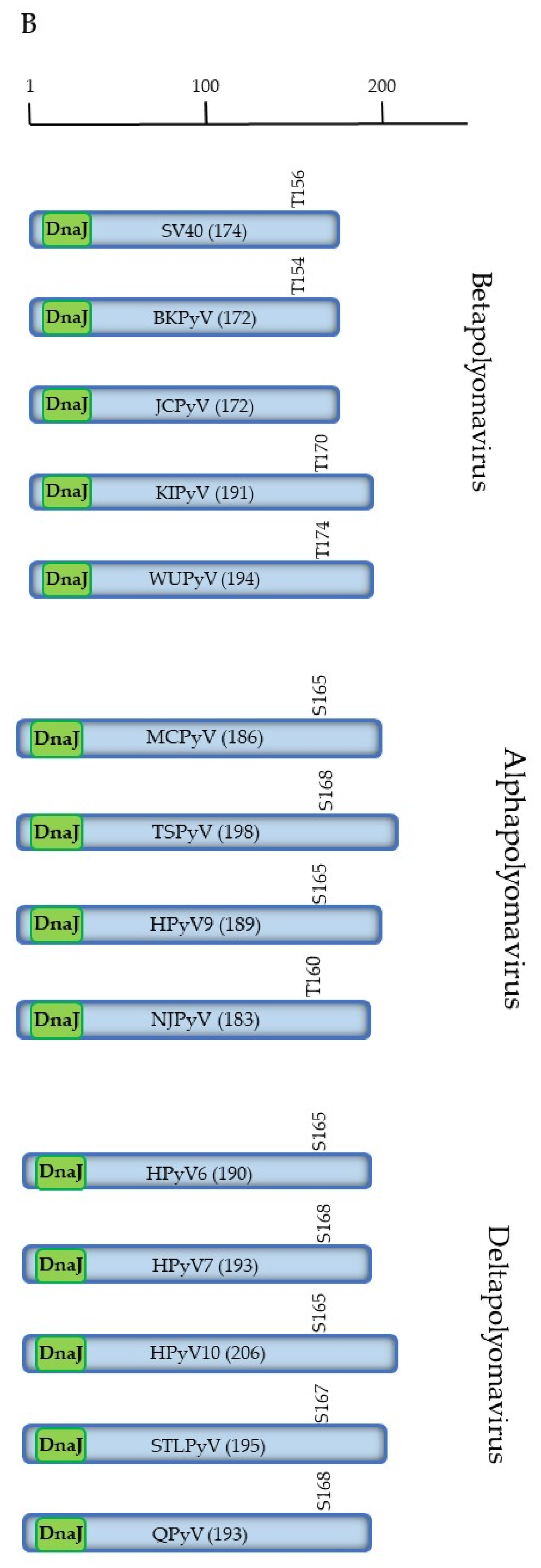
3. Phosphorylation of sTAg

3.1. Function of SV40 sTAg

SV40 sTAg is generated through alternative splicing of the early transcript, and the N-terminal 82 amino acids are identical to LTAg [61]. sTAg can transactivate different promoters and exerts an auxiliary role in the transformation of certain cell types by LTAg, but in some cells, sole expression of sTAg is sufficient for transformation [141,142]. The major contribution of sTAg in transformation is its ability to inhibit protein phosphatase PP2A activity, thereby perturbing the phosphorylation pattern of cellular proteins, but also affecting phosphorylation of LTAg [143].
Although it is assumed that sTAg of HPyV possesses the same properties as SV40 sTAg, MCPyV sTAg exerts different functions. It can fully transform Rat-1 and NIH3T3 mouse fibroblasts and also interacts with PP4. This interaction has an inhibitory effect on the NFκB signaling pathway and promotes microtubule destabilization, cell mobility, and filopodium formation (reviewed in [144]).

3.2. Putative Phosphorylation Sites of SV40 sTAg

Reports describing SV40 sTAg as a phosphoprotein are lacking. Since the first 82 amino acids are shared with LTAg and these are not phosphorylated in LTAg, it is unlikely that phosphorylation of the N-terminal region of sTAg occurs. In agreement with this assumption was the finding that a fragment comprising the 82 N-terminal aa of LTAg (which is identical to sTAg) did not show phosphorylation [145]. However, it cannot be ruled out that the configuration of full-length sTAg allows phosphorylation of this part of the protein, whereas in LTAg, the three-dimensional structure prevents phosphorylation of the first 82 residues because of the accessibility/inaccessibility of protein kinases. Bona fide phosphorylation sites in the unique sequence of sTAg are not known. Putative phosphorylation sites for SV40 sTAg include S10, S22, S57, T81, S87, Y96, S108, T167, and T168 (Supplementary Table S2). PKA (S10) and PKC (T57 and T167) may mediate the phosphorylation of these sites as their predicted score is relatively high (>0.750; with 0.000 = no similarity with the protein kinase consensus and 1.000 = completely identical to the consensus motif).
Phosphorylation of HPyV sTAg has, to the best of our knowledge, not been reported. Predicted phosphoacceptor sites with putative protein kinases that may mediate their phosphorylation are summarized in Supplementary Table S2. Conserved residues between all HPyVs or between species from the Alpha-, Beta-, and Deltapolyomavirus genera are depicted in Supplementary Figure S2. Residue T156 in SV40 sTAg is conserved in all HPyV as either T or S, except in JCPyV sTAg, which has A in this position. In SV40, T156 is part of a PKC consensus motif, whereas in BKPyV, the corresponding residue (T154) is not part of a protein kinase recognition site. The corresponding site in KIPyV (T170), WUPyV (T174), and STLPyV (S167) is a putative target for PKC (Supplementary Table S2). Alpha- and Betapolyomaviruses have conserved S or T in sTAg: S86 and S92 in NJPyV and TSPyV, respectively; T90 in MCPyV and HPyV10, T91 (HPyV6, HPyV7, HPyV9, QPyV), and T92 in STLPyV. However, no or weak consensus motifs are found for protein kinases, except for T90 in MCPyV sTAg and T91 in HPyV6, which might be sites for PKC (Supplementary Table S2). SV40 sTAg Y139 is conserved in sTAg of all human Alpha- and Betapolyomaviruses but is not predicted to be phosphoacceptor site for the protein kinases ATM, CDK1, CDK5, CKI, CKII, CaMKII, DNAPK, EGFR, GSK3, INSR, p38MAPK, PKA, PKB, PKC, PKG, RSK, and SRC used in the Netphos 3.1 algorithm [72,73].
The residues Y96 and S108 are conserved in sTAg of BKPyV and JCPyV. T167 is conserved in all human Betapolyomaviruses (BKPyV, JCPyV, KIPyV, WUPyV), in the Deltapolyomaviruses HPyV6, HPyV10, QPyV, and HPyV7 (in the latter residue 167 is Ser), and in the Alphapolyomavirus MCPyV.
The residues T156L157 in SV40 sTAg are conserved in the corresponding sites of the other HPyVs as T/SL/F, except JCPyV, which contains the amino acid sequence AL (Supplementary Figure S2).

4. Gaps in Knowledge and Future Research Directions

Our knowledge of HPyV LTAg and sTAg phosphorylation is, at best, limited. Phosphorylation of these PyV regulatory proteins has mainly been studied in SV40 and mouse polyomavirus (MPyV). Most of these studies were performed in the 1980s–1990s and mainly identified phosphoacceptor sites based on Edman degradation of 32P-labeled proteins, whereas protein kinases that catalyze phosphorylation are poorly characterized [74,75,76,146,147]. Comparing the known phosphorylation sites in SV40 and MPyV with the amino acid sequences of HPyV LTAg revealed that only Thr corresponding to T124 in SV40 LTAg (T278 in MPyV; [144]) is conserved (Supplementary Figure S1). As phosphorylation of this residue is crucial for SV40 and MPyV DNA replication [86,147], it may suggest a similar role for the HPyVs. Neither phosphorylation of SV40 nor MPyV sTAg has been reported, nor has this posttranslational modification been examined in HPyV sTAg. Currently, mass spectrometry (MS) is the method of choice for the identification of phosphomodification in a protein [148]. Such studies have only been performed on MCPyV LTAg and in vitro PKA-phosphorylated peptides derived from MCPyV LTAg [132,133,135]. Therefore, mass spectrometry studies on the LTAg and sTAg from HPyVs should be performed. This will allow identification of phosphoacceptor sites. Phosphospecific antibodies can then be generated to confirm the phosphorylation sites, to study subcellular-dependent and cell-cycle-dependent phosphorylation, and to compare the phosphorylation state of these proteins in healthy and diseased tissue. The latter may provide information about whether aberrant phosphorylation of LTAg and/or sTAg plays a role in HPyV pathogenesis and whether the phosphorylation pattern of LTAg/sTAg can be used as a biomarker. Studies with mutant proteins in which phosphoacceptor sites are substituted may provide information of the biological importance of these phosphoresidues. Another almost untouched research field is the characterization of protein kinases and protein phosphatases that are responsible for phosphorylation and dephosphorylation. Protein kinase analysis using purified LTAg and sTAg and a panel of protein kinases and protein phosphatases may lead to the identification of protein kinases and protein phosphatases responsible for LTAg and sTAg (de)phosphorylation. Phosphorylation of HPyV LTAg and sTAg may be sequential, where a phosphorylation is required before a subsequent phosphorylation can occur. Because posttranslational modifications, including phosphorylation, of viral proteins can enhance the pathogenic properties of the virus [19,21,149], it is highly relevant to study these in HPyVs, which are involved in diseases such as nephropathy, progressive multifocal leukoencephalopathy, and malignancies [127,130,150]. Our understanding of the phosphorylation of the LTAg and sTAg of HPyVs may help to develop new therapeutic strategies, and the phosphorylation state of these proteins may serve as clinical tools for disease diagnosis, prognosis, and treatment response assessment.

Supplementary Materials

The following supporting information can be downloaded at: https://www.mdpi.com/article/10.3390/v15112235/s1, Figure S1: Alignment of LTAg from SV40 and HPyVs; Figure S2: Alignment of sTAg from SV40 and HPyVs; Table S1: Putative phosphorylation sites in SV40 and HPyV LTAg and their protein kinases; Table S2: Putative phosphorylation sites in SV40 and HPyV sTAg and their protein kinases.

Author Contributions

Conceptualization, U.M. and V.P.; data collection, U.M., S.P., M.F., B.S. and V.P.; writing—original draft preparation, U.M., S.P., M.F., B.S. and V.P.; writing—review and editing, U.M., S.P., M.F., B.S. and V.P. All authors have read and agreed to the published version of the manuscript.

Funding

The APC was covered by funds from The University of Tromsø—The Arctic University of Norway.

Institutional Review Board Statement

Not applicable.

Informed Consent Statement

Not applicable.

Data Availability Statement

Not applicable.

Conflicts of Interest

The authors declare no conflict of interest.

References

  1. Denu, J.M.; Stuckey, J.A.; Saper, M.A.; Dixon, J.E. Form and function in protein dephosphorylation. Cell 1996, 87, 361–364. [Google Scholar] [CrossRef]
  2. Ubersax, J.A.; Ferrell, J.E., Jr. Mechanisms of specificity in protein phosphorylation. Nat. Rev. Mol. Cell. Biol. 2007, 8, 530–541. [Google Scholar] [CrossRef] [PubMed]
  3. Manning, G.; Whyte, D.B.; Martinez, R.; Hunter, T.; Sudarsanam, S. The protein kinase complement of the human genome. Science 2002, 298, 1912–1934. [Google Scholar] [CrossRef] [PubMed]
  4. Chen, M.J.; Dixon, J.E.; Manning, G. Genomics and evolution of protein phosphatases. Sci. Signal. 2017, 10, eaag1796. [Google Scholar] [CrossRef] [PubMed]
  5. Cohen, P. The origins of protein phosphorylation. Nat. Cell. Biol. 2002, 4, E127–E130. [Google Scholar] [CrossRef] [PubMed]
  6. Olsen, J.V.; Blagoev, B.; Gnad, F.; Macek, B.; Kumar, C.; Mortensen, P.; Mann, M. Global, in vivo, and site-specific phosphorylation dynamics in signaling networks. Cell 2006, 127, 635–648. [Google Scholar] [CrossRef]
  7. Cohen, P. The regulation of protein function by multisite phosphorylation—A 25 year update. Trends Biochem. Sci. 2000, 25, 596–601. [Google Scholar] [CrossRef] [PubMed]
  8. Nishi, H.; Hashimoto, K.; Panchenko, A.R. Phosphorylation in protein-protein binding: Effect on stability and function. Structure 2011, 19, 1807–1815. [Google Scholar] [CrossRef] [PubMed]
  9. Gjertsen, B.T.; Døskeland, S.O. Protein phosphorylation in apoptosis. Biochim. Biophys. Acta 1995, 1269, 187–199. [Google Scholar] [CrossRef] [PubMed]
  10. Humphrey, S.J.; James, D.E.; Mann, M. Protein Phosphorylation: A Major Switch Mechanism for Metabolic Regulation. Trends Endocrinol. Metab. 2015, 26, 676–687. [Google Scholar] [CrossRef] [PubMed]
  11. Ardito, F.; Giuliani, M.; Perrone, D.; Troiano, G.; Lo Muzio, L. The crucial role of protein phosphorylation in cell signaling and its use as targeted therapy (Review). Int. J. Mol. Med. 2017, 40, 271–280. [Google Scholar] [CrossRef]
  12. Bilbrough, T.; Piemontese, E.; Seitz, O. Dissecting the role of protein phosphorylation: A chemical biology toolbox. Chem. Soc. Rev. 2022, 51, 5691–5730. [Google Scholar] [CrossRef] [PubMed]
  13. Cohen, P. The role of protein phosphorylation in human health and disease. The Sir Hans Krebs Medal Lecture. Eur. J. Biochem. 2001, 268, 5001–5010. [Google Scholar] [CrossRef]
  14. Stebbing, J.; Lit, L.C.; Zhang, H.; Darrington, R.S.; Melaiu, O.; Rudraraju, B.; Giamas, G. The regulatory roles of phosphatases in cancer. Oncogene 2014, 33, 939–953. [Google Scholar] [CrossRef]
  15. Delom, F.; Chevet, E. Phosphoprotein analysis: From proteins to proteomes. Proteome Sci. 2006, 4, 15. [Google Scholar] [CrossRef] [PubMed]
  16. Yu, L.R.; Veenstra, T.D. Characterization of Phosphorylated Proteins Using Mass Spectrometry. Curr. Protein Pept. Sci. 2021, 22, 148–157. [Google Scholar] [CrossRef]
  17. Fulcher, A.J.; Jans, D.A. Regulation of nucleocytoplasmic trafficking of viral proteins: An integral role in pathogenesis? Biochim. Biophys. Acta 2011, 1813, 2176–2190. [Google Scholar] [CrossRef] [PubMed]
  18. Yang, F. Post-translational Modification Control of HBV Biological Processes. Front. Microbiol. 2018, 9, 2661. [Google Scholar] [CrossRef] [PubMed]
  19. Kumar, R.; Mehta, D.; Mishra, N.; Nayak, D.; Sunil, S. Role of Host-Mediated Post-Translational Modifications (PTMs) in RNA Virus Pathogenesis. Int. J. Mol. Sci. 2020, 22, 323. [Google Scholar] [CrossRef]
  20. Zhou, T.; Wang, M.; Cheng, A.; Yang, Q.; Tian, B.; Wu, Y.; Jia, R.; Chen, S.; Liu, M.; Zhao, X.X.; et al. Regulation of alphaherpesvirus protein via post-translational phosphorylation. Vet. Res. 2022, 53, 93. [Google Scholar] [CrossRef]
  21. Cheng, N.; Liu, M.; Li, W.; Sun, B.; Liu, D.; Wang, G.; Shi, J.; Li, L. Protein post-translational modification in SARS-CoV-2 and host interaction. Front. Immunol. 2023, 13, 1068449. [Google Scholar] [CrossRef] [PubMed]
  22. Zheng, K.; Ren, Z.; Wang, Y. Serine-arginine protein kinases and their targets in viral infection and their inhibition. Cell. Mol. Life Sci. 2023, 80, 153. [Google Scholar] [CrossRef] [PubMed]
  23. Guan, K.L.; Broyles, S.S.; Dixon, J.E. A Tyr/Ser protein phosphatase encoded by vaccinia virus. Nature 1991, 350, 359–362. [Google Scholar] [CrossRef] [PubMed]
  24. Liu, K.; Lemon, B.; Traktman, P. The dual-specificity phosphatase encoded by vaccinia virus, VH1, is essential for viral transcription in vivo and in vitro. J. Virol. 1995, 69, 7823–7834. [Google Scholar] [CrossRef]
  25. Guan, K.L.; Dixon, J.E. Bacterial and viral protein tyrosine phosphatases. Semin. Cell. Biol. 1993, 4, 389–396. [Google Scholar] [CrossRef]
  26. Leader, D.P. Viral protein kinases and protein phosphatases. Pharmacol. Ther. 1993, 59, 343–389. [Google Scholar] [CrossRef] [PubMed]
  27. Jacob, T.; Van den Broeke, C.; Favoreel, H.W. Viral serine/threonine protein kinases. J. Virol. 2011, 85, 1158–1173. [Google Scholar] [CrossRef]
  28. Segovia, D.; Haouz, A.; Porley, D.; Olivero, N.; Martínez, M.; Mariadassou, M.; Berois, M.; André-Leroux, G.; Villarino, A. OH1 from Orf Virus: A New Tyrosine Phosphatase that Displays Distinct Structural Features and Triple Substrate Specificity. J. Mol. Biol. 2017, 429, 2816–2824. [Google Scholar] [CrossRef]
  29. Polyomaviridae Study Group of the International Committee on Taxonomy of Viruses; Calvignac-Spencer, S.; Feltkamp, M.C.; Daugherty, M.D.; Moens, U.; Ramqvist, T.; Johne, R.; Ehlers, B. A taxonomy update for the family Polyomaviridae. Arch. Virol. 2016, 161, 1739–1750. [Google Scholar] [CrossRef]
  30. Moens, U.; Calvignac-Spencer, S.; Lauber, C.; Ramqvist, T.; Feltkamp, M.C.W.; Daugherty, M.D.; Verschoor, E.J.; Ehlers, B.; Ictv Report Consortium. ICTV Virus Taxonomy Profile: Polyomaviridae. J. Gen. Virol. 2017, 98, 1159–1160. [Google Scholar] [CrossRef]
  31. Gardner, S.D.; Field, A.M.; Coleman, D.V.; Hulme, B. New human papovavirus (B.K.) isolated from urine after renal transplantation. Lancet 1971, 1, 1253–1257. [Google Scholar] [CrossRef]
  32. Padgett, B.L.; Walker, D.L.; ZuRhein, G.M.; Eckroade, R.J.; Dessel, B.H. Cultivation of papova-like virus from human brain with progressive multifocal leucoencephalopathy. Lancet 1971, 1, 1257–1260. [Google Scholar] [CrossRef]
  33. Allander, T.; Andreasson, K.; Gupta, S.; Bjerkner, A.; Bogdanovic, G.; Persson, M.A.; Dalianis, T.; Ramqvist, T.; Andersson, B. Identification of a third human polyomavirus. J. Virol. 2007, 81, 4130–4136. [Google Scholar] [CrossRef]
  34. Gaynor, A.M.; Nissen, M.D.; Whiley, D.M.; Mackay, I.M.; Lambert, S.B.; Wu, G.; Brennan, D.C.; Storch, G.A.; Sloots, T.P.; Wang, D. Identification of a novel polyomavirus from patients with acute respiratory tract infections. PLoS Pathog. 2007, 3, e64. [Google Scholar] [CrossRef] [PubMed]
  35. Feng, H.; Shuda, M.; Chang, Y.; Moore, P.S. Clonal integration of a polyomavirus in human Merkel cell carcinoma. Science 2008, 319, 1096–1100. [Google Scholar] [CrossRef] [PubMed]
  36. Schowalter, R.M.; Pastrana, D.V.; Pumphrey, K.A.; Moyer, A.L.; Buck, C.B. Merkel cell polyomavirus and two previously unknown polyomaviruses are chronically shed from human skin. Cell Host Microbe 2010, 7, 509–515. [Google Scholar] [CrossRef]
  37. van der Meijden, E.; Janssens, R.W.; Lauber, C.; Bouwes Bavinck, J.N.; Gorbalenya, A.E.; Feltkamp, M.C. Discovery of a new human polyomavirus associated with trichodysplasia spinulosa in an immunocompromized patient. PLoS Pathog. 2010, 6, e1001024. [Google Scholar] [CrossRef]
  38. Scuda, N.; Hofmann, J.; Calvignac-Spencer, S.; Ruprecht, K.; Liman, P.; Kühn, J.; Hengel, H.; Ehlers, B. A novel human polyomavirus closely related to the african green monkey-derived lymphotropic polyomavirus. J. Virol. 2011, 85, 4586–4590. [Google Scholar] [CrossRef] [PubMed]
  39. Buck, C.B.; Phan, G.Q.; Raiji, M.T.; Murphy, P.M.; McDermott, D.H.; McBride, A.A. Complete genome sequence of a tenth human polyomavirus. J. Virol. 2012, 86, 10887. [Google Scholar] [CrossRef] [PubMed]
  40. Lim, E.S.; Reyes, A.; Antonio, M.; Saha, D.; Ikumapayi, U.N.; Adeyemi, M.; Stine, O.C.; Skelton, R.; Brennan, D.C.; Mkakosya, R.S.; et al. Discovery of STL polyomavirus, a polyomavirus of ancestral recombinant origin that encodes a unique T antigen by alternative splicing. Virology 2013, 436, 295–303. [Google Scholar] [CrossRef]
  41. Korup, S.; Rietscher, J.; Calvignac-Spencer, S.; Trusch, F.; Hofmann, J.; Moens, U.; Sauer, I.; Voigt, S.; Schmuck, R.; Ehlers, B. Identification of a novel human polyomavirus in organs of the gastrointestinal tract. PLoS ONE 2013, 8, e58021. [Google Scholar] [CrossRef] [PubMed]
  42. Mishra, N.; Pereira, M.; Rhodes, R.H.; An, P.; Pipas, J.M.; Jain, K.; Kapoor, A.; Briese, T.; Faust, P.L.; Lipkin, W.I. Identification of a novel polyomavirus in a pancreatic transplant recipient with retinal blindness and vasculitic myopathy. J. Infect. Dis. 2014, 210, 1595–1599. [Google Scholar] [CrossRef] [PubMed]
  43. Gheit, T.; Dutta, S.; Oliver, J.; Robitaille, A.; Hampras, S.; Combes, J.D.; McKay-Chopin, S.; Le Calvez-Kelm, F.; Fenske, N.; Cherpelis, B.; et al. Isolation and characterization of a novel putative human polyomavirus. Virology 2017, 506, 45–54. [Google Scholar] [CrossRef] [PubMed]
  44. Ondov, B.D.; Starrett, G.J.; Sappington, A.; Kostic, A.; Koren, S.; Buck, C.B.; Phillippy, A.M. Mash Screen: High-throughput sequence containment estimation for genome discovery. Genome Biol. 2019, 20, 232. [Google Scholar] [CrossRef]
  45. Lednicky, J.A.; Arrington, A.S.; Stewart, A.R.; Dai, X.M.; Wong, C.; Jafar, S.; Murphey-Corb, M.; Butel, J.S. Natural isolates of simian virus 40 from immunocompromised monkeys display extensive genetic heterogeneity: New implications for polyomavirus disease. J. Virol. 1998, 72, 3980–3990. [Google Scholar] [CrossRef]
  46. Sweet, B.H.; Hilleman, M.R. The vacuolating virus, S.V.40. Proc. Soc. Exp. Biol. Med. 1960, 105, 420–427. [Google Scholar] [CrossRef]
  47. Koliaskina, G.I. Virological and serological data on latent infection caused by OB40 (SV40) virus in macaca rhesus monkeys. Vopr. Virusol. 1963, 29, 450–452. [Google Scholar] [PubMed]
  48. Fagrouch, Z.; Karremans, K.; Deuzing, I.; van Gessel, S.; Niphuis, H.; Bogers, W.; Verschoor, E.J. Molecular analysis of a novel simian virus 40 (SV40) type in rhesus macaques and evidence for double infections with the classical SV40 type. J. Clin. Microbiol. 2011, 49, 1280–1286. [Google Scholar] [CrossRef]
  49. Gedvilaite, A.; Tryland, M.; Ulrich, R.G.; Schneider, J.; Kurmauskaite, V.; Moens, U.; Preugschas, H.; Calvignac-Spencer, S.; Ehlers, B. Novel polyomaviruses in shrews (Soricidae) with close similarity to human polyomavirus 12. J. Gen. Virol. 2017, 98, 3060–3067. [Google Scholar] [CrossRef] [PubMed]
  50. Kamminga, S.; van der Meijden, E.; Pesavento, P.; Buck, C.B.; Feltkamp, M.C.W. Serology Identifies LIPyV as a Feline Rather than a Human Polyomavirus. Viruses 2023, 15, 1546. [Google Scholar] [CrossRef]
  51. Prezioso, C.; Van Ghelue, M.; Pietropaolo, V.; Moens, U. Detection of Quebec Polyomavirus DNA in Samples from Different Patient Groups. Microorganisms 2021, 9, 1082. [Google Scholar] [CrossRef] [PubMed]
  52. DeCaprio, J.A.; Garcea, R.L. A cornucopia of human polyomaviruses. Nat. Rev. Microbiol. 2013, 11, 264–276. [Google Scholar] [CrossRef] [PubMed]
  53. Moens, U.; Krumbholz, A.; Ehlers, B.; Zell, R.; Johne, R.; Calvignac-Spencer, S.; Lauber, C. Biology, evolution, and medical importance of polyomaviruses: An update. Infect. Genet. Evol. 2017, 54, 18–38. [Google Scholar] [CrossRef] [PubMed]
  54. Prins, C.; Frisque, R.J. JC virus T′ proteins encoded by alternatively spliced early mRNAs enhance T antigen-mediated viral DNA replication in human cells. J. Neurovirol. 2001, 7, 250–264. [Google Scholar]
  55. Carter, J.J.; Daugherty, M.D.; Qi, X.; Bheda-Malge, A.; Wipf, G.C.; Robinson, K.; Roman, A.; Malik, H.S.; Galloway, D.A. Identification of an overprinting gene in Merkel cell polyomavirus provides evolutionary insight into the birth of viral genes. Proc. Natl. Acad. Sci. USA 2013, 110, 12744–12749. [Google Scholar] [CrossRef] [PubMed]
  56. van der Meijden, E.; Kazem, S.; Dargel, C.A.; van Vuren, N.; Hensbergen, P.J.; Feltkamp, M.C. Characterization of T Antigens, Including Middle T and Alternative T, Expressed by the Human Polyomavirus Associated with Trichodysplasia Spinulosa. J. Virol. 2015, 89, 9427–9439. [Google Scholar] [CrossRef] [PubMed]
  57. Ponder, B.A.; Robbins, A.K.; Crawford, L.V. Phophorylation of polyoma and SV40 virus proteins. J. Gen. Virol. 1977, 37, 75–83. [Google Scholar] [CrossRef] [PubMed]
  58. Chen, P.L.; Hsu, P.H.; Fang, C.Y.; Chang, C.F.; Ou, W.C.; Wang, M.; Chang, D. Phosphorylation of Ser-80 of VP1 and Ser-254 of VP2 is essential for human BK virus propagation in tissue culture. J. Gen. Virol. 2011, 92, 2637–2645. [Google Scholar] [CrossRef] [PubMed]
  59. Simmons, D.T. SV40 large T antigen functions in DNA replication and transformation. Adv. Virus Res. 2000, 55, 75–134. [Google Scholar]
  60. Ali, S.H.; DeCaprio, J.A. Cellular transformation by SV40 large T antigen: Interaction with host proteins. Semin. Cancer Biol. 2001, 11, 15–23. [Google Scholar] [CrossRef]
  61. Sullivan, C.S.; Pipas, J.M. T antigens of simian virus 40: Molecular chaperones for viral replication and tumorigenesis. Microbiol. Mol. Biol. Rev. 2002, 66, 179–202. [Google Scholar] [CrossRef]
  62. Fanning, E. Simian virus 40 large T antigen: The puzzle, the pieces, and the emerging picture. J. Virol. 1992, 66, 1289–1293. [Google Scholar] [CrossRef]
  63. Ahuja, D.; Sáenz-Robles, M.T.; Pipas, J.M. SV40 large T antigen targets multiple cellular pathways to elicit cellular transformation. Oncogene 2005, 24, 7729–7745. [Google Scholar] [CrossRef] [PubMed]
  64. White, M.K.; Khalili, K. Interaction of retinoblastoma protein family members with large T-antigen of primate polyomaviruses. Oncogene 2006, 25, 5286–5293. [Google Scholar] [CrossRef] [PubMed]
  65. An, P.; Sáenz Robles, M.T.; Pipas, J.M. Large T antigens of polyomaviruses: Amazing molecular machines. Annu. Rev. Microbiol. 2012, 66, 213–236. [Google Scholar] [CrossRef]
  66. Topalis, D.; Andrei, G.; Snoeck, R. The large tumor antigen: A “Swiss Army knife” protein possessing the functions required for the polyomavirus life cycle. Antivir. Res. 2013, 97, 122–136. [Google Scholar] [CrossRef]
  67. Simmons, D.T.; Gai, D.; Parsons, R.; Debes, A.; Roy, R. Assembly of the replication initiation complex on SV40 origin DNA. Nucleic Acids Res. 2004, 32, 1103–1112. [Google Scholar] [CrossRef]
  68. Sowd, G.A.; Fanning, E. A wolf in sheep’s clothing: SV40 co-opts host genome maintenance proteins to replicate viral DNA. PLoS Pathog. 2012, 8, e1002994. [Google Scholar] [CrossRef] [PubMed]
  69. Shah, K.V. SV40 and human cancer: A review of recent data. Int. J. Cancer 2007, 120, 215–223. [Google Scholar] [CrossRef]
  70. Rotondo, J.C.; Mazzoni, E.; Bononi, I.; Tognon, M.; Martini, F. Association Between Simian Virus 40 and Human Tumors. Front. Oncol. 2019, 9, 670. [Google Scholar] [CrossRef] [PubMed]
  71. Carbone, M.; Gazdar, A.; Butel, J.S. SV40 and human mesothelioma. Transl. Lung Cancer Res. 2020, 9, S47–S59. [Google Scholar] [CrossRef] [PubMed]
  72. Blom, N.; Gammeltoft, S.; Brunak, S. Sequence and structure-based prediction of eukaryotic protein phosphorylation sites. J. Mol. Biol. 1999, 294, 1351–1362. [Google Scholar] [CrossRef]
  73. Blom, N.; Sicheritz-Pontén, T.; Gupta, R.; Gammeltoft, S.; Brunak, S. Prediction of post-translational glycosylation and phosphorylation of proteins from the amino acid sequence. Proteomics 2004, 4, 1633–1649. [Google Scholar] [CrossRef]
  74. Scheidtmann, K.H.; Buck, M.; Schneider, J.; Kalderon, D.; Fanning, E.; Smith, A.E. Biochemical characterization of phosphorylation site mutants of simian virus 40 large T antigen: Evidence for interaction between amino- and carboxy-terminal domains. J. Virol. 1991, 65, 1479–1490. [Google Scholar] [CrossRef] [PubMed]
  75. Grässer, F.A.; Scheidtmann, K.H.; Tuazon, P.T.; Traugh, J.A.; Walter, G. In vitro phosphorylation of SV40 large T antigen. Virology 1988, 165, 13–22. [Google Scholar] [CrossRef] [PubMed]
  76. Scheidtmann, K.H.; Kaiser, A.; Carbone, A.; Walter, G. Phosphorylation of threonine in the proline-rich carboxy-terminal region of simian virus 40 large T antigen. J. Virol. 1981, 38, 59–69. [Google Scholar] [CrossRef]
  77. Cegielska, A.; Moarefi, I.; Fanning, E.; Virshup, D.M. T-antigen kinase inhibits simian virus 40 DNA replication by phosphorylation of intact T antigen on serines 120 and 123. J. Virol. 1994, 68, 269–275. [Google Scholar] [CrossRef] [PubMed]
  78. Schneider, J.; Fanning, E. Mutations in the phosphorylation sites of simian virus 40 (SV40) T antigen alter its origin DNA-binding specificity for sites I or II and affect SV40 DNA replication activity. J. Virol. 1988, 62, 1598–1605. [Google Scholar] [CrossRef] [PubMed]
  79. Scheidtmann, K.H.; Schickedanz, J.; Walter, G.; Lanford, R.E.; Butel, J.S. Differential phosphorylation of cytoplasmic and nuclear variants of simian virus 40 large T antigen encoded by simian virus 40-adenovirus 7 hybrid viruses. J. Virol. 1984, 50, 636–640. [Google Scholar] [CrossRef] [PubMed]
  80. Bierer, B.E.; Burakoff, S.J. T cell adhesion molecules. FASEB J. 1988, 2, 2584–2590. [Google Scholar] [CrossRef]
  81. Rihs, H.P.; Jans, D.A.; Fan, H.; Peters, R. The rate of nuclear cytoplasmic protein transport is determined by the casein kinase II site flanking the nuclear localization sequence of the SV40 T-antigen. EMBO J. 1991, 10, 633–639. [Google Scholar] [CrossRef]
  82. Chen, Y.R.; Lees-Miller, S.P.; Tegtmeyer, P.; Anderson, C.W. The human DNA-activated protein kinase phosphorylates simian virus 40 T antigen at amino- and carboxy-terminal sites. J. Virol. 1991, 65, 5131–5140. [Google Scholar] [CrossRef] [PubMed]
  83. Scheidtmann, K.H.; Virshup, D.M.; Kelly, T.J. Protein phosphatase 2A dephosphorylates simian virus 40 large T antigen specifically at residues involved in regulation of DNA-binding activity. J. Virol. 1991, 65, 209–2101. [Google Scholar] [CrossRef] [PubMed]
  84. Wang, Y.; Zhou, X.Y.; Wang, H.; Huq, M.S.; Iliakis, G. Roles of replication protein A and DNA-dependent protein kinase in the regulation of DNA replication following DNA damage. J. Biol. Chem. 1999, 274, 22060–22064. [Google Scholar] [CrossRef] [PubMed]
  85. Shi, Y.; Dodson, G.E.; Shaikh, S.; Rundell, K.; Tibbetts, R.S. Ataxia-telangiectasia-mutated (ATM) is a T-antigen kinase that controls SV40 viral replication in vivo. J. Biol. Chem. 2005, 280, 40195–40200. [Google Scholar] [CrossRef]
  86. McVey, D.; Brizuela, L.; Mohr, I.; Marshak, D.R.; Gluzman, Y.; Beach, D. Phosphorylation of large tumour antigen by cdc2 stimulates SV40 DNA replication. Nature 1989, 341, 503–507. [Google Scholar] [CrossRef]
  87. Fanning, E.; Nowak, B.; Burger, C. Detection and characterization of multiple forms of simian virus 40 large T antigen. J. Virol. 1981, 37, 92–102. [Google Scholar] [CrossRef] [PubMed]
  88. Walser, A.; Rinke, Y.; Deppert, W. Only a minor fraction of plasma membrane-associated large T antigen in simian virus 40-transformed mouse tumor cells (mKSA) is exposed on the cell surface. J. Virol. 1989, 63, 3926–3933. [Google Scholar] [CrossRef] [PubMed]
  89. Kalderon, D.; Richardson, W.D.; Markham, A.F.; Smith, A.E. Sequence requirements for nuclear location of simian virus 40 large-T antigen. Nature 1984, 311, 33–38. [Google Scholar] [CrossRef] [PubMed]
  90. Mann, K.; Hunter, T. Phosphorylation of SV40 large T antigen in SV40 nucleoprotein complexes. Virology 1980, 107, 526–532. [Google Scholar] [CrossRef] [PubMed]
  91. Fulcher, A.J.; Dias, M.M.; Jans, D.A. Binding of p110 retinoblastoma protein inhibits nuclear import of simian virus SV40 large tumor antigen. J. Biol. Chem. 2010, 285, 17744–17753. [Google Scholar] [CrossRef] [PubMed]
  92. Jans, D.A.; Ackermann, M.J.; Bischoff, J.R.; Beach, D.H.; Peters, R. p34cdc2-mediated phosphorylation at T124 inhibits nuclear import of SV-40 T antigen proteins. J. Cell Biol. 1991, 115, 1203–1212. [Google Scholar] [CrossRef]
  93. Hübner, S.; Xiao, C.Y.; Jans, D.A. The protein kinase CK2 site (Ser111/112) enhances recognition of the simian virus 40 large T-antigen nuclear localization sequence by importin. J. Biol. Chem. 1997, 272, 17191–17195. [Google Scholar] [CrossRef] [PubMed]
  94. Xiao, C.Y.; Hübner, S.; Jans, D.A. SV40 large tumor antigen nuclear import is regulated by the double-stranded DNA-dependent protein kinase site (serine 120) flanking the nuclear localization sequence. J. Biol. Chem. 1997, 272, 22191–22198. [Google Scholar] [CrossRef] [PubMed]
  95. Fulcher, A.J.; Roth, D.M.; Fatima, S.; Alvisi, G.; Jans, D.A. The BRCA-1 binding protein BRAP2 is a novel, negative regulator of nuclear import of viral proteins, dependent on phosphorylation flanking the nuclear localization signal. FASEB J. 2010, 24, 1454–1466. [Google Scholar] [CrossRef] [PubMed]
  96. Rihs, H.P.; Peters, R. Nuclear transport kinetics depend on phosphorylation-site-containing sequences flanking the karyophilic signal of the Simian virus 40 T-antigen. EMBO J. 1989, 8, 1479–1484. [Google Scholar] [CrossRef]
  97. Xiao, C.Y.; Jans, P.; Jans, D.A. Negative charge at the protein kinase CK2 site enhances recognition of the SV40 large T-antigen NLS by importin: Effect of conformation. FEBS Lett. 1998, 440, 297–301. [Google Scholar] [CrossRef] [PubMed]
  98. Jans, D.A.; Jans, P. Negative charge at the casein kinase II site flanking the nuclear localization signal of the SV40 large T-antigen is mechanistically important for enhanced nuclear import. Oncogene 1994, 9, 2961–2968. [Google Scholar] [PubMed]
  99. Dean, F.B.; Borowiec, J.A.; Eki, T.; Hurwitz, J. The simian virus 40 T antigen double hexamer assembles around the DNA at the replication origin. J. Biol. Chem. 1992, 267, 14129–14137. [Google Scholar] [CrossRef] [PubMed]
  100. Valle, M.; Gruss, C.; Halmer, L.; Carazo, J.M.; Donate, L.E. Large T-antigen double hexamers imaged at the simian virus 40 origin of replication. Mol. Cell. Biol. 2000, 20, 34–41. [Google Scholar] [CrossRef] [PubMed]
  101. Baumann, E.A. DNA-binding properties of phosphorylated and dephosphorylated D2-T antigen, a simian-virus-40 T-antigen-related protein. Eur. J. Biochem. 1985, 147, 495–501. [Google Scholar] [CrossRef] [PubMed]
  102. Prives, C.; Beck, Y.; Gidoni, D.; Oren, M.; Shure, H. DNA binding and sedimentation properties of SV40 T antigens synthesized in vivo and in vitro. Cold Spring Harb. Symp. Quant. Biol. 1980, 44, 123–130. [Google Scholar] [CrossRef]
  103. Scheidtmann, K.H.; Hardung, M.; Echle, B.; Walter, G. DNA-binding activity of simian virus 40 large T antigen correlates with a distinct phosphorylation state. J. Virol. 1984, 50, 1–12. [Google Scholar] [CrossRef] [PubMed]
  104. Simmons, D.T.; Chou, W.; Rodgers, K. Phosphorylation downregulates the DNA-binding activity of simian virus 40 T antigen. J. Virol. 1986, 60, 888–894. [Google Scholar] [CrossRef] [PubMed]
  105. Grässer, F.A.; Mann, K.; Walter, G. Removal of serine phosphates from simian virus 40 large T antigen increases its ability to stimulate DNA replication in vitro but has no effect on ATPase and DNA binding. J. Virol. 1987, 61, 3373–3380. [Google Scholar] [CrossRef] [PubMed]
  106. Mohr, I.J.; Stillman, B.; Gluzman, Y. Regulation of SV40 DNA replication by phosphorylation of T antigen. EMBO J. 1987, 6, 153–160. [Google Scholar] [CrossRef]
  107. Shaw, S.B.; Tegtmeyer, P. Binding of dephosphorylated A protein to SV40 DNA. Virology 1981, 115, 88–96. [Google Scholar] [CrossRef]
  108. Enserink, J.M.; Kolodner, R.D. An overview of Cdk1-controlled targets and processes. Cell Div. 2010, 5, 11. [Google Scholar] [CrossRef] [PubMed]
  109. McVey, D.; Ray, S.; Gluzman, Y.; Berger, L.; Wildeman, A.G.; Marshak, D.R.; Tegtmeyer, P. cdc2 phosphorylation of threonine 124 activates the origin-unwinding functions of simian virus 40 T antigen. J. Virol. 1993, 67, 5206–5215. [Google Scholar] [CrossRef] [PubMed]
  110. Barbaro, B.A.; Sreekumar, K.R.; Winters, D.R.; Prack, A.E.; Bullock, P.A. Phosphorylation of simian virus 40 T antigen on Thr 124 selectively promotes double-hexamer formation on subfragments of the viral core origin. J. Virol. 2000, 74, 8601–8613. [Google Scholar] [CrossRef]
  111. Moarefi, I.F.; Small, D.; Gilbert, I.; Höpfner, M.; Randall, S.K.; Schneider, C.; Russo, A.A.; Ramsperger, U.; Arthur, A.K.; Stahl, H.; et al. Mutation of the cyclin-dependent kinase phosphorylation site in simian virus 40 (SV40) large T antigen specifically blocks SV40 origin DNA unwinding. J. Virol. 1993, 67, 4992–5002. [Google Scholar] [CrossRef] [PubMed]
  112. Scheidtmann, K.H. Phosphorylation of simian virus 40 large T antigen: Cytoplasmic and nuclear phophorylation sites differ in their metabolic stability. Virology 1986, 150, 85–95. [Google Scholar] [CrossRef] [PubMed]
  113. Virshup, D.M.; Kauffman, M.G.; Kelly, T.J. Activation of SV40 DNA replication in vitro by cellular protein phosphatase 2A. EMBO J. 1989, 8, 3891–3898. [Google Scholar] [CrossRef] [PubMed]
  114. Virshup, D.M.; Russo, A.A.; Kelly, T.J. Mechanism of activation of simian virus 40 DNA replication by protein phosphatase 2A. Mol. Cell. Biol. 1992, 12, 4883–4895. [Google Scholar] [PubMed]
  115. Scheidtmann, K.H.; Mumby, M.C.; Rundell, K.; Walter, G. Dephosphorylation of simian virus 40 large-T antigen and p53 protein by protein phosphatase 2A: Inhibition by small-t antigen. Mol. Cell. Biol. 1991, 11, 1996–2003. [Google Scholar] [PubMed]
  116. Welcker, M.; Clurman, B.E. FBW7 ubiquitin ligase: A tumour suppressor at the crossroads of cell division, growth and differentiation. Nat. Rev. Cancer 2008, 8, 83–93. [Google Scholar] [CrossRef]
  117. Welcker, M.; Clurman, B.E. The SV40 large T antigen contains a decoy phosphodegron that mediates its interactions with Fbw7/hCdc4. J. Biol. Chem. 2005, 280, 7654–7658. [Google Scholar] [CrossRef] [PubMed]
  118. Kalderon, D.; Smith, A.E. In vitro mutagenesis of a putative DNA binding domain of SV40 large-T. Virology 1984, 139, 109–137. [Google Scholar] [CrossRef]
  119. Prives, C. The replication functions of SV40 T antigen are regulated by phosphorylation. Cell 1990, 61, 735–738. [Google Scholar] [CrossRef]
  120. Swenson, J.J.; Frisque, R.J. Biochemical characterization and localization of JC virus large T antigen phosphorylation domains. Virology 1995, 212, 295–308. [Google Scholar] [CrossRef]
  121. Swenson, J.J.; Trowbridge, P.W.; Frisque, R.J. Replication activity of JC virus large T antigen phosphorylation and zinc finger domain mutants. J. Neurovirol. 1996, 2, 78–86. [Google Scholar] [CrossRef]
  122. Tyagarajan, S.K.; Frisque, R.J. Stability and function of JC virus large T antigen and T′ proteins are altered by mutation of their phosphorylated threonine 125 residues. J. Virol. 2006, 80, 2083–2091. [Google Scholar] [CrossRef] [PubMed]
  123. Fuchs, S.Y.; Spiegelman, V.S.; Kumar, K.G. The many faces of beta-TrCP E3 ubiquitin ligases: Reflections in the magic mirror of cancer. Oncogene 2004, 23, 2028–2036. [Google Scholar] [CrossRef]
  124. Reviriego-Mendoza, M.M.; Frisque, R.J. Interaction and co-localization of JC virus large T antigen and the F-box protein β-transducin-repeat containing protein. Virology 2011, 410, 119–128. [Google Scholar] [CrossRef]
  125. Chang, Y.; Moore, P.S. Merkel cell carcinoma: A virus-induced human cancer. Annu. Rev. Pathol. 2012, 7, 123–144. [Google Scholar] [CrossRef]
  126. Starrett, G.J.; Buck, C.B. The case for BK polyomavirus as a cause of bladder cancer. Curr. Opin. Virol. 2019, 39, 8–15. [Google Scholar] [CrossRef]
  127. Klufah, F.; Mobaraki, G.; Liu, D.; Alharbi, R.A.; Kurz, A.K.; Speel, E.J.M.; Winnepenninckx, V.; Zur Hausen, A. Emerging role of human polyomaviruses 6 and 7 in human cancers. Infect. Agent Cancer. 2021, 16, 35. [Google Scholar] [CrossRef] [PubMed]
  128. Fang, C.Y.; Chen, H.Y.; Wang, M.; Chen, P.L.; Chang, C.F.; Chen, L.S.; Shen, C.H.; Ou, W.C.; Tsai, M.D.; Hsu, P.H.; et al. Global analysis of modifications of the human BK virus structural proteins by LC-MS/MS. Virology 2010, 402, 164–176. [Google Scholar] [CrossRef]
  129. Harris, K.F.; Christensen, J.B.; Imperiale, M.J. BK virus large T antigen: Interactions with the retinoblastoma family of tumor suppressor proteins and effects on cellular growth control. J. Virol. 1996, 70, 2378–2386. [Google Scholar] [CrossRef]
  130. Becker, J.C.; Stang, A.; DeCaprio, J.A.; Cerroni, L.; Lebbé, C.; Veness, M.; Nghiem, P. Merkel cell carcinoma. Nat. Rev. Dis. Primers 2017, 3, 17077. [Google Scholar] [CrossRef] [PubMed]
  131. Schrama, D.; Sarosi, E.M.; Adam, C.; Ritter, C.; Kaemmerer, U.; Klopocki, E.; König, E.M.; Utikal, J.; Becker, J.C.; Houben, R. Characterization of six Merkel cell polyomavirus-positive Merkel cell carcinoma cell lines: Integration pattern suggest that large T antigen truncating events occur before or during integration. Int. J. Cancer. 2019, 145, 1020–1032. [Google Scholar] [CrossRef] [PubMed]
  132. Schrama, D.; Hesbacher, S.; Angermeyer, S.; Schlosser, A.; Haferkamp, S.; Aue, A.; Adam, C.; Weber, A.; Schmidt, M.; Houben, R. Serine 220 phosphorylation of the Merkel cell polyomavirus large T antigen crucially supports growth of Merkel cell carcinoma cells. Int. J. Cancer. 2016, 138, 1153–1162. [Google Scholar] [CrossRef]
  133. Diaz, J.; Wang, X.; Tsang, S.H.; Jiao, J.; You, J. Phosphorylation of large T antigen regulates merkel cell polyomavirus replication. Cancers 2014, 6, 1464–1486. [Google Scholar] [CrossRef] [PubMed]
  134. Li, J.; Diaz, J.; Wang, X.; Tsang, S.H.; You, J. Phosphorylation of Merkel cell polyomavirus large tumor antigen at serine 816 by ATM kinase induces apoptosis in host cells. J. Biol. Chem. 2015, 290, 1874–1884. [Google Scholar] [CrossRef] [PubMed]
  135. Falquet, M.; Prezioso, C.; Ludvigsen, M.; Bruun, J.A.; Passerini, S.; Sveinbjørnsson, B.; Pietropaolo, V.; Moens, U. Regulation of Transcriptional Activity of Merkel Cell Polyomavirus Large T-Antigen by PKA-Mediated Phosphorylation. Int. J. Mol. Sci. 2023, 24, 895. [Google Scholar] [CrossRef]
  136. Kwun, H.J.; Chang, Y.; Moore, P.S. Protein-mediated viral latency is a novel mechanism for Merkel cell polyomavirus persistence. Proc. Natl. Acad. Sci. USA 2017, 114, E4040–E4047. [Google Scholar] [CrossRef]
  137. Nwogu, N.; Ortiz, L.E.; Kwun, H.J. Merkel Cell Polyomavirus Large T Antigen Unique Domain Regulates Its Own Protein Stability and Cell Growth. Viruses 2020, 12, 1043. [Google Scholar] [CrossRef]
  138. Alvarez Orellana, J.; Kwun, H.J.; Artusi, S.; Chang, Y.; Moore, P.S. Sirolimus and Other Mechanistic Target of Rapamycin Inhibitors Directly Activate Latent Pathogenic Human Polyomavirus Replication. J. Infect. Dis. 2021, 224, 1160–1169. [Google Scholar] [CrossRef] [PubMed]
  139. Caplan, S.; Hartnell, L.M.; Aguilar, R.C.; Naslavsky, N.; Bonifacino, J.S. Human Vam6p promotes lysosome clustering and fusion in vivo. J. Cell Biol. 2001, 154, 109–122. [Google Scholar] [CrossRef]
  140. Liu, X.; Hein, J.; Richardson, S.C.; Basse, P.H.; Toptan, T.; Moore, P.S.; Gjoerup, O.V.; Chang, Y. Merkel cell polyomavirus large T antigen disrupts lysosome clustering by translocating human Vam6p from the cytoplasm to the nucleus. J. Biol. Chem. 2011, 286, 17079–17090. [Google Scholar] [CrossRef]
  141. Moens, U.; Seternes, O.M.; Johansen, B.; Rekvig, O.P. Mechanisms of transcriptional regulation of cellular genes by SV40 large T- and small T-antigens. Virus Genes 1997, 15, 135–154. [Google Scholar] [CrossRef]
  142. Montano, X.; Millikan, R.; Milhaven, J.M.; Newsom, D.A.; Ludlow, J.W.; Arthur, A.K.; Fanning, E.; Bikel, I.; Livingston, D.M. Simian virus 40 small tumor antigen and an amino-terminal domain of large tumor antigen share a common transforming function. Proc. Natl. Acad. Sci. USA 1990, 87, 7448–7452. [Google Scholar] [CrossRef]
  143. Sontag, J.M.; Sontag, E. Regulation of cell adhesion by PP2A and SV40 small tumor antigen: An important link to cell transformation. Cell. Mol. Life Sci. 2006, 63, 2979–2991. [Google Scholar] [CrossRef]
  144. Pietropaolo, V.; Prezioso, C.; Moens, U. Merkel Cell Polyomavirus and Merkel Cell Carcinoma. Cancers 2020, 12, 1774. [Google Scholar] [CrossRef] [PubMed]
  145. Schwyzer, M.; Weil, R.; Frank, G.; Zuber, H. Amino acid sequence analysis of fragments generated by partial proteolysis from large simian virus 40 tumor antigen. J. Biol. Chem. 1980, 255, 5627–5634. [Google Scholar] [CrossRef] [PubMed]
  146. Hassauer, M.; Scheidtmann, K.H.; Walter, G. Mapping of phosphorylation sites in polyomavirus large T antigen. J. Virol. 1986, 58, 805–816. [Google Scholar] [CrossRef]
  147. Chatterjee, A.; Bockus, B.J.; Gjørup, O.V.; Schaffhausen, B.S. Phosphorylation sites in polyomavirus large T antigen that regulate its function in viral, but not cellular, DNA synthesis. J. Virol. 1997, 71, 6472–6478. [Google Scholar] [CrossRef] [PubMed]
  148. Daly, L.A.; Clarke, C.J.; Po, A.; Oswald, S.O.; Eyers, C.E. Considerations for defining +80 Da mass shifts in mass spectrometry-based proteomics: Phosphorylation and beyond. Chem. Commun. 2023, 59, 11484–11499. [Google Scholar] [CrossRef] [PubMed]
  149. Feng, T.; Zhang, J.; Chen, Z.; Pan, W.; Chen, Z.; Yan, Y.; Dai, J. Glycosylation of viral proteins: Implication in virus-host interaction and virulence. Virulence 2022, 13, 670–683. [Google Scholar] [CrossRef]
  150. Dalianis, T.; Hirsch, H.H. Human polyomaviruses in disease and cancer. Virology 2013, 437, 63–72. [Google Scholar] [CrossRef] [PubMed]
Figure 1. Conserved putative phosphoacceptor sites in LTAg and sTAg of HPyV. (A) The LTAg of SV40 and HPyV with the number of amino acid residues in parenthesis. The conserved Hsc70 binding domain (DnaJ), retinoblastoma family members binding motif LXCEX (pRb), the nuclear localization signal (NLS) and the DNA binding domain (DBD) are depicted. The proven phosphorylation sites in SV40 are shown on top of LTAg. The conserved residues and their corresponding residues in the LTAg of HPyV are shown in red. (B) Conserved putative phosphoacceptor site in SV40 and HPyV sTAg. SV40 T156 is conserved in all HPyVs except JCPyV. The number of amino acids is shown in parenthesis. The conserved Hsc70 binding domain (DnaJ) is indicated. The Betapolyomaviruses are shown first because they are most closely related to SV40.
Viruses 15 02235 g001aViruses 15 02235 g001b
Table 1. Functional phosphorylation sites in SV40 LTAg and effect of mutations in these sites compared to wild-type SV40 LTAg.
Residue MutantProtein KinaseSupport of Virion Production 1Transforming Activity 2DNA
Rep 3
Site I Binding 4Site II Binding 4Expression Levels 5Nuclear Localization 6ATPase
Activity 7
References
S106ACKI, CKII,
GSK3
comparablecomparablesamesamesameslightly reducedsameintact[74,75,78,79,80]
S106FCKI, CKII,
GSK3
NT 8reducedsameNTNTNTNTNT[74]
S111A/
S112C
CKII, DNAPKreducedcomparablereducedsamesamesamereducedintact[75,78,79,81,82]
S112DCKII, DNAPKcomparablecomparablecomparablesamesameNTenhancedNT[74,75,79,81,82]
S120AATM, CKI,
DNAPK
nonecomparablenonesamesamesamereducedintact[74,77,78,80,82,83,84,85]
S123ACKI, DNAPKnonecomparablenonesameenhancedsameSameintact[74,75,77,78,79,82,83]
T124ACKI, CDK1nonecomparablenonesamenoneslightly reducedincreasedintact[74,75,78,79,86]
T124ECKI, CDK1not testedcomparablelowsamenonesamesameintact[78]
S639A enhanced
reduced
enhancedreducedsamesamesamesameintact[74,78,79]
S665DNAPKNTNTNTNTNTNTNTNT[82]
S667DNAPKNTNTNTNTNTNTNTNT[82]
S676A 9CKIenhanced
reduced
enhancedreducedreducedsamesamesameintact[74,75,77,78,79]
S677ACKI, DNAPKreducedreducedsamenonesamesamesameintact[74,75,78,79,82,83,87]
S679ACKIreducedreducedenhancedsameenhancedsamesameintact[74,75,78,79,83,87]
T701A comparablecomparablesamesamesamesamesameintact[74,78,79]
1 Virion production in TC7 monkey cells compared to wild-type LTAg; 2 Rat-2 cells compared to wild-type LTAg; 3 viral DNA transfected in monkey cell lines or purified LTAg added to cell-free systems prepared from, e.g., HEK293 or Raji cells; 4 in vitro binding of immunoprecipitated LTAg or baculovirus-expressed recombinant LTAg to plasmid DNA containing either site I or site II; 5 Western blot of different cell types transfected with LTAg expression vectors; 6 immunostaining with anti-LTAg antibody or GFP fusion proteins; 7 LTAg incubated with 32P-dATP, and free 32P measured; 8 NT: not tested; 9 excluded as phosphorylation site by [74].
Disclaimer/Publisher’s Note: The statements, opinions and data contained in all publications are solely those of the individual author(s) and contributor(s) and not of MDPI and/or the editor(s). MDPI and/or the editor(s) disclaim responsibility for any injury to people or property resulting from any ideas, methods, instructions or products referred to in the content.

