Article

Enterococcus faecium Bacteriophage vB_EfaH_163, a New Member of the Herelleviridae Family, Reduces the Mortality Associated with an E. faecium vanR Clinical Isolate in a Galleria mellonella Animal Model

1
Department of Technology and Biotechnology of Dairy Products, Dairy Research Institute, IPLA-CSIC, 33300 Villaviciosa, Spain
2
Instituto de Investigación Sanitaria del Principado de Asturias (ISPA), 33011 Oviedo, Spain
3
Microbiology Laboratory, Hospital el Bierzo, 24404 Ponferrada, Spain
4
Microbiology Laboratory, Hospital Universitario de Cabueñes, 33394 Gijón, Spain
*
Author to whom correspondence should be addressed.
Viruses 2023, 15(1), 179; https://doi.org/10.3390/v15010179
Submission received: 20 October 2022 / Revised: 22 December 2022 / Accepted: 3 January 2023 / Published: 7 January 2023
(This article belongs to the Section Bacterial Viruses)

Abstract

:
The rise of antimicrobial resistant (AMR) bacteria is a major health concern, especially with regard to members of the ESKAPE group, to which vancomycin-resistant (VRE) Enterococcus faecium belongs. Phage therapy has emerged as a novel alternative for the treatment of AMR infections. This, however, relies on the isolation and characterisation of a large collection of phages. This work describes the exploration of human faeces as a source of new E. faecium-infecting phages. Phage vB_EfaH_163 was isolated and characterised at the microbiological, genomic, and functional levels. vB_EfaH_163 phage, a new member of Herelleviridae, subfamily Brockvirinae, has a dsDNA genome of 150,836 bp that does not harbour any virulence factors or antibiotic resistance genes. It infects a wide range of E. faecium strains of different origins, including VRE strains. Interestingly, it can also infect Enterococcus faecalis strains, even some that are linezolid-resistant. Its capacity to control the growth of a clinical VRE isolate was shown in broth culture and in a Galleria mellonella animal model. The discovery and characterisation of vB_EfaH_163 increases the number of phages that might be used therapeutically against AMR bacteria.

1. Introduction

Since the mid-20th century, antibiotics have provided a means of successfully treating bacterial infections. However, antibiotic-resistant bacteria have been isolated since their early use. Today, antimicrobial resistant (AMR) bacteria are a huge problem; in Europe alone they are responsible for 133,000 deaths in 2019 [1] and cost health services some €1500 million every year [2]. In this worrisome scenario, the World Health Organization (WHO) listed the most threatening AMR bacteria as Enterococcus faecium, Staphylococcus aureus, Klebsiella pneumoniae, Acinetobacter baumannii, Pseudomonas aeruginosa, and Enterobacter spp., together known as the ESKAPE group [3,4]. Given their impact in terms of nosocomial infections and global health, the WHO encouraged the scientific community to search for new ways to combat these pathogens [4].
Enterococcus faecium is a coccus-shaped, Gram-positive bacterium that is tolerant to harsh conditions and has a versatile metabolism, allowing it to adapt to a wide variety of environments [4,5]. Together with other species of the genus Enterococcus, such as Enterococcus faecalis, it is considered a commensal bacterium of the human gastrointestinal system [4,5,6]. However, it can behave as an opportunistic pathogen, causing root canal infections [7], persistent endodontic infections [7], urinary infections [8], pneumonia [9], endocarditis [5], and bacteraemia [10]. Indeed, E. faecium is currently behind 90% of enterococcal infections, mainly nosocomial [5]. Unfortunately, the treatment of E. faecium infections is becoming more difficult due to the increasing number of AMR strains [5].
Enterococcus spp. are intrinsically resistant to cephalosporins, to moderate levels of aminoglycosides, and to streptogramins [6,11]; ampicillin–aminoglycoside combination therapy was therefore used against them for decades [12]. However, E. faecium strains resistant to ampicillin are becoming ever more common [5], and over recent years vancomycin has become the treatment of choice. Worryingly, a sharp increase in vancomycin-resistant (VRE) isolates has now been noted [6,11,13]. This has led to the use of last resort antibiotics such as daptomycin (a lipopeptide antibiotic), linezolid (an oxazolidinone), and their combination with the above antibiotics [6,11,12]. However, not all strains are daptomycin-susceptible, and recently a linezolid-resistance mechanism has been described in E. faecium and resistant strains have been isolated [6,11,12,14]. Other antibiotics, such as fosfomycin or ertapenem, and new combinations with the above last resort antibiotics (for example daptomycin with ceftaroline ertapenem, linezolid with doxycycline, gentamycin, or tigecycline) are now being studied in vitro (no efficacy in vivo has yet been showed) [12]. Thus, serious E. faecium infections have a bad prognosis [5], and alternative treatments are needed. A similar scenario has been described for E. faecalis too, with an increase in the number of resistant isolates detected, including a strong increase in linezolid-resistant isolates [14].
Novel ways of combating ESKAPE microorganisms are now under study, including antimicrobial peptides, photodynamic light therapy, silver nanoparticles, and bacteriophage therapy [2,3]. Phage therapy would seem very promising [15]. It was in fact used in the last century before antibiotics were discovered [16], but given the antibiotic revolution, fell into neglect [3,17]. Phages are viruses that infect and kill bacteria [17]. Their use as therapeutic tools has several advantages: they are strongly host-specific and harmless to humans (only the targeted bacteria are disrupted); they resist adverse conditions; they are easy to isolate; they are found wherever their hosts exist; and they are cheaply and relatively easily raised in large numbers [18,19]. In addition, phage populations are automatically controlled. They are strict parasites, so the population increases when the host is present, and decreases when the host is absent [17]. However, before a phage can be proposed as a therapeutic candidate, it must be ensured that it carries no virulence or antibiotic resistance genes, integrases, or repressors of the lytic cycle (temperate phages need to be ruled out). Neither can they be transducer phages (to avoid gene transfer between hosts), and the more strains they infect, the better [3,19].
Different studies have reported the use of Enterococcus phages in different settings, including biofilms [4,20], root canal infections [21], a fibrin clot model [22], food production [23,24], and a mouse model [25,26]. There is even a case report of the use of phage therapy in a human patient with chronic prostatitis [27]. In the present work, a new bacteriophage, named here as Enterococcus faecium bacteriophage vB_EfaH_163, was isolated from human faeces. Its morphology, host range, one-step growth curve, and genome sequence were determined, and its ability to control VRE E. faecium in vitro and in vivo were examined. The results highlight the potential use of this phage for controlling VRE E. faecium and thus improving the treatment of infections caused by this bacterium.

2. Materials and Methods

Ethical approval for this study was obtained from the Bioethics Committee of the CSIC (Consejo Superior de Investigaciones Científicas) and from the Regional Ethics Committee for Clinical Research (Servicio de Salud del Principado de Asturias n° 353/19) in compliance with the Declaration of Helsinki. All experiments were carried out in accordance with approved guidelines and regulations.

2.1. Sample, Strains and Culture Conditions

A faecal sample was obtained from a volunteer and stored until being processed in a GutAlive device (Microviable Therapeutics, Gijón, Spain) at the IPLA facilities.
Five E. faecium strains, LMGY1, LMGY-10, AM, HF52, and LGM11397 of different origins (Table 1) were used for phage screening. In addition, a total of 77 E. faecium and 11 E. faecalis strains were challenged by the double-layer agar spot test to determine the host range of the vB_EfaH_163 phage (Table 1).
All bacteria were grown in M17 medium (Formedium, Swaffham, UK) supplemented with glucose (0.5% p/v) (GM17), plus 10 mM CaNO3 and 10 mM Mg2SO4 for the screening experiment (CaMg-GM17). Broth cultures and plates were incubated at 37 °C with no agitation.

2.2. Phage Titre Determination

Phage titres were determined by the double-layer agar technique using 10-fold serial dilutions. A 10 µL spot of serial phage dilution was placed on a 2% agar plate. After the drops dried, a second agar layer (5 mL of GM17 with 1.2% agar) mixed with 300 µL of an overnight culture of corresponding host strain was added. Plaques were counted after overnight incubation at 37 °C.

2.3. Phage Isolation and Propagation

Phage vB_EfaH_163 was isolated from the human faecal sample by enrichment culture and following the spot method in double-layer agar plates [23]. Enrichment cultures were inoculated with 100 µL of overnight cultures of the selected E. faecium strains and 100 µL of faecal sample processed following a previously described procedure [31]—except that after gradient centrifugation the supernatant was not discarded but used as a source of bacteriophages in enrichment cultures that were incubated overnight at 37 °C. The tubes were then centrifuged at 2000× g for 15 min (5910R Eppendorf benchtop centrifuge) and 100 µL of the supernatant added to 100 µL of overnight cultures of the same strain. Two such enrichment rounds were performed. Finally, 10 µL of the supernatant were spotted onto double-layered agar CaMg-GM17 plates covered with the second layer inoculated with the aforementioned E. faecium strains and incubated for 24 h at 37 °C. When an inhibition halo was observed, the source supernatant was streaked to obtain single plaques. For bacteriophage purification, a single plaque was picked up with a sterile tip, inoculated into 50 mL of CaMg-GM17 broth inoculated with the host strain, and incubated at 30 °C for 6 h. The culture was then centrifuged at 4000 rpm in a 5910R Eppendorf benchtop centrifuge, the phage titre determined, and the supernatant stored at 4 °C.
To obtain larger and more concentrated phage stocks, infections were performed in larger volumes of GM17 media (from 100 mL up to 600 mL) inoculated at 1% with an overnight culture of the host strain. After incubation at 25 °C for 1 h, phage vB_EfaH_163 was added at an MOI of 10 and incubated for 18 h at the same temperature. Finally, the culture was centrifuged for 15 min at 4000 rpm as above, the phage titre determined, and the supernatant stored at 4 °C.

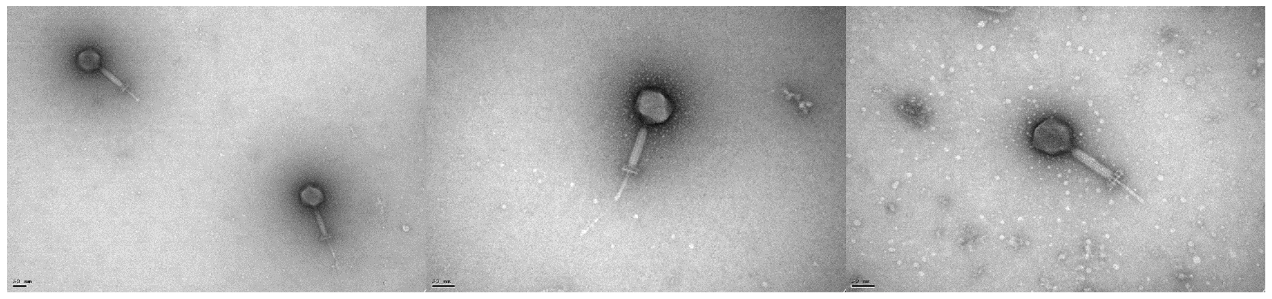
2.4. Electron Microscopy

Phage vB_EfaH_163 was concentrated using the PEG/NaCl method [32]. Electron microscopy images were obtained as previously described [33]: phage particles were stained with 2% uranyl acetate solution, and electron micrographs produced using a CCD Gatan Erlangshen ES 1000 W camera coupled to a JEOL JEM 1011 transmission electron microscope (JEOL USA, Inc., Peabody, MA, USA) operating at 100 kV (performed at the Electron Microscopy Service of the Biotechnology National Centre [CNB-CSIC], Madrid, Spain).

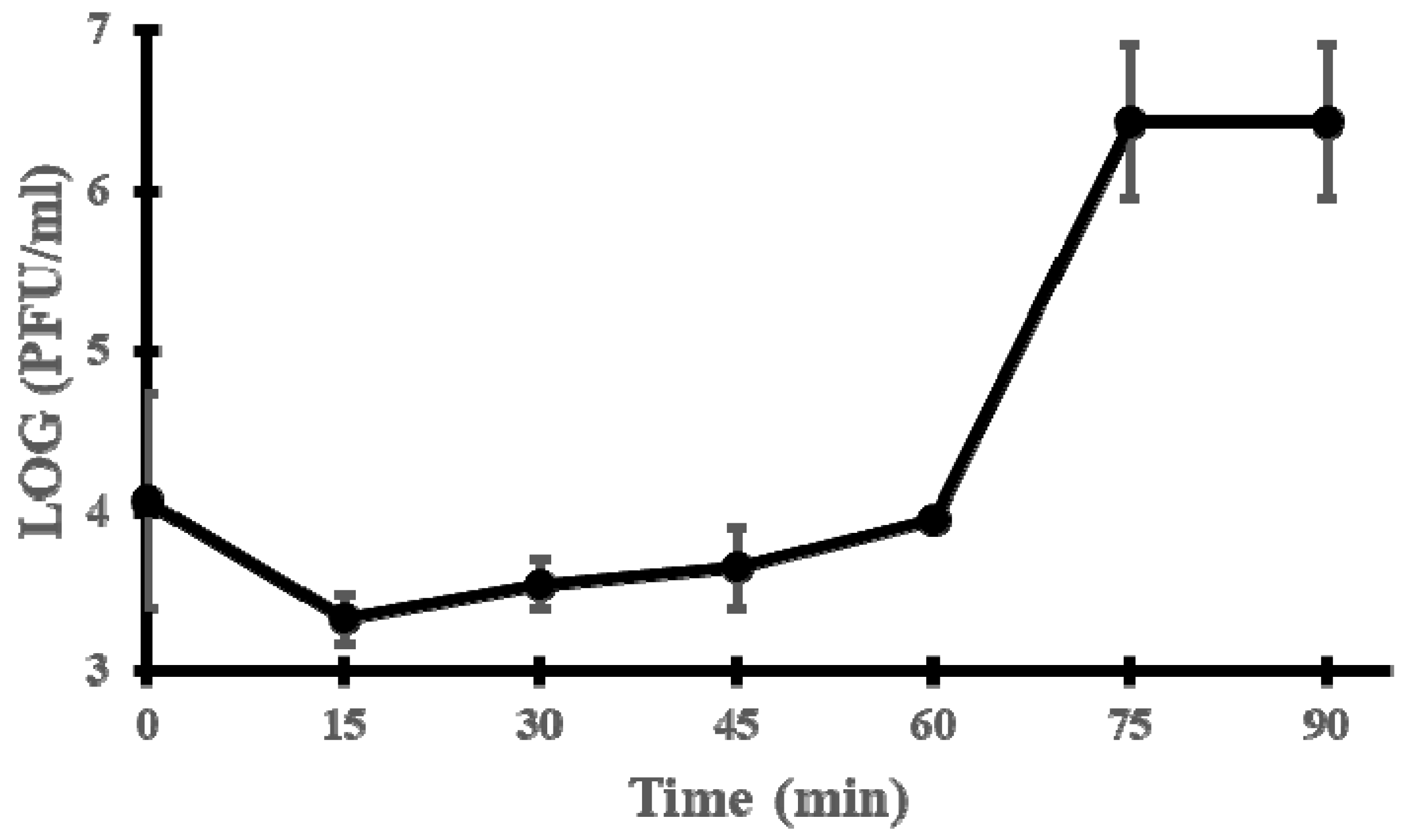
2.5. One-Step Growth Curve

To construct a one step growth curve, a 1% inoculum of an overnight culture of the host strain E. faecium LMGY1 was added to GM17 medium. After 1 h of incubation at 37 °C, phage vB_EfaH_163 was added at an MOI of 0.1 and a sample was examined for its phage titre. After 5 min of adsorption at 37 °C, cells were harvested by centrifugation (4000 rpm for 10 min in a 5910R Eppendorf benchtop centrifuge), the phage titre of the supernatant determined, and the cells resuspended in new medium and incubated at 37 °C. At regular intervals of 15 min, samples were collected for phage titration. This experiment was performed in triplicate. The burst time and latent period were calculated from the one step growth curve produced.

2.6. Phage Genome Sequencing and Analysis

Phage DNA was isolated from a concentrated phage suspension obtained following the PEG/NaCl method [32] as previously described [23]. A genomic library of 0.5 kbp was constructed and subjected to 150 paired-end sequencing (providing approximately 800-fold coverage) using an Illumina HiSeq 1000 System sequencer at GATC services (Eurofins Genomics, Ebersberg, Germany). Quality filtered reads without trimming were assembled using SPADES software [34]. Annotation was performed using the RAST, (with RASTtk pipeline) [35] and PATRIC [36] servers, and improved with BLAST analysis results [37]. Automatic and manual annotation was checked for the presence of virulence factors, including toxin-enconding genes, as well as antibiotic resistance genes. Moreover, PhageLeads [38] and the Resistance Gene Identifier from the Comprehensive Antibiotic Resistance Database (CARD) [39] were used to check the presence of temperate lifestyle genes, antimicrobial resistance, and virulence genes. The genome sequence was deposited in the European Nucleotide Archive (ENA) under accession number CAJDKA010000002.1. The packaging mechanism was determined in silico using the PhagTerm tool [40] at http://galaxy.pasteur.fr (accessed on 10 February 2022). To confirm the circularity of the genome, as well as the sequence of the redundancies, a PCR amplification using primers 163f (5’-GCCCAGAATACATCCGACAAG-3´) and 163r (5´-CCAAGCCCACAAGGAACCTCC-3,) located at both contigs ends and further sequencing of the obtained amplicon was performed.

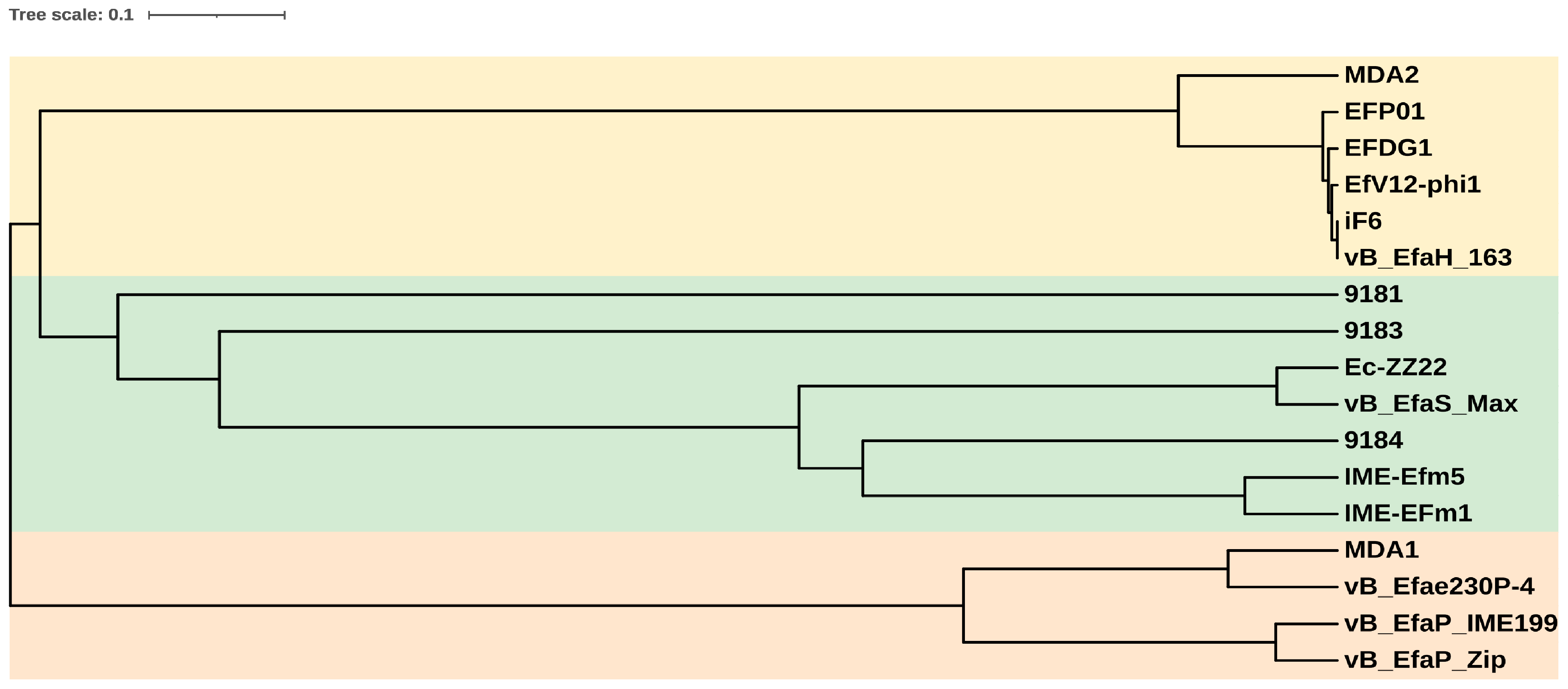
2.7. Phylogenetic Analysis

The amino acidic sequence of the major capsid protein of vB_EfaH_163 was aligned with those of other E. faecium-infecting phages for which complete genomes are available (Table 2). This was achieved using the unweighted pair group method with arithmetic means (UPGMA) and employing MAFFT v.7 software (https://mafft.cbrc.jp/alignment/server/ [41] accessed on 14 November 2022). The phylogenetic tree produced was visualized using the iTOL web server (https://itol.embl.de/ [42] accessed on 14 November 2022).

2.8. Technological Characterisation

The effect of different incubation temperatures on plaque formation was determined by calculating phage titres of the same phage suspension on plates incubated at different temperatures (22, 27, 32, 37, or 42 °C). The thermal stability of phage vB_EfaH_163 was tested by placing 100 µL of its suspension at different temperatures (room temperature [control], 40, 45, 50, 55, 60, 70, 80, 90 °C) for 15 min and determining phage titres. Finally, the pH stability of the phage was tested by mixing 10 µL of its suspension in 900 µL of PBS at different pHs (2–9). Phage titres were determined after 15 min of incubation. All experiments were performed in triplicate.

2.9. Functional Characterisation

2.9.1. Biocontrol of the E. faecium VR-13 vanR Clinical Isolate by vB_EfaH_163 Infection in Broth

E. faecium VR-13 was chosen as a VRE clinical strain to test the potential of phage vB_EfaH_163 as a biocontrol tool. To evaluate its effectiveness in broth, 10 mL of GM17 medium were inoculated with an overnight culture of E. faecium VR-13 to obtain an initial cell concentration of (104 CFU/mL). After 1 h of incubation at 37 °C, 100 µL of vB_EfaH_163 phage were added at different MOIs (0.1, 1, and 10). Total viable cells were then measured at 5 h, 7 h, and 24h. Three biological and two technical replicates for each condition were performed.

2.9.2. In Vivo Effectiveness of Phage Treatment in the Galleria mellonella Model

vB_EfaH_163 was evaluated for its capacity to eliminate E. faecium infection in vivo using a G. mellonella (wax moth larvae) model. TruLarv larvae obtained from BioSystems Technology (Exeter, UK) were stored at 15 °C until the day of the experiment. Larvae were considered dead only when there was no movement after stimulation or when melanisation was seen, and considered alive when there was movement and no melanisation [50,51].
To test the lethality of E. faecium VR-13, an overnight culture of this bacterial strain was centrifuged for 10 min at 3500 rpm and the pellet resuspended in the same volume of PBS. To determine what concentration of larvae to use in experiments, 10 µL of the above suspension were injected into the right pro-leg at test concentrations of 105–107 CFU/larva, as previously described [52]. A group of larvae was injected with PBS as a control (see Section 3.6 for details). After inoculation, the larvae were kept in Petri dishes at 37 °C, and the number of deaths monitored for five days.
To test the effectiveness of the vB_EfaH_163 phage against E. faecium VR-13, an overnight culture of this bacterial strain was centrifuged for 10 min at 3500 rpm and the pellet resuspended in the same volume of PBS. The bacterium was injected into the second-last right proleg at 105 CFU/larva. The control group was injected with PBS. After 1 h at 37 °C, 10 µL of the phage suspension (concentrated by the PEG-NaCl method and resuspended in sterile dH2O) was injected into the second last left pro-leg at an MOI of 0.1. At the same moment, control larvae were injected with sterile dH2O. After inoculation, all larvae were kept in Petri dishes at 37 °C, and the number of deaths monitored for five days.

2.10. Statistical Analysis

Means (± standard deviations) were calculated from at least three independent results and compared using the Student t-test. Significance was set at p < 0.05.
The survival curve for, and analysis of, the in vivo experiments in the G. mellonella model were performed using GraphPad Prism 6 following the method of Kaplan and Meier, calculating the 95% confidence interval for fractional survival at any given time. Survival curves were compared using the log-rank test. At least 10 larvae were used for each condition. Significance was set at p < 0.05.

3. Results

3.1. Phage vB_EfaH_163 Isolation

E. faecium bacteriophage vB_EfaH_163 was isolated from a human faecal sample processed following a previously described method to separate different components of the faecal stool and then isolate the faecal microbiota [31]. The method was adapted to recover the supernatant after the gradient separation of faecal components. An amount of 100 µL of this supernatant were added to CaMg-GM17 cultures, individually inoculated with one of the five potential host strains indicated in the Material and Methods section. After two rounds of enrichment culture, a growth inhibition halo was observed on E. faecium LMGY1 plates. The culture supernatant was then streaked to obtain isolated plaques and confirm that the inhibition halo was caused by a phage. Since only one type of plaque was observed it was assumed that only one phage was present in the sample. One of the plaques was used to infect E. faecium LMGY1 in broth to obtain a high titre bacteriophage stock for further characterisation. The efficiency of infection with the obtained stock was then determined in CaMg-GM17, Ca-GM17, Mg-GM17, and GM17 media. No significant differences were seen between the different media (data not shown), so the following experiments were all performed in GM17 medium.

3.2. Microbiological Characterisation of Phage vB_EfaH_163

To determine the host range of phage vB_EfaH_163, 77 E. faecium strains of different origin (Table 1) were subjected to the spot test. The phage infected about 50% of them—39 in total, of which 20 had been isolated from food, three from human faecal samples, and 16 of which were vancomycin-resistant clinical isolates (Table 1). In addition, since some E. faecium phages were recently shown capable of infecting some E. faecalis strains [4], 11 such strains of different origin were tested as possible host strains (Table 1). Two were susceptible to infection by the phage, notably one that was a linezolid-resistant clinical isolate.
Electron photomicrographs revealed phage vB_EfaH_163 to have typical myovirus morphology (Figure 1). It had an isometric head (around 81 ± 5 nm), a contracted tail sheath length of 106 ± 7 nm finished with a decorated baseplate, and a tail tube length of 74 ± 10 nm that accounted for a total tail length of 180 ± 17 nm (Figure 1). The release of DNA from some particles can be observed (Figure 1).
To better characterise the phage cycle, a one-step growth curve was constructed. The latent period was estimated to last around 60 min; the burst size was about 155 PFU per infected cell (Figure 2).

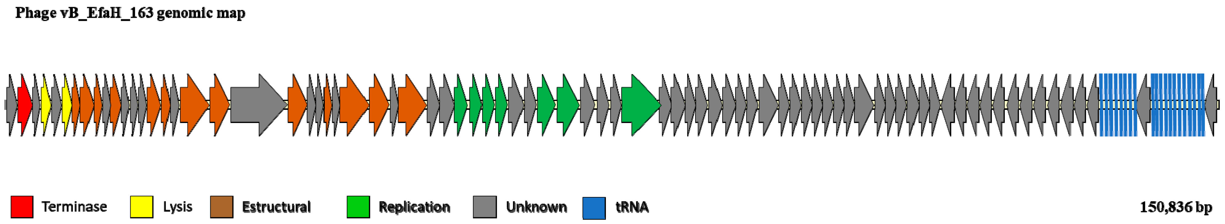
3.3. The vB_EfmH_163 Genome: Characterisation and Phylogenetic Analysis

Assembly of the corresponding Illumina reads revealed a single contig of 150,836 bp, representing the phage genome. The GC content was 37% similar to that of the bacterial host E. faecium (37.8%).
Annotation, combining the RAST, PATRIC, and BLAST results, revealed the existence of 186 orfs and 21 tRNAs. Most of these orfs (81%) showed no similarity to genes of a known function and were consequently annotated as coding for hypothetical phage proteins. Genes for which a putative function could be assigned were mainly related to basic phage functions such as packaging (large terminase unit), lysis (endolysins with a muramidase domain), structural proteins (portal, major head, tail sheaths, tail measure, base plate), or replication (helicase, primase, DNA polymerase) (Supplementary Table S1; Figure 3). These genes are functionally grouped into modules that are all transcribed in the same direction (Figure 3), except for a small group of orfs of unknown function, as well as the tRNA genes, which are divergently transcribed (Figure 3). No genes involved in the establishment of a lysogenic cycle were found, suggesting that vB_EfaH_163 is a lytic bacteriophage. No genes related to pathogenicity, virulence (including toxin-encoding), or AMR were detected either, supporting the idea that vB_EfaH_163 could be therapeutically used without danger. The vB_EfaH_163 DNA packaging mechanism was determined in silico using the Phageterm tool, suggesting a mechanism of long direct terminal repeats, similar to that seen in phage T5 [53]. The size of the terminal repeat was estimated at 2105 bp between positions 104,991 and 107,095 of the reported genome sequence (Accession number CAJDKA010000002.1). As most of the phages with this type of packaging have a circular genome, but the obtained genome was a linear contig, a PCR amplification using primers located at both contig ends was performed. The sequencing of the resulted amplicon allowed confirmation that the genome is circular and to correct the genome sequence.
BLASTn comparisons of the vB_EfaH_163 genome revealed the iF6, EfV12-phi1, and EFDG1 phage genomes to be the most similar (98.88%, 96.82%, and 95.75% similarity, respectively). These three phages belong to the Herelleviriade, subfamily Brockvirinae, genus Schiekvirus. The members of this family are characterised by their typical myovirus morphology, isometric head with rigid contractile tail, and the strong similarity of their genomes [54].
A phylogenetic tree based on the major capsid protein of the 17 E. faecium-infecting phages for which complete genomes are available in databases (Table 2) was made. Three clusters are visible (Figure 4). The vB_EfaH_163 phage grouped with five phages belonging to Herelleviridae, a second group was formed by Siphoviridae phages, and a third cluster by Podoviridae phages. Within the Herelleviridae phages, the phage MDA2 clustered in a different branch than the others, as it is classified as Kochikohdavirus [44], whereas the others are classified as belonging to Schiekvirus genus. The morphological characteristics, the similarity with the iF6, EfV12-phi1, and EFDG1 genomes, and the phylogenetic tree, all suggested phage vB_EfaH_163 to be a new member of Herelleviriade, subfamily Brockvirinae. Moreover, based on a higher than 95% similarity with the above mentioned phage genomes [55], the vB_EfaH_163 can be assumed to belong to the same genus, Schiekvirus.

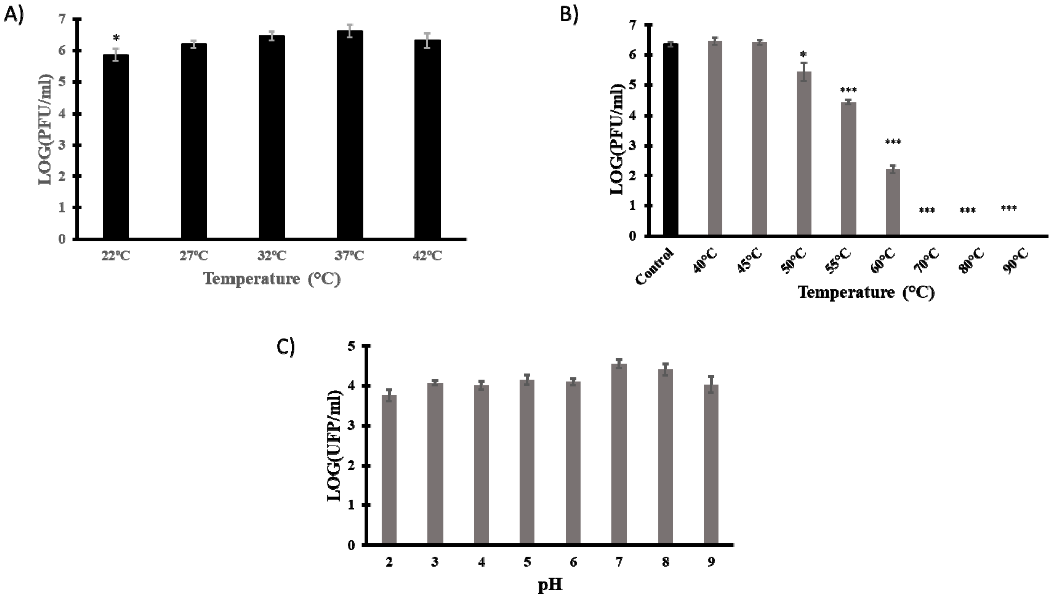
3.4. Technological Characterisation

Phages intended to be used as therapeutic agents must be able to withstand environmental temperatures and pHs. The effect of temperature on vB_EfaH_163 infection was therefore examined. Phage titre determinations were made in double-agar plates incubated at different temperatures (22, 27, 32, 37, and 42 °C). No significant differences were observed between plaque titres at 27, 32, 37, and 42 °C, but small differences were detected when the plates were incubated at 22 °C (Figure 5A).
Thermal and pH stability were then investigated. Phage titre determinations were made after 15 min incubation at different temperatures (room temperature, 40, 45, 50, 55, 60, 70, 80, and 90 °C) or different pHs (2, 3, 4, 5, 6, 7, 8, and 9). The phage titre did not significantly change at temperatures below 45 °C, but reductions of 1 log PFU/mL, 2 log PFU/mL, and 3 log PFU/mL were observed at 50, 55, and 60 °C, respectively. At temperatures above 70 °C, phage viability was lost (Figure 5B). Finally, given the slight and non-significant reduction in titre at all pHs tested (control pH = 7), phage vB_EfaH_163 can be deemed stable over the pH range studied (Figure 5C).

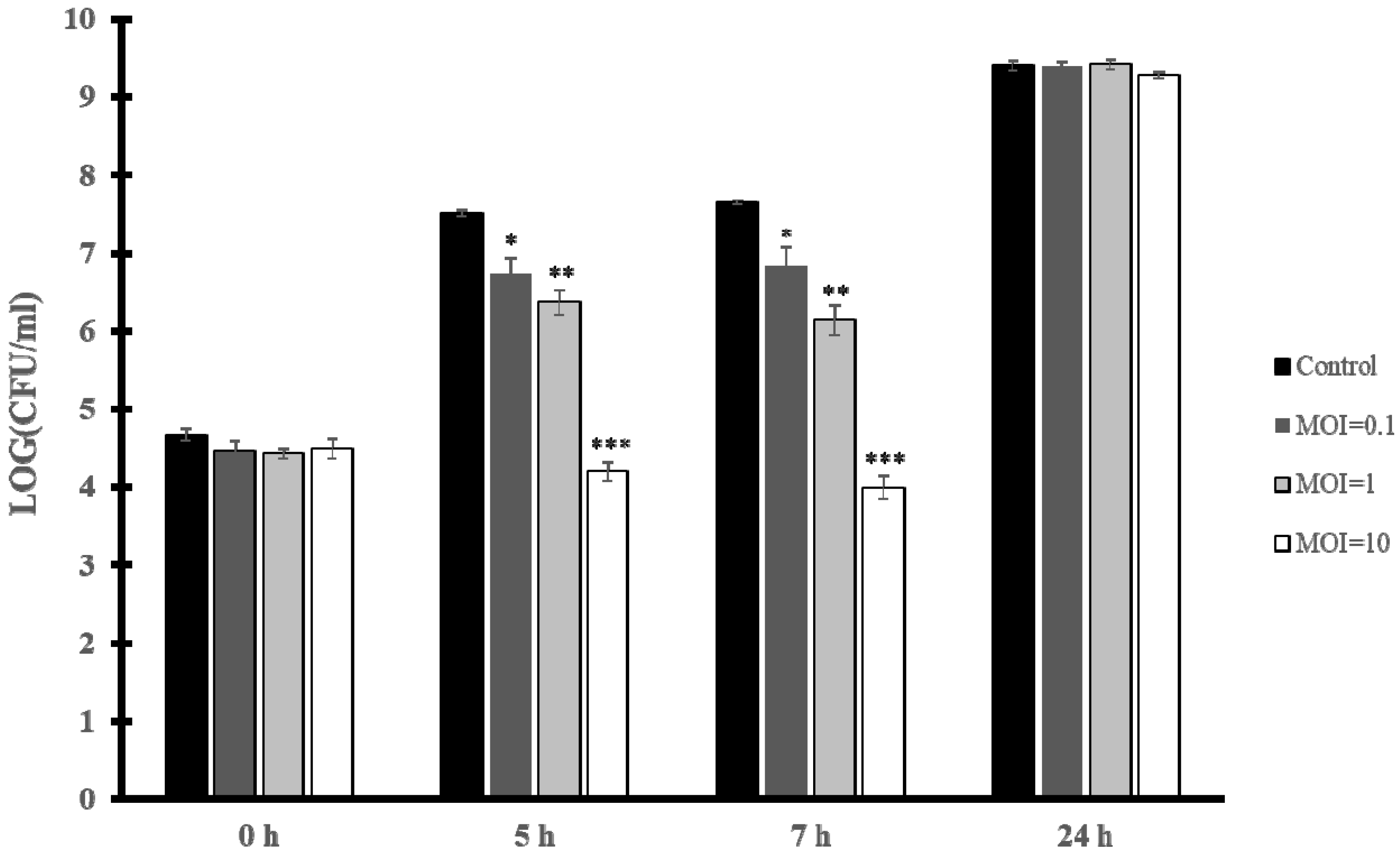
3.5. Biocontrol of E. faecium VR-13 by vB_EfaH_163

To test the capacity of vB_EfaH_163 to reduce the growth of E. faecium, biocontrol experiments were performed in culture broth with the VRE clinical strain E. faecium VR-13 as the target. An initial concentration of 104 CFU/mL was incubated at 37 °C and after 1 h, phage vB_EfaH_163 was added at an MOI of 0.1, 1, or 10 and bacterial growth was measured as viable cell counts. A significant reduction in the number of the viable cells was observed after 5 h and 7 h for all the MOIs assayed (Figure 6).
The reduction in the number of cells was greater the more phage particles were present in the assay (Figure 6). At 7 h post-infection a reduction of 1 log(CFU/mL) in viable cells was seen at MOI = 0.1, of 2 log(CFU/mL) at MOI = 1, and 4 log(CFU/mL) at MOI = 10. This confirms vB_EfaH_163 to show biocontrol capacity for a period lasting at least 7 h post-infection, although at 24 h the E. faecium VR-13 strain was able to grow to a concentration similar to that reached by the control (Figure 6).

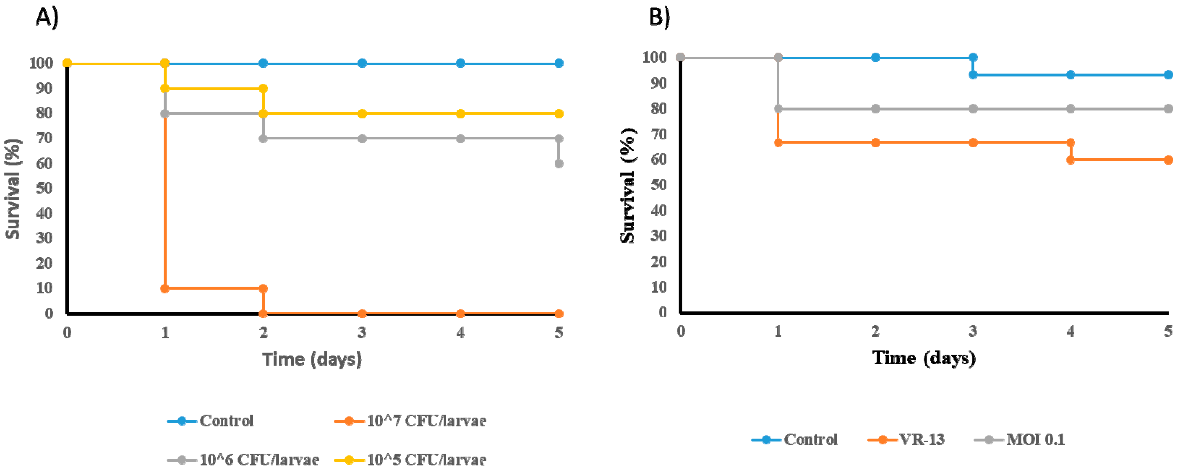
3.6. Reduction in the Mortality of Galleria mellonella Infected by E. faecium VR-13 due to Treatment with Phage vB_EfaH_163

The potential of vB_EfaH_163 to combat VRE E. faecium was also tested in a G. mellonella model. First, the lethality of VRE E. faecium VR-13 was determined by injecting moth larvae with concentrations of 105, 106, or 107 CFU of bacteria (PBS was used as a control). Larval survival was monitored for five days after infection (Figure 7A). After 48 h, all larvae injected with 107 CFU/larvae had died. After five days, mortalities of 20% and 30% were recorded for inoculum concentrations of 105 and 106 CFU/larvae, respectively.
A concentration of 106 CFU/larvae was selected for the challenge test with phage vB_EfaH_163 at MOI = 0.1 (Figure 7B). Infection with E. faecium VR-13 killed about 40% of the larvae after five days. Treatment with phage vB_EfaH_163 at MOI = 0.1 increased larval survival by 20%, although the observed differences were not statistically significant (Figure 7B).

4. Discussion

Vancomycin-resistance in E. faecium isolates is increasing [6,11,13]; new alternatives to combat infections caused by these bacteria are much needed [4]. Indeed, E. faecium is on the WHO’s priority list of ESKAPE microorganisms for which new therapies are required [11]. Phage therapy is a candidate. Phages are obligate parasites of bacteria found wherever their hosts are present [56]. Although enterococci have been documented in many different ecosystems, most of the phages that infect clinically important E. faecium strains have been isolated from sewage water. The present work explores human faecal samples as a source of new E. faecium-infecting phages with applications in phage therapy. Human faecal samples are complex matrices to work with, but the method designed by [31], with slight modifications, allowed samples to be easily cleaned and used in enrichment cultures. As a result, phage vB_EfaH_163 was isolated and further characterised. Host range analysis showed that this phage infects many E. faecium strains (51% of those tested; Table 1); this contrasts with the narrow host range described for phages of lactic acid bacteria, which is sometimes limited to just one strain [57], including those infecting Enterococci species [33]. The host range of vB_EfaH_163 includes 16 VRE E. faecium clinical isolates. Another phage able to infect VRE strains has been described, however its host range is narrower and limited to E. faecalis [58]. In this sense, it is noteworthy than in addition to E. faecium strains, the phage vB_EfaH_163 was able to infect two strains of E. faecalis (Table 1), one of them resistant to linezolid, a last resort antibiotic [14]. Although a rare event, other phages infecting E. faecium have been shown to infect some E. faecalis strains [4,20,44]. E. faecalis is the second most important species of Enterococcus that causes problematic nosocomial infections. Both species are similar in their microbiology and in the diseases they cause [11]. A phage that could be used to treat infections caused by both species would be a great boon.
The genome of vB_EfaH_163 is quite large at 150,836 bp. This is within the range of other E. faecium-infecting phages belonging to the Herelleviridae family, such as the 147,589 bp genome of EDFG1 [20] or the 156,592 bp genome of IF6 (Table 2). Most of the proteins encoded were annotated as hypothetical, with no putative function assigned. This is not uncommon when phage genomes are annotated; usually only a small percentage of orf products show homology with proteins available in databases, a consequence of wide bacteriophage diversity and the low levels of proteins characterised [59]. Those proteins with a putative function clustered into functional modules: packaging, lysis, structural (subclustered into head and tail proteins), and replication. This cluster organization might facilitate coordinated transcription, and could promote phage genome diversity via the exchange of similarly organised functional modules. Two putative lysins with similar catalytic domains (corresponding to muramidase activities) were encoded by the genome of vB_EfaH_163, (Supplementary Table S1; Figure 3). The structural module is composed of genes that code for several proteins similar to others of known function, such as portal protein, head mature protease, major capsid protein, tail sheath, and tail tape measure, and baseplate structure proteins (Supplementary Table S1; Figure 3). Interestingly, the encoding of a carbohydrate binding protein (Orf 28) next to a tail protein with a hydrolase domain (Orf 27) (Supplementary Table S1) indicates that both may be involved in the entry of viral DNA (via the recognition of a structure on the bacterial surface and the hydrolysis of the cell wall). Proteins with hydrolase domains at their tips, together with endolysins, have been proposed as antimicrobial enzybiotics against AMR bacteria [60]. In the opposite orientation to most of these functional modules, up to 21 tRNA genes were found lying close to other encoding proteins predicted to be involved in RNA metabolism (Supplementary Table S1; Figure 3). Although this seems to be a common feature in members of the Brockvirinae subfamily, their role in cell infection has not been established.
The predicted packaging system for vB_EfaH_163 (long direct terminal repeats) involves the recognition of specific sequences by the terminase, if phage DNA is to be encapsidated. This packaging system reduces the possibility of the undesirable host transduction of AMR, or virulence genes. In fact, no virulence factors, including toxin-encoding genes or antibiotic-resistance genes, were detected in the phage genome; it might, therefore, be safely used in phage therapy, although safety trials should be performed first.
A BLASTn comparison of the full phage genome revealed it to be very similar to those of the E. faecium phages EFDG1, IF6, and EfV12-phi1 (data not shown). These two phages and vB_EfaH_163 clustered with phages belonging to the family Herelleviridae, subfamily Brockvirinae, and genus Schiekvirus (Figure 4; Table 2). Members of the subfamily Brockvirinae are defined by a series of morphological and genomic characteristics: (1) all have a myovirus morphology with an isometric, icosahedral head joined to a uncontracted tail and a baseplate; (2) their genome is within the range of 125–170 kpb, it harbours a series of tRNA encoding genes, and most of the coding genes it contains are transcribed in the same direction; and (3) they have terminal repeat regions as a recognition sequence for DNA packaging [54]. All these characteristics are fulfilled by phage vB_EfaH_163: not only does it show typical myovirus morphology (Figure 2); it has a large genome of 150,836 bp (within the observed range of Brockvirinae subfamily members) with a series of 21 tRNA genes transcribed in the opposite direction to most of the genes encoded in the genome (Figure 3; Supplementary Table S1); and it has direct terminal repeats as predicted by the Phageterm tool. These characteristics, and its similarity to other Herelleviridae phages, classify phage vB_EfaH_163 within Herelleviridae, subfamily Brockvirinae, and genus Schiekvirus.
To our knowledge, neither the latent period nor burst size have been reported for the other E. faecium phages of the Herelleviridae family. Compared with E. faecium phages belonging to other families, the observed latent period for vB_EfaH_163 was high at 60 min compared, for example, with 10 min for the E. faecium Max phage [4]. However, vB_EfaH_163 has a much larger burst size (155 phages/infected bacterium) compared with the 38 phages/infected bacterium for Max [4], or the 60 phages/infected bacterium of phage IME-EF1 [61]. A larger burst size results in a larger number of phages that can infect a larger number of bacteria. However, the therapeutic success of a phage with a large burst size cannot be predicted [62].
To further assess the therapeutic potential of vB_EfaH_163, virion stability at different temperatures and pHs were tested. No significant difference was seen in terms of PFU/mL when incubations were performed within the range of human body temperature, not even at high fever temperatures. Therefore, it could also be useful in patients with fever. Moreover, this wide range of temperature could allow other future applications related to food safety, a field in which E. faecium is also relevant due to its ability to produce toxic biogenic amines [28,63]. In fact, the phage was active up to 60 °C, similar to the temperature stability described for the Max and Zip phages [4]. It was also stable over a wide range of pH values (from 2 to 9). Max and Zip phage stability is greatly reduced below pH 4, and undetectable at pH 3 for Zip, and at pH 2 for Max [4]. vB_EfaH_163 is also physicochemically stable over a wider range than other E. faecium phages. Together, these results confirm it to show promise for use in phage therapy.
The functionality of vB_EfaH_163 against clinical E. faecium isolates was tested against VRE E. faecium VR-13 isolated at the Hospital del Bierzo in northern Spain, a region with a high prevalence of VRE. Challenge experiments in broth revealed vB_EfaH_163 capable of reducing the growth of this bacterium in a concentration-dependent manner (Figure 6). However, the effect was temporary; strong growth inhibition was observed at 5–7 h, but the bacterium recovered and grew again at 24 h (Figure 6). The observed re-growth of the bacteria after 24 h could be considered a limitation for phage therapy. If the phage does not kill all the E. faecium cells during the treatment, a re-emergence of the infection could occur. This is a general drawback of phage therapy, and larger treatment periods, the use of phage cocktails, or a combination with antibiotic treatment have been suggested as possible solutions to improve effectiveness [7,45].
Although the G. mellonella animal model has been used to examine the virulence of VRE enterococci [64], the effectiveness of different antimicrobial treatments [65], and the potential of phage therapy against pathogens such as Staphylococcus aureus [66] and different Gram-negative species [50], it has not usually been employed for assessing the therapeutic potential of a phage infecting VRE E. faecium strains, although some reports can be found ([44]; Figure 7A). The virulence of this bacterium is similar to that reported for other VRE enterococcal strains of clinical origin [52,65]. The treatment of infected G. mellonella larvae with vB_EfaH_163 resulted in reduced mortality, reaching values similar to the uninfected control.
The G. mellonella model is a cost-effective model that can be used to evaluate the potential of phage therapy, including single phage, phage cocktail, and phage-antibiotic treatments, before scaling up to murine models. Previous comparative experiments have returned similar results with both types of models [51].
In summary, this work explores an alternative source, human stools, for screening for phages that can infect VRE enterococci. Phage vB_EfaH_163, a new member of Herelleviridae, subfamily Brockvirinae, and genus Schiekvirus shows features that render it a candidate for therapeutic use. It has a large host range, including clinical VRE E. faecium strains, and can even infect some E. faecalis strains of health concern, such as those resistant to last resort antibiotics. The phage’s packaging method reduces the chances of transduction, and no virulence or antibiotic resistance genes were found in its genome. It shows good virion stability and the capacity to control the growth of VRE E. faecium VR-13, both in broth and in a G. mellonella infection model. vB_EfaH_163 could increase phage therapy options for combating VRE enterococci, although safety and effectiveness studies should be performed first.

Supplementary Materials

The following are available online at https://www.mdpi.com/article/10.3390/v15010179/s1, Table S1: Genome features of the Enterococcus faecium bacteriophage vB_EfaH_163.

Author Contributions

Conceptualization, V.L.; Funding acquisition, M.A.A. and M.F.; Methodology, I.P., A.C., B.d.R., C.R.-L. and V.L.; Supervision, V.L.; Writing—original draft, I.P.; Writing—review and editing, I.P., B.d.R., M.A.A. and V.L. All authors have read and agreed to the published version of the manuscript.

Funding

This work was funded by the Plan for Science, Technology and Innovation of the Principality of Asturias 2018–2022, co-financed by FEDER (AYUD/2021/50916), and by the Spanish National Research Council (CSIC201870I091, CSIC).

Institutional Review Board Statement

The study was conducted according to the guidelines of the Declaration of Helsinki, and approved by the Bioethics Committee of the CSIC and the Regional Ethics Committee for Clinical Research (Servicio de Salud del Principado de Asturias n° 353/19. All experiments were performed in accordance with the approved guidelines and regulations.

Informed Consent Statement

Informed consent was obtained from all subjects involved in the study.

Data Availability Statement

Publicly available sequence dataset generated in this study can be in the European Nucleotide Archive (ENA) under accession number CAJDKA010000002.1.

Acknowledgments

Inés Pradal (JAEINT_19_01869) and Angel Casado (JAEINT_20_01539) were recipient of a JAE-Intro grant funded by CSIC. Angel Casado was the recipient of a predoctoral contract for training in research and teaching (Severo Ochoa Ref: BP20-031) financed by the PCTI. The authors are grateful to the Electron Microscopy Service at the National Biotechnology Centre (CNB-CSIC) for taking the electron photomicrographs. The authors also thank Adrian Burton for language and editing assistance.

Conflicts of Interest

The authors declare no conflict of interest. The funders had no role in the design of the study, in data collection, analysis or interpretation, in the writing of the manuscript, or in the decision to publish the results.

References

  1. Mestrovic, T.; Robles Aguilar, G.; Swetschinski, L.R.; Ikuta, K.S.; Gray, A.P.; Davis Weaver, N.; Han, C.; Wool, E.E.; Gershberg Hayoon, A.; Hay, S.I.; et al. The burden of bacterial antimicrobial resistance in the WHO European region in 2019: A cross-country systematic analysis. Lancet Public Health 2022, 2667, e897–e913. [Google Scholar] [CrossRef]
  2. Shankar, P.R. Book review: Tackling drug-resistant infections globally. Arch. Pharm. Pract. 2016, 7, 110. [Google Scholar] [CrossRef]
  3. Mulani, M.S.; Kamble, E.E.; Kumkar, S.N.; Tawre, M.S.; Pardesi, K.R. Emerging strategies to combat ESKAPE pathogens in the era of antimicrobial resistance: A review. Front. Microbiol. 2019, 10, 539. [Google Scholar] [CrossRef] [PubMed]
  4. Melo, L.D.R.; Ferreira, R.; Costa, A.R.; Oliveira, H.; Azeredo, J. Efficacy and safety assessment of two enterococci phages in an in vitro biofilm wound model. Sci. Rep. 2019, 9, 6643. [Google Scholar] [CrossRef] [PubMed] [Green Version]
  5. Dahl, A.; Bruun, N.E. Enterococcus faecalis infective endocarditis: Focus on clinical aspects. Expert Rev. Cardiovasc. Ther. 2013, 11, 1247–1257. [Google Scholar] [CrossRef]
  6. Zaheer, R.; Cook, S.R.; Barbieri, R.; Goji, N.; Cameron, A.; Petkau, A.; Polo, R.O.; Tymensen, L.; Stamm, C.; Song, J.; et al. Surveillance of Enterococcus spp. reveals distinct species and antimicrobial resistance diversity across a One-Health continuum. Sci. Rep. 2020, 10, 3937. [Google Scholar] [CrossRef] [Green Version]
  7. Stuart, C.; Schwartz, S.; Beeson, T.; Owatz, C. Enterococcus faecalis: Its role in root canal treatment failure and current concepts in retreatment. J. Endod. 2006, 32, 93–98. [Google Scholar] [CrossRef]
  8. Tebruegge, M.; Pantazidou, A.; Clifford, V.; Gonis, G.; Ritz, N.; Connell, T.; Curtis, N. The age-related risk of co-existing meningitis in children with urinary tract infection. PLoS ONE 2011, 6, e26576. [Google Scholar] [CrossRef] [Green Version]
  9. Li, F.; Wang, Y.; Sun, L.; Wang, X. Vancomycin-resistant Enterococcus faecium pneumonia in a uremic patient on hemodialysis: A case report and review of the literature. BMC Infect. Dis. 2020, 20, 167. [Google Scholar] [CrossRef]
  10. Chuang, Y.-C.; Lin, H.-Y.; Chen, P.-Y.; Lin, C.-Y.; Wang, J.-T.; Chang, S.-C. Daptomycin versus linezolid for the treatment of vancomycin-resistant enterococcal bacteraemia: Implications of daptomycin dose. Clin. Microbiol. Infect. 2016, 22, 890.e1–890.e7. [Google Scholar] [CrossRef]
  11. Torres, C.; Alonso, C.A.; Ruiz-Ripa, L.; Leon-Sampedro, R.; Del Campo, R.; Coque, T.M.; León-Sampedro, R.; Del Campo, R.; Coque, T.M. Antimicrobial resistance in Enterococcus spp. of animal origin. Microbiol. Spectr. 2018, 6, 185–227. [Google Scholar] [CrossRef] [PubMed]
  12. Palareti, G.; Legnani, C.; Cosmi, B.; Antonucci, E.; Erba, N.; Poli, D.; Testa, S.; Tosetto, A.; De Micheli, V.; Ghirarduzzi, A.; et al. Comparison between different D-Dimer cutoff values to assess the individual risk of recurrent venous thromboembolism: Analysis of results obtained in the DULCIS study. Int. J. Lab. Hematol. 2016, 38, 42–49. [Google Scholar] [CrossRef] [PubMed]
  13. Lam, P.W.; Kozak, R.A.; Eshaghi, A.; Avaness, M.; Salt, N.; Patel, S.N.; Simor, A.E.; Leis, J.A. Nosocomial outbreak of vanD -carrying vancomycin-resistant Enterococcus faecium. Infect. Control Hosp. Epidemiol. 2018, 39, 1266–1268. [Google Scholar] [CrossRef] [PubMed]
  14. Egan, S.A.; Shore, A.C.; O’Connell, B.; Brennan, G.I.; Coleman, D.C. Linezolid resistance in Enterococcus faecium and Enterococcus faecalis from hospitalized patients in Ireland: High prevalence of the MDR genes optrA and poxtA in isolates with diverse genetic backgrounds. J. Antimicrob. Chemother. 2020, 75, 1704–1711. [Google Scholar] [CrossRef] [PubMed]
  15. Sharma, S.; Chatterjee, S.; Datta, S.; Prasad, R.; Dubey, D.; Prasad, R.K.; Vairale, M.G. Bacteriophages and its applications: An overview. Folia Microbiol. 2017, 62, 17–55. [Google Scholar] [CrossRef]
  16. Cisek, A.A.; Dąbrowska, I.; Gregorczyk, K.P.; Wyżewski, Z. Phage therapy in bacterial infections treatment: One hundred years after the discovery of bacteriophages. Curr. Microbiol. 2017, 74, 277–283. [Google Scholar] [CrossRef] [Green Version]
  17. Elbreki, M.; Ross, R.P.; Hill, C.; O’Mahony, J.; McAuliffe, O.; Coffey, A. Bacteriophages and their derivatives as biotherapeutic agents in disease prevention and treatment. J. Viruses 2014, 2014, 382539. [Google Scholar] [CrossRef] [Green Version]
  18. Bolocan, A.S.; Upadrasta, A.; Bettio, P.H.A.; Clooney, A.G.; Draper, L.A.; Ross, R.P.; Hill, C. Evaluation of phage therapy in the context of Enterococcus faecalis and its associated diseases. Viruses 2019, 11, 366. [Google Scholar] [CrossRef] [Green Version]
  19. Fernández, L.; Gutiérrez, D.; García, P.; Rodríguez, A. The perfect bacteriophage for therapeutic applications—A quick guide. Antibiotics 2019, 8, 126. [Google Scholar] [CrossRef] [Green Version]
  20. Khalifa, L.; Shlezinger, M.; Beyth, S.; Houri-Haddad, Y.; Coppenhagen-Glazer, S.; Beyth, N.; Hazan, R. Phage therapy against Enterococcus faecalis in dental root canals. J. Oral Microbiol. 2016, 8, 32157. [Google Scholar] [CrossRef]
  21. Khalifa, L.; Brosh, Y.; Gelman, D.; Coppenhagen-Glazer, S.; Beyth, S.; Poradosu-Cohen, R.; Que, Y.-A.; Beyth, N.; Hazan, R. Targeting Enterococcus faecalis biofilms with phage therapy. Appl. Environ. Microbiol. 2015, 81, 2696–2705. [Google Scholar] [CrossRef] [PubMed] [Green Version]
  22. Khalifa, L.; Gelman, D.; Shlezinger, M.; Dessal, A.L.; Coppenhagen-Glazer, S.; Beyth, N.; Hazan, R. Defeating antibiotic- and phage-resistant Enterococcus faecalis using a phage cocktail in vitro and in a clot model. Front. Microbiol. 2018, 9, 326. [Google Scholar] [CrossRef] [PubMed] [Green Version]
  23. del Rio, B.; Sanchez-Llana, E.; Redruello, B.; Magadan, A.H.; Fernandez, M.; Martin, M.C.; Ladero, V.; Alvarez, M.A. Enterococcus faecalis bacteriophage 156 Is an effective biotechnological tool for reducing the presence of tyramine and putrescine in an experimental cheese model. Front. Microbiol. 2019, 10, 566. [Google Scholar] [CrossRef] [PubMed]
  24. Ladero, V.; Gomez-Sordo, C.; Sanchez-Llana, E.; del Rio, B.; Redruello, B.; Fernandez, M.; Martin, M.C.; Alvarez, M.A. Q69 (an E. faecalis-Infecting bacteriophage) as a biocontrol agent for reducing tyramine in dairy products. Front. Microbiol. 2016, 7, 445. [Google Scholar] [CrossRef] [PubMed]
  25. Biswas, B.; Adhya, S.; Washart, P.; Paul, B.; Trostel, A.N.; Powell, B.; Carlton, R.; Merril, C.R. Bacteriophage therapy rescues mice bacteremic from a clinical isolate of vancomycin-resistant Enterococcus faecium. Infect. Immun. 2002, 70, 1664. [Google Scholar] [CrossRef] [Green Version]
  26. Gelman, D.; Beyth, S.; Lerer, V.; Adler, K.; Poradosu-Cohen, R.; Coppenhagen-Glazer, S.; Hazan, R. Combined bacteriophages and antibiotics as an efficient therapy against VRE Enterococcus faecalis in a mouse model. Res. Microbiol. 2018, 169, 531–539. [Google Scholar] [CrossRef]
  27. Letkiewicz, S.; Międzybrodzki, R.; Fortuna, W.; Weber-Dąbrowska, B.; Górski, A. Eradication of Enterococcus faecalis by phage therapy in chronic bacterial prostatitis—Case report. Folia Microbiol. 2009, 54, 457–461. [Google Scholar] [CrossRef]
  28. Ladero, V.; Fernandez, M.; Calles-Enriquez, M.; Sanchez-Llana, E.; Canedo, E.; Martin, M.C.; Alvarez, M.A. Is the production of the biogenic amines tyramine and putrescine a species-level trait in enterococci? Food Microbiol. 2012, 30, 132–138. [Google Scholar] [CrossRef]
  29. Ladero, V.; Fernandez, M.; Alvarez, M.A. Isolation and identification of tyramine-producing enterococci from human fecal samples. Can. J. Microbiol. 2009, 55, 215–218. [Google Scholar] [CrossRef]
  30. Paulsen, I.T.; Banerjei, L.; Myers, G.S.; Nelson, K.E.; Seshadri, R.; Read, T.D.; Fouts, D.E.; Eisen, J.A.; Gill, S.R.; Heidelberg, J.F.; et al. Role of mobile DNA in the evolution of vancomycin-resistant Enterococcus faecalis. Science 2003, 299, 2071–2074. [Google Scholar] [CrossRef]
  31. Hevia, A.; Delgado, S.; Margolles, A.; Sánchez, B. Application of density gradient for the isolation of the fecal microbial stool component and the potential use thereof. Sci. Rep. 2015, 5, 16807. [Google Scholar] [CrossRef] [PubMed] [Green Version]
  32. Binetti, A.G.; del Rio, B.; Martin, M.C.; Alvarez, M.A. Detection and characterization of Streptococcus thermophilus bacteriophages by use of the antireceptor gene sequence. Appl. Environ. Microbiol. 2005, 71, 6096–6103. [Google Scholar] [CrossRef] [PubMed] [Green Version]
  33. del Rio, B.; Sánchez-Llana, E.; Martínez, N.; Fernández, M.; Ladero, V.; Alvarez, M.A. Isolation and characterization of Enterococcus faecalis-infecting bacteriophages from different cheese types. Front. Microbiol. 2021, 11, 592172. [Google Scholar] [CrossRef] [PubMed]
  34. Bankevich, A.; Nurk, S.; Antipov, D.; Gurevich, A.A.; Dvorkin, M.; Kulikov, A.S.; Lesin, V.M.; Nikolenko, S.I.; Pham, S.; Prjibelski, A.D.; et al. SPAdes: A new genome assembly algorithm and its applications to single-cell sequencing. J. Comput. Biol. 2012, 19, 455–477. [Google Scholar] [CrossRef] [Green Version]
  35. Aziz, R.K.; Bartels, D.; Best, A.A.; DeJongh, M.; Disz, T.; Edwards, R.A.; Formsma, K.; Gerdes, S.; Glass, E.M.; Kubal, M.; et al. The RAST Server: Rapid annotations using subsystems technology. BMC Genom. 2008, 9, 75. [Google Scholar] [CrossRef] [Green Version]
  36. Wattam, A.R.; Davis, J.J.; Assaf, R.; Boisvert, S.; Brettin, T.; Bun, C.; Conrad, N.; Dietrich, E.M.; Disz, T.; Gabbard, J.L.; et al. Improvements to PATRIC, the all-bacterial bioinformatics database and analysis resource center. Nucleic Acids Res. 2017, 45, D535–D542. [Google Scholar] [CrossRef] [PubMed]
  37. Altschul, S.F.; Madden, T.L.; Schaffer, A.A.; Zhang, J.; Zhang, Z.; Miller, W.; Lipman, D.J. Gapped BLAST and PSI-BLAST: A new generation of protein database search programs. Nucleic Acids Res. 1997, 25, 3389–3402. [Google Scholar] [CrossRef] [Green Version]
  38. Yukgehnaish, K.; Rajandas, H.; Parimannan, S.; Manickam, R.; Marimuthu, K.; Petersen, B.; Clokie, M.R.J.; Millard, A.; Sicheritz-Pontén, T. PhageLeads: Rapid assessment of phage therapeutic suitability using an ensemble machine learning approach. Viruses 2022, 14, 342. [Google Scholar] [CrossRef]
  39. Alcock, B.P.; Raphenya, A.R.; Lau, T.T.Y.; Tsang, K.K.; Bouchard, M.; Edalatmand, A.; Huynh, W.; Nguyen, A.-L.V.; Cheng, A.A.; Liu, S.; et al. CARD 2020: Antibiotic resistome surveillance with the comprehensive antibiotic resistance database. Nucleic Acids Res. 2019, 48, D517–D525. [Google Scholar] [CrossRef]
  40. Garneau, J.R.; Depardieu, F.; Fortier, L.-C.; Bikard, D.; Monot, M. PhageTerm: A tool for fast and accurate determination of phage termini and packaging mechanism using next-generation sequencing data. Sci. Rep. 2017, 7, 8292. [Google Scholar] [CrossRef]
  41. Katoh, K.; Rozewicki, J.; Yamada, K.D. MAFFT online service: Multiple sequence alignment, interactive sequence choice and visualization. Br. Bioinform. 2019, 20, 1160–1166. [Google Scholar] [CrossRef] [PubMed] [Green Version]
  42. Letunic, I.; Bork, P. Interactive tree of life (iTOL) v3: An online tool for the display and annotation of phylogenetic and other trees. Nucleic Acids Res. 2016, 44, W242–W245. [Google Scholar] [CrossRef] [PubMed]
  43. Wandro, S.; Oliver, A.; Gallagher, T.; Weihe, C.; England, W.; Martiny, J.B.H.; Whiteson, K. Predictable molecular adaptation of coevolving Enterococcus faecium and lytic phage EfV12-phi1. Front. Microbiol. 2019, 9, 3192. [Google Scholar] [CrossRef] [PubMed] [Green Version]
  44. El Haddad, L.; Angelidakis, G.; Clark, J.R.; Mendoza, J.F.; Terwilliger, A.L.; Chaftari, C.P.; Duna, M.; Yusuf, S.T.; Harb, C.P.; Stibich, M.; et al. Genomic and functional characterization of vancomycin-resistant enterococci-specific bacteriophages in the Galleria mellonella wax moth larvae model. Pharmaceutics 2022, 14, 1591. [Google Scholar] [CrossRef]
  45. Canfield, G.S.; Chatterjee, A.; Espinosa, J.; Mangalea, M.R.; Sheriff, E.K.; Keidan, M.; McBride, S.W.; McCollister, B.D.; Hang, H.C.; Duerkop, B.A. Lytic bacteriophages facilitate antibiotic sensitization of Enterococcus faecium. Antimicrob. Agents Chemother. 2021, 65, e00143-21. [Google Scholar] [CrossRef]
  46. Li, J.; Shi, H.; Zhao, C.; Hao, Y.; He, Y.; Sun, Y. Complete genome sequence of the siphoviral bacteriophage ec-zz2, which is capable of lysing Enterococcus faecium. Genome Announc. 2016, 4, e01167-16. [Google Scholar] [CrossRef] [Green Version]
  47. Wang, Y.; Wang, W.; Lv, Y.; Zheng, W.; Mi, Z.; Pei, G.; An, X.; Xu, X.; Han, C.; Liu, J.; et al. Characterization and complete genome sequence analysis of novel bacteriophage IME-EFm1 infecting Enterococcus faecium. J. Gen. Virol. 2014, 95, 2565–2575. [Google Scholar] [CrossRef] [Green Version]
  48. Gong, P.; Cheng, M.; Li, X.; Jiang, H.; Yu, C.; Kahaer, N.; Li, J.; Zhang, L.; Xia, F.; Hu, L.; et al. Characterization of Enterococcus faecium bacteriophage IME-EFm5 and its endolysin LysEFm5. Virology 2016, 492, 11–20. [Google Scholar] [CrossRef]
  49. Xing, S.; Zhang, X.; Sun, Q.; Wang, J.; Mi, Z.; Pei, G.; Huang, Y.; An, X.; Fu, K.; Zhou, L.; et al. Complete genome sequence of a novel, virulent Ahjdlikevirus bacteriophage that infects Enterococcus faecium. Arch. Virol. 2017, 162, 3843–3847. [Google Scholar] [CrossRef]
  50. Manohar, P.; Nachimuthu, R.; Lopes, B.S. The therapeutic potential of bacteriophages targeting gram-negative bacteria using Galleria mellonella infection model. BMC Microbiol. 2018, 18, 97. [Google Scholar] [CrossRef]
  51. Jeon, J.; Park, J.-H.; Yong, D. Efficacy of bacteriophage treatment against carbapenem-resistant Acinetobacter baumannii in Galleria mellonella larvae and a mouse model of acute pneumonia. BMC Microbiol. 2019, 19, 70. [Google Scholar] [CrossRef] [PubMed]
  52. Lagatolla, C.; Milic, J.; Imperi, F.; Cervoni, M.; Bressan, R.; Luzzati, R.; Di Bella, S. Synergistic activity of fosfomycin and chloramphenicol against vancomycin-resistant Enterococcus faecium (VREfm) isolates from bloodstream infections. Diagn. Microbiol. Infect. Dis. 2021, 99, 115241. [Google Scholar] [CrossRef] [PubMed]
  53. Fujisawa, H.; Morita, M. Phage DNA packaging. Genes Cells 2003, 2, 537–545. [Google Scholar] [CrossRef] [PubMed]
  54. Barylski, J.; Kropinski, A.M.; Alikhan, N.-F.; Adriaenssens, E.M. ICTV report consortium ICTV virus taxonomy profile: Herelleviridae. J. Gen. Virol. 2020, 101, 362. [Google Scholar] [CrossRef]
  55. Turner, D.; Adriaenssens, E.M.; Tolstoy, I.; Kropinski, A.M. Phage annotation guide: Guidelines for assembly and high-quality annotation. PHAGE 2021, 2, 170–182. [Google Scholar] [CrossRef] [PubMed]
  56. Chibani-Chennoufi, S.; Bruttin, A.; Dillmann, M.; Brüssow, H. Phage-host interaction: An ecological perspective. J. Bacteriol. 2004, 186, 3677–3686. [Google Scholar] [CrossRef] [Green Version]
  57. Kot, W.; Neve, H.; Heller, K.J.; Vogensen, F.K. Bacteriophages of leuconostoc, oenococcus, and weissella. Front. Microbiol. 2014, 5, 186. [Google Scholar] [CrossRef] [Green Version]
  58. Uchiyama, J.; Rashel, M.; Maeda, Y.; Takemura, I.; Sugihara, S.; Akechi, K.; Muraoka, A.; Wakiguchi, H.; Matsuzaki, S. Isolation and characterization of a novel Enterococcus faecalis bacteriophage φEF24C as a therapeutic candidate. FEMS Microbiol. Lett. 2008, 278, 200–206. [Google Scholar] [CrossRef] [Green Version]
  59. Yin, Y.; Fischer, D. Identification and investigation of ORFans in the viral world. BMC Genom. 2008, 9, 24. [Google Scholar] [CrossRef] [Green Version]
  60. Rodríguez-Rubio, L.; Gutiérrez, D.; Donovan, D.M.; Martínez, B.; Rodríguez, A.; García, P. Phage lytic proteins: Biotechnological applications beyond clinical antimicrobials. Crit. Rev. Biotechnol. 2015, 36, 542–552. [Google Scholar] [CrossRef]
  61. Wang, R.; Xing, S.; Zhao, F.; Li, P.; Mi, Z.; Shi, T.; Liu, H.; Tong, Y. Characterization and genome analysis of novel phage vB_EfaP_IME195 infecting Enterococcus faecalis. Virus Genes 2018, 54, 804–811. [Google Scholar] [CrossRef] [PubMed]
  62. Casey, E.; van Sinderen, D.; Mahony, J. In vitro characteristics of phages to guide ‘Real Life’ phage therapy suitability. Viruses 2018, 10, 163. [Google Scholar] [CrossRef] [Green Version]
  63. Ladero, V.; Calles-Enríquez, M.; Fernández, M.; Alvarez, M.A. Toxicological effects of dietary biogenic amines. Curr. Nutr. Food Sci. 2010, 6, 145–156. [Google Scholar] [CrossRef]
  64. McLaughlin, M.; Malczynski, M.; Qi, C.; Barajas, G.; Radetski, J.; Zembower, T.; Scheetz, M.H. Virulence of Vancomycin-resistant Enterococcus faecium according to linezolid resistance and clinical outbreak status. Antimicrob. Agents Chemother. 2013, 57, 3923–3927. [Google Scholar] [CrossRef] [Green Version]
  65. Skinner, K.; Sandoe, J.A.T.; Rajendran, R.; Ramage, G.; Lang, S. Efficacy of rifampicin combination therapy for the treatment of enterococcal infections assessed in vivo using a Galleria mellonella infection model. Int. J. Antimicrob. Agents 2017, 49, 507–511. [Google Scholar] [CrossRef] [Green Version]
  66. Tkhilaishvili, T.; Wang, L.; Tavanti, A.; Trampuz, A.; Di Luca, M. Antibacterial efficacy of two commercially available bacteriophage formulations, staphylococcal bacteriophage and pyo bacteriophage, against methicillin-resistant Staphylococcus aureus: Prevention and eradication of biofilm formation and control of a systemic infection of Galleria mellonella larvae. Front. Microbiol. 2020, 11, 110. [Google Scholar] [CrossRef] [PubMed]
Figure 1. Electron photomicrographs of phage vB_EfmH_163. The scale bar represents 50 nm.
Viruses 15 00179 g001
Figure 2. One-step growth curve of phage vB_EfmH_163. Log PFU/mL was measured every 15 min. Means and standard deviations of three independent replicates are shown.
Viruses 15 00179 g002
Figure 3. Representation of the 150,836 bp-long genome of phage vB_EfmH_163. Open reading frames are indicated by arrows pointing in the transcription direction, and coloured according to the functional module. Red: packaging; yellow: lysis genes; brown: structural genes; green: replication; grey: unknown function. tRNA genes are represented by thick blue lines.
Viruses 15 00179 g003
Figure 4. Phylogenetic tree of the E. faecium phages with complete genomes in databases, based on the amino acidic sequence of the major capsid proteins. Light yellow: Herelleviridae phages; light green: Siphoviridae phages; light orange: Podoviridae phages.
Viruses 15 00179 g004
Figure 5. Technological characterisation of phage vB_EfmH_163. The effect of incubation temperature (22, 27, 32, 37, and 42 °C) (A), thermal stability (15 min-treatment at 40, 45, 50, 55, 60, 70, 80, or 90 °C) (B) and pH stability (15 min treatment in PBS pH 2–9) (C) were assessed by measuring the phage titre (Log PFU/mL). Means and standard deviations of three independent replicates are shown. *: p < 0.05; ***: p < 0.001.
Viruses 15 00179 g005
Figure 6. Biocontrol assay. E. faecium VR-13 104 CFU/mL culture infected with phage vB_EfmH_163 at different MOIs (0.1, 1, and 10). Viable cells (CFU/mL) were counted at 5 h, 7 h, and 24 h post-infection. Means and standard deviations of three independent replicates are shown. *: p < 0.05; **: p < 0.01; ***: p < 0.001.
Viruses 15 00179 g006
Figure 7. In vivo experiments in Galleria mellonella. A suitable concentration of E. faecium VR-13 per larvae was determined. Control larvae were inoculated with 10 μl of PBS. (A) and the effect of the treatment with phage vB_EfmH_163 at MOI = 0.1 (B) tested. Control larvae were inoculated with 10 μl of dH2O. N = 10 for each experiment and condition.
Viruses 15 00179 g007
Table 1. E. faecium and E. faecalis strains used in this work for bacteriophage vB_EfmH_163 isolation and host range determination.
SpecieStrainOriginvB_EfmH_163
Infection
Reference
E. faeciumLMA2Camel milk+MicroMol
E. faeciumLMA3Camel milk+MicroMol
E. faeciumLMA4Camel milk+MicroMol
E. faeciumLMA5Camel milk+MicroMol
E. faeciumLMA6Camel milk+MicroMol
E. faeciumLMA8Camel milk+MicroMol
E. faeciumLMA9Camel milkMicroMol
E. faeciumLMA10Camel milk+MicroMol
E. faeciumLGMY-2Camel milk+MicroMol
E. faeciumLGMY-5Camel milk+MicroMol
E. faeciumLGMY-1Cow milk+MicroMol
E. faeciumLMGY-10Cow milkMicroMol
E. faeciumLGMY-6Sheep milk+MicroMol
E. faeciumLGMY-11Goat milk+MicroMol
E. faeciumC39Cheese[28]
E. faeciumAMCheese[28]
E. faecium103Cheese[28]
E. faeciumLGMY-12Date+MicroMol
E. faeciumLMGY-13Date+LGM
E. faeciumLGM11397MeatLGM
E. faeciumLGM14205MeatLGM
E. faeciumLGM20641MeatLGM
E. faeciumHF11Human[29]
E. faeciumHF14Human+[29]
E. faeciumHF24Human+[29]
E. faeciumHF52Human[29]
E. faeciumHF56Human+[29]
E. faeciumVR-1ClinicalBierzo Hospital
E. faeciumVR-2ClinicalBierzo Hospital
E. faeciumVR-3ClinicalBierzo Hospital
E. faeciumVR-4ClinicalBierzo Hospital
E. faeciumVR-5ClinicalBierzo Hospital
E. faeciumVR-6ClinicalBierzo Hospital
E. faeciumVR-7Clinical+Bierzo Hospital
E. faeciumVR-8Clinical+Bierzo Hospital
E. faeciumVR-9ClinicalBierzo Hospital
E. faeciumVR-10Clinical+Bierzo Hospital
E. faeciumVR-11ClinicalBierzo Hospital
E. faeciumVR-12ClinicalBierzo Hospital
E. faeciumVR-13Clinical+Bierzo Hospital
E. faeciumVR-14Clinical+Bierzo Hospital
E. faeciumVR-15ClinicalBierzo Hospital
E. faeciumVR-16Clinical+Bierzo Hospital
E. faeciumVR-17ClinicalBierzo Hospital
E. faeciumVR-18Clinical+Bierzo Hospital
E. faeciumVR-19ClinicalBierzo Hospital
E. faeciumVR-20ClinicalBierzo Hospital
E. faeciumVR-20bClinical+Bierzo Hospital
E. faeciumVR-22Clinical+Bierzo Hospital
E. faeciumVR-23Clinical+Bierzo Hospital
E. faeciumVR-24ClinicalBierzo Hospital
E. faeciumVR-25Clinical+Bierzo Hospital
E. faeciumVR-26Clinical+Bierzo Hospital
E. faeciumVR-27Clinical+Bierzo Hospital
E. faeciumVR-28ClinicalBierzo Hospital
E. faeciumVR-29ClinicalBierzo Hospital
E. faeciumVR-30Clinical+Bierzo Hospital
E. faeciumVR-31ClinicalBierzo Hospital
E. faeciumVR-32Clinical+Bierzo Hospital
E. faeciumVR-33ClinicalBierzo Hospital
E. faeciumVR-34Clinical+Bierzo Hospital
E. faeciumVR-35ClinicalBierzo Hospital
E. faeciumVR-36ClinicalBierzo Hospital
E. faeciumVR-37ClinicalBierzo Hospital
E. faeciumVR-38ClinicalBierzo Hospital
E. faeciumVR-39ClinicalBierzo Hospital
E. faeciumVR-40ClinicalBierzo Hospital
E. faeciumVR-41ClinicalBierzo Hospital
E. faeciumVR-42ClinicalBierzo Hospital
E. faeciumVR-43ClinicalBierzo Hospital
E. faeciumVR-44ClinicalBierzo Hospital
E. faeciumVR-45ClinicalBierzo Hospital
E. faeciumVR-46ClinicalBierzo Hospital
E. faeciumVR-47ClinicalBierzo Hospital
E. faeciumVR-48ClinicalBierzo Hospital
E. faeciumVR-49ClinicalBierzo Hospital
E. faeciumVR-50ClinicalBierzo Hospital
E. faecalisCECT481TType strainCECT
E. faecalis18aCheese[28]
E. faecalis23aCheese[28]
E. faecalisV63Cheese[28]
E. faecalis63cCheese[28]
E. faecalis52cCheese[28]
E. faecalisHFS56Human+[29]
E. faecalisHFS57Human[29]
E. faecalisVR-5ClinicalBierzo Hospital
E. faecalisVR-11ClinicalBierzo Hospital
E. faecalisoptra5Clinical+Bierzo Hospital
E. faecalisV583Clinical[30]
Bierzo Hospital: Strain isolated at the Microbiology unit at the El Bierzo Hospital, Spain; CECT: Colección Española de Cultivos Tipo; LMG: Laboratorium voor Microbiologie; MicroMol: Culture collection of the Molecular Microbiology Group at IPLA. Clinical origin refers to strains isolated from humans in a clinical ambient. Human origin refers to strains isolated from humans outside a clinical environment.
Table 2. Characteristics of the E. faecium phages used in the phylogenetic analysis. Genome size and accession number are shown.
PhageAccession
Number
HostFamilyGenome
Size
OriginReference
vB_EfmH_163CAJDKA010000002.1E. faecium
E. faecalis
Herelleviridae150,836Human faecal samplesThis work
EFDG1NC_029009E. faecium
E faecalis
Herelleviridae147,589Sewage effluents[21]
EfV12-phi1MH880817E. faeciumHerelleviridae152,770Sewage[43]
EFP01NC_047796.1E. faeciumHerelleviridae155,053Sewage-
iF6MT909815.1E. faeciumHerelleviridae156,592--
MDA2MW633168.1E. faeciumHerelleviridae140,226-[44]
9183MT939241.1E. faeciumSiphoviridae806,301Wastewater[45]
9181MT939240.1E. faeciumSiphoviridae71,854Wastewater[45]
vB_EfaS_MaxMK360024E. faecium
E. faecalis
Siphoviridae40,975Raw sewage from wastewater [4]
9184MT939242.1E. faeciumSiphoviridae44,108Wastewater[45]
Ec-ZZ2NC_031260E. faeciumSiphoviridae41,170Sewage[46]
IME-EFm1NC_024356E. faeciumSiphoviridae42,597Hospital sewage[47]
IME-EFm5NC_028826E. faeciumSiphoviridae42,265Hospital sewage[48]
vB_EfaP_ZipMK360025E. faecium
E. faecalis
Podoviridae18,742Raw sewage from wastewater [4]
vB_Efae230p-4NC_025467E. faeciumPodoviridae17,972--
vB_EfaP_IME199KT945995E. faeciumPodoviridae18,838Sewage[49]
MDA1MW623430.1E. faeciumPodoviridae18,058-[44]
-: information not available.
Disclaimer/Publisher’s Note: The statements, opinions and data contained in all publications are solely those of the individual author(s) and contributor(s) and not of MDPI and/or the editor(s). MDPI and/or the editor(s) disclaim responsibility for any injury to people or property resulting from any ideas, methods, instructions or products referred to in the content.

