Article

Isolation of Novel Xanthomonas Phages Infecting the Plant Pathogens X. translucens and X. campestris

1
Institute of Bio- and Geosciences, Department for Plant Sciences (IBG-2), Forschungszentrum Jülich, 52425 Jülich, Germany
2
Institute of Bio- and Geosciences, Department for Biotechnology (IBG-1), Forschungszentrum Jülich, 52425 Jülich, Germany
*
Author to whom correspondence should be addressed.
Viruses 2022, 14(7), 1449; https://doi.org/10.3390/v14071449
Submission received: 18 May 2022 / Revised: 22 June 2022 / Accepted: 28 June 2022 / Published: 30 June 2022
(This article belongs to the Section Bacterial Viruses)

Abstract

:
The genus of Xanthomonas contains many well-known plant pathogens with the ability to infect some of the most important crop plants, thereby causing significant economic damage. Unfortunately, classical pest-control strategies are neither particularly efficient nor sustainable and we are, therefore, in demand of alternatives. Here, we present the isolation and characterization of seven novel phages infecting the plant-pathogenic species Xanthomonas translucens and Xanthomonas campestris. Transmission electron microscopy revealed that all phages show a siphovirion morphology. The analysis of genome sequences and plaque morphologies are in agreement with a lytic lifestyle of the phages making them suitable candidates for biocontrol. Moreover, three of the isolated phages form the new genus “Shirevirus”. All seven phages belong to four distinct clusters underpinning their phylogenetic diversity. Altogether, this study presents the first characterized isolates for the plant pathogen X. translucens and expands the number of available phages for plant biocontrol.

1. Introduction

Pathogenic microbes represent a major factor hampering current food production and account for an annual loss of 10% of the global production [1]. Phage-based biocontrol for the treatment of bacterial infections was already assessed in trials by Mallman and Hemsworth in 1924 to treat black rot of cabbage shortly after the discovery of phages in general [2]. However, due to their high specificity, phages were replaced by broad-range antibiotics and cheaper copper treatments in the beginning of the last century. Nowadays, the massive spread of antibiotic-resistant bacteria has fostered a global surge in the reinvestigation of phage biology and phage-based biocontrol as an alternative to antibiotic treatments [3,4,5,6].
An overview of the economically most relevant plant pathogens is given in Mansfield et al. 2012 [7]. In recent years, several attempts have been made to isolate phages infecting plant pathogens, including Ralstonia solanacearum, Erwinia amylovora, Pseudomonas syringe spp, Xylella fastidiosa, and Xanthomonas spp. Multiple studies were conducted on phage biocontrol of Ralstonia solanacearum showing suppression of plant wilting on potato and tomato [8,9,10,11,12,13]. Erwinia amylovora, which developed resistance to streptomycin, has led to the evaluation of phage biocontrol with promising outcomes in some cases [14,15,16]. Due to the broad spectrum of plants infected by Pseudomonas syringae spp., multiple biocontrol trails have been conducted in the past, but with a special effort on citrus canker disease [17,18,19,20,21,22,23]. Xylella fastidiosa is a major threat to olive trees in Europe, where first phages have been isolated and tested [24,25,26].
The genus of Xanthomonas contains multiple gram-negative plant pathogens causing devastating losses in food production in a broad variety of important food crops, from the Poaceae family (including rice, sugar cane, and wheat) to the Brassicaceae family (including cabbages, broccoli, and oil seed). In this study, we focus on two important Xanthomonas pathogens: (1) Xanthomonas translucens pv. translucens (Xtt), causing bacterial leaf streak in cereals, and (2) Xanthomonas campestris pv. campestris (Xcc), the major cause of black rot disease in crucifers.
X. translucens (Xtt) infects cereals such as barley or wheat and, thereby, poses a threat to crop production, with annual losses reported from 10% reaching up to 40% in severe cases [27]. For other Xanthomonas species [6], first phages have been sequenced and morphologically characterized, but in the case of Xtt no phages are available in phage collections. There is only one report from 1953 on the isolation of four phages for different X. translucens pathovars [28], which were not sequenced nor further characterized so far. Therefore, the isolation of lytic phages for Xtt is required for the establishment of phage-based biocontrol strategies of this important pathogen. Xtt has been shown to be transmitted from leaf to seed, emphasizing that seed decontamination treatments are not always completely successful [27]. Outbreaks have been reported from many geographical regions. Nevertheless, they prevail in warmer regions. In the context of climate-change and worldwide rising average temperatures it is likely that the impact of these plant diseases will increase.
As a second host for phage isolation, we chose Xanthomonas campestris pv. campestris (Xcc), a vascular pathogen causing black rot of crucifers. The rod-shaped uniflagellar yellow colony forming bacteria causes V-shaped lesions in many Brassicaceae plants, of which B. oleracea (cabbage, cauliflower, and broccoli) is the economically most relevant [29,30]. Its main route of transmission is via the seeds, but it also was shown to be persistent in field soil [31]. For Xcc, phage isolates have been reported in the past (Caudovirales: FoX 1–7 [32]; phage PPDBI, phage PBR31, ϕL7, Phage Carpasina [33], XC1, Xccφ1 [34], Tubuvirales: ϕLf2, ϕLF), but there is still a great need for further phage isolates applicable for biocontrol. Of note, some of the previously isolated phages belong to the order of Tubuvirales, which are known to cause chronic infections of their bacterial host. Chronic infections typically have a fitness cost for the bacterium, but they do not kill the bacterial cell and are, therefore, not the ideal scenario for biocontrol.
One of the most important aspects of phage biocontrol is the isolation of phages with desirable traits, including a lytic lifestyle, the absence of virulence genes, and reasonable repression of host growth. Comprehensive characterization, genome analysis, and the analysis of the host range of newly isolated phages allow us to better understand how phages target their host bacterium. Altogether, these efforts are important to expand the “toolbox” for sustainable and targeted control of plant pathogens and allows researchers to select the phages with the most desirable traits.
Here we present the isolation, characterization, and genome analysis of seven novel Xanthomonas phages. Six were isolated using Xtt as host bacterium (Langgrundbatt 1, Langgrundbatt 2, Pfeifenkraut, Elanor, Laurilin and Mallos), and one of them (Seregon) was isolated using Xcc. Transmission electron microscopy and genome sequencing revealed that all seven phages show a siphovirion morphology and have a lytic lifestyle, making them suitable candidates for phage biocontrol.

2. Materials and Methods

2.1. Bacterial Strains and Growth Conditions

Xanthomonas translucens pv. translucens (DSM 18974) [27] and Xanthomonas campestris pv. Campestris [29,30,35] were used as host strains for phage isolation in this study. Cultures were grown on a nutrient agar plate and inoculated from single colonies in liquid media for overnight cultures. Nutrient broth (NB) or agar was used for culturing the bacterial strains at 30 °C.

2.2. Phage Isolation and Propagation

Phages were isolated from wastewater samples donated by the Forschungszentrum Jülich wastewater plant (50.902547168169825, 6.404891888790708—Jülich, Germany) as well as from soil samples (50.754354003126345, 6.366620681310555 within a 5 km radius—Eifel, Germany).
Virus particles within the samples were solubilized using phosphate-buffered saline (100 mM NaCl, 2.7 mM KCl, 10 mM Na2HPO4, 1.8 mM KH2PO4, 1 mM CaCl2, and 0.5 mM MgCl2) and incubated for 3 h on a rocking shaker (Heidolph Polymax 1040, Heidolph Instruments GmbH and Co. KG, Schwabach, Germany) at 30 rpm. Afterwards, the samples were centrifuged at 5000 g for 15 min to remove solid particles within the samples. The supernatants were filtered through 0.2 µM pore size membrane filters (Sarstedt; Filtropur S, PES). For each sample, an aliquot of 1 mL was mixed with 0.4% NB soft agar and 100 µL of a densely-grown overnight culture (OD600 of 1) of the host and directly plated on an NB agar plate according to a modified version of the double agar overlay [36]. Plates were stored at 30 °C for incubation overnight.
Phage enrichment was performed in 15 mL NB. Filtered supernatant solution (1 mL) and 1 mL of an overnight culture of the host was added. To adjust the enrichment culture to 1-fold NB, appropriate amounts of 5-fold concentrated NB were added to the sample. The culture was incubated at 30 °C and at 150 rpm overnight. Afterwards, the enrichment cultures were centrifuged at 5000× g for 25 min to collect the supernatant, which was subsequently filtered with 0.2 µM pore size membrane filters, to remove residual bacteria. Serial dilutions of the enrichment supernatant were spotted on double agar overlay plates containing the host bacterium. Plaques were typically visible after overnight incubation.
Purification of the phage samples was carried out by restreaking single plaques with an inoculation loop on a fresh double agar overlay at least three times. When a stable plaque morphology was observed after three restreakings, a sample was considered as a single phage isolate [36].
Harvesting of purified phage particles was performed after overnight incubation of a double agar overlay containing the purified phage. The top agar was solubilized by adding 5 mL SM buffer (100 mM NaCl, 8 mM MgSO4, and 100 mM Tris-HCl) and 2 h incubation on a rocking shaker. The solution was, subsequently, transferred into a falcon tube and centrifuged at 5000× g for 25 min to remove the residual amounts of top agar. The supernatant was filtered through 0.2 µM syringe filters and stored at 4 °C. For titer determination, a dilution series was spotted on overlay agar and the visible plaques at the highest dilution were counted.
Phage particles were either amplified on plates or in liquid cultures. For plate amplification, a double agar overlay was performed using 100 µL phage solution with a high titer (>108 Pfu/mL) added to the top agar and harvested as described above. For liquid propagation 50 mL medium was inoculated with 1 mL host overnight culture and 100 µL phage solution and incubated at 30 °C 150 rpm overnight. The cleared lysate cultures were centrifuged at 5000× g for 25 min to collect the supernatant, which was subsequently filtered with 0.2 µM pore size membrane filters. Subsequently, a 10% PEG enrichment according to [37] was performed to obtain very high titers.

2.3. Electron Microscopy Observation of Phage Virions

For electron microscopy of single phage particles, 3.5 µL purified phage suspension was fixated on a glow discharged (15 mA, 30 s) carbon coated copper grid (CF300-CU, carbon film 300 mesh copper) and stained with 2% (w/v) uranyl acetate. After air drying, the sample was analysed with a TEM Talos L120C (Thermo Scientific, Dreieich, Germany) at an acceleration of 120 kV.

2.4. Phage Infection Curves

Infection was performed in microtiter plates using the BioLector® microcultivation system (Beckmann Coulter GmbH, Krefeld, Germany). For cultivation, biological triplicates were conducted in 48-well FlowerPlates (Beckmann Coulter GmbH, Krefeld, Germany) at 30 °C and a constant shaking frequency of 1200 rpm. Backscatter was measured by excitation with light of a wavelength of 620 nm (filter module: λEx/λEm: 620 nm/620 nm, gain: 40) in 15 min intervals. Each well contained 1 mL host culture adjusted to an OD600 of 0.2 in NB and an addition of 2 mm MgCl2. Phages were added at a multiplicity of infection (MOI) of 1 or 0.1, respectively, and incubated for 15 min at room temperature without shaking to promote phage adsorption. Sampling was performed at the indicated time points. Subsequently, 3 µL of dilutions were spotted on NB double agar plates containing the isolation host.

2.5. Host Range Determination

The host range of the phages was determined on the following strains: Xanthomonas translucens pv. translucens (DSM 18974) [27] and Xanthomonas campestris pv. campestris [29,30], Pseudomonas syringae (DSM 50274) [38], Pseudomoas syringae pv tomato DC3000 (Pst) [39,40], Pseudomonas fluorescence, Azospirillum brasilense sp245 grown on NB and Sinorhizobium meliloti 1021, Herbaspirillum seropedicae, Pseudomonas koreensis (DSM 16610), and Bacillus subtilis grown on Lysogeny Broth (LB).
The host range was determined by spotting dilution series of the phage solution on bacterial lawns prepared as double agar overlays in triplicates. A species was considered as part of the host spectrum of the phage if single plaques were visible. The efficiency of plating (EOP) was calculated relative to the isolation host.

2.6. DNA Isolation

For isolation of phage DNA, 2 mL of phage solution was treated with 1 U/μL DNAse (Invitrogen, Carlsbad, CA, USA) to remove free DNA from the solution and incubated for 30 min at 37 °C. Afterwards, EDTA and proteinase K were added to the mixture at final concentrations of 50 mM. SDS was added to a final concentration of 1% (w/v) to remove structural proteins. The mixture was incubated at 56 °C for 1 h. Subsequently, phage DNA was separated by adding 250 μL of phenol:chloroform:isopropanol (25:24:1; v/v). The mixed solution was centrifuged at 16,000× g for 4 min and the upper phase containing the DNA was carefully transferred to new microcentrifuge tube. Afterwards two volumes of chilled 100% ethanol were added as well as sodium acetate to a final concentration of 0.3 M. The samples were stored for at least 1 h at −20 °C and centrifuged afterwards for 10 min at 16,000× g. The supernatant was discarded, and the pellet was washed with 70% ethanol. The pellet was air-dried and finally resuspended in 50 µL DNAse free water. The purified DNA was stored at 4 °C until further usage for sequencing [41].

2.7. DNA Sequencing and Genome Assembly

The DNA library was prepared using the NEBNext Ultra II DNA Library Prep Kit for Illumina, according to the manufacturer’s instructions, and shotgun-sequenced using the Illumina MiSeq platform with a read length of 2 × 150 bp (Illumina, San Diego, CA, USA). For each phage a subset of 100,000 reads were sampled, and a de novo assembly was performed with CLC genomics workbench 20.0.4 (QIAGEN, Hilden, Germany). Finally, contigs were manually curated and checked for coverage.

2.8. Gene Prediction and Functional Annotation

Open reading frames (ORFs) were predicted with PHANOTATE v.1.5.0 [42] and annotated against the custom databases (NCBI viral proteins, NCBI Refseq proteins, Kyoto Encyclopedia of Genes and Genomes (KEGG) [43], Phage Annotation Tools and Methods (PhAnToMe) (www.phantome.org), phage Virus Orthologous Groups (pVOG)) using the multiPhATE v.1.0 pipeline [44]. Additionally, all identified sequences were later curated manually using online NCBI Blast against the non-redundant (NR) database [45]. Conserved protein domains were further predicted using the batch function of NCBI Conserved Domain Database (CDD) [46] with the e-value cutoff of 0.01.
The annotated genomes were deposited on NCBI via BankIt portal under the following accession numbers: ON189042 (Langgrundblatt1), ON189043 (Langgrundblatt2), ON189044 (Pfeifenkraut), ON189045 (Elanor), ON189046 (Laurilin), ON189047 (Mallos), and ON189048 (Seregon).
Genome termini classes were determined using Phage Term [47] parameters were set by default. Phage lifestyle was predicted by the machine-learning-based program PhageAI [48] using default parameters and further confirmed by the absence of integrase genes inside the genomes.

2.9. Genome Comparison and Classification
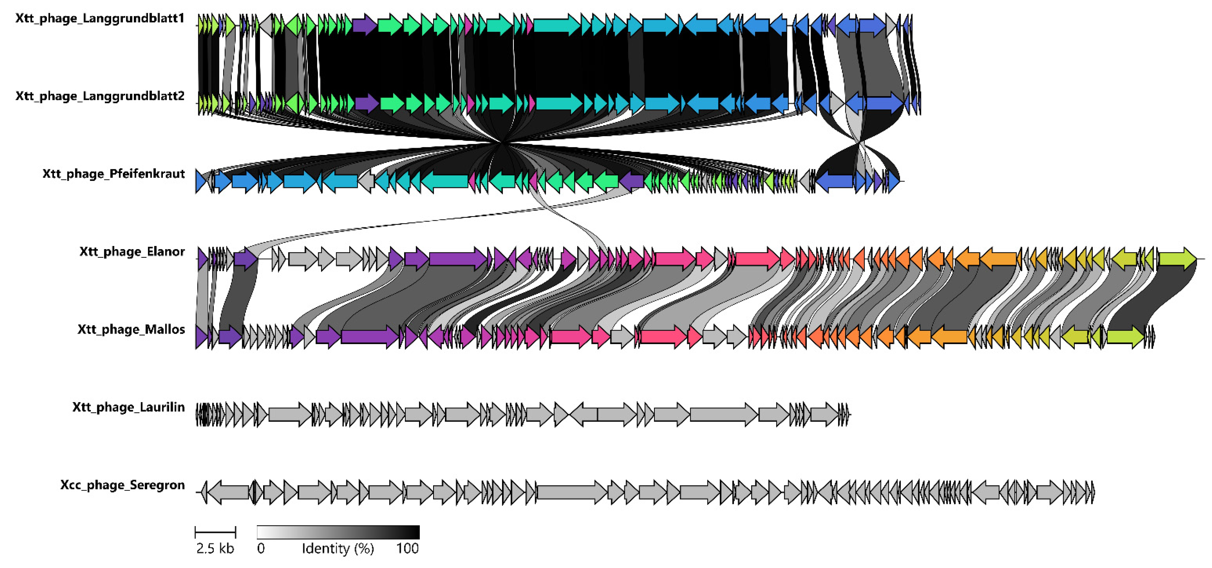
Genome maps were created using the R package gggenes (version 0.4.1) with fixed length parameters.
Novel phages were classified based on complete nucleotide sequences by comparing them against known sequenced phages infecting Xanthomonas, including closely related members recovered from the NCBI nucleotide blast searches, resulting in a total of 97 phage genomes. The 90 known genomes were downloaded from NCBI by their unique identifier. The average nucleotide identities (ANI) were calculated by pairwise comparison of the seven novel phages to the 90 reference genomes using the Perl program ClusterGenomes (https://github.com/simroux/ClusterGenomes) with default settings (80% coverage and 95% ANI). Further, we performed a clustering analysis using the VIRIDIC tool [49]. Heatmap clustering were displayed using the R package “pheatmap v.1.0.12”. In addition, more than 2000 proteobacteria-specific complete genomic sequences based on Virus–Host DB [50] information list of accession numbers were downloaded using the python program NCBI-genome-download (https://github.com/kblin/ncbi-genome-download). This set of sequences was clustered at 95% identity into 725 clusters using ClusterGenomes (https://github.com/simroux/ClusterGenomes) with default settings (80% coverage, 95% ANI). Lastly, a representative sequence from each 725 clusters, including seven novel phages with related phages obtained from blast-based searches, was analysed using k-mer clustering phylogeny (https://bioinformaticshome.com/bioinformatics_tutorials/R/phylogeny_estimation.html).

3. Results

3.1. Phage Isolation and Virion Morphology

Seven novel phages infecting Xanthomonas species were isolated from wastewater samples taken at Forschungszentrum Jülich (Germany) or from soil samples from the Eifel (Germany). The phages Langgrundbatt 1, Langgrundbatt 2, Pfeifenkraut, Elanor, Laurelin, and Mallos were isolated on Xanthomonas translucens pv. translucens (DSM 18974) (Figure 1A). Langgrundblatt 1 and 2 formed clear plaques with a mean diameter of 0.6 and 0.9 mm, respectively, but with considerable variation in plaque sizes. Phage Pfeifenkraut formed homogeneous round and clear plaques with a diameter of 1.3 mm. Phages Elanor, Laurilin, and Mallos formed clear irregular plaques with average diameter of 0.3 mm, 0.45 mm, and 0.65 mm, respectively (Figure 1B). The phage Seregon was isolated using Xanthomonas campestris pv. campestris as host. Sergon’s plaques had a diameter of 0.9 mm and were turbid in appearance. They were visible only after two days of incubation (Figure 1B).
TEM analysis of purified phage particles showed that all seven phages have an icosahedral capsid with sizes ranging 55–82 nm (Table S1) and a non-contractile tail 134–225 nm (Figure 1C). Therefore, based on their morphology the phages were classified as siphovirion phages.

3.2. Infection Curves and Host Range Determination

All isolated phages suppressed growth of the host culture when applied at a multiplicity of infection (MOI) of 0.1 or 1 (Figure 2). Interestingly, in some cases, the lower MOI had a stronger inhibitory effect on the host culture. Quantification of the phage titer by spotting dilution series on a bacterial lawn allowed us to visualize the amplification dynamics of the phages over the course of the experiment (Figure 2 right panel). Amplification was in several cases more pronounced at an MOI of 0.1.
In the context of plant biocontrol, the host range of phages represents an important parameter, since phages should not target plant growth promoting bacteria. Therefore, we assessed the host-range of the seven phages by spotting them on bacterial lawns of different Xanthomonas species (Xtt; Xcc), plant growth promoting bacteria (Pseudomonas fluorescence, Azospirillum brasilense, Sinorhizobium meliloti 1021, Herbaspirillum seropedicae, Pseudomonas koreenis, and Bacillus subtilis) and other plant pathogenic bacteria (Pseudomonas syringae pv lapsa, Pseudomonas syringae pv. tomato, and Agrobacterium tumefaciens). Xanthomonas campestris showed a slight susceptibility to the phages Langgrundblatt1, Langgrundblatt2, Pfeifenkraut, and Mallos. Vice versa, the phage Seregon isolated on Xcc showed lytic activity on X. translucens, but also at low levels. The EOPs are listed in Table S2. Interestingly, phage Mallos showed additional lytic activity on the plant pathogenic P. syringae strain DC3000, but also infected the plant growth-promoting P. fluorescens. This is making phage Mallos an omnilytic phage infecting distinct species and the phage with the broadest host range of the phages isolated in this study.

3.3. Genome Sequencing and Genome Features

All isolated phages were sequenced using Illumina Mi-Seq. short-read technology. Genomic features of the seven isolated phages are summarized in Table 1. Briefly, they show genome sizes from 40 to 62 kb, a GC content varies in the range of 53 to 64% and contain 56–89 annotated ORFs.
Phages Pfeifenkraut, Langgrundbatt 1, and Langgrundbatt 2 exhibit a remarkably low GC contend (53%) in contrast to their host Xtt (68%). Phage Laurelin differs from the other phages, in that its genome is the smallest, containing only 56 genes. Furthermore, it is the only one of the isolated phages containing 218 bp directed terminal repeats (DTRs). The genomic ends were determined using Phage Term [47]. In contrast, phages Langgrundbatt 1, Langgrundbatt 2, Pfeifenkraut, Elanor, and Mallos show a headful packing mechanism were the genome is translocated to the capsid at dedicated pac sites [51] resulting in variable genome ends. For the phage Seregon, cohesive ends with a length of 12 bp (5′ GGGGGCGCTGAC) were predicted. Since for plant biocontrol the phage lifestyle is of special importance, the lifestyle was predicted using PhageAI, a machine-learning tool which compares the genomes to over 20,000 publicly available phages [48]. All isolated phages were classified as virulent. This is further supported by the absence of integrase genes within the genomes.
The genome architecture for our isolated phages was very typical, in the sense that genes involved in the same function are clustered together featuring the typical modularity of phage genomes (Figure 3). These include units involved in DNA replication and repair, regulation, virion structure, and assembly (capsid, tail, and tail fibres), DNA packaging, and lysis. Further inspection of the phage genomes revealed that Langgrundblatt1, Langgrundblatt2, and Pfeifenkraut encode MazG nucleotide pyrophosphohydrolases [52], which, potentially, interfere with the hosts’ programmed cell death. The mechanism of abortive infection is widespread among bacteria and represents a typical antiviral strategy protecting the entire population by sacrificing a single cell [53,54]. Interestingly, phage Pfeifenkraut carries a group one Intron which have a limited distribution among bacteria but have been reported only in a few cases for phages infecting gram-negative bacteria [55,56].
With an average nucleotide identity of 97% Langgrundblatt1 and Langgrundblatt2 are members of the same species [57]. The genomes of the phages Elanor and Mallos share 75% sequence identity. Both phages contain a relatively high fraction of ORF encoding proteins of unknown functions (hypothetical proteins/CDS; 40/86 for Elanor and for Mallos 46/89) reflecting once more the significant amount of ‘dark matter’ harboured in phage genomes.
An important family of nucleoid-associated proteins involved in the silencing of foreign (e.g., phage) DNA are H-NS proteins [58]. Interestingly, phage Laurilin encodes an H-NS-binding protein which could function as inhibitor or of the host-encoded xenogeneic silencer protein [59]. NCBI protein blast revealed a broad distribution of homolog H-NS-binding proteins among phages of gram-negative bacteria. CDD blast revealed this protein as a hypothetical protein conserved in T7-like phages (cl10202).

3.4. Average Nucleotide Identity (ANI) Analysis

To analyze the phylogenetic relationship of our sequenced phages, we performed a comparison based on genome wide pairwise identity. Due to the high variability within phages genomes, traditional approaches often use single-gene phylogenies for the classification; however, no single gene is shared by all seven isolated phages. Only the large terminase shares sequence identity for five of the seven phages (dark purple arrow, Figure 4). Therefore, we selected all available genomic sequences for phages infecting Xanthomonas reviewed by Nakayinga et al. (2021) [6] and from literature [24]. The set was further expanded by unique entries of VirusHost DB [50], as well as by our phages and their closest relatives according to NCBI nucleotide blast, resulting in a total of 97 genomes. With those genomes we performed two independent clustering analysis (using VIRIDIC and ClusterGenomes), based on average nucleotide identity (ANI). The results of the clustering dendrogram created with VIRIDIC [49] show that our isolated phages fall into four distinct clusters within the Xanthomonas phages (Figure 5). The ANI-based clustering analysis showed that phages Langgrundblatt1, Langgrundblatt2, and Pfeifenkraut form a distinct cluster, resulting in the novel genus “Shirevirus”. Phage Laurilin clusters with three phages known to infect bacterial species of the genus Pseudomonas. Phage Elanor clusters together with the phages Bosa, Xp12, and Xoo-sp2. Interestingly, all members of this cluster infect Xanthomonas species that are pathogens of plants belonging to the Poaceae family. Seregon clusters together with phages infecting Xanthomonas and Xyllea [24]. This is supported by both clustering approaches (Table S1). Phage Mallos is a special case, while in VIRIDIC it clusters with the Elanor group and phage PaMx28 as a neighbour, in the Perl-based clustering it forms a group of its own. Altogether, our isolated phages display a broad diversity and cluster broadly among the known phages for Xanthomonas.
This diversity within our isolates is further supported by an additional clustering performed against 700 representative phages infecting proteobacteria retrieved from VirusHost DB [50] (supplementary Figure S1).

4. Discussion

Phage-based products bear a great potential for the sustainable and targeted treatment of bacterial infections in agriculture. However, to the best of our knowledge, no phage-based product was registered as plant protection product or biopesticide by the European Food Safety Authority until now [61]. Some of the main challenges to move bacteriophages towards application are: gaining an comprehensive understanding of the bacteria–phage–plant interaction on a molecular level, the development of standard operation procedures for the evaluation of novel phages as pest-control agents, and reliable ‘on-field’ application strategies [61].
While many of the early phage-isolation studies [2] were limited by the technical possibilities of their time. Out of 176 known phages infecting bacteria of the genus of Xanthomonas only roughly 100 are sequenced. Additionally, morphological characterization has become more feasible in recent years, with more access to high-resolution imaging devices. This information provides an important basis to potentially link phage morphological traits and binding preferences known to occur in certain virion morphotypes with biocontrol possibilities [62]. Last, but not least, phages for Xanthomonas are highly under-sampled given the fact that the number of known phages has almost doubled in the last decade, reaching over 14,244 complete sequenced phage genomes by the beginning of 2021 [63,64,65].
Here we report the isolation and characterization of seven novel Xanthomonas phages. Genomic analysis revealed typical arrangement of genes into clusters linked to functional units, but also a significant amount of ‘dark matter’ harboured in phage genomes. The phages Elanor, Mallos, and Seregon encode a Cas4 family exonuclease which normally plays a role in acquiring functional spacers in bacterial CRISPR immunity [66]. Nevertheless, cases have been reported were phage-derived Cas4-like proteins led to host spacer acquisition and, subsequently, autoimmunity of the bacterial host [67]. This way, the phage uses the bacterial genome as a decoy for its own immune system, thereby gaining time for the production of phage progeny.
An essential trait for phages used in biocontrol is a lytic lifestyle. Since temperate phages could equip their bacterial host with further virulence traits [68]. All our phages were predicted to have a lytic lifestyle by the machine-learning-based phage lifestyle determination tool PhageAI [48]. This was further underpinned by the absence of integrase genes within their genomes. These results are in line with genomic clustering analysis where they cluster with virulent phages. It has, however, to be noted, that phage Seregon forms turbid plaques on lawns (Figure 1). Therefore, further analysis is required to determine the lifestyle of this phage.
Characterization of the host range assay showed that phages Langgrundblat1, Langgrundblat2, Pfeifenkraut, Elanor, and Seregon are highly specific, only infecting their isolation host and the other plant-pathogenic Xanthomonads but featuring a significantly lower efficiency of plating. Nevertheless, these assays confirmed that they do not infect the majority of the here-tested plant growth-promoting bacteria (PGPB). This shows the advantage of phages as targeted plant biocontrol argent in contrast to other antimicrobials, which also broadly affect the beneficial part of the plant microbiome. Phage Laurilin displayed the highest specificity, only infecting its isolation host X. translucens. In contrast, phage Mallos displayed the broadest host range and also infected the plant pathogenic bacterium Pseudomonas syringae DC3000, making it an omnylitic phage infecting species of two different alphaproteobacterial genera. This makes sense in the context that Xanthomonas and Pseudomonas are among the most abundant genera in the plant phyllosphere [69]. In addition, a recent study of two phages infecting Pseudomonas syringae pv. tomato DC3000 also finds lytic activity on two Xanthomonas species [70].
ANI-based comparison of our isolates to phages from the literature [6,24] revealed that the phages are very diverse. Phages Pfeifenkraut, Langgrundblatt1, and Laggrundblatt2 cluster with each other but show very little homology with other described phages, therefore forming the new genus of “Shirevirus”. This highlights that there is a vast number of uncharacterized phages out there that can potentially be harnessed for biocontrol. This is also in line with a recent phage metagenomic study of wheat phyllosphere where Xanthomonas and Pseudomonas were the most abundant bacterial genera in the plant phyllosphere and 96.8% of the generated viral taxonomic units did represent phages which have not been isolated so far [69].
Our results suggest that our phages are suitable candidates for an application in planta and, thereby, will add up to the many phage biocontrol trials performed against a broad spectrum of different plant pathogenic bacteria in the recent years [4,5,6,61]. A comprehensive overview over the current phage isolates for Xanthomonas species is given by Nakayinga et al. (2021) [6]. Further the application of Xcc phages (FoX6 and FoX2) at different stages of plant lifecycle (seed, seedling, and field condition) was accessed recently, showing reduction of disease severity at all stages demonstrating the potential benefits of phages as biocontrol agents [32].
Recent trials aim at circumventing problems caused by classical chemical treatments against Xtt, for example, by using supernatants of lactic acid bacteria, Bacillus strains, or plant natural products [71]. The main downside of all these substances is that they—unlike phages—cannot multiply themselves in the presence of the pathogen. Combinatorial approaches using phages and plant-growth-promoting bacteria producing those chemically active compounds could overcome this hurdle and could be part of future integrated pest control strategies to maximize crop yield and protection. Here, the phages isolated and characterized in this study add suitable candidates to be benchmarked in future biocontrol experiments for the treatment of Xtt and Xcc infections in planta.
Additionally, by way of an example from personalized medicine, the treatment of bacterial infections in humans or animals is routinely based on the isolation of bacterial strains from the patient and isolation of specific phages for the respective pathogen [72,73,74]. This approach has shown promising results and demands for the regular update of phage cocktails in the clinic. It is a strong advantage of phage-based biocontrol, that this natural diversity can be harnessed. This is very likely also required for sustainably successful agricultural applications and would require phage isolation from plants growing exposed to pathogenic bacteria in field conditions.

Supplementary Materials

The following supporting information can be downloaded at: https://www.mdpi.com/article/10.3390/v14071449/s1, Figure S1: K-mer clustering dendrogram of Proteobacteria phages; Figure S2: Host range assay; Table S1: Phage particle size. Measurements of virion particles analyzed by Transmission electron microscopy (TEM); Table S2: Strains /Phages used in this study.

Author Contributions

Conceptualization, S.H.E., B.A. and J.F.; methodology, all; validation, all; formal analysis, all; investigation, S.H.E.; resources, B.A., U.S. and J.F.; data curation, S.H.E. and V.S.; writing—original draft preparation, S.H.E. and J.F.; writing—review and editing, all; visualization, S.H.E. and V.S.; supervision, B.A., U.S. and J.F.; project administration, J.F.; funding acquisition, B.A., U.S. and J.F. All authors have read and agreed to the published version of the manuscript.

Funding

We thank the European Research Council (ERC starting grant number 757563) for support. B.A. is part of the CEPLAS excellence cluster (Germany’s Excellence Strategy, EXC 2048).

Institutional Review Board Statement

Not applicable.

Informed Consent Statement

Not applicable.

Data Availability Statement

Not applicable.

Acknowledgments

The authors gratefully acknowledge the electron microscopy training and access time granted by the biological EM facility of the Ernst-Ruska Centre at Forschungszentrum Jülich. Further, the authors thank Franz Narberhaus (RUB, Bochum, North Rhine-Westphalia, Germany) for providing the X. campestris and the Agrobacterum tumefaciens C58 strains. We also thank Sebastian Schenk and the Ott (University of Freiburg, Freiburg, Baden-Wurttemberg, Germany) for sharing the P. syringae DC3000 strain. The X. translucens strain was kindly provided by the German Collection of Microorganisms and Cell Cultures (DSMZ, Braunschweig, Germany). We are also grateful to the entire Frunzke lab and the root group for fruitful discussions.

Conflicts of Interest

The authors declare no conflict of interest.

References

  1. Strange, R.N.; Scott, P.R. Plant Disease: A Threat to Global Food Security. Annu. Rev. Phytopathol. 2005, 43, 83–116. [Google Scholar] [CrossRef] [PubMed]
  2. Mallmann, W.; Hemstreet, S. Isolation of an inhibitory substance from plants. J. Agric. Res. 1924, 28, 599–602. [Google Scholar]
  3. Balogh, B.; Jones, J.B.; Iriarte, F.B.; Momol, M.T. Phage therapy for plant disease control. Curr. Pharm. Biotechnol. 2010, 11, 48–57. [Google Scholar] [CrossRef]
  4. Svircev, A.; Roach, D.; Castle, A. Framing the Future with Bacteriophages in Agriculture. Viruses 2018, 10, 218. [Google Scholar] [CrossRef] [PubMed] [Green Version]
  5. Buttimer, C.; McAuliffe, O.; Ross, R.P.; Hill, C.; O’Mahony, J.; Coffey, A. Bacteriophages and Bacterial Plant Diseases. Front. Microbiol. 2017, 8, 34. [Google Scholar] [CrossRef] [Green Version]
  6. Nakayinga, R.; Makumi, A.; Tumuhaise, V.; Tinzaara, W. Xanthomonas bacteriophages: A review of their biology and biocontrol applications in agriculture. BMC Microbiol. 2021, 21, 291. [Google Scholar] [CrossRef]
  7. Mansfield, J.; Genin, S.; Magori, S.; Citovsky, V.; Sriariyanum, M.; Ronald, P.; Dow, M.; Verdier, V.; Beer, S.V.; Machado, M.A.; et al. Top 10 plant pathogenic bacteria in molecular plant pathology: Top 10 plant pathogenic bacteria. Mol. Plant Pathol. 2012, 13, 614–629. [Google Scholar] [CrossRef] [Green Version]
  8. Fujiwara, A.; Fujisawa, M.; Hamasaki, R.; Kawasaki, T.; Fujie, M.; Yamada, T. Biocontrol of Ralstonia solanacearum by Treatment with Lytic Bacteriophages. Appl. Environ. Microbiol. 2011, 77, 4155–4162. [Google Scholar] [CrossRef] [Green Version]
  9. Bae, J.Y. Biocontrol Potential of a Lytic Bacteriophage PE204 against Bacterial Wilt of Tomato. J. Microbiol. Biotechnol. 2012, 22, 1613–1620. [Google Scholar] [CrossRef]
  10. Wei, C.; Liu, J.; Maina, A.N.; Mwaura, F.B.; Yu, J.; Yan, C.; Zhang, R.; Wei, H. Developing a bacteriophage cocktail for biocontrol of potato bacterial wilt. Virol. Sin. 2017, 32, 476–484. [Google Scholar] [CrossRef]
  11. Elhalag, K.; Nasr-Eldin, M.; Hussien, A.; Ahmad, A. Potential use of soilborne lytic Podoviridae phage as a biocontrol agent against Ralstonia solanacearum. J. Basic Microbiol. 2018, 58, 658–669. [Google Scholar] [CrossRef]
  12. Álvarez, B.; López, M.M.; Biosca, E.G. Biocontrol of the Major Plant Pathogen Ralstonia solanacearum in Irrigation Water and Host Plants by Novel Waterborne Lytic Bacteriophages. Front. Microbiol. 2019, 10, 2813. [Google Scholar] [CrossRef] [PubMed] [Green Version]
  13. Wang, X.; Wei, Z.; Yang, K.; Wang, J.; Jousset, A.; Xu, Y.; Shen, Q.; Friman, V.-P. Phage combination therapies for bacterial wilt disease in tomato. Nat. Biotechnol. 2019, 37, 1513–1520. [Google Scholar] [CrossRef] [PubMed]
  14. Boulé, J.; Sholberg, P.L.; Lehman, S.M.; O’gorman, D.T.; Svircev, A.M. Isolation and characterization of eight bacteriophages infecting Erwinia amylovora and their potential as biological control agents in British Columbia, Canada. Can. J. Plant Pathol. 2011, 33, 308–317. [Google Scholar] [CrossRef]
  15. Born, Y.; Fieseler, L.; Thöny, V.; Leimer, N.; Duffy, B.; Loessner, M.J. Engineering of Bacteriophages Y2:: dpoL1-C and Y2:: luxAB for Efficient Control and Rapid Detection of the Fire Blight Pathogen, Erwinia amylovora. Appl. Environ. Microbiol. 2017, 83, e00341-17. [Google Scholar] [CrossRef] [Green Version]
  16. Schwarczinger, I.; Nagy, J.K.; Künstler, A.; Szabó, L.; Geider, K.; Király, L.; Pogany, M. Characterization of Myoviridae and Podoviridae family bacteriophages of Erwinia amylovora from Hungary—Potential of application in biological control of fire blight. Eur. J. Plant Pathol. 2017, 149, 639–652. [Google Scholar] [CrossRef] [Green Version]
  17. Di Lallo, G.; Evangelisti, M.; Mancuso, F.; Ferrante, P.; Marcelletti, S.; Tinari, A.; Superti, F.; Migliore, L.; D’Addabbo, P.; Frezza, D.; et al. Isolation and partial characterization of bacteriophages infecting Pseudomonas syringae pv. actinidiae, causal agent of kiwifruit bacterial canker: Bacteriophages infecting P. syringae pv. actinidiae. J. Basic Microbiol. 2014, 54, 1210–1221. [Google Scholar]
  18. Frampton, R.A.; Taylor, C.; Moreno, A.V.H.; Visnovsky, S.B.; Petty, N.K.; Pitman, A.R.; Fineran, P.C. Identification of Bacteriophages for Biocontrol of the Kiwifruit Canker Phytopathogen Pseudomonas syringae pv. actinidiae. Appl. Environ. Microbiol. 2014, 80, 2216–2228. [Google Scholar] [CrossRef] [Green Version]
  19. Rombouts, S.; Volckaert, A.; Venneman, S.; Declercq, B.; Vandenheuvel, D.; Allonsius, C.N.; Van Malderghem, C.; Jang, H.B.; Briers, Y.; Noben, J.P.; et al. Characterization of Novel Bacteriophages for Biocontrol of Bacterial Blight in Leek Caused by Pseudomonas syringae pv. porri. Front. Microbiol. 2016, 7, 279. [Google Scholar] [CrossRef] [Green Version]
  20. Rabiey, M.; Roy, S.R.; Holtappels, D.; Franceschetti, L.; Quilty, B.J.; Creeth, R.; Sundin, G.W.; Wagemans, J.; Lavigne, R.; Jackson, R.W. Phage biocontrol to combat Pseudomonas syringae pathogens causing disease in cherry. Microb. Biotechnol. 2020, 13, 1428–1445. [Google Scholar] [CrossRef]
  21. Liu, Y.; Liu, M.; Hu, R.; Bai, J.; He, X.; Jin, Y. Isolation of the Novel Phage PHB09 and Its Potential Use against the Plant Pathogen Pseudomonas syringae pv. actinidiae. Viruses 2021, 13, 2275. [Google Scholar] [CrossRef] [PubMed]
  22. Song, Y.R.; Vu, N.T.; Park, J.; Hwang, I.S.; Jeong, H.J.; Cho, Y.S.; Oh, C.S. Phage PPPL-1, A New Biological Agent to Control Bacterial Canker Caused by Pseudomonas syringae pv. actinidiae in Kiwifruit. Antibiotics 2021, 10, 554. [Google Scholar] [CrossRef] [PubMed]
  23. Tarakanov, R.I.; Lukianova, A.A.; Evseev, P.V.; Toshchakov, S.V.; Kulikov, E.E.; Ignatov, A.N.; Miroshnikov, K.A.; Dzhalilov, F.S. Bacteriophage Control of Pseudomonas savastanoi pv. glycinea in Soybean. Plants 2022, 11, 938. [Google Scholar] [CrossRef] [PubMed]
  24. Ahern, S.; Das, M.; Bhowmick, T.S.; Young, R.; Gonzalez, C.F. Characterization of Novel Virulent Broad-Host-Range Phages of Xylella fastidiosa and Xanthomonas. J. Bacteriol. 2013, 196, 459–471. [Google Scholar] [CrossRef] [PubMed]
  25. Das, M.; Bhowmick, T.S.; Ahern, S.J.; Young, R.; Gonzalez, C.F. Control of Pierce’s Disease by Phage. PLoS ONE 2015, 10, e0128902. [Google Scholar] [CrossRef] [Green Version]
  26. Grace, E.R.; Rabiey, M.; Friman, V.; Jackson, R.W. Seeing the forest for the trees: Use of phages to treat bacterial tree diseases. Plant Pathol. 2021, 70, 1987–2004. [Google Scholar] [CrossRef]
  27. Sapkota, S.; Mergoum, M.; Liu, Z. The translucens group of Xanthomonas translucens: Complicated and important pathogens causing bacterial leaf streak on cereals. Mol. Plant Pathol. 2020, 21, 291–302. [Google Scholar] [CrossRef] [Green Version]
  28. Katznelson, H.; Sutton, M.D. Bacteriophage in Relation to Xanthomonas Translucens. Can. J. Bot. 1953, 31, 725–729. [Google Scholar] [CrossRef]
  29. Vicente, J.G.; Holub, E.B. Xanthomonas campestris pv. campestris (cause of black rot of crucifers) in the genomic era is still a worldwide threat to brassica crops: Xanthomonas campestris pv. campestris. Mol. Plant Pathol. 2013, 14, 2–18. [Google Scholar]
  30. An, S.Q.; Potnis, N.; Dow, M.; Vorhölter, F.J.; He, Y.Q.; Becker, A.; Teper, D.; Li, Y.; Wang, N.; Bleris, L.; et al. Mechanistic insights into host adaptation, virulence and epidemiology of the phytopathogen Xanthomonas. FEMS Microbiol. Rev. 2020, 44, 1–32. [Google Scholar] [CrossRef] [Green Version]
  31. Gazdik, F.; Magnus, S.; Roberts, S.J.; Baranski, R.; Cechova, J.; Pokluda, R.; Eichmeier, A.; Grzebelus, D.; Baranek, M. Persistence of Xanthomonas campestris pv. campestris in Field Soil in Central Europe. Microorganisms 2021, 9, 591. [Google Scholar] [CrossRef] [PubMed]
  32. Holtappels, D.; Fortuna, K.J.; Moons, L.; Broeckaert, N.; Bäcker, L.E.; Venneman, S.; Rombouts, S.; Lippens, L.; Baeyen, S.; Pollet, S.; et al. The potential of bacteriophages to control Xanthomonas campestris pv. campestris at different stages of disease development. Microb. Biotechnol. 2022, 15, 1762–1782. [Google Scholar] [CrossRef] [PubMed]
  33. Weiss, B.D.; Capage, M.A.; Kessel, M.; Benson, S.A. Isolation and characterization of a generalized transducing phage for Xanthomonas campestris pv. campestris. J. Bacteriol. 1994, 176, 3354–3359. [Google Scholar] [CrossRef] [PubMed] [Green Version]
  34. Papaianni, M.; Paris, D.; Woo, S.L.; Fulgione, A.; Rigano, M.M.; Parrilli, E.; Tutino, M.L.; Marra, R.; Manganiello, G.; Casillo, A.; et al. Plant Dynamic Metabolic Response to Bacteriophage Treatment After Xanthomonas campestris pv. campestris Infection. Front. Microbiol. 2020, 11, 732. [Google Scholar] [CrossRef]
  35. Moser, R.; Aktas, M.; Narberhaus, F. Phosphatidylcholine biosynthesis in X anthomonas campestris via a yeast-like acylation pathway: Methylated phospholipids in Xanthomonas. Mol. Microbiol. 2014, 91, 736–750. [Google Scholar] [CrossRef]
  36. Kauffman, K.M.; Polz, M.F. Streamlining standard bacteriophage methods for higher throughput. MethodsX 2018, 5, 159–172. [Google Scholar] [CrossRef]
  37. Yamamoto, K.R.; Alberts, B.M.; Benzinger, R.; Lawhorne, L.; Treiber, G. Rapid bacteriophage sedimentation in the presence of polyethylene glycol and its application to large-scale virus purification. Virology 1970, 40, 734–744. [Google Scholar] [CrossRef]
  38. Young, J.M.; Dye, D.W.; Bradbury, J.F.; Panagopoulos, C.G.; Robbs, C.F. A proposed nomenclature and classification for plant pathogenic bacteria. N. Z. J. Agric. Res. 1978, 21, 153–177. [Google Scholar] [CrossRef]
  39. Xin, X.-F.; He, S.Y. Pseudomonas syringae pv. tomato DC3000: A Model Pathogen for Probing Disease Susceptibility and Hormone Signaling in Plants. Annu. Rev. Phytopathol. 2013, 51, 473–498. [Google Scholar] [CrossRef]
  40. Schenk, S.T.; Hernández-Reyes, C.; Samans, B.; Stein, E.; Neumann, C.; Schikora, M.; Reichelt, M.; Mithöfer, A.; Becker, A.; Kogel, K.-H.; et al. N-Acyl-Homoserine Lactone Primes Plants for Cell Wall Reinforcement and Induces Resistance to Bacterial Pathogens via the Salicylic Acid/Oxylipin Pathway. Plant Cell 2014, 26, 2708–2723. [Google Scholar] [CrossRef] [Green Version]
  41. Hardy, A.; Sharma, V.; Kever, L.; Frunzke, J. Genome Sequence and Characterization of Five Bacteriophages Infecting Streptomyces coelicolor and Streptomyces venezuelae: Alderaan, Coruscant, Dagobah, Endor1 and Endor2. Viruses 2020, 12, 1065. [Google Scholar] [CrossRef] [PubMed]
  42. McNair, K.; Zhou, C.; Dinsdale, E.; Souza, B.; Edwards, R. PHANOTATE: A novel approach to gene identification in phage genomes. Bioinformatics 2019, 35, 4537–4542. [Google Scholar] [CrossRef] [PubMed] [Green Version]
  43. Kanehisa, M.; Furumichi, M.; Tanabe, M.; Sato, Y.; Morishima, K. KEGG: New perspectives on genomes, pathways, diseases and drugs. Nucleic Acids Res. 2017, 45, D353–D361. [Google Scholar] [CrossRef] [PubMed] [Green Version]
  44. Zhou, C.L.E.; Malfatti, S.; Kimbrel, J.; Philipson, C.; McNair, K.; Hamilton, T.; Edwards, R.; Souza, B. multiPhATE: Bioinformatics pipeline for functional annotation of phage isolates. Bioinformatics 2019, 35, 4402–4404. [Google Scholar] [CrossRef] [Green Version]
  45. Altschul, S.F.; Gish, W.; Miller, W.; Myers, E.W.; Lipman, D.J. Basic local alignment search tool. J. Mol. Biol. 1990, 215, 403–410. [Google Scholar] [CrossRef]
  46. Marchler-Bauer, A.; Lu, S.; Anderson, J.B.; Chitsaz, F.; Derbyshire, M.K.; DeWeese-Scott, C.; Fong, J.H.; Geer, L.; Geer, R.C.; Gonzales, N.R.; et al. CDD: A Conserved Domain Database for the functional annotation of proteins. Nucleic Acids Res. 2010, 39, D225–D229. [Google Scholar] [CrossRef] [Green Version]
  47. Garneau, J.R.; Depardieu, F.; Fortier, L.-C.; Bikard, D.; Monot, M. PhageTerm: A tool for fast and accurate determination of phage termini and packaging mechanism using next-generation sequencing data. Sci. Rep. 2017, 7, 8292. [Google Scholar] [CrossRef]
  48. Tynecki, P.; Guziński, A.; Kazimierczak, J.; Jadczuk, M.; Dastych, J.; Onisko, A. PhageAI—Bacteriophage Life Cycle Recognition with Machine Learning and Natural Language Processing. bioRxiv 2020. [Google Scholar] [CrossRef]
  49. Moraru, C.; Varsani, A.; Kropinski, A.M. VIRIDIC—A Novel Tool to Calculate the Intergenomic Similarities of Prokaryote-Infecting Viruses. Viruses 2020, 12, 1268. [Google Scholar] [CrossRef]
  50. Mihara, T.; Nishimura, Y.; Shimizu, Y.; Nishiyama, H.; Yoshikawa, G.; Uehara, H.; Hingamp, P.; Goto, S.; Ogata, H. Linking Virus Genomes with Host Taxonomy. Viruses 2016, 8, 66. [Google Scholar] [CrossRef]
  51. Fujisawa, H.; Morita, M. Phage DNA packaging. Genes Cells 1997, 2, 537–545. [Google Scholar] [CrossRef] [PubMed]
  52. Gross, M.; Marianovsky, I.; Glaser, G. MazG—A regulator of programmed cell death in Escherichia coli: MazG—PCD regulator in E. coli. Mol. Microbiol. 2006, 59, 590–601. [Google Scholar] [CrossRef] [PubMed]
  53. Harms, A.; Brodersen, D.E.; Mitarai, N.; Gerdes, K. Toxins, Targets, and Triggers: An Overview of Toxin-Antitoxin Biology. Mol. Cell 2018, 70, 768–784. [Google Scholar] [CrossRef] [PubMed] [Green Version]
  54. Lopatina, A.; Tal, N.; Sorek, R. Abortive Infection: Bacterial Suicide as an Antiviral Immune Strategy. Annu. Rev. Virol. 2020, 7, 371–384. [Google Scholar] [CrossRef]
  55. Bonocora, R.P.; Shub, D.A. A Self-Splicing Group I Intron in DNA Polymerase Genes of T7-Like Bacteriophages. J. Bacteriol. 2004, 186, 8153–8155. [Google Scholar] [CrossRef] [Green Version]
  56. Hausner, G.; Hafez, M.; Edgell, D.R. Bacterial group I introns: Mobile RNA catalysts. Mob. DNA 2014, 5, 8. [Google Scholar] [CrossRef] [Green Version]
  57. Adriaenssens, E.; Brister, J.R. How to Name and Classify Your Phage: An Informal Guide. Viruses 2017, 9, 70. [Google Scholar] [CrossRef] [Green Version]
  58. Navarre, W.W. The Impact of Gene Silencing on Horizontal Gene Transfer and Bacterial Evolution. In Advances in Microbial Physiology; Elsevier: Amsterdam, The Netherlands, 2016; Volume 69, pp. 157–186. [Google Scholar]
  59. Pfeifer, E.; Hünnefeld, M.; Popa, O.; Frunzke, J. Impact of Xenogeneic Silencing on Phage–Host Interactions. J. Mol. Biol. 2019, 431, 4670–4683. [Google Scholar] [CrossRef]
  60. Gilchrist, C.L.M.; Chooi, Y.-H. clinker & clustermap.js: Automatic generation of gene cluster comparison figures. Bioinformatics 2021, 37, 2473–2475. [Google Scholar] [CrossRef]
  61. Holtappels, D.; Fortuna, K.; Lavigne, R.; Wagemans, J. The future of phage biocontrol in integrated plant protection for sustainable crop production. Curr. Opin. Biotechnol. 2020, 68, 60–71. [Google Scholar] [CrossRef]
  62. Bertozzi Silva, J.; Storms, Z.; Sauvageau, D. Host receptors for bacteriophage adsorption. FEMS Microbiol. Lett. 2016, 363, fnw002. [Google Scholar] [CrossRef] [PubMed] [Green Version]
  63. Clokie, M.R.J.; Millard, A.D.; Letarov, A.V.; Heaphy, S. Phages in nature. Bacteriophage 2011, 1, 31–45. [Google Scholar] [CrossRef] [Green Version]
  64. Dion, M.B.; Oechslin, F.; Moineau, S. Phage diversity, genomics and phylogeny. Nat. Rev. Microbiol. 2020, 18, 125–138. [Google Scholar] [CrossRef] [PubMed]
  65. Cook, R.; Brown, N.; Redgwell, T.; Rihtman, B.; Barnes, M.; Clokie, M.; Stekel, D.J.; Hobman, J.; Jones, M.A.; Millard, A. INfrastructure for a PHAge REference Database: Identification of Large-Scale Biases in the Current Collection of Cultured Phage Genomes. PHAGE 2021, 2, 214–223. [Google Scholar] [CrossRef]
  66. Kieper, S.N.; Almendros, C.; Behler, J.; McKenzie, R.E.; Nóbrega, F.; Haagsma, A.C.; Vink, J.N.; Hess, W.R.; Brouns, S.J. Cas4 Facilitates PAM-Compatible Spacer Selection during CRISPR Adaptation. Cell Rep. 2018, 22, 3377–3384. [Google Scholar] [CrossRef]
  67. Hooton, S.P.T.; Connerton, I. Campylobacter jejuni acquire new host-derived CRISPR spacers when in association with bacteriophages harboring a CRISPR-like Cas4 protein. Front. Microbiol. 2015, 5, 744. [Google Scholar] [CrossRef] [Green Version]
  68. Wagner, P.L.; Waldor, M.K. Bacteriophage Control of Bacterial Virulence. Infect. Immun. 2002, 70, 3985–3993. [Google Scholar] [CrossRef] [Green Version]
  69. Forero-Junco, L.M.; Alanin, K.W.S.; Djurhuus, A.M.; Kot, W.; Gobbi, A.; Hansen, L.H. Bacteriophages Roam the Wheat Phyllosphere. Viruses 2022, 14, 244. [Google Scholar] [CrossRef]
  70. Korniienko, N.; Kharina, A.; Zrelovs, N.; Jindřichová, B.; Moravec, T.; Budzanivska, I.; Burketová, L.; Kalachova, T. Isolation and Characterization of Two Lytic Phages Efficient Against Phytopathogenic Bacteria from Pseudomonas and Xanthomonas Genera. Front. Microbiol. 2022, 13, 853593. [Google Scholar] [CrossRef]
  71. Mačionienė, I.; Čepukoit, D.; Šalomskienė, J.; Černauskas, D.; Burokienė, D.; Šalaševičienė, A. Effects of Natural Antimicrobials on Xanthomonas Strains Growth. Horticulturae 2021, 8, 7. [Google Scholar] [CrossRef]
  72. Mattila, S.; Ruotsalainen, P.; Jalasvuori, M. On-Demand Isolation of Bacteriophages Against Drug-Resistant Bacteria for Personalized Phage Therapy. Front. Microbiol. 2015, 6, 1271. [Google Scholar] [CrossRef] [PubMed] [Green Version]
  73. Schooley, R.T.; Biswas, B.; Gill, J.J.; Hernandez-Morales, A.; Lancaster, J.; Lessor, L.; Barr, J.J.; Reed, S.L.; Rohwer, F.; Benler, S.; et al. Development and Use of Personalized Bacteriophage-Based Therapeutic Cocktails to Treat a Patient with a Disseminated Resistant Acinetobacter baumannii Infection. Antimicrob. Agents Chemother. 2017, 61, e00954-17. [Google Scholar] [CrossRef] [PubMed] [Green Version]
  74. Greene, W.; Chan, B.; Bromage, E.; Grose, J.H.; Walsh, C.; Kortright, K.; Forrest, S.; Perry, G.; Byrd, L.; Stamper, M.A. The Use of Bacteriophages and Immunological Monitoring for the Treatment of a Case of Chronic Septicemic Cutaneous Ulcerative Disease in a Loggerhead Sea Turtle Caretta caretta. J. Aquat. Anim. Health 2021, 33, 139–154. [Google Scholar] [CrossRef] [PubMed]
Figure 1. Phage morphology of novel Xanthomonas phage isolates. (A) Plaque morphologies of the seven different phages infecting Xanthomonas translucens pv. translucens (DSM 18974) (Langgrundbatt 1, Langgrundbatt 2, Pfeifenkraut, Elanor, Laurelin, and Mallos) and Xanthomonas campestris pv. campestris (Seregon). Scale Bar: 2 cm; (B) Stereo microscopy of single plaques. Scale bar: 1 mm; (C) Transmission electron microscopy (TEM) images of virion particles. The phage isolates were negative stained with uranyl acetate. Scale bar: 100 nm.
Viruses 14 01449 g001
Figure 2. Infection curves of seven phages infecting Xtt (A) and Xcc (B). Full-grown overnight cultures of the host were adjusted to an OD600 of 0.2 and phages at the corresponding multiplicity of infection (MOI) were added. The infection was performed as biological triplicates. Backscatter was measured over time (left panels), as well as phage titers (right panels). Plaques of phage Seregon have a turbid appearance and are, therefore, hardly visible (see Figure 1B).
Viruses 14 01449 g002aViruses 14 01449 g002b
Figure 3. Genome map of the six Xanthomonas phages. Open reading frames (ORFs) were predicted using multiPhate [44], with Phanotate [42] and were later curated manually using NCBI Blast. Encoded protein domains were further predicted by using the batch function of NCBI Conserved Domain Database (CDD). Genome maps were created using the R package gggenes using fixed-length parameters.
Viruses 14 01449 g003
Figure 4. Genome comparison on the level of coding sequences. The coding sequences (CDS) of the isolated phages were compared using the clinker pipeline [60] to cluster them in groups by similarity (each colour represents one group) and per cent identity of the member of one group is indicated by grey values. The circular genomes are represented linearly and the direction of the arrows is in line with transcription direction of each CDS.
Viruses 14 01449 g004
Figure 5. Average nucleotide-based dendrogram analysis using 97 phage genomes of phages infecting Xanthomonas species. Genomes are derived from [6] or Virus Host DB (https://www.genome.jp/virushostdb/, accessed on 19 January 2022). Furthermore, 11 genomes were acquired from NCBI based on similarity to our seven novel phages.
Viruses 14 01449 g005
Table 1. Basic genomic features of the seven novel phages.
Phage NameAccession NumberReference HostGenome Size (Bp)GC Content (%)ORF Number aGenome Termini Class bLifestyle Prediction c
Langgrundblatt 1ON189042Xanthomonas translucens DSM 1897444.23953.367Headful
(pac)
virulent
Langgrundblatt 2ON189043Xanthomonas translucens DSM 1897444.76853.468Headful
(pac)
virulent
PfeifenkrautON189044Xanthomonas translucens DSM 1897443.79153.372Headful
(pac)
virulent
ElanorON189045Xanthomonas translucens DSM 1897462.34164.586Headful
(pac)
virulent
LaurelinON189046Xanthomonas translucens DSM 1897440.49857.456DTR (short)virulent
MallosON189047Xanthomonas translucens DSM 1897459.24261.888Headful
(pac)
virulent
SeregonON189048Xanthomonas campestris55.52763.272COS (5′)virulent
a Open reading frames (ORFs) were predicted using multiPhate [44] with Phanotate [42] and, subsequently, annotated against different customized databases (NCBI viral proteins, NCBI Refseq proteins, Kyoto Encyclopedia of Genes and Genomes (KEGG) [43], Phage Annotation Tools and Methods (PhAnToMe) (www.phantome.org), and phage Virus Orthologous Groups (pVOG)). Additionally, manually curated using NCBI Blastp. Encoded Protein Domains were further predicted by using the batch function of NCBI Conserved Domain Database (CDD). b Genome termini classes were determined using PhageTerm [47]. c Phage lifestyle was predicted by the machine-learning-based program PhageAI [48] and, further, confirmed by absence of intergrade genes inside the genomes.
Publisher’s Note: MDPI stays neutral with regard to jurisdictional claims in published maps and institutional affiliations.

