Article

Clinical Evaluation of a Fully-Automated High-Throughput Multiplex Screening-Assay to Detect and Differentiate the SARS-CoV-2 B.1.1.529 (Omicron) and B.1.617.2 (Delta) Lineage Variants

1
Institute of Medical Microbiology, Virology and Hygiene, University Medical Center Hamburg-Eppendorf (UKE), 20246 Hamburg, Germany
2
Leibniz Institute for Experimental Virology (HPI), Virus Genomics, 20251 Hamburg, Germany
*
Author to whom correspondence should be addressed.
Viruses 2022, 14(3), 608; https://doi.org/10.3390/v14030608
Submission received: 9 January 2022 / Revised: 1 March 2022 / Accepted: 12 March 2022 / Published: 15 March 2022
(This article belongs to the Topic Infectious Diseases)

Abstract

:
Background: The recently emerged SARS-CoV-2 B.1.1.529 lineage and its sublineages (Omicron variant) pose a new challenge to healthcare systems worldwide due to its ability to efficiently spread in immunized populations and its resistance to currently available monoclonal antibody therapies. RT-PCR-based variant tests can be used to screen large sample-sets rapidly and accurately for relevant variants of concern (VOC). The aim of this study was to establish and validate a multiplex assay on the cobas 6800/8800 systems to allow discrimination between the two currently circulating VOCs, Omicron and Delta, in clinical samples. Methods: Primers and probes were evaluated for multiplex compatibility. Analytic performance was assessed using cell culture supernatant of an Omicron variant isolate and a clinical Delta variant sample, normalized to WHO-Standard. Clinical performance of the multiplex assay was benchmarked against NGS results. Results: In silico testing of all oligos showed no interactions with a high risk of primer-dimer formation or amplification of human DNA/RNA. Over 99.9% of all currently available Omicron variant sequences are a perfect match for at least one of the three Omicron targets included in the multiplex. Analytic sensitivity was determined as 19.0 IU/mL (CI95%: 12.9–132.2 IU/mL) for the A67V + del-HV69-70 target, 193.9 IU/mL (CI95%: 144.7–334.7 IU/mL) for the E484A target, 35.5 IU/mL (CI95%: 23.3–158.0 IU/mL) for the N679K + P681H target and 105.0 IU/mL (CI95%: 80.7–129.3 IU/mL) for the P681R target. All sequence variances were correctly detected in the clinical sample set (225/225 Targets). Conclusion: RT-PCR-based variant screening compared to whole genome sequencing is both rapid and reliable in detecting relevant sequence variations in SARS-CoV-2 positive samples to exclude or verify relevant VOCs. This allows short-term decision-making, e.g., for patient treatment or public health measures.

1. Introduction

The SARS-CoV-2 B.1.1.529 lineage (including the BA.1, BA.2 and BA.3 sublines) was first identified in November 2021 through whole genome sequencing from clinical samples in Botswana and classified as variant of concern (VOC) “Omicron” by the World Health Organization (WHO) shortly thereafter [1]. The Omicron variant features an unusually large number of mutations compared to previously prevalent lineages, over 30 of which are located in the Spike-gene (S-gene) and significantly reduce the efficacy of neutralizing antibodies generated through past infection or vaccination [2,3]. The Omicron variant drove a steep new wave of infections within the South-African region, the United Kingdom, and Denmark and has since become dominant worldwide, likely due to its ability to efficiently infect populations with a high degree of pre-existing immunity to previously prevalent SARS-CoV-2 variants such as Beta and Delta [4]. More recently, the BA.2 and BA.1.1 sublines see continued expansion within Europe, the former of which lacks certain sequence variances in the Spike-Gene (notably del-HV69-70) and the latter featuring an additional one (R346K), both of which have been shown to affect antibody efficacy [5].
Rapid PCR typing assays are warranted when encountering new variants, specifically regarding time and cost compared to whole-genome sequencing, to provide fundamentals for quick decision-making in the clinic and public health policy. It has been noted early on, that Omicron variant samples will present with “S-gene target failure” on the Thermofisher TaqPath SARS-CoV-2 assay [6,7,8] due to the HV69-70 deletion (except BA.2), similar to Alpha (B.1.1.7) and other lineages. Apart from multiple deletions in the N-terminal domain [9], the Omicron variant offers a wide range of S-gene single nucleotide polymorphisms (SNP) in functionally relevant regions such as the receptor binding domain [10] and furin cleavage site [11], which are well known from previous variants. Such SNPs can be detected by RT-PCR through different methods in order to predict lineages based on sequencing and epidemiological data [12,13,14].
The aim of this study was to compile a multiplexed RT-PCR assay for detection of four different Spike-gene mutations in order to differentiate Omicron and Delta variant samples on a fully automated high-throughput platform. This can serve to provide clinicians and public health officials with timely information about the presence or absence of relevant variants in individual patients.

2. Material and Methods
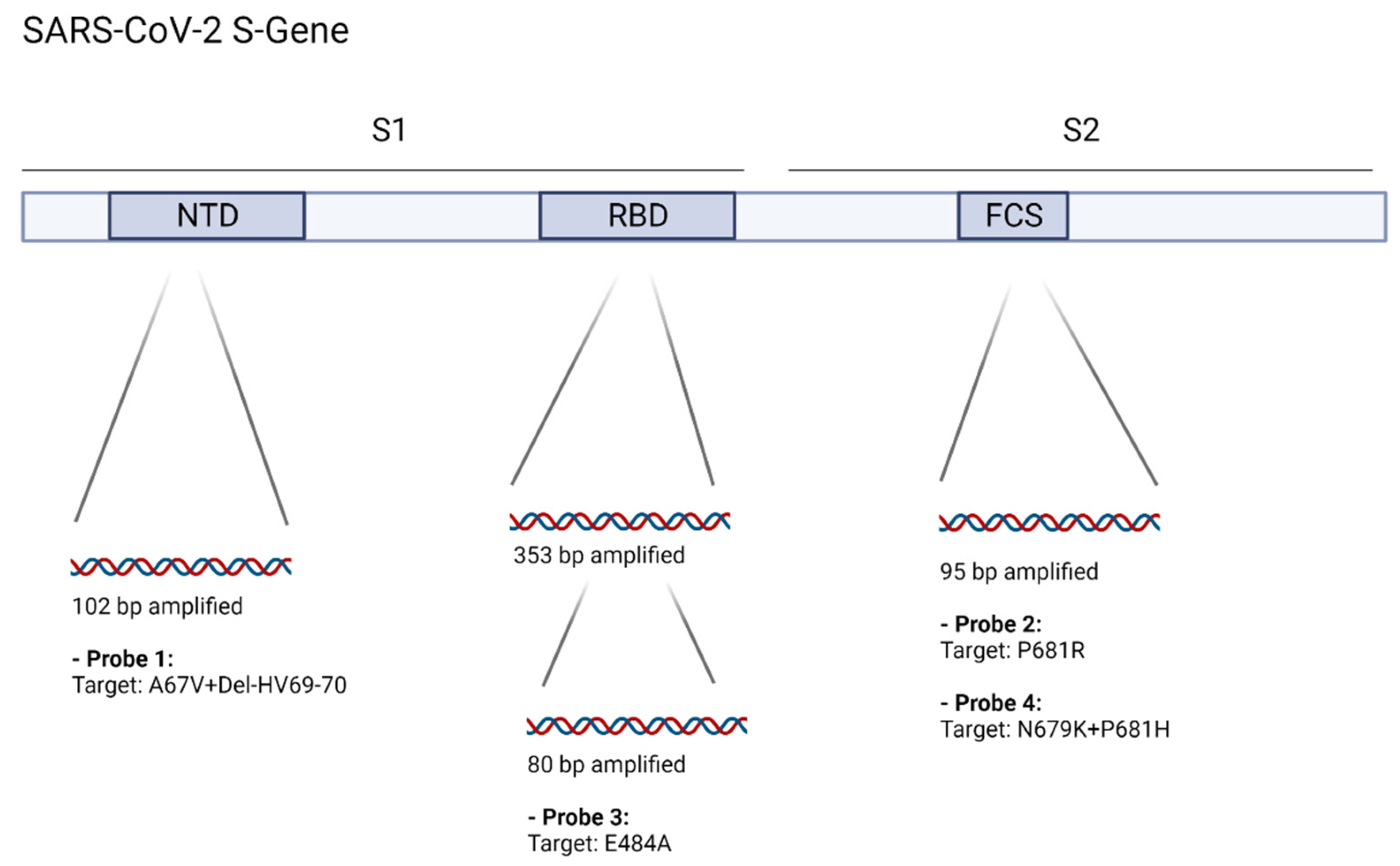
2.1. Assay Design

The Omicron variant features a number of mutations (SNPs and deletions) which have previously been found in other VOCs, e.g., del-HV69-70 and P681H (www.outbreak.info, accessed 24 December 2021). However, many of these are now accompanied by additional SNPs within potential probe regions, such as A67V or N679K. The inclusion of these additional variances allows for assays to be highly specific for the Omicron variant.
A set of previously described TaqMan-assays of our group [15,16] was modified for the respective target regions of the Omicron variant S-gene [16]. Briefly, assays were designed using PrimerQuest software (IDT) with probes being 12–20 bp in length, containing a triplet of locked nucleic acid (LNA)-bases at the SNP location and melting temperature being adjusted by including additional LNA-bases. If additional SNPs were present in the probe target region, these were covered with LNA-bases to improve discrimination. For the HV69-70 deletion, the probe sequence was modified to omit the affected bases and an LNA-base positioned at the A67V SNP (probe 1, “SDEL2”). For the furin-cleavage-site, probe-4 (“P681H”) also covers N679K with an LNA-base. By using this approach, we generated two Omicron “specific” targets (A67V + DEL69/70; and P681H + N679K). For sequence variants within the probe regions that are not covered by specific probes, blocker oligos were included in the assay [16].
In general, LNA bases allow for shorter Taqman-Probes by increasing melting temperatures, which is an established method for increasing sequence specificity [15,17]. 2′O-methyl RNA bases are placed at the penultimate position of every primer to reduce the formation of primer dimers. Blocker-oligos serve to depress off-target activity of specific probes, especially when discriminating for a single mismatch, thus allowing for lower RFI (relative fluorescence increase) thresholds.
The multiplex assay amplifies three regions of the SARS-CoV-2 S-gene: 102bp within the N-terminal domain (NTD) (probe 1: “SDEL2”), 353/80bp within the receptor-binding-domain (RBD) (probe 3, “E484A”), and 95bp at the furin-cleavage-site (probe 2: “P681R”, probe 4: “P681H”) (Figure 1). Primer sequences were modified with ambiguous bases to account for SNPs present in the B.1.1.529 lineage. For an overview about the presence or absence of relevant mutations in different VOCs, see Supplementary Table S1 or, e.g., the outbreak.info website (www.outbreak.info, accessed on 26 February 2022).
Figure 1. Target regions were chosen based on initial available Omicron variant sequences. The included primer sets are either still a 100% match or modified with ambiguous bases to amplify three target regions within the SARS-CoV-2 S-gene, containing deletions or SNPs in the Omicron variant. Four different LNA-Probes are used to specifically detect one or multiple sequence variances in order to allow classification. Delta variant sequences are expected to contain the P681R SNP (probe 2, “P681R”) but not the other tested variances. Omicron variant (Non-BA.2) sequences are expected to contain A67V + del-HV69-70 (probe 1, “SDEL2”), E484A (probe 3, “E484A”) and N679K + P681H (probe 4, “P681H”). The BA.2 lineage is expected to return negative for probe 1 but remain positive for the two other targets. NTD: N-terminal domain, RBD: receptor binding domain, FCS: Furin cleavage site. Image was created using Biorender software. All included oligos are listed in Table 1.
Viruses 14 00608 g001

2.2. Inclusivity and Compatibility

Inclusivity of the primer/probe sets was determined by Roche diagnostics (Pleasanton, CA, USA) as part of a Utility channel support request. Sequences were excluded from the analysis, if the target regions could not be analyzed due to poor sequence quality. All Omicron sequences available (GISAID) until 20 December 2021 were included (13,462 for the NTD assay, 3472 for the RBD, and 11,910 for the furin cleavage site). All three target regions could be analyzed in 2984 sequences. The results are included in Supplementary Table S2. Notably, for those sequences in which all three target regions could be analyzed (n = 2984), 99.93% contained a perfect match for at least one of the three Omicron variant targets.
Performance of multiplex RT-PCR assays is jeopardized by strong oligo–oligo interactions, unspecific amplification of primer-dimers and amplification of non-target sequences, e.g., from human DNA/RNA, among other factors. For oligo–oligo interactions and risk of primer-dimer formation, all oligo sequences were analyzed against each other using OligoAnalyzer software (IDT, Coralville, IA, USA) (Supplementary Table S3). Five interactions were analyzed further due to high binding energy (RBD-484-fwd: RBD-484-fwd (2×), P681-fwd: P681fwd, RBD-484-fwd: P681R-probe, RBD-484-fwd: P681H-probe) (Supplementary Figure S1). Of these, none show overlap at the 3′ ends of the respective oligos.
Matches in human DNA/RNA with a risk to lead to efficient amplification were analyzed using Primerblast (NCBI, USA). Two human RNA targets, 672bp and 969bp in length can potentially be amplified by included primers due to a perfect match of 10 or more bases at the 3′-ends (Supplementary Table S4).

2.3. Utility Channel Test Setup

Primers and Probes were added to MMX-R2 reagent and loaded into cobas omni Utility Channel cassettes (Roche diagnostics, Rotkreuz, Switzerland) according to instructions by the manufacturer. The test is referred to as SCOV2_OMIC_VOC-UCT. The complete run protocol is shown in Table 2.
All cobas omni Utility Channel tests contain a spike-in full-process-control (internal control, IC) by default. Primers and probes are preloaded in MMX-R2 reagent, and the spike-in RNA target is added automatically during extraction. The internal control is detected in Channel 5 for each reaction and is functionally identical to commercial CE-IVD tests by Roche Diagnostics on the cobas 6800/8800 instruments.
Loaded cassettes are placed into the system for processing. No further manual steps are needed to perform a test.

2.4. Analytic Sensitivity, Inter-, and Intra-Run Variance

For evaluation of analytic performance, cell culture supernatant of a clinical isolate of SARS-CoV-2 Omicron variant was used to create dilution series. Briefly, 500 µL medium (UTM) of a confirmed SARS-CoV-2 (Omicron variant) positive nasopharyngeal swab sample (UTM, Mantacc, Shenzhen, China) were used for virus isolation on Vero E6 cells (ATCC® CRL-1008) as described previously [18].
Lineage of the isolate was confirmed by NGS before and after virus isolation (GeneBank OL960487). SARS-CoV-2 WHO standard (NIBSC, Potters Bar, UK) was used in conjunction with the cobas SARS-CoV-2 CE-IVD test (Roche Diagnostics, Rotkreuz, Switzerland) to create a quantified reference stock (in IU/mL). For the P681R target, a clinical Delta-variant sample (confirmed by NGS) was normalized to WHO standard using the same method. Analytic limit of detection (LoD) was determined by 2-fold dilution series (8 steps, 8 repeats, performed on a Hamilton STARlet IVD liquid handler), run on a cobas6800 instrument with the SCOV2_OMIC_VOC-UCT. Then, 95% probability of detection and confidence intervals (CI95%) (probit analysis) were determined with MedCalc Software (Ostend, Belgium).
For inter- and intra-run variability, a set of 10-fold dilutions (4 steps, 5 repeats) were run with the test on three separate days.
For exclusivity testing, a panel of 27 external controls or clinical samples was tested, containing various respiratory viruses, notably endemic human coronaviruses, Middle East respiratory syndrome-related Coronavirus (MERS-CoV) and SARS-CoV (2003, Frankfurt-1, AY291315).

2.5. Clinical Performance

A total of 244 predetermined clinical remnant samples (by qPCR and NGS if positive for SARS-CoV-2 RNA) were run with the SCOV2_OMIC_VOC-UCT.
Of these samples, 51 were predetermined negative for SARS-CoV-2 RNA by the cobas SARS-CoV-2 CE-IVD test. A total of 75 samples were positive for non-Delta, non-Omicron SARS-CoV-2 lineages (notably 50 B.1.1.7 samples); 54 samples were predetermined Delta variant (B.1.617.2-like or AY.4-like lineages), and 64 samples were predetermined as Omicron variant (B.1.1.529 lineage, 42 BA.1-like and 22 BA.2-like). All lineages were assigned based on whole genome sequencing, carried out at the Leibniz Institute for Experimental Virology (HPI, Hamburg), as part of the Hamburg Genome Surveillance Project (https://www.hpi-hamburg.de/en/, accessed on 14 March 2022).

3. Results

3.1. Analytic Performance

Analytic LoD was determined by 2-fold dilution series (8 steps, 8 repeats) as 19.0 IU/mL (CI95%: 12.9–132.2 IU/mL) for target 1: A67V + del-HV69-70; 105.0 IU/mL (CI95%: 80.7–129.3 IU/mL) for target 2: P681R; 193.9 IU/mL (CI95%: 144.7–334.7 IU/mL) for target 3: E484A; and 35.5 IU/mL (CI95%: 23.3–158.0 IU/mL) for target 4: N679K +P681H (See Table 3). Probit plots are included in Supplementary Figure S2.
The multiplex test returned negative for all clinical and external control samples of the exclusivity set, notably, SARS-CoV, MERS-CoV, and endemic human coronaviruses (see Supplementary Table S5).

3.2. Clinical Performance

A total of 244 qPCR and NGS predetermined (if positive) clinical remnant samples were run with the test to assess clinical performance. A total of 193 clinical samples were positive for SARS-CoV-2 RNA. All four targets (225 sequence variances in total, SNPs, and deletions) were correctly identified by the multiplex assay (Table 4). The clinical sample set notably included SARS-CoV-2 lineages with P681H (non-N679K), Del-HV69-70 (non-A67V), E484K and E484Q sequence variances (e.g., B.1.1.7 (Alpha), B.1.351 (Beta), and B.1.617.1 (Kappa)). A complete list can be found in Table 5. Exemplary amplification curves are displayed in Supplementary Figure S3.

4. Discussion and Conclusions

In this study, we presented a multiplexed screening assay with three independent targets for the Omicron variant (first: A67V + del-HV69-70, second: E484A, and third: N679K + P681H) and one for the delta variant P681R, thus allowing easy discrimination between these two prevalent lineages.
The S-gene target dropout (due to the HV69-70 deletion) observed on the widely used TaqPath SARS-CoV-2 assay (Thermofisher) has reportedly been used for tracking the expansion of the B.1.1.7 lineage [6] and is now used again for the same purpose with the novel Omicron (Non-BA.2) variant [8]. However, due to the unspecific nature of assay dropouts and the prevalence of NTD deletions in non-Omicron variants, it is preferable to specifically detect SNPs or combinations of SNPs that are conserved within the lineage in question [16]. There now exists a wide range of lab-developed and commercial solutions for this purpose [12,13,14], although their suitability for application on a large scale varies as many available protocols are highly manual and require careful interpretation of every individual result. Furthermore, the Omicron variant is reported to feature a lot of heterogeneity within well-established target regions; this, however, seems to be largely a result of low sequence quality or faulty annotations as NGS struggles with large numbers of mismatches for amplicon-primers, especially within the RBD region.
For the multiplex-test described in this study, over 98% of available Omicron sequences have perfect sequence identity with the individual SNP/deletion-assays, when only including those that have valid sequences for the respective regions. Only 0.07% of currently available Omicron sequences would return negative on all three assays.
Of note, the BA.2 subline (sometimes dubbed the “stealth variant”) lacks the A67V and del-HV69-70 mutations, thus leading to a negative result for the NTD target, while retaining positivity for E484A and N679K + P681H; a pattern that can be utilized to distinguish contemporary BA.2 from other Omicron sublines.
Still, the evolution of the Omicron variant remains in an early stage, and distinct sublines may emerge, or individual SNPs may be lost within the coming months. It is therefore particularly important to continuously monitor emerging sequences for mismatches in the oligo-set. In any case, definitive assignment of lineages can only be carried out based on whole genome sequencing results; PCR based typing can and should be used as a complement but not replace NGS. Furthermore, it should be noted that the assay is not intended or suited as a first-line SARS-CoV-2 assay but as a typing-test following detection of SARS-CoV-2 RNA by established methods. Results of individual targets should be considered valid only if the clinical sample tested contains SARS-CoV-2 RNA at concentrations above the respective LoDs.
As such, the multiplex assay we present here can be an important asset allowing clinicians and public health officials to rapidly act on suspected cases or outbreaks. Rapid differentiation of newly diagnosed infections may become particularly relevant for this newly emerged lineage due to potential implications for treatment or quarantine mandates. For instance, the often life-saving monoclonal antibody preparations seem to have largely lost their effectiveness against the Omicron variant in cell culture neutralization experiments [2,3,19]. Further studies have demonstrated how pseudovirus neutralization of frequently used monoclonal antibodies such as Regencov is greatly diminished for the Omicron variant-Spike protein, while, e.g., Sotrovimab remains little affected for BA.1-Spike but substantially loses efficacy against BA.2-Spike [5]. Rapid variant screening may be of value for allocating monoclonal antibody treatments in the future. From a public health perspective, local Omicron variant outbreaks may pose a more urgent need for decisive intervention, due to Omicron’s ability to efficiently spread in pre-immunized populations [4]. It can further help classifying low viral-RNA load samples, for which NGS often fails to obtain viable sequences [20]. As described above, the A67V + del-HV69-70 and N679K + P681H components of the multiplex assay feature analytic sensitivities similar to currently available diagnostic SARS-CoV-2 tests (e.g., between 30–50 dcp/mL for the cobas SARS-CoV-2 test [21] and can detect relevant sequence variances even in samples with very low viral-RNA loads.
The adaptation of variant screening assays for automated high-throughput platforms further enables laboratories to efficiently predict SARS-CoV-2 lineages for large numbers of samples and in a timely manner, thus serving as an important complement to whole-genome sequencing based surveillance programs.

Supplementary Materials

The following supporting information can be downloaded at: https://www.mdpi.com/article/10.3390/v14030608/s1, Figure S1: Oligo-dimer alignments; Figure S2: Probit plots; Figures S3–S6 Multiplex assay amplification curves for various SARS-CoV-2 lineages; Table S1: Overview of current and former VOCs and distribution of relevant mutations; Table S2: Inclusivity analysis; Table S3: Oligo-dimerization table; Table S4: Amplification targets of the multiplex within human DNA/RNA; Table S5: Cross-reactivity panel.

Author Contributions

M.L., S.P., D.N., N.F., A.G. and M.A. conceptualized and supervised the study. D.N., M.G., H.T.T., C.W., K.G., T.G. and A.R. performed the experiments. D.N., M.L., S.P., N.F. and M.A. wrote and edited the manuscript. All authors have read and agreed to the published version of the manuscript.

Funding

This work was partially funded by a grant from the Sozialbehörde (Authority of Social Affairs), Free and Hanseatic City of Hamburg, given to N.F., M.A. and A.G. Further funding was institutional only.

Institutional Review Board Statement

This work was conducted in accordance with §12 of the Hamburg hospital law (§12 HmbKHG). The use of anonymized samples was approved by the ethics committee, Freie und Hansestadt Hamburg, PV5626.

Informed Consent Statement

Not applicable.

Data Availability Statement

In cases where sequencing was performed, all sequences were made publicly available at GISAID. All data is available upon request.

Acknowledgments

We thank Roche Diagnostics in Pleasanton CA for providing bioinformatics support for this project. We further want to thank Hubert Feucht of Aesculabor Hamburg GmbH and Nadia Wohlwend of Risch Group for providing clinical remnant samples.

Conflicts of Interest

M.L. received speaker honoraria and related travel expenses from Roche Diagnostics. All other authors declare no conflict of interest.

Abbreviations

LoDLimit of Detection
SNPsingle nucleotide polymorphism
RBDreceptor binding domain
ICinternal control; IVD
in-vitro diagnosticRFI, relative fluorescence increase
CIconfidence interval
NGSnext generation sequencing

References

  1. Gao, S.-J.; Guo, H.; Luo, G. Omicron variant (B.1.1.529) of SARS-CoV-2, a global urgent public health alert! J. Med. Virol. 2021, 94, 1255–1256. [Google Scholar] [CrossRef]
  2. Planas, D.; Saunders, N.; Maes, P.; Guivel-Benhassine, F.; Planchais, C.; Buchrieser, J.; Bolland, W.-H.; Porrot, F.; Staropoli, I.; Lemoine, F.; et al. Considerable escape of SARS-CoV-2 Omicron to antibody neutralization. Nature 2022, 602, 671–675. [Google Scholar] [CrossRef] [PubMed]
  3. Cao, Y.; Wang, J.; Jian, F.; Xiao, T.; Song, W.; Yisimayi, A.; Huang, W.; Li, Q.; Wang, P.; An, R. Omicron escapes the majority of existing SARS-CoV-2 neutralizing antibodies. Nature 2022, 602, 657–663. [Google Scholar] [CrossRef] [PubMed]
  4. Pulliam, J.R.C.; van Schalkwyk, C.; Govender, N.; von Gottberg, A.; Cohen, C.; Groome, M.J.; Dushoff, J.; Mlisana, K.; Moultrie, H. Increased risk of SARS-CoV-2 reinfection associated with emergence of the Omicron variant in South Africa. medRxiv 2021. [Google Scholar] [CrossRef]
  5. Iketani, S.; Liu, L.; Guo, Y.; Liu, L.; Huang, Y.; Wang, M.; Luo, Y.; Yu, J.; Yin, M.T.; Sobieszczyk, M.E.; et al. Antibody Evasion Properties of SARS-CoV-2 Omicron Sublineages. bioRxiv 2022. [Google Scholar] [CrossRef]
  6. Brown, K.A.; Gubbay, J.; Hopkins, J.; Patel, S.; Buchan, S.A.; Daneman, N.; Goneau, L.W. S-Gene Target Failure as a Marker of Variant B.1.1.7 Among SARS-CoV-2 Isolates in the Greater Toronto Area, December 2020 to March 2021. JAMA 2021, 325, 2115–2116. [Google Scholar] [CrossRef] [PubMed]
  7. Zhen, W.; Berry, G.J. Development of a New Multiplex Real-Time RT-PCR Assay for Severe Acute Respiratory Syndrome Coronavirus 2 (SARS-CoV-2) Detection. J. Mol. Diagn. 2020, 22, 1367–1372. [Google Scholar] [CrossRef] [PubMed]
  8. Li, A.; Maier, A.; Carter, M.; Guan, T.H. Omicron and S-gene target failure cases in the highest COVID-19 case rate region in Canada—December 2021. J. Med. Virol. 2022. [Google Scholar] [CrossRef] [PubMed]
  9. McCarthy, K.R.; Rennick, L.J.; Nambulli, S.; Robinson-McCarthy, L.R.; Bain, W.G.; Haidar, G.; Duprex, W.P. Recurrent deletions in the SARS-CoV-2 spike glycoprotein drive antibody escape. Science 2021, 371, 1139–1142. [Google Scholar] [CrossRef]
  10. Shang, E.; Axelsen, P.H. The Potential for SARS-CoV-2 to Evade Both Natural and Vaccine-induced Immunity. bioRxiv, 2020. [Google Scholar] [CrossRef]
  11. Lasek-Nesselquist, E.; Pata, J.; Schneider, E.; George, K.S. A tale of three SARS-CoV-2 variants with independently acquired P681H mutations in New York State. medRxiv 2021. [Google Scholar] [CrossRef]
  12. Boršová, K.; Paul, E.D.; Kováčová, V.; Radvánszka, M.; Hajdu, R.; Čabanová, V.; Sláviková, M.; Ličková, M.; Lukáčiková, L.; Belák, A.; et al. Surveillance of SARS-CoV-2 lineage B.1.1.7 in Slovakia using a novel, multiplexed RT-qPCR assay. Sci. Rep. 2021, 11, 20494. [Google Scholar] [CrossRef] [PubMed]
  13. Vogels, C.B.F.; Breban, M.I.; Ott, I.M.; Alpert, T.; Petrone, M.E.; Watkins, A.E.; Kalinich, C.C.; Earnest, R.; Rothman, J.E.; Goes de Jesus, J.; et al. Multiplex qPCR discriminates variants of concern to enhance global surveillance of SARS-CoV-2. PLoS Biol. 2021, 19, e3001236. [Google Scholar] [CrossRef] [PubMed]
  14. Bier, C.; Edelmann, A.; Theil, K.; Schwarzer, R.; Deichner, M.; Gessner, A.; Hiergeist, A.; Rentschler, U.; Gohl, P.; Kuchta, A.; et al. Multi-site Evaluation of SARS-CoV-2 Spike Mutation Detection Using a Multiplex Real-time RT-PCR Assay. medRxiv 2021. [Google Scholar] [CrossRef]
  15. You, Y.; Moreira, B.G.; Behlke, M.A.; Owczarzy, R. Design of LNA probes that improve mismatch discrimination. Nucleic Acids Res. 2006, 34, e60. [Google Scholar] [CrossRef] [PubMed] [Green Version]
  16. Nörz, D.; Grunwald, M.; Tang, H.T.; Olearo, F.; Günther, T.; Robitaille, A.; Fischer, N.; Grundhoff, A.; Aepfelbacher, M.; Pfefferle, S.; et al. Rapid Automated Screening for SARS-CoV-2 B.1.617 Lineage Variants (Delta/Kappa) through a Versatile Toolset of qPCR-Based SNP Detection. Diagnostics 2021, 11, 1818. [Google Scholar] [CrossRef] [PubMed]
  17. Braasch, D.A.; Corey, D.R. Locked nucleic acid (LNA): Fine-tuning the recognition of DNA and RNA. Chem Biol. 2001, 8, 1–7. [Google Scholar] [CrossRef] [Green Version]
  18. Brehm, T.T.; Pfefferle, S.; von Possel, R.; Kobbe, R.; Nörz, D.; Schmiedel, S.; Grundhoff, A.; Olearo, F.; Emmerich, P.; Robitaille, A.; et al. SARS-CoV-2 Reinfection in a Healthcare Worker Despite the Presence of Detectable Neutralizing Antibodies. Viruses 2021, 13, 661. [Google Scholar] [CrossRef] [PubMed]
  19. VanBlargan, L.A.; Errico, J.M.; Halfmann, P.J.; Zost, S.J.; Crowe, J.E.; Purcell, L.A.; Kawaoka, Y.; Corti, D.; Fremont, D.H.; Diamond, M.S. An infectious SARS-CoV-2 B.1.1.529 Omicron virus escapes neutralization by therapeutic monoclonal antibodies. Nat. Med. 2022. [Google Scholar] [CrossRef] [PubMed]
  20. Charre, C.; Ginevra, C.; Sabatier, M.; Regue, H.; Destras, G.; Brun, S.; Burfin, G.; Scholtes, C.; Morfin, F.; Valette, M.; et al. Evaluation of NGS-based approaches for SARS-CoV-2 whole genome characterisation. Virus Evol. 2020, 6, veaa075. [Google Scholar] [CrossRef] [PubMed]
  21. Manohar, C.; Sun, J.; Schlag, P.; Santini, C.; Fontecha, M.; Lötscher, P.; Bier, C.; Goepfert, K.; Duncan, D.; Spier, G.; et al. Agile design and development of a high throughput cobas® SARS-CoV-2 RT-PCR diagnostic test. medRxiv 2021. [Google Scholar] [CrossRef]
Table 1. Primer-, probe-, and blocker sequences of the multiplex assay are listed. Oligos were custom made by Ella Biotech (Fürstenfelbruck, Germany). Indicated final concentrations refer to the final oligo concentrations within the reaction mix. 2′O-methyl-RNA bases are indicated as “OMe-X”. LNA bases are indicated as “+X”).
Oligo TypeOligo NameSequence 5′–3′Final Concentration [nM]
PrimersNTD fwdTCA ACT CAG GAC TTG TTC T(OMe-U)A C400
NTD revTGG TAG GAC AGG GTT AT(OMe-C) AAA C400
RBD-452 fwdGAT T(+C)T AAG GTT GGT GG(2OMe-U) AAT400
RBD-484 fwdCTA TCA GGC CGG TAR (2OMe-C)A400
RBD-484-revGTC GGA AAC TAT ATG ATC GTA AA(OMe-G) G400
RBD-univ-revAGT TGC TGG TGC ATG TA(OMe-G) AA400
FCS fwdTGC AGG TAT ATG CGC TAG T(OMe-U)A400
FCS revGTG ACA TAG TGT AGG CAA TGA (OMe-U)G400
ProbesA67V-del69-70 probeAtto425- TGG TCC CAG A(+G)A T(+A)(+A) C(+A)T -BHQ150
E484A probeYakYellow- AT(+G) GTG TT(+G) (+C)(+A)G (+G)TT -BHQ150
P681R probeFAM- A(+T)T CT(+C) (+G)(+T)C GGC G -BHQ150
N679K-P681H probeAtto620- A(+G)T CT(+C) (+A)(+T)C GG(+C) G -BHQ250
BlockersE484WT blockerAT(+G) GTG T(+T)(+G) (+A)AG (+G)TT -C3-Spacer50
E484K blockerAT(+G) GTG T(+T)(+A) (+A)AG (+G)TT -C3-Spacer50
E484Q blockerAT(+G) GTG T(+T)(+C) (+A)AG (+G)TT -C3-Spacer50
P681WT blockerTAA (+T)TC T(+C)(+C) (+T)CG GCG -C3-Spacer50
The “NTD” primer set and “A67V-del69-70”-probe are derived from a published assay by Zhen et al. [7]. Oligos used in this study were custom made by Ella Biotech (Fürstenfeldbruck, Germany).
Table 2. Cobas omni Utility Channel run protocol for the SCOV2_OMIC_VOC-UCT. RFI (relative fluorescence increase) cut-offs are used to achieve specificity for a 100% sequence match for respective Taqman-probes.
Software Settings
Sample TypeSwab (400 µL)
Channels1: SDEL22: P681R3: E484A4: P681H5: IC
RFI1.82.52.82.52
PCR cycling conditions
UNG incubationPre-PCR step1st measurement2nd measurementCooling
No. of cyclesPredefined1545Predefined
No. of steps322
Temperature55 °C; 60 °C; 65 °C95 °C; 55 °C91 °C; 58 °C
Hold time120 s; 360 s; 240 s5 s; 30 s5 s; 25 s
Data acquisitionNoneEnd of each cycleEnd of each cycle
Table 3. LoDs were determined by serial dilution of a quantified Omicron variant cell culture stock and a clinical Delta variant sample, using the SARS-CoV-2 WHO standard (NBSCI, UK) as reference. Dilution series were generated automatically using a Hamilton STARlet IVD liquid handler. A 95% probability of detection was calculated using medcalc software. * P681R LoD constitutes a separate experiment using a quantified clinical sample of the B.1.617.2 lineage.
SARS-CoV-2 Omicron (B.1.1.529) and Delta (B.1.617.2) Variants
StepIU/mlSDEL2: Pos/RepE484A: Pos/RepP681H: Pos/RepP681R: Pos/Rep *
1500.008/88/88/88/8
2250.008/88/88/88/8
3125.008/84/88/87/8
462.508/84/87/88/8
531.258/81/87/84/8
615.637/80/86/85/8
77.815/80/85/80/8
83.913/80/82/80/8
Table 4. SCOV2_OMIC_VOC-UCT results for the clinical sample set. Each target/channel is analyzed individually.
TargetResultSNP PositiveSNP NegativeAgreement
A67V + del-HV69-70Positive420100%
Negative0202100%
P681RPositive550100%
Negative0189100%
E484APositive640100%
Negative0180100%
N679K + P681HPositive640100%
Negative0180100%
Table 5. Lineages included in the clinical sample set, as determined by whole genome sequencing.
Clinical Sample Set—Included Lineages
SNP SetLineageNumber
All negativeB.1.1.7-like (Alpha)50
B.1.17710
B.1.2216
B.1.1.296
C.36.31
B.1.351 (Beta)1
P681RB.1.617.2-like45
AY.4-like9
B.1.617.1 (Kappa)1
A67V, del-HV69-70
E484A
N679K, P681H
B.1.1.529 (Omicron)
BA.1-like
42
E484A
N679K, P681H
B.1.1.529 (Omicron)
BA.2-like
22
Publisher’s Note: MDPI stays neutral with regard to jurisdictional claims in published maps and institutional affiliations.

