Review

Host Molecule Incorporation into HIV Virions, Potential Influences in HIV Pathogenesis

Division of Immunology and Allergy, Lausanne University Hospital, University of Lausanne, 1011 Lausanne, Switzerland
*
Author to whom correspondence should be addressed.
Viruses 2022, 14(11), 2523; https://doi.org/10.3390/v14112523
Submission received: 10 October 2022 / Revised: 8 November 2022 / Accepted: 12 November 2022 / Published: 14 November 2022
(This article belongs to the Special Issue State-of-the-Art Virology Research in Switzerland)

Abstract

:
During the last phase of HIV viral production, nascent HIV virions acquire a fraction of the cellular lipid membrane to create the external lipid envelope, a process by which cellular proteins present on the surface of the infected cell can be incorporated along with Env trimers. Interestingly, several studies indicated that these incorporated host molecules could conserve their biological activity and consequently contribute to HIV pathogenesis either by enhancing the infectivity of HIV virions, their tissue tropism or by affecting immune cell functions. The following review will describe the main approaches used to characterize membrane bound host molecule incorporation into HIV virions, the proposed mechanisms involved, and the role of a non-exhaustive list of incorporated molecules.

1. Introduction

Since the beginning of the epidemic, more than 84.2 million individuals have been infected by HIV. In 2021, about 38.4 million people have already died from HIV-related diseases (https://www.who.int/gho/hiv/en/) (accessed on 15 August 2022) and 37.6 million people were living with HIV infection, but none of them were able to naturally clear the infection. Indeed, in the absence of treatment, 99% of HIV-infected people experience a gradual decline in their CD4 T cell count. When the CD4 T cell count drops to around 200 cells/mm3 of blood, these individuals enter the final stage of the infection, the acquired immunodeficiency syndrome (AIDS), eight to ten years after infection [1,2,3]. At that stage, the immune system collapses and is no longer able to protect individuals from opportunistic pathogens, and HIV-infected individuals ultimately die from infections or cancers [1,4].
The reasons for this extremely efficient way for HIV to resist to the pressure exerted by the immune system probably reside in complex immuno-virological mechanisms [5,6,7,8,9]. The functional impairment of HIV-specific CD4 and CD8 T cells (reviewed in Fenwick et al., 2019 [7]), the difficulty in generating broadly neutralizing antibodies by HIV-specific B cells (reviewed in Havenar-Daughton et al., 2017 [8]), as well as the ability of the virus to establish a viral reservoir capable of remaining in a latent state, and therefore invisible to the immune system (reviewed in Cohn et al., 2020 [9]) contribute to the persistence of the virus [7,8,9].
Since HIV is an enveloped virus, HIV virions can incorporate host molecules located either at the membrane or at the cytoplasmic levels during the production process [10,11,12,13,14,15,16]. Interestingly, several studies indicated that these incorporated molecules could still harbor biological activities [17,18,19].
A mathematical model estimated that between 100 million and 100 billion viruses could be produced and eliminated from the body of an HIV-infected individual every day [20]. Due to the extent of viruses produced and the capacity of HIV virions to incorporate host molecules during the budding phase, several studies focused on their potential impact during HIV pathogenesis. The present review therefore proposed to focus on the main experimental approaches used to identify membrane bound host molecules incorporated into HIV virions, the mechanisms involved in the incorporation process and the potential role of selected incorporated molecules in HIV pathogenesis.

2. Experimental Strategies to Characterize Host Proteins Incorporation into HIV Virions

The deep structural characterization of viruses has, for a long time, been limited by their relatively small size. However, in the last decades, the development of novel technological tools allowed for a better characterization of HIV virion structures as well as their protein compositions. Because each technique has its advantages and disadvantages; e.g., the sensitivity for mass spectrometry, the robustness for western blot and electron microscopy, and the small amount of required protein for immune capture (Figure 1), most recent studies proposed combined approaches to characterize the host protein spectra of HIV virions and their potential factors during HIV pathogenesis [21,22]. The following section will focus on the characteristics, advantages, and limitations of the major experimental strategies used to characterize HIV virion envelope composition.
Mass spectrometry (MS) (Figure 1a) or coupling methods such as liquid chromatography–MS (LC-MS) are considered gold standards for the characterization of the protein composition of several viral sources [22,23,24]. Indeed, the high sensitivity in determining the protein quantity and composition of several viruses has improved the understanding of the viral structure and host protein interactions [21,22,24]. In addition, MS is actually the only experimental strategy that does not require prior knowledge of a virus-incorporated host factor [22]. However, this technique has mostly been used on HIV virions produced in vitro from cell lines and not on HIV virions collected directly from ex vivo samples from body fluid studies [25]. Even though MS has been a pioneer tool in the identification of host molecules incorporated into HIV virions, no insights into the host protein profile of a single virion or the co-incorporation of host molecules or the precise localization into the lipid envelope have been obtained, as the analyses were performed on bulk viral preparations.
Over the past decades, electron microscopy (Figure 1b) became a powerful approach for the characterization of the structural organization of HIV virions. While mature HIV virions could be easily recognized by the specific conical shape of the capsid using electron microscopy, the characterization of host protein incorporation into the HIV virion lipid envelope requires the use of a specific immunogold-labeling [26,27]. Immunogold staining of cellular sections infected with viruses involves low level of fixation, which may result in the loss of HIV viral structure and limit the visual characterization of HIV virions in these preparations. The development of novel approaches to distinguish several proteins on an individual virion by electron microscopy was therefore developed and illustrated by the recent use of dual immunogold-labeling [19,28]. This approach is based on the use of two primary antibodies (e.g., p24, Env, or host proteins) labeled beads harboring distinct sizes, distinguishable by electron microscopy. This approach classically allows the determination of the co-expression of the two proteins (viral or host proteins) on individual HIV virions [28]. This approach may also provide further information on the localization (e.g., envelope, capsid, matrix) of cellular proteins into HIV virions.
The immuno-capture assay is a highly specific approach that includes two phases (Figure 1c). The first step consists of the capture of viral particles that can be achieved using either beads or plates coated with antibodies against the proteins of interest. The second step is usually based on the quantification of either p24 or HIV RNA to determine the quantity of viral particles captured by the specific antibodies [12,29,30]. Several studies used this approach to confirm the incorporation of host proteins (e.g., Intercellular Adhesion Molecules-1 (ICAM-1), HLA-DR, and α4β7) [12,29,30]. However, one of the limitations of this technique is the detection of only one protein at a time, preventing the study of the co-incorporation of various host molecules.
Until recently, the use of flow cytometry to characterize HIV virions was limited due to the difficulties of conventional flow cytometers to detect particles smaller than 400 nm, knowing that the size of HIV virions is comprised between 90–120 nm. However, in the last few years, many efforts were invested in the development of more sensitive instruments and the most recent studies are able to efficiently distinguish individual HIV virion using a new approach called flow virometry (Figure 1d) [31,32,33,34,35]. Most of these studies used fluorescent-labeled in vitro produced HIV virions to facilitate the discrimination between the background noise and viral populations [36,37]. To circumvent the small size of HIV virions and to increase the purity of viral preparations, one study proposed an indirect method by first isolating viral particles using an immuno-capture assay by small magnetic beads (15 nm) coated with gp120 antibodies and second, the HIV virion-bead complex detection using flow cytometry [32]. This study confirmed the presence of two cellular proteins known to be incorporated into HIV virions (i.e., HLA-DR, and LFA-1) and was able to distinguish distinct viral subpopulations based on the co-expression of these two proteins [32]. This method has several advantages; the estimation of the amount of cellular proteins expressed by a single virion as well as the potential co-expression of different proteins, allowing for the differentiation of distinct viral populations based on their host protein incorporation signatures. Indeed, the flow virometry method could represent a considerable advance in virology research and may echo the technological shift that flow cytometry has brought to cellular immunology.

3. Mechanisms of Host Protein Incorporation

Pioneer studies initially proposed that host protein incorporation resulted from a passive mechanism depending on the quantity of proteins expressed on the cell surface and located at close proximity to the budding site (Figure 2a) [38].
In agreement with this paradigm, initial findings proposed that during the viral budding process through the lipid rafts, proteins present on the cell surface were passively incorporated into the HIV virion envelope [39,40].
However, more recent studies have demonstrated that the protein levels on HIV-infected cell surfaces did not necessarily correlate with host protein profiles of HIV virions [11,41]. In this context, more recent studies proposed that in addition to a passive mechanism (Figure 2a) and to the enrichment of host molecule incorporation into HIV virions due to the composition of anchored proteins within the lipid raft (Figure 2b), host molecules could also be incorporated via an active process involving direct interactions between host molecules and viral proteins (Figure 2c) [42]. The next section will, therefore, underscore the proposed mechanisms of host protein incorporation and host protein exclusion [41,43].
HIV can actively remodel the lipid composition of the cell membrane at the budding site (Figure 2b). The lipid composition remodeling is usually divided into two phases; the first one consists of the formation of a liquid order (Lo) domain occurring when Gag polyproteins start to oligomerize at the cell membrane [44]. Indeed, Gag polyprotein has a high affinity for Phosphatidylinositol-4,5-bisphosphate (PIP2) that is enriched at the site of assembly and can mediate the clustering of specific GPI-anchored lipid raft within virions envelope to facilitate the incorporation of several proteins (e.g., CD59) [44,45,46]. The second phase is also mediated by Gag and relies on the curvature of the plasma membrane at the assembly site [44]. The enrichment of host proteins with high affinity to the Lo phase domain increases protein composition differences and membrane thickness compared to the bulk plasma membrane [44]. Consequently, these changes result in an increase in the line tension, therefore inducing the membrane domains to form circular zones, leading to the formation of budding vesicles enriched in host proteins with high affinity to the Lo domain [44,47]. In this context, while the remodeling of the lipid raft at the budding site involves an active mechanism mediated by viral proteins, the incorporation of host molecules might reflect an indirect consequence of this process.
The selective acquisition of host proteins might also occur as an active process involving the interaction with host molecules and viral proteins (e.g., Gag or Env). Gag polyprotein consists of four fragments: p17 (MA), p24 (CA), NC, and p6. The p17 domain was demonstrated to be crucial for the incorporation of Env, and p17 and NC have been shown to be potential players in protein incorporation [42,48,49,50]. Notably, the incorporation of host molecules (e.g., HLA-DR) has also been shown to be dependent on Env incorporation, even though this observation is still a matter of debate [51,52]. In this context, two independent studies indicated that the interaction of Gag polyprotein with host molecules (i.e., ICAM-1 and PD-L1) promotes their incorporation (Figure 2c) [19,42].
In these cases, the intracytoplasmic tail of host molecules was shown to interact with the matrix (MA) domains of Gag polyprotein thus, driving the active incorporation into HIV Virions [19,42,53]: Of note, the interaction between the host proteins and Gag can remain stable even after the complete maturation of HIV virions. A better understanding and characterization of specific motifs governing the interactions between Gag polyprotein and host molecules would be of great interest to prevent/force the incorporation of host molecules that might have a beneficial/detrimental role during HIV pathogenesis.
Interestingly, some host proteins (e.g., CD45, CD4, and CD80) can be actively excluded from HIV virion lipid envelopes [41,54,55]. Some authors proposed that this may rely on a mechanism similar to the one used by tetherin, an anti-viral protein known to prohibit HIV virions released during the late phase of the budding process [56]. In this model, the authors proposed that HIV Vpu protein could block the insertion of the GPI domain of tetherin into HIV virion envelope by preventing its co-localization with Gag at the plasma membrane, thus counteracting its potential anti-viral property on mature HIV virions [57]. Whether or not such a mechanism also occurs for other host molecules remains, however, to be determined.
HIV virions can use two distinct pathways to bud out from infected cells. HIV virions can bud using the lipid membrane at the surface of cells to create their own envelope (Figure 3a). This mechanism mostly occurs in lymphoid cells but can also happen in myeloid cells [58]. Furthermore, HIV virions can also assemble in virus containing compartment (VCC) in myeloid cells (Figure 3b) [59]. The different production pathways of HIV virions may partially explain the differences in host protein incorporation signatures observed between HIV virions produced in lymphoid vs. myeloid cell lineages [25].

4. Potential Influence of Host Protein Incorporation on HIV Pathogenesis

Upon incorporation into HIV virions, molecules can retain their biological activity and mediate some effects during the course of HIV infection. Depending on the nature of the host molecules incorporated, several potential impacts were proposed on HIV pathogenesis (e.g., HIV virions infectivity potential, cell attachment and immunoregulation) [12,17,19,29,30,60,61]. The following section will, therefore, underscore the major potential influence of host molecules incorporation on HIV pathogenesis.

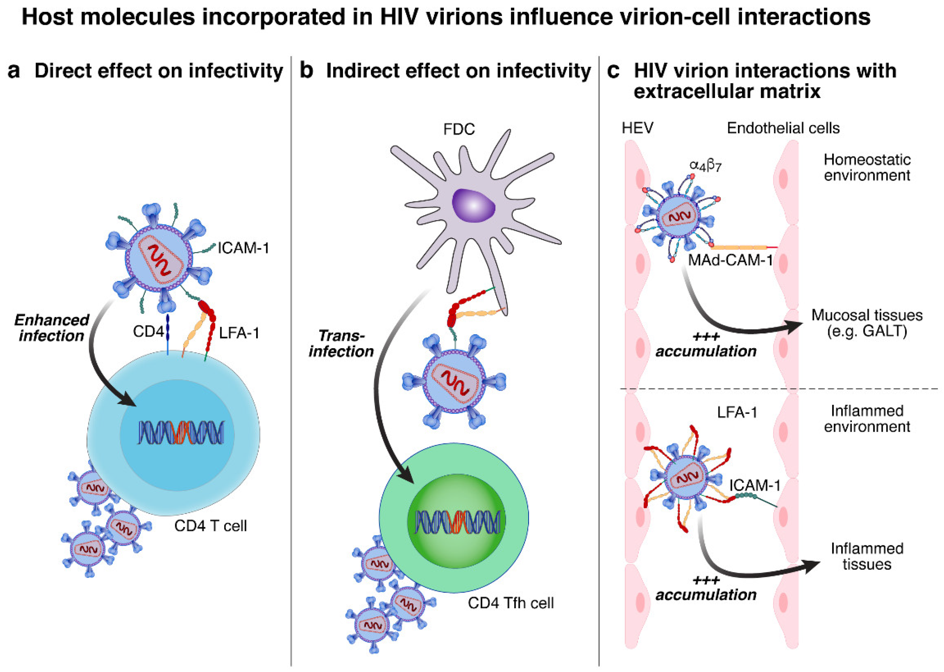
4.1. Potential Influence on Promoting HIV Virions Interaction with Susceptible Cells

HIV virion interaction with the host cell is mediated by the viral glycoprotein gp120 interacting with the cellular receptor CD4, resulting in conformational changes in gp120 and allowing the interaction with one of the two co-receptors, CCR5 or CXCR4 [62]. However, the adhesion to the target cell can be reinforced by host proteins incorporated into the HIV virion lipid envelope, which can increase the binding affinity [30,63]. In this regard, certain incorporated molecules were proposed to play a role in enhacing HIV infectivity by promoting the direct interaction between HIV virions and susceptible cells (e.g., HLA-DR) (Figure 4a), while others may increase HIV infectivity through indirect interaction with follicular dendritic cells (FDCs) (e.g., ICAM-1) (Figure 4b), or with the extracellular matrix of high endothelial venules (HEVs) (e.g., α4β7 or LFA-1) (Figure 4c) [12,17,29,30].
ICAM-1: ICAM-1 is an adhesion molecule expressed mainly on endothelial cells and is playing a crucial role in the migration of immune cells expressing LFA-1 to inflammation sites [64]. Furthermore, the interaction between ICAM-1 and LFA-1 is also known to create an immunological synapse between dendritic cells and T cells thus promoting their interaction [65]. ICAM-1 incorporation into HIV virions was proposed to be mediated following interactions with Gag viral protein [13] and promote HIV virion infectivity by increasing virus attachment to susceptible LFA-1+ cells, thereby promoting the first steps of HIV replication cycle [17,66,67]. Indeed, no increase in infectivity was observed when LFA-1 cells were infected with ICAM-1+ HIV virions, thus supporting these findings [68]. ICAM-1 incorporation might also promote HIV virions rolling on susceptible cells to maximize gp120-CD4 interaction, enhance the interaction with the co-receptor CXCR4, and induce cytoskeleton-remodeling [66,67,69]. Furthermore, ICAM-1 incorporation into HIV virions might also favor trans-infection from LFA-1+ cells (e.g., FDCs) to susceptible cells in LNs germinal centers (GC) [70,71].
α4β7: α4β7 is expressed on immune cells and has been described to play a fundamental role in T-cell homing towards the intestinal track through its interaction with MAdCAM-1 expressed on HEV in Peyer’s patches and the mesenteric lymph nodes [72]. A recent study based on SIV/SHIV-infected macaques demonstrated that the blockade of α4β7 reduced SIV/SHIV viral load both in plasma and gastrointestinal tissues and delayed the time to viral rebound after ART interruption [73,74]. The author subsequently proposed that α4β7 mAbs might have acted directly on α4β7+ HIV virions interrupting key events of viral infection [12]. Using an in vivo model, they showed that the incorporation of α4β7 into HIV virions might play a role in promoting HIV virions accumulation to MAdCAM-1+ on HEV [12]. Notably, a higher proportion of α4β7+ HIV virions was observed in plasma of viremic acute HIV-infected individuals compared to chronic HIV-infected individuals [12]. Based on these observations, a clinical trial using α4β7 mAbs in ART-treated HIV-infected individuals investigated the potential impact on HIV viral rebound. Unfortunately, this study did not achieve the same promising results as the one in macaques, and no delay in viral rebound was observed [75]. Therefore, the role of α4β7 mAbs in the therapeutic armamentarium for HIV cure remains debated.
Other adhesion molecules were shown to be incorporated into HIV virions (e.g., LFA-1 and CD62L) and it has been proposed that they play a role in enhancing HIV infectivity by promoting the interaction of HIV virions with susceptible cells through the interaction with their respective ligands (i.e., ICAM-1 and CD34/MAdCAM-1) in various settings (e.g., homeostatic vs. inflamed environment) [76,77,78,79].

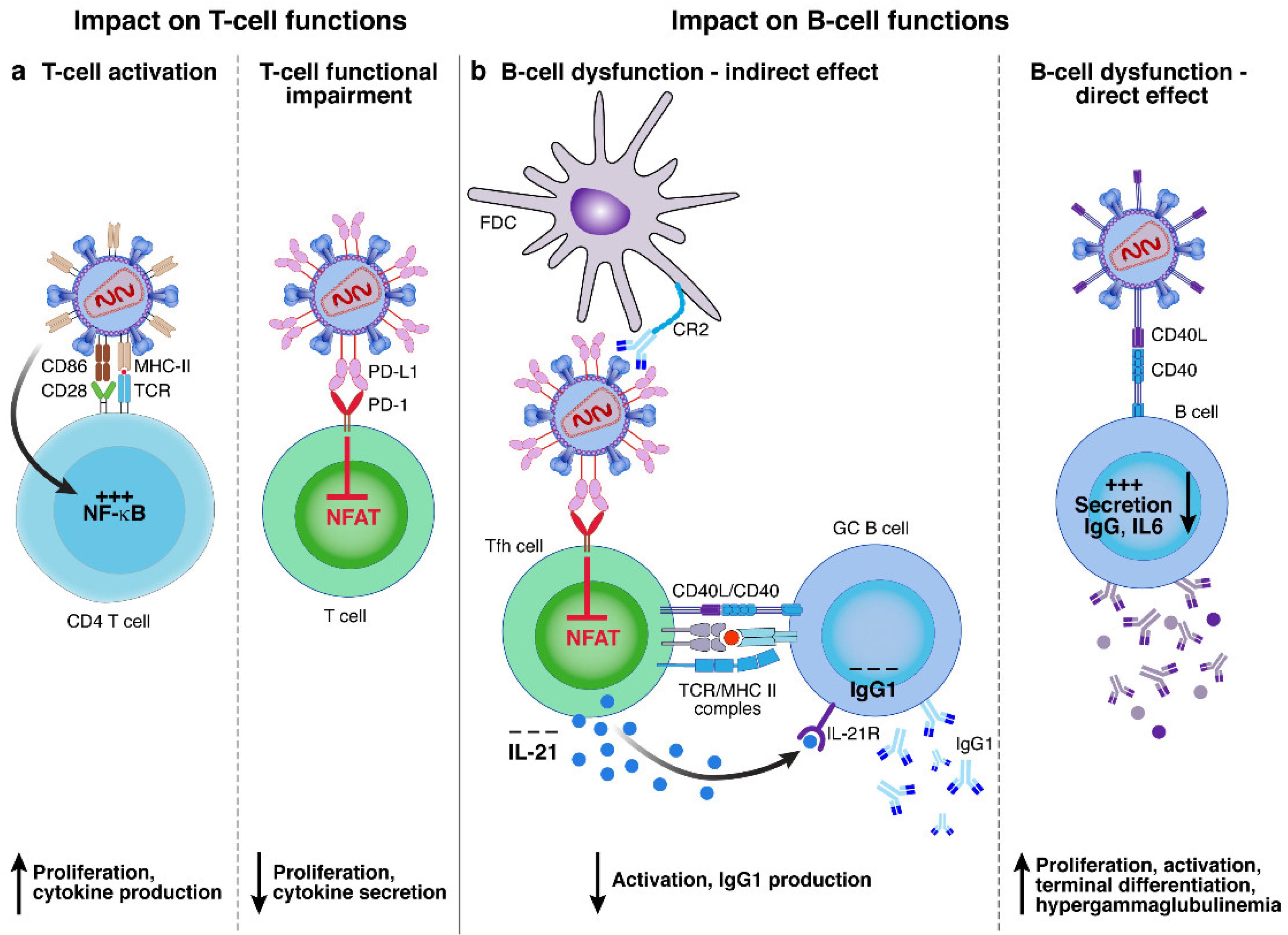
4.2. Influence of T and B Cell Responses

Various incorporated molecules were proposed to impact immune cell function/activation (Figure 5), either by promoting B and/or T-cell activation (e.g., CD40L and HLA-DR alone or co-incorporated with CD86) or functional impairment (e.g., PD-L1) [18,19,60,61,80].
HLA-DR: HLA-DR is one of the MHC class II molecules expressed mostly on antigen-presenting cells (APCs) and is able to present peptides to CD4 T cells [81]. HLA-DR is also known to be upregulated on activated CD4 T cells [82]. HLA-DR was one of the first host molecules described to be incorporated into HIV virions. In addition to its role in increasing HIV infectivity through the enhancement of HIV virion fusion by the interaction of HLA-DR+ HIV virions with CD4, HLA-DR incorporation was proposed to influence T-cell function/activation [30,38,52,83]. Interestingly, it was also suggested that HLA-DR+ HIV virions may have the capacity to present peptide epitopes to CD4 T cells and may modulate CD4 T-cell responses (Figure 5a) [60,61,63]. The consequences of this stimulation may depend on the co-incorporation of the co-stimulatory molecule CD86 and the nature of the peptide presented [60,61]. Indeed, some authors proposed that HLA-DR+ HIV virions might be sufficient to trigger T-cell proliferation, cytokine production, and apoptosis in vitro, while other studies proposed that HLA-DR+ HIV virions might not trigger complete CD4 T-cell activation and thus may prime these cells towards anergy [60,61,63]. Another study proposed that HLA-DR+ HIV virions trigger T-cell apoptosis by increasing Fas and FasL expression levels [84].
PD-L1: The interaction between PD-1 on T cells and PD-L1 on APCs contribute to regulate T-cell proliferation and cytokines production and is proposed to play a major role in T-cell functional impairment [85,86]. It was demonstrated that PD-L1 was actively incorporated into HIV virions from plasma of viremic HIV-infected individuals with a process involving, at least in part, the interaction with HIV p17 matrix proteins. In this study, PD-L1+ HIV virions were able to significantly reduce T follicular helper (Tfh) cell proliferation and IL-21 production in vitro (Figure 5a), through the partial inhibition of T-cell signaling pathway [19]. Interestingly, the impact of PD-L1+ HIV virions on Tfh cell function ultimately translated into reduced IgG1 production from GC B cells in vitro (Figure 5b) [19]. Therefore, in this study, the authors showed that PD-L1+ HIV virions may not only influence directly CD4 T-cell functions but also indirectly influence B-cell responses.
CD40L: CD40L is a co-stimularoy receptors expressed on T cells that play a fundamental role by interaction with CD40 on B cells during B-cell maturation and differentiation processes [87]. Several studies showed that CD40L+ HIV virions could interact with B cells and could induce unspecific B-cell proliferation, activation, and terminal differentiation as well as IgG production, and IL-6 secretion (Figure 5b) [18,80,88]. Interestingly, the quantity of IgG produced did not correlate with the presence or absence of Env but only depended on the presence of CD40L on HIV virion surface. The authors proposed that CD40L+ HIV virions might contribute to the hypergammaglobulinemia observed during the course of HIV infection [80,88]. At the intracellular level, CD40L+ HIV virions were able to induce an activation of the NF-κB pathway, which was similar to the one observed using CD40 antibody stimulation. Furthermore, CD40L+ HIV virions might also contribute to the increase the infection rate of CD4 T cells when co-cultured with tonsillar B cells, by promoting the secretion of pro-inflammatory cytokines by B cells, providing a favorable micro-environment for HIV replication [88].

5. Conclusions

During the budding process, HIV virions can passively and/or actively incorporate functionally active host molecules, which may play an active role during HIV pathogenesis. The development of novel experimental strategies and the advance of new technologies will soon uncover the complexity of this phenomenon, opening the way to a better understanding of HIV pathogenesis.

Funding

This work was funded by Swiss National Science Foundation Grant 320030_200912 and by Freedom Forever Association to M.P. and by an educational grant from FONDATION MACHAON to R.B. The funders had no role in study design, data collection and analysis, decision to publish, or preparation of the manuscript.

Institutional Review Board Statement

Not applicable.

Informed Consent Statement

Not applicable.

Data Availability Statement

Not applicable.

Acknowledgments

We are grateful to Aaron Weddle and John Weddle for their assistance with the figures.

Conflicts of Interest

The authors declare no conflict of interest.

References

  1. Pantaleo, G.; Graziosi, C.; Fauci, A.S. The immunopathogenesis of human immunodeficiency virus infection. N. Engl. J. Med. 1993, 328, 327–335. [Google Scholar] [PubMed]
  2. Guadalupe, M.; Reay, E.; Sankaran, S.; Prindiville, T.; Flamm, J.; McNeil, A.; Dandekar, S. Severe CD4+ T-cell depletion in gut lymphoid tissue during primary human immunodeficiency virus type 1 infection and substantial delay in restoration following highly active antiretroviral therapy. J. Virol. 2003, 77, 11708–11717. [Google Scholar] [CrossRef] [PubMed] [Green Version]
  3. Brenchley, J.M.; Schacker, T.W.; Ruff, L.E.; Price, D.A.; Taylor, J.H.; Beilman, G.J.; Nguyen, P.L.; Khoruts, A.; Larson, M.; Haase, A.T. CD4+ T cell depletion during all stages of HIV disease occurs predominantly in the gastrointestinal tract. J. Exp. Med. 2004, 200, 749–759. [Google Scholar] [CrossRef] [PubMed] [Green Version]
  4. Buchacz, K.; Lau, B.; Jing, Y.; Bosch, R.; Abraham, A.G.; Gill, M.J.; Silverberg, M.J.; Goedert, J.J.; Sterling, T.R.; Althoff, K.N. Incidence of AIDS-defining opportunistic infections in a multicohort analysis of HIV-infected persons in the United States and Canada, 2000–2010. J. Infect. Dis. 2016, 214, 862–872. [Google Scholar] [CrossRef] [Green Version]
  5. Geller, R.; Domingo-Calap, P.; Cuevas, J.M.; Rossolillo, P.; Negroni, M.; Sanjuán, R. The external domains of the HIV-1 envelope are a mutational cold spot. Nat. Commun. 2015, 6, 8571. [Google Scholar] [CrossRef] [Green Version]
  6. Simon, V.; Bloch, N.; Landau, N.R. Intrinsic host restrictions to HIV-1 and mechanisms of viral escape. Nat. Immunol. 2015, 16, 546–553. [Google Scholar] [CrossRef]
  7. Fenwick, C.; Joo, V.; Jacquier, P.; Noto, A.; Banga, R.; Perreau, M.; Pantaleo, G. T-cell exhaustion in HIV infection. Immunol. Rev. 2019, 292, 149–163. [Google Scholar] [CrossRef] [Green Version]
  8. Havenar-Daughton, C.; Lee, J.H.; Crotty, S. Tfh cells and HIV bnAbs, an immunodominance model of the HIV neutralizing antibody generation problem. Immunol. Rev. 2017, 275, 49–61. [Google Scholar] [CrossRef] [Green Version]
  9. Cohn, L.B.; Chomont, N.; Deeks, S.G. The biology of the HIV-1 latent reservoir and implications for cure strategies. Cell Host Microbe 2020, 27, 519–530. [Google Scholar] [CrossRef]
  10. Burnie, J.; Guzzo, C. The incorporation of host proteins into the external HIV-1 envelope. Viruses 2019, 11, 85. [Google Scholar] [CrossRef]
  11. Esser, M.T.; Graham, D.R.; Coren, L.V.; Trubey, C.M.; Bess, J.W., Jr.; Arthur, L.O.; Ott, D.E.; Lifson, J.D. Differential incorporation of CD45, CD80 (B7-1), CD86 (B7-2), and major histocompatibility complex class I and II molecules into human immunodeficiency virus type 1 virions and microvesicles: Implications for viral pathogenesis and immune regulation. J. Virol. 2001, 75, 6173–6182. [Google Scholar] [CrossRef] [PubMed] [Green Version]
  12. Guzzo, C.; Ichikawa, D.; Park, C.; Phillips, D.; Liu, Q.; Zhang, P.; Kwon, A.; Miao, H.; Lu, J.; Rehm, C. Virion incorporation of integrin α4β7 facilitates HIV-1 infection and intestinal homing. Sci. Immunol. 2017, 2, eaam7341. [Google Scholar] [CrossRef] [PubMed] [Green Version]
  13. Beauséjour, Y.; Tremblay, M.J. Envelope glycoproteins are not required for insertion of host ICAM-1 into human immunodeficiency virus type 1 and ICAM-1-bearing viruses are still infectious despite a suboptimal level of trimeric envelope proteins. Virology 2004, 324, 165–172. [Google Scholar] [CrossRef] [PubMed] [Green Version]
  14. Zennou, V.; Perez-Caballero, D.; Göttlinger, H.; Bieniasz, P.D. APOBEC3G incorporation into human immunodeficiency virus type 1 particles. J. Virol. 2004, 78, 12058–12061. [Google Scholar] [CrossRef] [PubMed] [Green Version]
  15. Sharma, S.; Lewinski, M.K.; Guatelli, J. An N-glycosylated form of SERINC5 is specifically incorporated into HIV-1 virions. J. Virol. 2018, 92, e00753-18. [Google Scholar] [CrossRef] [Green Version]
  16. Ott, D.E. Cellular proteins detected in HIV-1. Rev. Med. Virol. 2008, 18, 159–175. [Google Scholar] [CrossRef]
  17. Fortin, J.-F.; Cantin, R.; Lamontagne, G.; Tremblay, M. Host-derived ICAM-1 glycoproteins incorporated on human immunodeficiency virus type 1 are biologically active and enhance viral infectivity. J. Virol. 1997, 71, 3588–3596. [Google Scholar] [CrossRef] [Green Version]
  18. Imbeault, M.; Ouellet, M.; Giguère, K.; Bertin, J.; Bélanger, D.; Martin, G.; Tremblay, M.J. Acquisition of host-derived CD40L by HIV-1 in vivo and its functional consequences in the B-cell compartment. J. Virol. 2011, 85, 2189–2200. [Google Scholar] [CrossRef] [Green Version]
  19. Munoz, O.; Banga, R.; Schelling, R.; Procopio, F.A.; Mastrangelo, A.; Nortier, P.; Ohmiti, K.; Daraspe, J.; Cavassini, M.; Fenwick, C. Active PD-L1 incorporation within HIV virions functionally impairs T follicular helper cells. PLOS Pathog. 2022, 18, e1010673. [Google Scholar] [CrossRef]
  20. Perelson, A.S.; Neumann, A.U.; Markowitz, M.; Leonard, J.M.; Ho, D.D. HIV-1 dynamics in vivo: Virion clearance rate, infected cell life-span, and viral generation time. Science 1996, 271, 1582–1586. [Google Scholar] [CrossRef]
  21. Luo, Y.; Muesing, M.A. Mass spectrometry-based proteomic approaches for discovery of HIV–host interactions. Future Virol. 2014, 9, 979–992. [Google Scholar] [CrossRef] [PubMed] [Green Version]
  22. Saphire, A.C.; Gallay, P.A.; Bark, S.J. Proteomic analysis of human immunodeficiency virus using liquid chromatography/tandem mass spectrometry effectively distinguishes specific incorporated host proteins. J. Proteome Res. 2006, 5, 530–538. [Google Scholar] [CrossRef] [PubMed]
  23. Chertova, E.; Chertov, O.; Coren, L.V.; Roser, J.D.; Trubey, C.M.; Bess, J.W.; Sowder, R.C.; Barsov, E.; Hood, B.L.; Fisher, R.J. Proteomic and biochemical analysis of purified human immunodeficiency virus type 1 produced from infected monocyte-derived macrophages. J. Virol. 2006, 80, 9039–9052. [Google Scholar] [CrossRef] [PubMed] [Green Version]
  24. Zheng, J.; Sugrue, R.J.; Tang, K. Mass spectrometry based proteomic studies on viruses and hosts–A review. Anal. Chim. Acta 2011, 702, 149–159. [Google Scholar] [CrossRef] [PubMed]
  25. Santos, S.; Obukhov, Y.; Nekhai, S.; Bukrinsky, M.; Iordanskiy, S. Virus-producing cells determine the host protein profiles of HIV-1 virion cores. Retrovirology 2012, 9, 65. [Google Scholar] [CrossRef] [PubMed] [Green Version]
  26. Yi, H.; Strauss, J.D.; Ke, Z.; Alonas, E.; Dillard, R.S.; Hampton, C.M.; Lamb, K.M.; Hammonds, J.E.; Santangelo, P.J.; Spearman, P.W. Native immunogold labeling of cell surface proteins and viral glycoproteins for cryo-electron microscopy and cryo-electron tomography applications. J. Histochem. Cytochem. 2015, 63, 780–792. [Google Scholar] [CrossRef] [PubMed]
  27. Hammonds, J.; Wang, J.-J.; Yi, H.; Spearman, P. Immunoelectron microscopic evidence for Tetherin/BST2 as the physical bridge between HIV-1 virions and the plasma membrane. PLoS Pathog. 2010, 6, e1000749. [Google Scholar] [CrossRef] [Green Version]
  28. Nkwe, D.O.; Pelchen-Matthews, A.; Burden, J.J.; Collinson, L.M.; Marsh, M. The intracellular plasma membrane-connected compartment in the assembly of HIV-1 in human macrophages. BMC Biol. 2016, 14, 50. [Google Scholar] [CrossRef] [Green Version]
  29. Bounou, S.; Leclerc, J.E.; Tremblay, M.J. Presence of host ICAM-1 in laboratory and clinical strains of human immunodeficiency virus type 1 increases virus infectivity and CD4+-T-cell depletion in human lymphoid tissue, a major site of replication in vivo. J. Virol. 2002, 76, 1004–1014. [Google Scholar] [CrossRef] [Green Version]
  30. Cantin, R.; Fortin, J.-F.; Lamontagne, G.; Tremblay, M. The presence of host-derived HLA-DR1 on human immunodeficiency virus type 1 increases viral infectivity. J. Virol. 1997, 71, 1922–1930. [Google Scholar] [CrossRef]
  31. Tang, V.A.; Renner, T.M.; Fritzsche, A.K.; Burger, D.; Langlois, M.-A. Single-particle discrimination of retroviruses from extracellular vesicles by nanoscale flow cytometry. Sci. Rep. 2017, 7, 17769. [Google Scholar] [CrossRef] [PubMed] [Green Version]
  32. Arakelyan, A.; Fitzgerald, W.; Margolis, L.; Grivel, J.-C. Nanoparticle-based flow virometry for the analysis of individual virions. J. Clin. Investig. 2013, 123, 3716–3727. [Google Scholar] [CrossRef] [PubMed] [Green Version]
  33. Zamora, J.L.R.; Aguilar, H.C. Flow virometry as a tool to study viruses. Methods 2018, 134, 87–97. [Google Scholar] [CrossRef]
  34. Lippé, R. Flow virometry: A powerful tool to functionally characterize viruses. J. Virol. 2018, 92, e01765-17. [Google Scholar] [CrossRef] [PubMed] [Green Version]
  35. Gaudin, R.; Barteneva, N.S. Sorting of small infectious virus particles by flow virometry reveals distinct infectivity profiles. Nat. Commun. 2015, 6, 6022. [Google Scholar] [CrossRef] [Green Version]
  36. Arakelyan, A.; Fitzgerald, W.; King, D.F.; Rogers, P.; Cheeseman, H.M.; Grivel, J.-C.; Shattock, R.J.; Margolis, L. Flow virometry analysis of envelope glycoprotein conformations on individual HIV virions. Sci. Rep. 2017, 7, 948. [Google Scholar] [CrossRef] [Green Version]
  37. Burnie, J.; Persaud, A.T.; Thaya, L.; Liu, Q.; Miao, H.; Grabinsky, S.; Norouzi, V.; Lusso, P.; Tang, V.A.; Guzzo, C. P-selectin glycoprotein ligand-1 (PSGL-1/CD162) is incorporated into clinical HIV-1 isolates and can mediate virus capture and subsequent transfer to permissive cells. Retrovirology 2022, 19, 9. [Google Scholar] [CrossRef]
  38. Roy, J.; Martin, G.; Giguère, J.-F.; Bélanger, D.; Pétrin, M.; Tremblay, M.J. HIV type 1 can act as an APC upon acquisition from the host cell of peptide-loaded HLA-DR and CD86 molecules. J. Immunol. 2005, 174, 4779–4788. [Google Scholar] [CrossRef] [Green Version]
  39. Tardif, M.R.; Tremblay, M.J. Presence of host ICAM-1 in human immunodeficiency virus type 1 virions increases productive infection of CD4+ T lymphocytes by favoring cytosolic delivery of viral material. J. Virol. 2003, 77, 12299–12309. [Google Scholar] [CrossRef] [Green Version]
  40. CANTIN, R.; FORTIN, J.-F.; TREMBLAY, M. The amount of host HLA-DR proteins acquired by HIV-1 is virus strain-and cell type-specific. Virology 1996, 218, 372–381. [Google Scholar] [CrossRef]
  41. Nguyen, D.H.; Hildreth, J.E. Evidence for budding of human immunodeficiency virus type 1 selectively from glycolipid-enriched membrane lipid rafts. J. Virol. 2000, 74, 3264–3272. [Google Scholar] [CrossRef] [PubMed] [Green Version]
  42. Liao, Z.; Cimakasky, L.M.; Hampton, R.; Nguyen, D.H.; Hildreth, J.E. Lipid rafts and HIV pathogenesis: Host membrane cholesterol is required for infection by HIV type 1. AIDS Res. Hum. Retrovir. 2001, 17, 1009–1019. [Google Scholar] [CrossRef] [PubMed] [Green Version]
  43. Lallos, L.B.; Laal, S.; Hoxie, J.A.; Zolla-Pazner, S.; Bandres, J.C. Exclusion of HIV coreceptors CXCR4, CCR5, and CCR3 from the HIV envelope. AIDS Res. Hum. Retrovir. 1999, 15, 895–897. [Google Scholar] [CrossRef] [PubMed]
  44. Jalaguier, P.; Cantin, R.; Maaroufi, H.; Tremblay, M.J. Selective acquisition of host-derived ICAM-1 by HIV-1 is a matrix-dependent process. J. Virol. 2015, 89, 323–336. [Google Scholar] [CrossRef] [PubMed] [Green Version]
  45. Mariani, R.; Chen, D.; Schröfelbauer, B.; Navarro, F.; König, R.; Bollman, B.; Münk, C.; Nymark-McMahon, H.; Landau, N.R. Species-specific exclusion of APOBEC3G from HIV-1 virions by Vif. Cell 2003, 114, 21–31. [Google Scholar] [CrossRef] [Green Version]
  46. Sengupta, P.; Seo, A.Y.; Pasolli, H.A.; Song, Y.E.; Johnson, M.C.; Lippincott-Schwartz, J. A lipid-based partitioning mechanism for selective incorporation of proteins into membranes of HIV particles. Nat. Cell Biol. 2019, 21, 452–461. [Google Scholar] [CrossRef]
  47. Raghupathy, R.; Anilkumar, A.A.; Polley, A.; Singh, P.P.; Yadav, M.; Johnson, C.; Suryawanshi, S.; Saikam, V.; Sawant, S.D.; Panda, A. Transbilayer lipid interactions mediate nanoclustering of lipid-anchored proteins. Cell 2015, 161, 581–594. [Google Scholar] [CrossRef] [Green Version]
  48. Hogue, I.B.; Grover, J.R.; Soheilian, F.; Nagashima, K.; Ono, A. Gag induces the coalescence of clustered lipid rafts and tetraspanin-enriched microdomains at HIV-1 assembly sites on the plasma membrane. J. Virol. 2011, 85, 9749–9766. [Google Scholar] [CrossRef] [Green Version]
  49. Lipowsky, R. Budding of membranes induced by intramembrane domains. J. Phys. II 1992, 2, 1825–1840. [Google Scholar] [CrossRef]
  50. Cen, S.; Guo, F.; Niu, M.; Saadatmand, J.; Deflassieux, J.; Kleiman, L. The interaction between HIV-1 Gag and APOBEC3G. J. Biol. Chem. 2004, 279, 33177–33184. [Google Scholar] [CrossRef]
  51. Alce, T.M.; Popik, W. APOBEC3G is incorporated into virus-like particles by a direct interaction with HIV-1 Gag nucleocapsid protein. J. Biol. Chem. 2004, 279, 34083–34086. [Google Scholar] [CrossRef] [Green Version]
  52. Freed, E.O. HIV-1 assembly, release and maturation. Nat. Rev. Microbiol. 2015, 13, 484–496. [Google Scholar] [CrossRef]
  53. Poon, D.T.; Coren, L.V.; Ott, D.E. Efficient incorporation of HLA class II onto human immunodeficiency virus type 1 requires envelope glycoprotein packaging. J. Virol. 2000, 74, 3918–3923. [Google Scholar] [CrossRef] [Green Version]
  54. Martin, G.; Beauséjour, Y.; Thibodeau, J.; Tremblay, M.J. Envelope glycoproteins are dispensable for insertion of host HLA-DR molecules within nascent human immunodeficiency virus type 1 particles. Virology 2005, 335, 286–290. [Google Scholar] [CrossRef] [PubMed] [Green Version]
  55. Beauséjour, Y.; Tremblay, M.J. Interaction between the cytoplasmic domain of ICAM-1 and Pr55Gag leads to acquisition of host ICAM-1 by human immunodeficiency virus type 1. J. Virol. 2004, 78, 11916–11925. [Google Scholar] [CrossRef] [PubMed] [Green Version]
  56. Frank, I.; Stoiber, H.; Godar, S.; Stockinger, H.; Steindl, F.; Katinger, H.; Dierich, M.P. Acquisition of host cell-surface-derived molecules by HIV-1. AIDS 1996, 10, 1611–1620. [Google Scholar] [CrossRef]
  57. ORENTAS, R.J.; HILDRETH, J.E. Association of host cell surface adhesion receptors and other membrane proteins with HIV and SIV. AIDS Res. Hum. Retrovir. 1993, 9, 1157–1165. [Google Scholar] [CrossRef]
  58. Jouvenet, N.; Neil, S.J.; Zhadina, M.; Zang, T.; Kratovac, Z.; Lee, Y.; McNatt, M.; Hatziioannou, T.; Bieniasz, P.D. Broad-spectrum inhibition of retroviral and filoviral particle release by tetherin. J. Virol. 2009, 83, 1837–1844. [Google Scholar] [CrossRef] [Green Version]
  59. Perez-Caballero, D.; Zang, T.; Ebrahimi, A.; McNatt, M.W.; Gregory, D.A.; Johnson, M.C.; Bieniasz, P.D. Tetherin inhibits HIV-1 release by directly tethering virions to cells. Cell 2009, 139, 499–511. [Google Scholar] [CrossRef] [PubMed] [Green Version]
  60. Sundquist, W.I.; Kräusslich, H.-G. HIV-1 assembly, budding, and maturation. Cold Spring Harb. Perspect. Med. 2012, 2, a006924. [Google Scholar] [CrossRef]
  61. Nguyen, D.G.; Booth, A.; Gould, S.J.; Hildreth, J.E. Evidence that HIV budding in primary macrophages occurs through the exosome release pathway. J. Biol. Chem. 2003, 278, 52347–52354. [Google Scholar] [CrossRef] [PubMed] [Green Version]
  62. ROSSIO, J.L.; BESS JR, J.; HENDERSON, L.E.; CRESSWELL, P.; ARTHUR, L.O. HLA class II on HIV particles is functional in superantigen presentation to human T cells: Implications for HIV pathogenesis. AIDS Res. Hum. Retrovir. 1995, 11, 1433–1439. [Google Scholar] [CrossRef] [PubMed]
  63. Berger, E.A.; Murphy, P.M.; Farber, J.M. Chemokine receptors as HIV-1 coreceptors: Roles in viral entry, tropism, and disease. Annu. Rev. Immunol. 1999, 17, 657–700. [Google Scholar] [CrossRef] [PubMed]
  64. Bounou, S.; Dumais, N.; Tremblay, M.J. Attachment of human immunodeficiency virus-1 (HIV-1) particles bearing host-encoded B7-2 proteins leads to nuclear factor-κB-and nuclear factor of activated T cells-dependent activation of HIV-1 long terminal repeat transcription. J. Biol. Chem. 2001, 276, 6359–6369. [Google Scholar] [CrossRef] [PubMed] [Green Version]
  65. Singh, M.; Thakur, M.; Mishra, M.; Yadav, M.; Vibhuti, R.; Menon, A.M.; Nagda, G.; Dwivedi, V.P.; Dakal, T.C.; Yadav, V. Gene regulation of intracellular adhesion molecule-1 (ICAM-1): A molecule with multiple functions. Immunol. Lett. 2021, 240, 123–136. [Google Scholar] [CrossRef] [PubMed]
  66. Dustin, M.L. Cell adhesion molecules and actin cytoskeleton at immune synapses and kinapses. Curr. Opin. Cell Biol. 2007, 19, 529–533. [Google Scholar] [CrossRef] [PubMed] [Green Version]
  67. Tardif, M.R.; Tremblay, M.J. LFA-1 is a key determinant for preferential infection of memory CD4+ T cells by human immunodeficiency virus type 1. J. Virol. 2005, 79, 13714–13724. [Google Scholar] [CrossRef] [Green Version]
  68. Bounou, S.; Giguère, J.-F.; Cantin, R.; Gilbert, C.; Imbeault, M.; Martin, G.; Tremblay, M.J. The importance of virus-associated host ICAM-1 in human immunodeficiency virus type 1 dissemination depends on the cellular context. FASEB J. 2004, 18, 1294–1296. [Google Scholar] [CrossRef]
  69. Fortin, J.-F.; Cantin, R.; Tremblay, M.J. T cells expressing activated LFA-1 are more susceptible to infection with human immunodeficiency virus type 1 particles bearing host-encoded ICAM-1. J. Virol. 1998, 72, 2105–2112. [Google Scholar] [CrossRef] [Green Version]
  70. Chew, G.M.; Fujita, T.; Webb, G.M.; Burwitz, B.J.; Wu, H.L.; Reed, J.S.; Hammond, K.B.; Clayton, K.L.; Ishii, N.; Abdel-Mohsen, M. TIGIT marks exhausted T cells, correlates with disease progression, and serves as a target for immune restoration in HIV and SIV infection. PLoS Pathog. 2016, 12, e1005349. [Google Scholar] [CrossRef]
  71. Fujiwara, M.; Tsunoda, R.; Shigeta, S.; Yokota, T.; Baba, M. Human follicular dendritic cells remain uninfected and capture human immunodeficiency virus type 1 through CD54-CD11a interaction. J. Virol. 1999, 73, 3603–3607. [Google Scholar] [CrossRef] [PubMed] [Green Version]
  72. Ding, Y.; Xu, J.; Bromberg, J.S. Regulatory T cell migration during an immune response. Trends Immunol. 2012, 33, 174–180. [Google Scholar] [CrossRef] [PubMed] [Green Version]
  73. Byrareddy, S.N.; Arthos, J.; Cicala, C.; Villinger, F.; Ortiz, K.T.; Little, D.; Sidell, N.; Kane, M.A.; Yu, J.; Jones, J.W. Sustained virologic control in SIV+ macaques after antiretroviral and α4β7 antibody therapy. Science 2016, 354, 197–202. [Google Scholar] [CrossRef] [PubMed] [Green Version]
  74. Frank, I.; Cigoli, M.; Arif, M.S.; Fahlberg, M.D.; Maldonado, S.; Calenda, G.; Pegu, A.; Yang, E.S.; Rawi, R.; Chuang, G.-Y. Blocking α4β7 integrin delays viral rebound in SHIVSF162P3-infected macaques treated with anti-HIV broadly neutralizing antibodies. Sci. Transl. Med. 2021, 13, eabf7201. [Google Scholar] [CrossRef] [PubMed]
  75. Sneller, M.C.; Clarridge, K.E.; Seamon, C.; Shi, V.; Zorawski, M.D.; Justement, J.S.; Blazkova, J.; Huiting, E.D.; Proschan, M.A.; Mora, J.R. An open-label phase 1 clinical trial of the anti-α4β7 monoclonal antibody vedolizumab in HIV-infected individuals. Sci. Transl. Med. 2019, 11, eaax3447. [Google Scholar] [CrossRef]
  76. Liao, Z.; Roos, J.W.; Hildreth, J.E. Increased infectivity of HIV type 1 particles bound to cell surface and solid-phase ICAM-1 and VCAM-1 through acquired adhesion molecules LFA-1 and VLA-4. AIDS Res. Hum. Retrovir. 2000, 16, 355–366. [Google Scholar] [CrossRef]
  77. Gilbert, C.; Cantin, R.; Barat, C.; Tremblay, M.J. Human immunodeficiency virus type 1 replication in dendritic cell-T-cell cocultures is increased upon incorporation of host LFA-1 due to higher levels of virus production in immature dendritic cells. J. Virol. 2007, 81, 7672–7682. [Google Scholar] [CrossRef] [Green Version]
  78. Catalina, M.D.; Carroll, M.C.; Arizpe, H.; Takashima, A.; Estess, P.; Siegelman, M.H. The route of antigen entry determines the requirement for L-selectin during immune responses. J. Exp. Med. 1996, 184, 2341–2352. [Google Scholar] [CrossRef] [Green Version]
  79. Thibault, S.; Tardif, M.R.; Gilbert, C.; Tremblay, M.J. Virus-associated host CD62L increases attachment of human immunodeficiency virus type 1 to endothelial cells and enhances trans infection of CD4+ T lymphocytes. J. Gen. Virol. 2007, 88, 2568–2573. [Google Scholar] [CrossRef]
  80. Epeldegui, M.; Thapa, D.R.; De La Cruz, J.; Kitchen, S.; Zack, J.A.; Martínez-Maza, O. CD40 ligand (CD154) incorporated into HIV virions induces activation-induced cytidine deaminase (AID) expression in human B lymphocytes. PLoS ONE 2010, 5, e11448. [Google Scholar] [CrossRef]
  81. Gay, D.; Maddon, P.; Sekaly, R.; Talle, M.A.; Godfrey, M.; Long, E.; Goldstein, G.; Chess, L.; Axel, R.; Kappler, J. Functional interaction between human T-cell protein CD4 and the major histocompatibility complex HLA-DR antigen. Nature 1987, 328, 626–629. [Google Scholar] [CrossRef] [PubMed]
  82. Salgado, F.J.; Lojo, J.; Fernández-Alonso, C.M.; Viñuela, J.E.; Cordero, O.J.; Nogueira, M. Interleukin-dependent modulation of HLA-DR expression on CD4 and CD8 activated T cells. Immunol. Cell Biol. 2002, 80, 138–147. [Google Scholar] [CrossRef] [PubMed]
  83. Saarloos, M.-N.; Sullivan, B.L.; Czerniewski, M.A.; Parameswar, K.D.; Spear, G.T. Detection of HLA-DR associated with monocytotropic, primary, and plasma isolates of human immunodeficiency virus type 1. J. Virol. 1997, 71, 1640–1643. [Google Scholar] [CrossRef] [PubMed] [Green Version]
  84. Holm, G.H.; Gabuzda, D. Distinct mechanisms of CD4+ and CD8+ T-cell activation and bystander apoptosis induced by human immunodeficiency virus type 1 virions. J. Virol. 2005, 79, 6299–6311. [Google Scholar] [CrossRef] [Green Version]
  85. Topalian, S.L.; Hodi, F.S.; Brahmer, J.R.; Gettinger, S.N.; Smith, D.C.; McDermott, D.F.; Powderly, J.D.; Carvajal, R.D.; Sosman, J.A.; Atkins, M.B. Safety, activity, and immune correlates of anti–PD-1 antibody in cancer. N. Engl. J. Med. 2012, 366, 2443–2454. [Google Scholar] [CrossRef] [Green Version]
  86. Sharpe, A.H.; Pauken, K.E. The diverse functions of the PD1 inhibitory pathway. Nat. Rev. Immunol. 2018, 18, 153. [Google Scholar] [CrossRef]
  87. van Kooten, C.; Banchereau, J. Functions of CD40 on B cells, dendritic cells and other cells. Curr. Opin. Immunol. 1997, 9, 330–337. [Google Scholar] [CrossRef]
  88. Martin, G.; Roy, J.; Barat, C.; Ouellet, M.; Gilbert, C.; Tremblay, M.J. Human immunodeficiency virus type 1-associated CD40 ligand transactivates B lymphocytes and promotes infection of CD4+ T cells. J. Virol. 2007, 81, 5872–5881. [Google Scholar] [CrossRef]
Figure 1. Methods to characterize host protein incorporation into HIV virions: (a) mass spectrometry is the gold standard to deeply characterize protein compositions of viral preparations; (b) electron microscopy and more recently the use of immuno-gold labeling are becoming standard tools to characterize host protein interaction/incorporation in HIV virions; (c) the immuno-capture of HIV virions is a two-step process; first, the capture using beads or plates coated with mAbs and second, the detection of either HIV RNA or p24 to quantify HIV virions; and (d) flow virometry represents a promising tool, where HIV virions could be detected and characterized using fluorescently labeled mAbs.
Viruses 14 02523 g001
Figure 2. Mechanisms of host protein incorporation into HIV virions: (a) passive incorporation; no differences in protein (e.g., HLA-DR in blue) levels between the cellular and viral envelope: and (b) lipid composition remodeling (grey fragments) and surface curvature promote the incorporation of specific proteins (e.g., CD59 in red) that have a transmembrane domain with high affinity to the liquid-order phase generated by HIV virions. (c) Interaction between HIV proteins (e.g., Gag polyprotein) (violet) and the intracytoplasmic tail of host proteins (e.g., ICAM-1 in green).
Viruses 14 02523 g002
Figure 3. HIV virion assembly and maturation routes: The assembly of HIV virions might occur either (a) using the lipid membrane at the cell surface, mostly in lymphoid cells (e.g., HLA-DR in blue) or (b) using virus containing compartments, mostly in myeloid cells (e.g., CD63 in green).
Viruses 14 02523 g003
Figure 4. Host molecules incorporated into HIV virions influence virion-cell interactions. The incorporation of host molecules might influence HIV pathogenesis by promoting the interaction between HIV virions and susceptible cells. The incorporation of host molecules (e.g., ICAM-1) might promote HIV virion infectivity through: (a) the direct interaction with susceptible cells; and (b) trans-infection through the interaction with FDC. (c) Incorporated molecules (e.g., α4β7 and LFA-1) might promote HIV virion attachment to endothelial cells and thus enhancing the interaction with susceptible cells either in homeostatic or inflamed environments.
Viruses 14 02523 g004
Figure 5. Potential influence of host protein incorporation on T and B cell responses. The incorporation of host molecules might influence HIV pathogenesis by modulating T and B cell responses: (a) the co-incorporation of HLA-DR and CD86 into HIV virions may favor T-cell activation by increasing NF-κB pathway. The incorporation of PD-L1 into HIV virions may favor T-cell functional impairment by inhibiting NFAT pathway; and (b) the incorporation of PD-L1 impacts B-cell functions through an indirect mechanism, by impairment of Tfh cell functions, which in turn may fail to provide efficient B-cell support. The impact of incorporated molecules on B-cell functions might involve direct or indirect interactions. The incorporation of CD40L into HIV virions interacts with CD40+ B cells and may promote B-cell activation.
Viruses 14 02523 g005
Publisher’s Note: MDPI stays neutral with regard to jurisdictional claims in published maps and institutional affiliations.

