Article

Validation of Viral Inactivation Protocols for Therapeutic Blood Products against Severe Acute Respiratory Syndrome Coronavirus-2 (SARS-CoV-2)

1
International PhD Program in Biomedical Engineering, College of Biomedical Engineering, Taipei Medical University, Taipei 110, Taiwan
2
Department of Microbiology and Immunology, School of Medicine, College of Medicine, Taipei Medical University, Taipei 110, Taiwan
3
Graduate Institute of Medical Sciences, College of Medicine, Taipei Medical University, Taipei 110, Taiwan
4
Graduate Institute of Biomedical Materials and Tissue Engineering, College of Biomedical Engineering, Taipei Medical University, Taipei 110, Taiwan
*
Author to whom correspondence should be addressed.
Viruses 2022, 14(11), 2419; https://doi.org/10.3390/v14112419
Submission received: 25 September 2022 / Revised: 27 October 2022 / Accepted: 28 October 2022 / Published: 31 October 2022
(This article belongs to the Special Issue Virology Research in Taiwan)

Abstract

:
Therapeutic blood products including convalescent plasma/serum and immunoglobulins concentrated from convalescent plasma, such as intravenous immunoglobulins or hyperimmune globulins, and monoclonal antibodies are passive immunotherapy options for novel coronavirus disease 2019 (COVID-19). They have been shown to improve the clinical status and biological and radiological parameters in some groups of COVID-19 patients. However, blood products are still potential sources of virus transmission in recipients. The use of pathogen reduction technology (PRT) should increase the safety of the products. The purpose of this study was to determine the impact of solvent/detergents (S/D) procedures on SARS-CoV-2 infectivity elimination in the plasma of donors but also on COVID-19 convalescent serum (CCS) capacity to neutralize SARS-CoV-2 infectivity. In this investigation, S/D treatment for all experiments was performed at a shortened process time (30 min). We first evaluated the impact of S/D treatments (1% TnBP/1% TritonX-45 and 1% TnBP/1% TritonX-100) on the inactivation of SARS-CoV-2 pseudoparticles (SARS-CoV-2pp)-spiked human plasma followed by S/D agent removal using a Sep-Pak Plus C18 cartridge. Both treatments were able to completely inactivate SARS-CoV-2pp infectivity to an undetectable level. Moreover, the neutralizing activity of CCS against SARS-CoV-2pp was preserved after S/D treatments. Our data suggested that viral inactivation methods using such S/D treatments could be useful in the implementation of viral inactivation/elimination processes of therapeutic blood products against SARS-CoV-2.

1. Introduction

SARS-CoV-2 is a pathogenic virus responsible for COVID-19 [1]. The disease has become a serious public health concern on all continents, causing more than 6.5 million deaths as of 2022 [2]. The causative agent of COVID-19 belongs to the enveloped virus, single-stranded, and positive (+) sense RNA, encoding four structural proteins called spike (S), envelope (E), membrane (M), and nucleocapsid (N). All these proteins are essential for producing a structurally complete viral particle [3]. The S protein plays a key role in transmission [3,4]. It binds to the SARS-CoV-2 cell surface receptor, angiotensin-converting enzyme 2 (ACE-2), through the receptor-binding domain (RBD) of the spike protein S1 subunit to mediate viral entry [5]. Many neutralizing antibodies (nAbs) are also directed against the different regions of this protein [6]. Although the RBD protein is the immunodominant protein of SARS-CoV-2, evidence exists for a substantial role of other regions of S-protein in antigenicity [7]. There was a good correlation between the neutralizing effects of COVID-19 convalescent plasma (CCP) and anti-S Ab [8], anti-S1, or anti-RBD IgG contents [9,10,11].
Since the appearance of the disease, several strategies have been implemented against SARS-CoV-2, including therapy through CCP transfusion or infusion of Ig concentrated from CCP [12,13,14]. Like any other therapy, CCP may induce side effects in some patients, but a large number of randomized clinical studies has concluded that CCP exhibits the same safety profile than that of standard plasma for transfusion, including in children [15,16,17,18,19,20]. Therapy based on CCP has shown beneficial effects in COVID-19 disease management for severely [21,22] or critically [22] ill patients. Several clinical studies, but not all [15,20], have demonstrated an effective reduction of viral load [23,24,25], clinical outcome improvement [14], and decreased mortality rate [24,25]. In spite of uncertainties existing on the efficacy of CCP in late-stage COVID-19 disease, there is a growing consensus that passive polyclonal immunotherapy using CCP has emerged as possible and promising treatment for COVID-19 treatment in some patient populations, most specifically when they are administered early in the progression of the disease (before seroconversion) and in immunocompromised patients [13,20,26]. Passive polyclonal immunotherapies may provide therapeutic benefit to patients with preexisting immunosuppression who have weak responses to SARS-CoV-2 vaccines and are at high risk of COVID-19 complications [27]. RCTs have evaluated the efficacy of CCP in hospitalized patients with preexisting immunosuppression [28,29], and data suggest decreased mortality linked to CCP transfusion compared to standard of care or placebo, supporting “CCP transfusion in addition to the usual standard of care for hospitalized patients with pre-existing immunosuppression, as a weak recommendation with moderate certainty of evidence” by the Association for the Advancement of Blood and Biotherapies (AABB) [30]. Recently, the CORIPLASM study [31] identified that in hospitalized COVID-19 patients with underlying immunosuppression, CCP transfusion was associated with reduced mortality compared to the standard of care. CCP may therefore represent a realistic treatment option for immunocompromised patients affected by SARS-CoV-2 and its variants. Several regulatory agencies, including the US FDA, authorize the evaluation of CCP in immunocompromised patients [32].
The administration of convalescent products can certainly exert a potent efficacy due to their contents in specific nAbs against SARS-CoV-2 that are in the form of IgG, IgM, and IgA classes [9,10]. According to the initial US Food and Drugs Administration (FDA) recommendation, nAb titer in therapeutic CCP should be at ≥1:160 [33], although different cutoffs have been suggested later due to manufacturer differences [34].
To our knowledge, there has been no reported case of SARS-COV transmission after CCP transfusion [35], but some studies detected the presence of SARS-CoV-2 genome in CCP [36]. Although the presence of the genome does not mean infectivity, it is preferable anyway to use strategies to increase the safety of CCP against any virus transmissible by transfusion of CCP. The approaches to minimize the risk of residual transfusion of transmissible viruses by plasma therapeutic products while maintaining a high titer of functional Abs against SARS-CoV-2 should be demonstrated to support the validity of the therapy, as they still remain challenges that need to be addressed.
Since the onset of the disease, viral inactivation methods of SARS-CoV-2 have been studied within the context of ensuring safe handling of biological samples (plasma and serum) taken from potentially infected individuals. Among them are treatments with detergents alone [37,38] and solvent/detergents (S/D) [39]. However, these protocols, specifically evaluated for biological samples, do not take into account the specific processing conditions of the licensed S/D methods applied to plasma for transfusion. Most importantly, nothing is also known about the impact of the S/D treatment on the neutralizing activity of Abs in COVID-19 convalescent blood products. Today the impact of different pathogen reduction technology (PRT) using photochemical treatments has been evaluated on SARS-CoV-2 inactivation and maintenance of the immunological properties of CCP, in particular nAbs [40,41]. Moreover, therapy based on convalescent plasma (CP) or hyperimmune globulins (HIG) could be associated with some side effects such as transfusion-related acute lung injury (TRALI), transfusion-associated circulatory overload (TACO), hemolytic transfusion reactions, Ab-dependent enhancement, and allergic reactions [42]. Some of these adverse effects (TRALI, TACO, and allergic reactions) were observed following the CCP transfusion [43]. S/D-treated plasma exhibits major advantages in reducing most of these side effects compared to freshly frozen plasma (FFP) or plasma treated with other PRT [44].
Our research work aims (a) to address the capacity of the S/D treatment to inactivate SARS-CoV-2, and (b) to unveil its impact on the neutralization capacity of COVID-19 convalescent serum (CCS). To achieve this goal and compare the impacts, we employed inactivation protocols using the organic solvent Tri-n-butyl phosphate (TnBP) combined with the detergents (TritonX-45 or Triton X-100), followed by Sep-Pak C18 Plus Light Cartridge filtration to achieve S/D removal as used for industrial applications.

2. Materials and Methods

2.1. Reagents and Chemicals

Dulbecco’s modified Eagle’s medium (DMEM), fetal bovine serum (FBS), gentamycin, and amphotericin B were obtained from GIBCO-Invitrogen (Carlsbad, CA, USA). Dulbecco’s phosphate-buffered saline (PBS) was obtained from Hyclone (Logan, UT, USA). Cell Counting Kit-8 (CCK-8) and Triton X-45 were purchased from Sigma–Aldrich. Polyethylenimine (PEI) transfection reagent was obtained from Polysciences (Warrington, PA, USA). Tri-n-butyl phosphate (TnBP) and Triton X-100 were from Merck KGaA (Darmstadt, Germany).

2.2. Cell Culture

Human hepatoma cells Huh-7 and human embryonic kidney HEK 293FT cells (Invitrogen/Thermo Fisher Scientific) were maintained in DMEM containing 10% FBS, 1% gentamycin, and 1% amphotericin B and incubated at 37 °C in 5% CO2 incubator as previously described [45]. HEK 293FT cells were additionally supplemented with 0.5 mg/mL G418 sulfate (InvivoGen; San Diego, CA, USA) [45]. A basal medium of DMEM containing 2% FBS with antibiotics was used in all viral infection assays.

2.3. Normal Plasma and Convalescent Serum Preparation

FFP was obtained from the Taipei Blood center (Guandu, Taiwan Blood Services Foundation, Taipei, Taiwan). The plasma was prepared from donations collected from volunteers who fulfilled the donation requirements in the presence of citrate anticoagulant solution. Patient-derived CCS was obtained from the National Health Research Institute Biobank (Miaoli, Taiwan). All samples were stored at −80 °C until the day of the experiment.

2.4. Preparation of SARS-CoV-2 Pseudoparticles (SARS-CoV-2pp)

The production of SARS-CoV-2pp was performed using a previously described method [46] with a few modifications. Briefly, HEK 293FT cells were seeded in 10 cm dishes (5 × 10⁶ cells/dish) and co-transfected with 10 μg Env-defective human immunodeficiency virus (HIV) proviral construct that encodes firefly luciferase [47] and 10 μg SARS-CoV-2 S plasmid using the PEI transfection reagent. The SARS-CoV-2 S expressor plasmid is generated based on the first isolate Wuhan-Hu-1 (GenBank: MN908947.3) modified with 19aa truncation at its cytoplasmic tail [48] through gene synthesis (Genomics, New Taipei City, Taiwan) and standard cloning techniques. The cells were incubated overnight before the transfection mixture was removed and replaced with DMEM supplemented with 2% FBS. SARS-CoV-2pp in supernatants were collected at 72 h and 96 h post-transfection by PEG-8000 (Sigma) concentration and resuspended in PBS.
Pseudovirus titers were determined by analysis of luciferase activity after infection of Huh-7 cells. Briefly, the serial dilution of SARS-CoV-2pp stock solution was used to infect the monolayer cells (1 × 10⁴ cells/well of a 96-well plate) for 2 h, followed by washing twice with PBS, and was overlaid with basal medium. The plate was incubated for 72 h, and the cells were harvested and assayed for luciferase activity using the Luciferase Assay System (Promega) according to the manufacturer’s instructions. A luciferase reporter signal of at least 1 log10 higher than the mock controls was deemed positive for successful SARS-CoV-2pp entry and was then used to determine the 50% tissue culture infectious dose (TCID50) based on the Reed–Muench method [24].

2.5. S/D Inactivation Protocols of the SARS-CoV-2pp-Spiked FFP

Normal FFP was spiked with SARS-CoV-2pp at a ratio of 9:1 (normal plasma:SARS-CoV-2pp). SARS-CoV-2pp-spiked FFP was treated with S/D agents according to the previously published conditions [49]. Briefly, the samples were mixed with S/D at a final concentration of 1% (v/v) each: 1% TnBP + 1% TritonX-45 (S/D45) or 1% TnBP + 1% Triton X-100 (S/D100). The S/D-treated SARS-CoV-2pp-spiked plasma was incubated at 31°C for 30 min before reverse-phase chromatography using a Sep-Pak C18 Plus Light Cartridge, 130 mg Sorbent, 55–105 µm (Waters Corporation, Milford, MA, USA) to stop the reaction and remove S/D.

2.6. Cytotoxicity Assay

Cytotoxicity of FFP and S/D treatments (S/D45 or S/D100) with or without C18 cartridge filtration was tested in Huh-7 cells using the CCK-8 assay. Huh-7 cells (1 × 10⁴ cells/well of a 96-well plate) were treated with 10X diluted experimental samples comprising normal FFP, normal FFP + S/D, or normal FFP + S/D followed by C18 cartridge filtration. The diluted samples were added to the cells. After 72 h of incubation, 10 µL of CCK-8 solution was added to each well. The plate was further incubated for 2 h followed by the measurement of absorbance at 450 nm with a spectrophotometer. Cell viability was normalized to the Mock treatment group.

2.7. Infectivity Assay

The filtered S/D-treated SARS-CoV-2pp-spiked FFP was diluted 10X in basal medium for infectivity assay to determine the inactivation ability of S/D treatment. Huh-7 cells (1 × 10⁴ cells/well of a 96-well plate), which are susceptible to SARS-CoV-2 infection [50], were infected with the 10X diluted samples (final multiplicity of infection (MOI) of SARS-CoV-2pp was 0.005) for 1.5 h at 37 °C, before the inoculum was removed, the cells were washed twice with PBS and overlaid with basal medium. Following another 72 h of incubation, the cells were harvested and assayed for luciferase activity as described above. Cells fixed with 4% paraformaldehyde (PFA) before SARS-CoV-2pp infection, thus precluding viral entry, served as a negative control.

2.8. Effect of S/D Inactivation Protocols on the Neutralizing Activity of CCS

The impact of S/D treatments (S/D45 or S/D100) and C18 cartridge filtration on SARS-CoV-2 nAbs was evaluated using patient-derived CCS. CCS was diluted 9X in PBS and treated with S/D and C18 cartridge filtration as described above. The treated CCS was then diluted 10X in basal medium (final dilution 1:90), mixed with SARS-CoV-2pp (final MOI 0.025), and preincubated at 37 °C for 1 h in an Eppendorf tube. The CCS-virus mixture was then used to infected Huh-7 cells (1 × 10⁴ cells/well of a 96-well plate) for 2 h at 37 °C. After the inoculum was removed, the cells were washed twice with PBS and overlaid with basal medium. Following another 72 h of incubation, the cells were harvested and assayed for luciferase activity as described above.

2.9. Statistical Analysis

Statistical analysis was performed by one-way ANOVA followed by post hoc tests. Data are presented as mean ± standard deviation (SD). p values less than 0.05 were considered statistically significant.

3. Results

3.1. Production of SARS-CoV-2pp

To evaluate the efficacy of S/D treatment plus C18 filtration protocol for inactivating potential SARS-CoV-2 particles in CCP, we produced lentivirus-based SARS-CoV-2pp to spike normal FFP as a surrogate model. As pseudoviruses mimic the virion surface of the native virus, they are often used as a surrogate model for examining the initial steps of viral infection (e.g., viral entry). They therefore represent a good model for testing neutralizing antibodies [51] and viral inactivation protocols [52]. The SARS-CoV-2pp were generated by transfecting HEK 293FT cells with a luciferase reporter-tagged HIV-1-based lentiviral backbone along with a plasmid encoding the SARS-CoV-2 S protein. The titer was determined based on the luciferase signals yielded after Huh-7 cells infection with the serial dilution of SARS-CoV-2pp stock. As shown in Figure 1, SARS-CoV-2pp could successfully enter Huh-7 cells and produce luciferase signals, which correlate with the dilution of viral titer. The SARS-CoV-2pp stock was then used for the following experiments.

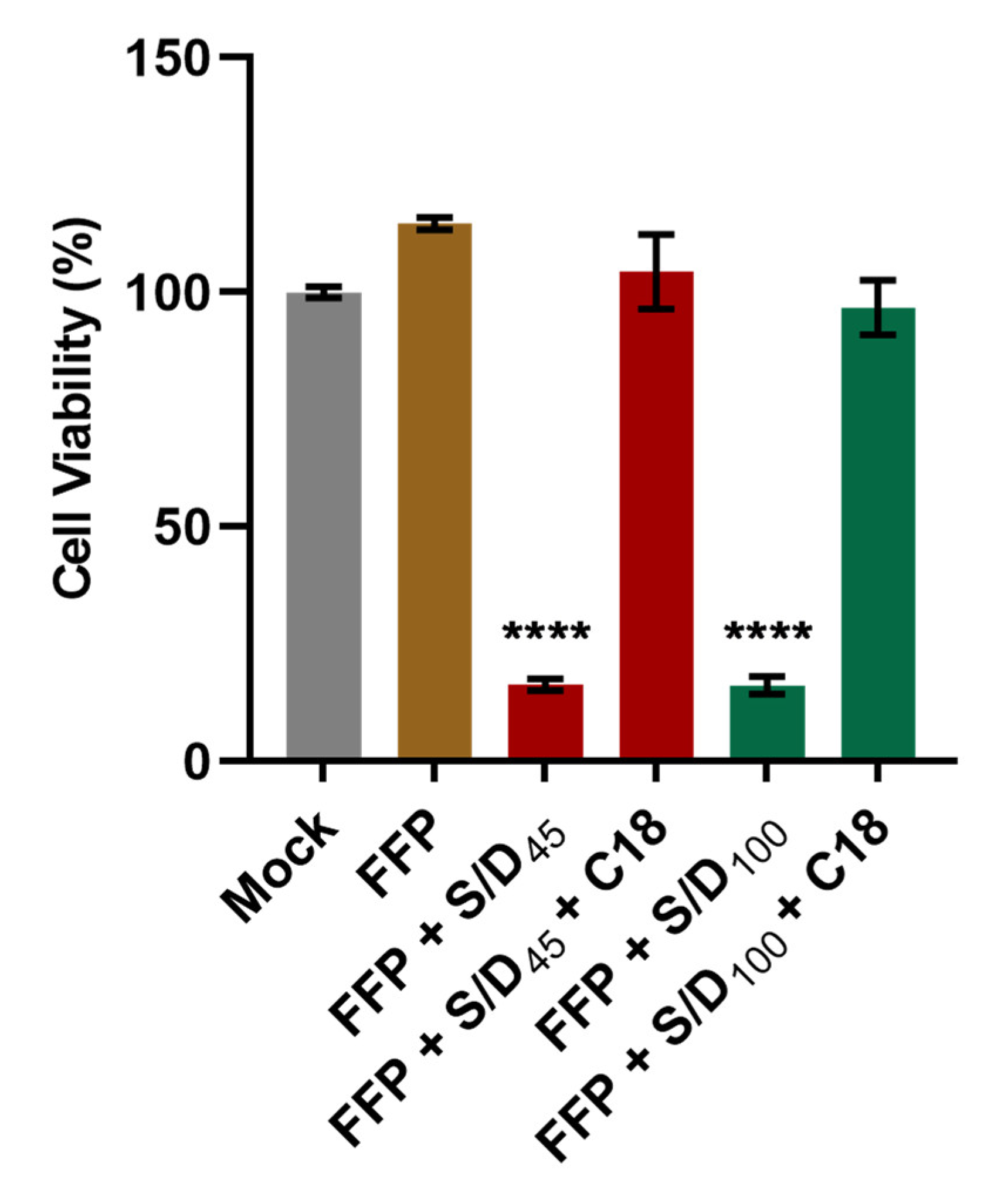
3.2. Elimination of S/D Cytotoxicity by C18 Filtration in S/D-Treated Plasma

Before the infectivity experiment, we tested the elimination of the toxicity associated with both S/D combinations (S/D45 or S/D100) in plasma by using C18 reverse phase chromatography cartridges (Sep-Pak Plus C18 cartridge) to avoid any toxic effects related to the samples during the infectivity assays in cell cultures. To confirm the clearance of S/D by C18 filtration in plasma after treatment, we performed a cell viability assay using FFP treated with S/D (S/D45 or S/D100) with or without C18 filtration in Huh-7 cells. As shown in Figure 2, while significant cell death was generated with the samples of FFP treated with S/D (S/D45 or S/D100), no cytotoxicity in Huh-7 cells was observed with the samples of FFP alone and C18 filtrates of FFP treated with S/D. These results indicate that the filtration through the hydrophobic C18 could effectively eliminate the toxicity of each S/D combination from plasma after treatment.

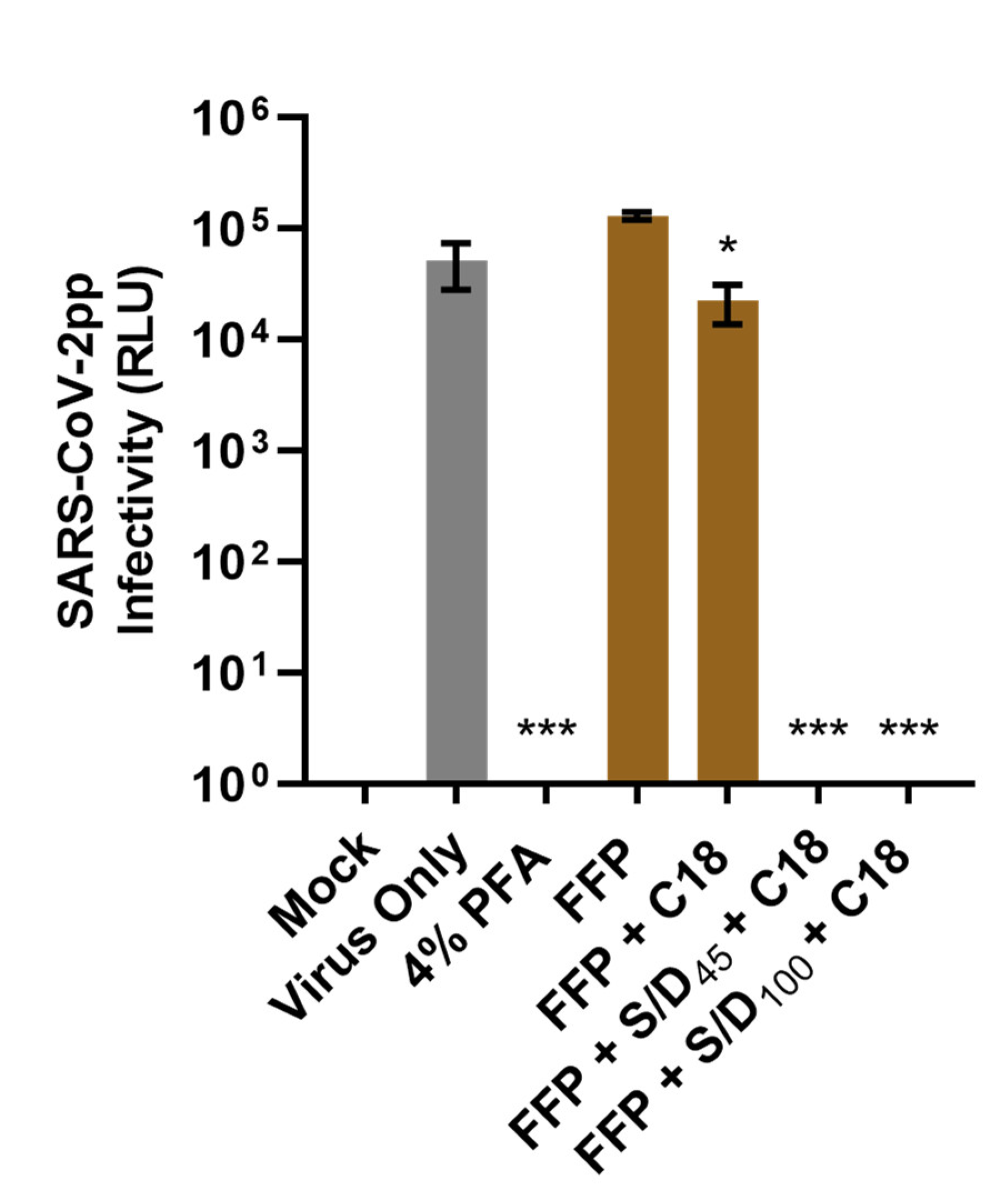
3.3. S/D Inactivation Protocols Efficiently Eliminate SARS-CoV-2pp Infectivity in Human Plasma

To demonstrate whether S/D treatments can eliminate SARS-CoV-2 infectivity in human plasma, we used S/D-treated or -untreated SARS-CoV-2pp-spiked FFP to infect Huh-7 cells. Both S/D treatments followed by C18 filtration of plasmas completely eliminated the infectivity of SARS-CoV-2pp, resulting in an undetectable level of luciferase reporter signals, similar to the Mock and PFA fixation control (Figure 3). These data show the treatments of plasma with S/D (S/D45 or S/D100) followed by C18 filtration could efficiently abolish SARS-CoV-2pp infectivity (decrease over 4 log10). In addition, the infectivity of SARS-CoV-2pp-spiked FFP treated with C18 cartridge filtration (FFP + C18) was modestly decreased, suggesting that the C18 filtration could additionally decrease SARS-CoV-2pp in the plasma.

3.4. The Neutralizing Activity of COVID-19 Convalescent Serum Is Preserved Following the S/D Inactivation Protocols

To verify whether the neutralizing activity of convalescent serum could be preserved following the S/D treatment, we next treated a patient-derived CCS with the S/D inactivation protocol. As shown in Figure 4, CCS (CCS Only) significantly reduced SARS-CoV-2pp infectivity, and CCS treated with the S/D inactivation protocols (CCS + S/D100 + C18 and CCS + S/D45 + C18) could also produce the same effect. No statistically significant difference in luciferase activity was observed between the S/D-treated and -untreated CCS samples. Our results suggested that both S/D inactivation protocols preserved the neutralizing activity of the SARS-CoV-2 nAbs in the CCS.

4. Discussion

SARS-CoV-2 pseudoviruses are useful for virological applications such as viral entry studies, drug screening, Abs detection, and the investigation of the S protein [53]. Here, we have shown the utility of SARS-CoV-2 S-pseudotyped lentiviral particles (Figure 1) for validation of viral inactivation protocols for therapeutic blood products. The development of the pseudoviral particle neutralization test (PPNT) is effective to quantify the neutralization titers of SARS-CoV-2 nAbs in the CCP safely in typical biosafety level (BSL)-2 laboratory facilities of blood centers, which will allow them to select the CCP units for therapeutic use [41].
S/D treatment is a standard method used to inactivate viruses in human blood products [54]. Such a viral inactivation method can safely inactivate all lipid-enveloped viruses in plasma [44], which includes coronaviruses. The action of S/D treatment is fast, and its inactivation ability can usually be achieved within the first 15 min [55]. The mechanism consists in disrupting the lipid membrane of enveloped viruses [56]. As such, the nonionic detergents used in the S/D treatment protocol could effectively disrupt the viral membrane of SARS-CoV-2pp (Figure 5) and inactivate the particle for infection. Since the target of S/D treatment is on the lipid viral membrane, such inactivation activity should not be affected by the mutations of viral proteins in newly emerging variants, even if their binding stability to the ACE2 receptor were enhanced [57]. In addition, the S/D methods are attractive in the manufacturing process of intravenous immunoglobulins (IVIg). They appear to have no detrimental effect on the binding capacity and functions of IgG [56] with good recovery of the product during the process [58]. The S/D procedures commonly incorporated in the purification process of the immunoglobulins consist of using the combination of 0.3–1% TnBP with one, sometimes two, detergents such as Tween-80, Triton X-100, or sodium cholate. Most processes are carried out at 20–35 °C for at least 1–6 h [59]. In the past, S/D (1% TnBP/TritonX-100) treatment has been found effective in inactivating SARS-COV (≥5.75 ± 0.25 log10 after 30 min incubation period) in immunoglobulin preparations [60]. Two main combinations of S/D (typically 1% TnBP/1% TritonX-100 and 1% TnBP/1% TritonX-45) are also described in the treatment of plasma for transfusion. Tritons (Triton X-100 or Triton X-45) are nonionic detergents belonging to octylphenoxy polyoxyethylene ethers. They have the same hydrophobic moiety, but the hydrophilic moiety contains a different number of oxyethylene units [61]. The combination of 1% TnBP/1% Triton X-100 was the first viral inactivation method implemented on a large pool of donations on an industrial scale [62], whereas the combination of 1% TnBP/1% Triton X-45 followed by oil extraction and filtration was incorporated in a closed bag system for the treatment of a mini pool of plasma [44].
Since its implementation in the processes of preparing plasma products and plasma, S/D technology has contributed to stopping the transmission of viral infections (e.g., hepatitis B and C, HIV) by plasma products [62,63,64]. Additionally, S/D treatment is known to be effective in inactivating (re)emerging lipid-enveloped viruses that remain a challenge for blood transfusion services such as West Nile virus, dengue virus, Zika virus, and Chikungunya virus [65]. During the COVID-19 pandemic, many regulatory authorities recommended the implementation of PRT to maximize the pathogen safety of CCP [66]. Our experiment has shown that the treatments of SARS-CoV-2pp-spiked plasma with S/D combinations (S/D45 or S/D100) eliminated SARS-CoV-2pp infectivity to the undetectable level (Figure 3). We also demonstrated the C18 filtration, which is used to remove the reactants of S/D agents for industrial plasma for transfusion, could additionally reduce SARS-CoV-2pp infectivity. In a study published by Horowitz et al., they also found that C18 column could remove 1 log10 TCID50 of HIV [62].
Other PRTs using photochemical treatments, such as methylene blue/visible light (MB + visible light), amotosalen/UVA (A + UVA), and riboflavin/UVB (R + UVB), are available in blood centers for single-donor plasma donation treatments. Their mechanisms of action lead to inactivating viruses by alteration of nucleic acids [49]. It has been shown that MB + visible light reduced SARS-COV (≥3.1 log10) [67] and MERS-COV (≥3.3 log10) [68] in plasma. R + UVB treatment could reduce ≥4.0 log10 for MERS-COV [69] and ≥3.40 log10 [70] or ≥4.79 for SARS-CoV-2 [66] in plasma. Similarly, A+ UVA reduced >3.32 ± 0.2 for SARS-CoV-2 [71] and ≥4.67 ± 0.25 for MERS-COV [72] in treated plasma. Our results using the S/D treatment followed by C18 filtration represent an additional viral inactivation protocol against SARS-CoV-2.
There are currently in the literature controversial data on the immunological properties of Abs in CCP treated with various PRT available in blood centers using either viral neutralization assays or high-throughput serology. Kostin et al. found in general a significant decrease in nAbs in CCP in all three photochemical PRT. They concluded that MB + visible light or A + UVA treatments seem to preserve the immunological properties of nAbs in CCP compared to R + UVB treatment [40,66]. However, Franchini M et al. observed no statistically significant difference between preinactivation and postinactivation nAb titers during A + UVA treatment [73]. Yonemura et al. also suggested that the functionality of nAbs in CCP did not alter after R + UVB treatment [41]. Moreover, the investigation of the impact of MB + visible light treatment on the immunological properties of Abs against Ebolavirus in plasma showed the preservation of the binding ability of IgM and IgG to their epitopes or the Fc receptor interaction of IgG [74]. A slight reduction [75] or no reduction [76] of Ebolavirus nAbs were found in CP treated with A + UVA treatment. In addition to the aforementioned PRT, S/D treatments with Triton X-45 or Triton X-100 are regarded to have minimal impact on the functionality and neutralizing activity of fractionated immunoglobulins [59,77], but data on anti-SARS-CoV-2 are lacking. To clarify this, we demonstrated that 30 min of S/D treatments (S/D45 or S/D100), which is enough to inactivate SARS-CoV-2pp, does not impact the functionality of SARS-CoV-2 nAbs in CCS (Figure 4). As such, the S/D inactivation protocol presents a useful strategy for removal of SARS-CoV-2 infectivity while preserving anti-SARS-CoV-2 nAbs functionality in CCP/CCS-based blood products.

5. Conclusions

Our investigation demonstrated that S/D treatments with TnBP and Triton X-45 or Triton X-100 followed by C18 filtration effectively removed SARS-CoV-2pp infectivity from plasma without destroying the functionality of nAbs against SARS-CoV-2. We believe S/D treatments could be useful in viral inactivation/elimination processes of therapeutic blood products against SARS-CoV-2.

Author Contributions

Conceptualization, W.F.B., T.B. and L.-T.L.; methodology, W.F.B., C.-H.L., T.B. and L.-T.L.; formal analysis, W.F.B., C.-H.L., T.B. and L.-T.L.; investigation, W.F.B., C.-H.L. and Y.-T.H.; resources, T.B. and L.-T.L.; writing—original draft preparation, W.F.B. and C.-H.L.; writing—review and editing, C.-H.L., T.B. and L.-T.L.; supervision, T.B. and L.-T.L.; funding acquisition, L.-T.L. All authors have read and agreed to the published version of the manuscript.

Funding

L.-T.L. is supported by funding from the Ministry of Science and Technology of Taiwan (MOST110-2320-B-038-041-MY3). W.F.B. received a PhD fellowship from Taipei Medical University and additional financial support from T.B. laboratory.

Institutional Review Board Statement

The study was conducted in accordance with the Declaration of Helsinki and approved by the Joint Institutional Review Board of Taipei Medical University (N201802052 and N202006017).

Informed Consent Statement

For FFP, voluntary nonremunerated blood donors provided informed consent that part of their donation could be used for research purpose intended to improve virus safety of blood transfusion. Patient consent for CCS was waived according to the TMU-Joint Institutional Review Board regulations.

Data Availability Statement

Data are contained within the article.

Acknowledgments

We would like to thank The National Health Research Institute Biobank and its cooperative institutions for providing the biological specimen and related clinical data (all are deidentified) for our research. National Health Research Institute Biobank is supported by grants from Ministry of Health and Welfare, Ministry of Science and Technology, and National Health Research Institutes, Taiwan.

Conflicts of Interest

The authors declare no conflict of interest.

References

  1. Astuti, I.; Ysrafil. Severe Acute Respiratory Syndrome Coronavirus 2 (SARS-CoV-2): An overview of viral structure and host response. Syndr. Clin. Res. Rev. 2020, 14, 407–412. [Google Scholar] [CrossRef] [PubMed]
  2. Center for Systems Science and Engineering at Johns Hopkins University. COVID-19 Dashboard. Available online: https://coronavirus.jhu.edu/map.html (accessed on 1 August 2022).
  3. Hadi, J.; Dunowska, M.; Wu, S.; Brightwell, G. Control Measures for SARS-CoV-2: A Review on Light-Based Inactivation of Single-Stranded RNA Viruses. Pathogens 2020, 9, 737. [Google Scholar] [CrossRef] [PubMed]
  4. Jiang, S.; Hillyer, C.; Du, L. Neutralizing Antibodies against SARS-CoV-2 and Other Human Coronaviruses. Trends Immunol. 2020, 41, 355–359. [Google Scholar] [CrossRef] [PubMed]
  5. Salazar, E.; Kuchipudi, S.V.; Christensen, P.A.; Eagar, T.; Yi, X.; Zhao, P.; Jin, Z.; Long, S.W.; Olsen, R.J.; Chen, J.; et al. Convalescent plasma anti-SARS-CoV-2 spike protein ectodomain and receptor-binding domain IgG correlate with virus neutralization. J. Clin. Investig. 2020, 130, 6728–6738. [Google Scholar] [CrossRef] [PubMed]
  6. Du, L.; Yang, Y.; Zhang, X. Neutralizing antibodies for the prevention and treatment of COVID-19. Cell Mol. Immunol. 2021, 18, 2293–2306. [Google Scholar] [CrossRef]
  7. Harvey, W.T.; Carabelli, A.M.; Jackson, B.; Gupta, R.K.; Thomson, E.C.; Harrison, E.M.; Ludden, C.; Reeve, R.; Rambaut, A.; Consortium, C.-G.U.; et al. SARS-CoV-2 variants, spike mutations and immune escape. Nat. Rev. Microbiol. 2021, 19, 409–424. [Google Scholar] [CrossRef]
  8. Wang, P.; Liu, L.; Nair, M.S.; Yin, M.T.; Luo, Y.; Wang, Q.; Yuan, T.; Mori, K.; Solis, A.G.; Yamashita, M.; et al. SARS-CoV-2 neutralizing antibody responses are more robust in patients with severe disease. Emerg. Microbes Infect. 2020, 9, 2091–2093. [Google Scholar] [CrossRef]
  9. Beaudoin-Bussières, G.; Laumaea, A.; Anand, S.P.; Prévost, J.; Gasser, R.; Goyette, G.; Medjahed, H.; Perreault, J.; Tremblay, T.; Lewin, A.; et al. Decline of humoral responses against SARS-CoV-2 Spike in convalescent individuals. mBio 2020, 11, e02590-20. [Google Scholar] [CrossRef]
  10. Suthar, M.S.; Zimmerman, M.; Kauffman, R.; Mantus, G.; Linderman, S.; Vanderheiden, A.; Nyhoff, L.; Davis, C.; Adekunle, S.; Affer, M.; et al. Rapid generation of neutralizing antibody responses in COVID-19 patients. Cell Rep. Med. 2020, 1, 100040. [Google Scholar] [CrossRef]
  11. Maor, Y.; Cohen, D.; Paran, N.; Israely, T.; Ezra, V.; Axelrod, O.; Shinar, E.; Izak, M.; Rahav, G.; Rahimi-Levene, N.; et al. Compassionate use of convalescent plasma for treatment of moderate and severe pneumonia in COVID-19 patients and association with IgG antibody levels in donated plasma. EClinicalMedicine 2020, 26, 100525. [Google Scholar] [CrossRef]
  12. Burnouf, T.; Gathof, B.; Bloch, E.M.; Bazin, R.; de Angelis, V.; Patidar, G.K.; Rastvorceva, R.M.G.; Oreh, A.; Goel, R.; Rahimi-Levene, N.; et al. Production and Quality Assurance of Human Polyclonal Hyperimmune Immunoglobulins Against SARS-CoV-2. Transfus. Med. Rev. 2022, 36, 125–132. [Google Scholar] [CrossRef] [PubMed]
  13. Perricone, C.; Triggianese, P.; Bursi, R.; Cafaro, G.; Bartoloni, E.; Chimenti, M.S.; Gerli, R.; Perricone, R. Intravenous Immunoglobulins at the Crossroad of Autoimmunity and Viral Infections. Microorganisms 2021, 9, 121. [Google Scholar] [CrossRef] [PubMed]
  14. Bloch, E.M.; Shoham, S.; Casadevall, A.; Sachais, B.S.; Shaz, B.; Winters, J.L.; van Buskirk, C.; Grossman, B.J.; Joyner, M.; Henderson, J.P.; et al. Deployment of convalescent plasma for the prevention and treatment of COVID-19. J. Clin. Investig. 2020, 130, 2757–2765. [Google Scholar] [CrossRef] [PubMed] [Green Version]
  15. Piechotta, V.; Iannizzi, C.; Chai, K.L.; Valk, S.J.; Kimber, C.; Dorando, E.; Monsef, I.; Wood, E.M.; Lamikanra, A.A.; Roberts, D.J.; et al. Convalescent plasma or hyperimmune immunoglobulin for people with COVID-19: A living systematic review. Cochrane Database Syst. Rev. 2021, 5, CD013600. [Google Scholar] [CrossRef]
  16. Valk, S.J.; Piechotta, V.; Chai, K.L.; Doree, C.; Monsef, I.; Wood, E.M.; Lamikanra, A.; Kimber, C.; McQuilten, Z.; So-Osman, C.; et al. Convalescent plasma or hyperimmune immunoglobulin for people with COVID-19: A rapid review. Cochrane Database Syst. Rev. 2020, 5, CD013600. [Google Scholar] [CrossRef]
  17. Bar, K.J.; Shaw, P.A.; Choi, G.H.; Aqui, N.; Fesnak, A.; Yang, J.B.; Soto-Calderon, H.; Grajales, L.; Starr, J.; Andronov, M.; et al. A randomized controlled study of convalescent plasma for individuals hospitalized with COVID-19 pneumonia. J. Clin. Investig. 2021, 131, e155114. [Google Scholar] [CrossRef]
  18. Hirsch, C.; Park, Y.S.; Piechotta, V.; Chai, K.L.; Estcourt, L.J.; Monsef, I.; Salomon, S.; Wood, E.M.; So-Osman, C.; McQuilten, Z. SARS-CoV-2-neutralising monoclonal antibodies to prevent COVID-19. Cochrane Database Syst. Rev. 2022, 6, CD014945. [Google Scholar]
  19. Recovery Collaborative Group. Convalescent plasma in patients admitted to hospital with COVID-19 (RECOVERY): A randomised controlled, open-label, platform trial. Lancet 2021, 397, 2049–2059. [Google Scholar] [CrossRef]
  20. Focosi, D.; Franchini, M.; Pirofski, L.A.; Burnouf, T.; Paneth, N.; Joyner, M.J.; Casadevall, A. COVID-19 Convalescent Plasma and Clinical Trials: Understanding Conflicting Outcomes. Clin. Microbiol. Rev. 2022, 35, e0020021. [Google Scholar] [CrossRef]
  21. Valdez-Cruz, N.A.; Garcia-Hernandez, E.; Espitia, C.; Cobos-Marin, L.; Altamirano, C.; Bando-Campos, C.G.; Cofas-Vargas, L.F.; Coronado-Aceves, E.W.; Gonzalez-Hernandez, R.A.; Hernandez-Peralta, P.; et al. Integrative overview of antibodies against SARS-CoV-2 and their possible applications in COVID-19 prophylaxis and treatment. Microb. Cell Fact. 2021, 20, 88. [Google Scholar] [CrossRef]
  22. Oliveira, F.A.; Nucci, M.P.; Rego, G.N.A.; Alves, A.D.H.; Marti, L.C.; Nucci, L.P.; Mamani, J.B.; Gamarra, L.F. Convalescent plasma therapy in COVID-19 critically ill patients during advanced phases of clinical trials and their preliminary results. Einstein 2021, 19, eRW6186. [Google Scholar] [CrossRef] [PubMed]
  23. Dulipsingh, L.; Ibrahim, D.; Schaefer, E.J.; Crowell, R.; Diffenderfer, M.R.; Williams, K.; Lima, C.; McKenzie, J.; Cook, L.; Puff, J.; et al. SARS-CoV-2 serology and virology trends in donors and recipients of convalescent plasma. Transfus. Apher. Sci. 2020, 59, 102922. [Google Scholar] [CrossRef] [PubMed]
  24. Mosnier, L.O. How much clotting is enough? Blood 2020, 136, 651–652. [Google Scholar] [CrossRef]
  25. Zeng, Q.L.; Yu, Z.J.; Gou, J.J.; Li, G.M.; Ma, S.H.; Zhang, G.F.; Xu, J.H.; Lin, W.B.; Cui, G.L.; Zhang, M.M.; et al. Effect of Convalescent Plasma Therapy on Viral Shedding and Survival in Patients With Coronavirus Disease 2019. J. Infect. Dis. 2020, 222, 38–43. [Google Scholar] [CrossRef] [PubMed]
  26. Selvi, V. Convalescent Plasma: A Challenging Tool to Treat COVID-19 Patients-A Lesson from the Past and New Perspectives. Biomed. Res. Int. 2020, 2020, 2606058. [Google Scholar] [CrossRef] [PubMed]
  27. Boyarsky, B.J.; Werbel, W.A.; Avery, R.K.; Tobian, A.A.R.; Massie, A.B.; Segev, D.L.; Garonzik-Wang, J.M. Antibody Response to 2-Dose SARS-CoV-2 mRNA Vaccine Series in Solid Organ Transplant Recipients. JAMA 2021, 325, 2204–2206. [Google Scholar] [CrossRef] [PubMed]
  28. Abdelhady, H.; Abdelrazik, M.; Abdi, Z.; Abdo, D.; Abdulle, A.; Abel, L.; Abouzeenni, S.; Abrahamson, G.; Abusamra, Y.; Adams, L.; et al. Effect of Convalescent Plasma on Organ Support-Free Days in Critically Ill Patients With COVID-19: A Randomized Clinical Trial. JAMA 2021, 326, 1690–1702. [Google Scholar] [CrossRef]
  29. Gharbharan, A.; Jordans, C.C.E.; GeurtsvanKessel, C.; den Hollander, J.G.; Karim, F.; Mollema, F.P.N.; Stalenhoef-Schukken, J.E.; Dofferhoff, A.; Ludwig, I.; Koster, A.; et al. Effects of potent neutralizing antibodies from convalescent plasma in patients hospitalized for severe SARS-CoV-2 infection. Nat. Commun. 2021, 12, 3189. [Google Scholar] [CrossRef]
  30. Expert Panel Updates Guidelines for Clinical CCP Use. Available online: https://www.aabb.org/news-resources/news/article/2022/04/13/aabb-expert-panel-updates-guidelines-for-clinical-ccp-use (accessed on 27 July 2022).
  31. Lacombe, K.; Hueso, T.; Porcher, R.; Mekinian, A.; Chiarabini, T.; Georgin-Lavialle, S.; Ader, F.; Saison, J.; Martin Blondel, G.; de Castro, N.; et al. COVID-19 convalescent plasma for hospitalizedCOVID-19 patients not requiring assisted ventilation. A randomized controlled trial. Vox Sang 2022, 117, 74–75. [Google Scholar]
  32. FDA. Regulatory Update: FDA Issues Revised CCP Guidance, AABB Updates CCP Toolkit. Available online: https://www.aabb.org/news-resources/news/article/2022/01/11/regulatory-update-fda-issues-revised-ccp-guidance-aabb-updates-ccp-toolkit (accessed on 27 July 2022).
  33. Larkey, N.E.; Ewaisha, R.; Lasho, M.A.; Roforth, M.M.; Granger, D.; Jerde, C.R.; Wu, L.; Gorsh, A.; Klassen, S.A.; Senefeld, J.W.; et al. Limited Correlation between SARS-CoV-2 Serologic Assays for Identification of High-Titer COVID-19 Convalescent Plasma Using FDA Thresholds. Microbiol. Spectr. 2022, 10, e0115422. [Google Scholar] [CrossRef]
  34. US Food and Drugs Administration. Convalescent Plasma EUA Letter of Authorization 12282021. Available online: https://www.fda.gov/media/141477/download (accessed on 25 September 2022).
  35. Chang, L.; Yan, Y.; Wang, L. Coronavirus Disease 2019: Coronaviruses and Blood Safety. Transfus. Med. Rev. 2020, 34, 75–80. [Google Scholar] [CrossRef] [PubMed]
  36. AlShehry, N.; Zaidi, S.Z.A.; AlAskar, A.; Al Odayani, A.; Alotaibi, J.M.; AlSagheir, A.; Al-Eyadhy, A.; Balelah, S.; Salam, A.; Zaidi, A.R.Z.; et al. Safety and Efficacy of Convalescent Plasma for Severe COVID-19: Interim Report of a Multicenter Phase II Study from Saudi Arabia. Saudi J. Med. Med. Sci. 2021, 9, 16–23. [Google Scholar] [CrossRef] [PubMed]
  37. Welch, S.R.; Davies, K.A.; Buczkowski, H.; Hettiarachchi, N.; Green, N.; Arnold, U.; Jones, M.; Hannah, M.J.; Evans, R.; Burton, C.; et al. Analysis of Inactivation of SARS-CoV-2 by Specimen Transport Media, Nucleic Acid Extraction Reagents, Detergents, and Fixatives. J. Clin. Microbiol. 2020, 58, e01713-20. [Google Scholar] [CrossRef] [PubMed]
  38. Patterson, E.I.; Prince, T.; Anderson, E.R.; Casas-Sanchez, A.; Smith, S.L.; Cansado-Utrilla, C.; Solomon, T.; Griffiths, M.J.; Acosta-Serrano, Á.; Turtle, L.; et al. Methods of Inactivation of SARS-CoV-2 for Downstream Biological Assays. J. Infect. Dis. 2020, 222, 1462–1467. [Google Scholar] [CrossRef]
  39. Liew, O.W.; Fanusi, F.; Ng, J.Y.X.; Ahidjo, B.A.; Ling, S.S.M.; Lilyanna, S.; Chong, J.P.C.; Lim, A.E.S.; Lim, W.Z.; Ravindran, S.; et al. Immunoassay-Compatible Inactivation of SARS-CoV-2 in Plasma Samples for Enhanced Handling Safety. ACS Omega 2022, 7, 25510–25520. [Google Scholar] [CrossRef] [PubMed]
  40. Kostin, A.I.; Lundgren, M.N.; Bulanov, A.Y.; Ladygina, E.A.; Chirkova, K.S.; Gintsburg, A.L.; Logunov, D.Y.; Dolzhikova, I.V.; Shcheblyakov, D.V.; Borovkova, N.V.; et al. Impact of pathogen reduction methods on immunological properties of the COVID-19 convalescent plasma. Vox Sang 2021, 116, 665–672. [Google Scholar] [CrossRef]
  41. Yonemura, S.; Hartson, L.; Dutt, T.S.; Henao-Tamayo, M.; Goodrich, R.; Marschner, S. Preservation of neutralizing antibody function in COVID-19 convalescent plasma treated using a riboflavin and ultraviolet light-based pathogen reduction technology. Vox Sang 2021, 116, 1076–1083. [Google Scholar] [CrossRef]
  42. Cagdas, D. Convalescent plasma and hyperimmune globulin therapy in COVID-19. Expert Rev. Clin. Immunol. 2021, 17, 309–316. [Google Scholar] [CrossRef]
  43. Joyner, M.J.; Wright, R.S.; Fairweather, D.; Senefeld, J.W.; Bruno, K.A.; Klassen, S.A.; Carter, R.E.; Klompas, A.M.; Wiggins, C.C.; Shepherd, J.R.; et al. Early safety indicators of COVID-19 convalescent plasma in 5000 patients. J. Clin. Investig. 2020, 130, 4791–4797. [Google Scholar] [CrossRef]
  44. Hellstern, P.; Solheim, B.G. The Use of Solvent/Detergent Treatment in Pathogen Reduction of Plasma. Transfus. Med. Hemother 2011, 38, 65–70. [Google Scholar] [CrossRef]
  45. Kuo, Y.T.; Liu, C.H.; Corona, A.; Fanunza, E.; Tramontano, E.; Lin, L.T. The Methanolic Extract of Perilla frutescens Robustly Restricts Ebola Virus Glycoprotein-Mediated Entry. Viruses 2021, 13, 1793. [Google Scholar] [CrossRef] [PubMed]
  46. Hung, T.C.; Jassey, A.; Liu, C.H.; Lin, C.J.; Lin, C.C.; Wong, S.H.; Wang, J.Y.; Yen, M.H.; Lin, L.T. Berberine inhibits hepatitis C virus entry by targeting the viral E2 glycoprotein. Phytomedicine 2019, 53, 62–69. [Google Scholar] [CrossRef] [PubMed]
  47. Connor, R.I.; Chen, B.K.; Choe, S.; Landau, N.R. Vpr is required for efficient replication of human immunodeficiency virus type-1 in mononuclear phagocytes. Virology 1995, 206, 935–944. [Google Scholar] [CrossRef] [PubMed] [Green Version]
  48. Giroglou, T.; Cinatl, J., Jr.; Rabenau, H.; Drosten, C.; Schwalbe, H.; Doerr, H.W.; von Laer, D. Retroviral vectors pseudotyped with severe acute respiratory syndrome coronavirus S protein. J. Virol. 2004, 78, 9007–9015. [Google Scholar] [CrossRef] [Green Version]
  49. Chou, M.L.; Burnouf, T.; Chang, S.P.; Hung, T.C.; Lin, C.C.; Richardson, C.D.; Lin, L.T. TnBPTriton X-45 treatment of plasma for transfusion efficiently inactivates hepatitis C virus. PLoS ONE 2015, 10, e0117800. [Google Scholar] [CrossRef] [Green Version]
  50. Sanclemente-Alaman, I.; Moreno-Jimenez, L.; Benito-Martin, M.S.; Canales-Aguirre, A.; Matias-Guiu, J.A.; Matias-Guiu, J.; Gomez-Pinedo, U. Experimental Models for the Study of Central Nervous System Infection by SARS-CoV-2. Front. Immunol. 2020, 11, 2163. [Google Scholar] [CrossRef]
  51. Sholukh, A.M.; Fiore-Gartland, A.; Ford, E.S.; Miner, M.D.; Hou, Y.J.; Tse, L.V.; Kaiser, H.; Zhu, H.; Lu, J.; Madarampalli, B.; et al. Evaluation of Cell-Based and Surrogate SARS-CoV-2 Neutralization Assays. J. Clin. Microbiol. 2021, 59, e0052721. [Google Scholar] [CrossRef]
  52. Zucker, I.; Lester, Y.; Alter, J.; Werbner, M.; Yecheskel, Y.; Gal-Tanamy, M.; Dessau, M. Pseudoviruses for the assessment of coronavirus disinfection by ozone. Env. Chem. Lett. 2021, 19, 1779–1785. [Google Scholar] [CrossRef]
  53. Hu, J.; Gao, Q.; He, C.; Huang, A.; Tang, N.; Wang, K. Development of cell-based pseudovirus entry assay to identify potential viral entry inhibitors and neutralizing antibodies against SARS-CoV-2. Genes Dis. 2020, 7, 551–557. [Google Scholar] [CrossRef]
  54. Darnell, M.E.R.; Taylor, D.R. Evaluation of inactivation methods for severe acute respiratory syndrome coronavirus in noncellular blood products. Transfusion 2006, 46, 1770–1777. [Google Scholar] [CrossRef] [Green Version]
  55. Barahona Afonso, A.F.; Joao, C.M. The Production Processes and Biological Effects of Intravenous Immunoglobulin. Biomolecules 2016, 6, 15. [Google Scholar] [CrossRef] [PubMed] [Green Version]
  56. Mould, G.P.; Sutton, J.A.; Matejtschuk, P.; Gascoigne, E.W.; Dash, C.H. Dash. Solvent detergent treatment does not alter the tolerance or uptake of human normal. Vox Sang 2001, 80, 151–158. [Google Scholar] [CrossRef] [PubMed]
  57. Koehler, M.; Ray, A.; Moreira, R.A.; Juniku, B.; Poma, A.B.; Alsteens, D. Molecular insights into receptor binding energetics and neutralization of SARS-CoV-2 variants. Nat. Commun. 2021, 12, 6977. [Google Scholar] [CrossRef] [PubMed]
  58. Buchacher, A.; Iberer, G. Purification of intravenous immunoglobulin G from human plasma-aspects of yield and virus safety. Biotechnol. J. 2006, 1, 148–163. [Google Scholar] [CrossRef] [PubMed]
  59. Radosevich, M.; Burnouf, T. Intravenous immunoglobulin G: Trends in production methods, quality control and quality assurance. Vox Sang 2010, 98, 12–28. [Google Scholar] [CrossRef]
  60. Rabenau, H.F.; Biesert, L.; Schmidt, T.; Bauer, G.; Cinatl, J.; Doerr, H.W. SARS-coronavirus (SARS-CoV) and the safety of a solvent/detergent (S/D) treated immunoglobulin preparation. Biologicals 2005, 33, 95–99. [Google Scholar] [CrossRef]
  61. Kang, B.; Zhang, X.; Wu, Z.; Qi, H.; Wang, Z. Solubilization capacity of nonionic surfactant micelles exhibiting strong influence on export of intracellular pigments in Monascus fermentation. Microb. Biotechnol. 2013, 6, 540–550. [Google Scholar] [CrossRef]
  62. Horowitz, B.; Bonomo, R.; Prince, A.M.; Chin, S.N.; Brotman, B.; Shulman, R.W. Solvent/detergent-treated plasma: A virus-inactivated substitute for fresh frozen plasma. Blood 1992, 79, 826–831. [Google Scholar] [CrossRef] [Green Version]
  63. Piët, M.P.; Chin, S.; Prince, A.M.; Brotman, B.; Cundell, A.M.; Horowitz, B. The use of tri(n-butyl)phosphate detergent mixtures to inactivate hepatitis viruses and human immunodeficiency virus in plasma and plasma’s subsequent fractionation. Transfusion 1990, 30, 591–598. [Google Scholar] [CrossRef]
  64. Toth, D. Neutralization Of SARS-CoV-2 Variants By A Human Polyclonal Antibody Therapeutic (COVID-HIG, NP-028) With High Neutralizing Titers To SARS-CoV-2. bioRxiv 2022. [Google Scholar] [CrossRef]
  65. Burnouf, T. An overview of plasma fractionation. Ann. Blood 2018, 3, 33. [Google Scholar] [CrossRef]
  66. Focosi, D.; Franchini, M. Impact of pathogen-reduction technologies on COVID-19 convalescent plasma potency. Transfus. Clin. Biol. 2021, 3188, 3. [Google Scholar] [CrossRef] [PubMed]
  67. Eickmann, M.; Gravemann, U.; Handke, W.; Tolksdorf, F.; Reichenberg, S.; Muller, T.H.; Seltsam, A. Inactivation of three emerging viruses-severe acute respiratory syndrome coronavirus, Crimean-Congo haemorrhagic fever virus and Nipah virus-in platelet concentrates by ultraviolet C light and in plasma by methylene blue plus visible light. Vox Sang 2020, 115, 146–151. [Google Scholar] [CrossRef] [PubMed] [Green Version]
  68. Eickmann, M.; Gravemann, U.; Handke, W.; Tolksdorf, F.; Reichenberg, S.; Muller, T.H.; Seltsam, A. Inactivation of Ebola virus and Middle East respiratory syndrome coronavirus in platelet concentrates and plasma by ultraviolet C light and methylene blue plus visible light, respectively. Transfusion 2018, 58, 2202–2207. [Google Scholar] [CrossRef] [PubMed] [Green Version]
  69. Keil, S.D.; Bowen, R.; Marschner, S. Inactivation of Middle East respiratory syndrome coronavirus (MERS-CoV) in plasma products using a riboflavin-based and ultraviolet light-based photochemical treatment. Transfusion 2016, 56, 2948–2952. [Google Scholar] [CrossRef] [Green Version]
  70. Keil, S.D.; Ragan, I.; Yonemura, S.; Hartson, L.; Dart, N.K.; Bowen, R. Inactivation of severe acute respiratory syndrome coronavirus 2 in plasma and platelet products using a riboflavin and ultraviolet light-based photochemical treatment. Vox Sang 2020, 115, 495–501. [Google Scholar] [CrossRef]
  71. Azhar, E.I.; Hindawi, S.I.; El-Kafrawy, S.A.; Hassan, A.M.; Tolah, A.M.; Alandijany, T.A.; Bajrai, L.H.; Damanhouri, G.A. Amotosalen and ultraviolet A light treatment efficiently inactivates severe acute respiratory syndrome coronavirus 2 (SARS-CoV-2) in human plasma. Vox Sang 2021, 116, 673–681. [Google Scholar] [CrossRef]
  72. Hindawi, S.I.; Hashem, A.M.; Damanhouri, G.A.; El-Kafrawy, S.A.; Tolah, A.M.; Hassan, A.M.; Azhar, E.I. Inactivation of Middle East respiratory syndrome-coronavirus in human plasma using amotosalen and ultraviolet A light. Transfusion 2018, 58, 52–59. [Google Scholar] [CrossRef]
  73. Franchini, M.; Baldanti, F.; Percivalle, E.; Ferrari, A.; Biasi, P.; Liumbruno, G.M.; Glingani, C. Impact of pathogen reduction technology on neutralizing antibody titer in COVID-19 convalescent plasma. Transfus. Clin. Et Biol. 2022, 29, 101. [Google Scholar] [CrossRef]
  74. Raster, J.; Zimmermann, K.; Wesche, J.; Aurich, K.; Greinacher, A.; Selleng, K. Effect of Methylene Blue Pathogen Inactivation on the Integrity of Immunoglobulin M and G. Transfus. Med. Hemother 2021, 48, 148–152. [Google Scholar] [CrossRef]
  75. Geisen, C.; Kann, G.; Strecker, T.; Wolf, T.; Schuttfort, G.; van Kraaij, M.; MacLennan, S.; Rummler, S.; Weinigel, C.; Eickmann, M.; et al. Pathogen-reduced Ebola virus convalescent plasma: First steps towards standardization of manufacturing and quality control including assessment of Ebola-specific neutralizing antibodies. Vox Sang 2016, 110, 329–335. [Google Scholar] [CrossRef] [PubMed]
  76. Dean, C.L.; Hooper, J.W.; Dye, J.M.; Zak, S.E.; Koepsell, S.A.; Corash, L.; Benjamin, R.J.; Kwilas, S.; Bonds, S.; Winkler, A.M.; et al. Characterization of Ebola convalescent plasma donor immune response and psoralen treated plasma in the United States. Transfusion 2020, 60, 1024–1031. [Google Scholar] [CrossRef] [PubMed]
  77. Yang, Y.H.; Ngo, C.; Yeh, I.N.; Uemura, Y. Antibody Fc functional activity of intravenous immunoglobulin preparations treated with solvent-detergent for virus inactivation. Vox Sang 1994, 67, 337–344. [Google Scholar] [CrossRef] [PubMed]
Figure 1. Titration of SARS-CoV-2 pseudoparticles (SARS-CoV-2pp). The serial dilution of SARS-CoV-2pp stock was used to infect Huh-7 cells for 2 h. Viral infectivity was measured by luciferase reporter activity (relative light units; RLU) after 72 h incubation. Data shown are collected in triplicate and expressed as mean ± standard deviation (SD). Paraformaldehyde (PFA; 4%) fixed cells infected with 10× diluted SARS-CoV-2pp stock was included as a negative control.
Viruses 14 02419 g001
Figure 2. C18 filtration efficiently removes S/D cytotoxicity. Huh-7 cells were treated with basal medium (Mock), freshly frozen plasma (FFP), plasma treated with 1% TnBP/1% Triton X-45 (FFP + S/D45), plasma treated with 1% TnBP/1% Triton X-45 followed by C18 cartridge filtration (FFP + S/D45 + C18), plasma treated with 1% TnBP/1% Triton X-100 (FFP + S/D100), or plasma treated with 1% TnBP/1% Triton X-100 followed by C18 cartridge filtration (FFP + S/D100 + C18) for 72 h before cell viability was measured by CCK-8 assay. Data shown were collected in duplicate and expressed as mean ± standard deviation (SD). Statistical significance was analyzed by one-way ANOVA with Dunnett’s multiple comparisons test (**** p ≤ 0.0001 compared to Mock).
Viruses 14 02419 g002
Figure 3. S/D treatments completely inactivate SARS-CoV-2pp from plasma. Huh-7 cells were infected with SARS-CoV-2pp-spiked freshly frozen plasma (FFP), SARS-CoV-2pp-spiked FFP treated with C18 cartridge filtration (FFP + C18), SARS-CoV-2pp-spiked FFP treated with 1% TnBP/1% Triton X-45 followed by C18 cartridge filtration (FFP + S/D45 + C18), or SARS-CoV-2pp-spiked FFP treated with 1% TnBP/1% Triton X-100 followed by C18 cartridge filtration (FFP + S/D100 + C18) for 1.5 h. Viral infectivity was measured by luciferase reporter activity (relative light units; RLU) after 72 h incubation. Data shown are collected in triplicate and expressed as mean ± standard deviation (SD). Statistical significance was analyzed by one-way ANOVA with Dunnett’s multiple comparisons test (* p ≤ 0.05 and *** p ≤ 0.001 compared to Virus Only).
Viruses 14 02419 g003
Figure 4. Neutralization efficacy of SARS-CoV-2 nAbs in COVID-19 convalescent serum (CCS) were preserved after the S/D inactivation protocols. Patient-derived CCS was treated with the S/D inactivation protocols (CCS + S/D100 + C18 or CCS + S/D45 + C18) or left untreated (CCS Only) before incubation with SARS-CoV-2pp at 37 °C for 1 h in an Eppendorf tube. The CCS–virus mixture was then used to infect Huh-7 cells, and viral infectivity was measured by luciferase reporter activity (relative light units; RLU) after 72 h incubation. Data shown were collected in triplicate and expressed as mean ± standard deviation (SD). Statistical significance was analyzed by one-way ANOVA with Tukey’s multiple comparisons test (** p ≤ 0.01 and *** p ≤ 0.001 compared to Virus Only).
Viruses 14 02419 g004
Figure 5. Schematic illustration of S/D treatment’s impact on inactivating SARS-CoV-2pp. The intact lentivirus-based SARS-CoV-2pp is inactivated after S/D treatment, which disrupts the lipid membrane of enveloped viruses, thus destabilizing particle integrity and preventing viral infection. Created with BioRender.com.
Viruses 14 02419 g005
Publisher’s Note: MDPI stays neutral with regard to jurisdictional claims in published maps and institutional affiliations.

