Article

Colletotrichum Species Causing Cyclocarya paliurus Anthracnose in Southern China

by
Xiang-Rong Zheng
1,2,†,
Mao-Jiao Zhang
1,† and
Feng-Mao Chen
1,*
1
Collaborative Innovation Center of Sustainable Forestry in Southern China, College of Forestry, Nanjing Forestry University, Nanjing 210037, China
2
College of Landscape Architecture, Jiangsu Vocational College of Agriculture and Forestry, Zhenjiang 212400, China
*
Author to whom correspondence should be addressed.
These authors contributed equally to this work.
Forests 2024, 15(3), 490; https://doi.org/10.3390/f15030490
Submission received: 31 January 2024 / Revised: 28 February 2024 / Accepted: 4 March 2024 / Published: 6 March 2024
(This article belongs to the Special Issue Forest Tree Diseases Genomics: Growing Resources and Applications)

Abstract

:
Cyclocarya paliurus, native to China, is a medicinal and edible plant with important health benefits. Anthracnose is an emerging disease in southern China that causes severe economic losses and poses a great threat to the C. paliurus tea industry. However, to date, the species diversity of pathogens causing C. paliurus anthracnose has remained limited. From 2018 to 2022, a total of 331 Colletotrichum isolates were recovered from symptomatic leaves in eight major C. paliurus planting provinces of southern China. Phylogenetic analyses based on nine loci (ITS, GAPDH, ACT, CHS-1, TUB, CAL, HIS3, GS and ApMat) coupled with phenotypic characteristics revealed that 43 representative isolates belonged to seven known Colletotrichum species, including C. brevisporum, C. fructicola, C. gloeosporioides sensu stricto, C. godetiae, C. nymphaeae, C. plurivorum and C. sojae. Pathogenicity tests demonstrated that all species described above were pathogenic to wounding detached leaves of C. paliurus, with C. fructicola being the most aggressive species. However, C. brevisporum, C. plurivorum and C. sojae were not pathogenic to the intact plant of C. paliurus. These findings reveal the remarkable species diversity involved in C. paliurus anthracnose and will facilitate further studies on implementing effective control of C. paliurus anthracnose in China.

1. Introduction

Cyclocarya paliurus (Batal.) Iljinsk., commonly called “sweet tea” in China, is the sole extant species belonging to the Juglandaceae family, and is naturally distributed in the central southern mountains [1]. In Chinese folk medicine, leaves of C. paliurus have been used in traditional tea or medicine for the treatment of diabetes mellitus or obesity for more than 1000 years [2]. In recent years, considerable attention has been given to C. paliurus because pharmacological studies have suggested that its leaves exhibit hypoglycaemic [3], hypolipidemic [4], antioxidant [5], anti-HIV-1 [6] and anticancer [7] properties. Consequently, C. paliurus leaves were investigated as a substitute for common tea (Camellia sinensis) and authorized as a new food raw material by the National Health and Family Planning Commission of China in 2013 [8]. During the past few years, large-scale plantings of C. paliurus have been established for leaf-harvesting to meet the increasing demand for tender C. paliurus leaves for tea production or medical use in China [9]. The cultivation of C. paliurus is beneficial for the national economy and livelihoods of local farmers but also leads to infectious diseases.
The destructive pathogens causing C. paliurus anthracnose were attributed exclusively to Colletotrichum spp. within the C. gloeosporioides species complex [10], which are also responsible for anthracnose on numerous tree species and crops in subtropical and tropical regions. Although historical data are unavailable, it has been recently reported in Jiangsu Province that the incidence of C. paliurus anthracnose can reach 64% in some newly established plantations and can also result in mortality of branches and even plants in severe cases [10]. In the presence of appropriate temperatures and high moisture conditions in the fields of southern China, Colletotrichum spp. can form fruiting bodies and spread rapidly; thus, anthracnose leads to significant losses in yield and economy, ultimately posing a major threat to the C. paliurus tea industry in China [11].
C. paliurus anthracnose is considered an emerging and serious disease since multiple Colletotrichum species can coexist on a single host plant, even within the same lesion [10]. Hence, accurate identification of the Colletotrichum spp. associated with C. paliurus anthracnose is highly important for understanding its epidemiology and effective application of management strategies. Identification and circumscription of Colletotrichum spp. have historically been based on symptoms in particular hosts, host range and a series of morphological features [12]. Nevertheless, the use of these conventional criteria has failed to delimit Colletotrichum spp. due to phenotypic variations in the same species under different environmental conditions [12,13]. According to Liu et al. [14], the current classification system of Colletotrichum comprises 15 species complexes, all of which can be differentiated from each other by utilizing the internal transcribed spacer (ITS) region alone, whereas the species-within-species complex can be resolved by sequence differences in additional genes, such as five loci (GADPH, CHS-1, HIS3, ACT and TUB) that have been used for the C. acutatum (Acutatum) and C. orchidearum (Orchidearum) species complexes [15,16], while two additional loci (GS and ApMat) have been employed for the C. gloeosporioides (Gloeosporioides) species complex [13,17].
Anthracnose has increasingly aroused concern among growers in the C. paliurus tea-producing areas of southern China. Hence, the objectives of this study were (i) to investigate the diversity of Colletotrichum species associated with C. paliurus anthracnose among the major production provinces in southern China based on morphological features and phylogenetic analyses and (ii) to determine the distribution and pathogenicity of these Colletotrichum species in the region.

2. Materials and Methods

2.1. Sample Collection and Fungal Isolation

From July to October in 2018–2022, C. paliurus leaves exhibiting typical anthracnose symptoms (Figure 1) were collected from the eight main C. paliurus tea-producing provinces (Fujian, Guangxi, Guizhou, Hubei, Hunan, Jiangxi, Sichuan, and Zhejiang; Table A3) of southern China. One commercial plantation was surveyed per location/county. Before sampling, the disease incidence was estimated by randomly counting and rating 100 plants after zigzag walking throughout the orchards. In total, 83 leaf samples were obtained (Table A3). Symptomatic leaves were examined with a ZEISS Stereo Microscope (Discovery V20, Carl Zeiss, Oberkochen, Germany) to observe asexual or sexual fungal structures for preliminary identification. Foliar fragments (lesion margin; 4 mm in side length) without sporulation were surface-sterilized (1% NaClO for 45 s, followed by 70% ethanol for 45 s, rinsed in sterile distilled water three times and dried), placed to potato dextrose agar (PDA; 200 g/L of potato; 20 g/L of glucose; 20 g/L of agar; Solarbio, Beijing, China) plates supplemented with 100 μg/mL ampicillin, and incubated at 25 °C in the dark. For symptomatic leaves with sporulation, conidial suspensions were collected by rinsing fruiting bodies with sterile distilled water, diluted to a concentration of 1 × 10⁴ cfu/mL, and coating them on the surface of 2% water agar (WA; Solarbio, China) [18]. The edges of the emerging myceliawere were transferred onto fresh PDA plates, and pure cultures were obtained by single spore (conidium or ascospore) isolation following the methods of Cai et al. [19]. Representative isolates were deposited at Nanjing Forestry University (NJFU) and the Microbiological Culture Collection Centre at Jiangsu Vocational College of Agriculture and Forestry (JSAFC).

2.2. Molecular Identification

2.2.1. DNA Extraction

Aerial mycelia of each single-spore isolate were collected with a sterile scalpel from a 5-day-old colony and placed in a sterile 2 mL centrifuge tube. Total genomic DNA was extracted using a Genomic DNA Extraction Kit (D2300, Solarbio, Beijing, China) following the manufacturer’s instructions. DNA concentrations were quantified using a Nanodrop 2000 spectrophotometer (Thermo Scientific, Waltham, MA, USA), and the DNA was manually diluted to 100 ng/μL for polymerase chain reaction (PCR) amplification.

2.2.2. Multigene Amplification and Sequencing

As an initial analysis of genetic diversity, portions of the ITS and GADPH loci were amplified from all the isolates to select representative sequences for further multilocus phylogenetic analysis The genetic loci and primers used for amplification and sequencing are listed in Table 1.
The procedure and conditions for PCR amplification were adopted from Zheng et al. [10], except for HIS3, for which the annealing temperature was 55 °C. The amplification products were visualized on a 1.2% agarose gel after electrophoresis (120 V, 20 min), and positive amplicons were purified and sequenced by Sangon Biotechnology Company (Shanghai, China). The forward and reverse sequences of all representative isolates were assembled, and consensus sequences were deposited in GenBank (Table A1 and Table A2).

2.2.3. Phylogenetic Analyses

Reference sequences from authentic specimens of the Gloeosporioides, Acutatum, Magnum and Orchidearum complexes were retrieved from GenBank and aligned with sequences generated in the present study to construct phylogenetic trees. Monilochaetes infuscans (CBS 869.96) was included as the outgroup taxon. Sequence alignments of each locus were performed with BioEdit (version 7.1.9) and optimized by manual adjustment to allow for maximum alignment. Subsequently, multiple loci were concatenated with SequenceMatrix 1.8 [29].
The concatenated sequences of different gene combinations were used to infer phylogenetic relationships under the maximum-likelihood (ML) and Bayesian inference (BI) criteria, implemented in MEGA X [30] and MrBayes 3.2.6 [31], respectively. MEGA was first used to determine the best model of nucleotide substitution for the combined dataset using the Akaike Information Criterion (AIC). The ML analysis utilized the nearest-neighbor-interchange (NNI) heuristic search method, with clade stability assessed by 1000 bootstrap replicates [30]. For BI, two independent analyses were conducted with four Markov chains, evaluating 3 × 10⁶ generations, with samples taken every 1000th generation. Posterior probabilities (PPs) were calculated after discarding the first 25% of generations as burn-in. A PP equal to 1.00 and bootstrap values (Bv) greater than 85% were taken as evidence for branch support. The consensus tree was visualized using FigTree (version 1.3.1).

2.3. Phenotypic Analysis

For macroscopic and microscopic characterization of representative Colletotrichum isolates, mycelial blocks (2–3 mm2) were aseptically removed from the edge of actively growing cultures, transferred to fresh PDA and synthetic nutrient-poor agar (SNA [18]) plates and incubated as described above. The culture characteristics were recorded at 6 days after inoculation, and images of the upper and lower surfaces of the colonies were taken. The colony diameters on the PDA plates were measured at 24 h intervals to calculate the mean daily growth (mm/d). The experiment was performed as a randomized complete block, with three replicates for each isolate. Conidial and ascospore suspensions of each selected isolate were prepared in sterile water from conidial masses and ascomata on PDA plates, respectively. Conidial appressoria were induced via a previously published technique [32]. The conidiophores were observed on the colonies grown on PDA or SNA plates. At least 100 measurements were conducted for each Colletotrichum fungal structure (conidia, appressoria and ascospores) with a ZEISS fluorescence microscope (Axio Imager A2m, Carl Zeiss, Germany) using differential interference contrast.

2.4. Pathogenicity Tests

Three representative isolates of each identified Colletotrichum species were selected to confirm their pathogenicity on detached leaves and whole plants of C. paliurus using the mycelial plug method because some Colletotrichum species showed no satisfactory sporulation on culture media. Prior to inoculation, asymptomatic leaves of C. paliurus were surface-disinfected and air-dried as described above.
To inoculate the detached C. paliurus leaves, both wounding and nonwounding techniques were utilized. A mycelial plug (5 mm in diameter) was prepared from a fresh colony as mentioned above, and the plug was adhered to the adaxial surface of each leaf, which was punctured with a hot-top needle (0.5-mm in diameter) or left unwounded. A noncolonized PDA plug was used to treat the control leaves. All the inoculated leaves were then placed in sterilized transparent containers (260 × 260 × 30 mm) with a layer of moist absorbent paper to maintain high relative humidity (RH). The containers were sealed with parafilm and maintained in a growth chamber at 25 °C with a 12 h photoperiod [33]. The experiment was conducted in three replicates for each treatment, and the entire experiment was repeated twice.
Plant inoculations were performed on newly developed leaves on potted seedlings of C. paliurus using the wounding method as described above. C. paliurus seedlings treated with noncolonized PDA plugs were used as controls. All the inoculated seedlings were subsequently placed in an incubator (25 °C, 12 h photoperiod, 90%–95% RH). Three replicates were performed for each treatment, and the entire experiment was repeated twice.
Inoculated leaves were monitored and recorded for symptom development of anthracnose for up to three weeks. Disease incidence (percentage of infected leaves) was evaluated at 10 days post inoculation (dpi), and severity was assessed by measuring lesion length in two perpendicular directions at 15 dpi. To fulfil Koch’s postulates, all Colletotrichum isolates used in pathogenicity tests were reisolated from the infected leaves and their identity were confirmed according to cultural characteristics and GADPH sequences as described above. In addition, inoculated leaves bearing typical Colletotrichum conidial masses or ascomata were collected and prepared in accordance with Fu et al. [18]. Photomicrographs were taken under a ZEISS fluorescence microscope (Stereo Discovery V20, Carl Zeiss, Germany).

2.5. Data Analyses

The data used for the statistical analyses of the morphological characteristics and virulence of Colletotrichum species are presented as the mean ± standard error (SE) or standard deviation (SD) and were analyzed using Origin 2021. Differences between treatments were evaluated using one-way analysis of variance (ANOVA) in SPSS 26.0 software. When ANOVA revealed significant differences, the treatment means were compared according to Tukey’s honestly significant difference test (p = 0.05).

3. Results

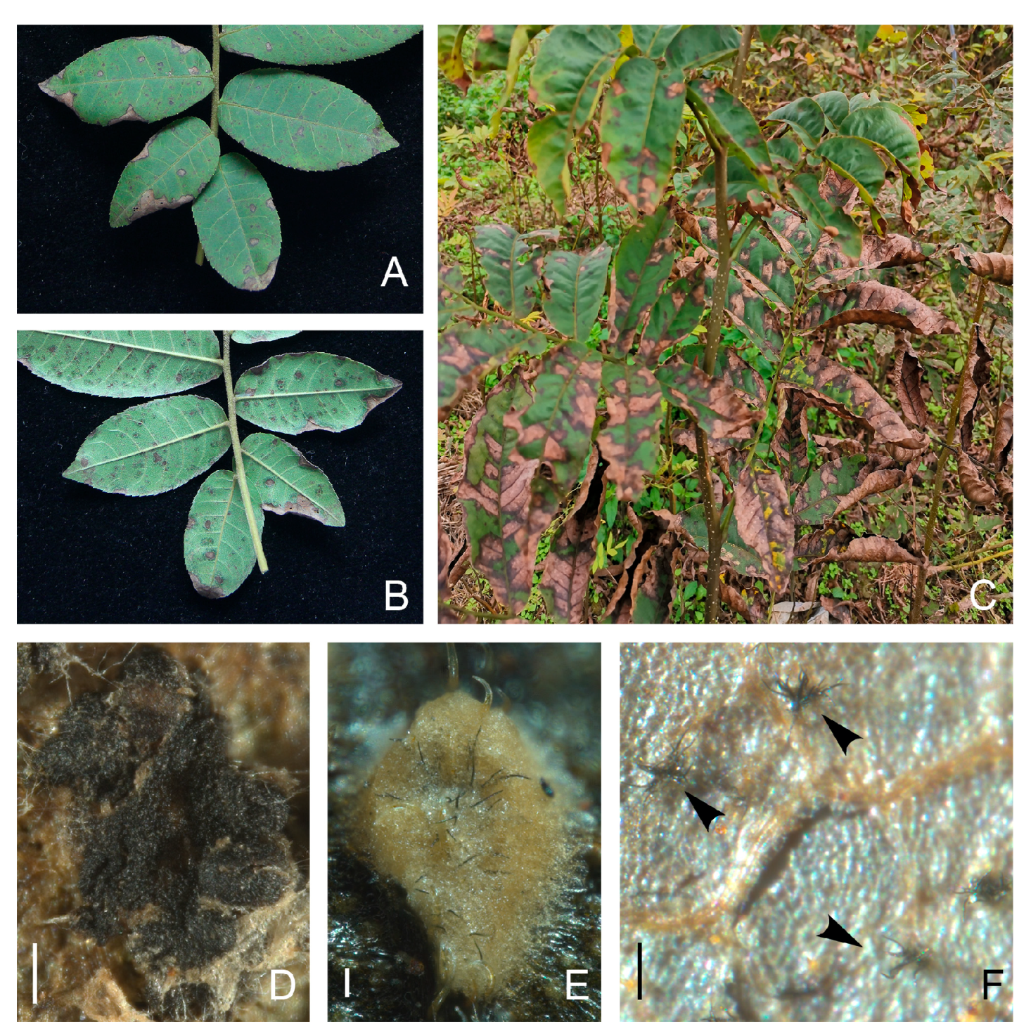
3.1. Symptomatology and Fungal Isolation

The typical symptoms of C. paliurus anthracnose observed in the present study were initially circular or irregularly shaped black-brown spots that gradually enlarged and then collapsed into necrotic lesions, turning grey, white or brown in the middle and dark brown at the edges (Figure 1A,B). Severe infection resulted in extensive early defoliation and eventually the death of the whole plant (Figure 1C). Under high-humidity conditions, typical structures of Colletotrichum, such as ascomata (Figure 1D), conidiomata (Figure 1E), and setae (Figure 1F), appeared on these lesions. In total, 337 isolates were recovered from symptomatic C. paliurus leaves that were collected in eight surveyed provinces of southern China. According to ITS sequence alignment, 331 isolates were identified as Colletotrichum spp. Other isolates belonging to Pestalotiopsis, Alternaria and Phomopsis were also isolated, but those were not further studied for the time being (Table A3).

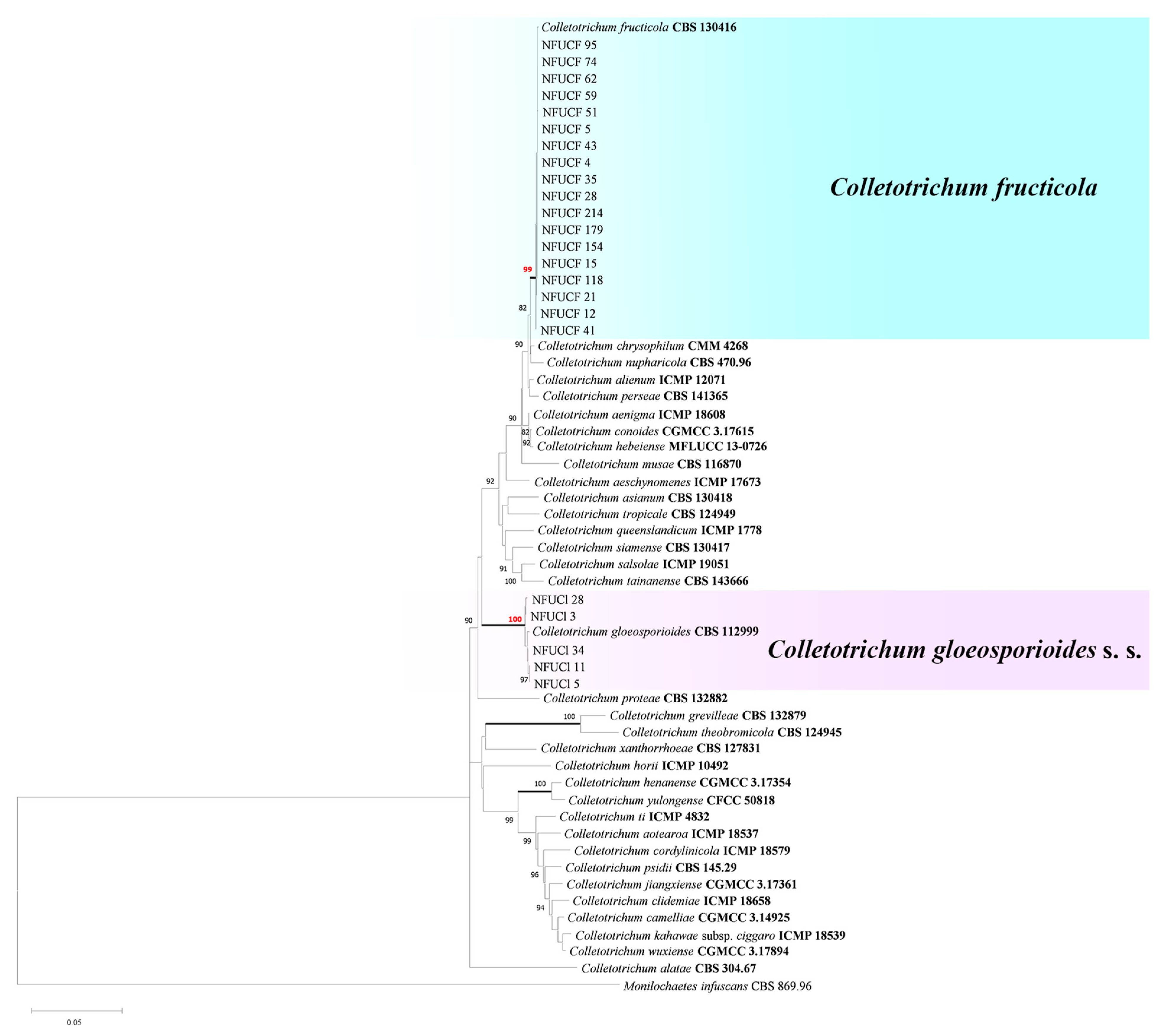
3.2. Molecular Identification and Phylogenetic Analyses

Based on the alignment of ITS and GADPH sequences and cultural characteristics, all Colletotrichum isolates were grouped into the Gloeosporioides (249 isolates), Acutatum (37 isolates), Orchidearum (32 isolates) and Magnum complexes (13 isolates). Subsequently, a subset of 43 isolates representing different geographic origins, phenotypic characteristics (conidial shape and size) and genetic diversity (ITS and GADPH sequence analysis) was selected for further investigation (Table A1 and Table A2).
For isolates in the Gloeosporioides complex, phylogenetic analyses of eight concatenated loci (ITS, GADPH, CHS-1, ACT, TUB, CAL, GS and ApMat) sequences were carried out with corresponding sequences from 39 authentic specimens (Table A1). The concatenated matrixes of the aligned dataset were composed of 3537 characters and gaps in the alignment. The GTR+G model was selected based on the AIC to reconstruct the ML tree. For BI analysis, the corresponding models were selected by MrModeltest: GTR+I+G for ITS; K80+G for GAPDH and ApMat; HKY+I+G for CHS-1; and GTR+G for ACT, TUB, CAL and GS. The isolates in the Gloeosporioides complex were clustered into two well-supported clades (Bv > 99% and Bayesian PP = 1.00): 18 isolates were grouped into the C. fructicola clade, and five were clustered with C. gloeosporioides s. s. (Figure 2).
To identify the Colletotrichum species within the Acutatum, Magnum and Orchidearum complexes, a dataset of six combined genes (ITS, GADPH, CHS-1, ACT, TUB and HIS3) from 49 authentic specimens was used, and the dataset comprised 1905 characters after alignment. The ML tree was reconstructed utilizing the GTR+G+I model. The best models for BI were found by MrModeltest: GTR+I+G for ITS, CHS-1 and HIS3, K80+G for GADPH, GTR+G for ACT, and HKY+I+G for TUB. The isolates in the Acutatum complex could be well defined as C. nymphaeae and C. godetiae because five isolates clustered together with the C. nymphaeae ex-type strain CBS 515.78 with strong support (99% Bv/1.00 PP), and five isolates clustered in another highly supported clade (99% BP/1.00 PP) with the C. godetiae type strain CBS 133.44. Among the isolates in the Orchidearum complex, five were grouped in C. plurivorum Damm, Alizadeh & Toy. Sato (98% Bv/1.00 PP), whereas the other five isolates were clustered with C. sojae Damm & Alizadeh (99% Bv/1.00 PP). Additionally, the remaining five isolates clustered with the C. brevisporum authentic strain BCC 38876 with high support (99% Bv/1.00 PP) in the Magnum clade (Figure 3).

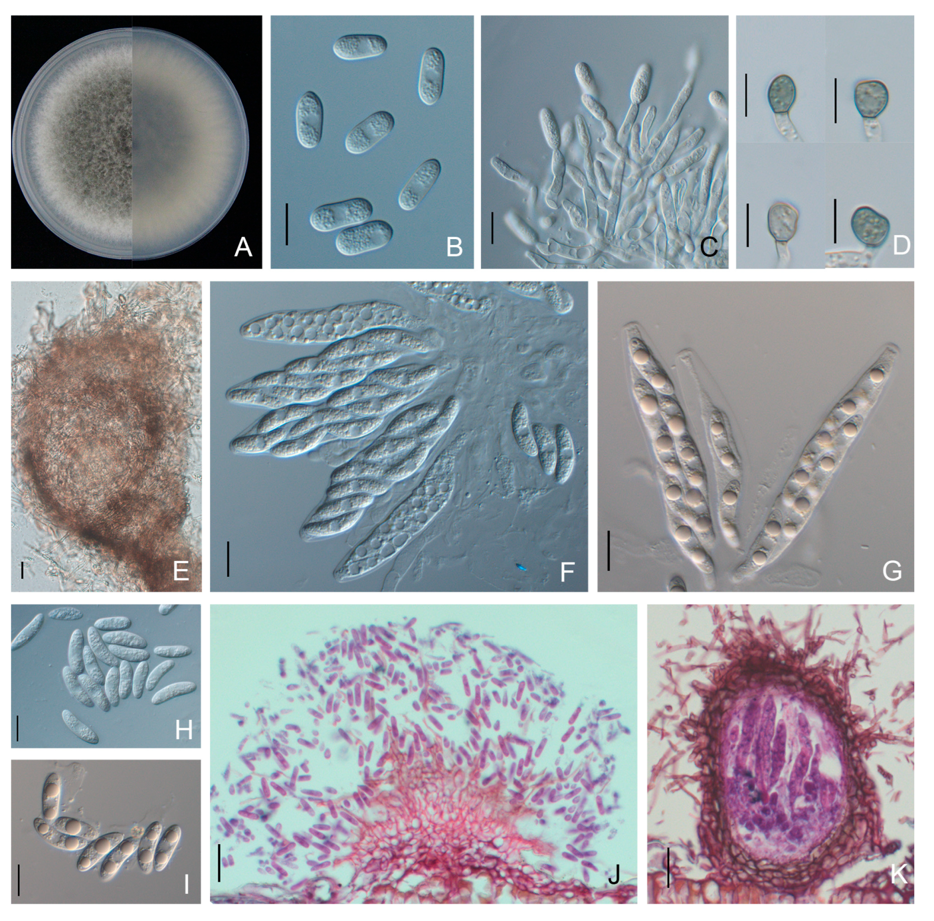
3.3. Morphological Characteristics

Colonies of C. brevisporum isolates on PDA were dark grey with grey aerial mycelium and edges (Figure 4). Yellowish conidial conidiomata formed across the colony after 14 days of incubation at 25 °C. The conidia were cylindrical to clavate, smooth-walled, hyaline, aseptate, and rounded at both ends (few one end rounded to acute), measuring 10.6 to 17.3 × 5.0 to 6.8 μm (average 14.1 ± 1.2 × 5.8 ± 0.3 μm). The appressoria were globose, puce, with an entire or lobed margin, and 7.5 to 17.5 × 5.6 to 13.2 μm (average 10.5 ± 1.7 × 8.9 ± 0.9 μm) in size (Table 2). Conidiophores and setae formed from a brown stroma. The setae were dark brown, straight to slightly curved, opaque, tip acute, and base cylindrical (Figure 4). The mycelial growth rate was 12.7 ± 0.2 mm per day on PDA at 25 °C (Table 2).
Colonies of C. fructicola isolates were olive-grey with whitish edges on PDA, and the average growth rate was 14.4 ± 0.2 mm/day. Conidia were produced as brick-red masses and were hyaline, smooth-walled, aseptate, cylindrical with rounded ends, and 10.3 to 22.5 × 4.4 to 7.9 μm (average 13.5 ± 1.8 × 5.8 ± 0.5 μm) in size. Conidiophores were hyaline, simple to 2-septate, and unbranched. Appressoria were greyish brown to black and formed singularly, with ovoid to slightly irregular outlines, measuring 6.5 to 16.0 × 4.5 to 9.1 μm (average 9.5 ± 1.6 × 7.0 ± 0.9 μm). Asci were clavate, fasciculate and 8-spored. Ascospores were smooth-walled, hyaline, aseptate, partly guttulate, curved fusoid with rounded ends and 12.3 to 23.2 × 3.8 to 6.5 μm (average 17.7 ± 1.7 × 5.0 ± 0.6 μm) in size (Figure 5, Table 2).
Colonies of C. gloeosporioides s. s. on PDA were white to off-white with dense aerial mycelia and edges, and the average growth rate was 12.6 ± 0.5 mm/day. Conidia were cylindrical, straight with a few slightly curved, aseptate, hyaline, rounded at both ends, and were 13.1 to 22.7 × 4.5 to 6.3 μm (average 15.9 ± 1.1 × 5.5 ± 0.4 μm) in size. Brown-colored appressoria were ovoid to slightly irregular, with an entire margin measuring 7.2 to 12.5 × 6.0 to 10.3 μm (average 9.6 ± 1.0 × 7.2 ± 0.9 μm) (Figure 6, Table 2). Conidiophores and setae formed from a dark brown stroma. Setae were dark brown, straight to slightly curved, and opaque, with acute tip and cylindrical base (Figure 6).
The C. godetiae isolates exhibited dense and white colonies on PDA, and the average growth rate was 8.4 ± 0.2 mm/day. Conidia were produced in orange conidiomata and were aseptate, hyaline and fusiform, with one end rounded and one end rounded to acute, measuring 12.6 to 20.7 × 3.8 to 6.8 μm (average 15.9 ± 1.3 × 5.1 ± 0.4 μm). Conidiophores and setae formed from a brown stroma. Setae were dark brown, straight or curved, and opaque, with acute tip and cylindrical base. Sexual morphs were not observed (Figure 7). Appressoria were greyish brown to black, ovoid to globose, with entire or lobed margins, and 7.6 to 13.2 × 4.9 to 8.7 μm (average 9.5 ± 1.0 × 6.4 ± 0.7 μm) in size (Figure 7, Table 2).
Colletotrichum nymphaeae colonies on PDA were dense, olive-grey with a white margin after 6 days of incubation, and had similar morphological features to those of C. godetiae. Conidia were fusiform with one end rounded and one end rounded to acute, measuring 11.1 to 18.0 × 4.0 to 6.9 μm (average 14.5 ± 1.9 × 5.5 ± 0.9 μm). Appressoria were greyish brown to black, ovoid, with smooth margins, and 7.0 to 11.9 × 5.0 to 8.9 μm (average 9.1 ± 1.3 × 7.0 ± 1.1 μm) in size (Figure 8, Table 2). The average growth rate of Colletotrichum nymphaeae isolates on PDA was 9.8 ± 0.2 mm per day (Table 2).
Colonies of C. plurivorum isolates on PDA were olive-grey with white margins, and the average growth rate was 11.1 ± 0.1 mm/day. Conidia were aseptate, hyaline, cylindrical with rounded ends, and 12.1 to 20.2 × 5.0 to 7.7 μm (average 14.9 ± 1.6 × 6.2 ± 0.6 μm) in size. Conidiophores were hyaline, unbranched, and formed from a brown stroma. Setae were dark brown, straight and opaque, with acute tip and cylindrical base (Figure 9). Appressoria were globose to ovoid, puce, with an entire or lobed margin, and 8.6 to 20.5 × 6.4 to 12.5 μm (average 12.4 ± 2.2 × 9.2 ± 1.2 μm) in size. Ascomata were semi-immersed in agar medium, subglobose to pyriform, and dark brown. Asci were clavate or fasciculate, and eight-spored (Figure 9). Ascospores were hyaline, smooth-walled, aseptate, fusiform to curved fusoid, and rounded at both ends, measuring 13.6 to 23.0 × 5.0 to 9.3 μm (average 18.0 ± 1.6 × 7.0 ± 0.8 μm) (Figure 9, Table 2).
Colletotrichum sojae colonies on PDA were light orange-red with whitish aerial mycelia and edges, and the average growth rate was 14.7 ± 0.7 mm per day. Asexual morphs were not observed. Ascomata formed on PDA or SNA after two weeks of inoculation, which were subglobose to pyriform, dark brown, ostiolate, and semi-immersed in the agar medium. Asci were clavate or fasciculate and eight-spored (Figure 10). Ascospores were hyaline, aseptate, smooth-walled and curved fusoid with rounded ends, had granular content and measured 13.2 to 32.1 × 3.0 to 6.8 μm (average 24.4 ± 4.4 × 5.0 ± 0.7 μm). Appressoria were puce, ovoid with an entire or lobed margin and 7.2 to 18.0 × 5.7 to 9.4 μm (average 11.1 ± 1.7 × 7.4 ± 0.7 μm) in size (Figure 10, Table 2).

3.4. Pathogenicity Tests

The data from the pathogenicity tests are given in Table 3. Representative isolates of all seven Colletotrichum species produced typical symptoms of anthracnose on detached C. paliurus leaves, while the corresponding mock controls remained asymptomatic up to 10 dpi. Inoculation with C. fructicola, C. godetiae, C. gloeosporioides s. s., C. nymphaeae and C. sojae isolates led to the development of anthracnose symptoms on leaves through both wounding and nonwounding methods, whereas C. brevisporum and C. plurivorum ones exhibited weaker virulence and were only capable of infecting wounded leaves.
All isolates of Colletotrichum species showed a higher incidence and severity of disease on wounded leaves than on nonwounded leaves. Moreover, the isolates of the different species displayed distinct levels of aggressiveness. Among them, isolates of C. fructicola exhibited the highest aggressiveness on both detached leaves and intact plants. At 3 dpi, symptoms began to appear around the inoculation site, and then the lesion expanded rapidly. Dark-brown necrotic lesions were observed with typical Colletotrichum acervuli or ascomata after 15 dpi. The average lesion diameters (mean ± SE) were 25.3 ± 0.8 mm, 20.1 ± 1.3 mm and 9.4 ± 1.0 mm on wounded detached leaves, nonwounded detached leaves and intact plants, respectively. The virulence of C. gloeosporioides s. s., C. godetiae and C. nymphaeae isolates was weaker than that of C. fructicola isolates. In contrast, C. brevisporum, C. plurivorum and C. sojae were weakly aggressive to C. paliurus leaves, and at 15 dpi, the symptoms on nonwounded leaves and intact plants did not markedly spread or remained asymptomatic. There was no significant difference in pathogenicity between different strains of the same Colletotrichum species. Re-isolation from infected leaves was successful and confirmed by morphological and molecular identification, thus fulfilling Koch’s postulates.

4. Discussion

Anthracnose is the most prevalent foliar disease in all major C. paliurus-growing areas in southern China, causing enormous pecuniary losses under humid conditions and disease-favorable temperatures. Unfortunately, the species diversity of C. paliurus anthracnose pathogens in southern China remains largely unclear. In the present study, we collected and characterized 331 Colletotrichum isolates from eight C. paliurus planting provinces and identified seven species belonging to the Gloeosporioides, Acutatum, Magnum and Orchidearum complexes, demonstrating that diverse Colletotrichum species complexes can infect C. paliurus.
The ascomycete genus Colletotrichum includes important phytopathogens that cause anthracnose worldwide. Among them, three species belonging to the Gloeosporioides complex have been identified to induce C. paliurus anthracnose in China [10], whereas C. fructicola and C. gloeosporioides s. s. were identified in this study. The composition of Colletotrichum spp. causing C. paliurus anthracnose has been reported only in Jiangsu Province, where the Gloeosporioides complex was consistently reported as the most dominant instigator. Nevertheless, based on extensively collected samples, we found that the Gloeosporioides complex was not the only species complex causing C. paliurus anthracnose.
Taxonomic studies of Colletotrichum species have focused on disentangling intraspecific or specific taxa, traditionally according to phenotypic differences, mainly characteristics of cultural morphology, growth rate and microstructure morphs [34,35]. However, environmental factors and cultural conditions have major impacts on the stability of phenotypic traits. Furthermore, the morphological characteristics of Colletotrichum spp. within the species complex largely overlap; thus, phenotypical criteria are not adequate for a precise identification [34].
In terms of molecular characterization, for several fungi, the ITS region has been proposed as a universal DNA marker [36]; however, previous studies have proven that Colletotrichum species cannot be efficiently distinguished by ITS alone. Consequently, other loci such as GADPH, GS and ApMat must be considered. Hyde et al. [37] suggested that the GADPH gene is the most variable marker across multiple Colletotrichum species complexes. Several studies have recommended the use of the ApMat marker for the delimitation of cryptic species within the Gloeosporioides complex, yet Tovar-Pedraza [38] reported that C. jiangxiense and C. kahawae in the Gloeosporioides complex cannot be distinguished from each other by only ApMat sequence data, and their identification requires GS- and ApMat-concatenated phylogenetic analysis. In the present work, phylogenetic analyses of eight loci (ITS, GAPDH, ACT, CHS-1, TUB, CAL, GS and ApMat) in the Gloeosporioides complex and six loci (ITS, GAPDH, ACT, CHS-1, TUB, CAL and HIS3) in the other species complexes revealed that 43 representative isolates belonged to seven known Colletotrichum species, including C. brevisporum, C. fructicola, C. gloeosporioides s. s., C. godetiae, C. nymphaeae, C. plurivorum and C. sojae (Figure 2 and Figure 3). Furthermore, the morphological groups identified based on colony features, asexual or sexual morphs, and typical Colletotrichum conidial masses or ascomata that developed on inoculated leaves were entirely consistent with the results of the molecular data.
Pathogenicity tests revealed that all seven Colletotrichum species were pathogenic to wounding detached leaves of C. paliurus. When the foliar tissue was wounded, the incidence and severity of disease increased significantly. These results suggest that wounds may play an important role for pathogen penetration into the host. On average, species within the Gloeosporioides and Acutatum complexes produced larger lesions than those in the Magnum and Orchidearum complexes, which may be one of the notable factors contributing to the prevalence of the Gloeosporioides complex. Moreover, different Colletotrichum species had various degrees of aggressiveness on C. paliurus leaves. C. fructicola in the Gloeosporioides complex was the most aggressive species. Thus, a species-specific diagnosis is highly important for the prediction of relative aggressiveness; the species complex alone is not a sufficient indicator of pathogenicity or disease risk.
A previous study demonstrated that C. fructicola was the most common pathogen causing C. paliurus anthracnose in Jiangsu Province, China [10]. Similarly, in the present work, the dominant causal agent associated with C. paliurus anthracnose was C. fructicola on the basis of the highest isolation rate and aggressiveness levels. C. fructicola was originally isolated from coffee berries in Thailand [39]. It has been subsequently reported that C. fructicola could cause serious anthracnose infections in Australia, Brazil, China, Malaysia and the USA [18,40,41,42,43]. C. fructicola has been found on a broad range of host plants, such as fruit trees and economically important crops, including apple (Malus spp.), Citrus spp., mango (Mangifera indica), peach (Prunus persica), pear (Pyrus spp.), strawberry (Fragaria × ananassa) and tea (Camellia sinensis), possibly due to its parasitic and endophytic lifestyle [18,40,44,45,46,47,48,49].

5. Conclusions

This study presents the first large-scale survey of Colletotrichum species associated with C. paliurus anthracnose in southern China. It offers novel insights into the disease’s aetiology, including the first report of C. brevisporum, C. godetiae, C. nymphaeae, C. plurivorum and C. sojae associated with C. paliurus anthracnose. Considering the occurrence of several species involved in C. paliurus anthracnose, future research should take into account that the effective control of this disease may depend on the individual characteristics of each Colletotrichum species and their distribution in the C. paliurus planting areas. Furthermore, in view of the dominance of C. fructicola in major planting regions and its greater aggressiveness than other species, more epidemiological studies are needed to elucidate this pathological system.

Author Contributions

Conceptualization, F.-M.C.; methodology, X.-R.Z.; software, X.-R.Z.; validation, M.-J.Z.; formal analysis, M.-J.Z.; investigation, X.-R.Z.; resources, F.-M.C.; data curation, M.-J.Z.; writing—original draft preparation, X.-R.Z.; writing—review and editing, X.-R.Z. and M.-J.Z.; visualization, X.-R.Z. and M.-J.Z.; supervision, F.-M.C.; project administration, F.-M.C.; funding acquisition, F.-M.C. All authors have read and agreed to the published version of the manuscript.

Funding

This research was funded by Postgraduate Research and Practice Innovation Program of Jiangsu Province, grant number KYCX20_0875 and KYCX23_1222.

Data Availability Statement

Data are contained within the article and Appendix A.

Conflicts of Interest

The authors declare no conflict of interest.

Appendix A

Table A1. Isolates of Colletotrichum from leaves of wheel wingnut and various hosts examined in this study.
SpeciesCulture/Isolate aHostLocationGenBank Accession Number b
ITS GAPDH CHS ACT TUB CAL GS ApMat
Colletotrichum aenigma ICMP 18608 Persea americana Israel JX010244 JX010044 JX009774 JX009443 JX010389 JX009683 JX010078 KM360143
C. aeschynomenes ICMP 17673 Aeschynomene virginica USA JX010176 JX009930 JX009799 JX009483 JX010392 JX009721 JX010081 KM360145
C. alatae CBS 304.67 Dioscorea alata India JX010190 JX009990 JX009837 JX009471 JX010383 JX009738 JX010065 KC888932
C. alienum ICMP 12071 Malus domestica New Zealand JX010251 JX010028 JX009882 JX009572 JX010411 JX009654 JX010101 KM360144
C. aotearoa ICMP 18537 Coprosma sp. New Zealand JX010205 JX010005 JX009853 JX009564 JX010420 JX009611 JX010113 KC888930
C. asianum CBS 130418 Coffea arabica Thailand FJ972612 JX010053 JX009867 JX009584 JX010406 FJ917506 JX010096 FR718814
C. camelliae CGMCC 3.14925 Camellia sinensis China KJ955081 KJ954782 MZ799255 KJ954363 KJ955230 KJ954634 KJ954932 KJ954497
C. chrysophilumCMM 4268Musa sp.BrazilKX094252KX094183KX094083KX093982KX094285KX094063KX094204KX094325
C. clidemiae ICMP 18658 Clidemia hirta USA JX010265 JX009989 JX009877 JX009537 JX010438 JX009645 JX010129 KC888929
C. conoidesCGMCC 3.17615Chili pepperChinaKP890168KP890162KP890156KP890144KP890174KP890150--
C. cordylinicola ICMP 18579 Cordyline fruticosa Thailand JX010226 JX009975 JX009864 HM470235 JX010440 HM470238 JX010122 JQ899274
C. fructicolaCBS 130416Coffea arabicaThailandJX010165JX010033JX009866FJ907426JX010405FJ917508JX010095JQ807838
NFUCF-4Cyclocarya paliurusSichuan, ChinaOR056200OR069484OR073817OR096449OR073835OR096522OR098645OR105821
NFUCF-5Cy. paliurusGuizhou, ChinaOR056201OR069485OR073818OR096450OR073836OR096523OR098646OR105822
NFUCF-12Cy. paliurusSichuan, ChinaOR056202OR069486OR073819OR096451OR073837OR096524OR098647OR105823
NFUCF-15 cCy. paliurusGuangxi, ChinaOR056203OR069487OR073820OR096452OR073838OR096525OR098648OR105824
NFUCF-21Cy. paliurusFujian, ChinaOR056204OR069488OR073821OR096453OR073839OR096526OR098649OR105825
NFUCF-28Cy. paliurusHubei, ChnaOR056205OR069489OR073822OR096454OR073840OR096527OR098650OR105826
NFUCF-35Cy. paliurusJiangxi, ChinaOR056206OR069490OR073823OR096455OR073841OR096528OR098651OR105827
NFUCF-41Cy. paliurusGuangxi, ChinaOR056207OR069491OR073824OR096456OR073842OR096529OR098652OR105828
NFUCF-43Cy. paliurusFujian, ChinaOR056208OR069492OR073825OR096457OR073843OR096530OR098653OR105829
NFUCF-51Cy. paliurusJiangxi, ChinaOR056209OR069493OR073826OR096458OR073844OR096531OR098654OR105830
NFUCF-59Cy. paliurusGuizhou, ChinaOR056210OR069494OR073827OR096459OR073845OR096532OR098655OR105831
NFUCF-62 cCy. paliurusHunan, ChinaOR056211OR069495OR073828OR096460OR073846OR096533OR098656OR105832
NFUCF-74Cy. paliurusGuangxi, ChinaOR056212OR069496OR073829OR096461OR073847OR096534OR098657OR105833
NFUCF-95Cy. paliurusZhejiang, ChinaOR056213OR069497OR073830OR096462OR073848OR096535OR098658OR105834
NFUCF-118Cy. paliurusGuizhou, ChinaOR056214OR069498OR073831OR096463OR073849OR096536OR098659OR105835
NFUCF-154Cy. paliurusHunan, ChinaOR056215OR069499OR073832OR096464OR073850OR096537OR098660OR105836
NFUCF-179Cy. paliurusJiangxi, ChinaOR056216OR069500OR073833OR096465OR073851OR096538OR098661OR105837
NFUCF-214 cCy. paliurusZhejiang, ChinaOR056217OR069501OR073834OR096466OR073852OR096539OR098662OR105838
C. gloeosporioidesCBS 112999Citrus sinensisItalyJX010152JX010056JX009818JX009531JX010445JX009731JX010085JQ807843
NFUCl-3Cy. paliurusGuangxi, ChinaOR064046OR069502OR073853OR096419OR096467OR096540OR098663OR105839
NFUCl-5 cCy. paliurusJiangxi, ChinaOR064047OR069503OR073854OR096420OR096468OR096541OR098664OR105840
NFUCl-11 cCy. paliurusGuizhou, ChinaOR064048OR069504OR073855OR096421OR096469OR096542OR098665OR105841
NFUCl-28Cy. paliurusGuizhou, ChinaOR064049OR069505OR073856OR096422OR096470OR096543OR098666OR105842
NFUCl-34 cCy. paliurusHunan, ChinaOR064050OR069506OR073857OR096423OR096471OR096544OR098667OR105843
C. grevilleae CBS 132879 Grevillea sp. Italy KC297078 KC297010 KC296987 KC296941 KC297102 KC296963 KC297033 -
C. hebeienseMFLUCC13–0726Vitis viniferaChinaKF156863KF377495KF289008KF377532KF288975---
C. henanense CGMCC 3.17354 Ca. sinensis China KJ955109 KJ954810 MZ799256 KM023257 KJ955257 KJ954662 KJ954960 KJ954524
C. horii ICMP 10492 Diospyros kaki Japan GQ329690 GQ329681 JX009752 JX009438 JX010450 JX009604 JX010137 JQ807840
C. jiangxiense CGMCC 3.17361 Ca. sinensis China KJ955149KJ954850MZ799257KJ954427OK236389KJ954701KJ955000KJ954561
C. kahawae subsp. ciggaro ICMP 18539 Olea europaea Australia JX010230 JX009966 JX009800 JX009523 JX010434 JX009635 JX010132 -
C. musae CBS 116870 Musa sp. USA JX010146 JX010050 JX009896 JX009433 HQ596280 JX009742 JX010103 KC888926
C. nupharicola CBS 470.96 Nuphar lutea USA JX010187 JX009972 JX009835 JX009437 JX010398 JX009663 JX010088 JX145319
C. perseaeCBS 141365AvocadoIsraelKX620308KX620242MZ799260KX620145KX620341KX620206KX620275KX620177
C. proteae CBS 132882 Protea sp. South Africa KC297079 KC297009 KC296986 KC296940 KC297101 KC296960 KC297032 -
C. psidii CBS 145.29 Psidium sp. Italy JX010219 JX009967 JX009901 JX009515 JX010443 JX009743 JX010133 KC888931
C. queenslandicum ICMP 1778 Carica papaya Australia JX010276 JX009934 JX009899 JX009447 JX010414 JX009691 JX010104 KC888928
C. salsolae ICMP 19051 Salsola tragus Hungary JX010242 JX009916 JX009863 JX009562 JX010403 JX009696 JX010093 KC888925
C. siamense CBS 130417 Coffea arabica Thailand JX010171 JX009924 JX009865 FJ907423 JX010404 FJ917505 JX010094 JQ899289
C. tainanenseCBS 143666Capsicum annuumChinaMH728818MH728823MH805845MH781475MH846558-MH748259MH728836
C. theobromicola CBS 124945 Theobroma cacao Panama JX010294 JX010006 JX009869 JX009444 JX010447 JX009591 JX010139 KC790726
C. ti ICMP 4832 Cordyline sp. New Zealand JX010269 JX009952 JX009898 JX009520 JX010442 JX009649 JX010123 KM360146
C. tropicale CBS 124949 Theobroma cacao Panama JX010264 JX010007 JX009870 JX009489 JX010407 JX009719 JX010097 KC790728
C. wuxienseCGMCC 3.17894Camellia sinensisChinaKU251591KU252045KU251939KU251672KU252200KU251833KU252101KU251722
C. xanthorrhoeae CBS 127831 Xanthorrhoea preissii Australia JX010261 JX009927 JX009823 JX009478 JX010448 JX009653 JX010138 KC790689
C. yulongenseCFCC 50818Vaccinium dunalianumChinaMH751507MK108986MH793605MH777394MK108987MH793604MK108988-
Monilochaetes infuscansCBS 869.96Ipomoea batatasSouth AfricaJQ005780JX546612JQ005801JQ005843JQ005864---
a Culture numbers in bold type represent ex-type or other authentic specimens. CBS 869.96 (Monilochaetes infuscans) was added as an outgroup. b Sequences in italics were generated in this study. “-”indicates missing data. c Isolates used for macroscopic and microscopic characterization and virulence tests.
Table A2. Strains of Colletotrichum excluded from the C. gloeosporioides species complex. Details are provided about clade, host and location, and GenBank accessions of the sequences generated.
SpeciesCulture/Isolate aCladeHostLocationGenBank Accession Number b
ITSGAPDHCHS-1HIS3ACTTUB2
C. abscissumCOAD 1877AcutatumCitrus sinensis cv. PeraBrazilKP843126KP843129KP843132KP843138KP843141KP843135
C. acerbum CBS 128530 Acutatum Malus domestica New Zealand JQ948459 JQ948790 JQ949120 JQ949450 JQ949780 JQ950110
C. acutatum CBS 112996 Acutatum Carica papaya Australia JQ005776 JQ948677 JQ005797 JQ005818 JQ005839 JQ005860
C. australe CBS 116478 Acutatum Trachycarpus fortunei South Africa JQ948455 JQ948786 JQ949116 JQ949446 JQ949776 JQ950106
C. brevisporumBCC 38876MagnumNeoregalia sp.ThailandJN050238JN050227MZ799287MZ673841JN050216JN050244
NFUCB-2 cMagnumCyclocarya paliurusHunan, ChinaOR064061OR069517OR073868OR096507OR096434OR096482
NFUCB-6 cMagnumCy. PaliurusHunan, ChinaOR064062OR069518OR073869OR096508OR096435OR096483
NFUCB-9MagnumCy. PaliurusHunan, ChinaOR064063OR069519OR073870OR096509OR096436OR096484
NFUCB-11MagnumCy. PaliurusHunan, ChinaOR064064OR069520OR073871OR096510OR096437OR096485
NFUCB-12 cMagnumCy. PaliurusGuizhou, ChinaOR064065OR069521OR073872OR096511OR096438OR096486
C. brisbanense CBS 292.67 Acutatum Capsicum annuum Australia JQ948291 JQ948621 JQ948952 JQ949282 JQ949612 JQ949942
C. cacaoCBS 119297MagnumTheobroma cacaoCosta RicaMG600772MG600832MG600878MG600916MG600976MG601039
C. cairnsenseBRIP 63642AcutatumCapsicum annuumAustraliaKU923672KU923704KU923710KU923722KU923716KU923688
C. cattleyicolaCBS 170.49OrchidearumCattleya sp.BelgiumMG600758MG600819MG600866MG600905MG600963MG601025
C. chrysanthemi IMI 364540 Acutatum Chrysanthemum coronarium China JQ948273 JQ948603 JQ948934 JQ949264 JQ949594 JQ949924
C. cliviicolaCBS 125375OrchidearumClivia miniataChinaMG600733MG600795MG600850MG600892MG600939MG601000
C. cosmi CBS 853.73 Acutatum Cosmos sp. Netherlands JQ948274 JQ948604 JQ948935 JQ949265 JQ949595 JQ949925
C. costaricense CBS 330.75 Acutatum Coffea arabica, cv. Typica Costa Rica JQ948180 JQ948510 JQ948841 JQ949171 JQ949501 JQ949831
C. cuscutae IMI 304802 Acutatum Cuscuta sp. Dominica JQ948195 JQ948525 JQ948856 JQ949186 JQ949516 JQ949846
C. eriobotryaeGLMC 1935AcutatumEriobotrya japonicaChinaMF772487MF795423MN191653MN191658MN191648MF795428
C. fioriniaeCBS 128517AcutatumFiorinia externaUSAJQ948292JQ948622JQ948953JQ949283JQ949613JQ949943
C. godetiae CBS 133.44 Acutatum Clarkia hybrida cv. Kelvon Glory Denmark JQ948402 JQ948733 JQ949063 JQ949393 JQ949723 JQ950053
NFUCo-1 cAcutatumCy. paliurusGuizhou, China OR064051 OR069507 OR073858 OR096497 OR096424 OR096472
NFUCo-4 cAcutatumCy. paliurusJiangxi, China OR064052 OR069508 OR073859 OR096498 OR096425 OR096473
NFUCo-5 cAcutatumCy. paliurusHunan, China OR064053 OR069509 OR073860 OR096499 OR096426 OR096474
NFUCo-8AcutatumCy. paliurusHunan, China OR064054 OR069510 OR073861 OR096500 OR096427 OR096475
NFUCo-11AcutatumCy. paliurusHunan, China OR064055 OR069511 OR073862 OR096501 OR096428 OR096476
C. guajavae IMI 350839 Acutatum Psidium guajava India JQ948270 JQ948600 JQ948931 JQ949261 JQ949591 JQ949921
C. indonesiense CBS 127551 Acutatum Eucalyptus sp. Indonesia JQ948288 JQ948618 JQ948949 JQ949279 JQ949609 JQ949939
C. javanenseCBS 144963AcutatumCapsicum annuumIndonesiaMH846576MH846572MH846573MH846571MH846575MH846574
C. johnstonii CBS 128532 Acutatum Solanum lycopersicum New Zealand JQ948444 JQ948775 JQ949105 JQ949435 JQ949765 JQ950095
C. kinghornii CBS 198.35 Acutatum Phormium sp. UK JQ948454 JQ948785 JQ949115 JQ949445 JQ949775 JQ950105
C. laticiphilum CBS 112989 Acutatum Hevea brasiliensis India JQ948289 JQ948619 JQ948950 JQ949280 JQ949610 JQ949940
C. limetticola CBS 114.14 Acutatum Citrus aurantifolia USA, Florida JQ948193 JQ948523 JQ948854 JQ949184 JQ949514 JQ949844
C. lobatumIMI 79736MagnumPiper catalpaefoliumTrinidadMG600768MG600828MG600874MG600912MG600972MG601035
C. lupini CBS 109225 Acutatum Lupinus albus Ukraine JQ948155 JQ948485 JQ948816 JQ949146 JQ949476 JQ949806
C. magnumCBS 519.97MagnumCitrullus lanatusUSAMG600769MG600829MG600875MG600913MG600973MG601036
C. melonis CBS 159.84 Acutatum Cucumis melo Brazil JQ948194 JQ948524 JQ948855 JQ949185 JQ949515 JQ949845
C. merremiaeCBS 124955MagnumMerremia umbellataPanamaMG600765MG600825MG600872MG600910MG600969MG601032
C. monsteraeLC13871OrchidearumMonstera deliciosaChinaMZ595897MZ664121MZ799351MZ673917MZ664195MZ674015
C. musicolaCBS 132885OrchidearumMusa sp.MexicoMG600736MG600798MG600853MG600895MG600942MG601003
C. nymphaeae CBS 515.78 Acutatum Nymphaea alba Netherlands JQ948197 JQ948527 JQ948858 JQ949188 JQ949518 JQ949848
NFUCN-2 cAcutatumCy. paliurusGuangxi, ChinaOR064071OR069527OR073878OR096517OR096444OR096492
NFUCN-5 cAcutatumCy. paliurusHunan, ChinaOR064072OR069528OR073879OR096518OR096445OR096493
NFUCN-7AcutatumCy. paliurusHunan, ChinaOR064073OR069529OR073880OR096519OR096446OR096494
NFUCN-11 cAcutatumCy. paliurusGuizhou, ChinaOR064074OR069530OR073881OR096520OR096447OR096495
NFUCN-14AcutatumCy. paliurusJiangxi, ChinaOR064075OR069531OR073882OR096521OR096448OR096496
C. orchidearumCBS 135131OrchidearumDendrobium nobileNetherlandsMG600738MG600800MG600855MG600897MG600944MG601005
C. panamenseCBS 125386MagnumMerremia umbellataPanamaMG600766MG600826MG600873MG600911MG600970MG601033
C. paxtonii IMI 165753 Acutatum Musa sp. Saint Lucia JQ948285 JQ948615 JQ948946 JQ949276 JQ949606 JQ949936
C. phormii CBS 118194 Acutatum Phormium sp. Germany JQ948446 JQ948777 JQ949107 JQ949437 JQ949767 JQ950097
C. piperisIMI 71397OrchidearumPiper nigrumMalaysiaMG600760MG600820MG600867MG600906MG600964MG601027
C. plurivorumCBS 125474OrchidearumCoffea sp.VietnamMG600718MG600781MG600841MG600887MG600925MG600985
NFUCP-3 cOrchidearumCy. paliurusGuizhou, ChinaOR064066OR069522OR073873OR096512OR096439OR096487
NFUCP-5 cOrchidearumCy. paliurusHunan, ChinaOR064067OR069523OR073874OR096513OR096440OR096488
NFUCP-7OrchidearumCy. paliurusHunan, ChinaOR064068OR069524OR073875OR096514OR096441OR096489
NFUCP-8OrchidearumCy. paliurusHunan, ChinaOR064069OR069525OR073876OR096515OR096442OR096490
NFUCP-13 cOrchidearumCy. paliurusJiangxi, ChinaOR064070OR069526OR073877OR096516OR096443OR096491
C. pyricola CBS 128531 Acutatum Pyrus communis New Zealand JQ948445 JQ948776 JQ949106 JQ949436 JQ949766 JQ950096
C. reniformeLC8230OrchidearumSmilax cocculoidesChinaMZ595847MZ664110MZ799290MZ673867MZ664145MZ673968
C. rhombiforme CBS 129953 Acutatum Olea europaea Portugal JQ948457 JQ948788 JQ949118 JQ949448 JQ949778 JQ950108
C. salicis CBS 607.94 Acutatum Salix sp. Netherlands JQ948460 JQ948791 JQ949121 JQ949451 JQ949781 JQ950111
C. schimaeLC13880AcutatumSchima sp.ChinaMZ595885MZ664105MZ799347MZ673905MZ664183MZ674003
C. scovillei CBS 126529 Acutatum Capsicum sp. Indonesia JQ948267 JQ948597 JQ948928 JQ949258 JQ949588 JQ949918
C. simmondsii CBS 122122 Acutatum Carica papaya Australia JQ948276 JQ948606 JQ948937 JQ949267 JQ949597 JQ949927
C. sloanei IMI 364297 Acutatum Theobroma cacao Malaysia JQ948287 JQ948617 JQ948948 JQ949278 JQ949608 JQ949938
C. sojaeATCC 62257OrchidearumGlycine maxUSAMG600749MG600810MG600860MG600899MG600954MG601016
NFUCS-1 cOrchidearumCy. paliurusJiangxi, ChinaOR064056OR069512OR073863OR096502OR096429OR096477
NFUCS-3 cOrchidearumCy. paliurusHunan, ChinaOR064057OR069513OR073864OR096503OR096430OR096478
NFUCS-6OrchidearumCy. paliurusHunan, ChinaOR064058OR069514OR073865OR096504OR096431OR096479
NFUCS-10 cOrchidearumCy. paliurusGuizhou, ChinaOR064059OR069515OR073866OR096505OR096432OR096480
NFUCS-15OrchidearumCy. paliurusFujian, ChinaOR064060OR069516OR073867OR096506OR096433OR096481
C. tamarilloi CBS 129814 Acutatum Solanum betaceum Colombia JQ948184 JQ948514 JQ948845 JQ949175 JQ949505 JQ949835
C. vittalenseCBS 181.82OrchidearumTheobroma cacaoIndiaMG600734MG600796MG600851MG600893MG600940MG601001
C. walleri CBS 125472 Acutatum Coffea sp. Vietnam JQ948275 JQ948605 JQ948936 JQ949266 JQ949596 JQ949926
Monilochaetes infuscansCBS 869.96outgroupIpomoea batatasSouth AfricaJQ005780JX546612JQ005801JQ005822JQ005843JQ005864
a Culture numbers in bold type represent ex-type or other authentic specimens. CBS 869.96 (Monilochaetes infuscans) was added as an outgroup. b Sequences in italics were generated in this study. c Isolates used for phenotypic analysis and virulence tests.
Table A3. Location information and incidence rate statistics of the investigated area.
ProvinceCounty/LocationLeaf
Samples
Latitude (N)Longitude (E)
FujianXiapu427°03′08″119°56′33″
Jianyang627°33′08″117°47′03″
GuangxiLongsheng 726°01′13″109°55′08″
GuizhouLipin1226°06′50″109°11′08″
HubeiYidu630°26′04″111°19′54″
Sui632°11′43″113°16′11″
HunanJianghua Yao nationality1324°54′01″112°06′43″
JiangxiJinggangshan 626°42′03″114°17′47″
Shangrao728°49′54″118°11′07″
SichuanXuyong528°09′11″105°23′54″
ZhejiangLanxi1129°08′50″119°23′28″

References

  1. Zhao, W.; Tang, D.; Yuan, E.; Wang, M.; Zhang, Q.; Liu, Y.; Shen, B.; Chen, J.; Yin, Z. Inducement and cultivation of novel red Cyclocarya paliurus callus and its unique morphological and metabolic characteristics. Ind. Crops Prod. 2020, 147, 30–33. [Google Scholar] [CrossRef]
  2. Yang, Z.; Wang, J.; Li, J.; Xiong, L.; Chen, H.; Liu, X.; Wang, N.; Ouyang, K.; Wang, W. Antihyperlipidemic and hepatoprotective activities of polysaccharide fraction from Cyclocarya paliurus in high-fat emulsion-induced hyperlipidaemic mice. Carbohydr. Polym. 2018, 183, 11–20. [Google Scholar] [CrossRef] [PubMed]
  3. Li, Q.; Hu, J.; Nie, Q.; Chang, X.; Fang, Q.; Xie, J.; Li, H.; Nie, S.P. Hypoglycemic mechanism of polysaccharide from Cyclocarya paliurus leaves in type 2 diabetic rats by gut microbiota and host metabolism alteration. Sci. China Life Sci. 2020, 64, 117–132. [Google Scholar] [CrossRef] [PubMed]
  4. Shen, Y.; Peng, Y.; Zhu, X.; Li, H.; Zhang, L.; Kong, F.; Wang, J.; Yu, D. The phytochemicals and health benefits of Cyclocarya paliurus (Batalin) Iljinskaja. Front. Nutr. 2023, 10, 1158158. [Google Scholar] [CrossRef] [PubMed]
  5. Lei, X.; Hu, W.B.; Yang, Z.W.; Chen, H.; Wang, N.; Liu, X.; Wang, W. Enzymolysis-ultrasonic assisted extraction of flavanoid from Cyclocarya paliurus (Batal) Iljinskaja: HPLC profile, antimicrobial and antioxidant activity. Ind. Crops Prod. 2019, 130, 615–626. [Google Scholar]
  6. Tu, W.C.; Luo, H.R.; Yuan, E.; Sakah, J.; Yang, Q.Y.; Xiao, W.L.; Zheng, Y.T.; Liu, M.F. Triterpene constituents from the fruits of Cyclocarya paliurus and their anti-HIV-1 IIIB activity. Nat. Prod. Res. 2022, 37, 1787–1796. [Google Scholar] [CrossRef] [PubMed]
  7. Zhou, M.; Quek, S.Y.; Shang, X.; Fang, S. Geographical variations of triterpenoid contents in Cyclocarya paliurus leaves and their inhibitory effects on HeLa cells. Ind. Crops Prod. 2021, 162, 113314. [Google Scholar] [CrossRef]
  8. Li, X.; Fu, X.; Shang, X.; Yang, W.; Fang, S. Natural population structure and genetic differentiation for heterodicogamous plant: Cyclocarya paliurus (Batal.) Iljinskaja (Juglandaceae). Tree Genet. Genomes 2017, 13, 80. [Google Scholar] [CrossRef]
  9. Zheng, X.R.; Liu, C.L.; Zhang, M.J.; Shang, X.L.; Fang, S.Z.; Chen, F.M. First report of leaf blight of Cyclocarya paliurus caused by Nigrospora sphaerica in China. Crop Prot. 2020, 140, 105453. [Google Scholar] [CrossRef]
  10. Zheng, X.R.; Zhang, M.J.; Shang, X.L.; Fang, S.Z.; Chen, F.M. Etiology of Cyclocarya paliurus Anthracnose in Jiangsu Province, China. Front. Plant Sci. 2021, 11, 613499. [Google Scholar] [CrossRef]
  11. Wang, Y.; Chen, J.; Xu, X.; Cheng, J.; Zheng, L.; Huang, J.; Li, D.W. Identification and Characterization of Colletotrichum Species Associated with Anthracnose Disease of Camellia oleifera in China. Plant Dis. 2020, 104, 474–482. [Google Scholar] [CrossRef] [PubMed]
  12. Hyde, K.; Cai, L.; Cannon, P.; Crouch, J.A.; Crous, P.; Damm, U.; Goodwin, P.H.; Chen, H.; Johnston, P.; Jones, E.; et al. Colletotrichum—Names in current use. Fungal Divers. 2009, 39, 147–182. [Google Scholar]
  13. Weir, B.S.; Johnston, P.R.; Damm, U. The Colletotrichum gloeosporioides species complex. Stud. Mycol. 2012, 73, 115–180. [Google Scholar] [CrossRef]
  14. Liu, F.; Ma, Z.Y.; Hou, L.; Diao, Y.; Wu, W.; Damm, U.; Song, S.; Cai, L. Updating species diversity of Colletotrichum, with a phylogenomic overview. Stud. Mycol. 2022, 101, 1–56. [Google Scholar] [CrossRef]
  15. Damm, U.; Sato, T.; Alizadeh, A.; Groenewald, J.Z.; Crous, P.W. The Colletotrichum dracaenophilum, C. magnum and C. orchidearum species complexes. Stud. Mycol. 2019, 92, 1–46. [Google Scholar] [CrossRef] [PubMed]
  16. Damm, U.; Cannon, P.F.; Woudenberg, J.H.C.; Crous, P.W. The Colletotrichum acutatum species complex. Stud. Mycol. 2012, 73, 37–113. [Google Scholar] [CrossRef] [PubMed]
  17. Liu, F.; Weir, B.; Damm, U.; Crous, P.; Wang, Y.; Liu, B.; Wang, M.; Zhang, M.; Cai, L. Unravelling Colletotrichum species associated with Camellia: Employing ApMat and GS loci to resolve species in the C. gloeosporioides complex. Persoonia 2016, 35, 63–86. [Google Scholar] [CrossRef] [PubMed]
  18. Fu, M.; Crous, P.W.; Bai, Q.; Zhang, P.F.; Xiang, J.; Guo, Y.S.; Zhao, F.F.; Yang, M.M.; Hong, N.; Xu, W.X.; et al. Colletotrichum species associated with anthracnose of Pyrus spp. in China. Persoonia 2019, 42, 1–35. [Google Scholar] [CrossRef]
  19. Cai, L.; Hyde, K.; Taylor, P.; Weir, B.; Waller, J.; Abang, M.; Zhang, J.Z.; Yang, Y.L.; Phoulivong, S.; Liu, Z.Y. A polyphasic approach for studying Colletotrichum. Fungal Divers. 2009, 39, 183–204. [Google Scholar]
  20. Gardes, M.; Bruns, T. ITS primers with enhanced specificity for basidiomycetes—Application to the identification of mycorrhizae and rusts. Mol. Ecol. 1993, 2, 113–118. [Google Scholar] [CrossRef]
  21. White, T.; Bruns, T.; Lee, S.; Taylor, J.; Innis, M.; Gelfand, D.; Sninsky, J. Amplification and direct sequencing of fungal ribosomal RNA genes for phylogenetics. In PCR Protocols: A Guide to Methods and Applications; Academic Press: Cambridge, MA, USA, 1990; pp. 315–322. [Google Scholar]
  22. Guerber, J.C.; Liu, B.; Correll, J.C.; Johnston, P.R. Characterization of diversity in Colletotrichum acutatum sensu lato by sequence analysis of two gene introns, mtDNA and intron RFLPs, and mating compatibility. Mycologia 2003, 95, 872–895. [Google Scholar] [CrossRef] [PubMed]
  23. Carbone, I.; Kohn, L.M. A method for designing primer sets for speciation studies in filamentous ascomycetes. Mycologia 1999, 91, 553–556. [Google Scholar] [CrossRef]
  24. Crous, P.W.; Groenewald, J.Z.; Risede, J.M.; Simoneau, P.; Hywel-Jones, N.L. Calonectria species and their Cylindrocladium anamorphs: Species with sphaeropedunculate vesicles. Stud. Mycol. 2004, 50, 415–430. [Google Scholar]
  25. O’Donnell, K.; Cigelnik, E. Two divergent intragenomic rDNA ITS2 types within a monophyletic lineage of the fungus Fusarium are nonorthologous. Mol. Phylogenet. Evol. 1997, 7, 103–116. [Google Scholar] [CrossRef]
  26. Glass, N.L.; Donaldson, G.C. Development of primer sets designed for use with the PCR to amplify conserved genes from filamentous ascomycetes. Appl. Environ. Microbiol. 1995, 61, 1323–1330. [Google Scholar] [CrossRef]
  27. O’Donnell, K.; Nirenberg, H.I.; Aoki, T.; Cigelnik, E. A Multigene phylogeny of the Gibberella fujikuroi species complex: Detection of additional phylogenetically distinct species. Mycoscience 2000, 41, 61–78. [Google Scholar] [CrossRef]
  28. Silva, D.N.; Talhinhas, P.; Varzea, V.; Cai, L.; Paulo, O.S.; Batista, D. Application of the Apn2/MAT locus to improve the systematics of the Colletotrichum gloeosporioides complex: An example from coffee (Coffea spp.) hosts. Mycologia 2012, 104, 396–409. [Google Scholar] [CrossRef]
  29. Vaidya, G.; Lohman, D.J.; Meier, R. SequenceMatrix: Concatenation software for the fast assembly of multi-gene datasets with character set and codon information. Cladistics 2011, 27, 171–180. [Google Scholar] [CrossRef]
  30. Kumar, S.; Stecher, G.; Li, M.; Knyaz, C.; Tamura, K. MEGA X: Molecular Evolutionary Genetics Analysis across Computing Platforms. Mol. Biol. Evol. 2018, 35, 1547–1549. [Google Scholar] [CrossRef]
  31. Ronquist, F.; Teslenko, M.; Mark, P.; Ayres, D.; Darling, A.; Höhna, S.; Larget, B.; Liu, L.; Suchard, M.; Huelsenbeck, J. MrBayes 3.2: Efficient Bayesian Phylogenetic Inference and Model Choice Across a Large Model Space. Syst. Biol. 2012, 61, 539–542. [Google Scholar] [CrossRef]
  32. Zheng, X.R.; Zhang, M.J.; Qiao, Y.H.; Li, R.; Alkan, N.; Chen, J.Y.; Chen, F.M. Cyclocarya paliurus Reprograms the Flavonoid Biosynthesis Pathway Against Colletotrichum fructicola. Front. Plant Sci. 2022, 13, 933484. [Google Scholar] [CrossRef] [PubMed]
  33. He, L.F.; Li, X.X.; Gao, Y.Y.; Li, B.X.; Mu, W.; Liu, F. Characterization and Fungicide Sensitivity of Colletotrichum spp. from Different Hosts in Shandong, China. Plant Dis. 2019, 103, 34–43. [Google Scholar] [CrossRef] [PubMed]
  34. Freeman, S.; Katan, T.; Shabi, E. Characterization of Colletotrichum Species Responsible for Anthracnose Diseases of Various Fruits. Plant Dis. 1998, 82, 596–605. [Google Scholar] [CrossRef] [PubMed]
  35. Freeman, S.; Horowitz, S.; Sharon, A. Pathogenic and Nonpathogenic Lifestyles in Colletotrichum acutatum from Strawberry and other Plants. Phytopathology 2001, 91, 986–992. [Google Scholar] [CrossRef] [PubMed]
  36. Schoch, C.; Seifert, K.; Huhndorf, S.M.; Robert, V.; Spouge, J.; Levesque, C.; Chen, W.; Janzen, D.; Consortium, A. Nuclear ribosomal internal transcribed spacer (ITS) region as a universal DNA barcode marker for Fungi. Proc. Natl. Acad. Sci. USA 2012, 109, 6241–6246. [Google Scholar] [CrossRef] [PubMed]
  37. Hyde, K.D.; Nilsson, R.H.; Alias, S.A.; Ariyawansa, H.A.; Blair, J.E.; Cai, L.; Cock, A.W.A.M.; Dissanayake, A.J.; Glockling, S.L.; Goonasekara, I.D.; et al. One stop shop: Backbones trees for important phytopathogenic genera: I. Fungal Divers. 2014, 67, 21–125. [Google Scholar] [CrossRef]
  38. Tovar-Pedraza, J.M.; Mora-Aguilera, J.A.; Nava-Díaz, C.; Lima, N.B.; Michereff, S.J.; Sandoval-Islas, J.S.; Câmara, M.P.S.; Téliz-Ortiz, D.; Leyva-Mir, S.G. Distribution and Pathogenicity of Colletotrichum Species Associated with Mango Anthracnose in Mexico. Plant Dis. 2020, 104, 137–146. [Google Scholar] [CrossRef]
  39. Prihastuti, H.; McKenzie, E.; Hyde, K.; Cai, L.; Hu, M.; Hyde, E. Characterization of Colletotrichum species associated with coffee berries in northern Thailand. Fungal Divers. 2009, 39, 89–109. [Google Scholar]
  40. Wang, W.; de Silva, D.D.; Moslemi, A.; Edwards, J.; Ades, P.K.; Crous, P.W.; Taylor, P.W.J. Colletotrichum Species Causing Anthracnose of Citrus in Australia. J. Fungi 2021, 7, 47. [Google Scholar] [CrossRef]
  41. Eaton, M.; Edwards, S.; Inocencio, H.; Machado, F.; Nuckles, E.; Farman, M.; Gauthier, N.; Vaillancourt, L. Diversity and Cross-Infection Potential of Colletotrichum Causing Fruit Rots in Mixed-Fruit Orchards in Kentucky. Plant Dis. 2020, 105, 1115–1128. [Google Scholar] [CrossRef]
  42. Moreira, R.R.; Peres, N.A.; May, D.M.L.L. Colletotrichum acutatum and C. gloeosporioides Species Complexes Associated with Apple in Brazil. Plant Dis. 2019, 103, 268–275. [Google Scholar] [CrossRef] [PubMed]
  43. Noor, N.M.; Zakaria, L. Identification and characterization of Colletotrichum spp. associated with chili anthracnose in peninsular Malaysia. Eur. J. Plant Pathol. 2018, 151, 961–973. [Google Scholar] [CrossRef]
  44. Zhang, L.; Li, X.; Zhou, Y.; Tan, G.; Zhang, L. Identification and characterization of Colletotrichum species associated with Camellia sinensis anthracnose in Anhui province, China. Plant Dis. 2020, 105, 2649–2657. [Google Scholar] [CrossRef]
  45. Jian, Y.; Li, Y.; Tang, G.; Zheng, X.; Khaskheli, M.I.; Gong, G. Identification of Colletotrichum Species Associated with Anthracnose Disease of Strawberry in Sichuan Province, China. Plant Dis. 2021, 105, 3025–3036. [Google Scholar] [CrossRef] [PubMed]
  46. Chen, Y.; Fu, D.; Wang, W.; Gleason, M.L.; Zhang, R.; Liang, X.; Sun, G. Diversity of Colletotrichum Species Causing Apple Bitter Rot and Glomerella Leaf Spot in China. J. Fungi 2022, 8, 740. [Google Scholar] [CrossRef] [PubMed]
  47. Tan, Q.; Schnabel, G.; Chaisiri, C.; Yin, L.; Yin, W.; Luo, C. Colletotrichum Species Associated with Peaches in China. J. Fungi 2022, 8, 313. [Google Scholar] [CrossRef] [PubMed]
  48. Mo, J.; Zhao, G.; Li, Q.; Solangi, G.S.; Tang, L.; Guo, T.; Huang, S.; Hsiang, T. Identification and Characterization of Colletotrichum Species Associated with Mango Anthracnose in Guangxi, China. Plant Dis. 2018, 102, 1283–1289. [Google Scholar] [CrossRef] [PubMed]
  49. Wan, Y.; Jin, G.; Li, D.; Wu, S.; Zhu, L. First report of Colletotrichum fructicola causing leaf spots on Liriodendron chinense × tulipifera in China. Forest Pathol. 2022, 52, e12779. [Google Scholar] [CrossRef]
Figure 1. Typical symptoms of Cyclocarya paliurus anthracnose. (A) Front and (B) reverse view of irregular necrotic lesions on leaves; (C) field symptoms; (D) Colletotrichum fruiting bodies of ascomata and (E,F) Acervuli developed on diseased leaf tissues, with arrows point to setae. Scale bars: (D) =200 μm; (E) =50 μm; (F) =100 μm.
Forests 15 00490 g001
Figure 2. Phylogram tree inferred from a maximum likelihood analysis based on eight-gene combined dataset (ITS, GADPH, CHS-1, ACT, TUB, CAL, GS and ApMat) alignments of the Colletotrichum gloeosporioides species complex. Bootstrap support values (Bv) above 80% are shown at the nodes. Branches in bold represent strong support (posterior probability values = 1.00) confirmed by Bayesian analysis. Ex-type or other authoritative cultures are emphasized in bold font. The tree was rooted to Monilochaetes infuscans (CBS 869.96). The scale bar indicates the average number of expected changes per site.
Forests 15 00490 g002
Figure 3. Phylogenetic tree resulting from maximum likelihood analysis using the six-gene combined dataset (ITS, GADPH, CHS-1, ACT, TUB and HIS3) alignments of the Colletotrichum acutatum (Acutatum), C. magnum (Magnum) and C. orchidearum (Orchidearum) species complexes. Bootstrap support values (Bv) above 80% are shown at the nodes. Branches in bold represent strong support (posterior probability values = 1.00) confirmed by Bayesian analysis. Ex-type or other authoritative cultures are emphasized in bold font. The tree was rooted with Monilochaetes infuscans (CBS 869.96). The scale bar indicates the average number of expected changes per site.
Forests 15 00490 g003
Figure 4. Morphological features of Colletotrichum brevisporum isolate NFUCB-6 from Cyclocarya paliurus: (A) front and back views of a 6-d-old PDA culture; (B) conidia; (C,D) conidiomata produced on PDA and SNA, respectively; (E) conidiophores; (F) appressoria; (G) setae; (H,I) section view of acervuli produced on a Cyclocarya paliurus leaf. Scale bars: (B,EG) =10 μm; (C) =200 μm; (D) =500 μm; (H,I) =20 μm.
Forests 15 00490 g004
Figure 5. Morphological features of Colletotrichum fructicola isolate NFUCF-62 from Cyclocarya paliurus. (A) Front and back views of a 6-d-old PDA culture; (B) conidia; (C) conidiophores; (D) appressoria; (E) ascomata; (F,G) asci; (H,I) ascospores; (J,K) section view of acervuli and ascomata produced on a Cyclocarya paliurus leaf, respectively. Scale bars: (BD,FI) =10 μm; (E,J,K) =20 μm.
Forests 15 00490 g005
Figure 6. Morphological features of Colletotrichum gloeosporioides sensu stricto isolate NFUCl-5 from Cyclocarya paliurus. (A) Front and back views of a 6-d-old PDA culture; (B) conidia; (C) appressoria; (D) setae; (E) conidiophores; (F) conidiomata produced on SNA; (G,H) section view of acervuli and ascomata produced on a Cyclocarya paliurus leaf, respectively. Scale bars: (BE) =10 μm; (F) =200 μm; (G,H) =20 μm.
Forests 15 00490 g006
Figure 7. Morphological features of Colletotrichum godetiae isolate NFUCo-1 from Cyclocarya paliurus. (A) Front and back views of a 6-day-old PDA culture; (B) conidia; (C,D) conidiomata produced on SNA and PDA, respectively; (E) conidiophores; (F) setae; (G) appressoria; (H) section view of acervuli produced on a Cyclocarya paliurus leaf. Scale bars: (B,EG) =10 μm; (C) =200 μm; (D) =500 μm; (H) =20 μm.
Forests 15 00490 g007
Figure 8. Morphological features of Colletotrichum nymphaeae isolate NFUCN-2 from Cyclocarya paliurus. (A) Front and back views of a 6-d-old PDA culture; (B,C) conidiomata produced on PDA and SNA, respectively; (D) conidia; (E) appressoria; (F) conidiophores; (G) sectional view of acervuli produced on a Cyclocarya paliurus leaf. Scale bars: (B,C) =200 μm; (DF) =10 μm; (G) =20 μm.
Forests 15 00490 g008
Figure 9. Morphological features of Colletotrichum plurivorum isolate NFUCP-13 from Cyclocarya paliurus. (A) Front and back view of 6-day-old PDA culture; (B) ascomata produced on SNA; (C) conidia; (D) appressoria; (E) conidiophores; (F) setae; (G) ascospores; (H) asci; (I) section view of ascomata produced on Cyclocarya paliurus leaf. Scale bars: (B) =500 μm; (CH) =10 μm; (I) =20 μm.
Forests 15 00490 g009
Figure 10. Morphological features of Colletotrichum sojae isolate NFUCS-10 from Cyclocarya paliurus. (A) Front and back views of a 6-day-old PDA culture; (B,C) ascomata produced on PDA and SNA, respectively; (D) ascomata; (E) asci; (F) appressoria; (G) ascospores; (H) section view of ascomata produced on a Cyclocarya paliurus leaf. Scale bars: (B) =500 μm; (C) =200 μm; (D,H) =20 μm; (EG) =10 μm.
Forests 15 00490 g010
Table 1. Genetic loci and primers used in this study.
LociProduct NamePrimerDirectionSequence (5′-3′)Reference
ITSInternal transcribed spacerITS1FForwardCTTGGTCATTTAGAGGAAGTAAGardes and Bruns [20]
ITS4ReverseTCCTCCGCTTATTGATATGCWhite et al. [21]
GAPDHGlyceraldehyde-3-phosphate dehydrogenaseGDF1ForwardGCCGTCAACGACCCCTTCATTGAGuerber et al. [22]
GDR1ReverseGGGTGGAGTCGTACTTGAGCATGTGuerber et al. [22]
CHS-1Chitin synthase 1CHS-79FForwardTGGGGCAAGGATGCTTGGAAGAAGCarbone and Kohn [23]
CHS-354RReverseTGGAAGAACCATCTGTGAGAGTTGCarbone and Kohn [23]
HIS3histone H3CYLH3FForwardAGGTCCACTGGTGGCAAGCrous et al. [24]
CYLH3RReverseAGCTGGATGTCCTTGGACTGCrous et al. [24]
ACTActinACT-512FForwardATGTGCAAGGCCGGTTTCGCCarbone and Kohn [23]
ACT-783RReverseTACGAGTCCTTCTGGCCCATCarbone and Kohn [23]
TUBβ-tubulinT1ForwardAACATGCGTGAGATTGTAAGTO’Donnell and Cigelnik [25]
Bt-2bReverseACCCTCAGTGTAGTGACCCTTGGCGlass and Donaldson [26]
CALCalmodulinCL1AForwardGATCAAGGAGGCCTTCTCO’Donnell et al. [27]
CL2AReverseTTTTTGCATCATGAGTTGGACO’Donnell et al. [27]
GSGlutamine synthetaseGSLF2ForwardTACACGAGSAAAAGGATACGCLiu et al. [17]
GSLR1ReverseAGRCGCACATTGTCAGTATCGLiu et al. [17]
ApMatApn2-Mat1-2 intergenic
spacer
AM-FForwardTCATTCTACGTATGTGCCCGSilva et al. [28]
AM-RReverseCCAGAAATACACCGAACTTGCSilva et al. [28]
Table 2. Phenotypic and morphological characteristics of representative isolates from Cyclocarya paliurus of the seven Colletotrichum species identified in the present study.
SpeciesColony AppearanceGrowth Rate (mm/d) aConidiaAppressoriaAscospores
Length (μm) bWidth (μm) bShapeLength (μm) bWidth (μm) bShapeLength (μm) bWidth (μm) bShape
Colletotrichum brevisporumDense, dark-grey with the grey aerial mycelium and edges12.7 ± 0.2 B14.1 ± 1.2
(10.6–17.3)
5.8 ± 0.3
(5.0–6.8)
Cylindrical10.5 ± 1.7
(7.5–17.5)
8.9 ± 1.4
(5.6–13.2)
Globose, entire or lobed margin///
C. fructicolaDense, olive-grey with the white edge hyphae14.4 ± 0.2 A13.5 ± 1.8
(10.3–22.5)
5.8 ± 0.5
(4.4–7.9)
Cylindrical9.5 ± 1.6
(6.5–16.0)
7.0 ± 0.9
(4.5–9.1)
Ovoid to slightly irregular17.7 ± 1.7
(12.3–23.2)
5.0 ± 0.6
(3.8–6.5)
Curved fusoid
C. gloeosporioidesDense, white with whitish aerial mycelium and edges12.6 ± 0.5 B15.9 ± 1.1
(13.1–22.7)
5.5 ± 0.4
(4.5–6.3)
Cylindrical9.6 ± 1.0
(7.2–12.5)
7.2 ± 0.9
(6.0–10.3)
Ovoid to slightly irregular///
C. godetiae Dense, white hyphae, lack of aerial mycelium8.4 ± 0.2 E15.9 ± 1.3
(12.6–20.7)
5.1 ± 0.4
(3.8–6.8)
Fusiform9.5 ± 1.0
(7.6–13.2)
6.4 ± 0.7
(4.9–8.7)
Ovoid to globose///
C. nymphaeae Dense, olive-grey with white margin, lack of aerial mycelium9.8 ± 0.2 D14.5 ± 1.9
(11.1–18.0)
5.5 ± 0.9
(4.0–6.9)
Fusiform9.1 ± 1.3
(7.0–11.9)
7.0 ± 1.1
(5.0–8.9)
Ovoid, with smooth margin///
C. plurivorumDense, olive-grey with the white edge hyphae11.1 ± 0.1 C14.9 ± 1.6
(12.1–20.2)
6.2 ± 0.6
(5.0–7.7)
Cylindrical12.4 ± 2.2
(8.6–20.5)
9.2 ± 1.2
(6.4–12.5)
Globose, entire or lobed margin18.0 ± 1.6
(13.6–23.0)
7.0 ± 0.8
(5.0–9.3)
Fusiform to curved fusoid
C. sojaeDense, light orange-red with the whitish aerial mycelium and edges14.7 ± 0.7 A///11.1 ± 1.7
(7.2–18.0)
7.4 ± 0.7
(5.7–9.4)
Ovoid, entire or lobed margin24.4 ± 4.4
(13.2–32.1)
5.0 ± 0.7
(3.0–6.8)
Curved fusoid
a Data are mean ± standard deviation. Means with different letters indicate mean lesion lengths that are significantly different (p < 0.05). b Data are mean ± standard deviation, with ranges in parentheses. / means data were absent.
Table 3. Results of pathogenicity tests of Colletotrichum isolates artificially inoculated on Cyclocarya paliurus.
Detached Leaves aIntact Plant a
WoundingNonwoundingWounding
SpeciesDisease Incidence (%)Lesion Diameter (cm)Disease Incidence (%)Lesion Diameter (cm)Disease Incidence (%)Lesion Diameter (cm)
Colletotrichum brevisporum66.7 ± 10.35.4 ± 1.1 D
C. fructicola100.0 ± 0.025.3 ± 0.8 A100.0 ± 0.020.1 ± 1.3 a88.7 ± 7.29.4 ± 1.0 a
C. gloeosporioides100.0 ± 0.018.0 ± 1.0 B100.0 ± 0.017.7 ± 0.9 a66.7 ± 8.76.2 ± 1.1 ab
C. godetiae 100.0 ± 0.021.8 ± 1.0 AB100.0 ± 0.016.9 ± 1.2 a66.3 ± 12.26.1 ± 1.1 ab
C. nymphaeae 100.0 ± 0.019.2 ± 0.7 B55.2 ± 11.13.6 ± 0.8 b33.0 ± 12.02.3 ± 0.9 b
C. plurivorum66.2 ± 8.76.6 ± 1.2 CD
C. sojae100.0 ± 0.010.4 ± 0.7 C38.5 ± 13.22.3 ± 0.7 b
a Data are means (±standard error) of two repeated experiments. Means with different letters indicate mean lesion lengths that are significantly different (p < 0.05). – indicates no symptom developed on inoculated site.
Disclaimer/Publisher’s Note: The statements, opinions and data contained in all publications are solely those of the individual author(s) and contributor(s) and not of MDPI and/or the editor(s). MDPI and/or the editor(s) disclaim responsibility for any injury to people or property resulting from any ideas, methods, instructions or products referred to in the content.

