1. Introduction
The large genus
Phytophthora de Bary includes several invasive plant pathogens that represent an increasing threat to forest ecosystems and agriculture productions worldwide [
1,
2,
3,
4]. Over the last 20 years, scientific interest in this group of oomycetes has increased rapidly in forest pathology and this has led to the discovery of several new species and pathosystems [
5,
6,
7,
8].
Most of the known
Phytophthora species have a soilborne or waterborne lifestyle, due to the production of persistent sporangia and the release of motile zoospores [
9,
10]. The majority of
Phytophthora species are necrotrophic or hemibiotrophic pathogens, able to cause root rot diseases in herbaceous and woody plant hosts; whereas a few species, especially those strongly associated with water habitats, can also survive as saprophytes [
11]. The main symptoms caused by pathogenic
Phytophthora species with a soilborne lifestyle include fine root losses, root rot, collar necrosis and stem bleeding cankers. Plants with root and collar infections show nonspecific secondary symptoms at the canopy level, such as epicormic shoots and sudden death [
1,
12].
Conversely,
Phytophthora species with an airborne or mixed airborne and soilborne lifestyle have the ability to produce caducous sporangia and infect fruits, leaves, shoots, twigs and branches, causing necrosis, rots and an anticipated loss of organs [
1,
13,
14,
15]. Caducous sporangia can act directly as infective propagules or release motile zoospores [
1]. Aerial
Phytophthora infection can occur actively via lenticels or stomata in the epigeal organs of the host [
16]. The ability to produce caducous sporangia is a feature common in the species belonging to clades 1, 3, 4 and 8 [
17]. Within this last clade, one of the most aggressive species is
Phytophthora ramorum, known to cause leaf blight, shoot blight and bleeding cankers on forest and ornamental plant species in the temperate areas of North America and Europe [
5,
18,
19]. Other species belonging to clade 8, such as
P. foliorum and
P. hibernalis have been reported as airborne pathogens on
Rhododendron and
Citrus spp. [
20,
21,
22]. In agriculture and horticulture, species of clades 1 and 4, such as
P. cactorum,
P. infestans,
P. nicotianae and
P. palmivora, are well known to cause leaf, stem and fruit diseases on many herbaceous and wood crops [
23,
24,
25,
26,
27,
28,
29].
Clade 3 includes a few cryptic species characterized by a partial aerial lifestyle with a relatively low optimum temperature for growth and a common association with native forest species [
14,
30,
31]. In particular,
Phytophthora pseudosyringae is emerging as an invasive pathogen on a broad number of hosts at global scale [
14,
32,
33,
34].
In Europe, aerial
Phytophthora diseases have been studied mainly on agricultural crops [
1,
26,
35,
36,
37,
38] and to a much lesser extent on forest trees, especially in subalpine ecosystems [
14]. Alpine and subalpine regions are important biodiversity hotspots for the flora, including a large number of plants and many endemisms in very confined environments and extreme conditions [
39]. Due to the huge floristic diversity in small spatial scales, mountain forests could represent useful models to understand the ecological and evolutionary host–pathogen dynamics and to conserve pristine ecosystems [
40,
41].
Therefore, given the growing expansion of Phytophthora diseases in subalpine ecosystems in Italy and Slovenia and the still limited information available about these pathosystems, a study was conducted to isolate, identify and characterize the main pathogens associated with these new and emerging diseases.
3. Results
3.1. Symptomatology
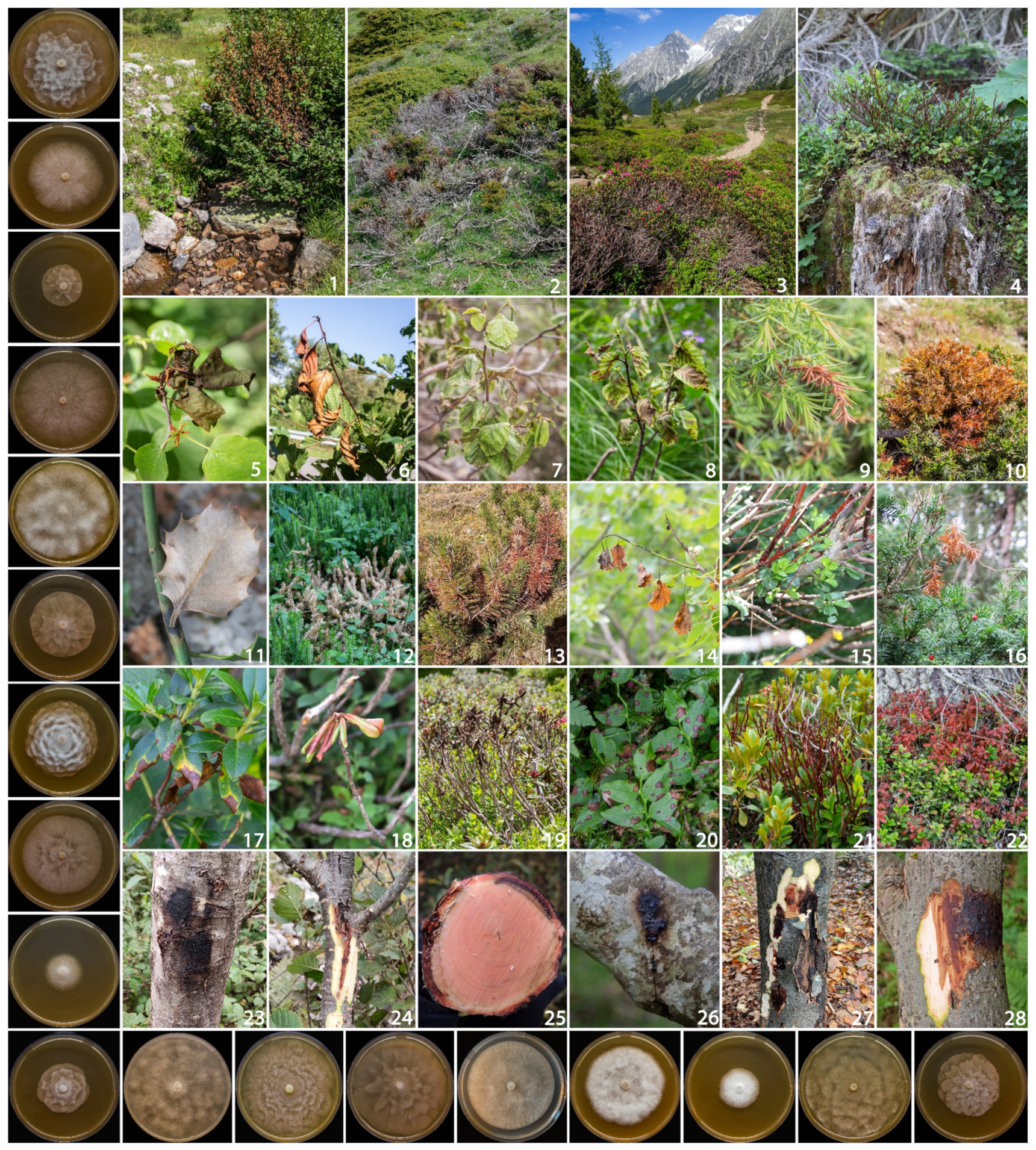
Monitoring surveys conducted in 54 sites distributed in Italy and Slovenia allowed the occurrence of
Phytophthora-related diseases to be detected in several plants typical of the alpine and subalpine climate. Disease incidence was highest in shrub vegetation, alpine heathlands and along the mountain riparian systems, ranging from 25 to 100%, with a mortality rate between 5 and 45% (
Table 3). The most impacted ecosystems were heathlands dominated by common juniper and blueberry, and alder riparian systems (
Figure 1). In these ecosystems,
Phytophthora outbreaks showed an epidemic trend with a high mortality rate.
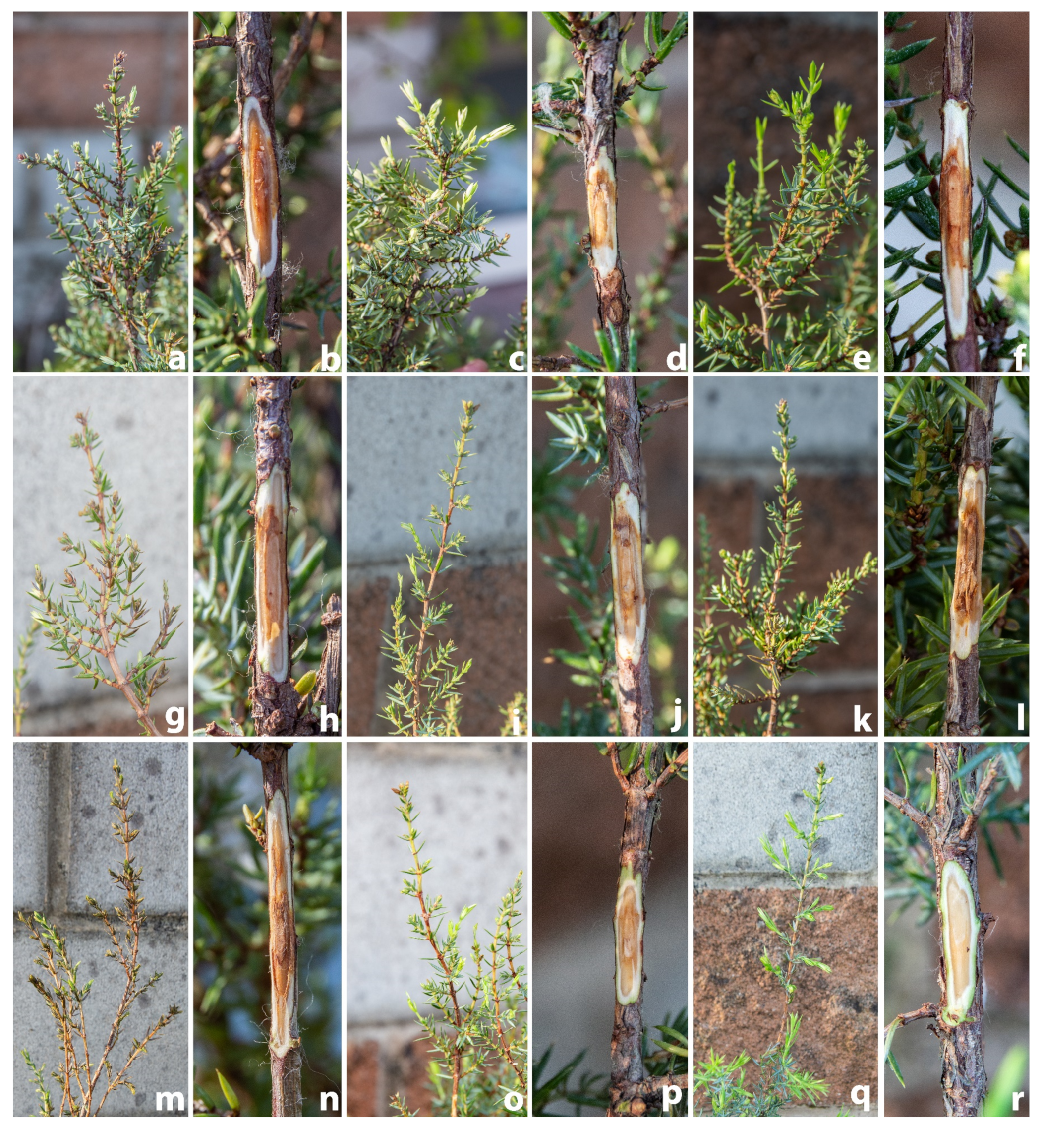
Many of the aerial
Phytophthora symptoms observed were new and involved various plant organs such as leaves (moist necrotic lesions), fruit (rot), twigs (wilting and shoot blights). Moreover, on tree and shrub species stem and branches extensive bleeding cankers were observed (
Figure 1). Cankers and necrosis progressively girdled the circumference of the branch, causing partial or total death of the crown.
On shrubs and heath formations, the disease was initially observed in small areas and progressively spread in a concentric manner affecting more plant species (
Figure 1).
3.2. Aetiology
Isolations performed on 397 samples yielded a total of 360 Phytophthora isolates. Based on morphological features and ITS sequence data, 17 known Phytophthora species were identified, namely: P. pseudosyringae (201 isolates), P. plurivora (54), P. gonapodyides (21), P. ilicis (20), P. alpina (17), P. acerina (11), P. cactorum (7), P. pseudocryptogea (6), P. cambivora (5), P. idaei (4), P. psychrophila (3), P. bilorbang (2), P. chlamydospora (2), P. hedraiandra (1), P. kelmanii (1), P. rosacearum (1) and P. syringae (1).
In addition, three isolates obtained from necrotic tissues of Alnus viridis, Juniperus communis and Rhododendron ferrugineum could not be assigned to any known Phytophthora species and are therefore described here as Phytophthora pseudogregata sp. nov.
The assemblage and distribution of
Phytophthora species was very variable among hosts and geographic areas. The 33 plant species monitored were divided into three main categories: small trees (
Table 4), shrubs/heathland species (
Table 5) and herbaceous/perennial plant species (
Table 6).
The most common and widespread
Phytophthora species detected in this study was
P. pseudosyringae. This species was isolated from 25 out of the 33 hosts, in 36 sites distributed in all monitored geographic regions. Together with
P. cactorum, it is the only species detected in all three types of hosts, while the other
Phytophthora species were isolated from only one or two types (
Figure 2).
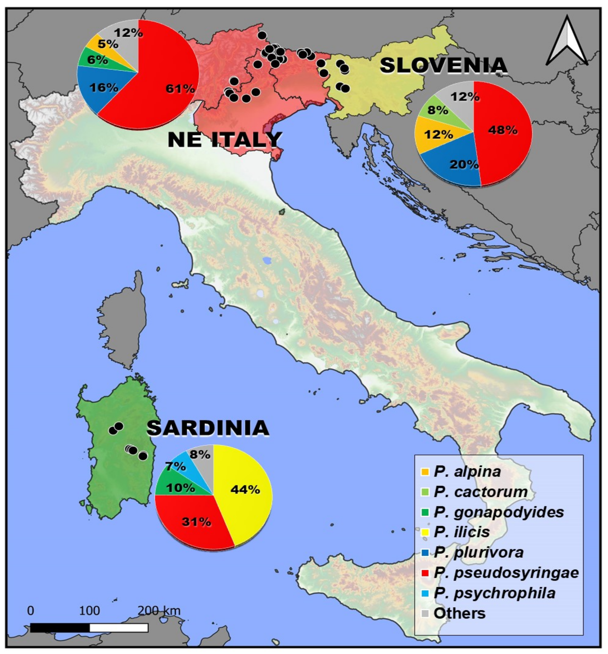
Phytophthora plurivora was the second most-isolated species, obtained from 12 hosts in 24 sites.
Phytophthora pseudosyringae and
P. plurivora were the most frequently isolated species in NE Italy and Slovenia (
Figure 2). In addition to these two species, some species belonging to clade 1, such as
P. alpina and
P. cactorum, were frequently isolated from different hosts in the NE Alps. In the mountainous areas of Sardinia, in addition to
P. pseudosyringae, other two species
P. ilicis and
P. psychrophila belonging to clade 3 were constantly isolated (
Figure 2).
As regards the distribution within
Phytophthora clades, clade 6 is the most represented in terms of species (five species) followed by clade 1 (4), clade 3 (3) and clade 8 (3). Only one or two species were obtained for clades 2 and 7. Overall, 56 new host–pathogen associations were detected (
Table 4,
Table 5 and
Table 6).
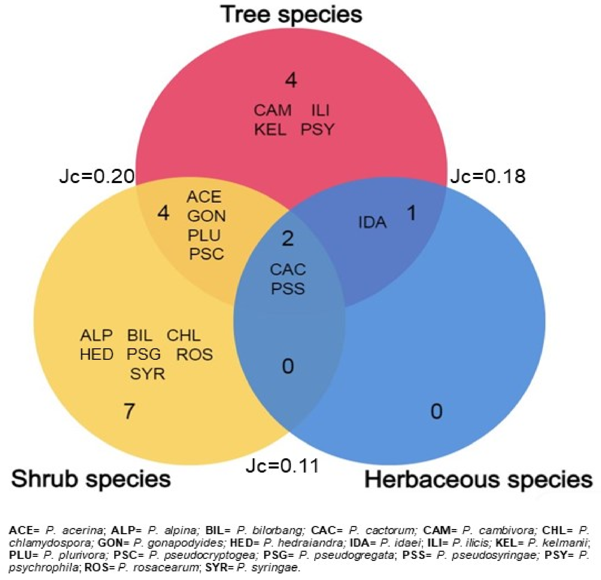
3.3. Structure and Diversity of Phytophthora Communities
The diversity indices of the
Phytophthora assemblages detected in the subalpine vegetation varied among the three categories of hosts, but in general they displayed high diversity and richness and moderate evenness, with the exception of the shrub
Phytophthora community dominated by
P. pseudosyringae (
Table 7).
Tree and shrub species displayed the highest number of taxa and Shannon index (H) values. As regards the degree of similarity between the three
Phytophthora communities, the Jaccard similarity index (Jc) was variable between 0.11 and 0.20. Only two
Phytophthora species,
P. pseudosyringae and
P. cactorum, were isolated from all host groups. Relationships among the three categories of hosts are shown in
Figure 3.
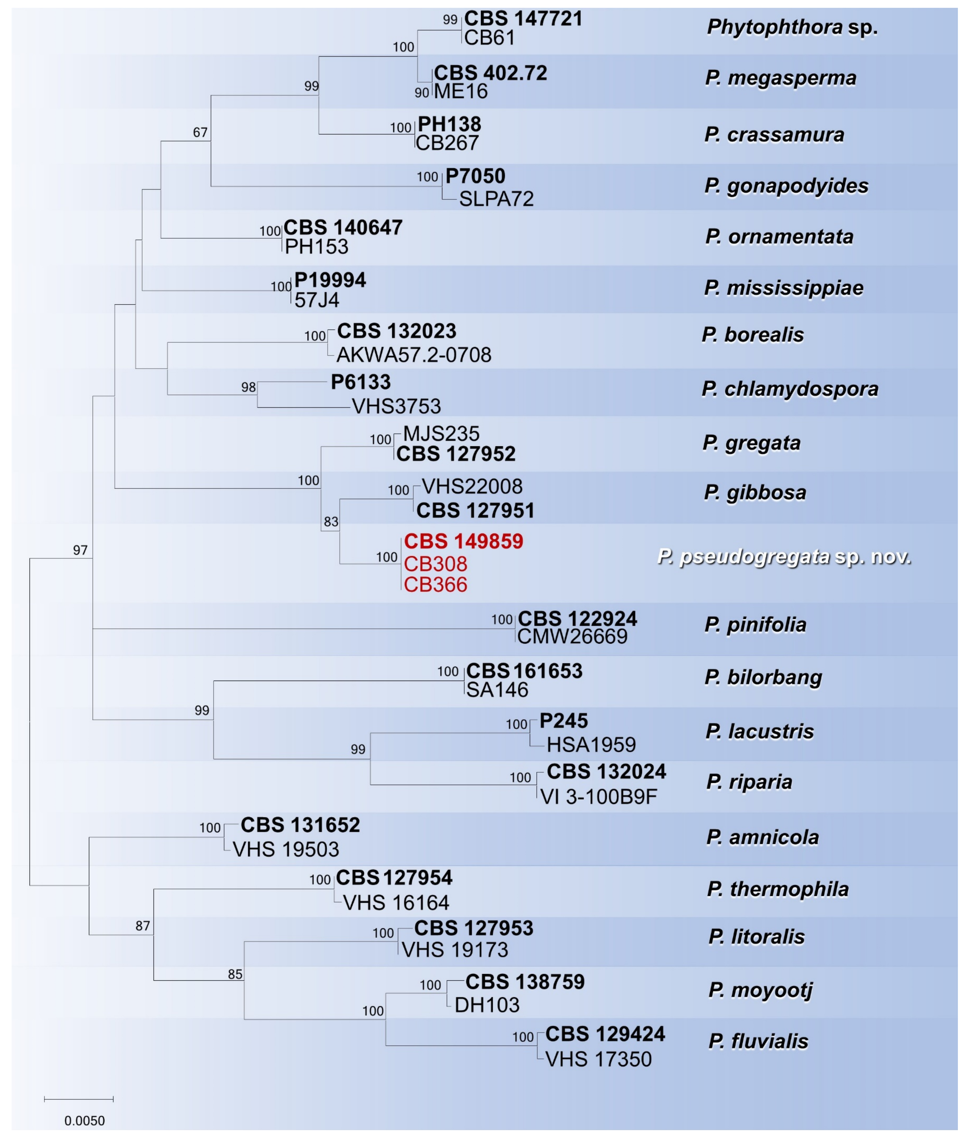
3.4. DNA Phylogeny
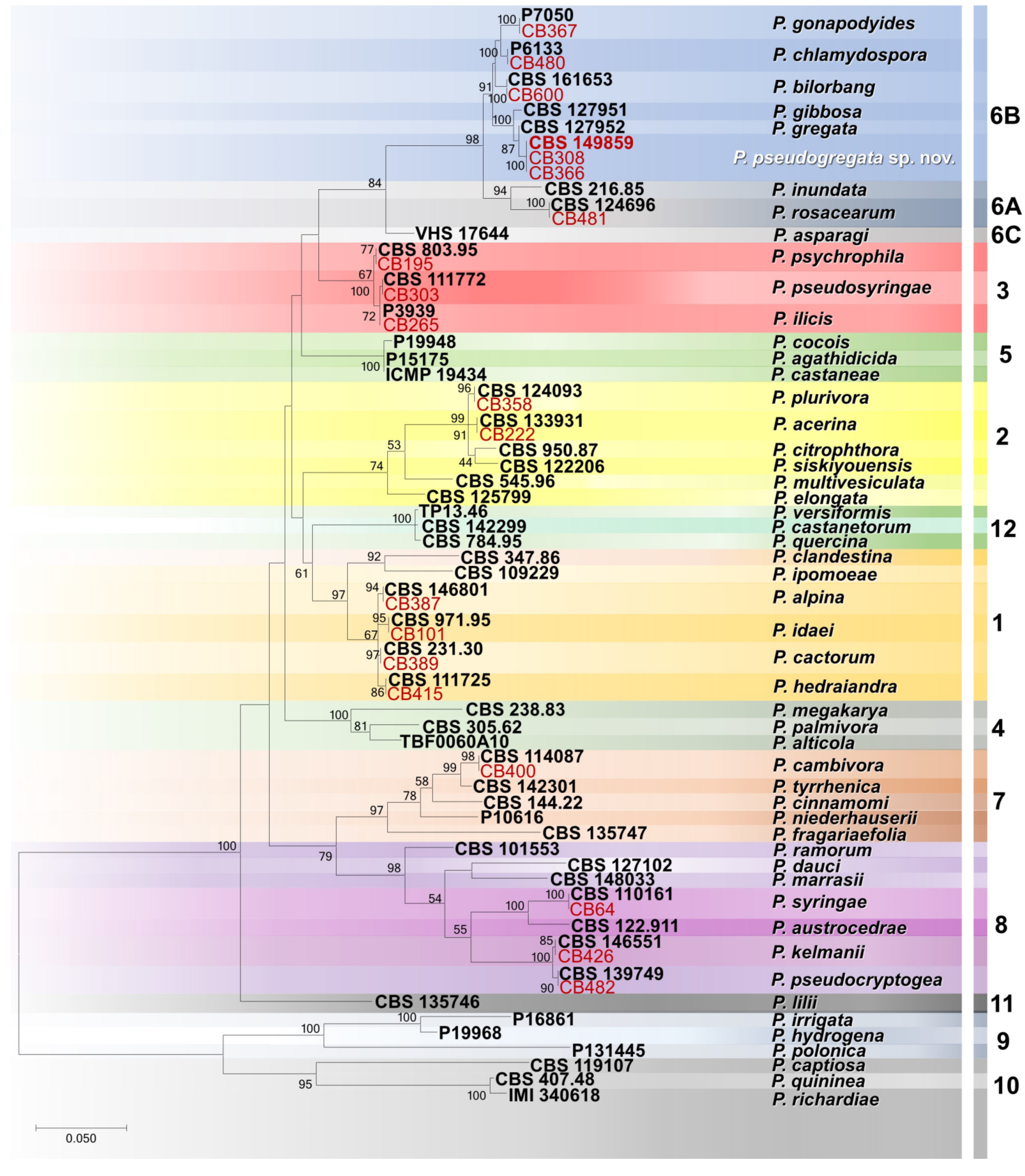
Phylogenetic relationships among the
Phytophthora isolates obtained in this study were elucidated using ITS sequences (
Figure 4). In particular, the 20 isolates included in the phylogenetic analysis were distributed in 18 terminal clades, 17 of which belong to formally described species (
Figure 4). Instead, three isolates clustered together in a separate and well-supported terminal clade (ML bootstrap = 100%) representing a previously unrecognized species closely related to
P. gregata, which is described here as
Phytophthora pseudogregata sp. nov. (
Figure 4).
To resolve the phylogenetic position of
P. pseudogregata within subclade 6b, a concatenated nuclear and mitochondrial dataset (the length of the final alignment was 2129 bp) was analysed. Individual gene phylogenies revealed no major conflicts, thus indicating that the three loci (ITS, Btub and
cox1) could be combined. The ML analysis resolved the positions of all formally described
Phytophthora species in subclade 6b, accommodating the isolates
P. pseudogregata in a terminal clade sister to
P. gibbosa (
Figure 5).
Phytophthora pseudogregata is separated by the two closely related species,
P. gregata and
P. gibbose, by three, two, and 18 bp and by eight, three, and 17 bp in ITS, Btub, and
cox1 loci, respectively.
3.5. Taxonomy
Phytophthora pseudogregata Bregant, Ogris, Meli and Linaldeddu sp. nov.
MycoBank: MB849354
Etymology: the name refers to the morphological similarity to Phytophthora gregata.
Holotype: CBS H-25226
Host/distribution: Alnus viridis, Juniperus communis and Rhododendron ferrugineum with foliar necrosis and shoot blight symptoms in Italy and Slovenia.
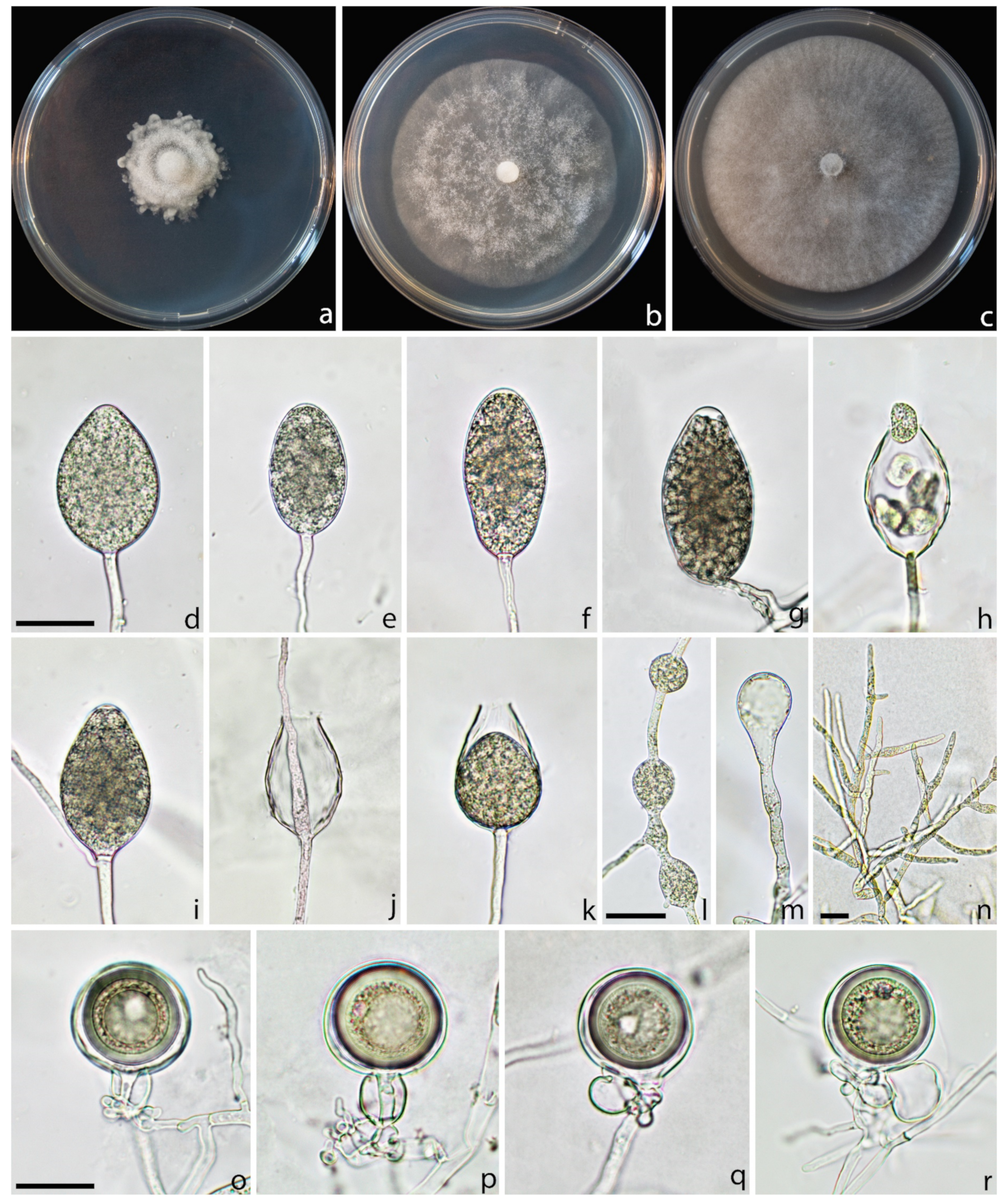
Description: Sporangia were produced on CA plugs flooded in unsterile pond water after 36–72 h of incubation at 25 °C on simple sporangiophores. Sporangia were persistent, mostly nonpapillate (80%), rarely semipapillate (20%), from ovoid to obpyriform, sometimes ellipsoid, borne terminally on unbranched sporangiophores, average 50.3 ± 6.5 × 29.9 ± 3.8 µm (total range 32.1–65.1 × 22.1–38.4 µm), with a length/breadth ratio of 1.7 ± 0.2 (
n = 50) (
Figure 6d–g). Zoospores were abundantly produced in liquid cultures after 24–36 h at 25 °C in the dark (
Figure 6h). Sporangia proliferated, usually externally and rarely internally, in both a nested and extended way (
Figure 6i–k). Hyphal swellings were not formed on solid agar and rarely in pond water, they were globose to subglobose, mostly intercalary catenulate, rarely terminal (
Figure 6l–m). Chlamydospores were not observed. All isolates produced gametangia in single culture on carrot agar after 7–10 days at 20 °C in the dark. Oogonia were smooth-walled, borne mainly terminally, with an average diameter of 33.2 ± 3.4. Oospores were spherical and usually aplerotic and 29.0 ± 3.7 µm in diameter. Antheridia were mostly amphigynous (58%), less frequently paragynous (42%) hyaline, rounded, club-shaped, or irregular: average 16.2 ± 2.7 × 12.5 ± 2.2 µm (
Figure 6o–r).
Cultural characteristics: colony growth pattern cottony on PDA with an irregular border, with an indistinct pattern on MEA and CA. On PDA, growth was slow, whereas on MEA and CA, colonies reached a diameter of 55 and 70 mm in 7 days at 23 °C, respectively.
Cardinal temperatures for growth: minimum <2 °C, maximum 32 °C, and optimum 23 °C. Isolates failed to grow at 34 °C, and mycelium did not resume growth when plates were moved to 20 °C.
Material examined: ITALY: Borso del Grappa, isolated from a necrotic shoot of Juniperus communis, 13 June 2022, collected by Letizia Meli, isolated by Carlo Bregant, HOLOTYPE CBS H-25226, a dried culture on CA, culture ex-holotype CB234 = CBS 149859. ITALY: San Nicolò di Comelico, isolated from necrotic leaves of Rhododendron ferrugineum, 3 July 2021, collected and isolated by C. Bregant (isolate CB308). SLOVENIA: Bohinj, isolated from a necrotic branch of Alnus viridis, 7 October 2021, collected by C. Bregant and Nikica Ogris and isolated by C. Bregant (isolate CB366).
Notes:
Phytophthora pseudogregata belongs to subclade 6b. The closest species are
P. gregata and
P. gibbosa, from which it differs through a combination of unique morphological features (
Table 8) and sequence data such as sporangia size and proliferation, oogonia and antheridia shapes and cardinal temperature values, as well as a total of 23 (
P. gregata) and 28 (
P. gibbosa) fixed nucleotide differences in the ITS, Btub, and
cox1 sequences.
3.6. Pathogenicity
All
Phytophthora species proved to be pathogenic on
Juniperus communis. At the end of the experimental period, inoculated seedlings showed dark brown inner bark lesions that spread up and down from the inoculation point (
Figure 7).
Among the different species assayed, the length of the necrotic lesion differed significantly (
Table 9). The lesions caused by
P. pseudosyringae were significantly larger than those caused by other species (
Table 9). Lesions caused by
P. pseudosyringae,
P. plurivora and
P. acerina progressively girdled the twigs causing shoot blight, browned foliage and wilting symptoms.
Control seedlings, inoculated with sterile PDA plugs, remained symptomless; in only two twigs, a small light brown discoloration was observed restricted to the inoculation point.
All eight Phytophthora species were successfully re-isolated from the necrotic inner bark lesions of all seedlings, thus fulfilling Koch’s postulates. No Phytophthora or other fungal isolates were obtained from control plants.
4. Discussion
This study represents the most comprehensive investigation to date on aerial diseases caused by Phytophthora species on mountain vegetation in Italy and Slovenia. The results obtained have allowed us to clarify both symptomatology and aetiology of the emerging pathosystems affecting mountain and subalpine formations. The progressive spread of several airborne Phytophthora species is causing the destruction of vast ecosystems and compromising the biodiversity of these ecologically fragile habitats.
Based on combined sequence data and micromorphological features, 18 Phytophthora species belonging to six out the 12 major Phytophthora phylogenetic clades were identified from a collection of 397 symptomatic samples collected from 33 herbaceous and woody hosts. These included: P. acerina, P. alpina, P. bilorbang, P. cactorum, P. cambivora, P. chlamydospora, P. gonapodyides, P. hedraiandra, P. idaei, P. ilicis, P. plurivora, P. pseudocryptogea, P. pseudosyringae, P. psychrophila, P. rosacearum and P. syringae. In addition, three isolates described here as Phytophthora pseudogregata sp. nov. were isolated and characterized.
The most frequently isolated
Phytophthora species belong mainly to clades 1 and 3. These species are characterized by the ability to produce caducous sporangia useful for aerial infections [
1]. Furthermore, the relatively low cardinal temperatures for growth suggest that these species have a great potential to threaten mountain vegetation [
14,
31,
55].
In particular, in Northeast Italy a higher number of species belonging to the ITS clade 1 was isolated (
P. alpina,
P. cactorum,
P. hedraiandra and
P. idaei), while in Sardinia, clade 3 was dominant, with three species,
P. pseudosyringae,
P. ilicis and
P. psychrophila. Overall,
P. pseudosyringae (clade 3) was the most frequent species in terms of number of hosts infected and distribution among sites. Two hundred and one out of the 360 isolates obtained in this study belonged to this species. In particular,
P. pseudosyringae have been detected in 36 sites and 25 hosts of all three plant categories investigated: trees, shrubs and herbaceous plants (17 new host–pathogen associations). The wide spread of
P. pseudosyringae in different mountain and subalpine formations and its involvement in several new diseases highlight the polyphagous nature of this invasive pathogen and its aerial lifestyle. This agrees with previous studies conducted in mountain environments in Asia, Europe, North and South America [
14,
32,
34,
55,
56,
57,
58,
59,
60,
61].
Phytophthora pseudosyringae is the key species in the aetiology of aerial infections detected in high-altitude shrubs and heaths such as blueberry, dwarf pine, juniper, rhododendron and alpine willow formations; these shrubs are characterized by creeping behaviour with very limited heights above the ground, this habitus could favour the attack of
Phytophthora sporangia and zoospores. The attacks of
P. pseudosyringae on
Vaccinium myrtillus (leaf necrosis and shoot blight) were particularly severe, confirming the susceptibility of this small shrub as previously reported in Ireland [
33,
62]. Many aspects regarding the infectivity and survival of
P. pseudosyringae sporangia in the infected tissues fallen to the ground in subalpine areas remain to be clarified. At the same time the ability of oospores to persist for years and their infectivity in environments where the pathogen is subjected to extreme low temperatures need further investigations. Probably the survival of this species in cold habitats is guaranteed by the production of very large and thick wall chlamydospores. In fact, unlike what was reported in previous studies and in the original description, all isolates of
P. pseudosyringae examined produced a large amount of globose chlamydospores on CA both in solid and liquid culture. The chlamydospores were mainly terminal and 76.6 ± 22.02 (range 39.9–102.3,
n = 25) μm in diameter. Based on the wide variation in morphological characters found in this study, the description of
P. pseudosyringae needs to be redefined. Undoubtedly, increased inoculum in the litter due to the diseased fallen leaves not only could represent an increased risk of outbreaks but also a faster disease progression in these habitats [Bregant & Linaldeddu, unpublished]. In the pathogenicity test,
P. pseudosyringae shows high aggressivity on common juniper, producing wood necrosis and shoot blight after four weeks from the inoculation.
The other two species in clade 3 were isolated only in the mountain area of Sardinia.
Phytophthora psychrophila have been isolated from bleeding cankers of
Quercus pubescens, confirming the affinity of this pathogen towards oak species [
63]; the geographic distribution and impact of this species is still unknown; there have been a few reports of it in European and American natural forests and nurseries [
4,
31,
64].
Phytophthora ilicis has been known for a long time as a specific pathogen of
Ilex aquifolium in the mountains of the Mediterranean basin and a few other areas of Europe and North America [
55,
65,
66,
67].
Four species belonging to subclade 1a have been isolated in the northeastern Alps.
Phytopththora alpina shows the highest ability to survive in extremely cold conditions due to the low temperature values for growth and the high production of caducous sporangia and chlamydospores [
14]; in addition to
Alnus viridis, its discovery on three new hosts in Italy and Slovenia suggest that this recently described species is well adapted to affect typical alpine and subalpine shrubs. The second most common species in subclade 1a was
P. cactorum, an invasive and polyphagous pathogen widespread from tropical to temperate climates where it is responsible for severe diseases on agriculture crops and forest trees [
1,
29,
68]. The occurrence of
P. cactorum in cold areas has recently been reported in Europe and Australia [
4,
14,
60,
69]. Together,
P. pseudosyringae and
P. cactorum are the two species obtained from all three plant types.
In addition to the numerous new host–pathogen associations (
Table 4,
Table 5 and
Table 6), some species detected such as
P. hedraiandra and
P. idaei are reported for the first time in natural ecosystems in Europe. Previous studies have ascertained the involvement of these two pathogens in root and foliar disease in agriculture and ornamental nurseries;
P. idaei appears restricted to the genus
Rubus [
70], while
P. hedraiandra has a wider range of ornamental hosts [
71,
72,
73,
74,
75]. Although, in the original description
P. idaei is reported to have persistent-sporangia, the Italian isolates obtained in this study showed a moderate production of caducous sporangia.
The second most common species obtained in this study was
P. plurivora. Isolates of this species were obtained from 54 symptomatic samples of 12 plant species including eight new hosts.
Phytophthora plurivora resides in clade 2 and is common in forest ecosystems of Central Europe; from a recent population study it is considered to be originally of this continent and spread to others by human activities [
76]. This agrees with the results of this and previous studies [
8,
14] given the wide distribution of this pathogen in various extreme and nonhumanized natural environments. While the distribution and impact of
P. plurivora is well studied, little is known about its closely related species,
P. acerina. To date, this species appears widespread in agricultural systems, nurseries, forests and ornamental trees in northern Italy and Sardinia, and much rarer worldwide [
4,
77,
78,
79]. Both
P. acerina and
P. plurivora were already known as primary pathogens involved in common and grey alder decline in Italy [
14]. Isolates of
Phytophthora acerina obtained in this study confirm a single polymorphism in the ITS region between northern Italy and Sardinia populations [
14].
Among the other
Phytophthora species isolated in this study, five belong to clade 6, including the newly described species
P. pseudogregata. Clade 6 encompasses species very common in European forests, such as
P. bilorbang and
P. gonapodyides and species with more limited or still unknown distribution, such as
P. amnicola and
P. rosacearum [
8]. Some species in this clade are reported as saprophyte or occasionally weak opportunistic pathogens [
11,
54,
80,
81]; the involvement of five species of this clade in the aetiology of aerial infections on mountain vegetation highlight the ecological versatility of these organisms. The ability of
P. bilorbang,
P. gonapodyides and
P. pseudogregata to reproduce the symptoms observed in nature on common juniper suggest their active role in the aetiology of the emerging disease affecting woody trees in mountain areas.
Phytophthora pseudogregata resides in subclade 6b; it is closely related to
P. gregata and
P. gibbosa, from which it can be distinguished by unique morphological features and sequence data.
Phytophthora gregata was originally described in 2011 in Australia in wet native forests and in Tasmania associated with dying alpine heathland vegetation [
54,
69] and then recently reported in the Czech Republic and Finland [
60,
82], whereas
P. gibbosa is known to occur only in Australia associated with dying native vegetation on seasonally wet sites [
54].
Sub-clade 6b is larger and contains several described species (
P. amnicola,
P. borealis,
P. chlamydospora,
P. bilorbang,
P. crassamura,
P. fluvialis,
P. gibbosa,
P. gonapodyides,
P. gregata,
P. lacustris,
P. litoralis,
P. megasperma,
P. mississippiae,
P. moyootj,
P. ornamentata,
P. pinifolia,
P. pseudogregata,
P. riparia and
P. thermophila), some not formally described species and a few hybrids [
83]. Most of the species in this sub-clade have been described in the last 12 years, the only species known until 2011 were
P. gonapodyides and
P. megasperma [
1]. The majority of species in sub-clade 6b, including
P. pseudogregata, have been described in forest ecosystems, underlining the key role played by natural areas in exploring the biodiversity of the
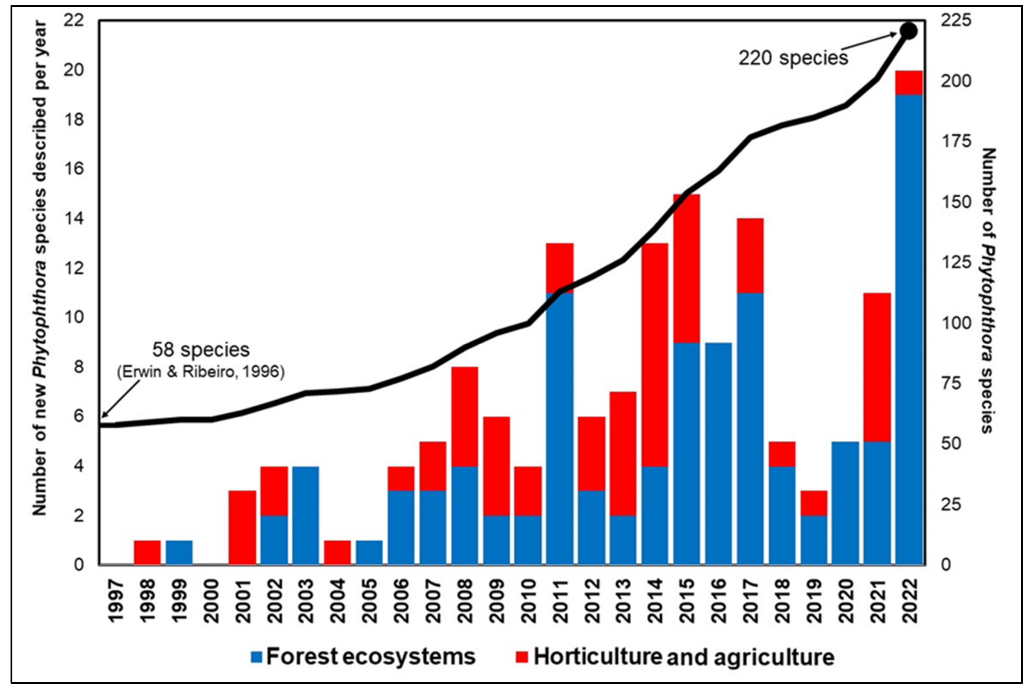
Phytophthora genus, which currently includes 220 species (
Figure 8).
Finally, three species of clade 8 (
P. kelmanii,
P. pseudocryptogea and
P. syringae) and one from clade 7 (
P. cambivora) have been isolated, mainly from stem bleeding cankers of small trees and shrubs. While
P. kelmanii and
P. syringae have a very limited distribution,
P. pseudocryptogea is widespread along the Alps. The large range of growth temperatures and polyphagous nature explain it being widespread in Italian ecosystems spanning from Mediterranean areas to the tree line in the Dolomites [
4,
14,
78]. Both mating types of
P. cambivora occurred in the NE Alps (A2 on
Alnus incana in Slovenia and A1 on
Laburnum alpinum and
Sorbus aucuparia in Italy).

 ,
, 


