Article

The New Report of Root Rot on Fatsia japonica Caused by Phytophthora nicotianae in China

1
Co-Innovation Center for the Sustainable Forestry in Southern China, Nanjing Forestry University, Nanjing 210037, China
2
School of Food Science, Nanjing Xiaozhuang University, 3601 Hongjin Avenue, Nanjing 211171, China
*
Authors to whom correspondence should be addressed.
Forests 2023, 14(7), 1459; https://doi.org/10.3390/f14071459
Submission received: 17 June 2023 / Revised: 11 July 2023 / Accepted: 13 July 2023 / Published: 17 July 2023
(This article belongs to the Special Issue Forest Tree Diseases Genomics: Growing Resources and Applications)

Abstract

:
As an ornamental plant, Fatsia japonica has been widely used in gardens. From April 2021 to 2022, a disease that caused the wilting and root rot of F. japonica in a large area was observed, which eventually led to the plants wilting and dying, while the leaves did not fall off. This disease greatly reduced the landscape effect of plants. An oomycete species was isolated from the roots of the infected plants. This colony morphology was slightly radial to stellate, and the aerial mycelium was flocculent. Oval sporangia with papillae, apical chlamydospores and zoospores formed in sporangia were observed. The morphological characteristics were consistent with Phytophthora. For accurate identification, the internal transcribed spacer (ITS), cytochrome oxidase subunit II (COXII) and large ribosomal subunit (LSU) genes were amplified and sequenced. The species was identified as Phytophthora nicotianae using phylogenetic analysis. Finally, the disease was reproduced by inoculating healthy F. japonica with a zoospore suspension; the symptoms were consistent with those of natural infections, and the isolate obtained from artificially infected plants had the same morphological characteristics as the inoculated isolate. The results demonstrated that P. nicotianae is the pathogenic factor of root rot. of F. japonica. This is the first report of root rot on F. japonica caused by P. nicotianae in China.

1. Introduction

Fatsia japonica Decne. et Planch. (Syn. Aralia japonica Thunb. and A. sieboldii Anon.) is a member of the Araliaceae Juss family, which grows wild on Japanese islands and is widely cultivated as a decorative plant [1]. Often grown as a foliage houseplant for cooling situations, it is also a very successful shade-tolerant garden plant. Fatsia japonica is a well-known ornamental and potentially medicinal plant [2]. Its leaves, roots and barks have a certain medicinal value. The root bark has the medicinal efficacy of resolving phlegm and relieving cough, promoting blood circulation and removing blood stasis. Other benefits include dispersing wind and removing dampness, as well as removing stasis to ease pain; it can be used to treat traumatic injuries, cough and phlegm, rheumatic arthralgia and gout [3]. F. japonica is also widely planted in cities south of the Yangtze River in China [4].
The Phytophthora, classified in the family Pythiaceae, order Peronosporales, phylum Oomycota and kingdom Stramenopila, is distributed all around the world [5]. This genus was established by Anton de Bary in 1876 [6] and can be divided into 12 major phylogenetic branches, with many sub-branches [7,8,9]. In 1983, only 43 species of Phytophthora were identified, and later, Erwin and Ribeiro described around 60 species in 1996 [10]. By 2008, the number of species was close to 100 [11]. At present, more than three hundred species are described on www.mycobank.org (accessed on 9 May 2023). The species of the genus Phytophthora are characterized by having aseptate hyphae [12]. It produces asexual organs, namely sporangia, that can be oval, inverted pear-shaped or lemon-shaped. The differentiation of the contents inside the sporangium produces zoospores with double flagella, which are finally released through the top of the sporangium. Phytophthora species are homothallic or heterothallic, and sexual spores are obtained by the fertilization of male and female organs, namely oospores [13]. According to some researchers [10,14], most pathogens can survive in the soil for a long time without a host and will have a certain impact on the part of the plant in contact with the ground to destroy the plant roots. The effect of Phytophthora on plants can be seen by its name. In Greek, Phytophthora means plant destroyer [14,15,16], which can cause diseases in herbaceous and woody plants, mostly dicotyledon plants [10,13,17,18,19,20,21]. Major diseases caused by Phytophthora spp. are crown and root rot, stem rot, foot rot, stem canker, leaf blight, fruit brown rot and late blight [22,23,24,25,26,27,28]. They can infect cultivated and spontaneous plants worldwide, causing serious agricultural losses and destroying natural forest ecosystems [29]. Among the ten important pathogenic oomycetes causing plant oomycete diseases in the world, P. infestans, P. ramorum, P. sojae, P. capsici, P. cinnamomi and P. nicotianae are included [30]. In the 1840s, P. infestans caused potato late blight, causing famine in Ireland. In 2020, Linaldeddu et al. found that P. pini can cause crown blight and root rot in four Olea europaea L. forests in the Veneto region of Italy [29]. In 2023, Hrabetova, M. et al. found that there were dark brown to black necrotic lesions in the rhizome of Buxus sempervirens, resulting in the death of the whole plant [31]. Some diseases caused by Phytophthora have reached epidemic status [13,32,33,34]. For example, the host range of P. romorum is pretty wide, and it has become an epidemic disease in many places. It can infect a variety of ornamental plants and woody trees, causing serious losses to agricultural and forestry crop production, and has been listed as a quarantine object by many countries [35]. P. cinnamomi has been found in Fujian [36,37,38], Jiangsu and Zhejiang [38], Hainan [39], Shanxi [40], Shanghai [41] and other provinces. It poses a threat to the production of chestnut, kiwifruit, blueberry, avocado, ornamental trees and the health of natural forests [42]. The annual loss of agriculture and forestry caused by epidemics worldwide is huge. Once pathogens are established in the environment, the eradication of pathogens requires huge manpower and costs, and annual economic losses and governance costs can be as high as USD 10 billion. [43].
In the past, the traditional identification of Phytophthora was mainly based on morphological characteristics, including sporangium, sporangium peduncle, chlamydospores, sexual organs, hyphae and colonies. Due to the large morphological variation of Phytophthora pathogens, the basis of some classifications and identifications is not stable, so the classification and identification of the Phytophthora species is difficult [44,45]. Therefore, the identification of Phytophthora must also be combined with molecular biology methods, and the results are more reliable. The rDNA-ITS sequence is one of the most widely used target genes for the identification of Phytophthora. However, for some closely related species or sister species of Phytophthora, it is sometimes difficult to distinguish them by a single rDNA-ITS sequence, which requires the analysis of other conserved target genes to identify the species more accurately [46]. According to the sequencing of the mitochondrial DNA (mt DNA) coding region of the Phytophthora species, the COXII (Mitochondrially encoded cytochrome oxidase II) gene is suitable for the broad-spectrum phylogenetic analysis of Phytophthora [47]. Studies have shown that the LSU (Large subunit) gene also has a good resolution for the Phytophthora species and is used for the study of oomycete phylogeny [46].
From April 2021 to 2022, several diseases of Fatsia japonica were found in the campus of Nanjing Forestry University, China. We dug up dozens of seriously diseased F. japonica roots under the dormitory building. The symptoms were that the whole plant was wilting, the leaves were low and dead without falling off and the roots were rotten and black after digging them up. The main purpose of this study is to isolate and identify the pathogenic factors of F. japonica root rot using a pathogenicity test, morphological characteristics and a phylogenetic analysis. It provides a reference for the study of diseases of F. japonica.

2. Materials and Methods

2.1. Disease Investigation and Isolation

In May 2022, the diseased roots of dozens of F. japonica were dug up under several dormitory buildings of Nanjing Forestry University (Geographic coordinates: 31°14′ N, 118°22′ E). The roots were washed thoroughly with clean water and then cut into 30 pieces of 3 mm in size, surface-disinfested by immersion in 75% ethanol for 30 s followed by 1% NaClO for 90 s, rinsed three times with sterile water, then dried on sterilized filter paper and plated onto clarified 10% V8 juice agar (cV8A) [48] that was amended with pimaricin (20 mg/L), ampicillin (125 mg/L), rifampicin (10 mg/L) and pentachloronitrobenzene (20 mg/L). They were incubated at 26 °C (Incubator MIR-553, Sanyo, Osaka, Japan) for three days. Then, these hyphae tips were transferred to a fresh V8 plate to obtain pure cultures.

2.2. Morphological Identification

Three strains of pathogenic bacteria were selected, and the colony morphology of the isolates was observed with a clear 10% V8 solid medium. The colony plugs with a diameter of 0.6 × 0.6 cm were punched on the edge with a sterile puncher and inoculated in the middle of the culture dish. The culture dish was placed in a dark incubator at 26 °C. The structure, color and morphology of the colonies were observed and recorded.
To observe the morphology of various spores, several colony agar blocks taken off with a sterile puncher were placed in a liquid V8 at 26 °C for 3 days with a 12/12 h light–dark cycle, then liquid V8 was replaced with sterile water and 3–5 drops of soil extract (100 g (3~10 cm deep) of surface soil from a fertile vegetable garden was collected, and 100 mL of tap water was added, fully stirred and precipitated for several hours; the supernatant was filtered with ordinary filter paper to remove the coarse particles of the soil and was repeatedly filtered twice with a 0.22 μm microporous membrane) to stimulate sporangial production [49]. Species were identified based on morphological characteristics (colony morphology, color and texture, sporangia, chlamydospores and zoospores) of the three isolates on V8. The sporangia, chlamydospores and zoospores were measured using a Zeiss Axio Imager A2 m microscope (Carl Zeiss, Oberkochen, Germany) for morphological description and size measurement (n = 50).

2.3. DNA Extraction and PCR Amplifcation

The traditional morphological identification method is uncertain due to the influence of external factors, so the identification of Phytophthora nicotianae must also be combined with molecular biology methods, and the results are more reliable. rDNA-ITS is one of the most widely used target genes for the identification of P. nicotianae because of its multiple copies and fast coding [50]. In addition to the rDNA-ITS gene, the COXII gene of the Phytophthora species can also be used as a target gene for the identification of P. nicotianae [51]. For molecular identification, the DNA of representative isolates was extracted from the mycelium cultured for 3 days using the CTAB method [52]. The internal transcribed spacer (ITS) region, large subunit (LSU) and mitochondrially encoded cytochrome oxidase II (COXII) genes were amplified using the primer pairs ITS1/ITS4 [53], LROR-O/LR6-O [46] and FM82/FM80 [54], respectively. The primers and PCR conditions are shown in Table 1. For PCR amplification of 50 μL, the reaction system is as follows: primers (10 μmol/L) each 2 μL, Taq DNA Polymerase 25 μL, template DNA (100 ng/μL) 2 μL and ddH2O 19 μL. The PCR amplification products were purified with agarose gel electrophoresis and sent to Shanghai Jieli Biotechnology Co., Ltd. (Nanjing, China) for amplicon sequencing.

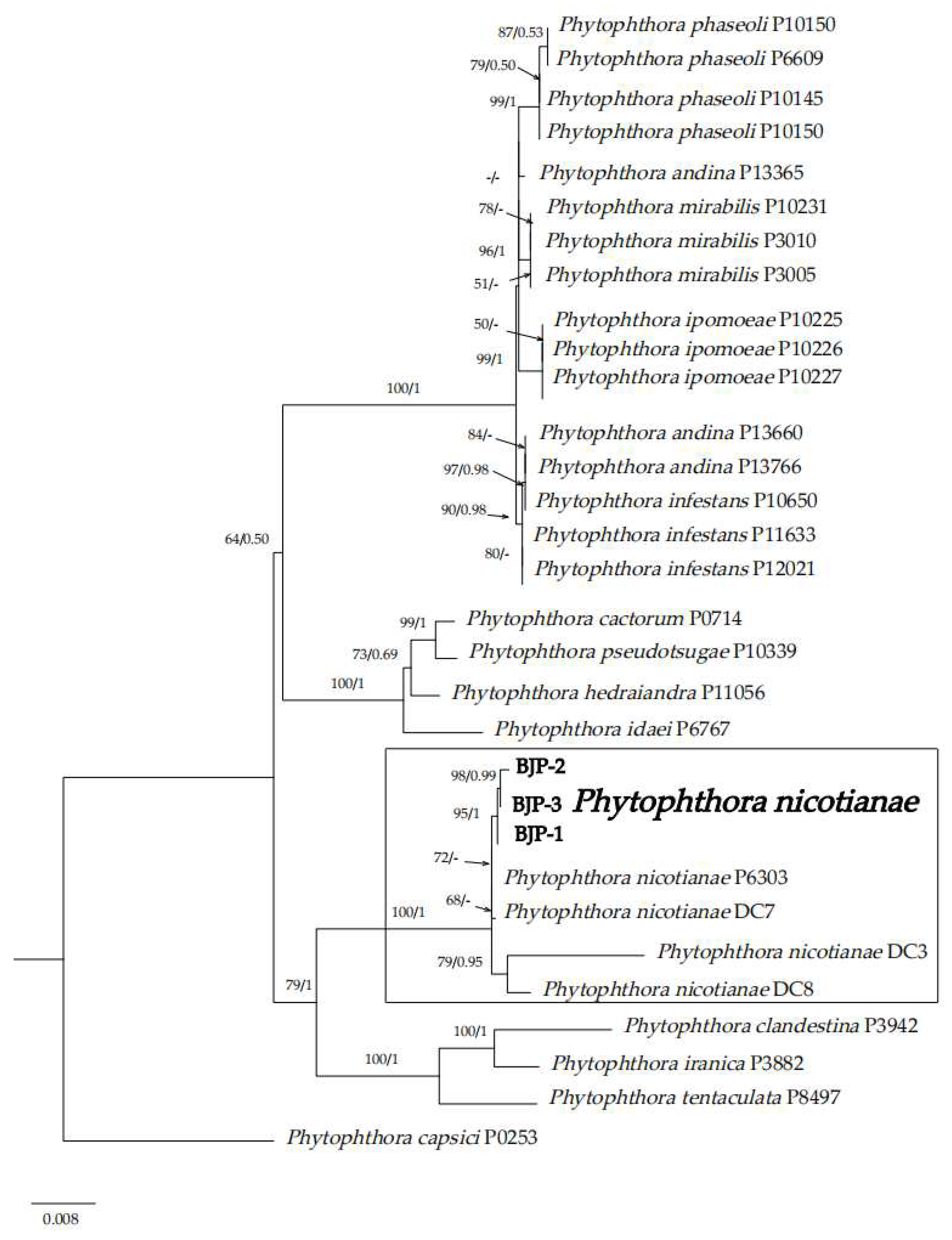
2.4. Phylogenetic Analyses

For further identification, the extracted DNA sequences were subjected to BLASTn search in the NCBI database to retrieve orthologous sequences with high similarity, and these sequences were submitted to NCBI/Gen Bank to obtain the registration number. Multiple gene sequence analyses used ClustalW Multiple Alignment in the bioinformatics software BioEdit ver.7.0.9.1 [55] for multiple sequence alignment. After editing and cutting, the first base of each pair of gene sequences was the same base, and the tail was the same. To ensure maximum sequence similarity, Maximum Likelihood (ML) and Bayesian Inference (BI) phylogenetic analyses were performed based on ITS, LSU and COXII multi-locus tandem sequences in PhyloSuite ver.1.2.2 software [56]. Concatenate Sequence was used to concatenate the sequences of ITS, LSU and COXII. ML phylogenetic analysis of multi-gene tandem sequences was performed using IQ-TREE ver.1.6.8 [57]. The best nucleotide substitution model was statistically selected using ModelFinder [58] and was based on the AIC (Akaike information criterion) standard. The general time reversible (GTR) nucleotide substitution model was used, and the site difference ratio was set to invgamma. The bootstrap (BS) test was used to calculate the branch support rate with 1000 replicates. MrBayes ver.3.2.6 [59] was also used to perform BI phylogenetic analysis of interspecific relationships. Statistical selection of the best nucleotide substitution model was performed using ModelFinder and was based on the BIC (Bayesian information criterion) criteria. The Markov Chain Monte Carlo (MCMC) algorithm was used for operation. The operation lasts for more than 2 × 10⁶ generations. Sampling is performed every 1000 generations until the average standard deviation of split frequencies is less than 0.01. The posterior probabilities (PP) of each branch were calculated. Finally, FigTree ver.1.4.4 (http://tree.bio.ed.ac.uk/software/figtree/ (accessed on 13 May 2023)) software can be used to view the tree file and beautify it, utilizing drawing tools to trim and embellish the tree diagram.

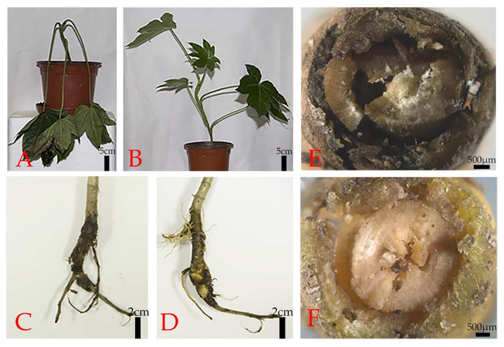
2.5. Pathogenicity Assays

To fulfill Koch’s postulates, one-year-old F. japonica potted seedlings (30 cm tall, n = 12) were placed in a greenhouse (temperature: 25 °C, 90% relative humidity, daylight: 14 h) for the pathogenicity tests. Healthy roots of F. japonica were dug up to expose root balls, which were wounded before inoculations with a sterile needle. Every plant was inoculated with 10 mL of zoospore suspension (106 zoospores/mL) that was mixed into sterile pot soil (approximately 500 g). The sterile water inoculation treatment was used as a blank control, and each treatment was repeated three times.

3. Results

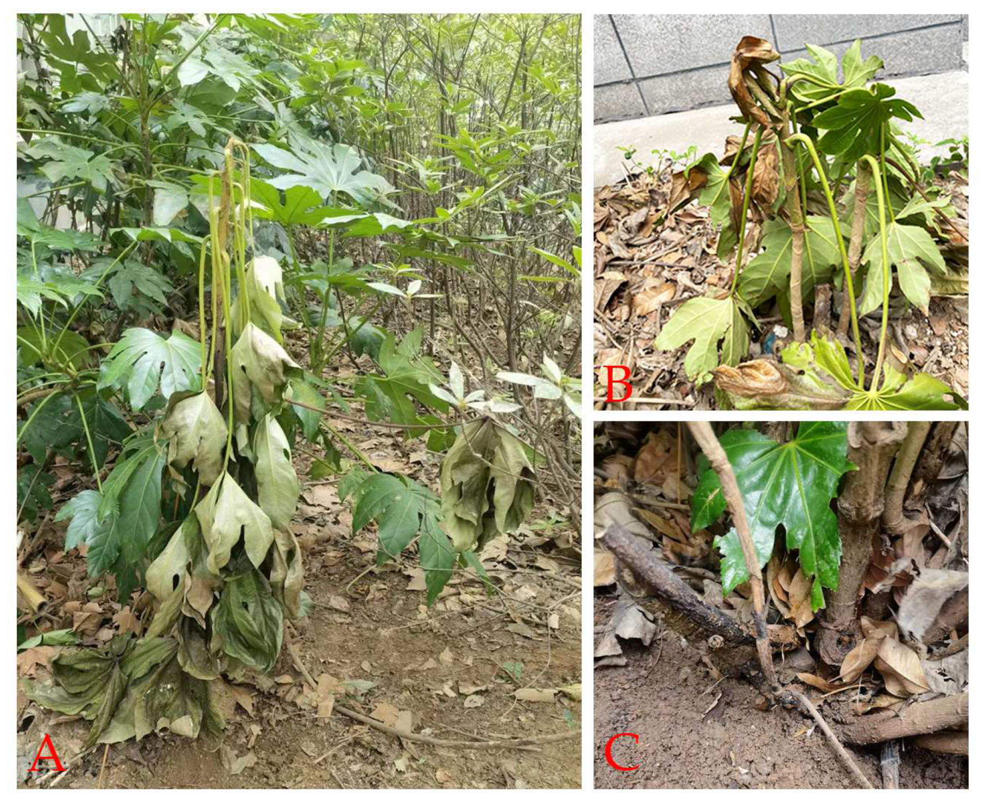
3.1. Natural Symptoms

From 2021 to 2022, a survey was conducted in the Xuanwu District of Nanjing City. It was found that the disease also widely occurred in some nurseries in the Xuanwu District, including Nanjing Forestry University, with an incidence of about 40%. In the early stage of the disease, the base of the petiole drooped, but there was no obvious necrosis. In the late stage of the disease, the affected plants wilted. The leaves were shrunk and yellowed, and the underground roots rotted, but the base leaves did not fall off (Figure 1).

3.2. Morphological Characteristics

By isolation, Phytophthora-like mycelium appeared in 80% of the samples. The colony morphology of all isolates was slightly radial and star-shaped hyphae, an irregular colony shape; the cotton flocculent aerial mycelium was exuberant and dense, and the reverse was white (Figure 2A,B). Three representative isolates (BJP-1, BJP-2 and BJP-3) were randomly selected and preserved in the collection of Nanjing Forestry University. Sporangia produced in 10% of the liquid V8 were ovate to suborbicular or elliptic in shape, with a smooth surface, containing protoplasts and immature zoospores. Figure 2 shows the following: the sporangium was nearly spherical and 27.9 ± 6.6 µm × 25.9 ± 6.8 µm in size (n = 30) (Figure 2C); sporangia produced in 10% liquid V8 zoospores (Figure 2D,E); an empty sporangium (Figure 2E); zoospores were suborbicular (Figure 2F) and 6.9–9.3 µm in diameter (n = 30); chlamydospores were spherical, terminal and 25.2 ± 0.3 µm in diameter (n = 30) (Figure 2G).

3.3. Molecular Biology Identification

The genomic DNA of three representative isolates was amplified using three genes, and the bands obtained with gel electrophoresis were in line with the expected size. Then, the amplified sequences were subjected to BLAST alignment analysis in NCBI, and the results are listed in Table 2. These sequences amplified in this study have been registered in GenBank (http://www.ncbi.nlm.nih.gov (accessed on 5 May 2023)). A total of 14 closely related species and 17 isolate sequences were downloaded as references. The GenBank accession of the sequences of Phytophthora are shown in Table 3. Based on the tandem sequences of the three genes, under the AIC standard, the Maximum Likelihood method development tree was constructed, and the Bayesian development tree was constructed under the BIC standard. The tree structure of the two is the same.
Table 2. BLAST results based on the ITS, LSU and COXII gene amplification sequences of three representative isolates in this experiment.
IsolateDNA TargetGenBank Accession No.Blast Match Sequence
Reference Accession No.Sequence Identity (%)
BJP-1ITSOP735506P. nicotianae B2 (MT472132.1)99.88% (801/802)
LSUOP738518P. nicotianae 22F9 (KX250514.1)100% (1246/1246)
COXIIOP743911P. nicotianae P6303 (GU318304.1)99.73% (733/735)
BJP-2ITSOP735507P. nicotianae B2 (MT472132.1)99.63% (802/805)
LSUOP738516P. nicotianae 22F9 (KX250514.1)100% (1248/1248)
COXIIOP743912P. nicotianae P6303 (GU318304.1)99.66% (880/883)
BJP-3ITSOP735526P. nicotianae B2 (MT472132.1)100% (803/803)
LSUOP738517P. nicotianae 22F9 (KX250514.1)100% (1245/1245)
COXIIOP743913P. nicotianae P6303 (GU318304.1)99.61% (758/761)
ML and BI analysis produced a basically consistent tree topology, indicating that the evolutionary relationship of Phytophthora isolates was statistically supported. A consensus tree with RAxML bootstrap ratio (BP) and Bayesian posterior probability (BPP) was generated from ML and BI (Figure 3). Phylogenetic analysis showed that the isolates BJP-1, BJP-2 and BJP-3 were clustered on the branch of P6303 (BP/BPP = 100%/1).

3.4. Pathogenicity Tests of Isolates

The results showed that 25 days after inoculation, all inoculated seedlings (n = 9) showed the same root rot symptoms as those observed in plants with natural infections (Figure 4A,C,E). In contrast, the control seedlings (n = 3) did not show symptoms (Figure 4B,D,F). The pathogen was re-isolated from all inoculated plants, and the experiment was repeated three times. Based on the morphological and molecular characters, the isolates were identified as P. nicotianae.

4. Discussion

Fatsia japonica is not only a widely planted ornamental foliage plant but also has great value in medical medicine. With the extensive cultivation of F. japonica, several diseases have emerged, which caused a reduction in its beauty, vitality and longevity. Anthracnose on F. japonica caused by Colletotrichum fructicola, C. karstii and C. gloeosporioides has been reported in China [13,60,61]. Botryosphaeria dothidea caused stem canker and leaf wilt on F. japonica in Iran and China, respectively [62,63]. Leaf blight on F. japonica caused by Alternaria panax and P. cactorum has been reported in Europe and Korea [64,65]. But, there are few reports on root diseases of F. japonica.
Phytophthora nicotianae was first described by De Haan in 1896. In the past, P. nicotianae was considered to infect tobacco only under natural conditions and could not infect other plants. Subsequent researchers isolated P. nicotianae from fruit trees, crops, herbs, ornamental plants, shrubs and other plants, indicating that P. nicotianae is not specific to tobacco and can infect a variety of host plants other than tobacco under certain conditions [35]. With a wide host range, more than 255 species have become one of the most destructive plant pathogens of oomycetes in the world [66]. P. nicotianae was also reported under the name of P. parasitica, and these two names are often used as synonyms [67]. It preferentially infects roots and the stem basal region of the plant, although all the parts of the plant can be infected [68]. For instance, P. nicotianae was reported to cause asparagus spear and root rot in China [69], cherry stem rot and leaf necrosis in China [70], strawberry crown and leather rot in Florida [71], foot rot of citrus in Texas [72], Dianthus chinensis root rot and foliage blight in China [73], brown rot of citrus fruits in California [74], bud rot disease of Washingtonia palms in Saudi Arabia [75], Catharanthus roseus leaf blight in Bangladesh [76] and Phytophthora blight disease on konjac in Yunan [51]. Recently, it has been reported to cause root and crown rot of paulownia and sago palms in Italy [77,78]. These last reports further expand the list of known hosts of P. nicotianae. The above examples indicate that P. nicotianae poses a potential threat to many plant species in nature, and there may be many natural hosts in nature that have not been discovered yet.
In this study, we investigated several areas in the Xuanwu District of Nanjing City and found that 40% of Fatsia japonica were also persecuted by root rot. This disease is a serious threat to the cultivation of F. japonica, which can lead to the death of the whole plant, reduce the ornamental value of F. japonica, and even affect the growth of the plant. According to the investigation of many places in Nanjing, the isolation and identification data of samples were collected. The isolation rate of Phytophthora nicotianae can reach 60%, in the samples that were collected. It was found that the disease was prone to occur in humid conditions with insufficient light, especially after rain. Although its natural transmission rate is very slow at present, with the increase in the number of plants, the incidence of the disease is expected to increase in the next few years, which will have a serious impact on the ecological and economic value of F. japonica. Therefore, based on the above reasons, we must attach great importance to the occurrence of this disease and take appropriate strategies to prevent the spread of the disease and its harm.

5. Conclusions

In summary, this study clarified the cause of disease in Fatsia japonica. Through the preliminary observation of symptoms of the whole plant, the diseased tissue was isolated; the isolated pathogen was identified with the morphology of mycelium, sporangia and chlamydospores; and the phylogenetic tree was constructed using ITS, COXII and LSU multi-gene series. Finally, it was determined that the disease was caused by Phytophthora nicotianae. The results of this study will contribute to a comprehensive and systematic understanding of the disease. The detailed descriptions, molecular data and pathogenicity studies of new diseases can provide new disease resources for plant pathologists and mycologists and can help identify diseases more accurately. On the other hand, this study can provide a theoretical basis for the future study of the pathogenic mechanism and prevention of P. nicotianae.

Author Contributions

Conceptualization, T.D. and T.L.; methodology, J.Z. and T.X.; software, J.Z.; validation, J.Z., T.X. and X.X.; formal analysis, J.Z.; investigation, T.D.; resources, T.D.; data curation, J.Z.; writing—original draft preparation, J.Z.; writing—review and editing, T.D.; visualization, J.Z.; supervision, T.L.; project administration, T.D.; funding acquisition, T.D. The co-authors finish the proofreading. All authors have read and agreed to the published version of the manuscript.

Funding

This work was supported by the National Key R&D Program of China (No. 2021YFD1400100, No. 2021YFD1400103), the Natural Science Foundation of Jiangsu Province (BK20221426), the Jiangsu University Natural Science Research Major Project (21KJA220003), the Qinglan Project of 2020 and the Priority Academic Program Development of Jiangsu Higher Education Institutions.

Institutional Review Board Statement

Not applicable for studies not involving humans or animals.

Informed Consent Statement

Not applicable.

Data Availability Statement

All data generated or analyzed during this study are included in this article.

Conflicts of Interest

The authors declare no conflict of interest.

References

  1. Grishkovets, V.I.; Sobolev, E.A.; Shashkov, A.S.; Chirva, V.Y. Triterpene glycosides of Fatsia japonica. I. Isolation and structure of glycosides from Fatsia japonica seeds. Chem. Nat. Compd. 2000, 36, 166–169. [Google Scholar] [CrossRef]
  2. Chen, Q.Y.; Feng, X.L.; Meng, Z.; Yang, B.X.; Gao, C.X.; Zhang, L.; Tian, J.K. The complete chloroplast genome sequence of Fatsia japonica (Apiales: Araliaceae) and the phylogenetic analysis. Mitochondrial DNA A DNA Mapp. Seq. Anal. 2016, 27, 3050–3051. [Google Scholar] [CrossRef]
  3. Liang, Z.Y.; Gan, X.J.; Wang, D.P.; Liao, G.C.; Chen, H.P. Analysis and Comparison of Chemical Constituents of Essential oils from Stems, Leaves and Flowers (Fruits) of Fatsia japonica. J. Anhui Agric. Sci. 2012, 40, 8473–8475. [Google Scholar] [CrossRef]
  4. Xu, X.L.; Xiao, Q.G.; Yang, C.L.; Liu, Y.G. First Report of Anthracnose Caused by Colletotrichum karstii on Fatsia japonica in Sichuan, China. Plant Dis. 2020, 105, 216. [Google Scholar] [CrossRef] [PubMed]
  5. Hieno, A.; Li, M.Z.; Afandi, A.; Otsubo, K.; Suga, H.; Kageyama, K. Detection of the Genus Phytophthora and the Species Phytophthora nicotianae by LAMP with a QProbe. Plant Dis. 2020, 104, 2469–2480. [Google Scholar] [CrossRef] [PubMed]
  6. De Bary, H.A. Researches into the nature of the potato–fungus, Phytophthora infestans. J. Roy. Agric. Soc. 1876, 12, 239–269. Available online: https://BiotaNZ.landcareresearch.co.nz/references/1cb0e861-36b9-11d5-9548-00d0592d548c (accessed on 13 May 2023).
  7. Yang, X.; Tyler, B.M.; Hong, C.X. An expanded phylogeny for the genus Phytophthora. IMA Fungus 2017, 8, 355–384. [Google Scholar] [CrossRef] [Green Version]
  8. Jung, T.; Scanu, B.; Bakonyi, J.; Seress, D.; Kovács, G.; Durán, A.; von Stowasser, E.S.; Schena, L.; Mosca, S.; Thu, P.; et al. Nothophytophthora gen. nov. a new sister genus of Phytophthora from natural and semi-natural ecosystem. Pers. -Mol. Phylogeny Evol. Fungi 2017, 39, 143–174. [Google Scholar] [CrossRef] [PubMed] [Green Version]
  9. Chen, Q.; Bakhshi, M.; Balci, Y.; Broders, K.; Cheewangkoon, R.; Chen, S.; Fan, X.; Gramaje, D.; Halleen, F.; Jung, M.H.; et al. Genera of phytopathogenic fungi: GOPHY 4. Stud. Mycol. 2022, 101, 417–564. [Google Scholar] [CrossRef]
  10. Erwin, D.C.; Ribeiro, O.K. Phytophthora Diseases Worldwide; American Phytopathological Society (APS) Press: St. Paul, MN, USA, 1996; p. 592. [Google Scholar]
  11. Gallegly, M.E.; Hong, C.X. Phytophthora: Identifying Species by Morphology and DNA Fingerprints. J. Phytopathol. 2008, 157, 520. [Google Scholar] [CrossRef]
  12. Van Tran, Q.; Ha, C.V.; Vvedensky, V.V.; Linh Le, T.T.; Han, V.C. Pathogenicity and fungicide sensitivity of Phytophthora parvispora, a new pathogen causing gummosis and root rot disease on citrus trees. Microb. Pathog. 2023, 175, 105986. [Google Scholar] [CrossRef] [PubMed]
  13. Ho, H.H. The taxonomy and biology of Phytophthora and Pythium. J. Bacteriol. Mycol. 2018, 6, 40–45. [Google Scholar] [CrossRef] [Green Version]
  14. Agrios, G.N. Plant Pathology, 5th ed.; Academic Press: Cambridge, MA, USA, 2005; p. 922. [Google Scholar]
  15. Fernandes, P.; Colavolpe, M.B.; Serrazina, S.; Costa, R.L. European and American chestnuts: An overview of the main threats and control efforts. Front. Plant Sci. 2022, 13, 844–951. [Google Scholar] [CrossRef] [PubMed]
  16. Colavolpe, M.B.; Vaz Dias, F.; Serrazina, S.; Malhó, R.; Lourenço Costa, R. Castanea crenata Ginkbilobin-2-like Recombinant Protein Reveals Potential as an Antimicrobial against Phytophthora cinnamomi, the Causal Agent of Ink Disease in European Chestnut. Forests 2023, 14, 785. [Google Scholar] [CrossRef]
  17. Jung, T.; Vettraino, A.M.; Cech, T.; Vannini, A. The impact of invasive Phytophthora species on European forests. In Phytophthora: A Global Perspective; Lamour, K., Ed.; Plant Protection Series 2; CABI: Wallingford, UK, 2013; pp. 146–158. [Google Scholar]
  18. Jung, T.; Orlikowski, L.; Henricot, B.; Abad-Campos, P.; Aday, A.G.; Aguín Casal, O.; Bakonyi, J.; Cacciola, S.O.; Cech, T.; Chavarriaga, D.; et al. Widespread Phytophthora infestations in European nurseries put forest, semi-natural and horticultural ecosystems at high risk of Phytophthora diseases. For. Pathol. 2016, 46, 134–163. [Google Scholar] [CrossRef] [Green Version]
  19. Jung, T.; Pérez–Sierra, A.; Durán, A.; Horta Jung, M.; Balci, Y.; Scanu, B. Canker and decline diseases caused by soil- and airborne Phytophthora species in forests and woodlands. Persoonia 2018, 40, 182–220. [Google Scholar] [CrossRef] [Green Version]
  20. Milenković, I.; Keča, N.; Karadžić, D.; Radulović, Z.; Nowakowska, J.A.; Oszako, T.; Sikora, K.; Corcobado, T.; Jung, T. Isolation and Pathogenicity of Phytophthora Species from Poplar Plantations in Serbia. Forests 2018, 9, 330. [Google Scholar] [CrossRef] [Green Version]
  21. Sun, F.; Sun, S.; Yang, Y.; Zhou, B.; Duan, C.; Shan, W.; Zhu, Z. A Novel Disease of Mung Bean, Phytophthora Stem Rot Caused by a New Forma Specialis of Phytophthora vignae. Plant Dis. 2021, 105, 2160–2168. [Google Scholar] [CrossRef]
  22. Nguyen, T.D.; Burgess, T.; Dau, V.T.; Le, V.Q.; Trinh, T.L.; Pham, T.L.; Burgess, L.W. Phytophthora stem rot of purple passionfruit in Vietnam. Australas. Plant Dis. Notes 2015, 10, 35. [Google Scholar] [CrossRef] [Green Version]
  23. Browne, G.T.; Ott, N.J.; Forbes, H.; Yaghmour, M.; Milliron, L.K. First report of Phytophthora chlamydospora causing crown and root rot on almond in California. Plant Dis. 2020, 104, 2033. [Google Scholar] [CrossRef] [Green Version]
  24. Abuley, I.K.; Hansen, J.G. An epidemiological analysis of the dilemma of plant age and late blight (Phytophthora infestans) susceptibility in potatoes. Eur. J. Plant Pathol. 2021, 161, 645–663. [Google Scholar] [CrossRef]
  25. Chang, L.; Liu, K.; Wang, L.G.; Yang, X.L.; Sun, H. First report of Phytophthora nicotianae causing stem canker of Catalpa bungei (Chinese catalpa) in China. Plant Dis. 2021, 106, 1309. [Google Scholar] [CrossRef]
  26. Elliott, M.; Rollins, L.; Bourret, T.; Chastagner, G. First report of leaf blight caused by Phytophthora ramorum on periwinkle (Vinca minor) in Washington State, USA. Plant Dis. 2021, 105, 2023. [Google Scholar] [CrossRef]
  27. Merga, J.; Shamil, A. Characterization of Phytophthora capsici Foot Rot Disease of Black Pepper in Ethiopia. J. Plant Pathol. Microbiol. 2021, 12, 537. [Google Scholar]
  28. Pane, A.; Li Destri Nicosia, M.G.; Cacciola, S.O. First Report of Phytophthora citrophthora Causing Fruit Brown Rot of Feijoa in Italy. Plant Dis. 2001, 85, 97. [Google Scholar] [CrossRef] [PubMed]
  29. Linaldeddu, B.T.; Bregant, C.; Montecchio, L.; Favaron, F.; Sella, L. First report of Phytophthora acerina, P. pini, and P. plurivora causing root rot and sudden death of olive trees in Italy. Plant Dis. 2020, 104, 996. [Google Scholar] [CrossRef]
  30. Kamoun, S.; Furzer, O.; Jones, J.D.G.; Judelson, H.S.; Ali, G.S.; Dalio, R.J.D.; Roy, S.G.; Schena, L.; Zambounis, A.; Panabières, F.; et al. The Top 10 oomycete pathogens in molecular plant pathology. Mol. Plant Pathol. 2015, 16, 413–434. [Google Scholar] [CrossRef] [PubMed]
  31. Hrabetova, M.; Mrazkova, M.; Cerny, K. First Report of Phytophthora occultans Causing Dieback of Buxus sempervirens in the Czech Republic. Plant Dis. 2022. online ahead of print. [Google Scholar] [CrossRef]
  32. Gregory, P.H. Some major epidemics caused by Phytophthora. In Phytophthora: Its Biology, Taxonomy, Ecology, and Pathology; Erwin, D.C., Bartnicki-Garcia, S., Eds.; American Phytopathological Society (APS) Press: St. Paul, MN, USA, 1983; pp. 271–278. [Google Scholar]
  33. Werres, S.; Marwitz, R.; In’T Veld, W.A.M.; De Cock, A.W.A.M.; Bonants, P.J.M.; De Weerdt, M.; Themann, K.; Ilieva, E.; Baayen, R.P. Phytophthora ramorum sp. nov., a new pathogen on Rhododendron and Viburnum. Mycol. Res. 2001, 105, 1155–1165. [Google Scholar] [CrossRef]
  34. Rizzo, D.M.; Garbelotto, M.; Davidson, J.M.; Slaughter, G.W.; Koike, S.T. Phytophthora ramorum as the cause of extensive mortality of Quercus spp. and Lithocarpus densiflorus in California. Plant Dis. 2002, 86, 205–214. [Google Scholar] [CrossRef] [Green Version]
  35. Huo, X. Pathogen Identification and Fungicides Screening of Bletilla Striata Blight; Guangxi University: Nanning, China, 2019; Volume 1, p. 82. Available online: https://kns.cnki.net/kcms/detail/detail.aspx?FileName=1019215854.nh&DbName=CMFD2020 (accessed on 15 May 2023).
  36. Lan, C.Z.; Ruan, H.C.; Yao, J.A. First report of Phytophthora cinnamomi causing root and stem rot of blueberry (Vaccinium corymbosum) in China. Plant Dis. 2016, 100, 2537. [Google Scholar] [CrossRef]
  37. Lan, C.Z.; Ruan, H.C.; Yao, J.A. First report of Phytophthora cinnamomi causing root rot of Castanea mollissima (Chinese chestnut) in China. Plant Dis. 2016, 100, 1248. [Google Scholar] [CrossRef]
  38. Zheng, X.B.; Lu, J.Y. Studies on Phytophthora species in Fujian, Zhejiang, Jiangsu Provinces and Shanghai, China. Acta Mycol. Sin. 1989, 8, 161–168. [Google Scholar] [CrossRef]
  39. Zeng, H.C.; Ho, H.H.; Zheng, F.C. A survey of Phytophthora species on Hainan Island of South China. J. Phytopathol. 2009, 157, 33–39. [Google Scholar] [CrossRef]
  40. Bi, X.Q.; Hieno, A.; Otsubo, K.; Kageyama, K.; Liu, G.; Li, M.Z. A multiplex PCR assay for three pathogenic Phytophthora species related to kiwifruit diseases in China. J. Gen. Plant Pathol. 2019, 85, 12–22. [Google Scholar] [CrossRef]
  41. Zhou, X.G.; Zhu, Z.Y.; Lu, C.P.; Wang, S.J.; Ko, W.H. Phytophthora cinnamomi in Shanghai and it possible origin. Mycopathologia 1992, 120, 29–32. [Google Scholar] [CrossRef]
  42. Dai, T.; Wang, A.; Yang, X.; Yu, X.; Tian, W.; Xu, Y.; Hu, T. PHYCI_587572: An RxLR Effector Gene and New Biomarker in A Recombinase Polymerase Amplification Assay for Rapid Detection of Phytophthora cinnamomi. Forests 2020, 11, 306. [Google Scholar] [CrossRef] [Green Version]
  43. Pimentel, D.; Zuniga, R.; Morrison, D. Update on the environmental and economic costs associated with alien-invasive species in the United States. Ecol. Econ. 2005, 52, 273–288. [Google Scholar] [CrossRef]
  44. Zheng, X.B. Phytophthora and Methods in Phytophthoa; China Agriculture Press: Beiing, China, 1997. (In Chinese) [Google Scholar]
  45. Martin, F.N.; Abad, Z.G.; Balci, Y.; Ivors, K. Identification and Detection of Phytophthora: Reviewing Our Progress, Identifying Our Needs. Plant Dis. 2012, 96, 1080–1103. [Google Scholar] [CrossRef] [Green Version]
  46. Blair, J.E.; Coffey, M.D.; Park, S.-Y.; Geiser, D.M.; Kang, S. A multi-locus phylogeny for Phytophthora utilizing markers derived from complete genome sequences. Fungal Genet. Biol. 2008, 45, 266–277. [Google Scholar] [CrossRef]
  47. Kroon, L.P.N.M.; Bakker, F.T.; van den Bosch, G.B.M.; Bonants, P.J.M.; Flier, W.G. Phylodenetic analsis of Phytophthora species based on mitochondrial and nuclear DNA seouens. Fungal Genet. Biol. 2004, 41, 766–782. [Google Scholar] [CrossRef] [PubMed]
  48. Jung, T.; Blaschke, H.; Neumann, P. Isolation, identification and pathogenicity of Phytophthora species from declining oak stands. Eur. J. For. Pathol. 1996, 26, 253–272. [Google Scholar] [CrossRef]
  49. Chen, Z.P.; Yang, X.; Xue, J.X.; Jiao, B.B.; Li, Y.X.; Xu, Y.; Dai, T.T. First Report of Phytopythium helicoides Causing Crown and Root Rot on Rhododendron pulchrum in China. Plant Dis. 2021, 105, 713. [Google Scholar] [CrossRef]
  50. dos Santos, A.F.; Luz, E.D.M.N.; de Souza, J.T. Phytophthora nicotianae: Agente Etiologico da Gomose da Acacia-Negra no Brasil I. Fitopatol. Bras. 2005, 30, 81–83. [Google Scholar] [CrossRef]
  51. Sun, D.W.; Cao, J.F.; Pei, W.H.; Yin, G.F.; Ma, J.Q.; Pan, K.H.; Wu, K.; Zhao, Q.; Duan, Z.; Yang, M.; et al. ldentification of a new Phytophthora blight disease on konjac in Yunnan. Phytopathol. Res. 2015, 45, 84–87. [Google Scholar] [CrossRef]
  52. Damm, U.; Mostert, L.; Crous, P.W.; Fourie, P.H. Novel Phaeoacremonium species associated with necrotic wood of Prunus trees. Persoonia 2008, 20, 87–102. [Google Scholar] [CrossRef] [Green Version]
  53. White, T.J.; Bruns, T.; Lee, S.; Taylor, J. Amplification and direct sequencing of fungal ribosomal RNA genes for phylogenetics. In PCR Protocols: A Guide to Methods and Applications; Innis, M.A., Gelfand, D.H., Sninsky, J.J., White, T.J., Eds.; Academic Press, Inc.: New York, NY, USA, 1990; pp. 315–322. [Google Scholar] [CrossRef]
  54. Martin, F.N.; and Tooley, P.W. Phylogenetic Relationships among Phytophthora Species Inferred from Sequence Analysis of Mitochondrially Encoded Cytochrome Oxidase I and II Genes. Mycologia 2003, 95, 269–284. [Google Scholar] [CrossRef]
  55. Hall, T.A. Bioedit: A user-friendly biological sequence alignment editor and analysis program for windows 95/98/ nt. Nucleic Acids Symp. Ser. 1999, 41, 95–98. [Google Scholar]
  56. Zhang, D.; Gao, F.; Jakovli´c, I.; Zou, H.; Zhang, J.; Li, W.X.; Wang, G.T. PhyloSuite: An integrated and scalable desktop platform for streamlined molecular sequence data management and evolutionary phylogenetics studies. Mol. Ecol. Resour. 2020, 20, 348–355. [Google Scholar] [CrossRef] [PubMed]
  57. Nguyen, L.-T.; Schmidt, H.A.; Von Haeseler, A.; Minh, B.Q. IQ-TREE: A fast and effective stochastic algorithm for estimating maximum-likelihood phylogenies. Mol. Biol. Evol. 2015, 32, 268–274. [Google Scholar] [CrossRef]
  58. Kalyaanamoorthy, S.; Minh, B.Q.; Wong, T.K.F.; Von Haeseler, A.; Jermiin, L.S. ModelFinder: Fast model selection for accurate phylogenetic estimates. Nat. Methods 2017, 14, 587–589. [Google Scholar] [CrossRef] [Green Version]
  59. Ronquist, F.; Teslenko, M.; van der Mark, P.; Ayres, D.L.; Darling, A.; Höhna, S.; Larget, B.; Liu, L.; Suchard, M.A.; Huelsenbeck, J.P. MrBayes 3.2: Efficient Bayesian phylogenetic inference and model choice across a large model space. Syst. Biol. 2012, 61, 539–542. [Google Scholar] [CrossRef] [Green Version]
  60. Wang, G.L. Study on Scab-anthranoge of Fatsia japonica. J. Zhejiang For. Sci. Technol. 2007, 27, 64–67. Available online: https://kns.cnki.net/kcms/detail/detail.aspx?FileName=ZJLK200705016&DbName=CJFQ2007 (accessed on 15 May 2023).
  61. Shi, N.N.; Du, Y.X.; Chen, F.R.; Ruan, H.C.; Yang, X.J. First report of leaf spot caused by Colletotrichum fructicola on Japanese Fatsia (Fatsia japonica) in Fujian Province in China. Plant Dis. 2017, 101, 1552. [Google Scholar] [CrossRef]
  62. Li, Y.L.; Wang, S.B.; Wang, Y.H.; Lin, Q.K.; Zhou, Z. First Report of Botryosphaeria dothidea Causing a Leaf Wilt on Fatsia japonica in Henan Province, China. Plant Dis. 2018, 102, 450. [Google Scholar] [CrossRef]
  63. Mehrabi-Koushki, M.; Artand, S.; Ahmadpour, S.A. Botryosphaeria dothidea causes stem canker on Fatsia japonica in Iran. Australas. Plant Dis. Notes 2021, 16, 31. [Google Scholar] [CrossRef]
  64. Garibaldi, A.; Gilardi, G.; Gullino, M.L. First Report of Alternaria Leaf Blight of Aralia japonica Caused by Alternaria panax in Europe. Plant Dis. 2004, 88, 82. [Google Scholar] [CrossRef]
  65. Deng, J.X.; Paul, N.C.; Park, M.S.; Yu, S.H. Molecular characterization, morphology, and pathogenicity of Alternaria panax from araliaceous plants in Korea. Mycol. Prog. 2013, 12, 383–396. [Google Scholar] [CrossRef]
  66. Panabieres, F.; Ali, G.S.; Allagui, M.B.; Dalio, R.J.D.; Gudmestad, N.C.; Kuhn, M.-L.; Roy, S.G.; Schena, L.; Zampounis, A. Phytophthora nicotianae diseases worldwide: New knowledge of a long-recognised pathogen. Phytopathol. Mediterr. 2016, 55, 20–40. [Google Scholar] [CrossRef]
  67. Cline, E.T.; Farr, D.F.; Rossman, A.Y. A synopsis of Phytophthora with accurate scientific names, host range, and geographic distribution. Plant Health Prog. 2008, 9, 32. [Google Scholar] [CrossRef]
  68. Falcón, A.B.; Cabrera, J.C.; Costales, D.; Ramírez, M.A.; Cabrera, G.; Toledo, V.; Martínez-Téllez, M.A. The effect of size and acetylation degree of chitosan derivatives on tobacco plant protection against Phytophthora parasitica nicotianae. World J. Microbiol. Biotechnol. 2008, 24, 103–112. [Google Scholar] [CrossRef] [Green Version]
  69. Li, D.L.; Huo, C.; Cao, J.F.; Rajput, N.A.; Zhao, Z.J. First Report of Phytophthora nicotianae Causing Asparagus Spear and Root Rot in China. Plant Dis. 2018, 102, 1042. [Google Scholar] [CrossRef]
  70. Yu, X.L.; Liu, X.Q.; Wang, P.S.; Wang, Y.Z. First Report of Cherry Stem Rot and Leaf Necrosis Disease Caused by Phytophthora nicotianae in Yantai, China. Plant Dis. 2015, 99, 284. [Google Scholar] [CrossRef] [PubMed]
  71. Marin, M.V.; Seijo, T.E.; Zuchelli, E.; Peres, N.A. Resistance to Mefenoxam of Phytophthora cactorum and Phytophthora nicotianae Causing Crown and Leather Rot in Florida Strawberry. Plant Dis. 2021, 105, 3490–3495. [Google Scholar] [CrossRef]
  72. Chaudhary, S.; Laughlin, D.A.; Setamou, M.; da Graça, J.V.; Kunta, M.; Alabi, O.J.; Crosby, K.M.; Ong, K.L.; Ancona, V. Incidence, Severity, and Characterization of Phytophthora Foot Rot of Citrus in Texas and Implications for Disease Management. Plant Dis. 2020, 104, 2455–2461. [Google Scholar] [CrossRef]
  73. Xu, J.Y.; Yang, X.; Wu, C.P.; Zhou, Z.W.; Chen, Z.P.; Dai, T.T. First Report of Phytophthora nicotianae Causing Dianthus chinensis Root Rot and Foliage Blight in China. Plant Dis. 2022, 106, 2002. [Google Scholar] [CrossRef] [PubMed]
  74. Hao, W.; Forster, H.; Belisle, R.J.; Adaskaveg, J. New Preharvest Treatments and Strategies in Managing Phytophthora Brown Rot of Citrus in California. Plant Dis. 2022. online ahead of print. [Google Scholar] [CrossRef] [PubMed]
  75. El Meleigi, M.A.; Omar, A.F.; Al Rokibah, A.A.; Alsohim, A.; Al Jamhan, K.A.; Sukar, N.A. Molecular identification andpathogenicity of Phytophthora nicotianae caused bud rot disease of Washingtonia palms in Saudi Arabia and use of Lysobacter enzymogenes, as abioagent in an in vitro study. Egypt. J. Biol. Pest. Control. 2019, 29, 3. [Google Scholar] [CrossRef]
  76. Farhana, L.; Nangia-Makker, P.; Arbit, E.; Shango, K.; Sarkar, S.; Mahmud, H.; Hadden, T.; Yu, Y.; Majumdar, A.P. Bile acid: A potential inducer of colon cancer stem cells. Stem Cell. Res. Ther. 2016, 7, 181. [Google Scholar] [CrossRef] [Green Version]
  77. Aloi, F.; Riolo, M.; La Spada, F.; Bentivenga, G.; Moricca, S.; Santilli, E.; Pane, A.; Faedda, R.; Cacciola, S.O. Phytophthora Rootand Collar Rot of Paulownia, a New Disease for Europe. Forests 2021, 12, 1664. [Google Scholar] [CrossRef]
  78. Aloi, F.; Parlascino, R.; ContiTaguali, S.; Faedda, R.; Pane, A.; Cacciola, S.O. Phytophthora pseudocryptogea, P. nicotianae and P. multivora Associated to Cycas revoluta: First Report World wide. Plants 2023, 12, 1197. [Google Scholar] [CrossRef] [PubMed]
Figure 1. Symptoms of root rot on F. japonica. (A,B) Field infection symptoms of F. japonica. (C) Field symptoms of root and crown rot on F. japonica.
Forests 14 01459 g001
Figure 2. Morphological characters of P. nicotianae from F. japonica. (A,B) Colony morphology of three-day-old isolate BJP-1 grown on V8A. (C) Sporangium. (D) Sporangium releasing zoospores. (E) Empty sporangium. (F) Zoospore. (G) Chlamydospore.
Forests 14 01459 g002
Figure 3. Maximum Likelihood and Bayesian analyses of Phytophthora species constructed using the concatenated dataset (ITS, LSU and COXII). Phytophthora nicotianae (BJP-1, BJP-2 and BJP-3) found in this study formed a monophyletic clade with other isolates of the same species. Bootstrap support values (ML ≥ 50) and Bayesian posterior probability (PP ≥ 0.50) were shown at the nodes. The scale bar shows the predicted number of substitutions per nucleotide position. Phytophthora capsici was used as an outgroup.
Forests 14 01459 g003
Figure 4. Pathogenicity of P. nicotianae on the roots of F. japonica inoculated artificially with zoospores. (A) Symptoms of F. japonica 25 days post inoculation of roots with a zoospore suspension. (B) Control plant treated with sterile water. (C) Root rot symptoms after inoculation with P. nicotianae. (D) Healthy root tissues of a control plant. (E) Cross-section of basal stem of a diseased plant inoculated with zoospore suspension of P. nicotianae. (F) Cross-section of basal stem of a healthy control plant.
Forests 14 01459 g004
Table 1. List of primers for PCR amplification in this article.
LocusPrimerSequence (5′-3′)PCR ConditionsReference
The internal transcribed spacer (ITS)ITS1TCCGTAGGTGAACCTGCGG94 °C, 3 min; (94 °C, 30 s, 55 °C, 30 s; 72 °C, 45 s) × 35; 72 °C,
10 min
[53]
ITS4TCCTCCGCTTATTGATATGC
Large subunit (LSU)LROR-OACCCGCTGAACTYAAGC94 °C, 3 min; (94 °C, 30 s; 52 °C, 30 s; 72 °C, 45 s) × 35; 72 °C,
10 min
[46]
LR6-OCGCCAGACGAGCTTACC
Mitochondrially encoded cytochrome oxidase II (COXII)FM82TTGGCAATTAGGTTTTCAAGATCC94 °C, 3 min; (94 °C, 30 s; 52 °C, 30 s; 72 °C, 45 s) × 35; 72 °C,
10 min
[54]
FM80AATATCTTTATGATTTGTTGAAA
Table 3. NCBI accession numbers for sequences used in phylogenetic study.
Phytophthora SpeciesIsolateGenBank Accession Numbers 1
ITSLSUCOXII
P. nicotianaeP6303JN699566EU080603GU318304
P. nicotianaedc3MZ557793MZ573546MZ573545
P. nicotianaedc7MZ519893MZ573547MZ540768
P. nicotianaedc8MZ557794MZ573549MZ573548
P. cactorumP0714HQ261514EU080282GU221951
P. hedraiandraP11056FJ802065EU080077JF771449
P. idaeiP6767HQ261579EU080134GU222032
P. pseudotsugaeP10339HQ261654EU080431GU222121
P. clandestinaP3942HQ261538EU079871GU221981
P. iranicaP3882HQ261598EU080116GU222048
P. tentaculataP8497HQ261717EU079960GU222150
P. andinaP13365FJ801734EU080187GU318297
P13660FJ801748-GU221934
P13766FJ801753-JQ439407
P. infestansP10650HQ261589EU079630GU318302
P11633FJ802075-JF771479
P12021GU258555-JF771480
P. ipomoeaeP10225HQ261597EU080835GU222045
P10226HQ261596EU080842GU222046
P10227HQ261595EU080849GU222047
P. mirabilisP3005HQ261622EU079780GU222077
P. phaseoliP6609HQ261640,EU079918GU222106
P10145HQ261642EU080753GU222104
P10150HQ261641EU080766GU222105
P. capsiciP0253FJ801244EU080856GU318299
1: ITS: internal transcribed spacer region of the rDNA; LSU: large subunit; COXII: mitochondrially encoded cytochrome oxidase II.
Disclaimer/Publisher’s Note: The statements, opinions and data contained in all publications are solely those of the individual author(s) and contributor(s) and not of MDPI and/or the editor(s). MDPI and/or the editor(s) disclaim responsibility for any injury to people or property resulting from any ideas, methods, instructions or products referred to in the content.

