Article

Characterization of Anatomical and Non-Anatomical Properties for the Identification of Six Commercial Wood Species from Vietnamese Plantation Forests

1
Department of Forest Biomaterials Engineering, College of Forest and Environmental Sciences, Kangwon National University, Chuncheon 24341, Republic of Korea
2
Institute of Forest Science, Kangwon National University, Chuncheon 24341, Republic of Korea
3
Board Research Team, Dongwha Enterprise Co. Ltd., Incheon 22300, Republic of Korea
4
Faculty of Forestry, Thai Nguyen University of Agriculture and Forestry, Thai Nguyen 24100, Vietnam
*
Author to whom correspondence should be addressed.
These authors contributed equally to this work.
Forests 2023, 14(3), 496; https://doi.org/10.3390/f14030496
Submission received: 23 January 2023 / Revised: 16 February 2023 / Accepted: 22 February 2023 / Published: 2 March 2023
(This article belongs to the Special Issue Wood Quality and Mechanical Properties)

Abstract

:
This study investigated the anatomical and non-anatomical characteristics of six wood species, Acacia mangium, Acacia hybrid, Dillenia pentagyna, Anacardium occidentale, Hevea brasiliensis, and Melaleuca cajuputi, from a plantation in Vietnam. The anatomical characteristics and non-anatomical characteristics were observed following the International Association of Wood Anatomists (IAWA) list. All species showed diffuse porosity and non-septate fibers. Exclusively solitary vessels were only observed in M. cajuputi. Vestured pits were observed in A. mangium, A. hybrid, and M. cajuputi, and tyloses were found in A. occidentale and H. brasiliensis. We observed vasicentric axial parenchyma in A. mangium, A. hybrid, A. occidentale, and H. brasiliensis, whereas diffuse axial parenchyma was observed in D. pentagyna and M. cajuputi. Further, prismatic crystals in the axial parenchyma cells existed in A. mangium, A. hybrid, and H. brasiliensis, and raphides in ray cells were observed in D. pentagyna. Silica bodies in ray cells were found in A. occidentale and M. cajuputi. H. brasiliensis exhibited the greatest vessel diameter and ray height, with D. pentagyna exhibiting the greatest fiber length and wall thickness. All the species showed considerable differences in heartwood fluorescence, water and ethanol extract colors, and froth test reactions.

1. Introduction

Vietnam has a forest area of 14,745,201 ha, which consists of 10,171,757 ha of natural forest and 4,573,444 ha of plantation forest [1]. In 2021, Vietnam’s total wood production surpassed 32 million m3, and the value of forestry exports was 15.87 billion USD, with 14.72 billion USD for wood and wood products and 1.15 billion USD for non-timber forestry products [2,3]. Therefore, the forestry sector plays an important role in the national economic and rural development of Vietnam.
According to the Forest Science Institute of Vietnam [4], Vietnam’s forests have a high level of biodiversity, with approximately 15,000 species of plants. Among these species, several tree species, such as Acacia mangium, Acacia hybrid, Hevea brasiliensis, Dillenia pentagyna, Anacardium occidentale, and Melaleuca cajuputi, are useful resources for wood products. Acacia spp. is the predominant species in Vietnam plantation forests for pulp products, sawn wood, woodchips, and veneer production [5,6,7]. Similar to Acacia spp., H. brasiliensis wood is commonly used for sawn wood, veneer, and pulp and paper materials [8]. D. pentagyna wood is utilized as sawn wood for the furniture industry and veneer for plywood [9,10]. A. occidentale wood is a common material for pulp and paper materials, charcoal, and firewood [11]. M. cajuputi wood is used for sawn wood and fuelwood [10,12].
Several studies have reported the anatomical characteristics of Vietnamese wood species. Oskolski [13] compared the anatomical features of 31 species of Schefflera from Vietnam, Australia, Oceania, South America, and Africa, as well as Tupidanthus calyptratus and Scheffleropsis hemiepiphytica from Vietnam. Ickert-Bond [14] reported the anatomical features of the mature and twig wood of Pinus krempfii from Vietnam. Kim et al. [15] compared the variation in wood properties, such as microfibril angle and fiber and vessel element lengths, of six naturals A. hybrid clones from Vietnam. Recently, Van Duong et al. [16] investigated the radial variation in cell morphology of Melia azedarach from Northern Vietnam, and Phuong et al. [17] developed an application using artificial intelligence to identify 50 popular timber species from Vietnam. Savero et al. [18] compared the anatomical features of Vietnamese A. mangium and A. hybrid.
Owing to the limited domestic wood supply for industries, Korea meets 84.8% of its total wood demand through imports from New Zealand, China, South America, and Southeast Asia, including Indonesia and Vietnam [19,20]. Korea has also invested in overseas plantation forests to secure a sustainable wood supply [21,22]. In total, this systematic plantation approach has been adopted by 34 companies in 14 countries, including Vietnam [23,24]. Korea has also invested in approximately 22,000 ha of overseas plantation forests in Vietnam, which is the second largest among the 14 countries and is primarily dominated by acacia and rubber trees [25].
So far, however, there is still a lack of study on the anatomical characteristics of A. mangium, A. hybrid, D. pentagyna, A. occidentale, H. brasiliensis, and M. cajuputi growing in Vietnamese plantation forests. Additionally, there is still no information regarding the non-anatomical characteristics of those species grown in Vietnam. As summarized by a few researchers [26,27], the non-anatomical characteristics such as heartwood fluorescence, the color and fluorescence of water and ethanol extracts, and the froth test, are rapid and easy methods to verify the identification of hardwood species. Therefore, this study aimed to investigate the anatomical and non-anatomical characteristics of the six commercial wood species (A. mangium, A. hybrid, D. pentagyna, A. occidentale, H. brasiliensis, and M. cajuputi) from the plantations in Vietnam to provide identification keys and quality indices for an effective utilization of those species.

2. Materials and Methods

2.1. Materials

Wood disks from six Vietnamese commercial hardwood species were used in the present study. A. mangium, A. hybrid, and D. pentagyna were collected from the plantation of Dongwha Vietnam Co., Ltd. (Song Cong City, Thai Nguyen Province, Vietnam; 21°30′17.74″ N, 105°50′48.102″ E). A. occidentale, H. brasiliensis, and M. cajuputi were obtained from the plantation of VRG Dongwha (Chon Thanh District, Binh Phuoc Province, Vietnam; 11°28′56.104″ N, 106°36′16.133″ E). Table 1 presents the fundamental sample information.

2.2. Methods

2.2.1. Sample Preparation for Microscopy

Specimens were prepared from four directions of the near bark (2–10 mm from the bark) of wood disks, with dimensions of 10 × 10 × 10 mm3 (longitudinal × tangential × radial). The specimens were softened by heating a 50:50 mixture of glycerin and water for 7 h. Slices of 15–20 µm thickness from the cross, tangential, and radial sections were obtained using a sliding microtome (MSL-H, Nippon Optical Works Co. Ltd., Nagano, Japan). Slices were stained using 1% safranin and light-green solutions and then dehydrated with a succession of increasing alcohol concentrations (50%, 70%, 90%, 95%, and 99%) and xylene. Canada balsams were used to create permanent slides.
For the fiber length measurement, 20 × 5 × 5 mm3 specimens were prepared in four directions near the bark on wood disks. The specimens were soaked in Schultz’s solution following the method [28] for 3 d and then heated at 60 °C for 1 h.

2.2.2. Analysis of Qualitative and Quantitative Anatomical Characteristics

Qualitative and quantitative anatomical characteristics were evaluated according to the International Association of Wood Anatomists (IAWA) list of microscopic features for hardwood identification [27]. The optical micrographs were observed using an optical microscope (Eclipse E600, Nikon Corp., Tokyo, Japan) and analyzed with an image analysis system (i-Solution Lite, IMT i-Solution Inc., Burnaby, BC, Canada).
For quantitative anatomical characteristics, the tangential vessel diameter and the vessel number were measured on the cross-section from 50 cells and 25 areas of 1 mm², respectively. Ray heights and ray number per millimeter were measured on the tangential section from 50 rays and 25 times, respectively. The length of 50 fibers for the fiber dimensions was measured, and the radial and tangential fiber diameters, fiber lumen diameters, and wall thicknesses were measured from 50 cells on the cross-section.

2.2.3. Analysis of Non-Anatomical Characteristics

Non-anatomical characteristics such as the heartwood fluorescence, color and fluorescence of water and ethanol extracts, and froth test were evaluated according to the IAWA lists [27].
Fluorescence characteristics were observed from freshly trimmed transverse heartwood surfaces (planned or scraped) in a darkened room. The transverse surfaces were exposed to longwave ultraviolet light (VL-6LC, 365 nm, 6 W; Vilber, Collégien, France) at a distance of less than 100 mm.
To observe the color and fluorescence of water and ethanol extracts, thin heartwood shavings of each species were prepared and placed in a 20 mL vial. The wood shavings were covered with distilled water for water extracts and 95% ethanol for ethanol extracts. The vials were shaken for 10 to 15 s and then exposed to longwave ultraviolet light to observe the fluorescence of the extract. After determining the fluorescence, the vials were heated on a hotplate until the solution boiled, and the color was observed immediately.
Froth tests were performed using thin heartwood shavings. The shavings in a 20 mL vial were covered with distilled water and shaken for 15 s. The vial was allowed to stand for approximately 1 min after shaking. The test was positive when the froth completely covered the surface of the solution, weakly positive when the froth did not cover the entire surface, and negative if the froth was absent.

3. Results and Discussion

3.1. Qualitative and Quantitative Anatomical Characteristics

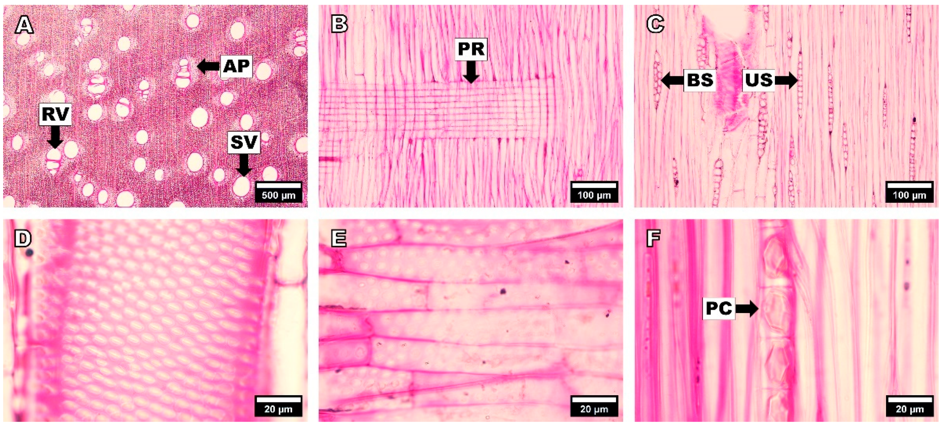
3.1.1. Acacia mangium

Figure 1 displays optical micrographs of the cross, radial, and tangential sections of A. mangium wood. In the cross-section, A. mangium showed indistinct growth ring boundaries. Wood porosity was diffuse with solitary and radial multiples vessels (Figure 1A). The arrangement of axial parenchyma was aliform, vasicentric, and confluent (Figure 1A). Tyloses were absent.
In the radial section, A. mangium showed a simple perforation plate, all body ray cells procumbent (Figure 1B), and distinct borders of vessel-ray pits, which were similar to intervessel pits (Figure 1E). Prismatic crystals were observed in the chambered axial parenchyma cells (Figure 1F).
In the tangential section, uniseriate and biseriate rays (Figure 1C) and fibers with simple to minutely bordered pits, which were non-septate (Figure 1C), were observed. Intervessel pits with a polygonal shape were alternated and vestured (Figure 1D).
For quantitative anatomical characteristics, the tangential vessel diameter and vessel numbers of A. mangium were 155.1 (116–200) µm and 8.7 (5–15)/mm², respectively. The fiber length, fiber diameter, lumen diameter, and fiber wall thickness were 882.0 (630–1300) µm, 14.1 (9–20) µm, 10.9 (7–17) µm, and 1.62 (1.14–2.37) µm, respectively. The lumen diameter was more than three times wider than the double-wall thickness of the fiber, which is categorized as a very thin-walled fiber according to the IAWA list [27]. The ray height and rays per millimeter were 200.0 (129–365) µm and 6.5 (3–11)/mm, respectively.
The qualitative anatomical characteristics of A. mangium in the present study are consistent with those reported in previous studies. As reported by Sahri et al. [29], Ogata et al. [30], Kim et al. [31], Andianto et al. [32], and Savero et al. [18], the wood porosity of A. mangium was diffused with solitary and radial multiple vessels. A. mangium also showed aliform and vasicentric axial parenchyma, non-septate fibers, and mostly uniseriate rays. In addition, prismatic crystals existed in the axial parenchyma of A. mangium.
The quantitative anatomical characteristics of A. mangium in the present study were also consistent with those reported in previous studies. Sahri et al. [29] reported that A. mangium from Malaysia had tangential vessel diameters of 132–167 µm and vessel numbers of 4–8/mm². Ogata et al. [30] reported these values to be 150–280 µm and 4–9/mm², respectively. Moreover, Andianto et al. [32] reported that A. mangium from Indonesia had tangential vessel diameters of 201 µm. Savero et al. [18] reported that the tangential vessel diameters and vessel numbers of A. mangium from Vietnam were 149 µm and 9/mm², respectively. However, in terms of fiber dimensions, our values were lower than those reported in the previous studies. The previously reported fiber length and fiber wall thickness of A. mangium were 934–1018 µm and 3.3–4.3 µm [29], 700–1400 µm and 1.5–2.5 µm [30], and 1215–1240 µm and 3.7 µm [32], respectively.

3.1.2. Acacia hybrid

Optical micrographs of the cross, radial, and tangential sections of A. hybrid are presented in Figure 2. In the cross-section, A. hybrid showed indistinct growth ring boundaries and was diffuse-porous with solitary and radial multiple vessels (Figure 2A). The arrangement of axial parenchyma was aliform, vasicentric, and confluent (Figure 2A). Tyloses were not observed in the vessels.
In the radial section, the rays were composed of procumbent body ray cells (Figure 2B). Vessel-ray pits showed distinct borders, which were similar to intervessel pits (Figure 2E). A. hybrid showed simple perforation plates. There were prismatic crystals in the chambered axial parenchyma cells (Figure 2F).
In the tangential section, the rays were uniseriate and biseriate (Figure 2C). Fibers with simple to minutely bordered pits, which were non-septate, were also present (Figure 2C). The intervessel pits showed an alternate arrangement with a polygonal shape, and these pits were vestured (Figure 2D).
The tangential vessel diameters and vessel numbers in A. hybrid were 145.1 (86–261) µm and 7.1 (4–11)/mm², respectively. The fiber length was 853.2 (509–1347) µm. Fiber diameter, lumen diameter, and fiber wall thickness were 15.7 (11–21) µm, 12.4 (8–17) µm, and 1.66 (0.98–2.45) µm, respectively. The lumen diameter was greater than three times of the double-wall fiber thickness, showing a very thin-walled fiber according to the IAWA list [27]. The ray height and number were 239.9 (141–487) µm and 5.5 (3–9)/mm, respectively.
The anatomical characteristics of A. hybrid in the present study were consistent with those of a few previous studies. As reported by Praptoyo [33], Nirsatmanto et al. [34], and Savero et al. [18], A. hybrid exhibited diffuse porosity with solitary and radial multiple vessels, with vasicentric axial parenchyma and uniseriate and biseriate rays. Praptoyo [33] reported that A. hybrid from Indonesia had a vessel tangential diameter of 113–200 µm and vessel numbers of 4–7/mm², respectively. Moreover, Nirsatmanto et al. [34] and Savero et al. [18] reported these values to be 115–145 µm and 5–9/mm² and 133 µm and 7/mm² in A. hybrid from Indonesia and Vietnam, respectively. Moreover, the previously reported value of the fiber length and fiber wall thickness of A. hybrid was 1030–1040 µm and 2.0–5.2 µm [33] and 780–850 µm and 1.2–1.8 µm [34], respectively. These results indicate that, in terms of fiber dimensions, A. hybrid in the present study showed a lower fiber dimension than in previous studies.

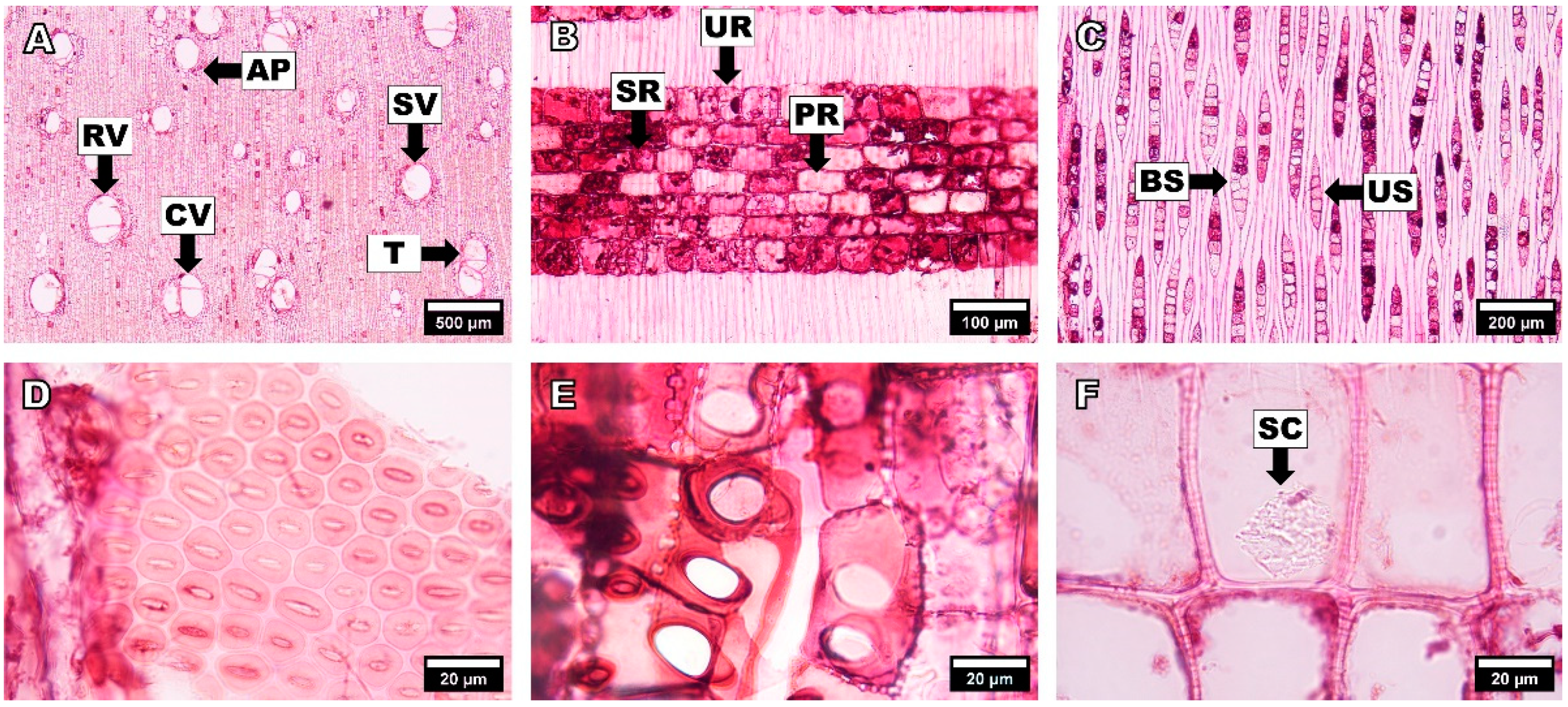
3.1.3. Dillenia pentagyna

Figure 3 shows optical micrographs of the cross, radial, and tangential sections of D. pentagyna. In the cross-section, D. pentagyna had indistinct growth ring boundaries and diffuse porosity with solitary and tangential multiples vessels (Figure 3A). The axial parenchyma was diffuse, diffuse-in-aggregate, and scanty paratracheal (Figure 3A). Tyloses were absent in the vessels.
In the radial section, the rays were composed of procumbent body ray cells with over four rows of upright and/or square marginal cells (Figure 3B). The perforated plates were scalariform. Disjunctive ray parenchyma cell walls were also observed. Vessel-ray pits were much reduced in borders to apparently simple with rounded or angular shapes (Figure 3E). Raphides were observed in the body ray cells (Figure 3F).
In the tangential section, the ray width was uniseriate and multiseriate with 4–7 seriates (Figure 3C). Non-septate fibers with bordered pits in both the radial and tangential walls were present (Figure 3C). The intervessel pits were arranged in a scalariform and opposite manner (Figure 3D).
The quantitative anatomical characteristics of D. pentagyna were observed as follows: tangential diameter of vessel lumina of 163.9 (91–216) µm, vessels per square millimeter of 9.1 (6–15)/mm², fiber length of 1405.0 (944–1791) µm, fiber diameter of 48.8 (39–59) µm, fiber lumen diameter of 23.5 (18–33) µm, and a fiber wall thickness of 12.7 (9–16) µm. The fiber lumen diameter was almost equal to the double-wall thickness of the fiber, which is classified as thin- to thick-walled according to the IAWA list [27]. The ray height was greater than 1 mm.
According to Martawijaya et al. [35], Ogata et al. [30], and Itoh et al. [10], Dillenia spp. are diffuse-porous with exclusively solitary vessels and non-septate fibers with large bordered pits. The axial parenchyma was diffuse, reticulate, and scanty and paratracheal. The rays consisted of two distinct sizes: uniseriate and broad rays. Raphides were present in the ray cells.
Studies on the quantitative anatomical characteristics of Dillenia spp. are relatively few. Martawijaya et al. [35] reported that the vessel lumina diameter, vessels per square millimeter, and fiber length of Dillenia spp. grown in Indonesia were 100–200 µm, 3–7/mm², and 2852 µm, respectively. Ogata et al. [30] reported these parameters to be 40–240 µm, 4–11/mm², and 2200–3000 µm, respectively. These results indicate that our findings on vessel lumina diameter and vessels per square millimeter were in line with those of previous studies, whereas the fiber length determined in the present study was shorter than that in previous studies. These differences in anatomical characteristics could be attributed to different growing sites.

3.1.4. Anacardium occidentale

Figure 4 shows optical micrographs of the cross, radial, and tangential sections of A. occidentale. In the cross-section, A. occidentale had indistinct growth ring boundaries, diffuse-porous with partly solitary, radial multiples, and very small cluster vessels (Figure 4A). The arrangement of axial parenchyma was vasicentric, aliform, lozenge-aliform, and confluent (Figure 4A). Tyloses were also present in the vessels (Figure 4A).
In the radial section, the rays consisted of procumbent, square, and upright cells (Figure 4B). We also observed vessel-ray pits with significantly reduced borders that were apparently simple with rounded or angular shapes (Figure 4E); moreover, simple perforated plates were also observed. Silica bodies were observed in the ray cells (Figure 4F).
In the tangential section, the rays were mostly uniseriate and sometimes biseriate (Figure 4C). The fibers were non-septate with simple to minutely bordered pits (Figure 4C). Intervessel pits with alternating polygonal shapes were observed (Figure 4D).
In terms of quantitative anatomical characteristics, the tangential diameter of vessel lumina and vessels per square millimeter in A. occidentale were 155.2 (69–284) µm and 4.9 (1–8)/mm², respectively. Fiber properties of A. occidentale were observed as follows: length, 622.5 (427–1058) µm; diameter, 14.2 (11–18) µm; lumen diameter, 11.4 (9–15) µm; and wall thickness, 1.39 (1.06–1.75) µm. The fiber was classified as a very thin-walled fiber according to the IAWA list [27]. The ray height and rays per millimeter were 374.6 (158–843) µm and 10.6 (6–15)/mm², respectively.
As reported by Terrazas Salgado [36] and Gupta and Agarwal [37], A. occidentale comprises diffuse-porous wood with solitary and radial multiple vessels. Tyloses are present in the vessels. The fibers were non-septate with vasicentric, aliform, lozenge-aliform, and confluent axial parenchyma. The rays were uniseriate and biseriate, with silica bodies in the ray cells. Terrazas Salgado [36] reported the vessel lumina tangential diameter, vessels per square millimeter, and fiber length of A. occidentale to be 22–330 µm, 5–14/mm², and 607–1228 µm, respectively. Moreover, Gupta and Agarwal [37] reported the values to be 54–200 µm, 3–13/mm², and 481–856 µm, respectively.

3.1.5. Hevea brasiliensis

Optical micrographs of the cross, radial, and tangential sections of H. brasiliensis are shown in Figure 5. In the cross-section, H. brasiliensis had indistinct growth ring boundaries and was diffuse-porous with solitary, radial multiples, and very small cluster vessels (Figure 5A). The axial parenchyma was vasicentric and banded, including reticulate, narrow, and marginal bands (Figure 5A). Tyloses were rarely found (Figure 5A).
In the radial section, the rays comprised procumbent body ray cells with mostly 2–4 rows of upright and/or square marginal cells (Figure 5B). Vessel-ray pits showed significantly reduced borders and appeared simple with rounded or angular shapes (Figure 5E). Simple perforated plates were also observed. Prismatic crystals were observed in the chambered axial parenchyma cells (Figure 5F).
In the tangential section, H. brasiliensis showed uniseriate rays, a smaller multiseriate ray with 2–3 seriates, and larger multiseriate rays with 4–5 seriates (Figure 5C). Fibers with simple to minutely bordered pits, which were non-septate, were observed (Figure 5C). The intervessel pits were arranged alternately in a polygonal shape (Figure 5D).
The quantitative anatomical characteristics of H. brasiliensis were as follows: tangential diameter of vessel lumina, 173.6 (82–260) µm; vessels per square millimeter, 4.8 (1–11)/mm²; fiber length, 1287.7 (782–2793) µm; fiber diameter, 17.2 (14–23) µm; fiber lumen diameter, 9.9 (7–15) µm, and fiber wall thickness, 3.66 (2.78–5.04) µm. The fiber lumen diameter was classified into a thin- to thick-walled fiber according to the IAWA list [27]. H. brasiliensis had a ray height of 479.8 (135–878) µm and ray number of 8.4 (6–11)/mm.
As reported by Ogata et al. [30] and Perdigão et al. [38], the wood porosity of H. brasiliensis diffuses with solitary and radial multiples vessels. It has non-septate fibers and vasicentric, reticulate, and banded axial parenchyma. The rays are uniseriate and multiseriate. There were prismatic crystals in the axial parenchyma. However, with respect to the existence of tyloses, there have been controversial findings. Ogata et al. [30] reported that tyloses were present in H. brasiliensis, whereas Perdigão et al. [38] reported that tyloses were absent.
In terms of quantitative anatomical characteristics, Ogata et al. [30] showed that tangential vessel lumina diameters, vessels per square millimeter, and the fiber length of H. brasiliensis were 100–280 µm, 1–4/mm², and 800–1600 µm, respectively. Perdigão et al. [38] reported tangential vessel diameters of 71–249 µm and vessel numbers of 1–15/mm².

3.1.6. Melaleuca cajuputi

Optical micrographs of the cross, radial, and tangential sections of M. cajuputi are shown in Figure 6. In the cross-section, M. cajuputi had indistinct growth ring boundaries. Wood porosity was diffuse, with exclusively solitary vessels (90% or more) (Figure 6A). The axial parenchyma was diffuse, confluent, and had narrow bands or lines up to three cells wide (Figure 6A). Tyloses were not observed in the vessels.
In the radial section, the body ray cells were procumbent, with one row of upright and/or square marginal cells (Figure 6B). The vessel-ray pits showed significantly reduced borders compared to simple and rounded or angular pits (Figure 6E). The perforated plates were simple. Silica bodies were observed in the ray cells (Figure 6B,F).
In the tangential section, the ray was exclusively uniseriate (Figure 6C). The fibers were non-septate with bordered pits in both the radial and tangential walls (Figure 6C). Intervessel pits showed alternating arrangements and were vestured (Figure 6D).
The tangential vessel diameter and vessel numbers for M. cajuputi were 119.6 (51–166) µm and 9.2 (5–16)/mm², respectively. The fiber length, fiber diameter, lumen diameter, and fiber wall thickness were 821.5 (597–1224) µm, 15.3 (9–19) µm, 8.7 (5–12) µm, and 3.26 (2.29–4.18) µm, respectively. The lumen diameter was less than three times wider than the double-wall thickness of the fiber, which is categorized as a thin- to thick-walled fiber according to the IAWA list [27]. The ray height and rays per millimeter were 259.0 (71–470) µm and 11.1 (8–15)/mm, respectively.
Ingle and Dadswell [39], Ogata et al. [30], and Itoh et al. [10] reported that Melaleuca spp. are diffuse-porous with exclusively solitary vessels. It had non-septate fibers with distinctly bordered pits. The axial parenchyma was diffuse and had a narrow, discontinuous band. The rays were exclusively uniseriate and silica bodies were present in the ray cells. Ingle and Dadswell [39] reported that the vessel diameter, vessel numbers, and fiber length of Melaleuca spp. were 45–180 µm, 5–20/mm², and 430–1130 µm, respectively. Moreover, Ogata et al. [30] reported these values to be 130–200 µm, 11–15/mm², and 800–1200 µm, respectively.

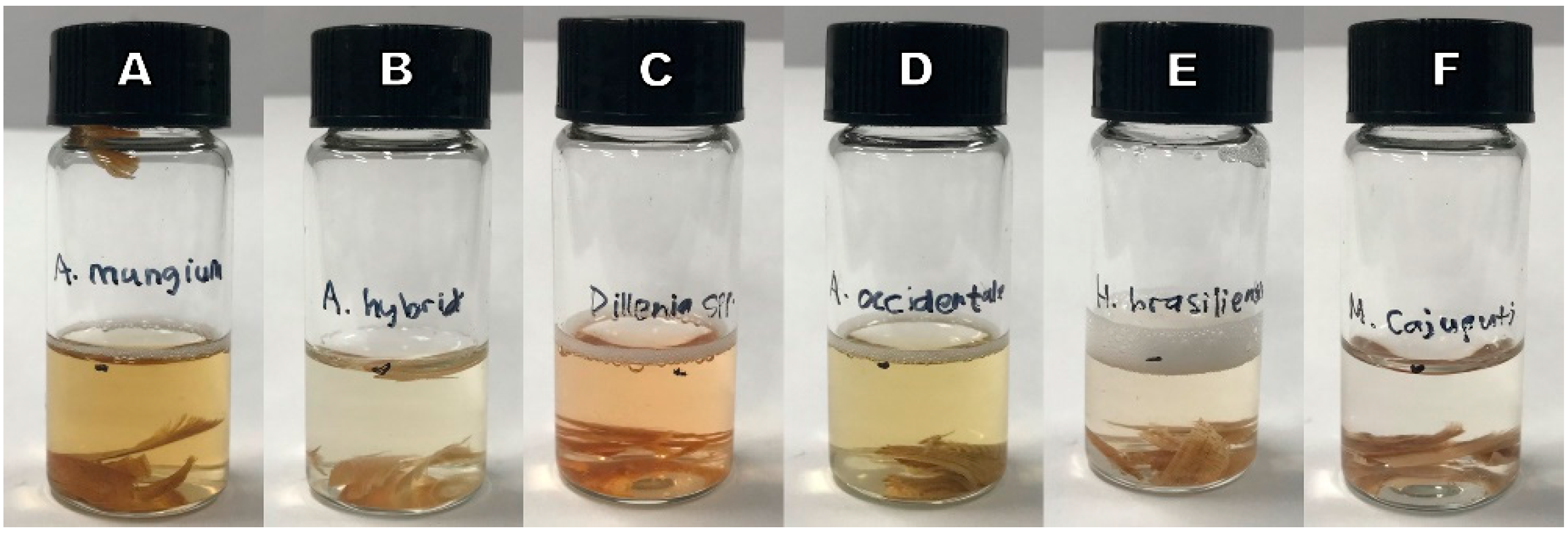
3.2. Non-Anatomical Characteristics

3.2.1. Acacia mangium

Of the non-anatomical characteristics of A. mangium, the heartwood presented yellow or green fluorescence (Figure 7A). The water- and ethanol-extracted samples showed a brown and yellow color (Figure 8) with no fluorescence. The froth test showed a weakly positive reaction (Figure 9A).
The heartwood fluorescence, water extract color and fluorescence, and froth test results of A. mangium in the present study are in line with Acacia spp. of previous studies [26,27,40].

3.2.2. Acacia hybrid

The heartwood of A. hybrid showed a yellow or green fluorescence color similar to that of A. mangium (Figure 7B), and the extracts from water and ethanol showed no fluorescence. However, the color of A. hybrid water and ethanol extracts differed from that of A. mangium, displaying shades of yellow (Figure 8). The difference in extract color between A. hybrid and A. mangium may be due to the extractives content of both species. According to Moya et al. [41], the lightness (L*) parameters in wood color showed a significantly negative correlation with extractives content in the wood, whereas the red-green (a*) parameters had a positive correlation with extractives content. Froth test results were negative (Figure 9B). The difference in the froth reaction between A. hybrid and A. mangium could be attributed to the saponin content in the heartwood [27].
The present study showed similar fluorescence characteristics for the heartwood and extracts from water and ethanol to Acacia spp. In previous studies [26,27], Acacia spp. showed yellow or green fluorescence from the heartwood and no fluorescence from water and ethanol extracts. However, the froth test showed a weakly positive reaction, which was different from that of A. hybrid in the present study.

3.2.3. Dillenia pentagyna

The heartwood of D. pentagyna was not fluorescent (Figure 7C). The water extract presented purple fluorescence and a red or orange color (Figure 8A), whereas the ethanol extract showed purple fluorescence and shades of red color (Figure 8B). The froth test showed a weakly positive reaction (Figure 9C).
The results of the present study on heartwood fluorescence are consistent with those of a previous study. As reported by Avella et al. [42], 27 Dilleniaceae species had no heartwood fluorescence.

3.2.4. Anacardium occidentale

Anacardium occidentale heartwood exhibited green or blue fluorescence (Figure 7D). The water extract showed a yellow color (Figure 8A) without fluorescence, whereas the ethanol extract presented shades of yellow color (Figure 8B) without fluorescence. The froth test was weakly positive (Figure 9D), similar to that of A. mangium and D. pentagyna.
As reported by the IAWA committee [27], the heartwood of Anacardium spp. shows yellow or green fluorescence, which is in line with the results of the present study.

3.2.5. Hevea brasiliensis

The heartwood of H. brasiliensis exhibited no fluorescence (Figure 7E). Both the water and ethanol extracts showed shades of red color (Figure 8) with blue fluorescence. A large amount of froth was observed, indicating a positive reaction for the froth test (Figure 9E).
Silva Guzmán et al. [43] reported that H. brasiliensis from Mexico was non-fluorescent for the heartwood, displayed blue or green fluorescence for the water extract test and purple fluorescence for the ethanol extract test, and had a negative reaction for the froth test.

3.2.6. Melaleuca cajuputi

Blue or purple fluorescence was observed for the heartwood of M. cajuputi (Figure 7F). In the water and ethanol extracts, M. cajuputi was neither fluorescent nor colorless (Figure 8). In addition, the froth test result was negative (Figure 9F).

3.3. Summary of Anatomical and Non-Anatomical Characteristics

The anatomical and non-anatomical characteristics of the six Vietnamese wood species based on the IAWA feature lists are presented in Table 2 and Table 3, respectively.

4. Conclusions

Regarding anatomical characteristics, all species showed similar characteristics in a few parameters such as indistinct growth ring, diffuse porosity, non-septate fibers, vessel diameter of 100–200 µm, and ray number of 4–12/mm. All the species showed distinctive characteristics in most of the evaluated parameters, especially in axial parenchyma, ray width and composition, and mineral inclusion.
In terms of non-anatomical characteristics, all species had distinctive heartwood fluorescence and color in the water and ethanol extracts. The fluorescence of the water and ethanol extracts was only observed for D. pentagyna and H. brasiliensis. The froth test showed a positive reaction for H. brasiliensis, a weakly positive reaction for A. mangium, D. pentagyna, and A. occidentale, and a negative reaction for A. hybrid and M. cajuputi.
In summary, the six wood species from the plantation in Vietnam exhibited distinctive differences in anatomical and non-anatomical characteristics. Our results can be used as identification keys and quality indices for the effective utilization of each species.

Author Contributions

Conceptualization, A.M.S., J.H.K. and N.H.K.; methodology, A.M.S., J.H.K., B.D.P., D.P., S.H.P., D.V.D. and N.H.K.; software, A.M.S.; validation, N.H.K.; formal analysis, A.M.S. and J.H.K.; investigation, A.M.S.; resources, N.H.K.; data curation, A.M.S. and J.H.K.; writing—original draft preparation, A.M.S. and J.H.K.; writing—review and editing, A.M.S., J.H.K., B.D.P., D.V.D. and N.H.K.; visualization, A.M.S.; supervision, N.H.K.; project administration, J.H.K.; funding acquisition, N.H.K. All authors have read and agreed to the published version of the manuscript.

Funding

This research was supported by the Science and Technology Support Program through Dongwha Enterprise Co. Ltd.; the National Research Foundation of Korea (NRF) funded by the Ministry of Science and ICT (MSIT) (NRF-2019K1A3A9A01000018, NRF-2022R1A2C1006470); the Basic Science Research Program through the NRF funded by the Ministry of Education (NRF-2018R1A6A1A03025582), and the R&D Program for Forest Science Technology (Project No. 2021350C10-2223-AC03) provided by the Korea Forest Service (Korea Forestry Promotion Institute).

Institutional Review Board Statement

Not applicable.

Informed Consent Statement

Not applicable.

Data Availability Statement

The datasets generated and analyzed during the current study are not publicly available, but are available from the corresponding author upon reasonable request.

Conflicts of Interest

The authors declare no conflict of interest.

References

  1. Hong, B. Ministry of Agriculture and Rural Development: Announcement of Forest Status in 2021. Available online: https://www.vietnamplus.vn/bo-nong-nghiepphat-trien-nong-thon-cong-bo-hien-trang-rung-nam-2021/810161.vnp (accessed on 16 August 2022). (In Vietnamese).
  2. Ministry of Agriculture and Rural Development. Report on the Implementation of the Agriculture and Rural Areas Development Plan in 2021 and Implementation in 2022; MARD: Hanoi, Vietnam, 2021. (In Vietnamese) [Google Scholar]
  3. Ministry of Agriculture and Rural Development. Vietnam’s Forestry Export in 2021. Available online: https://www.mard.gov.vn/en/Pages/vietnam%E2%80%99s-forestry-export-up-20-percent-in-2021.aspx (accessed on 25 August 2022).
  4. Forest Science Institute of Vietnam. Vietnam Forestry Outlook Study, Working Paper Series; Food and Agriculture Organization of the United Nations (FAO): Bangkok, Thailand, 2009. [Google Scholar]
  5. Sein, C.C.; Mitlöhner, R. Acacia Mangium Willd: Ecology and Silviculture in Vietnam; Center for International Forestry Research (CIFOR): Bogor, Indonesia, 2011; ISBN 978-602-8693-73-8. [Google Scholar]
  6. Sein, C.C.; Mitlöhner, R. Acacia hybrid: Ecology and Silviculture; Center for International Forestry Research (CIFOR): Bogor, Indonesia, 2011; ISBN 978-602-8693-71-4. [Google Scholar]
  7. Tenorio, C.; Moya, R.; Quesada-Pineda, H.J. Kiln drying of Acacia mangium wood: Colour, shrinkage, warp, split, and check in dried lumber. J. Trop. For. Sci. 2017, 24, 125–139. [Google Scholar]
  8. Teoh, Y.P.; Don, M.M.; Ujang, S. Assessment of the properties, utilization, and preservation of rubberwood (Hevea brasiliensis): A case study in Malaysia. J. Wood Sci. 2011, 57, 255–266. [Google Scholar] [CrossRef]
  9. Van Sam, H.; Nanthavong, K.; Kessler, P.J.A. Trees of Laos and Vietnam: A field guide to 100 economically or ecologically important species. Blumea 2004, 49, 201–349. [Google Scholar] [CrossRef] [Green Version]
  10. Itoh, T.; Pan, B.; Sano, Y.; Baas, P.; Luo, J.; Li, D.; Cui, Y. Anatomical Database and Atlas of Chinese Woods; Kaiseisha Press: Otsu, Japan, 2022; ISBN 978-4-86099-198-2. [Google Scholar]
  11. Lim, T.K. Edible Medicinal and Non-Medicinal Plants; Springer: Berlin/Heidelberg, Germany, 2012; Volume 1, ISBN 978-90-481-8661-7. [Google Scholar]
  12. Nuyim, T. Potentiality of Melaleuca cajuputi Powell Cultivation to Develop for Economic Plantation Purpose; Royal Forest Department: Bangkok, Thailand, 1998. [Google Scholar]
  13. Oskolski, A.A. Wood anatomy of Schefflera and related taxa (Araliaceae). IAWA J. 1995, 16, 159–190. [Google Scholar] [CrossRef]
  14. Ickert-Bond, S. Reexamination of wood anatomical features in Pinus krempfii (Pinaceae). IAWA J. 2001, 22, 355–365. [Google Scholar] [CrossRef] [Green Version]
  15. Kim, N.T.; Ochiishi, M.; Matsumura, J.; Oda, K. Variation in wood properties of six natural Acacia hybrid clones in Northern Vietnam. J. Wood Sci. 2008, 54, 436–442. [Google Scholar] [CrossRef]
  16. Van Duong, D.; Schimleck, L.; Dinh, T.T.; Tran, C.V. Radial variation in cell morphology of Melia azedarach planted in Northern Vietnam. MADERAS Cienc. Tecnol. 2021, 23, 1–10. [Google Scholar] [CrossRef]
  17. Phuong, L.X.; Tuong, V.M.; Loan, N.T.; Hai, D.T.; Tuyen, L.M. Develop an app to quickly identify popular timber species traded in Hanoi, Vietnam. In Proceedings of the 9TH Hardwood Conference, Sopron, Hungary, 24–25 June 2021. [Google Scholar]
  18. Savero, A.M.; Kim, J.H.; Purusatama, B.D.; Prasetia, D.; Park, S.H.; Kim, N.H. A comparative study on the anatomical characteristics of Acacia mangium and Acacia hybrid grown in Vietnam. Forests 2022, 13, 1700. [Google Scholar] [CrossRef]
  19. Korea Forest Service. Roadmap for Wood Supply in 2020; Department of Forest Resources in Korea Forest Service: Daejeon, Korea, 2020. (In Korean) [Google Scholar]
  20. Kim, J.H.; Jang, J.H.; Ryu, J.Y.; Hwang, W.J.; Febrianto, F.; Kim, N.H. Comparison of anatomical characteristics of white jabon and red jabon grown in Indonesia. J. Korean Wood Sci. Technol. 2013, 41, 327–336. [Google Scholar] [CrossRef] [Green Version]
  21. Kim, J.H.; Jang, J.H.; Ryu, J.Y.; Febrianto, F.; Hwang, W.J.; Kim, N.H. Physical and mechanical properties of major plantation and promising tree species grown in Indonesia (I). J. Korean Wood Sci. Technol. 2014, 42, 467–476. [Google Scholar] [CrossRef]
  22. Park, S.H.; Jang, J.H.; Qi, Y.; Hidayat, W.; Febrianto, F.; Kim, N.H. Color change of major wood species planted in Indonesia by ultraviolet radiation. J. Korean Wood Sci. Technol. 2016, 44, 9–18. [Google Scholar] [CrossRef] [Green Version]
  23. Kim, J.H.; Kim, D.H.; Kim, S.H.; Suri, I.F.; Purusatama, B.D.; Jo, J.I.; Lee, H.S.; Hidayat, W.; Febrianto, F.; Lee, S.H.; et al. Comparison of anatomical features in the three Syzygium species. BioResources 2021, 16, 3631–3642. [Google Scholar] [CrossRef]
  24. Park, S.H.; Jang, J.H.; Hidayat, W.; Qi, Y.; Febrianto, F.; Kim, N.H. Combustion properties of major wood species planted in Indonesia. J. Korean Wood Sci. Technol. 2015, 43, 768–776. [Google Scholar] [CrossRef]
  25. Korea Forest Service. Developing Overseas Forest Resources in 2019. Available online: https://english.forest.go.kr/kfsweb/kfi/kfs/cms/cmsView.do?cmsId=FC_002092&mn=UENG_06_04_01 (accessed on 25 June 2022).
  26. Dyer, S.T. Wood fluorescence of indigenous South African trees. IAWA Bull. 1988, 9, 75–87. [Google Scholar] [CrossRef]
  27. IAWA Committee. IAWA list of microscopic features for hardwood identification. IAWA Bull. 1989, 10, 219–332. [Google Scholar]
  28. Park, S.J.; Lee, J.Y.; Cho, N.S.; Cho, B.M. Wood Science Experiment Book; Gwangil Munhwasa: Seoul, Korea, 1993; p. 54. (In Korean) [Google Scholar]
  29. Sahri, H.M.; Ibrahim, F.H.; Shukor, N.A.A. Anatomy of Acacia mangium grown in Malaysia. IAWA J. 1993, 14, 245–251. [Google Scholar] [CrossRef]
  30. Ogata, K.; Fuji, T.; Abe, H.; Baas, P. Identification of the Timbers of Southeast Asia and the Western Pacific; Kaiseisha Press: Otsu, Japan, 2008; ISBN 978-4-86099-244-6. [Google Scholar]
  31. Kim, J.H.; Jang, J.H.; Kwon, S.M.; Febrianto, F.; Kim, N.H. Anatomical properties of major planted and promising species growing in Indonesia. J. Korean Wood Sci. Technol. 2012, 40, 244–256. [Google Scholar] [CrossRef] [Green Version]
  32. Andianto; Yuniarti, K.; Saputra, N.A.; Saputra, I.S. Fiber dimension and anatomy of Acacia mangium wood from two mother trees. In Proceedings of the International Conference on Forest Products (ICFP) 2020: 12th International Symposium of IWORS, Bogor, Indonesia, 1 September 2020; 2020; Volume 935. [Google Scholar] [CrossRef]
  33. Praptoyo, H. Variation of anatomical features between two Acacia hybrids (Acacia mangium x auriculiformis) and (Acacia auriculiformis x mangium) from Wonogiri. In Proceedings of the 7th International Symposium of IWoRS, Bandung, Indonesia, 5–6 November 2015; pp. 23–31. [Google Scholar]
  34. Nirsatmanto, A.; Sunarti, S.; Praptoyo, H. Wood anatomical structures of tropical acacias and its implication to tree breeding. Int. J. For. Hortic. 2017, 3, 9–16. [Google Scholar] [CrossRef]
  35. Martawijaya, A.; Kartasujana, I.; Mandang, Y.I.; Prawira, S.A.; Kadir, K. Indonesian Wood Atlas; Ministry of Forestry: Bogor, Indonesia, 2005; Volume II. (In Indonesian) [Google Scholar]
  36. Terrazas Salgado, T. Wood Anatomy of the Anacardiaceae: Ecological and Phylogenetic Interpretation. Ph.D. Dissertation, University of North Carolina, Chapel Hill, NC, USA, 1994. [Google Scholar]
  37. Gupta, S.; Agarwal, M. Wood anatomy of Anacardiaceae from India with special reference of systematic position of Rhus. IAWA J. 2008, 29, 79–106. [Google Scholar] [CrossRef]
  38. Perdigão, C.R.V.; Júnior, M.M.B.; Gonçalves, T.A.P.; Araujo, C.S.; Mori, F.A.; Barbosa, A.C.M.C.; de Souza, F.I.B.; Motta, J.P.; Melo, L.E.L. Forestry control in the Brazilian Amazon I: Wood and charcoal anatomy of three endangered species. IAWA J. 2020, 41, 490–509. [Google Scholar] [CrossRef]
  39. Ingle, H.D.; Dadswell, H.E. The anatomy of timbers of the south-west Pacific area, III. Myrtaceae. Aust. J. Bot. 1953, 1, 353–401. [Google Scholar] [CrossRef]
  40. Quirk, J.T. Data for a computer-assisted wood identification system, 1. Commercial legumes of tropical Asia and Australia. IAWA Bull. 1983, 4, 118–130. [Google Scholar] [CrossRef]
  41. Moya, R.; Fallas, R.S.; Bonilla, P.J.; Tenorio, C. Relationship between wood color parameters measured by the CIELab system and extractive and phenol content in Acacia mangium and Vochysia guatemalensis from fast-growth plantations. Molecules 2012, 17, 3639–3652. [Google Scholar] [CrossRef] [PubMed] [Green Version]
  42. Avella, T.; Dechamps, R.; Bastin, M. Fluorescence study of 10,610 woody species from the Tervuren (Tw) collection, Belgium. IAWA Bull. 1988, 9, 346–352. [Google Scholar] [CrossRef]
  43. Silva Guzmán, J.A.; Richter, H.G.; Rodríguez Anda, R.; Fuentes Talavera, F.J. Wood fluorescence of commercial timbers marketed in Mexico. IAWA J. 2008, 29, 311–322. [Google Scholar] [CrossRef] [Green Version]
Figure 1. Optical micrographs of Acacia mangium. Cross-section (A), radial section (B), tangential section (C), intervessel pits (D), vessel-ray pits (E), and mineral inclusions (F). Solitary vessel (SV), radial multiple vessel (RV), axial parenchyma (AP), procumbent body ray (PR), uniseriate ray (US), biseriate ray (BS), and prismatic crystal (PC).
Forests 14 00496 g001
Figure 2. Optical micrographs of Acacia hybrid. Cross-section (A), radial section (B), tangential section (C), intervessel pits (D), vessel-ray pits (E), and mineral inclusions (F). Solitary vessel (SV), radial multiple vessel (RV), axial parenchyma (AP), procumbent body ray (PR), uniseriate ray (US), biseriate ray (BS), and prismatic crystal (PC).
Forests 14 00496 g002
Figure 3. Optical micrographs of Dillenia pentagyna Cross-section (A), radial section (B), tangential section (C), intervessel pits (D), vessel-ray pits (E), and mineral inclusions (F). Solitary vessel (SV), tangential multiple vessel (TV), axial parenchyma (AP), procumbent body ray (PR), square body ray (SR), upright body ray (UR), uniseriate ray (US), multiseriate ray (MS), and raphides (RC).
Forests 14 00496 g003
Figure 4. Optical micrographs of Anacardium occidentale. Cross-section (A), radial section (B), tangential section (C), intervessel pits (D), vessel-ray pits (E), and mineral inclusions (F). Solitary vessel (SV), radial vessel (RV), cluster vessel (CV), axial parenchyma (AP), tyloses (T), procumbent body ray (PR), square body ray (SR), upright body ray (UR), uniseriate ray (US), biseriate ray (BS), and silica bodies (SC).
Forests 14 00496 g004
Figure 5. Optical micrographs of Hevea brasiliensis. Cross-section (A), radial section (B), tangential section (C), intervessel pits (D), vessel-ray pits (E), and mineral inclusions (F). Solitary vessel (SV), radial vessel (RV), cluster vessel (CV), axial parenchyma (AP), tyloses (T), procumbent body ray (PR), square body ray (SR), upright body ray (UR), uniseriate ray (US), multiseriate ray (MS), and prismatic crystal (PC).
Forests 14 00496 g005
Figure 6. Optical micrographs of Melaleuca cajuputi. Cross-section (A), radial section (B), tangential section (C), intervessel pits (D), vessel-ray pits (E), and mineral inclusions (F). Solitary vessel (SV), axial parenchyma (AP), procumbent body ray (PR), square body ray (SR), upright body ray (UR), uniseriate ray (US), and silica bodies (SC).
Forests 14 00496 g006
Figure 7. Heartwood fluorescence of Acacia mangium (A), Acacia hybrid (B), Dillenia pentagyna (C), Anacardium occidentale (D), Hevea brasiliensis (E), and Melaleuca cajuputi (F). Disks (A,B,DF) and chips (C).
Forests 14 00496 g007
Figure 8. Water (A) and ethanol (B) extract color for Acacia mangium (1), Acacia hybrid (2), Dillenia pentagyna (3), Anacardium occidentale (4), Hevea brasiliensis (5), and Melaleuca cajuputi (6).
Forests 14 00496 g008
Figure 9. Froth tests of Acacia mangium (A), Acacia hybrid (B), Dillenia pentagyna (C), Anacardium occidentale (D), Hevea brasiliensis (E), and Melaleuca cajuputi (F).
Forests 14 00496 g009
Table 1. Fundamental information of six wood species samples.
Scientific NameLocationSample TypesDiameter (cm)Density (g/cm3)
A. mangiumPlantation by Dongwha Vietnam Co., Ltd., Vietnam (21°30′17.74″ N, 105°50′48.102″ E)Disk13.20.50
A. hybridDisk12.20.50
D. pentagynaChips-0.75
A. occidentalePlantation by VRG Dongwha, Vietnam (11°28′56.104″ N, 106°36′16.133″ E)Disk7.80.52
H. brasiliensisDisk16.60.71
M. cajuputiDisk6.50.74
Table 2. Anatomical characteristics of Vietnamese wood species based on International Association of Wood Anatomist (IAWA) feature lists.
ParametersVietnamese Wood Species
A. mangiumA.hybridD. pentagynaA. occidentaleH. brasiliensisM. cajuputi
Growth ringsIndistinct or absent (2)
PorosityWood diffuse-porous (5)
Vessel groupingsMultiples (10)Exclusively solitary (9)
Perforation platesSimple (13)Scalariform (14)Simple (13)
Intervessel pitsAlternate (22) with polygonal shape (23)Scalariform (20) and opposite (21)Alternate (22) with polygonal shape (23)Alternate (22)
Vestured pitsPresent (29)AbsentPresent (29)
Vessel-ray pittingDistinct borders, similar to intervessel pits (30)Much reduced borders, pits rounded or angular (31)
Vessel diameter100–200 µm (42)
Vessel number5–20/mm² (47)≤ 5/mm² (46)5–20/mm2 (47)
TylosesAbsentPresent (56)Absent
Fiber pitsSimple to minutely bordered pits (61)Distinctly bordered pits (62) and pits common in both radial and tangential walls (63)Simple to minutely bordered pits (61)Distinctly bordered pits (62) and pits common in both radial and tangential walls (63)
Septate fibersNon-septate (66)
Fiber wall thicknessVery thin-walled (68)Thin- to thick-walled (69)Very thin-walled (68)Thin- to thick-walled (69)
Fiber length≤ 900 µm (71)900–1600 µm (72)≤900 µm (71)900–1600 µm (72)≤900 µm (71)
* Axial parenchymaVasicentric (79), aliform (80), and confluent (83)Diffuse (76), diffuse-in-aggregates (77), and scanty paratracheal (78)Vasicentric (79), aliform (80), lozenge-aliform (81), and confluent (83)Vasicentric (79), reticulate (87), in narrow bands (86), and in marginal (89).Diffuse (76), confluent (83), and in narrow bands (86)
Ray width1 to 3 cells (97)1 to 3 cells (97) and larger rays commonly 4- to 10-seriate (98)1 to 3 cells (97) 1 to 3 cells (97) and larger rays commonly 4- to 10-seriate (98)Exclusively uniseriate (96)
Ray compositionAll cells procumbent (104)Procumbent with over 4 rows of upright and/or square marginal cells (108)Procumbent, square, and upright cells mixed throughout the ray (109)Procumbent with mostly 2–4 rows of upright and/or square marginal cells (107)Procumbent with one row of upright and/or square marginal cells (106)
Ray number4–12/mm (115)
Mineral inclusionsPrismatic crystals (142)Raphides (149)Silica (160)Prismatic crystals (142)Silica (160)
Note: The numbers in parentheses denote the IAWA feature list for hardwood identification. * Bold letters indicate the most common axial parenchyma.
Table 3. Non-anatomical characteristics of Vietnamese wood species based on IAWA feature lists.
ParametersVietnamese Wood Species
A. mangiumA. hybridD. pentagynaA. occidentaleH. brasiliensisM. cajuputi
Heartwood fluorescenceYellow or green (204)Not fluorescentGreen or blue (204)Not fluorescentBlue or purple (204)
Water extract fluorescenceNot fluorescentPurple (205)Not fluorescentBlue (205)Not fluorescent
Water extract colorBrown (206)Shades of yellow (208)Red or orange (207)Yellow (208)Shades of red (207)Colorless (206)
Ethanol extract fluorescenceNot fluorescentPurple (210)Not fluorescentBlue (210) Not fluorescent
Ethanol extract colorYellow (208)Shades of yellow (208)Shades of red (207)Shades of yellow (208)Shades of red (207)Colorless (206)
Froth testWeakly positive (215)NegativeWeakly positive (215)Positive (215)Negative
Note: The numbers in parentheses denote the IAWA feature list for hardwood identification.
Disclaimer/Publisher’s Note: The statements, opinions and data contained in all publications are solely those of the individual author(s) and contributor(s) and not of MDPI and/or the editor(s). MDPI and/or the editor(s) disclaim responsibility for any injury to people or property resulting from any ideas, methods, instructions or products referred to in the content.

