Article

Mycobiome of Post-Agricultural Soils 20 Years after Application of Organic Substrates and Planting of Pine Seedlings

1
Museum and Institute of Zoology, Polish Academy of Science, ul. Wilcza 64, 00-679 Warszawa, Poland
2
Faculty of Physics, Warsaw University of Technology, ul. Koszykowa 75, 00-662 Warszawa, Poland
3
Departement of Ecology, Forest Research Institute, ul. Braci Leśnej 3, 05-090 Sękocin Stary, Poland
4
Department of Biochemistry and Pharmacogenomics, Faculty of Pharmacy, Medical University of Warsaw, Banacha 1 Street, 02-097 Warsaw, Poland
5
Ukrainian Research Institute of Forestry and Forest Melioration Named after G. M. Vysotsky, Pushkinska Street, 86, 61024 Kharkiv, Ukraine
6
Department of Forest Entomology and Pathology, Faculty of Forestry and Wood Technology, Poznań University of Life Sciences, Wojska Polskiego 71c, 60-625 Poznań, Poland
7
Department of Plant Protection, Division of Plant Pathology and Mycology, Wrocław University of Environmental and Life Sciences, Grunwaldzki Square 24a, 50-363 Wroclaw, Poland
8
Institute for Sustainable Plant Protection (IPSP), National Research Council of Italy (CNR), Strada delle Cacce 73, 10135 Torino, Italy
9
Departement of Forest Protection, Forest Research Institute, ul. Braci Leśnej 3, 05-090 Sękocin Stary, Poland
*
Author to whom correspondence should be addressed.
Forests 2023, 14(1), 36; https://doi.org/10.3390/f14010036
Submission received: 29 November 2022 / Revised: 20 December 2022 / Accepted: 21 December 2022 / Published: 25 December 2022
(This article belongs to the Special Issue Biological Control in Forests Protection)

Abstract

:
A 20-year study of a pine stand on post-agricultural land showed that woody debris in the form of organic matter can be successfully used to restore symbiotic mycorrhizal communities, as is the case with forest soils. Woody substrates restored organic matter in soils altered by long agricultural use and had a positive effect on the composition of mycobiota antagonistic to pathogens, especially to Heterobasidion annosum, the causal agent of the dangerous disease root and stump rot of many forest tree species, including stands of Pinus sylvestris (L.). In a study that started in 2001 in the forest district of Czarne Człuchowskie (northern Poland), the following organic materials were used: wood residues (W), sawdust (S), bark compost (B), and compost applied to the root zone during planting (G). The organic materials were spread in the form of mulch over the entire area during planting. After twenty years, it was found that the substrates used provided suitable growth conditions for mycobiome useful for pines. The addition of organic matter did not change the alpha biodiversity of the soil, but in the long term led to significant changes in the composition of mycobiota (beta biodiversity). The changes in the soil after the addition of organic material naturally accelerated the formation of the forest habitat. A number of fungi evolved that degraded added lignin and cellulose while being antagonists of H. annosum and other pine pathogens. In particular, the well-known hyperpathogens of the genus Trichoderma played an important role by promoting resistance of the soil environment to pathogens. Soil enrichment by bark compost and wood residues increased the relative abundance of Trichoderma more than fourfold.Mycorrhizal fungi became dominant in soil enriched with organic matter. After enriching the soil with bark compost, the relative abundance of Amphinema and Inocybe increased to 5%. The relative abundance of Russula in soil enriched with wood residues and sawdust increased to 9% and 5%, respectively. Mycorrhizal fungi, e.g., of the genus Ąmanita, Rusula, which formed root mycorrhizae, not only increased the root receiving area many times over, but also protected the roots (mechanically and chemically from pathogens). Altogether, the observed positive changes increase the chances that the first generation of pines will survive on the ground.

1. Introduction

The circular economy recycles the waste produced, thus protecting the environment. Forestry can also join this trend by reforesting former agricultural land [1]. From an ecological point of view, these soils have been impoverished by many years of agricultural use. The first generation of Scots pine (Pinus sylvestris L.) on formerly agricultural land is doomed to die already at the age of the young trees (about 40 years) due to increased activity of insect pests and fungal pathogens (short-term forecast 2002). Of the latter, the root and butt rot pathogen of pines Heterobasidion annosum (Fr.) Bref. is the most dangerous in post-agricultural soils, where it has no natural counterparts (fungi and bacteria) and may cause severe losses on a massive scale [2]. Therefore, the restoration of a stable forest on post-agricultural land depends primarily on soil conditions [3]. Post-agricultural soils do not yet function as forest soils, and measures to accelerate processes for their improvement, especially their biological properties, are important [4]. Post-agricultural soils lack sufficient humus, and it is the organic horizon that has a decisive influence on most soil properties, especially on their retention, microbiological properties, and fertility. Organic matter improves moisture conditions, increases biodiversity, and provides a constant supply of nutrients from decomposing organic matter to soil mineral horizons and subsequently to growing trees [5]. Soil revitalization issues on post-agricultural land have been addressed in many research papers, with the prevailing view being that they are necessary to improve the fertility of poor post-agricultural soils and to initiate the development of forest ecosystems that are more resistant to disease threats. Converting land use from agriculture to forestry is a difficult and time-consuming process. Post-agricultural soils under long-term management are highly modified and are fundamentally different from forest soils [6,7].
Some of the wood wastes generated in technological processes, such as sawdust and residues from timber harvesting, have been used to accelerate the transformation of post-agricultural soils into forest soils [8], as well as for organic fertilization of land formerly used for agriculture [9]. Sawdust has also been used in nursery substrates as a carrier of antagonistic fungi (e.g., Trichoderma sp.) to increase the biological activity of the soil against pathogens of roots [10].
This study aimed to evaluate the changes in the soil 20 years after the application of various organic substances in a pine stand established on former arable land in the Czarne Człuchowskie forestry. One of the objectives was to verify whether the changes in mycobiota biodiversity are in the direction of the formation of mycorrhizal fungal communities characteristic of forest ecosystems. We also wanted to use a powerful tool, namely, next-generation sequencing (NGS), to find out to what extent the addition of various organic substances has a long-term effect on the composition of the soil mycobiome.

2. Materials and Methods

Test plots of 0.3 ha of Scots pine in the Czarne Człuchowskie forest district (subdistrict Brzezie, fresh forest site) were used for soil revitalization in 2001 (after autumn preparation by full plowing). The following substrates were applied separately on each experimental plot: bark compost (B), wood residues (W), gravel from bark compost (G), and sawdust (S). No treatments were applied to the control plot (C). In 2021, after 20 years, an identification of fungal DNA from soil was performed (using NGS).

2.1. Mulching of the Soil

The restoration of the soil’s organic horizon and the changes in its physicochemical properties were observed in designated experimental plots (Figure 1).

2.2. DNA Extraction

For genetic analyses, 12 cylinders of 100 cm 3 of soil were taken, with pooled samples from the Czarne Człuchowskie forest district. They were taken after removing the organic layer of the soil from a depth of about 5 cm. Three replicates of each were prepared, for a total of 36 samples. The soil-containing samples were thoroughly mixed before collection of approximately 1 g.
For each sample, DNA was extracted from the soil using a NucleoSpin Soil Kit (Macherey-Nagel, Düren, Germany). Briefly, 0.5–1.0 g of soil was added to 2 mL Eppendorf tubes containing ceramic beads (1.4 mm), suspended in 500 μ L of SL1 extraction buffer, and vortexed at 10,000 rpm for 10 min. The further steps were performed following the manufacturer’s instructions. DNA was eluted in a final volume of 100 μ L and stored at –20 °C before further analysis.
The DNA concentrations and purity of all samples were measured with a NanoDrop (Thermo Fisher Scientific, Waltham, MA, USA). The concentration of extracted DNA ranged from 11 ng/ μ L up to ∼100 ng/ μ L, and the 260/280 ratio was in the range of 1.86–2.07. The DNA concentration of all samples was normalized to 10 ng/ μ L for amplicon PCR.

2.3. Libraries Preparation

Multiplexed amplicon libraries were constructed according to the two-step PCR protocol described by Rettel et al. [11]. This method consists of dual PCR amplification. The first PCR uses amplicon-specific primers, including an Illumina adapter overhang (amplicon PCR), and the second, cycle-limited PCR is used for the incorporation of Illumina index adapters for multiplexing (index PCR). The ITS2 primers (Table 1) contain a Nextera-Illumina-Adapter overhang (underlined) and the marker-specific sequence.
Briefly, amplicon PCR was conducted as described below. Primers were diluted to a final concentration of 20 pmol/ μ L, and the reverse primers (ITS4-cwmix1 and ITS4-cwmix2) were mixed at an equimolar concentration to improve taxonomic coverage of the fungal kingdom. The PCR cocktail of 20 μ L reaction volume comprised 10 μ L KAPA HiFi HotStart ReadyMix (Roche, Basel, Switzerland), 1 μ L of forward primer, and 1 μ L of reverse primer mix at 20 pmol/ μ L, 2 μ L of nuclease-free water (Qiagen, Hilden, Germany), and 2 μ L of template DNA (20 ng). PCR reactions were carried out with the following program on a Veriti 96-Well Thermal Cycler (ThermoFisher Scientific, Waltham, MA, USA): initial denaturation for 3 min at 95 °C followed by 30 cycles of 30 s at 95 °C, 30 s at 57 °C, 1.5 min at 70 °C, and a final elongation cycle for 5 min at 72 °C. The first PCR product was purified with CleanNGS (CleanNA, Waddinxveen, The Netherlands). Following purification, 2 μ L of the first PCR product was PCR amplified for final library construction containing the index using the NEBNext Multiplex Oligos for Illumina 96 Index Primers (New England Biolabs, UK). The cycle condition for the second PCR was the following: initial denaturation for 3 min at 95 °C followed by 8 cycles of 30 s at 95 °C, 30 s at 55 °C, 30 sat 72 °C, and a final elongation cycle for 5 min at 72 °C. The second PCR product was purified the same way as the first PCR product.
The resulting PCR products were pooled, and the final purified product was then quantified using qPCR according to the qPCR Quantification Protocol Guide (KAPA Library Quantification Kits for Illumina Sequencing platforms, Roche, Basel, Switzerland). The paired-end (2 × 300 bp with V3 chemistry) sequencing was performed using the MiSeq platform (Illumina, San Diego, CA, USA). Sequences were submitted in the NCBI Sequence Read Archive (SRA) under the study accession number PRJNA905069.

2.4. Processing and Analysis of Sequencing Data

Reads quality was checked at FastQC [14] and filtered using the Trimmomatic (version 0.38) [15] to exclude low-quality reads (Q < 20, sequences with any ambiguous (N) bases, more than six homopolymers). The chimera sequences identified by Mothur 1.31.2 [16] were discarded.
To analyze community composition and assign taxonomic affiliations to the amplicon sequences, we used the software pipeline CCMetagen v1.2.3—(ConClave-based Metagenomics) [17] that utilizes the ConClave sorting scheme [18]. Taxonomic assignment was carried out utilizing the entire NCBI nucleotide collection. The criteria used for taxonomic assignment in CCMetagen were as follows: species-level similarity threshold of 98.41%, genus-level of 96.31%, family-level of 88.51%, order-level of 81.21%, class-level of 80.91%, and phylum-level of 50% [19]. To account for differences in sequencing depth, rarefaction was performed in Mothur implemented in Galaxy v22.05 [16]. Fungal OTUs shared among experiment variants were analyzed using the Venn Diagram software [20].

2.5. Analysis of Fungal Biodiversity
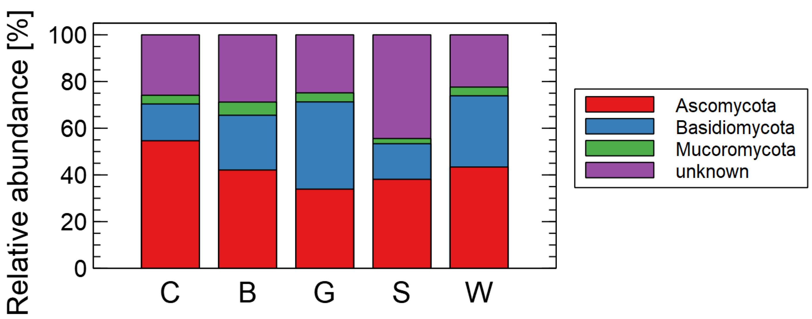
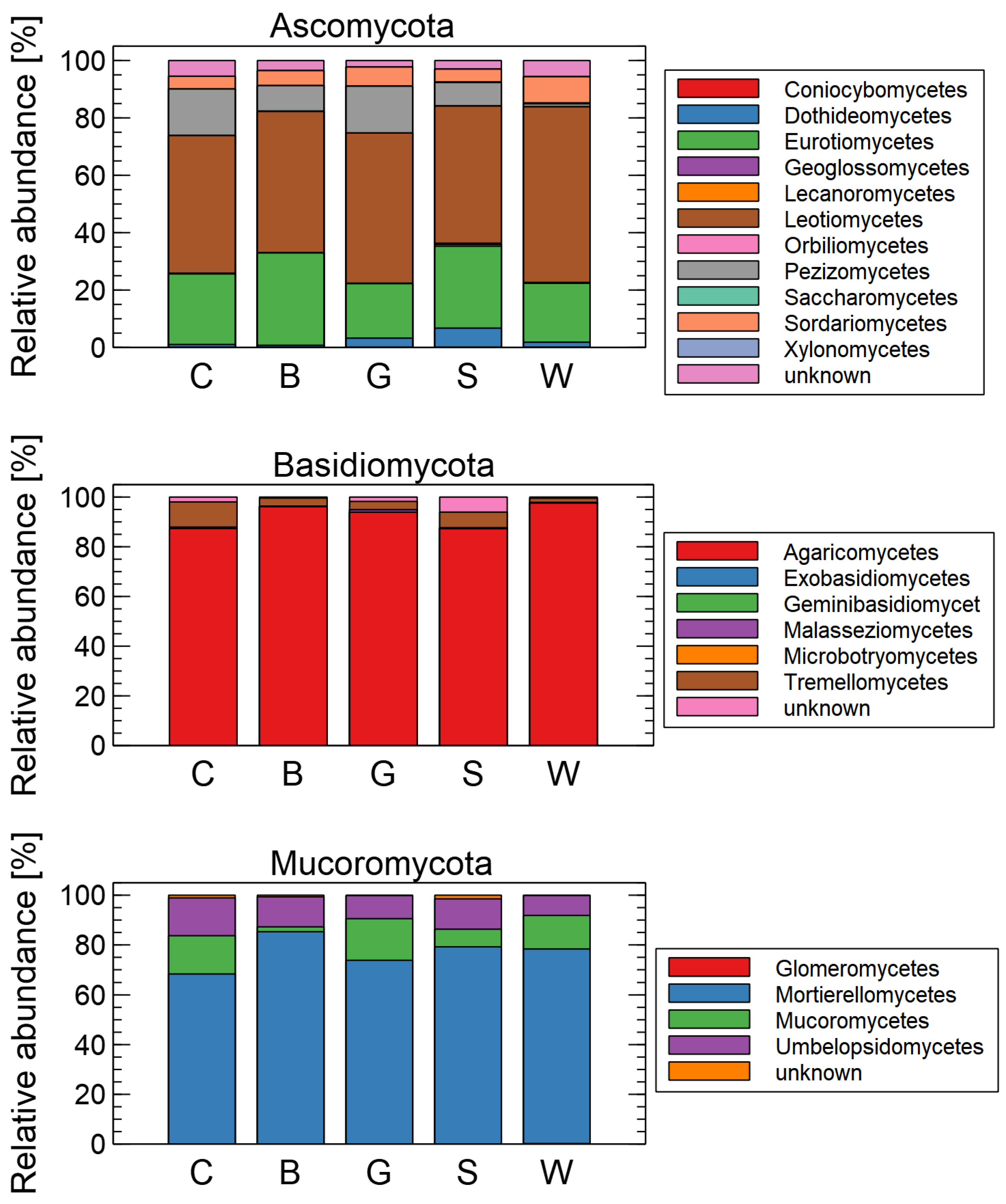
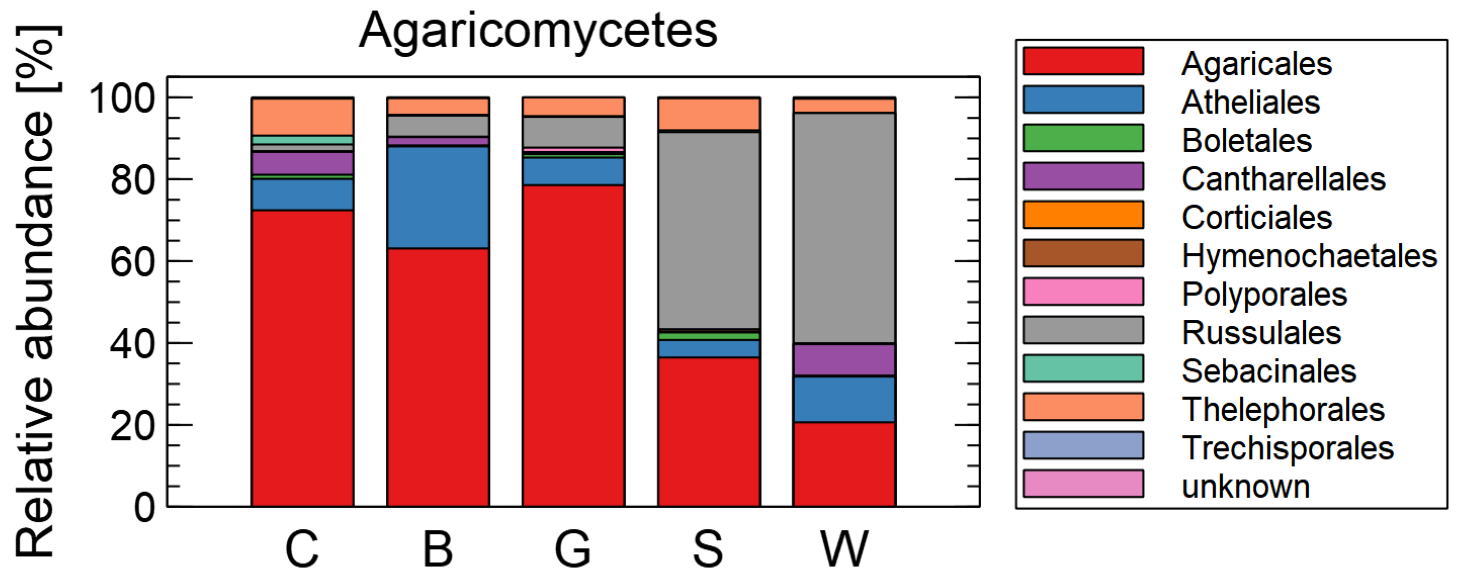
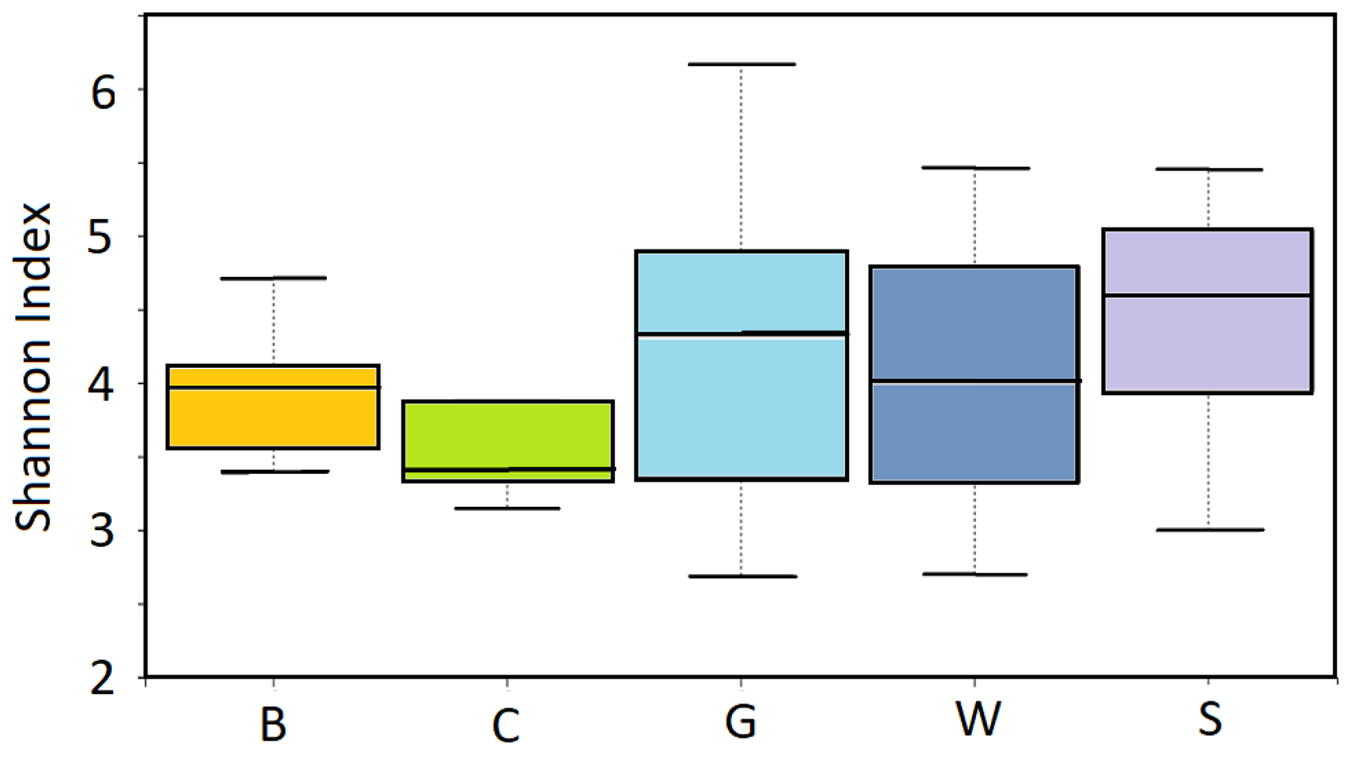
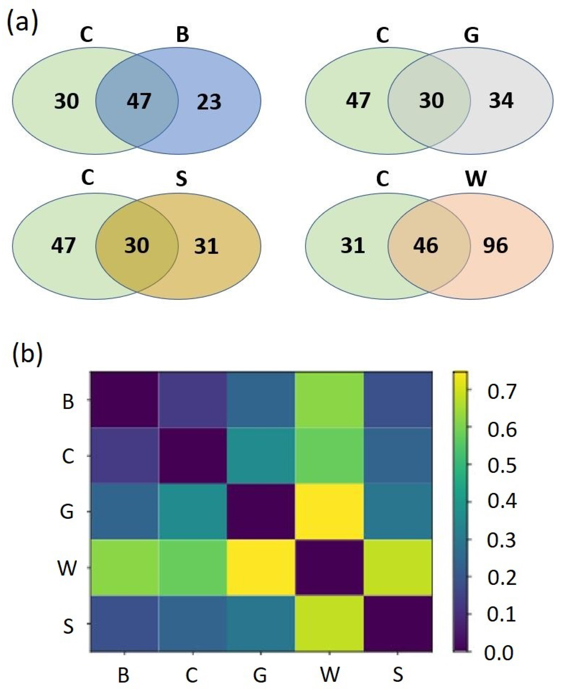
Alpha diversity of the fungal communities was estimated by the Shannon diversity index. Evaluation of beta diversity was calculated using Bray–Curtis dissimilarity index. For the calculations, we used the Scikit-bio 0.5.8 [21] software package. The assignment of ecological roles was based on FUNGuild [22].

3. Results

Effects of Mulching on the Soil Mycobiome

Obtained sequence reads were filtered and low-quality reads were discarded. High-quality reads were checked for chimera, and the chimera was also removed. Obtained high-quality free-of-chimera reads were assigned to OTU. OTUs with reads number below 100 were discarded from further analysis. The curated datasets comprised 1,697,460 reads The per-sample values ranged between 138,800 and 917,700.
Amplicons were assigned to 186 OTUs (Supplementary Table OTU). Approximately half of them were identified to genus or species level. There were 84 genera and 45 species found. Soil fungal communities were strongly dominated by the diverse Ascomycota and Basidiomycota (125 and 51 of total OTUs, respectively). Mucorromycota was represented by 11 OTUs.
Ascomycota was represented by 11 classes. The most numerous were Leotiomycetes (34 OTUs), Sordariomycetes (28 OTUs), Eurotiomycetes (22 OTUs), Dothideomycetes (19 OTUs), and Pezizomycetes (10 OTUs). Basidiomycota were represented by six classes, and almost all of them belonged to Agaricomycetes (42 OTUs). Similarly, Basidiomycota Mucorromycota were represented by four classes: Glomeromycetes, Mortierellomycetes, Mucoromycetes, and Umbelopsidomycetes, each of them containing a single OTU.
The most taxonomically rich phylum was also the most abundant in analyzed soil. Relative abundance of reads number belonging to Ascomycota ranged from 33.9% to 54.6%, Basidiomycota from 15.3 % to 37.3%, and Mucorromycota from 2.2% to 5.6%. Other phyla (Chytridiomycota, Cryptomycota, and Zoopogomycota) encountered below 0.01% reads. Besides known fungi phyla, a lot of reads belonging to unclassified fungi (22.4%–44.4%) were detected. Relative abundance of fungi at phylum, class, order, and family level is presented in Figure 2, Figure 3 and Figure 4, respectively.
Within the Ascomycota, two classes are the most numerous: Leotiomycetes and Eurothiomycetes. Leotiomycetes make up about half (C—48%; B—50%; G—48%; S-57; W—61%), and Eurothiomycetes about one quarter (C—25%; B—32%; G—25%; S—26%; W—21%) of Ascomycota OTUs, respectively. In Basidiomycota, Agaricomycetes dominate;they account for over 87% of all Basidiomycota OTUs reads (C—87%; B—96%; G—87%; S—87%; W—98%). Simillarly, in Mucoromycota, Mortierellales dominate(C—69%; B—85%; G—69%; S—79%; W—78%) (Figure 3).
In Agaricomycetes, there are the most abundant orders: Agaricales and Russulales. In control, bark compost, and bark compost gravel, relative abundance of Agaricales is 72%, 63%, and 79%, respectively. In sawdust and wood residues, there are 36% and 21% Agaricales, respectively. The opposite relationship occurs in Russulales. While in the control they are <1%, in wood-residues-enriched soil, their relative abundance is about 54%, making them the most abundant order of Agaricomycetes.
The performed analysis indicates that OTUs belonging to ten–twelve genera make up from 33% to 39% of all reads shown in Table 2.
The addition of organic matter did not change the alpha biodiversity of the soil. The Shannon diversity index H (Figure 5) ranged from 3.41 to 4.53 and was not statistically significantly different between studied soil conditions. Instead of changes in biodiversity, enrichment of the soil with organic substrates led to long-term significant changes in mycobiota composition. The changes occur at qualitative and quantitative levels. Changes at qualitative level are presented in Figure 6a, and at quantitative level, in Figure 6b.
In control soil, 77 OTUs were detected, 35 of which were assigned to genus and 22 to species level. The addition of sawdust, which enriched the soil with organic material consisting mainly of cellulose and lignin, slightly decreased diversity (61 OTUs, 30 genera, 10 species, respectively), but mainly led to mycobiome reconstruction. Moreover, in soil enriched by sawdust, 31 OTUs not detected in control soil appeared, among them Aspergillus terreus, Yarrowia lipolytica, Podospora sp., Pseudaegerita sp., Pseudocamaropycnis sp., and Rickenella sp. Importantly, a relative abundance of Cenococcum geophilum (the only species of the Cenococcum genus; Gloniaceae) increased twofold to approximately 2% of total fungal reads.
Enrichment of soil with sawdust leads not only to appearance of new species but also to the disappearance of some of the species present in the control plot (Absidia glauca, Chaetomium crispatum, Cortinarius diasemospermus, Cortinarius parvannulatus, Entoloma sanvitalense, Metapochonia suchlasporia, Oidiodendron chlamydosporicum, Penicillium raphiae, Phialocephala cf. fortinii, Phialocephala fortinii, Pseudogymnoascus pannorum, Sagenomella striatispora, Trichoderma polysporum, Umbelopsis isabellina, Wilcoxina rehmii). Especially strong changes in relative abundance were detected for Cortinarius parvannulatus (control—2%, not detected in sawdust) and Wilcoxina sp. (6.0% and 0.1%, respectively) (Table 2).
Enrichment of soil with bark compost also did not significantly change biodiversity (70 OTUs, 37 genera, 19 species), but, similar to sawdust, it significantly changed mycobiota composition. In bark-compost-enriched soil (but not in control soil plots), we detected Amphinema sp. (5% of total reads), two species of Oidiodendron (O. pilicola and O. tenuissimum), Acephala macrosclerotiorum, Apiotrichum sp., Chalara sp., Desmazierella acicola, Hebeloma sp., Hydnotrya sp., Hygrophorus sp., Lophodermium sp., Metarhizium anisopliae, Mycena sp., Penicillium thomii, Phoma sp., Rhizoscyphus sp., Trichoderma sp., and Tricholoma sp.; altogether 23 OTUs. Very abundant were Penicillium sp. (10% of total reads) which, together with eight other genera (Amphinema 5%, Cortinarius parvannulatus 1%, Hyaloscypha 4%, Inocybe 5%, Mortierella 2%, Oidiodendron 5%, Sagenomella 2%, Tricholoma 1%), made up 35% of total reads (Table 2).
The appearance of 23 new OTUs was associated with the disappearance of numerous OTUs present in control soil (Absidia cylindrospora, Absidia glauca, Capronia sp., Collarina sp., Cortinarius diasemospermus, Entoloma sanvitalense, Fusarium sp., Helvella sp., Humicola sp., Metapochonia suchlasporia, Neonectria sp., Pseudogymnoascus pannorum, Rhizoscyphus cf. ericae, Trichoderma polysporum, Varicosporium sp., Wilcoxina sp. and uncultured Leptosphaeriaceae, Malassezia, Venturiaceae, Wilcoxina).
Bark compost gravel had a similar effect on mycoflora to bark compost, but additional species were detected (Cadophora sp., Suillus sp., Sordaria sp., Cenococcum geophilum and Lophodermium conigenum. Acephala macrosclerotiorum, Amphinema sp., Chalara sp., Hebeloma sp., Hydnotrya sp., Metarhizium anisopliae, Mycena sp., Oidiodendron pilicola, Oidiodendron tenuissimum, Phoma sp., Rhizoscyphus sp. and Trichoderma asperellum ).
The biggest changes in soil mycobiota were found in soil enriched by wood residues. A total of 23 genera (Absidia sp., Articulospora sp., Botryosphaeria sp., Chaetothyriales sp., Cordyceps sp., Didymella sp., Diplodia sp., Erysiphe sp., Lactarius sp., Lasiodiplodia sp., Lecanicillium sp., Neofusicoccum sp., Paecilomyces sp., Paraphaeosphaeria sp., Peniophora sp., Phyllosticta sp., Pithomyces sp., Pochonia sp., Preussia sp., Pseudotomentella sp., Rhizopogon sp., Starmerella sp., Tylospora sp.) and some species (Fusarium merismoides, Penicillium canescens, P. ochrochloron, P. soppii, P. spinulosum) were detected only in this experiment variant. The appearance of so many fungi genera was associated with the disappearance of a small number of genera (Cortinarius sp., Entoloma sp., Helvella sp., Humicola sp., Ilyonectria sp., Rhizoscyphus sp. Wilcoxina sp.) and Oidiodendron chlamydosporicum species present in control. Quantitative analysis of abundance also showed that wood-residues-enriched soil has the biggest difference to control and to other types or organic supplements. The Bray–Curtis index for wood-residues-enriched soil ranged from 0.57–0.74, while for other organic supplements was in the range of 0.13–0.35.
The assignment of fungal ecological roles revealed that the number of mycorrhizal fungi OTUs increased from 11 in control to 24 and 21 after soil enrichment with bark compost or wood residues, respectively (Table 3). The addition of wood residues also increased the number of saprotrophs (from 20 to 31). This significant increase of mycorrhizal fungiand saprotroph in wood-residues-enriched soil was associated also with an increase of plant pathogens OTUs.

4. Discussion

4.1. Advantages of Adding Organic Substances

The inadequate state of biological properties of soils, especially the lack of naturally occurring antagonists of H. annosum, the cause of root and butt rot, is the main cause of stand health problems on former agricultural lands. Restoration of forests in such areas encounters numerous difficulties, and the death of pine stands is often caused by an infestation of the aforementioned fungal pathogen. Post-agricultural soils transferred for afforestation differ from forest soils mainly by the absence of an organic horizon [23]. In our experiment, we proved that we can positively modify the biodiversity of soil by adding organic matter that accumulated on the surface of forest soils (with varying degrees of decomposition), and determine its biological properties. We have showed that it provides a source of energy for the development of microorganisms, including mycorrhizal fungi. Soils destined for reforestation after agriculture are generally completely degraded and deficient. The conversion of agricultural soils to the forest is a slow process, and without silvicultural measures such as ours, it is difficult to achieve stable stands. Our goal was to set these natural processes in motion to ensure sustainable and stable forest ecosystems for future generations. Forests grow best when the species composition of the stands is compatible with the forest habitat type, including relevant soil properties. This is favored by beneficial fungi (antagonistic and mycorrhizal). Similar beneficial effects of revitalizing soils with sawdust were described in 1999 [3]. Our experimental plots were established in 2001, applying shredded wood residues, bark compost, and sawdust to restore the organic horizon [24,25]. The initial results of these studies were already been presented [23,25,26,27], but we now have the opportunity to show the changes that have taken place after more than 20 years. The present study shows positive changes in the DNA of soil microbiota. It turned out that the earlier enrichment of afforested post-agricultural land with organic matter in the form of compost, wood waste, or sawdust improved the fertility of these poor soils and stimulated beneficial microbial processes in the soil environment, in particular increasing the proportion of antagonists, which increased the resistance of roots to pathogen attack.
It is known that post-agricultural soils lack organic matter, including lignin and hemicellulose tissues that make up the structure of wood, including roots, which are the main substrate and energy source for the development of microorganisms, including fungi, especially basidiomycetes, which degrade the cellulose–lignin complex [27].
Hydrolysis of cellulose requires synergism of several organisms, including bacteria, actinobacteria, filamentous fungi, plants, and animals [28]. Among these organisms, filamentous fungi stand out, with the genera Penicillium, Trichoderma, and Aspergillus known as models for cellulase production at laboratory and industrial scales [29]. In our study, Penicillum accounts for between 4% and 10% of sequence reads, and the highest abundance was found in soils enriched with wood residues (10%) (Table 2). Aspergillus is much less abundant, but after soil enrichment, the abundance of this genus increased from 0.02% to 0.3% (sawdust, bark compost gravel). Soil enrichment also increased the abundance of Trichoderma from 0.2% in control to 0.9% in compost and wood residues and it is fungi, bacteria, and other microorganisms that are involved in the processes of creating the specific structure of the forest soil, giving it its normal biological activity. These fungi have an antagonistic effect against pathogenic fungi, especially H. annosum.
Even in the first years after planting, the structure and proportion of arbuscular mycorrhizal symbionts and saprotrophic fungi in the soil after planting are important for the condition of future stands [30,31,32]. Saprotrophs, the main decomposers, produce a wide range of extracellular enzymes that allow them to degrade the recalcitrant fraction of tree biomass. Mycorrhizal fungi play a pivotal role in the mobilization and sequestration of nitrogen and phosphorus and are responsible for the significant transport of carbon [33]. In the juvenile life stage of forest trees, great importance is attached to the creation of conditions conducive to the establishment of mycorrhizal symbioses [34,35]. By using molecular biology methods (NGS), we were able to demonstrate the biodiversity of the mycobiomes. After a 20-year process of transformation of the mycorrhizal community, which was no longer present in the soils after agricultural use, its composition was typicalfor the forest environment. Although we are unable to determine exactly when this occurred, we believe that the fungal spores were transferred along with organic residues. Without our intervention, these processes would likely have continued, as evidenced by analyses of control soils (still resembling post-farming soil) or after the application of sawdust and/or compost (less-transformed soils).

4.2. Protective Effects of Identified Mycorrhizae against Heterobasidion spp. and Other Pathogens

Colonization with ectomicorrhizal fungi showed clear protective effects against Heterobasidion spp., but also against other root pathogenic fungi (Rhizoctonia solani, Fusarium damping-off, Ilyonectria destructans) in both pine and spruce [36,37,38]. Thus, even in root tips that were heavily covered with dense M. bicolor hyphae, fungal colonization may have created a physical protective barrier around the roots in addition to direct antagonism of M. bicolor against Heterobasidion [39].
Among the numerous microorganisms found after treatment were those that support plant growth in poor soils, such as Pezizella ericae, which forms ericoid mycorrhiza. The hyphae of P. ericae secrete phosphatases that convert phosphorus from organic matter and polyphosphates into a plant-available state. Thanks to this phenomenon, fungi assimilate nitrogen from nitrates, ammonium, free amino acids, and various organic polymers through the release of proteases and chitin, which is decomposed by chitinases, and released to plants. They can also decompose pectins and lignins, releasing carbon [40]. We also found Meliniomyces bicolor and the closely related Cadophora finlandica [41], both colonizing the roots of many northern temperate forest trees, such as pine, spruce, and birch. Both form the well-defined and characteristic ectomycorrhizal morphotype Piceirhiza bicolorata on pine roots [42]. However, in northern temperate and boreal forests, M. bicolor can also form ericoid mycorrhizas with shrubs of the Ericaceae family, such as Vaccinium spp. Indeed, M. bicolor in vitro behaves similar to a typical ericoid mycorrhizal fungus by inducing a significantly higher growth rate in its Ericaceae host compared to uninfected seedlings and by causing a measurable mutual transfer of carbon and nitrogen [43].
Other fungal species Rhizopogon usually significantly affect the growth of bioassay seedlings [44]. It was found that the growth of seedlings colonized by Rhizopogon or Meliniomyces species was significantly improved compared to uncolonized seedlings [44]. In China, the positive effect of Rhizopogon on host plant growth and its crucial role in seedling establishment and forest regeneration of endangered Chinese Douglas-fir was established [44].
The analysis of soil microflora (bacteria and microscopic fungi) by the dilution method did not allow to separate healthy and infected stands [45]. However, using the soil clots’ overgrowth method, it was found that more cellulose-degrading bacteria were present in sample plots with healthy trees. Antagonistic microflora of H. annosum was found in all analyzed soils. Both isolated ascomycetes and Penicillium spp. showed antagonism to H. annosum. Microscopic fungi of the genera Verticillium, Aureobasidium, and Rhizopus were found only in the soil of healthy spruce stands [45].
Another species, Penicillium striatisporum Pst10, was isolated from the rhizosphere of chili peppers [46]. An experiment was conducted in which this isolate showed very strong antagonistic effects on mycelial growth of Phytophthora spp., Cladosporium cucumerium, and Sclerotinia sclerotiorum. In vitro assays tested the toxicity of sterilized liquid culture filtrates (SLCF) of Pst10 grown in potato dextrose broth against mycelial growth of Phytophthora capsici and sporangia/spore formation or germination. The SLCF completely inhibited mycelial growth and resulted in abnormal mycelium even at a 100-fold dilution. A 20-fold dilution of SLCF inhibited the formation and germination of sporangia and spores [46].
Velmalas et al. [39] investigated whether root colonization by ectomycorrhizal fungi (EMF) can alter the susceptibility of spruce (Picea abies) seedlings to root rot or necrotic leaf disease pathogens. First, spruce seedlings were inoculated by different EMFs and challenged with Heterobasidion isolates in Triaxenix tubes. The ascomycete EMF Meliniomyces bicolor, which showed strong antagonistic properties against the root rot pathogen Heterobasidion in vitro, effectively protected spruce seedlings against root rot. Second, spruce seedlings inoculated with M. bicolor or the forest humus were exposed to necrotrophic foliar pathogens under conventional forest nursery conditions on peat substrates. Post-winter infection with Botrytis cinerea was mild, and the extent of needle damage was independent of substrate and colonization by EMF. The severity of needle damage caused by pathogen Gremminiella abietina was high in seedlings in substrates with high nutrient availability and seedlings with well-established EMF communities. These results indicate that M. bicolor can protect spruce seedlings in axenic cultures against Heterobasidion root rot, but cannot induce systemic protection against foliar pathogens. It was also noted that nonsterile inoculum sources, such as forest humus, should not be considered for use under greenhouse conditions because they may predispose seedlings to unintended needle damage.
It appears that an extract of Oidiodendron truncatum, the other species of fungus found, could be used against white-nose syndrome (Pseudogymnoascus destructans), a devastating disease of hibernating bats in the USA. Rusman et al. [47] isolated 14 terpenes and three anthraquinone metabolites. Ten of these compounds were already described in the literature, but the structures of seven analogous terpenes were new. In addition, this was the first report of 4-chlorophyscione from a natural source that had previously been identified as a semisynthetic product. The compounds PR 1388 and LL -Z1271 α were the only inhibitors of P. destructans (MIC = 7.5 and 15 μ g/mL, respectively). Of two nematicides, 4-hydroxyphenylacetic acid (4-HPA) (1) and oidiolactone D (2), isolated from cultures of the fungus Oidiodendron sp., compound 2 showed nematicidal activities against the root lesion nematode, Pratylenchus penetrans, and the pine wood nematode, Bursaphelenchus xylophilus. This finding is very important for the health of pines planted on former agricultural lands [48].

4.3. Protective Effect of Identified Mycorrhizal Fungi on Plants against Drought

Fungi of the genus Oidiodendron, which are abundant in the soils we studied (Table 2), form ericoid mycorrhizae (ErM) and are considered an important factor in increasing crop yield and drought resistance [49]. Oidiodendron has not been studied from the point of view of antagonism in relation to diseases of the root system, e.g., Heterobasidion sp. [50]. Antagonism of Oidiodendron sp. from Sitka spruce ectomycorrhiza in relation to the species pathogenic for plants Phytophthora cinnamomi was confirmed [51]. The benefits of ErM fungi were also investigated by inoculating well-watered and severely drought-stressed plants with Oidiodendron maius. The results showed that the fungi significantly increased the biomass of stems and roots of lingonberry [49]. In the face of climate change, such a strategy to help plants cope with water scarcity is very important. In the cited study, ErM also significantly increased chlorophyll content probably because of a better water supply. Similarly, inoculation with Lachnum pygmaeum improved drought resistance, promoted root growth, and increased root wet weight by 1157%. While drought reduced chlorophyll content and soluble sugar content in the plant, ErM significantly increased their content after inoculation. At the same time, inoculation with the fungus L. pygmaeum decreased the content of malondialdehyde (a marker of oxidative stress) but increased the activity of superoxide dismutase (an important antioxidant for protection against oxidative stress). Overall, these results suggest that the successful coexistence of ErM fungi and lingonberry mitigates the negative effects of drought stress through higher secondary metabolites and photosynthetic pigment synthesis [49]. Similar effects were obtained by inoculating plants with Oidiodendron maius and Phialocephala fortinii [52]. It promoted the growth of Vaccinium corymbosum. In conclusion, inoculation of plants with three root symbionts (O. maius, Hymenoscyphus sp., and P. fortinii) and with the endophytic fungus Xylaria sp. increased plant height in laboratory experiments. On a semiindustrial scale, inoculation improved plant biomass and growth performance. Supplementing root-associated fungal communities with a mixture of ericoid mycorrhizal fungi and endophytic fungi could therefore provide an alternative to conventional fertilization and pesticide use in large-scale blueberry production [52]. This confirms the extensive changes observed in our experiment with pine and organic material.
In addition, the species we identified, Cenococcum geophilum (Table 2), is a cosmopolitan ectomycorrhizal fungus known for its broad habitat range [53] and may be the dominant ectomycorrhizal fungus in both arctic and temperate and subtropical forests [53,54,55]. It is particularly notable for its drought resistance [56,57]. However, this fungus is not restricted to dry sites, as it has also been observed in moist, poorly drained soils [55]. Cenococcum geophilum has a pioneer ability, although it also occurs in mature stands [58,59]. The ability of sclerotia to survive for several years may provide sufficient inoculum for effective colonization of host species [60]. The ectomycorrhiza of this fungus has been demonstrated on over 200 tree species from 40 different angiosperm and gymnosperm genera [61].
Thanks to the mycorrhizae, biennial seedlings of Tilia cordata growing on sandy soils can survive periods when soil water potential drops to −18 to −55 bar [62]. Their mycorrhiza, formed by Cenococcum geophilum, were shown to remain alive. Water loss measurements of seedlings under similar conditions decrease from 16.0 to 26.2 mg/h when the soil is moist to 4.1 to 4.6 mg/h when the soil is dry. Calculation of the volume of mycorrhiza indicates that their water content would support transpiration for periods not exceeding 1 h. Survival of mycorrhiza over long dry periods is apparently related to the ability of the fungal partner to resist desiccation [62].

4.4. Identified Fungi as a Potential Source of Bioactive Molecules

Fungi are considered an important source of bioactive molecules, often effective against other fungi and/or bacteria, and are therefore potential candidates in the search for new antibiotics. The fruiting bodies of sixteen different fungal species of the Basidiomycota were collected in the Italian Alps. Fungal species were identified by internal transcribed spacer sequencing (ITS). Most of the species belong to the genera Cortinarius, Mycena, and Ramaria, whose metabolite content has hardly been studied so far. The crude extracts obtained from the above fungi were tested for their inhibitory activity against five human pathogens: Candida albicans, C. glabrata, Staphylococcus aureus, Pseudomonas aeruginosa, and Klebsiella pneumoniae. Twelve crude extracts showed activity against P. aeruginosa. The highest activity was shown by some Cortinarius species, such as C. nanceiensis [63].
Six of the fungi [64] examined in this study [64] were known to produce pigments with antibacterial activity. The presence of emodin, physcion, torosachryson, hypericin, or skyrin would likely contribute to the antibacterial activity observed in Cortinarius persplendidus, C. austrovenetus, and C. [D. canaria]. However, nearly colorless extracts also showed antibacterial activity, suggesting that previously unidentified fungal constituents may also possess antibacterial activity. Australian fungi of the genus Cortinarius are promising sources of natural products for further drug discovery because of the high biological diversity and unique evolutionary lineages found only in this region. This is accompanied by a large proportion of bioactive species and a great diversity of chemical constituents [64].

4.5. Antioxidant Capacity and Antimicrobial Activities of Some Identified Fungi

In the study of Türkoğlu et al. [65], usingextracts obtained with ethanol, the antioxidant capacity and antimicrobial activities of Russula delica were demonstrated. The researchers used four complementary assay systems, namely, DPPH radical scavenging, β -carotene/linolenic acid systems, total phenolic compounds, and total flavonoid concentration. The inhibition levels of the ethanolic extract of R. delica and the standards (BHA and α -tocopherol) increased in parallel with the increase in the concentration in the linoleic acid system. The presence of the flavonoid quercetin and the phenolic compound pyrocatechol in the extract was detected. The antimicrobial activity of R. delica was tested in vitro by agar well diffusion method, and the study confirmed the antibacterial activity of R. delica extract. Therefore, the extracts could be suitable as antimicrobial and antioxidant agents in the food industry [65].
Similar results were obtained with another fungal species of this genus (Russula griseocarnosa) [66]. They proved that the mushroom contains bioactive substances such as phytochemicals, e.g., phenols, flavonoids, ergosterol, and β -carotene. The main constituent in (R. griseocarnosa) was quercetin (95.82 μ g/g) [66]. Thanks to its properties and activity, the mushroom can be used as a natural immunostimulant and antioxidant [67].

4.6. The Role of Mycorrhizal Fungi in the Nutrient Cycle of Pine Trees

The fungal species we detected in the soil samples Tricholoma matsutake (Supplementary table OTU) forms a symbiotic association with conifers, forming mycelial aggregations called “shiro”, characterized by different chemical and physical properties to the nearby forest soil. The fungal diversity that lives in shiro soils plays a key role in nutrient cycling [68]. Zhou et al. [68] combined phospholipid fatty acid (PLFA) analysis and NGS sequencing to determine fungal biomass and community structure. It was found that T. matsutake dominated in the shiro, which had significantly lower saprotrophic fungal biomass compared to the non-shiro soil. Fungal diversity was negatively correlated with the relative abundance of T. matsutake in the shiro soil. The fungal community in the shiro was characterized by similar fungal species composition in most of the forest type samples. It appears that T. matsutake coexisted with a particular fungal community due to competition or nutrient interactions. However, Oidiodendron sp. was positively correlated with the abundance of T. matsutake, which is common in Shiro. In contrast, Helotiales and Mortierella were negatively correlated with T. matsutake, both of which commonly inhabit non-shiro soil but do not occur in shiro soil. Zhou et al. [68] concluded that T. matsutake produces a dominance effect that shapes the fungal community and diversity in shiro soils across different forest types [68]. In other studies with the genus Tricholoma, the active mycorrhizal zone of shiro was revealed, in which T. matsutake showed antimicrobial activities [69], and as a result, the abundance of bacteria and sporulating fungi decreased within the zone but increased outside the zone
The information on the mentioned fungus is inconclusive, as it was also considered a pathogen. This was due to inoculation studies in which it was found that the needles of pine seedlings inoculated with T. matsutake were yellow, while the needles of uninoculated pine seedlings were green [70]. It was concluded that T. matsutake was a parasitic species. Ogawa [70] also reported the parasitic tendency of the species, as the hyphae of this fungus penetrated the cells of the roots, while no fungal sheaths or Hartig nets were formed. Only Eto [71] reported that the vegetative hyphae of T. matsutake covered the surface and invaded the intercellular spaces of the roots when an isolate of this species was inoculated into sterile pine seedlings in a closed pot. The author also reported that the addition of iron citrate to the culture medium improved mycelial growth and mycorrhizal synthesis [72]. Yamada et al. [73] reported that a fungal mantle and a Hartig net developed on the roots of Pinus densiflora seedlings cultured on sterile vermiculite with nutrient solution after inoculation with T. matsutake culture. This was the first report of the formation of a typical EM by T. matsutake. The growth of pines was not changed, or was only slightly improved, after the formation of EM, indicating that this fungus is not parasitic. Improved growth of pines after inoculation with T. matsutake was also reported by [74], who did not observe a typical fungal coat but noted the presence of a Hartig net [75].

4.7. The Role of Micorrhizal Fungi in the Nutrient Cycle of Pine Trees

In our experiment, Wilcoxina sp., T. terrestris, and Piloderma sp. were the most effective colonizers, and in the treatments, they dominated the rhizosphere root systems and positively affected the growth of P. sylvestris. Some other studies are in line with our research. Velmala et al. [76] demonstrated that ECM colonization improved shoot biomass production. The shoot height of slow-growing seedlings was also greater with increasing ECM fungal colonization, and marginally improved with increasing richness. ECM improved aboveground growth, which appeared to be related to a high abundance of Wilcoxina sp. We also obtained similar results in our experiment in a fertile habitat. This is probably due to the chitinase activity of Wilcoxina sp. and also to the high potential activity of glucose-releasing cellulases and hemicellulases, but again not to the hydrolytic activity involved in the mobilization of P and the release of amino acids.
A high amount of Wilcoxina sp. resulted in relatively high production of chitinase, cellulases, and hemicellulases [76]. In the article in question, the high activity of chitinase and cellulases was strongly positively related to the nitrogen content of the needles. This was also consistent with the field study of Jones et al. [77], in which the abundance of Wilcoxina sp. was associated with high N accumulation in both shoots and roots. Based on the [76] measurements of potential enzyme activities, Wilcoxina sp. effectively degrades chitin, a structural fungal cell wall polysaccharide and a rich organic N reservoir in the soil of boreal forests. Endophytic fungi such as Penicillium spp. may play an important role in plant survival by enhancing nutrient uptake and producing growth-promoting metabolites such as gibberellins and auxins. The plant-growth-promoting ability of this fungal strain could contribute to the maintenance and revegetation of rapidly eroding dune flora [45]. Recently mentioned studies showed that Penicillium citrinum can produce the mycotoxin citrinin and cellulose digesting enzymes such as cellulase and endoglucanase as well as xylulase. A new aspect of the above research is the discovery of the ability of this fungus to produce gibberellins (GA5).
The discovery of filamentous fungi in our soil samples is not surprising, as recent scientific reports based on DNA or RNA analysis demonstrated the widespread occurrence of Mortierella in various environments [78]. Some strains of this genus belong to the plant-growth-promoting fungi (PGPF) and are found in soil, the rhizosphere, and plant tissues. These microorganisms are often found even in extremely hostile environments and are responsible for improving access to bioavailable forms of P and Fe in soils, synthesizing phytohormones and 1-aminocyclopropane-1-carboxylate (ACC) deaminase, and, last but not least, protecting crops from pathogens. The association with crops confirms the specificity of these soils, and the identification of these fungi in our samples confirms the origin of the studied soils. In addition, previous reports classified Mortierella spp. as saprotrophic microorganisms isolated from agricultural soils, and today their status as very valuable decomposers of forest litter is confirmed. The ability to survive in very unfavorable environmental conditions and the use of carbon sources contained in polymers such as cellulose, hemicellulose, and chitin make these fungi efficient colonizers of different environments. The growing interest in the application of Mortierella spp. in agriculture is mainly due to the potential benefits of this genus in increasing nutrient uptake efficiency, beneficial effects in protecting plants from adverse conditions, and reducing the use of chemical fertilizers and pesticides. In addition, the activities of Mortierella species influence the soil microbiota and support the performance of beneficial microorganisms, which significantly increase crop yield [78].

4.8. The Role of Antagonistic Fungi in Biological Protection of Pines from Pathogens

Trichoderma spp. were found only in the rhizosphere of spruce in healthy stands [79]. From this literature, it appears that Trichoderma fungi, which constitute at least 70% of the total amount of microscopic fungi, protect roots of woody plants [79]. Penicillium spp. and Mucor spp. in soil were more abundant in stunted stands, while Trichoderma spp. was found mainly in healthy stands. Fewer cellulose-degrading bacteria were found in the soil of plots with dead spruce trees than in plots with healthy spruce trees. In our case, microflora antagonistic to H. annosum was found in all studied soils after treatment. The isolated ascomycetes, as well as Trichoderma spp. and Penicillium spp., showed strong antagonism, with 90% of the antagonists inhibiting the growth of both the S and P groups of H. annusum [79]. Microscopic fungi of the genera Verticillium, Aureobasidium, and Rhizopus were also isolated in our experiment. In the root rhizosphere, fungi of the genus Trichoderma were detected only in healthy spruce stands [79] and, in our case, after enrichment with organic material. It is commonly believed that the increase in the number and activity of Trichoderma occurs at a higher temperature (15–32 °C), pH in the range of 5.5–8.5, moderate soil moisture, high content of organic matter, and no mechanical interference with the soil.

4.9. Fungi Identified as Pathogens for Animals and Humans

Pathogenic organisms were also found in the soil samples tested (Supplementary Table OTU), and not just for plants. A filamentous fungus that caused a fatal systemic infection in a dog was identified as a new species Sagenomella chlamydospora [80]. When the case was originally reported, the fungus was identified as Paecilomyces sp. This study highlights how difficult identifying the causative agent of infection can be when dealing with an unusual microorganism. This is the first time this genus has been implicated in infections in animals, including humans. Recently, Garcia et al. [81] reported and illustrated a case of disseminated mycosis in a dog (a German shepherd). The fungus was isolated on postmortem examination from lesions in numerous organs, such as the kidneys, mitral valve, abdominal aorta, and intervertebral discs. The diagnosis was difficult because clinical symptoms were very nonspecific [80]. Thus, the presence of these fungi confirms the agricultural origin of the soils and the great potential of the NGS method to detect and identify them.
Pseudogymnoascus destructans causes white-nose syndrome (WNS) in North American bats, which has resulted in a dramatic decrease in the bat population in the United States and Canada. We found Pseudogymnoascus pannorum, which is the nearest fungal relative of P. destructans, with wider psychrophilic–physiological growth range, and ability to cause skin diseases in canines and humans [82,83].
Somewhat surprising is the presence of pathogens in our soil samples that cause the recent emergence of white-nose syndrome (WNS), a fungal disease that causes unprecedented mortality in hibernating bats [84]. Research in eastern North America has uncovered a gap in knowledge about fungal communities associated with bats and their hibernacula. American researchers used culture techniques to examine the diversity of fungi in soil samples collected from 24 bat hibernacula in the eastern United States [84].
Similar studies [85] were also carried out in Poland, and the occurrence of this species was found in the bats’ hibernation sites. However, no disease symptoms were found in bats.
Fungi of the genus Apiotrichum identified in the soil samples are urease-positive, unencapsulated basidiomycetous yeasts characterized by the development of hyaline, septate hyphae that fragment into oval or rectangular arthroconidia. This makes morphological identification difficult, and we were able to detect their presence, probably thanks to molecular identification. All species are resistant to echinocandins and other classes of antifungal agents [86]; perhaps this is the reason why they have survived in post-agricultural soils. Apiotrichum species are frequently isolated from human and animal sources as well as from clinical samples, and some of them have been associated with opportunistic infections °C [87]. It is also worth mentioning that the pathogenic species all can grow at 37 °C.
Another fungal species identified in our research is Exophiala jeanselmei (Supplementary table OTU)—a dematiaceous hyphomycete commonly found in soil, decaying vegetation, and rotting wood [88,89]. Traumatic inoculation of E. jeanselmei can lead to a variety of subcutaneous infections, including mycetoma, chromoblastomycosis, or phaeohyphomycosis [90]. In immunosuppressed organ transplant recipients, E. jeanselmei is the most common dematiaceous fungus associated with skin infections, occurring in up to 32% of cases, with an associated mortality rate of up to 18%. Exophiala jeanselmei is the causative agent of maduromycosis [88], a usually asymptomatic disease manifested by black or brown macular lesions that enlarge by peripheral extension. The lesion is darkest at the periphery and has very distinct margins. Exophiala jeanselmei is clinically defined as a rare causative agent of subcutaneous lesions of traumatic origin that eventually cause eumycetoma [90].

4.10. Seedborne Fungi

The fungus we identified in the soil, Caloscypha fulgens, produces bulging infectious beads on the surface of the seed within 1 to 3 days after inoculation, and over the next 10 days, several straight penetration holes form in the hull just below each bead [91]. During the penetration period, the growth of the fungus is restricted to the penetration holes and tissue interstices until it reaches the endosperm and embryo and spreads its branches. Occasionally, it penetrates the seed through slits in the integument, but never through the micropyle. The fungus invades from infected seeds through intact or split seed coats and the micropyle [91].

4.11. Edible Ectomycorrhizal Fungi

Hydnum is a fungal genus with considerable ecological and economic importance, as it includes several edible ectomycorrhizal fungi. Different species of Hydnum can be found among plants from several families, such as Pinaceae, Fagaceae, Dipterocarpaceae, and Myrtaceae. Previous studies showed that they can form ectomycorrhizal relationships with these plant families, making them very important for maintaining forest ecosystems [92,93].
Hygrophorus species are distributed worldwide and are mostly found in forests with pines or with ectomycorrhizal (ECM) angiosperms [94]. Hygrophorus are essential components of ECM communities in temperate regions of the Northern Hemisphere [95]. Recently, a new edible species, H. parvirussula, was described from southwestern China [96], belonging to the Hygrophorus section Pudorini [97]. It is not entirely clear why it was found in Poland (initial report); perhaps it was imported, or perhaps it already existed but was not yet discovered without NGS testing.

4.12. Identified Fungi Occurred as Pathogenic to Various Plant Species

Karadžić et al. [98] consider Cenangium ferruginosum as one of the most dangerous pathogenic fungi on pines. Pathogenicity tests conducted with mycelial plugs in summer confirmed that C. ferruginosum is not only a saprophyte but also a pathogen. Once infected and colonized by pathogens that cause bark necrosis in the crown, trees are severely stressed and may soon be attacked by secondary invaders, such as Ips acuminatus Gyll., Ips sexdentates Börn, Tomicus piniperda L., Tomicus minor Htg., and Buprestidae, or by other pathogens, such as Armillaria sp. or Ophiostoma sp. Cenangium ferruginosum is an important pathogen in some countries of Southern Europe [98,99]. In Central Europe, it used to be considered a saprophyte rather than a pathogen [100].

4.13. Other Identified Fungi Specific for Different Environments

Solicoccozyma terricola M, the yeast strain isolated from a soil sample from blueberry cultivation in Miedzyrzec Podlaski in Poland, can cleave phosphorus–nitrogen and nitrogen–carbon bonds into N-phosphonomethylglycine (PMG, glyphosate) [101]. This was the first report of PMG degradation as a phosphorus and nitrogen source by a yeast strain isolated from glyphosate-contaminated soil, and in this way, the above studies filled a gap in PMG biodegradation research between bacteria and filamentous fungi. The release of inorganic phosphate into the culture medium confirmed the progress of xenobiotic biodegradation, suggesting that the tested strain degrades this compound independently of phosphate cell status and may contribute to the development of effective methods of soil and water bioremediation in the future [101]. If so, it was also likely involved in improving soil properties to support pine growth in our experiment.
Compounds obtained from another fungal species we identified, S. terricola (it grows on lignocellulose), could be suggested as an additional source of oleochemicals [102]. Given the rising prices of fossil fuels and the environmental problems associated with their use, the use of compounds produced by S. terricola on lignocellulose could be a promising option as an additional oleochemical source, especially for biodiesel production [102].
The presence of the genus Collarina Jullien, 1886 (Cribrilinidae Hincks, 1879) [103] was surprising, since only two species were previously known from the Atlantic–Mediterranean region, C. balzaci [104], synonym of Collarina cribrosa. It is exclusively epiphytic (mainly on Posidonia oceanica (L.) Delile, 1813 and brown algae), its lifecycle is adapted to ephemeral hosts, and it can reproduce dramatically under unusual environmental conditions (Gulf of Gabes, chemical disturbances) on Posidonia leaves in association with diatoms [105].

5. Conclusions

Wood waste, sawdust, or bark compost in the form of organic material can be successfully used to accelerate the natural processes of formation of mycobiota characteristic for forest soils.
The wood substrate in degraded post-agricultural soils rebuilt the organic layer after 20 years, which had a positive effect on the composition of mycobiota.
The substrates used provided suitable growth conditions (moisture content, nutrient availability) for many fungal species beneficial to planted pines.
Fungal biodiversity was restored, which (as in forest soils) became antagonistic to pathogens, e.g., Heterobasidion annosum, which is dangerous to pines growing on former farmland.
Beneficial fungi included those that form mycorrhizal associations with pine roots, e.g., from the genera Amphinema, Inocybe, or Rusula, thus protecting them (mechanically and chemically) from pathogens and drought (as they increase the surface area of the fungal roots many times over). Fungi with antagonistic properties to H. annosum included those of the genera Penicilium or Trichoderma.
In general, the rhizosphere of treated pines was dominated by fungi of the genus Basidiomycota, and the control area by Ascomycota. The latter was dominated by pathogens of, e.g., pine needles and shoots, such as Cenanglium ferruginosum.

Author Contributions

Conceptualization, T.M. and T.O.; methodology, T.M. and I.O.; software, T.M. and P.B.; validation, TM., P.B. and D.B.; formal analysis, S.M.; investigation, I.O. and D.B.; resources, T.O. and I.O.; data curation, T.M., P.B. and V.D.; writing—original draft preparation, T.M., P.B. and T.O.; writing—review and editing, J.B.-B., W.P. and S.M.; visualization, P.B. and D.B.; supervision, T.O.; project administration, T.O. and P.B.; funding acquisition, T.O. and I.O. All authors have read and agreed to the published version of the manuscript.

Funding

This research received no external funding.

Institutional Review Board Statement

Not applicable.

Informed Consent Statement

Not applicable.

Data Availability Statement

Not applicable.

Conflicts of Interest

The authors declare no conflict of interest.

References

  1. Sierota, Z.; Malecka, M. Ocena zmian w drzewostanie sosnowym na gruncie porolnym po 30 latach od wykonania pierwszych ciec pielegnacyjnych bez zabiegu ochronnego przeciw hubie korzeni. Sylwan 2003, 147, 19–26. [Google Scholar]
  2. Małecka, M.; Sierota, Z. Ocena zagrożenia i ryzyka rozwoju huby korzeni w drzewostanie na gruncie porolnym. Sylwan 2003, 147, 12–25. [Google Scholar]
  3. Sierota, Z.; Kwaśna, H. Changes in fungal communities in abandoned farmland soil enriched with pine sawdust. Folia For. Pol. Ser. A For. 1998, 40, 85–94. [Google Scholar]
  4. Sierota, Z.; Kwaśna, H. Ocena mikologiczna zmian zachodzących w glebie gruntu porolnego po dodaniu trocin iglastych. Sylwan 1999, 143, 57–66. [Google Scholar]
  5. Kwaśna, H.; Brzeski, M.; Sierota, Z. Mikroorganizmy środowiska glebowego odłogujących gruntów porolnych–zmiany w zbiorowiskach grzybów i nicieni po dodaniu trocin iglastych. In Drobnoustroje Środowiska Glebowego–Aspekty Fizjologiczne, Biochemiczne, Genetyczne; Dahm, W.H., Pokojska, A., Eds.; Wyd. A. Marszałek: Toruń, Poland, 2001; pp. 57–66. [Google Scholar]
  6. Paul, K.I.; Polglase, P.J.; Nyakuengama, J.; Khanna, P. Change in soil carbon following afforestation. For. Ecol. Manag. 2002, 168, 241–257. [Google Scholar] [CrossRef]
  7. Wall, A.; Hytönen, J. Soil fertility of afforested arable land compared to continuously. Plant Soil 2005, 275, 247–260. [Google Scholar] [CrossRef]
  8. Olejarski, I. Wplyw zabiegow agrotechnicznych na niektore wlasciwosci gleb oraz stan upraw sosnowych na pozarzyskach wielkoobszarowych. Pr. Inst. Badaw. LeŚNictwa Ser. A 2003, 2, 47–77. [Google Scholar]
  9. Kwaśna, H.; Sierota, Z.; Bateman, G.L. Fungal communities in fallow soil before and after amending with pine sawdust. Appl. Soil Ecol. 2000, 14, 177–182. [Google Scholar] [CrossRef]
  10. Duda, B.; Sierota, Z. Survival of Scots pine seedlings after biological and chemical control of damping-off in plastic greenhouses. Eur. J. For. Pathol. 1987, 17, 110–117. [Google Scholar] [CrossRef]
  11. Retter, A.; Nilsson, R.H.; Bourlat, S.J. Exploring the taxonomic composition of two fungal communities on the Swedish west coast through metabarcoding. Biodivers. Data J. 2019, 7. [Google Scholar] [CrossRef] [Green Version]
  12. Tedersoo, L.; Anslan, S.; Bahram, M.; Põlme, S.; Riit, T.; Liiv, I.; Kõljalg, U.; Kisand, V.; Nilsson, H.; Hildebrand, F.; et al. Shotgun metagenomes and multiple primer pair-barcode combinations of amplicons reveal biases in metabarcoding analyses of fungi. MycoKeys 2015, 10, 1–43. [Google Scholar] [CrossRef]
  13. Wurzbacher, C.; Nilsson, R.H.; Rautio, M.; Peura, S. Poorly known microbial taxa dominate the microbiome of permafrost thaw ponds. ISME J. 2017, 11, 1938–1941. [Google Scholar] [CrossRef] [Green Version]
  14. FastQC Project. Available online: https://www.bioinformatics.babraham.ac.uk/projects/fastqc/ (accessed on 1 November 2022).
  15. Bolger, A.M.; Lohse, M.; Usadel, B. Trimmomatic: A flexible trimmer for Illumina sequence data. Bioinformatics 2014, 30, 2114–2120. [Google Scholar] [CrossRef] [Green Version]
  16. Schloss, P.D.; Westcott, S.L.; Ryabin, T.; Hall, J.R.; Hartmann, M.; Hollister, E.B.; Lesniewski, R.A.; Oakley, B.B.; Parks, D.H.; Robinson, C.J.; et al. Introducing mothur: Open-Source, Platform-Independent, Community-Supported Software for Describing and Comparing Microbial Communities. Appl. Environ. Microbiol. 2009, 75, 7537–7541. [Google Scholar] [CrossRef] [Green Version]
  17. Marcelino, V.R.; Clausen, P.T.L.C.; Buchmann, J.P.; Wille, M.; Iredell, J.R.; Meyer, W.; Lund, O.; Sorrell, T.C.; Holmes, E.C. CCMetagen: Comprehensive and accurate identification of eukaryotes and prokaryotes in metagenomic data. Genome Biol. 2020, 21. [Google Scholar] [CrossRef]
  18. Clausen, P.T.; Aarestrup, F.M.; Lund, O. Rapid and precise alignment of raw reads against redundant databases with KMA. BMC Bioinform. 2018, 19, 1–8. [Google Scholar] [CrossRef]
  19. Vu, D.; Groenewald, M.; Szöke, S.; Cardinali, G.; Eberhardt, U.; Stielow, B.; de Vries, M.; Verkleij, G.; Crous, P.; Boekhout, T.; et al. DNA barcoding analysis of more than 9 000 yeast isolates contributes to quantitative thresholds for yeast species and genera delimitation. Stud. Mycol. 2016, 85, 91–105. [Google Scholar] [CrossRef]
  20. Calculate and Draw Custom Venn Diagrams. Available online: http://bioinformatics.psb.ugent.be/webtools/Venn (accessed on 1 November 2022).
  21. Scikit-Bio Software Package Project. Available online: http://scikit-bio.org/docs/0.5.8/ (accessed on 28 November 2022).
  22. Nguyen, D.; Boberg, J.; Ihrmark, K.; Stenström, E.; Stenlid, J. Do foliar fungal communities of Norway spruce shift along a tree species diversity gradient in mature European forests? Fungal Ecol. 2016, 23, 97–108. [Google Scholar] [CrossRef] [Green Version]
  23. Małecka, M.; Hilszczańska, D. Changes in ectomycorrhizal structure of Scots pine growing on abandoned farmland soil enriched with organic substrates. Sylwan 2014, 158, 243–250. [Google Scholar]
  24. Olejarski, I.; Walendzik, J. Warunki wzrostu upraw lesnych w Bornem Sulinowie. Głos Lasu 2002, 08, 20–22. (In Polish) [Google Scholar]
  25. Olejarski, I. Wykorzystanie pozostalosci zrebowych do nawozenia organicznego gruntow (in Polish). Postępy Techniki w Leśnictwie 2005, 92, 20–24. [Google Scholar]
  26. Oszako, T.; Olejarski, I. Inicjowanie procesow przeksztalcenia gleb porolnych w gleby lesne poprzez wykorzystanie pozostalosci zrebowych, kompostow i trocin. Pr. Inst. Badaw. LeŚNictwa Ser. A 2003, 1, 76–79. (In Polish) [Google Scholar]
  27. Malecka, M.; Hilszczanska, D. Wpływ odłogowania i dodatku trocin iglastych do gleby porolnej na jej właściwości chemiczne i zbiorowisko grzybów ektomykoryzowych 15-letniej sosny zwyczajnej. Leśne Pr. Badaw. 2015, 76, 265–272. [Google Scholar]
  28. Kuhad, R.C.; Gupta, R.; Singh, A. Microbial cellulases and their industrial applications. Enzym. Res. 2011, 2011, 1–10. [Google Scholar] [CrossRef] [Green Version]
  29. de França Passos, D.; Pereira, N., Jr.; de Castro, A.M. A comparative review of recent advances in cellulases production by Aspergillus, Penicillium and Trichoderma strains and their use for lignocellulose deconstruction. Curr. Opin. Green Sustain. Chem. 2018, 14, 60–66. [Google Scholar] [CrossRef]
  30. Hilszczańska, D.; Sierota, Z. Wpływ inokulum mikoryzowego grzyba Thelephora terrestri na wzrost sadzonek sosny zwyczajnej Pinus sylvestris L. II. Badania polowe. Sylwan 2006, 150, 20–28. [Google Scholar]
  31. Kwaśna, H.; Lakomy, P.; Gornowicz, R.; Mikicinski, A.; Behnke-Borowczyk, J.; Galazka, S. Struktura zbiorowisk grzybow i bakterii w glebie 1-rocznej uprawy i 10-letniego mlodnika w zaleznosci od sposobu przygotowania gleby. Sylwan 2015, 159, 71–81. [Google Scholar]
  32. Kwaśna, H.; Behnke-Borowczyk, J.; Gornowicz, R.; Łakomy, P. Effects of preparation of clearcut forest sites on the soil mycobiota with consequences for Scots pine growth and health. For. Pathol. 2019, 49, e12494. [Google Scholar] [CrossRef]
  33. Baldrian, P. Forest microbiome: Diversity, complexity and dynamics. FEMS Microbiol. Rev. 2017, 41, 109–130. [Google Scholar] [CrossRef] [Green Version]
  34. Hilszczanska, D. Wplyw podlozy szkolkarskich na rozwoj mikoryz sosny Pinus sylvestris L. Sylwan 2000, 144, 93–97. [Google Scholar]
  35. Klimek, V.M.; Fircanis, S.; Maslak, P.; Guernah, I.; Baum, M.; Wu, N.; Panageas, K.; Wright, J.J.; Pandolfi, P.P.; Nimer, S.D. Tolerability, pharmacodynamics, and pharmacokinetics studies of depsipeptide (romidepsin) in patients with acute myelogenous leukemia or advanced myelodysplastic syndromes. Clin. Cancer Res. 2008, 14, 826–832. [Google Scholar] [CrossRef] [Green Version]
  36. Buscot, F.; Weber, G.; Oberwinkler, F. Interactions between Cylindrocarpon destructans and ectomycorrhizas of Picea abies with Laccaria laccata and Paxillus involutes. Trees 1992, 6, 83–90. [Google Scholar] [CrossRef]
  37. Martín-Pinto, P.; Pajares, J.; Díez, J. In vitro effects of four ectomycorrhizal fungi, Boletus edulis, Rhizopogon roseolus, Laccaria laccata and Lactarius deliciosus on Fusarium damping off in Pinus nigra seedlings. New For. 2006, 32, 323–334. [Google Scholar] [CrossRef]
  38. Zhang, R.Q.; Tang, M.; Chen, H.; Tian, Z.Q. Effects of ectomycorrhizal fungi on damping-off and induction of pathogenesis-related proteins in Pinus tabulaeformis seedlings inoculated with Amanita vaginata. For. Pathol. 2011, 41, 262–269. [Google Scholar] [CrossRef]
  39. Velmala, S.; Vuorinen, I.; Uimari, A.; Piri, T.; Pennanen, T. Ectomycorrhizal fungi increase the vitality of Norway spruce seedlings under the pressure of Heterobasidion root rot in vitro but may increase susceptibility to foliar necrotrophs. Fungal Biol. 2018, 122, 101–109. [Google Scholar] [CrossRef] [PubMed]
  40. Peterson, R.L.; Massicotte, H.B.; Melville, L.H. Mycorrhizas: Anatomy and Cell Biology; NRC Research Press: Ottawa, ON, Canada, 2004. [Google Scholar]
  41. Wilcox, H.; Wang, C. Ectomycorrhizal and ectendomycorrhizal associations of Phialophora finlandia with Pinus resinosa, Picea rubens, and Betula alleghaniensis. Can. J. For. Res. 1987, 17, 976–990. [Google Scholar] [CrossRef]
  42. Agerer, R. Studies on ectomycorrhizae. X. Mycorrhizae formed by Cortinarius obtusus and C. venetus on spruce. Mycologia 1987, 79, 524–539. [Google Scholar] [CrossRef]
  43. Grelet, G.A.; Johnson, D.; Paterson, E.; Anderson, I.C.; Alexander, I.J. Reciprocal carbon and nitrogen transfer between an ericaceous dwarf shrub and fungi isolated from Piceirhiza bicolorata ectomycorrhizas. New Phytol. 2009, 182, 359–366. [Google Scholar] [CrossRef]
  44. Wen, Z.; Shi, L.; Tang, Y.; Hong, L.; Xue, J.; Xing, J.; Chen, Y.; Nara, K. Soil spore bank communities of ectomycorrhizal fungi in endangered Chinese Douglas-fir forests. Mycorrhiza 2018, 28, 49–58. [Google Scholar] [CrossRef]
  45. Furtak, K.; Grządziel, J.; Gałązka, A.; Niedźwiecki, J. Analysis of soil properties, bacterial community composition, and metabolic diversity in fluvisols of a floodplain area. Sustainability 2019, 11, 3929. [Google Scholar] [CrossRef] [Green Version]
  46. Ma, Y.; Chang, Z.z.; Zhao, J.t.; Zhou, M.g. Antifungal activity of Penicillium striatisporum Pst10 and its biocontrol effect on Phytophthora root rot of chilli pepper. Biol. Control 2008, 44, 24–31. [Google Scholar] [CrossRef]
  47. Rusman, Y.; Wilson, M.B.; Williams, J.M.; Held, B.W.; Blanchette, R.A.; Anderson, B.N.; Lupfer, C.R.; Salomon, C.E. Antifungal norditerpene oidiolactones from the fungus Oidiodendron truncatum, a potential biocontrol agent for White-Nose Syndrome in bats. J. Nat. Prod. 2020, 83, 344–353. [Google Scholar] [CrossRef]
  48. Ohtani, K.; Fujioka, S.; Shimada, A.; Kimura, Y. Nematicidal activities of 4-hydroxyphenylacetic acid and oidiolactone D produced by the fungus Oidiodendron sp. Z. Naturforschung C 2011, 66, 31–34. [Google Scholar] [CrossRef]
  49. Lou, H.; Guo, C.; Fan, B.; Fu, R.; Su, H.; Zhang, J.; Sun, L. Lingonberry (Vaccinium vitis-idaea L.) Interact With Lachnum pygmaeum to Mitigate Drought and Promote Growth. Front. Plant Sci. 2022, 13. [Google Scholar] [CrossRef]
  50. Kwaśna, H.; Kotynska, U.; Łakomy, P.; Mallett, K. Stimulation of Armillaria rhizomorph formation by oak root fungi. Acta Mycol. 2001, 36, 257–272. [Google Scholar] [CrossRef] [Green Version]
  51. Schild, D.E.; Kennedy, A.; Stuart, M. Isolation of symbiont and associated fungi from ectomycorrhizas of Sitka spruce. For. Pathol. 1988, 18, 51–61. [Google Scholar] [CrossRef]
  52. Ważny, R.; Jędrzejczyk, R.J.; Rozpądek, P.; Domka, A.; Turnau, K. Biotization of highbush blueberry with ericoid mycorrhizal and endophytic fungi improves plant growth and vitality. Appl. Microbiol. Biotechnol. 2022, 106, 4775–4786. [Google Scholar] [CrossRef]
  53. Trappe, J.M. Mycorrhizal host and distribution of Cenococcum graniforme. Lloydia 1964, 27, 100–106. [Google Scholar]
  54. Molina, R.; Trappe, J.M. Patterns of ectomycorrhizal host specificity and potential among Pacific Northwest conifers and fungi. For. Sci. 1982, 28, 423–458. [Google Scholar]
  55. Trappe, J.M. Cenococcum Graniforme—Its Distribution, Ecology, Mycorrhiza Formation, and Inherent Variation; University of Washington: Seattle, WI, USA, 1962. [Google Scholar]
  56. Pigott, C. Fine structure of mycorrhiza formed by Cenococcum geophilum Fr. on Tilia cordata Mill. New Phytol. 1982, 92, 501–512. [Google Scholar] [CrossRef]
  57. Coleman, M.D.; Bledsoe, C.S.; Lopushinsky, W. Pure culture response of ectomycorrhizal fungi to imposed water stress. Can. J. Bot. 1989, 67, 29–39. [Google Scholar] [CrossRef]
  58. de Román, M.; de Miguel, A.M. Post-fire, seasonal and annual dynamics of the ectomycorrhizal community in a Quercus ilex L. forest over a 3-year period. Mycorrhiza 2005, 15, 471–482. [Google Scholar] [CrossRef]
  59. Torres, P.; Honrubia, M. Changes and effects of a natural fire on ectomycorrhizal inoculum potential of soil in a Pinus halepensis forest. For. Ecol. Manag. 1997, 96, 189–196. [Google Scholar] [CrossRef]
  60. Shaw, C.G., III; Sidle, R.C. Evaluation of planting sites common to a southeast Alaska clear-cut. II. Available inoculum of the ectomycorrhizal fungus Cenococcum geophilum. Can. J. For. Res. 1983, 13, 9–11. [Google Scholar] [CrossRef]
  61. Fernández-Toirán, L.; Águeda, B. Fruitbodies of Cenococcum geophilum. Mycotaxon 2007, 100, 109–114. [Google Scholar]
  62. Pigott, C. Survival of mycorrhiza formed by Cenococcum geophilum Fr. in dry soils. New Phytol. 1982, 92, 513–517. [Google Scholar] [CrossRef]
  63. Clericuzio, M.; Bivona, M.; Gamalero, E.; Bona, E.; Novello, G.; Massa, N.; Dovana, F.; Marengo, E.; Robotti, E. A Systematic Study of the Antibacterial Activity of Basidiomycota Crude Extracts. Antibiotics 2021, 10, 1424. [Google Scholar] [CrossRef]
  64. Beattie, K.D.; Rouf, R.; Gander, L.; May, T.W.; Ratkowsky, D.; Donner, C.D.; Gill, M.; Grice, I.D.; Tiralongo, E. Antibacterial metabolites from Australian macrofungi from the genus Cortinarius. Phytochemistry 2010, 71, 948–955. [Google Scholar] [CrossRef]
  65. Türkoğlu, A.; Duru, M.E.; Mercan, N. Antioxidant and antimicrobial activity of Russula delica Fr: An edidle wild mushroom. Eurasian J. Anal. Chem. 2007, 2, 54–67. [Google Scholar] [CrossRef]
  66. Chen, X.H.; Xia, L.X.; Zhou, H.B.; Qiu, G.Z. Chemical composition and antioxidant activities of Russula griseocarnosa sp. nov. J. Agric. Food Chem. 2010, 58, 6966–6971. [Google Scholar] [CrossRef]
  67. Nandi, A.K.; Samanta, S.; Maity, S.; Sen, I.K.; Khatua, S.; Devi, K.S.P.; Acharya, K.; Maiti, T.K.; Islam, S.S. Antioxidant and immunostimulant β-glucan from edible mushroom Russula albonigra (Krombh.) Fr. Carbohydr. Polym. 2014, 99, 774–782. [Google Scholar] [CrossRef]
  68. Zhou, J.; Gui, H.; Yang, S.; Yang, X.; Shi, L. Fungal interactions matter: Tricholoma matsutake domination affect fungal diversity and function in mountain forest soils. Biology 2021, 10, 1051. [Google Scholar] [CrossRef]
  69. Ohara, H.; Hamada, M. Disappearance of bacteria from the zone of active mycorrhizas in Tricholoma matsutake (S. Ito et Imai) Singer. Nature 1967, 213, 528–529. [Google Scholar] [CrossRef]
  70. Ogawa, M. Microbial ecology of mycorrhizal fungus, Tricholoma matsutake Sing. in pine forest 1. Fungal colony (“Shiro”) of T. matsutake. Bull. For. For. Prod. Res. Inst. 1975, 272, 79–121. [Google Scholar]
  71. Eto, S. Cultivation of the pine seedlings infected with Tricholoma matsutake by use of in vitro mycorrhizal synthesis. Bull. Hiroshima Prefect. For. Exp. Stn. 1990, 24, 1–6. [Google Scholar]
  72. Eto, S. Cultivation of the pine seedlings formed ectomycorrhizae with Tricholoma matsutake in plant culture flasks. Bull. Hiroshima Prefect. For. Exp. Stn. 1999, 24, 1–6. (In Japanese) [Google Scholar]
  73. Yamada, A.; Maeda, K.; Ohmasa, M. Ectomycorrhiza formation of Tricholoma matsutake isolates on seedlings of Pinus densiflora in vitro. Mycoscience 1999, 40, 455–463. [Google Scholar] [CrossRef]
  74. Guerin-Laguette, A.; Shindo, K.; Matsushita, N.; Suzuki, K.; Lapeyrie, F. The mycorrhizal fungus Tricholoma matsutake stimulates Pinus densiflora seedling growth in vitro. Mycorrhiza 2004, 14, 397–400. [Google Scholar] [CrossRef] [PubMed]
  75. Yamanaka, T.; Yamada, A.; Furukawa, H. Advances in the cultivation of the highly-prized ectomycorrhizal mushroom Tricholoma matsutake. Mycoscience 2020, 61, 49–57. [Google Scholar] [CrossRef]
  76. Velmala, S.M.; Rajala, T.; Heinonsalo, J.; Taylor, A.F.; Pennanen, T. Profiling functions of ectomycorrhizal diversity and root structuring in seedlings of Norway spruce (Picea abies) with fast-and slow-growing phenotypes. New Phytol. 2014, 201, 610–622. [Google Scholar] [CrossRef]
  77. Jones, M.D.; Grenon, F.; Peat, H.; Fitzgerald, M.; Holt, L.; Philip, L.J.; Bradley, R. Differences in 15N uptake amongst spruce seedlings colonized by three pioneer ectomycorrhizal fungi in the field. Fungal Ecol. 2009, 2, 110–120. [Google Scholar] [CrossRef]
  78. Ozimek, E.; Hanaka, A. Mortierella species as the plant growth-promoting fungi present in the agricultural soils. Agriculture 2020, 11, 7. [Google Scholar] [CrossRef]
  79. Arhipova, N.; Gaitnieks, T.; Vulfa, L.; Nikolajeva, V.; Balasova, I. Estimation of factors influencing development of Heterobasidion annosum in Spruce stands. Proc. Latv. Univ. Agric. 2008. [Google Scholar]
  80. Gené, J.; Blanco, J.L.; Cano, J.; García, M.E.; Guarro, J. New filamentous fungus Sagenomella chlamydospora responsible for a disseminated infection in a dog. J. Clin. Microbiol. 2003, 41, 1722–1725. [Google Scholar] [CrossRef] [Green Version]
  81. Garcia, M.; Caballero, J.; Toni, P.; Garcia, I.; Martinez de Merlo, E.; Rollan, E.; Gonzalez, M.; Blanco, J. Disseminated mycoses in a dog by Paecilomyces sp. J. Vet. Med. Ser. A 2000, 47, 243–249. [Google Scholar] [CrossRef]
  82. Christen-Zaech, S.; Patel, S.; Mancini, A.J. Recurrent cutaneous Geomyces pannorum infection in three brothers with ichthyosis. J. Am. Acad. Dermatol. 2008, 58, S112–S113. [Google Scholar] [CrossRef]
  83. Gianni, C.; Caretta, G.; Romano, C. Skin infection due to Geomyces pannorum var. pannorum. Mycoses 2003, 46, 430–432. [Google Scholar] [CrossRef]
  84. Lorch, J.M.; Lindner, D.L.; Gargas, A.; Muller, L.K.; Minnis, A.M.; Blehert, D.S. A culture-based survey of fungi in soil from bat hibernacula in the eastern United States and its implications for detection of Geomyces destructans, the causal agent of bat white-nose syndrome. Mycologia 2013, 105, 237–252. [Google Scholar] [CrossRef] [Green Version]
  85. Kokurewicz, T.; Ogórek, R.; Pusz, W.; Matkowski, K. Bats Increase the Number of Cultivable Airborne Fungi in the “Nietoperek” Bat Reserve in Western Poland. Microb. Ecol. 2016, 72, 36–48. [Google Scholar] [CrossRef] [Green Version]
  86. Arendrup, M.C.; Boekhout, T.; Akova, M.; Meis, J.F.; Cornely, O.A.; Lortholary, O.; study group, E.E.; ECMM. ESCMID and ECMM joint clinical guidelines for the diagnosis and management of rare invasive yeast infections. Clin. Microbiol. Infect. 2014, 20, 76–98. [Google Scholar] [CrossRef]
  87. Correa-Galeote, D.; Argiz, L.; Mosquera-Corral, A.; Del Rio, A.V.; Juarez-Jimenez, B.; Gonzalez-Lopez, J.; Rodelas, B. Structure of fungal communities in sequencing batch reactors operated at different salinities for the selection of triacylglyceride-producers from a fish-canning lipid-rich waste stream. New Biotechnol. 2022, 71, 47–55. [Google Scholar] [CrossRef] [PubMed]
  88. Agger, W.A.; Andes, D.; Burgess, J.W. Exophiala jeanselmei infection in a heart transplant recipient successfully treated with oral terbinafine. Clin. Infect. Dis. 2004, 38, e112–e115. [Google Scholar] [CrossRef] [PubMed] [Green Version]
  89. Marcio, N.; Tiyomi, A.; Gloria, B.; Fernanda, S.; Revankar, S.G.; Brian, L.W.; Sutton, D.A.; Patterson, T.F. Nosocomial outbreak of Exophiala jeanselmei fungemia associated with contamination of hospital water. Clin. Infect. Dis. 2002, 34, 1475–1480. [Google Scholar]
  90. Badali, H.; Najafzadeh, M.; Esbroeck, M.v.; Enden, E.v.d.; Tarazooie, B.; Meis, J.; Hoog, G.d. The clinical spectrum of Exophiala jeanselmei, with a case report and in vitro antifungal susceptibility of the species. Med. Mycol. 2010, 48, 318–327. [Google Scholar] [CrossRef] [PubMed] [Green Version]
  91. Woods, T.; Farris, S.; Sutherland, J. Penetration of Sitka spruce by the pathogenic fungus Caloscypha fulgens. Can. J. Bot. 1982, 60, 544–553. [Google Scholar] [CrossRef]
  92. Feng, B.; Wang, X.H.; Ratkowsky, D.; Gates, G.; Lee, S.S.; Grebenc, T.; Yang, Z.L. Multilocus phylogenetic analyses reveal unexpected abundant diversity and significant disjunct distribution pattern of the Hedgehog Mushrooms (Hydnum L.). Sci. Rep. 2016, 6, 1–11. [Google Scholar] [CrossRef] [Green Version]
  93. Xu, F.; Zhang, Y.Z.; Zhang, Y.H.; Guan, G.Y.; Zhang, K.P.; Li, H.J.; Wang, J.J. Mushroom poisoning from Inocybe serotina: A case report from Ningxia, northwest China with exact species identification and muscarine detection. Toxicon 2020, 179, 72–75. [Google Scholar] [CrossRef]
  94. Bas, C.; Kuyper, T.W.; Noordeloos, M.; Vellinga, E. Flora Agaricina Neerlandica; AA Balkema Publishers: Rotterdam, The Netherlands; Brookfield, VT, USA, 1988; Volume 1. [Google Scholar]
  95. Tedersoo, L.; May, T.W.; Smith, M.E. Ectomycorrhizal lifestyle in fungi: Global diversity, distribution, and evolution of phylogenetic lineages. Mycorrhiza 2010, 20, 217–263. [Google Scholar] [CrossRef]
  96. Huang, H.Y.; Yang, S.D.; Zeng, N.K.; Zhang, G.L.; Hu, Y.; Tang, L.P. Hygrophorus parvirussula sp. nov., a new edible mushroom from southwestern China. Phytotaxa 2018, 373, 139–146. [Google Scholar] [CrossRef]
  97. Naseer, A.; Khalid, A.N.; Healy, R.; Smith, M.E. Two new species of Hygrophorus from temperate Himalayan Oak forests of Pakistan. MycoKeys 2019, 56, 33. [Google Scholar] [CrossRef]
  98. Karadžić, D.; Milijašević, T. The most important parasitic and saprophytic fungi in Austrian pine and Scots pine plantations in Serbia. Glas. Šumar. Fak. 2008, 97, 147–170. [Google Scholar] [CrossRef]
  99. Santamaria, O.; Tejerina, L.; Pajares, J.; Diez, J. Effects of associated fungi Sclerophoma pythiophila and Cenangium ferruginosum on Gremmeniella abietina dieback in Spain. For. Pathol. 2007, 37, 121–128. [Google Scholar] [CrossRef]
  100. Kunca, A.; Leontovyč, R. Pines dieback caused by Cenangium ferruginosum Fr. in Slovakia in 2012. Folia Oecol. 2013, 40, 220–224. [Google Scholar]
  101. Stosiek, N.; Terebieniec, A.; Ząbek, A.; Młynarz, P.; Cieśliński, H.; Klimek-Ochab, M. N-phosphonomethylglycine utilization by the psychrotolerant yeast Solicoccozyma terricola M 3.1. 4. Bioorg. Chem. 2019, 93, 102866. [Google Scholar] [CrossRef]
  102. Aiello, D.; Sannino, C.; Giannoni, T.; Fabbrizi, G.; Gelosia, M.; Nicolini, A.; Turchetti, B.; Cotana, F.; Buzzini, P. Triacyl Glycerols from Yeast-Catalyzed Batch and Fed-Batch Bioconversion of Hydrolyzed Lignocellulose from Cardoon Stalks. Fermentation 2021, 7, 315. [Google Scholar] [CrossRef]
  103. Berning, B.; Jones, M.E.S.; Vieira, L.M. Revision of the European species of the genus Hincksina Norman, 1903 (Bryozoa, Cheilostomatida, Flustridae). Zootaxa 2021, 5081, 333–352. [Google Scholar] [CrossRef]
  104. Audouin, J. Explication sommaire des planches de polypes de I’Egypte et de la Syrie, publieers par Jules-Cesar Savigny. Descr. Égypte Hist. Nat. 1826, 1, 225–244. [Google Scholar]
  105. Harmelin, J.G.; Bishop, J.D.; Madurell, T.; Souto, J.; Jones, M.E.S.; Zabala, M. Unexpected diversity of the genus Collarina Jullien, 1886 (Bryozoa, Cheilostomatida) in the NE Atlantic-Mediterranean region: New species and reappraisal of C. balzaci (Audouin, 1826) and C. fayalensis Harmelin, 1978. Zoosystema 2019, 40, 385–418. [Google Scholar] [CrossRef]
Figure 1. Soil mulching in Czarne Człuchowskie forest district: (a) pine bark compost—B, (b) logging wood residues—W, (c) pine sawdust—S.
Forests 14 00036 g001
Figure 2. Relative abundance of detected fungi phylum versus various studied soil conditions: C—control, B—bark compost, G—bark compost gravel, S—sawdust, W—wood residues.
Forests 14 00036 g002
Figure 3. Relative abundance of detected fungi class versus various studied soil conditions: C—control, B—bark compost, G—bark compost gravel, S—sawdust, W—wood residues. The fungal phylum for which percentage is calculated is marked above subfigures.
Forests 14 00036 g003
Figure 4. Relative abundance of detected fungi order of selected class Agaricomycetes of phylum Basidomycota versus various studied soil conditions: C—control, B—bark compost, G—bark compost gravel, S—sawdust, W—wood residues.
Forests 14 00036 g004
Figure 5. Alpha diversity of detected fungi based on Shannon index versus various studied soil conditions: B—bark compost, C—control, G—bark compost gravel, W—wood residues, S—sawdust.
Forests 14 00036 g005
Figure 6. (a) Venn diagram showing the number of common OTUs between the soil conditions studied. (b) Bray–Curtis dissimilarity index. Comparison of species detected in studied soil conditions: C—control, B—bark compost, G—bark compost gravel, S—sawdust, W—wood residues.
Forests 14 00036 g006
Table 1. Sequence of the forward primer ITS3-Mix2 [12] and reverse primers ITS4-cwmix1 and ITS4-cwmix2 [13] for amplification of the fungal ITS2 region. Nextera-Illumina-Adapter overhang sequence is underlined.
Primer NamePrimer Sequence
ITS3-Mix2TCGTCGGCAGCGTCAGATGTGTATAAGAGACAGAACCAWCGATGAAGAACGCAG
ITS4-cwmix1GTCTCGTGGGCTCGGAGATGTGTATAAGAGACAGAATCCTCCGCTTAYTGATATGC
ITS4-cwmix2GTCTCGTGGGCTCGGAGATGTGTATAAGAGACAGAATCCTCCGCTTATTRATATGC
Table 2. Relative abundance of the most abundant fungi genera (>1% in at least one experimental condition).
GenusFungi Relative Abundance (%)
ControlBarkWoodSawdustBark Compost
CompostResiduesGravel
Amphinema0.005.010.000.000.00
Apiotrichum0.000.100.080.001.02
Cenangium0.100.090.400.102.01
Cenococcum0.000.000.002.010.00
Collarina0.060.001.010.000.00
Cortinarius3.012.030.010.080.00
Exophiala0.700.301.020.700.30
Geomyces1.010.300.800.092.01
Hyaloscypha1.104.000.211.030.20
Hydnum0.300.202.030.000.00
Hygrophorus0.000.100.900.001.04
Inocybe0.905.022.000.007.01
Meliniomyces6.100.401.018.022.00
Mortierella2.032.000.801.010.52
Oidiodendron6.005.007.005.032.03
Penicillium5.0210.016.037.014.04
Russula0.100.109.045.041.00
Sagenomella6.042.000.702.000.81
Solicoccozyma1.030.600.300.510.40
Tricholoma2.001.021.021.024.02
Wilcoxina6.010.100.040.112.00
Table 3. Number of fungal functional group OTUs in different soil conditions.
Functional Group
Soil ConditionMycorrizalSaprotrophPlant Pathogen
Control11204
Bark compost24153
Bark compost gravel16142
Sawdust12143
Wood residues21318
Disclaimer/Publisher’s Note: The statements, opinions and data contained in all publications are solely those of the individual author(s) and contributor(s) and not of MDPI and/or the editor(s). MDPI and/or the editor(s) disclaim responsibility for any injury to people or property resulting from any ideas, methods, instructions or products referred to in the content.

