Article

Loss of Gramicidin Biosynthesis in Gram-Positive Biocontrol Bacterium Aneurinibacillus migulanus (Takagi et al., 1993) Shida et al. 1996 Emend Heyndrickx et al., 1997 Nagano Impairs Its Biological Control Ability of Phytophthora

1
Marine Biodiscovery Centre, Department of Chemistry, University of Aberdeen, Old Aberdeen AB24 3UE, UK
2
NextBiotech, 98 Rue Ali Belhouane, Agareb 3030, Tunisia
3
East Azerbaijan Agricultural and Natural Resources Research and Education Centre, Plant Protection Research Department, Agricultural Research, Education and Extension Organization (AREEO), Tabriz 5355179854, Iran
4
Laboratory of Applied Microbiology, Department of Microbiology, Faculty of Natural and Life Sciences, University Ferhat Abbas of Setif, Setif 19000, Algeria
5
Department of Forest Protection, Forest Research Institute in Sekocin Stary, 05-090 Raszyn, Poland
6
Laboratory of Soil Biology, University of Neuchatel, 2000 Neuchatel, Switzerland
*
Author to whom correspondence should be addressed.
Forests 2022, 13(4), 535; https://doi.org/10.3390/f13040535
Submission received: 9 February 2022 / Revised: 15 March 2022 / Accepted: 28 March 2022 / Published: 30 March 2022
(This article belongs to the Special Issue Biological Control in Forests Protection)

Abstract

:
The soil-borne species Aneurinibacillus migulanus (A. migulanus) strains Nagano and NCTC 7096 were shown to be potent biocontrol agents active against several plant diseases in agricultural and forest ecosystems. Both strains produce the cyclic peptide gramicidin S (GS) that was described as the main weapon inhibiting some gram-negative and gram-positive bacteria and fungus-like organisms along with the production of biosurfactant and hemolysis activities. However, the contribution of the cyclic peptide gramicidin S (GS) to the biocontrol ability of A. migulanus has never been studied experimentally. In this paper, using a mutant of the A. migulanus Nagano strain (E1 mutant) impaired in GS biosynthesis we evaluated the contribution of GS in the biocontrol potential of A. migulanus against Phytophthora spp. The two strains of A. migulanus, Nagano and NCTC 7096, were tested in a pilot study for the inhibition of the growth of 13 Phytophthora species in dual culture assays. A. migulanus Nagano was significantly more inhibitory than NCTC 7096 to all species. Additionally, using apple infection assays, P. rosacearum MKDF-148 and P. cryptogea E2 were shown to be the most aggressive on apple fruits displaying clear infection halos. Therefore, the three A. migulanus strains, Nagano, NCTC 7096, and E1, were used in apple infection experiments to check their effect on infection ability of these two Phytophthora species. Treatment with A. migulanus Nagano significantly reduced the severity of symptoms in apple fruits compared with NCTC 7096. A. migulanus E1 mutant showed total loss of biocontrol ability suggesting that GS is a major actor in the biocontrol ability of A. migulanus Nagano strain.

1. Introduction

The biocontrol of plant diseases in agricultural and forest ecosystems was recently favored over other means to cope with plant pathogens [1,2,3,4,5,6,7]. Environmental [3,7], multi-tactic [4,6,8], and economic reasons were always acknowledged for using biocontrol versus chemical control [9,10,11,12,13]. Biological control agents act via a range of modes of action including (1) induced resistance and priming of the host plant [14], (2) indirect interaction with pathogens and competition with plant pathogens [15], and (3) direct interaction with pathogens mainly through hyperparasitism [16] and antibiosis by antimicrobial metabolites [13,17,18] among other modes of action and the mixed modes of action [19,20,21].
The soil-borne species A. migulanus strains Nagano and NCTC 7096 were shown to be potent biocontrol agents against several plant diseases in agricultural and forest ecosystems. Alenezi et al. [22] showed the efficiency of A. migulanus strains Nagano and NCTC 7096 in the control of forest trees’ pathogens such as Dothistroma septosporum. Both strains produce the cyclic peptide gramicidin S (GS) that was described as the main weapon inhibiting some gram-negative and gram-positive bacteria and fungus-like organisms along with the production of biosurfactant and hemolysis activities [17,22,23,24]. Gramicidin S (GS) is a cationic cyclic decapeptide with the primary structure [cyclo-(Val-Orn-Leu-d-Phe-Pro)2] active against many gram-positive and some gram-negative bacteria [17]. Gramicidins act through forming small pores in the cell membrane [17]. The full genome sequence of both strains, A. migulanus Nagano and NCTC 7096, was determined [25,26].
GS-deficient mutants in A. migulanus were reported [27]. Five different mutant classes were discovered including the E1 mutant that is unable to synthetize GS or d-phenylalanyl-l-propyl diketopiperazine (DKP). The contribution of gramicidins to the biocontrol ability in A. migulanus have never been investigated. In this manuscript and using A. migulanus Nagano and NCTC 7096 as well as A. migulanus Nagano mutant E1 we investigated the contribution of gramicidins to the biocontrol ability of A. migulanus. Our results unambiguously documented that gramicidins are major actors of the biocontrol ability of A. migulanus.

2. Materials and Methods

2.1. Growth Conditions for Bacteria

A. migulanus Nagano (including the E1 mutant) and NCTC 7096 strains were obtained from the Institute of Biological and Environmental Sciences’ (University of Aberdeen, Aberdeen, UK) culture collection and the National Collection of Type Cultures (NCTC, Porton Down, Salisbury, UK), respectively. The different strains were cultured on tryptone soya agar media following the close recommendations of Alenezi et al. [17]. Growth curves of bacteria and enumeration were also carried out according to Alenezi et al. [17].

2.2. Growth Conditions for Phytophthora Species

All Phytophthora species used in the present study are described in Table 1. All strains were maintained on potato dextrose agar (PDA; Oxoid, Basingstoke, Hants, UK) for 10 days at 25 °C. All cultures were routinely subcultured at 15-day intervals.

2.3. Screening for Biosurfactant Activity Using Surface Tension Method

Surface tension measurements were conducted according to Alenezi et al. [17]. Briefly, 16-hour bacterial suspension cultures of A. migulanus Nagano or NCTC 7096 strains (4 mL) were mixed with 400 mL of TSB in 1-L conical flasks. Flasks were then incubated at 37 °C with continuous shaking at 180 rpm for 48 h. Then 1 mL of the resulting cultures was centrifuged (10,000× g for 2 min) and the supernatant was collected. The resulting cell-free supernatant was transferred to a 5-cm-diameter concave glass dish. After 5 min of incubation, a thin ring was immersed and pulled out of the sample. The maximum force necessary to remove the ring was recorded and expressed in mNm−1. Negative controls were carried out using distilled water and TSB. At least five replicates were conducted for each measurement.

2.4. Co-Cultivation of A. migulanus and Plant Pathogens

A. migulanus Nagano and NCTC 7096 were tested for activity against 13 species of Phytophthora (Table 1). The method was adapted and modified from Alenezi et al. [17]. Briefly, 90-mm-diameter Petri dishes of PDA were divided by drawing a line 20 mm from the edge of the dish. Five replicate cultures were used per Phytophthora species for each bacterial strain. An aliquot (10 µL) of overnight A. migulanus culture was inoculated linearly on the agar immediately over the line, and we left the bacterial line to dry for 3 min under sterilized condition. The line drawn was on the bottom of the Petri dish, and the density (colony-forming units (CFU)) of bacteria was 108 CFU mL−1. Cultures were incubated at 37 °C for 48 h before subculturing a Phytophthora species to the dish by transferring a 6-mm-diameter disc to the opposite edge of the PDA. All cultures were incubated at 25 °C, except those of P. psychrophila, which were incubated at 20 °C. Graduated digital calipers were used to measure the distance in mm between bacterial strains and mycelial growth (inhibition zone) at time intervals of 2 days.

2.5. Effects of A. migulanus Treatment on Phytophthora Disease Severity on Apple Fruits

The ability of A. migulanus strains (Nagano, NCTC7096, and E1) to inhibit the development of disease was tested using a modified pathogen-baiting technique based on apples [28]. Granny Smith apples were washed with 70% ethanol followed by distilled water. The core of the tissue was then removed approximately 16 mm in depth aseptically with a sterilized 18-mm cork borer. The wound was inoculated with 500 µL of washed bacterial cell suspension (Nagano, NCTC, and E1), and the excised core was replaced. Apples were incubated at room temperature overnight before the inoculation with 6-mm-diameter mycelial discs of the Phytophthora species previously grown on 5% V8 medium [29]. For each test of the Phytophthora species, 10 replicate apples were inoculated with the pathogens. Negative controls were treated with 500 µL of distilled water. Treated apples were incubated in the dark at 25 °C, and the amount of rot that occurred was assessed by measuring the diameter of the externally visible lesions using graduated digital calipers at 48-h intervals. After 6 days, whole apples were weighed before excising and weighing the rotten tissues to calculate the proportion of rot.

2.6. Preparation of Gramicidin S Standard Curve

Gramicidin hydrochloride was obtained from Sigma-Aldrich (Sigma-Aldrich, Buchs, Switzerland). A 1-mM GS hydrochloride solution was obtained by dissolving commercial GS in 10 mL of sterile distilled water and filtering it through a 0.22-µm membrane filter (Millipore, Scotland, UK). The dilutions of 10, 30, 50, 100, 300, 500, and 700 µM were then prepared: 1 mL of each dilution was submitted to LC-MS analysis and the peaks’ areas were determined by the total ion current. At least five replicates of each solution were performed, and the calibration curve was prepared.

2.7. Extraction of Gramicidin S from A. migulanus Cultures

Overnight, A. migulanus Nagano or NCTC 7096 cultures were transferred to 250-mL Erlenmeyer flasks filled with 100 mL of TSB. Flasks were then incubated at 37 °C, with shaking at 180 rpm. After overnight incubation, 1 mL of the resulting cultures was centrifuged (3000× g for 10 min) and the supernatant was removed. The pellet was then dissolved in 1 mL of ethanol and incubated in a water bath at 70 °C for 15 min. After cooling to room temperature, the tubes were centrifuged in the same conditions as above, the ethanol extract was transferred to a new tube, and the ethanol was evaporated to dryness at room temperature by rotary evaporation at 45 °C. The residue was dissolved in 1 mL of ethanol and subjected to LC-MS analysis.

2.8. LC-MS Analysis

A reversed-phase column (Pursuit XRs ULTRA 2.8, C18, 100 mm × 2 mm, Agilent Technologies, Cheadle, UK) was used to perform HPLC analysis with the following conditions: a sample injection volume of 20 µL and column temperature of 30 °C. The mobile phases selected were 0.1% formic acid in water (A) and 0.1% formic acid in MeOH (B). For separation, a gradient program was used at a flow rate of 1 mL/min. The elution conditions employed a gradient of 100% solvent A/0% solvent B (25 min) to 0% solvent A/100% solvent B (5 min) over 20 min. MS was operated in the positive ion mode in a mass range of m/z 100–2000. A Thermo Instruments ESI-MS system (LTQ XL/LTQ Orbitrap Discovery, Waltham, MA, USA) connected to a Thermo Instruments HPLC system (Accela PDA detector, Accela PDA autosampler and Accela Pump) was used to generate high-resolution mass spectral data.

2.9. Statistical Analysis

Graphs were plotted using Sigma plot 12.0. The t-tests and analysis of variance (ANOVA, Tukey test) were used to compare the inhibition ability of two bacterial treatments against Phytophthora spp. in vitro and in planta. All statistical tests were carried out using Minitab 17.

3. Results

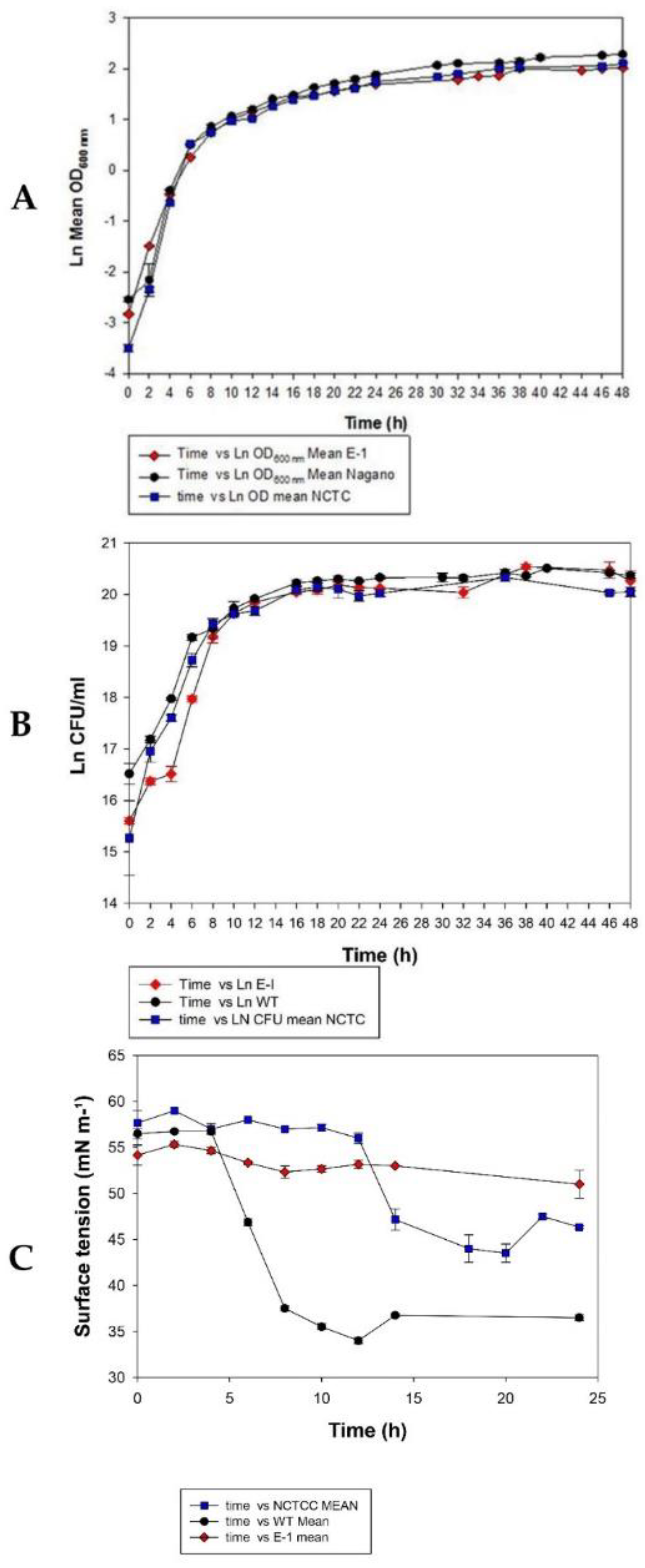
3.1. Growth Characteristics of A. migulanus Nagano, NCTC 7096, and E1 Mutant

A. migulanus Nagano, NCTC 7096, and E1 mutant strains grew vigorously in TSB broth. The exponential phase of growth was observed after 2 h of subculturing. After 48 h of growth, the maximum number of CFU was reached with a density of 108 mL−1. Cultures of A. migulanus Nagano, NCTC 7096, and E1 mutant entered approximately 8 h after subculturing using both CFU counts and OD measurements. It was obvious that there was no significant difference among the growth of the Nagano, NCTC 7096, and E1 mutant strains (p > 0.05) (Figure 1A,B).

3.2. Comparison of Biosurfactant Activity of A. migulanus Nagano, NCTC 7096, and E1 Mutant Strains Using Surface Tension Method

Surface tension of the culture fluids of the A. migulanus Nagano strain declined severely after 5 h of subculturing (Figure 1C). The stabilizing surface tension value was approximately 36 mNm−1 after 12–24 h of growth. The culture fluids of the NCTC 7096 strain showed a surface tension that began to decline after 12 h of subculturing with a final reduction to approximately 44 mNm−1, whereas the surface tension of the culture fluids of the A. migulanus Nagano E1 mutant did not show any decline over all the culture period, with a constant value of 54 mNm−1.

3.3. Gramicidin Production by A. migulanus Strains

A. migulanus Nagano and NCTC 7096 both were proven as effective in producing GS in contrast to the A. migulanus Nagano E1 mutant (Supplementary Table S1).

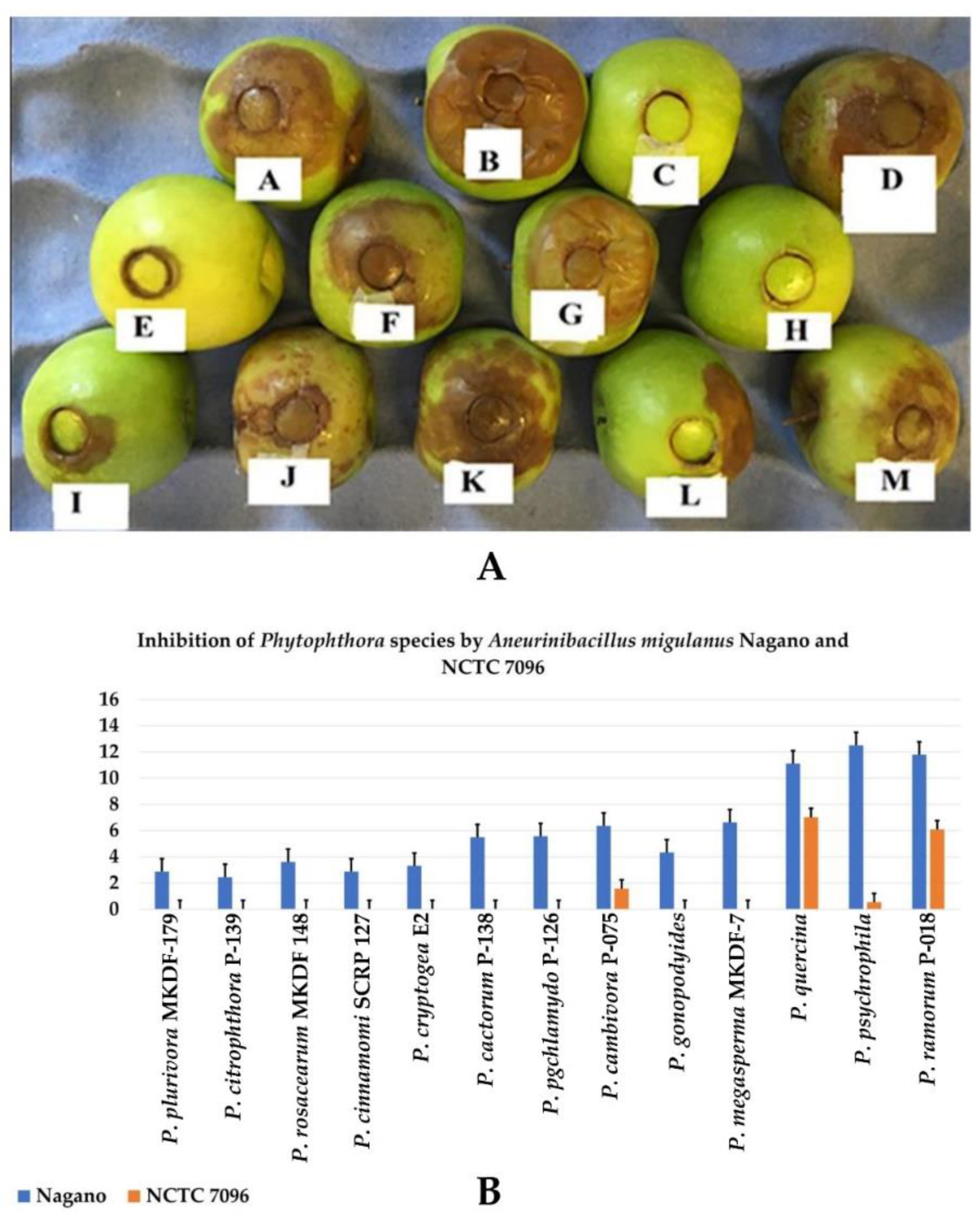
3.4. Effect of A. migulanus Nagano and NCTC 7096 Strains on Apple Infection of Phytophthora Species

All Phytophthora species were more significantly inhibited by A. migulanus Nagano compared to NCTC 7096 (p < 0.05; Figure 2). Species belonging to clade 6 of Phytophthora were the most sensitive Phytophthora species to A. migulanus Nagano, whereas clade 2 species were the least sensitive (Figure 2). The highest inhibition caused by A. migulanus Nagano was on P. psychrophila at 20 °C, whereas the least inhibition was against P. citrophthora P-139 (Figure 2). Mutant E1 failed to inhibit the growth of all tested Phytophthora species (Figure 2).

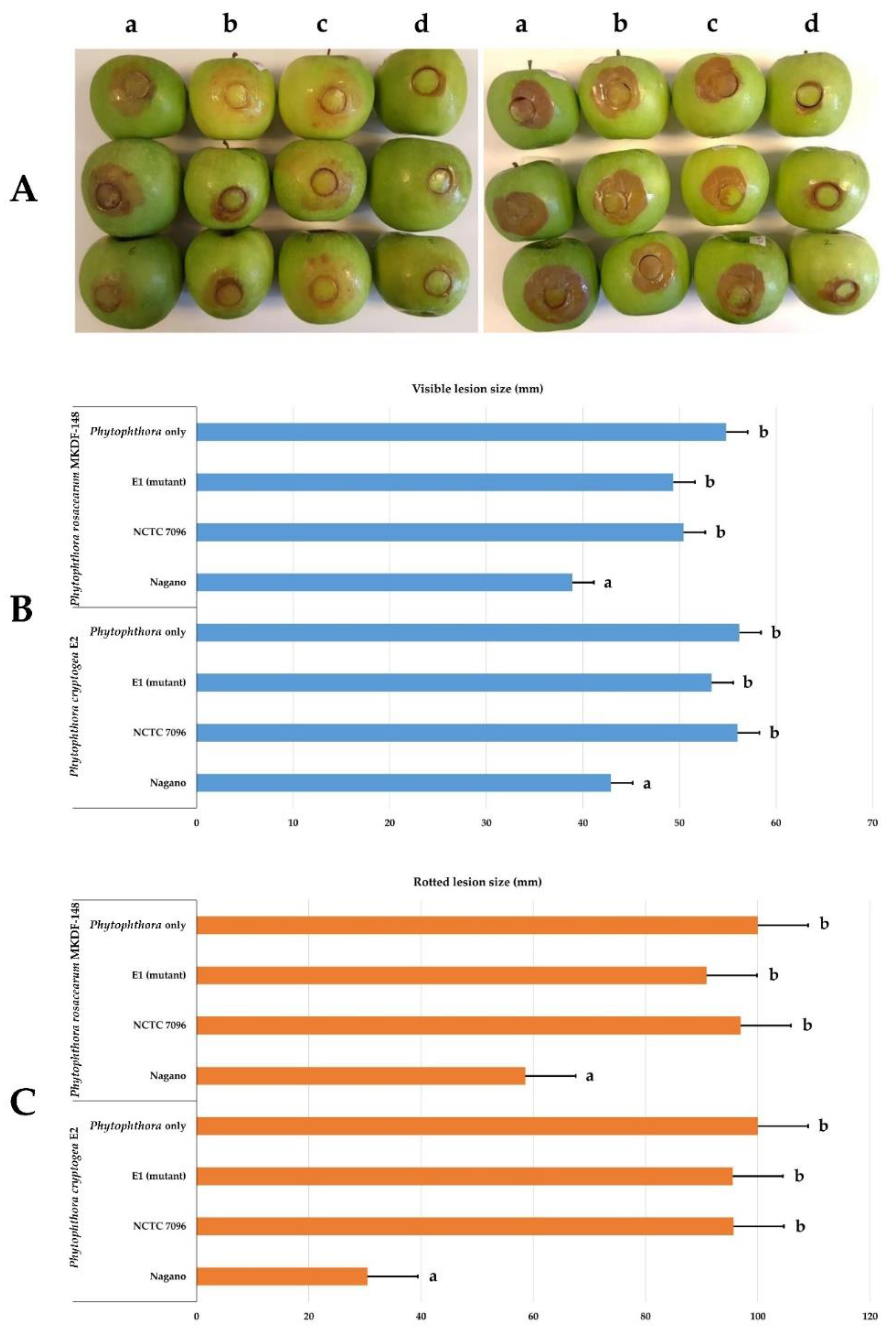
3.5. Effect of A. migulanus Nagano, NCTC 7096, and E1 Mutant Strains on Apple Infection of Phytophthora Rosacearum and Phytophthora Cryptogea Species

A. migulanus Nagano significantly reduced the external visible rot lesion sizes occurring in apples 6 days after inoculation with P. rosacearum MKDF-148 or P. cryptogea E2 (p = 0.001 and 0.01, respectively) compared with both apples being treated with A. migulanus NCTC 7096, positive controls, and mutant E1 (Figure 2 and Figure 3). In contrast, there was no significant difference in the size of the rot lesions in the NCTC 7096, positive control, and mutant E1 as a negative control (Phytophthora inoculated apples) with both Phytophthora spp. (Figure 2).
A. migulanus Nagano significantly reduced the amount of rot occurring in apples 6 days after inoculation with P. rosacearum MKDF-148 or P. cryptogea E2 (p = 0) compared with both apples being treated with A. migulanus NCTC 7096 and positive controls (Figure 2). In contrast, there was no significant difference in the quantity of rot in the NCTC 7096, positive control, and mutant E1 in apples with both Phytophthora species (Figure 2).

4. Discussion

Neither the use of the genetic resistance of the host nor the use of environmentally damaging synthetic pesticides succeeded in providing sustainable agricultural and forestry practices [3,7,30]. Therefore, biological control is more and more considered as a sound alternative of current practices for the management of plant pathogens in agricultural and forest ecosystems [6,8,11,13,31].
Biocontrol agents can act and positively impact plant health through well-established mechanisms, namely, antibiosis [2,18,32,33], induction of systemic resistance [34,35], and competition for nutrients and niches [4,5,10,13,17,36]. However, it is more and more documented that biocontrol is a multifactorial and complex process that has been until now under-investigated. Therefore, studies targeting the precise and fine study of biocontrol mechanisms are warranted towards the development of sustainable agricultural practices based on the use of biocontrol agents.
A. migulanus was shown to be a potent biocontrol agent effective in inhibiting various plant pathogens in agricultural and forest ecosystems [37]. Detailed studies of A. migulanus suggests that the bacteria act through the biosynthesis of a potent cyclic peptide, GS [17,22]. GS was shown to have a direct effect on pathogen spore germination and viability [17]. Apart from direct antibiosis, Seddon et al. [38,39] suggested that GS biosurfactant activity increases the rate of evaporation from the plant surface, thereby reducing periods of surface wetness inhibiting indirectly pathogen spore germination and viability. Indirect proof of the importance of GS in the interaction between A. migulanus and plant pathogens and, therefore, its biocontrol potential arises from the whole genome sequencing of A. migulanus Nagano and NCTC 7096 [25,26]. Up to 8% of their genome was dedicated to the biosynthesis of an impressive array of antimicrobial compounds, notably GS [17]. The availability of the A. migulanus Nagano GS mutant strain (E1 mutant) prompted us to study, for the first time, the importance of GS in the biocontrol ability of A. migulanus Nagano. LC-MS analysis of GS production in all A. migulanus tested strains allowed us to conclude that A. migulanus Nagano and NCTC 7096 both proved to be effective in producing GS in contrast to the A. migulanus Nagano E1 mutant, which failed to produce significant amounts of GS (Table 2, Supplementary Tables S2 and S3). It is worth noting that there was a clear correlation between GS production by the A. migulanus strains and biocontrol ability in a previous study by Alenezi et al. [17]. These results were confirmed in this study including the E1 mutant. Using a surface tension assay, we were able to prove that the surface tension of the culture fluids of the A. migulanus Nagano strain declined severely after 5 h of subculturing.
The stabilizing surface tension value was approximately 36 mNm−1 after 12–24 h of growth. The culture fluids of the NCTC 7096 strain showed a surface tension that began to decline after 12 h of subculturing, with a final reduction to approximately 44 mNm−1, whereas the surface tension of the culture fluids of the A. migulanus Nagano E1 mutant did not show any decline over all the culture periods, with a constant value of 54 mNm−1. This result is in agreement with the report of Alenezi et al. [17], where, additionally, the A. migulanus Nagano dried significantly faster on the tomato leaf surface than did the A. migulanus NCTC 7096 and E1 mutant [17]. This result clearly documented that the E1 mutant was impaired in the production of GS and its associated biosurfactant activity. Additional confirmation of this result was obtained using blood Agar and oil spreading methods applied to the A. migulanus Nagano, NCTC 7096, and E1 mutant strains. Although both strains, A. migulanus Nagano and NCTC 7096, were able to spread oil on the surface of water, the E1 mutant was not able do so, suggesting that the E1 mutant lost totally its biosurfactant activity. It is worth noticing that the diameter of the oil spread was greater for A. migulanus Nagano than for NCTC 7096 (p = 0.00; two-sample t-test), as reported by Alenezi et al. [17].
Using a set of 13 Phytophthora species, namely, P. cactorum, P. plurivora, P. citrophthora, P. psychrophila, P. quercina, P. taxon Pgchlamydo, P. gonapodyides, P. megasperma, P. rosacearum, P. cinnamomi, P. cambivora, P. cryptogea, and P. ramorum, we unambiguously documented that all Phytophthora species were more significantly inhibited by A. migulanus Nagano compared to NCTC 7096 (p < 0.05). This result is in agreement with further reports [17,22], suggesting the strain level biocontrol abilities in A. migulanus. Interestingly, species belonging to clade 6 of Phytophthora were the most sensitive Phytophthora species to A. migulanus Nagano, whereas clade 2 species were the least sensitive (data not shown). We speculated that clade-specific determinants are targeted by A. migulanus. However, this needs further targeted studies to determine the reasons of clade specificity of Phytophthora to A. migulanus. Our results also clearly document that the highest inhibition caused by A. migulanus Nagano was on P. psychrophila at 20 °C, whereas the least inhibition was against P. citrophthora P-139. Based on these results, two phytophthora species highly sensitive to A. migulanus (P. rosacearum and P. cryptogea) were selected for the rest of the study. These two species have also the advantage of being the most aggressive on apple fruits, displaying clear infection halos. It is worth noting that the A. migulanus Nagano mutant E1 failed to inhibit the growth of all the tested Phytophthora species (Figure 3). Further experiments using the combination of the two Phytophthora species with A. migulanus Nagano, NCTC 7096, and E1 mutants clearly indicated that the A. migulanus Nagano E1 mutant impaired in the biosynthesis of GS was unable to inhibit the growth of both Phytophthora species. A. migulanus Nagano significantly reduced the external visible rot lesion sizes occurring in apples 6 days after inoculation with P. rosacearum MKDF-148 or P. cryptogea E2 (p = 0.001 and 0.01, respectively) compared with both apples being treated with A. migulanus NCTC 7096, positive controls, and mutant E1 (Figure 1 and Figure 2).

5. Conclusions

The present work revealed a negative correlation between GS production in the culture fluids of A. migulanus and its biocontrol ability. This discovery provides a direct proof of the importance of GS for the biocontrol ability of A. migulanus. It also paves the way towards the development of effective biocontrol agents either in a homologous or heterologous way by overexpressing the GS biosynthetic cluster. Ongoing results that will be published in a future manuscript confirm this speculation and provide a model for further similar investigations in closely related species and genera.

Supplementary Materials

The following supporting information can be downloaded at https://www.mdpi.com/article/10.3390/f13040535/s1. Table S1: NCTC 7096 gramicidins’ mass spectrometric peptide fragmentation. For GS-1141, GS-1155, and GS-1169, where () represents the presence of each fragment and () represents the absence and () represents the actual mass. Table S2: E1 mutant (non-gramicidin’s producer) gramicidins’ mass spectrometric peptide fragmentation. For GS-1141, GS-1155, and GS-1169, where () represents the presence of each fragment, () represents the absence and () represents the actual mass.

Author Contributions

Conceptualization, F.N.A., A.C.B., H.C.-S., A.S. and L.B.; methodology, F.N.A., A.C.B., M.J., H.C.-S., A.S. and L.B.; software, F.N.A.; validation, F.N.A., H.C.-S. and L.B.; formal analysis, F.N.A., A.C.B., L.B. and M.J.; investigation, F.N.A., A.S. and L.B.; resources, T.O. and L.B.; data curation, F.N.A., A.C.B., M.J. and L.B.; writing—original draft preparation, F.N.A.; writing—review and editing, L.B.; visualization, A.C.B., F.N.A. and L.B.; supervision, L.B.; project administration, L.B.; funding acquisition, F.N.A. and L.B. All authors have read and agreed to the published version of the manuscript.

Funding

There is no funder about this work.

Institutional Review Board Statement

Not applicable.

Informed Consent Statement

Not applicable.

Conflicts of Interest

The authors declare no conflict of interest.

References

  1. Belbahri, L.; Lara, E. SSU rRNA reveals major trends in oomycete evolution. Fungal Divers. 2011, 49, 93–100. [Google Scholar] [CrossRef]
  2. Mefteh, F.B.; Daoud, A.; Chenari Bouket, A.; Alenezi, F.N.; Luptakova, L.; Rateb, M.E.; Kadri, A.; Gharsallah, N.; Belbahri, L. Fungal Root Microbiome from Healthy and Brittle Leaf Diseased Date Palm Trees (Phoenix dactylifera L.) Reveals a Hidden Untapped Arsenal of Antibacterial and Broad Spectrum Antifungal Secondary Metabolites. Front. Microbiol. 2017, 8, 307. [Google Scholar] [CrossRef]
  3. Cherrad, S.; Charnay, A.; Hernandez, C.; Steva, H.; Belbahri, L.; Vacher, S. Emergence of boscalid-resistant strains of Erysiphe necator in French vineyards. Microbiol. Res. 2018, 216, 79–84. [Google Scholar] [CrossRef]
  4. Slama, H.; Cherif-Silini, H.; Chenari Bouket, A.; Qader, M.; Silini, A.; Yahiaoui, B.; Alenezi, F.; Luptakova, L.; Triki, M.; Vallat, A.; et al. Screening for Fusarium antagonistic bacteria from contrasting niches designated the endophyte Bacillus halotolerans as plant warden against Fusarium. Front. Microbiol. 2019, 9, 3236. [Google Scholar] [CrossRef] [Green Version]
  5. Slama, H.B.; Triki, M.A.; Chenari Bouket, A.; Ben Mefteh, F.; Alenezi, F.N.; Luptakova, L.; Cherif-Silini, H.; Vallat, A.; Oszako, T.; Gharsallah, N.; et al. Screening of the High-Rhizosphere Competent Limoniastrum monopetalum’ Culturable Endophyte Mi-crobiota Allows the Recovery of Multifaceted and Versatile Biocontrol Agents. Microorganisms 2019, 7, 249. [Google Scholar] [CrossRef] [Green Version]
  6. Silini, H.C.; Silini, A.; Chenari Bouket, A.; Alenezi, F.N.; Luptakova, L.; Bouremani, N.; Nowakowska, J.A.; Oszako, T.; Belbahri, L. Tailoring Next Generation Plant Growth Promoting Microorganisms as Versatile Tools beyond Soil Desalinization: A Road Map towards Field Application. Sustainability 2021, 13, 4422. [Google Scholar] [CrossRef]
  7. Solla, A.; Moreno, G.; Malewski, T.; Jung, T.; Klisz, M.; Tkaczyk, M.; Siebyla, M.; Pérez, A.; Cubera, E.; Hrynyk, H.; et al. Phosphite Spray for the Control of Oak Decline Induced by Phytophthora in Europe. For. Ecol. Manag. 2021, 485, 118938. [Google Scholar] [CrossRef]
  8. Balla, A.; Silini, A.; Cherif-Silini, H.; Chenari Bouket, A.; Moser, W.K.; Nowakowska, J.A.; Oszako, T.; Benia, F.; Belbahri, L. The Threat of Pests and Pathogens and the Potential for Biological Control in Forest Ecosystems. Forests 2021, 12, 1579. [Google Scholar] [CrossRef]
  9. Silini, H.C.; Thissera, B.; Chenari Bouket, A.; Saadaoui, N.; Silini, A.; Eshelli, M.; Alenezi, F.N.; Vallat, A.; Luptakova, L.; Yahiaoui, B.; et al. Durum Wheat Stress Tolerance Induced by Endophyte Pantoea agglomerans with Genes Contributing to Plant Functions and Secondary Metabolite Arsenal. Int. J. Mol. Sci. 2019, 20, 3989. [Google Scholar] [CrossRef] [Green Version]
  10. Cheffi, M.; Chenari Bouket, A.; Alenezi, F.N.; Luptakova, L.; Belka, M.; Vallat, A.; Rateb, M.E.; Tounsi, S.; Triki, M.A.; Belbahri, L. Olea europaea L. Root Endophyte Bacillus velezensis OEE1 Counteracts Oomycete and Fungal Harmful Pathogens and Harbours a Large Repertoire of Secreted and Volatile Metabolites and Beneficial Functional Genes. Microorganisms 2019, 7, 314. [Google Scholar] [CrossRef] [Green Version]
  11. Dhouib, H.; Zouari, I.; Abdallah, D.B.; Belbahri, L.; Taktak, W.; Triki, M.A.; Tounsi, S. Potential of a novel endophytic Bacillus velezensis in tomato growth promotion and protection against Verticillium wilt disease. Biol. Control 2019, 139, 104092. [Google Scholar] [CrossRef]
  12. Kerbab, S.; Silini, A.; Chenari Bouket, A.; Cherif-Silini, H.; Eshelli, M.; El Houda Rabhi, N.; Belbahri, L. Mitigation of NaCl Stressin Wheat by Rhizosphere Engineering Using Salt Habitat Adapted PGPR Halotolerant Bacteria. Appl. Sci. 2021, 11, 1034. [Google Scholar] [CrossRef]
  13. Oszako, T.; Voitka, D.; Stocki, M.; Stocka, N.; Nowakowska, J.A.; Linkiewicz, A.; Hsiang, T.; Belbahri, L.; Berezovska, D.; Malewski, T. Trichoderma asperellum efficiently protects Quercus robur leaves against Erysiphe alphitoides. Eur. J. Plant Pathol. 2021, 159, 295–308. [Google Scholar] [CrossRef]
  14. Roylawar, P.; Khandagale, K.; Randive, P.; Shinde, B.; Murumkar, C.; Ade, A.; Singh, M.; Gawande, S.; Morelli, M. Piriformospora indica Primes Onion Response against Stemphylium Leaf Blight Disease. Pathogens 2021, 10, 1085. [Google Scholar] [CrossRef]
  15. Vick, S.H.W.; Fabian, B.K.; Dawson, C.J.; Foster, C.; Asher, A.; Hassan, K.A.; Midgley, D.J.; Paulsen, I.T.; Tetu, S.G. Delving into defence: Identifying the Pseudomonas protegens Pf-5 gene suite involved in defense against secreted products of fungal, oomycete and bacterial rhizosphere competitors. Microb. Genom. 2021, 7, 000671. [Google Scholar] [CrossRef]
  16. Sandhu, S.K.; Morozov, A.Y.; Holt, R.D.; Barfield, M. Revisiting the Role of Hyperparasitism in the Evolution of Virulence. Am. Nat. 2021, 197, 216–235. [Google Scholar] [CrossRef]
  17. Alenezi, F.N.; Rekik, I.; Chenari Bouket, A.; Luptakova, L.; Weitz, H.J.; Rateb, M.E.; Jaspars, M.; Woodward, S.; Belbahri, L. Increased Biological Activity of Aneurinibacillus migulanus Strains Correlates with the Production of New Gramicidin Secondary Metabolites. Front. Microbiol. 2017, 8, 517. [Google Scholar] [CrossRef] [Green Version]
  18. Thissera, B.; Alhadrami, H.A.; Hassan, M.H.A.; Hassan, H.M.; Behery, F.A.; Bawazeer, M.; Yaseen, M.; Belbahri, L.; Rateb, M.E. Induction of Cryptic Antifungal Pulicatin Derivatives from Pantoea Agglomerans by Microbial Co-Culture. Biomolecules 2020, 10, 268. [Google Scholar] [CrossRef] [Green Version]
  19. Köhl, J.; Kolnaar, R.; Ravensberg, W.J. Mode of action of microbial biological control agents against plant diseases: Relevance beyond efficacy. Front. Plant Sci. 2019, 10, 845. [Google Scholar] [CrossRef] [Green Version]
  20. Belbahri, L.; Alenezi, F.N.; Luptakova, L.; Rateb, M.E.; Woodward, S. Complete genome sequence of Aneurinibacillus migulanus E1, a gramicidin S- and D-phenylalanyl-L-propyl diketopiperazine-deficient mutant. Genome Announc. 2015, 3, e01441-15. [Google Scholar] [CrossRef] [Green Version]
  21. Slama, H.; Cherif-Silini, H.; Chenari Bouket, A.; Silini, A.; Alenezi, F.N.; Luptakova, L.; Vallat, A.; Belbahri, L. Biotechnology and bioinformatics of endophytes in biocontrol, bioremediation, and plant growth promotion. In Endophytes: Mineral Nutrient Management, Volume 3. Sustainable Development and Biodiversity; Maheshwari, D.K., Dheeman, S., Eds.; Springer: Cham, Switzerland, 2021; Volume 26, pp. 181–205. [Google Scholar]
  22. Alenezi, F.N.; Fraser, S.; Bełka, M.; Doğmubbbs, T.H.; Hečkova, Z.; Oskay, F.; Belbahri, L.; Woodward, S. Biological control of Dothistroma needle blight on pine with Aneurinibacillus migulanus. For. Pathol. 2016, 46, 555–558. [Google Scholar] [CrossRef]
  23. Alenezi, F.N.; Rekik, I.; Belka, M.; Ibrahim, A.F.; Luptakova, L.; Woodward, S.; Belbahri, L. Strain-level diversity of secondary metabolism in the biocontrol species Aneurinibacillus migulanus. Microbiol. Res. 2016, 182, 116–124. [Google Scholar] [CrossRef]
  24. Sellami, M.; Khlifi, A.; Frikha, F.; Miled, N.; Belbahri, L.; Ben Rebah, F. Agro-industrial waste-based growth media optimization for biosurfactant production by Aneurinibacillus migulanus. J. Microbiol. Biotechnol. Food Sci. 2016, 5, 578–583. [Google Scholar] [CrossRef] [Green Version]
  25. Alenezi, F.N.; Weitz, H.J.; Belbahri, L.; Nidhal, J.; Luptakova, L.; Jaspars, M.; Woodward, S. Draft genome sequence of Aneurinibacillus migulanus NCTC 7096. Genome Announc. 2015, 3, e00234-15. [Google Scholar] [CrossRef] [Green Version]
  26. Alenezi, F.N.; Weitz, H.J.; Belbahri, L.; Ben Rebah, H.; Luptakova, L.; Jaspars, M.; Woodward, S. Draft genome sequence of Aneurinibacillus migulanus strain nagano. Genome Announc. 2015, 3, e00232-15. [Google Scholar] [CrossRef] [Green Version]
  27. Shimura, K.; Iwaki, M.; Kanda, M.; Hori, K.; Kaji, E.; Hasegawa, S.; Saito, Y. On the enzyme system obtained from some mutants of Bacillus brevis deficient in gramicidin S formation. Biochim. Biophys. Acta 1974, 338, 577–587. [Google Scholar] [CrossRef]
  28. Belbahri, L.; Calmin, G.; Sanchez-Hernandez, E.; Oszako, T.; Lefort, F. Pythium sterilum sp. nov. isolated from Poland, Spain and France: Its morphology and molecular phylogenetic position. FEMS Microbiol. Lett. 2006, 255, 209–214. [Google Scholar] [CrossRef] [Green Version]
  29. Prospero, S.; Vercauteren, A.; Heungens, K.; Belbahri, L.; Rigling, D. Phytophthora diversity and the population structure of Phytophthora ramorum in Swiss ornamental nurseries. Plant Pathol. 2013, 62, 1063–1071. [Google Scholar] [CrossRef]
  30. Naseri, B.; Younesi, H. Beneficial microbes in biocontrol of root rots in bean crops: A meta-analysis (1990–2020). Physiol. Mol. Plant Pathol. 2021, 116, 101712. [Google Scholar] [CrossRef]
  31. Belbahri, L.; Chenari Bouket, A.; Rekik, I.; Alenezi, F.N.; Vallat, A.; Luptakova, L.; Petrovova, E.; Oszako, T.; Cherrad, S.; Vacher, S.; et al. Comparative Genomics of Bacillus amyloliquefaciens Strains Reveals a Core Genome with Traits for Habitat Adaptation and a Secondary Metabolites Rich Accessory Genome. Front. Microbiol. 2017, 8, 1438. [Google Scholar] [CrossRef]
  32. Mefteh, F.B.; Daoud, A.; Chenari Bouket, A.; Thissera, B.; Kadri, Y.; Cherif-Silini, H.; Eshelli, M.; Alenezi, F.N.; Vallat, A.; Oszako, T.; et al. Date Palm Trees Root-Derived Endophytes as Fungal Cell Factories for Diverse Bioactive Metabolites. Int. J. Mol. Sci. 2018, 19, 1986. [Google Scholar] [CrossRef] [Green Version]
  33. Thissera, B.; Hallyburton, I.; Ngwa, C.J.; Cherif-Silini, H.; Hassane, A.S.I.; Anderson, M.; Campbell, L.A.; Mutter, N.; Eshelli, M.; Abdelmohsen, U.R.; et al. Potent antiplasmodial alkaloids from the rhizobacterium Pantoea agglomerans as hemozoin modulators. Bioorg. Chem. 2021, 115, 105215. [Google Scholar] [CrossRef]
  34. Biessy, A.; Filion, M. Biological control of potato common scab by plant-beneficial bacteria. Biol. Control 2022, 165, 104808. [Google Scholar] [CrossRef]
  35. Qiu, Y.; Yan, H.-H.; Sun, S.-M.; Wang, Y.-Q.; Zhao, X.-R.; Wang, H.-Y. Use of Bacillus velezensis SDTB022 against tobacco black shank (TBS) and the biochemical mechanism involved. Biol. Control 2022, 165, 104785. [Google Scholar] [CrossRef]
  36. Dimkic, I.; Janakiev, T.; Petrovic, M.; Degrassi, G.; Fira, D. Plant-associated Bacillus and Pseudomonas antimicrobial activities in plant disease suppression via biological control mechanisms—A review. Physiol. Mol. Plant Pathol. 2022, 117, 101754. [Google Scholar] [CrossRef]
  37. Schuster, C.; Schmitt, A. Efficacy of a bacterial preparation of Aneurinibacillus migulanus against downy mildew of cucumber (Pseudoperonospora cubensis). Eur. J. Plant Pathol. 2017, 151, 439–450. [Google Scholar] [CrossRef]
  38. Seddon, B.; Edwards, S.; Markellou, E.; Malathrakis, N. Bacterial antagonist-fungal pathogen interactions on the plant aerial surface. Multitrophic Interact. Terr. Syst. 1997, 1, 5–25. [Google Scholar]
  39. Seddon, B.; McHugh, R.; Schmitt, A. Brevibacillus brevis—A novel candidate biocontrol agent with broad-spectrum antifungal activity. In Proceedings of the BCPC Conference: Pests and Diseases. Proceedings of the International Conference Held at the Brighton Hilton Metropole Hotel, Brighton, UK, 13–16 November 2000; Volume 2. [Google Scholar]
Figure 1. The comparison of growth rates among the three strains of A. migulanus. (A) Optical density (OD600nm) measurements for (●) A. migulanus Nagano, () NCTC 7096, and () E1. (B) Colony forming units (CFU) per mL for (●) A. migulanus Nagano, () NCTC 7096, and () E1. Vertical bars represent standard errors of the means of three independent replicates. (C) Aneurinibacillus migulanus biosurfactant effects on surface tension of culture fluids and time course of changes in surface tension of (●) A. migulanus Nagano, () NCTC 7096, and () E1 in culture fluids. Vertical bars represent standard errors of the means (N = 3).
Forests 13 00535 g001
Figure 2. (A) In vivo pathogenicity assay of Phytophthora species on apple fruits; (A) P. cryptogea E2, (B) P. rosacearum MKDF-148, (C) P. quercina, (D) P. plurivora MKDF-179, (E) P. cinnamomi SCRP 127, (F) P. cambivora P-075, (G) P. taxon Pgchlamydo P-126, (H) P. psychrophila, (I) P. megasperma MKDF-7, (J) P. citrophthora P-139, (K) P. cactorum P-138, (L) P. gonapodyides, (M) P. ramorum P-018 (7 days after inoculation at 20 °C). (B) Inhibition of Phytophthora species by A. migulanus in screening assay. Vertical bars represent standard errors of the means (N = 5).
Forests 13 00535 g002
Figure 3. (A) Granny Smith apples inoculated with P. cryptogea (left side) and P. rosacearum (right side); (a) inoculated with P. cryptogea or P. rosacearum (positive control), (b) pre-treated with A. migulanus E1, (c) pre-treated with A. migulanus NCTC 7096, (d) pre-treated with A. migulanus Nagano (6 days after inoculation). (B) Effects of A. migulanus Nagano, NCTC 7096, and mutant E1 on lesion sizes in apples by Phytophthora cryptogea E2 and P. rosacearum MKDF-148 (6 days after inoculation). (C) Effects of A. migulanus Nagano, NCTC 7096, and mutant E1 on the amount of rot caused by Phytophthora cryptogea E2 and P. rosacearum MKDF-148 (6 days after inoculation). Bars labeled with different letters represent significant differences among the treatments at p < 0.05 using the Tukey’s HSD test. In each bar groups, bars labeled with the same letter are not significantly different from each other according to Tukey’s HSD at p < 0.05.
Forests 13 00535 g003
Table 1. Sources of Phytophthora species used in dual culture antagonism tests with A. migulanus.
CladePhytophthora SpeciesStrainOrigin
1P. cactorumP-138Kent
2P. plurivora
P. citrophthora
MKDF-179
P-139
Macedonia
Kent
3P. psychrophilaSpain
4P. quercinaSpain
6P. taxonPgchlamydo
P.gonapodyides
P. megasperma
P. rosacearum
P-126

MKDF-7
MKDF-148
Kent
Spain
Macedonia
Macedonia
7P. cinnamomi
P. cambivora
SCRP 127
P-075
Dundee
Unknown
8P. cryptogea
P. ramorum
E2
P-018
Aberdeen
Kent
Table 2. Nagano gramicidins’ mass spectrometric peptide fragmentation. For GS-1141, GS-1155, and GS-1169, where () represents the presence of each fragment, () represents the absence and () represents the actual mass.
TitleNterm_modaa1aa2aa3aa4aa5aa6aa7aa8aa9aa10adduct
TermLFPVKLFPVK[M + H]+
Mass1.007825113.0841147.068497.0527699.06841128.095113.0841147.068497.0527699.06841128.094961169.745
b-series1.007825114.0919261.1603358.2131457.2815585.3764698.4605845.5289942.58171041.651169.745
Actual mass -261.1606358.2188457.2803585.3751698.4605845.5264942.57621041.6481
y-series1.0078251169.7451056.661909.5926812.5398713.4714585.3764472.2924325.224228.1712129.102785
Actual mass -1056.6727909.5892812.5347713.4695585.3751472.2917325.2228228.1687
Nterm_modaa1aa2aa3aa4Ornaa6aa7aa8aa9aa10adduct
TermLFPVKLFPVK[M + H]+
Mass1.007825113.0841147.068497.0527699.06841114.0793113.0841147.068497.0527699.06841128.094961155.729
b-series1.007825114.0919261.1603358.2131457.2815571.3608684.4448831.5132928.5661027.6341155.7294
Actual mass -261.1598358.2122457.2795571.3612684.4433831.5117928.55951027.6339
y-series1.0078251155.7291042.645895.5769798.5241699.4557585.3764472.2924325.224228.1712129.102785
Actual mass 1042.6502895.5762798.5214699.4553585.3752472.2917325.2232228.1713
Nterm_modaa1aa2aa3aa4Ornaa6aa7aa8aa9Ornadduct
TermLFPVKLFPVK[M + H]+
Mass1.007825113.0841147.068497.0527699.06841114.0793113.0841147.068497.0527699.06841113.938461141.573
b-series1.007825114.0919261.1603358.2131457.2815571.3608684.4448831.5132928.5661027.6341141.5729
Actual mass 261.1598358.2108457.2784571.3607684.4442831.5147928.56531027.6261
y-series1.0078251141.7141028.63881.5613784.5085685.4401571.3608458.2767311.2083214.1555115.087135
Actual mass 1028.6317881.5618784.5082685.4402571.3607458.2810311.2080214.1554
Publisher’s Note: MDPI stays neutral with regard to jurisdictional claims in published maps and institutional affiliations.

