Article

Mangrove Dieback and Leaf Disease in Sonneratia apetala and Sonneratia caseolaris in Vietnam

1
Agriculture and Forest Sciences, Murdoch University, Murdoch 6150, Australia
2
Silviculture Research Institute, Vietnamese Academy of Forest Sciences, Duc Thang, Bac Tu Liem, Ha Noi 11910, Vietnam
3
Institute of Ecology and Works Protection, Vietnam Academy for Water Resources, 267 Chua Boc, Dong Da, Ha Noi 11515, Vietnam
4
Institute of Biotechnology, Vietnam Academy of Science and Technology, 18 Hoang Quoc Viet, Nghia Do, Cau Giay, Ha Noi 11350, Vietnam
5
Forest Protection Research Centre, Vietnamese Academy of Forest Sciences, Duc Thang, Bac Tu Liem, Ha Noi 11910, Vietnam
*
Author to whom correspondence should be addressed.
Forests 2021, 12(9), 1273; https://doi.org/10.3390/f12091273
Submission received: 24 July 2021 / Revised: 13 September 2021 / Accepted: 14 September 2021 / Published: 17 September 2021
(This article belongs to the Special Issue Abiotic and Biotic Stress in Forest and Plantation Trees)

Abstract

:
Even though survival rates for mangrove restoration in Vietnam have often been low, there is no information on fungal pathogens associated with mangrove decline in Vietnam. Therefore, this research was undertaken to assess the overall health of mangrove afforestation in Thanh Hoa Province and fungal pathogens associated with tree decline. From a survey of 4800 Sonneratia trees, the incidence of disorders was in the order of pink leaf spot > shoot dieback > black leaf spot for S. caseolaris and black leaf spot > shoot dieback > pink leaf spot for S. apetala. Approximately 12% of S. caseolaris trees had both pink leaf spot and shoot dieback, while only 2% of S. apetala trees had black leaf spot and shoot dieback. Stem and leaf samples were taken from symptomatic trees and fungi were cultured in vitro. From ITS4 and ITS5 analysis, four main fungal genera causing leaf spots and shoot dieback on the two Sonneratia species were identified. The most frequently isolated fungal taxa were Curvularia aff. tsudae (from black leaf spot), Neopestalotiopsis sp.1 (from stem dieback), Pestalotiopsis sp.1 (from pink leaf spot), and Pestalotiopsis sp.4a (from black leaf spot). The pathogenicity of the four isolates was assessed by under-bark inoculation of S. apetala and S. caseolaris seedlings in a nursery in Thai Binh Province. All isolates caused stem lesions, and Neopestalotiopsis sp.1 was the most pathogenic. Thus, investigation of fungal pathogens and their impact on mangrove health should be extended to other afforestation projects in the region, and options for disease management need to be developed for mangrove nurseries.

1. Introduction

Mangroves provide crucial environmental services including habitat for birds, fish, and invertebrates. They are responsible for coastal protection from hurricanes, floods, sea-level rise, wave action, and erosion [1,2]. Mangrove systems are among the most threatened ecosystems globally [3,4] due to the fact of increasing urban and industrial development [5,6,7]. In addition, abiotic factors, such as hurricanes, lightning strikes, salinity, and flooding, are reported to be affecting the health of mangroves [2,8]. Extreme weather, such as drought, hot water, hot air, and the temporary drop in sea level, have contributed to mangrove dieback [9]. For example, approximately 7400 ha of mangroves in northern Australia died in the summer of 2015–2016 [10]. As well as primary pathogens and insects, abiotic stress can expose trees to a variety of opportunistic pests that can cause tree declines [11,12]. In addition, primary pathogens and insects can cause tree degradation. Unlike in terrestrial ecosystems, there are fewer reports of pathogenic fungi in mangroves [13,14,15], and these have mostly been descriptive and focus on the morphology and taxonomy of mangrove fungi [16,17,18,19,20]. A range of fungal pathogens have been associated with disease in mangroves (Table 1). Overall, given the pantropical distribution of mangroves, the number of investigations into mangrove health remain limited [21].
It is estimated that the total area of mangrove forests in Vietnam declined from 408,500 ha in 1943 [39] to 235,569 ha in 2019 [40]. According to recent studies, although many afforestation projects have been undertaken in Vietnam, the survival of out-planted nursery stock has often been unsatisfactory [41,42]. The few studies on mangrove damage or death in the country did not consider pathogens but focused on environmental processes and human activities such as industrial development, over-harvesting, and land conversion to agriculture. These included dieback of 20 year old Rhizophora apiculata in Ben Tre Province [43], where the disease was likely influenced by increased anthropogenic disturbances [44]. Other reports included mortality of mangroves in Quang Ngai [45] and in Ha Tinh [46] provinces. Large gaps remain in the documentation of mangrove diseases for most parts of northern Vietnam. As the average rate of success of mangrove restoration in northern Vietnam is approximately 30% [47], understanding whether mangrove diseases are a contributing factor could help improve the management of mangroves in future projects. Therefore, this study was undertaken to document the extent and impact of disease problems on afforested mangroves in Thanh Hoa Province. The objectives were to (i) assess the health of mangrove restoration and (ii) to identify likely causes of mangrove decline.

2. Materials and Methods

2.1. Study Area and Canopy Assessment

Thanh Hoa Province in north–central Vietnam was chosen because afforestation trials had been established in 2015, and remote sensing imagery showed that many plantings had been unsuccessful [47]. Thanh Hoa city (20.0° N, 105.5° E) has a tropical climate with intermittent rainfall concentrated in the summer (1447 mm/year), and the average annual temperature is 27.3 °C. Preliminary health surveys were randomly conducted in mangroves in two geographical areas (Table 2) to assess canopy conditions in 2018. This revealed shoot dieback and leaf spots as the main tree health problems. An afforestation plantation planted in April 2018 in Hoang Phu District was then chosen for a detailed study of these problems. The afforestation trial was established using Sonneratia caseolaris and Sonneratia apetala with single and mixed plantings at a spacing of 2 × 3 m. A rating system (Table 3) was used to quantify the condition of 4800 trees, 16 months after out-planting, in August 2019. Shoot dieback and foliar symptoms of pink and black spots are illustrated (Figure 1).

2.2. Sample Collection

During preliminary sampling in December 2018, 30 leaf and stem samples were collected at random from symptomatic trees in Hoang Phu and Sam Son districts (Table 1), and transported to the Plant Clinic, Forest Protection Research Center (Vietnamese Academy of Forest Sciences, Hanoi) where fungal cultures were obtained. In August 2019, more intensive sampling of two leaf spots (pink, black) and branch dieback was undertaken from the 2018 afforestation trials in the Hoang Phu District. Samples were collected from the two Sonneratia species from single and mixed plantings. A total of 105 samples were collected from each species comprising 70 leaf samples and 35 branch samples.

2.3. Fungal Isolation

Branches and leaves were surface sterilized with 70% ethanol for 1 min, rinsed with sterile water. Then 2 × 2 mm pieces containing leaf spots were excised, 1–2 cm lengths were cut from the junction of healthy and dead wood of branches, and the pieces were placed on sterile moist paper in Petri dishes. The Petri dishes were sealed with cling wrap and incubated at 25 °C in the dark. After 2–3 days, aerial mycelia of putative pathogens were transferred aseptically to potato dextrose agar (Gibco, ThermoFisher Scientific, Abingdon, UK) (PDA) supplemented with 0.75% streptomycin sulfate (strep) and incubated at 25 °C in the dark. When fungal cultures had grown several cm in diameter, hyphal tips were sub-cultured onto new PDA + strep Petri dishes. Isolates cultured on PDA at 25 °C were used to describe colony development and for DNA extraction. The morphology and size of spores were obtained using an OLYMPUS BX50 (Tokyo, Japan) compound microscope. A total of 227 fungal isolates were cultured from the 135 samples and grouped into 6 morphotypes. Of these, 9 isolates were selected for sequencing. All fungal cultures are lodged in the Vietnam National University of Agriculture.

2.4. Fungal Identification

  • DNA extraction, PCR amplification, and sequencing analysis
Fungal cultures obtained from symptomatic plant materials were grouped based on colony morphology, color, fruiting bodies, and spore characteristic and used for DNA extraction. DNA was extracted using a CTAB (cetyltrimethyl ammonium bromide, Novo Nordisk Pharmatech, Køge, Denmark) method [48] with minor modification. For each isolate, approximately 100 mg of fresh mycelia mat was transferred into a 1.5 mL Eppendorf tube, washed with distilled water, and homogenized with 500 μL extraction buffer (2 M NaCl, 25 mM EDTA, 100 mM Tris-HCl, 2% PVP, and 2% CTAB) using a plastic pestle. The genomic DNA was extracted with chloroform–isoamyl alcohol (24:1) two times and precipitated with cold propanol. DNA pellets were washed two times with 70% ethanol, air-dried for 30 min, resuspended in 50 μL of TE, and stored at −20 °C.
The primers ITS4 (forward primer) and ITS5 (reverse primer) [49,50] were used to amplify the ITS gene region. PCRs were performed in a final reaction volume of 25 μL containing 2.5 μL DreamTaq PCR X 10 buffer (Fermantas, Waltham, MA, USA), 0.5 μL dNTPS (10 mM each, Roche, Capetown, South Africa), 0.5 μL of each primer (20 μM), 0. 5 μL DreamTaq polymerase (5U/μL, Fermentas), and 0.5 μL DNA. The reactions were performed with the following steps: preliminarily denaturation step at 94 °C for 5 min, 35 cycles of denaturation at 94 °C for 40 s, annealing at 52 °C for 45 s, extension at 72 °C for 1 min, and then a final extension step for 5 min at 72 °C. The PCR products were purified from agarose gel using the PureLink Quick Gel Extraction Kit (Invitrogen, Waltham, MA, USA), estimated for the DNA concentration using agarose gel electrophoresis. The mixture of purified PCR product and primers was sent to 1st BASE in Singapore (https://order.base-asia.com/, accessed on 10 July 2021) for sequencing and reading.
  • Phylogenetic analyses
Consensus sequences of the ITS gene region were compared to data of previously published species obtained from GenBank (https://blast.ncbi.nlm.nih.gov/Blast.cgi, accessed on 10 July 2021). The obtained ITS consensus sequences were used to perform BLAST searches in GenBank and to identify isolates at the genus and species level, where possible. The highly similar sequences “megablast” option was selected. Sequences of closely related sister taxa to those obtained from mangroves were selected as outgroups in the individual sequence data sets. This information was used to generate data sets for further phylogenetic analyses. The basic sequence statistics, including nucleotide frequencies, transition/transversion (ns/nv) ratio, and variability in different regions of sequences, were computed by Molecular Evolutionary Genetics Analysis (MEGA X) [51], which is offline software that performs optimum alignment of sequences. The sequence data were analyzed by the neighbor-joining method using the NEIGBOR program unweighted pair group mean average (UPGMA) methods with MEGA X. Bootstrap values were equal to or greater than 50% derived from 1000 iterations. Aligned sequences were manually edited using BioEdit (Biological sequence alignment editor).

2.5. Pathogenicity Tests

Four sequenced isolates, P8 (MZ127510, Curvularia aff. tsudae), T28 (MZ127517, Neopestalotiopsis sp.1), P4 (MZ127507, Pestalotiopsis sp.1), and Pesta23 (MZ127514, Pestalotiopsis sp.4a) were chosen for pathogenicity trials on Sonneratia apetala and S. caseolaris in a mangrove nursery in Dong Hoang commune, Tien Hai District, Thai Binh Province. The fungi selected were representative of the most frequently isolated putative fungal pathogens. The preliminary health survey showed that the number of trees having pink or black leaf spot disorders was higher than trees with shoot dieback. Thus, one isolate T28 (MZ127517, Neopestalotiopsis sp.1, host source S. apetala) from dieback, two isolates from black spot symptoms (P8: MZ127510, Curvularia aff. tsudae, host source S. caseolaris; Pesta23: MZ127514, Pestalotiopsis sp.4a, host source S. apetala), and one isolate from pink spot symptoms (P4: MZ127507, Pestalotiopsis sp.1, host source S. caseolaris) were selected for the pathogenicity trial.
Pathogenicity was assessed on three-month-old seedlings with a stem diameter of 0.5–0.8 cm and a height of 50–70 cm. Ten seedlings of each host species was used for each fungus and the experiments were replicated four times. The inoculation method of Osorio et al. [28] was used. As inoculation on leaves in the field nursery was difficult to achieve, pathogenicity tests were conducted on the stem. Briefly, midway up each stem, a sterile knife was used to expose the cambium, and a 6 mm diameter plug of 6 day old colonized agar grown on PDA at 25 °C was placed, mycelial surface face down, on the cambium. Controls were inoculated with sterile agar plugs. The inoculated wounds were sealed with Parafilm® to reduce contamination and to prevent desiccation of the inoculum.
Lesion lengths (L) in the bark and cambium were measured 6 weeks after inoculation with five lesion classes as follows: L = 0 cm (nil), L ≤ 10 cm (weak), 10 < L ≤ 20 cm (average), 20 < L ≤ 30 cm (strong), and L > 30 cm (very strong). Five sections of each inoculated stem were retained for fungal re-isolation and re-sequencing to confirm Koch’s Postulates. Disease index (DI) was calculated as follows: DI = 0 4 nivi N in which: vi = class of disease scoring (lesion length); ni = number of seedlings observed in this class; N = total seedlings observed. Based on DI, pathogenicity was evaluated as: DI = 0 (no pathogenicity); 0 < DI ≤1 (weak); 1 < DI ≤ 2 (average); 2 < DI ≤ 3 (strong); 3 < DI ≤ 4 (very strong).

2.6. Statistical Analysis

Descriptive statistics for the preliminary health survey were analyzed using Excel. Other data were analyzed by non-parametric tests using the software SPSS v22 [52]. Non-parametric Kruskal–Wallis analyses of variance were used to determine whether the four isolates induced larger bark and cambium lesions than the controls at a significance threshold of p < 0.05.

3. Results

3.1. Tree Health Survey

During the preliminary assessment in December 2018, the three most commonly observed mangrove disorders were shoot dieback and pink and black leaf spots (Table S1, Figure 1). Therefore, these three disorders were then explored in detail.
From the canopy assessment (Table 3) of 4800 trees, the occurrence and extent of leaf spots and dieback were quantified (Table 4). In single stands, for dieback symptoms, on average, there was approximately 12.2% of trees with a dieback rating of 3 on S. caseolaris, and the values for rating 2 and rating 1 were 7.5% and 80.3%, respectively. The number of S. apetala trees with dieback symptoms with a rating of 3 was much lower at 1.4%. For mixed stands of the two species, the percentage of dieback symptoms with a rating of 3 was 10.1% for S. caseolaris and 1.5% for S. apetala, which were similar to the single species stands, being 13.6% and 1.3%, respectively. The number of S. caseolaris trees with pink leaf spot accounted for 33.3% (mixed stands) and 30.7% (single stands), while the values for S. apetala were 0.9% in both mixed and single stands. The incidence for black leaf spot was different from pink leaf spot. Approximately 35.2% of S. apetala trees had black leaf spot with a rating of 2, but for S. caseolaris it was only 1.1%. In the single stands, approximately 35% of S. apetala trees had black leaf spot, while the number for S. caseolaris was only 1.1%. Very few trees had all three disorders: 0.2% of S. caseolaris in mixed stands and 0.5% in single species stands, and 0.2% of S. apetala in single stands. However, the percentage of S. caseolaris with two symptoms (dieback and pink leaf spot) made up 11.6% (mixed stands) and 12.8% (single stands), while the numbers for S. apetala were <1% in both single and mixed stands. The joint incidence of dieback and leaf black spot was low but slightly higher in S. apetala (2.8%) than in S. caseolaris (0.5%).

3.2. Symptoms of Disease on Sonneratia Species

Typical symptoms of shoot dieback include yellowing, stunted shoots with leaves often cupped and with dead margins (Figure 1c). The bark and wood around the lesions were darker than usual. In trees with dieback, the canopies turned yellow, followed by wilting, leaf drying, leaf fall, and branch death. In some trees, new shoots proliferated from the trunk, but in others the trunks also died.
The most obvious symptoms of pink spot were circular lesions 3–5 mm in diameter on leaves which then turn dark grey (Figure 1d). Pink leaf spots can be single, but they often coalesce to cover the whole leaf. Leaf spots occur on both surfaces of the leaf and sometimes extend through to the opposite side of leaves. Black leaf spots began as small spots, stay separate, and then enlarged in size to 3–5 mm in diameter (Figure 1e). Black spots were present on the upper and/or lower surface.

3.3. Taxonomy

Phylograms were built using MEGA X 10.2.5 software (Molecular Evolutionary Genetics Analysis) across computing platforms [51] (Figure 2, Figure 3 and Figure 4). Bootstrap values were equal to or greater than 50% derived from 1000 iterations. The results show that isolate P8 groups with Curvularia studae with a 90% bootstrap value using Alternaria alternariacida as the outgroup. Isolate T28 lies within the Neopestalotiopsis mesopotamica branch with a 95% bootstrap value using Pseudopestalotiopsis theae as the outgroup. Isolates Pesta23 and Y6 lie in the branch with Pestalotiopsis australasiae and P. telopeae with a 61% bootstrap value; isolate Y61 lies within a sub-branch with a 74% bootstrap value from Pestalotiopsis lushanensis. Isolates P20, T27, P4, and P2 lie within the P. neolitseae and P. humus branch with P20 closest to P. neolitseae and P2 closest to P. humus. The findings are summarized (Table 5).

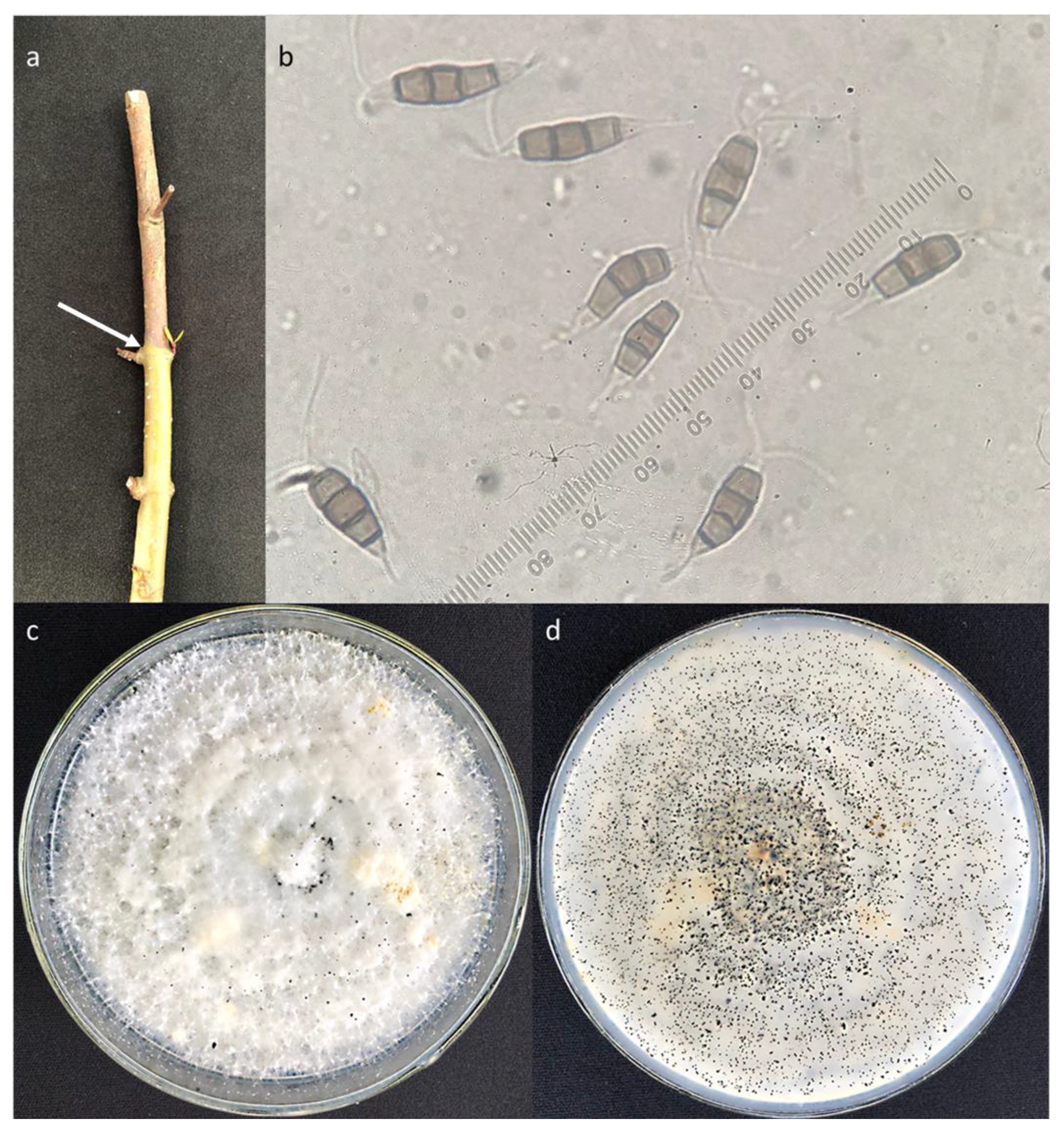
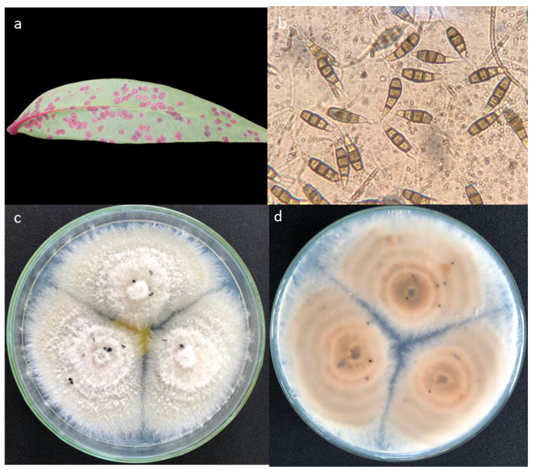
3.4. Morphology of the Main Taxa

Taxa that had a high frequency of isolation from field samples were selected for descriptions and for testing pathogenicity as follows:
  • Curvularia aff. tsudae
Associated with black leaf spots of S. apetala and S. caseolaris. Small black rufous spots appeared initially and then gradually enlarged, changing to tawny circular ring spots with a dark brown or black border and smooth edge (Figure 5a). Colonies on PDA reached 5 cm in diameter after 6 days at 25 °C. Young and old mycelium of isolate P8 (Curvularia aff. tsudae) (Figure 5) on PDA was dark grey, fluffy, cottony, reverse dark brown. Conidia and conidiophores were observed on the PDA plates after 14 days post-inoculation. Spores were 13.5–27 µm in length and 8.7–12.5 µm in width, brown, straight or slightly curved, septate, and unbranched.
  • Neopestalotiopsis sp.1
Associated with shoot dieback of S. apetala and S. caseolaris. Symptoms included yellowing of foliage, loss of leaves, canopy thinning, and dead branches (Figure 1c and Figure 6a). Dieback occurred on terminal shoots, lateral branches as well as the main stem. Colonies on PDA reached 6 cm in diameter after 5 days at 25 °C with light aerial mycelium on the surface (Figure 6c). Young and old mycelium of isolate T28 (Neopestalotiopsis sp.1) on PDA was milky white. Black and globular pycnidial conidiomata appeared after 14 days at 25 °C in the light. Conidia (Figure 6b) were 14.1–22.7 µm in length and 5.3–6.5 µm in width, fusiform, straight to slightly curved, four-celled, and periclinal walls were darker than the rest of the cell. The basal cell was conical and hyaline. Three median cells were dark brown, septa, and the periclinal walls darker than the rest of the cell. The apical cell was hyaline and filiform, with 2–4 apical appendages, 15.3–17.4 µm long.
  • Pestalotiopsis sp.1
Associated with pink leaf spot of S. apetala and S. caseolaris. Symptoms were small irregular-shaped spots, slightly sunken on the adaxial leaf surface, expanding over time (Figure 7a). Colonies on PDA reached 4–6 cm in diameter after four days at 25 °C in the light. Colonies were filamentous to circular, medium in density, aerial mycelium on the surface were flat or raised, with a filiform margin. Young and old mycelium of isolate P4 on PDA were milky white above and brown below (Figure 7c,d). The spores (Figure 7b) ranged from 13.5 to 22.5 µm in length and 5.4 to 6.3 µm in width, fusiform to clavate, straight to slightly curved, with three septa.
  • Pestalotiopsis sp.4a
Associated with black leaf spots of S. apetala and S. caseolaris (Figure 8a). Small black rufous spots appeared initially and then gradually enlarged, changing to tawny circular ring spots with a black border and smooth edge. Young and old mycelium of isolate Pesta23 on PDA were dark grey (Figure 8c,d). The length of conidia ranged from 13.2 to 21.4 µm in length and 5.0 to 6.1 µm in width, fusiform to clavate, straight to slightly curved, with four septa (Figure 8b). A separate group of 23 isolates was similar to Pesta23, except the mycelium was creamy white above and creamy brown below (not illustrated). Pesta23 and Y6 lie within the same arm of the phylogram in Figure 4, for convenience the mycelial types are referred to as Pestalotiopsis sp.2a for the dark mycelial isolates and Pestalotiopsis sp.2b for the light mycelial isolates.

3.5. Pathogenicity Trial

The pathogenicity trials showed that all four fungi produced larger lesions in the stems than the wounded control. Ten weeks after inoculation, five samples of each treatment were collected for re-isolation and identification to confirm Koch’s postulates. All four species were successfully re-isolated from stem lesions (data not shown).
Pathogenicity of Curvularia aff. tsudae (P8, MZ127510), Neopestalotiopsis sp.1 (T28, MZ127517), Pestalotiopsis sp.1 (P4, MZ127507), and Pestalotiopsis sp.4a (Pesta23, MZ127514) were significantly different from the control (PDA) (p < 0.001) when inoculated on seedlings of S. apetala and S. caseolaris. The pathogenicity of the isolates and the control fell into three groups: strong (one isolate), average (three isolates), and nil (control) (Table 6).

4. Discussion

This is the first study to explore whether fungal pathogens might be contributing to mangrove decline in Vietnam. The incidence of shoot dieback and leaf spots observed in young afforestation stands of two Sonneratia species suggests that diseases may be contributing to the poor survival rates in some mangrove restoration projects, particularly in central and north Vietnam where Sonneratia species have been widely planted. In the present study, 227 fungal isolates that were obtained from diseased tissue fell into nine morphotypes, and results from BLAST searches in GenBank for the ITS region indicated the presence of at least eight taxa within the plant pathogen genera Curvularia, Neopestalotiopsis, and Pestalotiopsis. Further collections are required before the unidentified new taxa can be described using more isolates to compare morphology and sequences.
Unlike for Vietnam, a range of mangrove disorders have been previously reported in other countries. Black leaf spot on Sonneratia apetala and small black leaf spot on S. caseolaris were associated with Olletotrichum lindemuthianum, Pestalotiopsis guepinii, and Phoma betae in Bangladesh [53]; Abbas et al. [54] described a black leaf spot on Conocarpus erectus caused by Alternaria alternata in Pakistan. Black leaf spot disease also occurs on Ceriops decandra, Osbornia octodonta, Rhizophora stylosa, and S. caseolaris associated with Pseudocercosporella in the Philippines [38]. Dark brown leaf spots caused by Orynespora cassiicola were found on Acanthus ilicifolius in China [22]. Regarding mangrove dieback, the high frequency of dieback and mortality were found in R. mangle in coastal mangrove forests in Puerto Rico [30,55]. Canker on the trunks, branches or twigs of S. apetala were observed in China [25], and top dieback was reported in Heritiera fomes in Bangladesh [24]. Symptoms of pink leaf spot disorders have so far not been recorded on mangroves.
More than 200 fungal species have been isolated and identified from mangroves [21,56]. Disease reports include branch/stem cankers, leaf spots, defoliation, dieback, and stem rot of mangroves [36,57,58,59,60,61,62,63,64,65,66,67,68,69,70,71]. For example, Diaporthe is known as an endophytic fungus on mangrove trees [69], and Pestalotiopsis is predominant in mangrove trees in China [72] and causes leaf spots on Rhizophora [35]. Pestalotiopsis microspora has been reported as an endophyte in Rhizophora mucronata [73] and Nigrospora oryzae was isolated from R. mucronata in Malaysia [71].
According to Shamsi et al. [53], fungi associated with black spot of S. apetala were Aspergillus fumigatus, Colletotrichum lindemuthianum, Pestalotiopsis guepinii, and Phoma betae. Fungi associated with small leaf spot of S. caseolaris were Curvularia fallax, Fusarium sp., Penicillium digitatum, and Phoma betae [53]. Alternaria alternata was considered to cause leaf spot on Conocarpus erectus in Pakistan [54].
In Vietnam, Curvularia species such as C. gloeosporioides are known to cause anthracnose disease in coffee [74], and C. lunata causes postharvest problems in rice [75] and dragon fruit [76]. Pestalotiopsis virgatula and P. clavispora were recorded as causal agents of postharvest rot diseases on rambutan [77], and an unidentified Pestalotiopsis was associated with crown rot disease of strawberry [78].
The source of the fungi causing leaf spots and shoot dieback of Sonneratia in afforestation in Vietnam is unknown. One possibility is that fungal spores are spread by wind from other mangrove stands in the area or infested fruits are carried by the tide to afforested sites. We observed some natural recruitment on site in the first year after planting Sonneratia. Thus, research on fungal pathogens should also be undertaken in old stands in the future. Another possibility is that disease might be carried from nurseries to the field. Two nurseries, one in Thai Binh Province and another in Thanh Hoa Province, provide seedlings for most mangrove afforestation projects in the north of Vietnam. This means that there is a risk of widespread infection if disease management in nurseries is poor.

5. Conclusions

This is the first report of diseases affecting mangrove afforestation in Vietnam. Neopestalotiopsis sp.1 and Pestalotiopsis sp.3 were associated with shoot dieback of Sonneratia apetala and S. caseolaris; Pestalotiopsis aff. humus, Pestalotiopsis aff. Neolitseae, and Pestalotiopsis sp.1 were associated with pink leaf spots; Curvularia aff. tsudae, Pestalotiopsis sp.2, and Pestalotiopsis sp.4 were associated with black leaf spots. Further work is required to characterize the fungi at the species level and to develop options for disease management.

Supplementary Materials

The following is available online at https://www.mdpi.com/article/10.3390/f12091273/s1, Table S1. Percentatage of Sonneratia apetala and S. caseolaris trees with stress symptoms in the preliminary survey in 2018.

Author Contributions

Conceptualization, H.T.T.N. and B.D.; Data curation, H.T.T.N.; Formal analysis, H.T.T.N.; Investigation, H.T.T.N., T.V.L., H.Q.N., and T.V.N.; Methodology, H.T.T.N.; Resources, T.V.L. and H.Q.N.; Software, D.H.L.; Supervision, B.D. and G.E.S.J.H.; Validation, B.D.; Visualization, G.E.S.J.H. and B.D.; Writing—original draft, H.T.T.N.; Writing—review and editing, B.D. All authors have read and agreed to the published version of the manuscript.

Funding

This research was funded by Murdoch University, Vietnamese Ministry of Education and Training, and the Institute of Ecology and Works Protection (Project “Integrated Solutions to Restore and Sustainably Develop Coastal Protection Forest in Thanh Hoa Province, code: DTĐL.CN-34/17”).

Institutional Review Board Statement

Not applicable.

Informed Consent Statement

Not applicable.

Data Availability Statement

Not applicable.

Acknowledgments

We thank Dang Ngoc Bich, Nguyen Nguyen Hang, Le Anh Tuan, Ta Van Van, Le Xuan Cuong, Pham Van Thach, and Le Van Duc (Forestry experts, Institute of Ecology and Works Protection, Vietnam Academy for Water Resources, 267 Chua Boc, Dong Da, Ha Noi, Vietnam) for assistance with collecting samples in the field; Forest Protection Research Centre, Vietnamese Academy of Forest Sciences, Duc Thang, Bac Tu Liem as well as the Vietnam National University of Agriculture for providing equipment for fungal isolation and also for the lodgment of cultures; 69 Institute, High Command of Protection the President Ho Chi Minh Mausoleum for providing microscopes; the Center for Ecology and Reservoir Protection (Institute of Ecology and Works Protection, Vietnam Academy for Water Resources, 267 Chua Boc, Dong Da, Ha Noi, Vietnam) for providing data on mangrove forests in Thanh Hoa Province.

Conflicts of Interest

The authors declare no conflict of interest. The funders had no role in the design of the study; the collection, analyses, or interpretation of data; the writing of the manuscript; or in the decision to publish the results.

References

  1. Sherman, R.E.; Fahey, T.J.; Martinez, P. Hurricane Impacts on a Mangrove Forest in the Dominican Republic: Damage Patterns and Early Recovery 1. Biotropica 2001, 33, 393–408. [Google Scholar] [CrossRef]
  2. Kathiresan, K.; Bingham, B.L. Biology of Mangroves and Mangrove Ecosystems. Adv. Mar. Biol. 2001, 40, 81–251. [Google Scholar]
  3. Taylor, M.; Ravilious, C.; Green, E.P. Mangroves of East Africa; UNEP–WCMC: Cambridge, UK, 2003. [Google Scholar]
  4. Martinuzzi, S.; Gould, W.A.; Lugo, A.E.; Medina, E. Conversion and Recovery of Puerto Rican Mangroves: 200 years of change. For. Ecol. Manag. 2009, 257, 75–84. [Google Scholar] [CrossRef]
  5. Abuodha, P.; Kairo, J. Human-induced Stresses on Mangrove Swamps along the Kenyan Coast. Hydrobiologia 2001, 458, 255–265. [Google Scholar] [CrossRef]
  6. Ellison, A.M. Mangrove Communities. In Marine Community Ecology; Sinauer Associates, Inc.: Sunderland, MA, USA, 2001; pp. 423–442. [Google Scholar]
  7. Spalding, M. World Atlas of Mangroves; Routledge: London, UK, 2010. [Google Scholar]
  8. Smith, T.J., III; Robblee, M.B.; Wanless, H.R.; Doyle, T.W. Mangroves, Hurricanes, and Lightning Strikes: Assessment of Hurricane Andrew Suggests an Interaction across Two Differing Scales of Disturbance. BioScience 1994, 44, 256–262. [Google Scholar] [CrossRef]
  9. Norm, D.; Jock, M.; John, K.; Grant, S.; Rob, C.; Apanie, W.; Ysabel, C. Assessing the Gulf of Carpentaria Mangrove Dieback 2017–2019; James Cook University: Douglas, Australia, 2020; p. 249. [Google Scholar]
  10. Van, O.P.; Duke, N. Extreme Weather Likely behind Worst Recorded Mangrove Dieback in Northern Australia. Conversation 2017, 14, 1–6. [Google Scholar]
  11. Gilbert, G.S. Evolutionary Ecology of Plant Diseases in Natural Ecosystems. Annu. Rev. Phytopathol. 2002, 40, 13–43. [Google Scholar] [CrossRef] [Green Version]
  12. Hulcr, J.; Dunn, R.R. The Sudden Emergence of Pathogenicity in Insect–fungus Symbioses Threatens Naive Forest Ecosystems. Proc. R. Soc. B Biol. Sci. 2011, 278, 2866–2873. [Google Scholar] [CrossRef] [Green Version]
  13. Hyde, K.; Cannon, P. Polystigma sonneratiae Causing Leaf Spots on The Mangrove Genus Sonneratia. Aust. Syst. Bot. 1992, 5, 415–420. [Google Scholar] [CrossRef]
  14. Jones, E.G.; Pang, K.-L. Tropical Aquatic Fungi. Biodivers. Conserv. 2012, 21, 2403–2423. [Google Scholar] [CrossRef]
  15. Jones, E.G.; Pang, K.-L.; Abdel-Wahab, M.A.; Scholz, B.; Hyde, K.D.; Boekhout, T.; Ebel, R.; Rateb, M.E.; Henderson, L.; Sakayaroj, J. An Online Resource for Marine Fungi. Fungal Divers. 2019, 96, 347–433. [Google Scholar] [CrossRef]
  16. Alias, S.; Jones, E. Colonization of Mangrove Wood by Marine Fungi at Kuala Selangor Mangrove Stand, Malaysia. Fungal Divers. 2000, 5, 9–21. [Google Scholar]
  17. Kohlmeyer, J.; Kohlmeyer, E. Marine Fungi From Tropical America and Africa. Mycologia 1971, 63, 831–861. [Google Scholar] [CrossRef] [PubMed]
  18. Jones, E.G.; Abdel-Wahab, M.A. Marine Fungi from the Bahamas Islands. Bot. Mar. 2005, 48, 356–364. [Google Scholar]
  19. Sarma, V.; Hyde, K.D. A review on Frequently Occurring Fungi in Mangroves. Fungal Divers 2001, 8, 1–34. [Google Scholar]
  20. Steinke, T. Mangrove Fungi on Dead Proproots of Rhizophora mucronata at Three Localites in South Africa. S. Afr. J. Bot. 2000, 66, 91–95. [Google Scholar] [CrossRef] [Green Version]
  21. Osorio, J.A.; Wingfield, M.J.; Roux, J. A Review of Factors Associated with Decline and Death of Mangroves, with Particular Reference to Fungal Pathogens. S. Afr. J. Bot. 2016, 103, 295–301. [Google Scholar] [CrossRef]
  22. Xie, S.; He, H.; Yang, R.; Xu, Z.; He, J.; Lu, H. First Report of Leaf Spot Caused by Corynespora cassiicola on Acanthus ilicifolius in China. Plant Dis. 2021, 105, 509. [Google Scholar] [CrossRef]
  23. Stevens, F.L. New or Noteworthy Porto Rican Fungi. Bot. Gaz. 1920, 70, 399–402. [Google Scholar] [CrossRef]
  24. Rahman, M.A. Top Dying of Sundri (Heritiera fomes) and Its Impact on the Regeneration and Management in the Mangrove Forests of Sunderbans in Bangladesh. In Proceedings of the Impact of Diseases and Insect Pests in Tropical Forests, Peechi City, India, 23–26 November 1993; pp. 117–133. [Google Scholar]
  25. Yan, T.L.; Zhu, X.J.; Yang, Z.D.; Long, Q.H.; Yang, H.; Chang, S.Q. New Records of Celoporthe guangdongensis and Cytospora rhizophorae on Mangrove Apple in China. Biodivers. Data J. 2020, 8, 15. [Google Scholar]
  26. McMillan, J.R. Studies of a Recently Described Cercospora on Rhizophora mangle. Plant Dis. Report. 1964, 48, 909–991. [Google Scholar]
  27. Creager, D.B. A New Cercospora on Rhizophora mangle. Mycologia 1962, 54, 536–539. [Google Scholar] [CrossRef]
  28. Osorio, J.A.; Crous, C.J.; Wingfield, M.J.; De Beer, Z.W.; Roux, J. An assessment of Mangrove Diseases and Pests in South Africa. For. Int. J. For. Res. 2017, 90, 343–358. [Google Scholar] [CrossRef] [Green Version]
  29. Osorio, J.A.; Crous, C.J.; De Beer, Z.W.; Wingfield, M.J.; Roux, J. Endophytic Botryosphaeriaceae, Including Five New Species, Associated with Mangrove Trees in South Africa. Fungal Biol. 2017, 121, 361–393. [Google Scholar] [CrossRef] [PubMed] [Green Version]
  30. Wier, A.M.; Tattar, T.A.; Klekowski, E.J., Jr. Disease of Red Mangrove (Rhizophora mangley) in Southwest Puerto Rico Caused by Cytospora rhizophorae 1. Biotropica 2000, 32, 299–306. [Google Scholar]
  31. Kohlmeyer, J. Ecological Notes on Fungi in Mangrove Forests. Trans. Br. Mycol. Soc. 1969, 53, 237–250. [Google Scholar] [CrossRef]
  32. Teas, H.; McEwan, R. An Epidemic Dieback Gall Disease of Rhizophora mangroves in The Gambia, West Africa. Plant Dis. 1982, 66, 522–523. [Google Scholar] [CrossRef]
  33. Sakayaroj, J.; Preedanon, S.; Suetrong, S.; Klaysuban, A.; Jones, E.G.; Hattori, T. Molecular Characterization of Basidiomycetes Associated with The Decayed Mangrove Tree Xylocarpus granatum in Thailand. Fungal Divers. 2012, 56, 145–156. [Google Scholar] [CrossRef]
  34. Pegg, K.; Gillespie, N.; Forsberg, L. Phytophthora sp. Associated with Mangrove Death in Central Coastal Queensland. Australas. Plant Pathol. 1980, 9, 6–7. [Google Scholar] [CrossRef]
  35. Xu, J.; Kjer, J.; Sendker, J.; Wray, V.; Guan, H.; Edrada, R.; Lin, W.; Wu, J.; Proksch, P. Chromones From the Endophytic Fungus Pestalotiopsis sp. Isolated from the Chinese Mangrove Plant Rhizophora mucronata. J. Nat. Prod. 2009, 72, 662–665. [Google Scholar] [CrossRef]
  36. Olexa, M.; Freeman, T. Occurrence of Three Unrecorded Diseases on Mangroves in Florida. In Proceedings of the International Symposium on Biology and Management of Mangroves, Honolulu, HI, USA, 8–11 October 1974; pp. 688–692. [Google Scholar]
  37. Shivas, R.; Young, A.; Crous, P. Pseudocercospora avicenniae RG Shivas, AJ Young & Crous, sp. nov. Persoonia 2009, 23, 193. [Google Scholar]
  38. Rafael, A.; Calumpong, H.P. Fungal Infections of Mangroves in Natural Forests and Reforestation Sites from Philippines. Aquac. Aquar. Conserv. Legis. 2019, 12, 2062–2074. [Google Scholar]
  39. Hong, P.N.; San, H.T. Mangroves of Vietnam; IUCN: Bangkok, Thailand, 1993; pp. 150–180.
  40. Ministry of Agriculture and Rural Development. Announcement of National Forest Status in 2019; MARD: Hanoi, Vietnam, 2020.
  41. Hai, N.; Dell, B.; Phuong, V.; Harper, R. Towards a More Robust Approach for The Restoration of Mangroves in Vietnam. Ann. For. Sci. 2020, 77, 1–18. [Google Scholar] [CrossRef] [Green Version]
  42. Nguyen, H.H.; Nghia, N.H.; Nguyen, H.T.T.; Le, A.T.; Tran, L.T.N.; Duong, L.V.K.; Bohm, S.; Furniss, M.J. Classification Methods for Mapping Mangrove Extents and Drivers of Change in Thanh Hoa Province, Vietnam During 2005–2018. For. Soc. 2020, 4, 225–242. [Google Scholar] [CrossRef]
  43. Vietnam Admistration of Forestry. Protective Forest in Coastal Is Unexplained Death. Available online: http://bentre.tintuc.vn/tin-tuc/ben-tre-rung-phong-ho-dac-dung-ven-bien-chet-chua-ro-nguyen-nhan.html (accessed on 1 July 2021).
  44. Baotainguyenvamoitruong. Proposing Scientific Research to Find the Cause of Death of Mangroves in Ben Tre. Available online: https://baotainguyenmoitruong.vn/moi-truong/ben-tre-de-xuat-nghien-cuu-khoa-hoc-tim-nguyen-nhan-rung-chet-1258495.html (accessed on 1 July 2021).
  45. Sggp.org.vn. Death of Rhizophora in Mass: Identify reasons (Vietnamese). Available online: http://www.sggp.org.vn/vu-cay-duoc-chet-hang-loat-xac-dinh-loai-sinh-vat-la-gay-hai-451729.html (accessed on 1 July 2021).
  46. Antv.gov.vn. Death of Mangroves in Mass in Ha Tinh (Vietnamese). Available online: http://www.antv.gov.vn/tin-tuc/xa-hoi/rung-cay-ngap-man-o-ha-tinh-chet-hang-loat-186342.html (accessed on 1 July 2021).
  47. Nguyen, H.T.T.; Hardy, G.E.; Le, T.V.; Nguyen, H.Q.; Nguyen, H.H.; Nguyen, T.V.; Dell, B. Mangrove Forest Landcover Changes in Coastal Vietnam: A Case Study from 1973 to 2020 in Thanh Hoa and Nghe An Provinces. Forests 2021, 12, 637. [Google Scholar] [CrossRef]
  48. Doyle, J.J.; Doyle, J.L. A Rapid DNA Isolation Procedure for Small Quantities of Fresh Leaf Tissue. Phytochem. Bull. 1987, 19, 11–15. [Google Scholar]
  49. Khan, A.M.; Bhadauria, S. Molecular Characterization of Keratin Degrading Fungi Isolated from Semi-arid Soil by PCR Using ITS4 and ITS5 Primers. J. King Saud Univ.-Sci. 2019, 31, 1418–1423. [Google Scholar] [CrossRef]
  50. White, T.J.; Bruns, T.; Lee, S.; Taylor, J. Amplification and Direct Sequencing of Fungal Ribosomal RNA Genes for Phylogenetics. In PCR Protocols: A Guide to Methods and Applications; Academic Press: New York, NY, USA, 1990; Volume 18, pp. 315–322. [Google Scholar]
  51. Kumar, S.; Stecher, G.; Li, M.; Knyaz, C.; Tamura, K. MEGA X: Molecular Evolutionary Genetics Analysis Across Computing Platforms. Mol. Biol. Evol. 2018, 35, 1547–1549. [Google Scholar] [CrossRef]
  52. Allen, P.; Bennett, K.; Heritage, B. SPSS Statistics Version 22: A Practical Guide; Cengage Learning Australia Pty Limited: Sydney, Australia, 2014; pp. 200–250. [Google Scholar]
  53. Shamsi, S.; Hosen, S.; Ahmed, A. Fungi Associated with Leaves of Sonneratia apetala Buch. Ham and Sonneratia caseolaris (L.) Engler from Rangabali Coastal Zone of Bangladesh. Dhaka Univ. J. Biol. Sci. 2018, 27, 155–162. [Google Scholar] [CrossRef]
  54. Abbas, M.F.; Rafiq, M.; Al-Sadi, A.M.; Alfarraj, S.; Alharbi, S.A.; Arif, M.; Ansari, M.J. Molecular Characterization of Leaf Spot caused by Alternaria alternata on Buttonwood (Conocarpus erectus L.) and Determination of Pathogenicity by a Novel Disease Rating Scale. PLoS ONE 2021, 16, e0251471. [Google Scholar] [CrossRef]
  55. Tattar, T.; Klekowski, E.; Stern, A. Dieback and Mortality in Red Mangrove, Rhizophora mangle L., in Southwest Puerto Rico. Arboric. J. 1994, 18, 419–429. [Google Scholar] [CrossRef]
  56. Liu, A.; Wu, X.; Xu, T. Research advances in endophytic fungi of mangrove. J. Appl. Ecol. 2007, 18, 912–918. [Google Scholar]
  57. Suryanarayanan, T.; Kumaresan, V. Endophytic fungi of some halophytes from an estuarine mangrove forest. Mycol. Res. 2000, 104, 1465–1467. [Google Scholar] [CrossRef]
  58. Chaeprasert, S.; Piapukiew, J.; Whalley, A.J.; Sihanonth, P. Endophytic Fungi from Mangrove Plant Species of Thailand: Their Antimicrobial and Anticancer Potentials. Bot. Mar. 2010, 53, 555–564. [Google Scholar] [CrossRef]
  59. Xing, X.; Guo, S. Fungal Endophyte Communities in four Rhizophoraceae Mangrove Species on The South Coast of China. Ecol. Res. 2011, 26, 403–409. [Google Scholar] [CrossRef]
  60. Khan, A.A.H. Endophytic Fungi and Their Impact on Agroecosystems. In Medicinal Plants: Biodiversity, Sustainable Utilization and Conservation; Springer: Singapore, 2020; pp. 443–499. [Google Scholar]
  61. Nurunnabi, T.R.; Sabrin, F.; Sharif, D.I.; Nahar, L.; Sohrab, M.H.; Sarker, S.D.; Rahman, S.M.; Billah, M.M. Antimicrobial Activity of Endophytic Fungi Isolated From the Mangrove Plant Sonneratia apetala (Buch.-Ham) from the Sundarbans Mangrove Forest. Adv. Tradit. Med. 2020, 40, 1–7. [Google Scholar] [CrossRef]
  62. Goudarzi, A.; Moslehi, M. Distribution of a Devastating Fungal Pathogen in Mangrove Forests of Southern Iran. Crop Prot. 2020, 128, 104987. [Google Scholar] [CrossRef]
  63. Debbab, A.; Aly, A.H.; Proksch, P. Mangrove Derived Fungal Endophytes–a Chemical and Biological Perception. Fungal Divers. 2013, 61, 1–27. [Google Scholar] [CrossRef]
  64. Ebrahim, W.; Aly, A.H.; Mándi, A.; Totzke, F.; Kubbutat, M.H.; Wray, V.; Lin, W.H.; Dai, H.; Proksch, P.; Kurtán, T. Decalactone Derivatives from Corynespora cassiicola, an Endophytic Fungus of the Mangrove Plant Laguncularia racemosa. Eur. J. Org. Chem. 2012, 2012, 3476–3484. [Google Scholar] [CrossRef] [Green Version]
  65. Cheng, Z.S.; Tang, W.C.; Xu, S.L.; Sun, S.F.; Huang, B.Y.; Yan, X.; Chen, Q.J.; Lin, Y.C. First Report of An endophyte (Diaporthe phaseolorum var. sojae) from Kandelia candel. J. For. Res. 2008, 19, 277–282. [Google Scholar] [CrossRef]
  66. Xing, X.; Chen, J.; Xu, M.; Lin, W.; Guo, S. Fungal Endophytes Associated with Sonneratia (Sonneratiaceae) Mangrove Plants on the South Coast of China. For. Pathol. 2011, 41, 334–340. [Google Scholar] [CrossRef]
  67. Olexa, M.; Freeman, T. A Gall Disease of Red Mangrove Caused by Cylindrocarpon didymum. Plant Dis. Report. 1978, 62, 283–286. [Google Scholar]
  68. Chi, W.-C.; Chen, W.; He, C.-C.; Guo, S.-Y.; Cha, H.-J.; Tsang, L.M.; Ho, T.W.; Pang, K.-L. A Highly Diverse Fungal Community Associated with Leaves of the Mangrove Plant Acanthus ilicifolius var. xiamenensis Revealed by Isolation and Metabarcoding Analyses. PeerJ 2019, 7, e7293. [Google Scholar] [CrossRef] [Green Version]
  69. Cui, H.; Lin, Y.; Luo, M.; Lu, Y.; Huang, X.; She, Z. Diaporisoindoles A–C: Three isoprenylisoindole alkaloid derivatives from the mangrove endophytic fungus Diaporthe sp. SYSU-HQ3. Org. Lett. 2017, 19, 5621–5624. [Google Scholar] [CrossRef] [PubMed]
  70. Norphanphoun, C.; Jayawardena, R.; Chen, Y.; Wen, T.; Meepol, W.; Hyde, K. Morphological and Phylogenetic Characterization of Novel Pestalotioid Species Associated with Mangroves in Thailand. Mycosphere 2019, 10, 531–578. [Google Scholar] [CrossRef]
  71. Hamzah, T.N.T.; Lee, S.Y.; Hidayat, A.; Terhem, R.; Faridah-Hanum, I.; Mohamed, R. Diversity and Characterization of Endophytic Fungi Isolated from the Tropical Mangrove Species, Rhizophora mucronata, and Identification of Potential Antagonists Against the Soil-Borne Fungus, Fusarium solani. Front. Microbiol. 2018, 9, 1707. [Google Scholar] [CrossRef] [PubMed]
  72. Zhou, J.; Diao, X.; Wang, T.; Chen, G.; Lin, Q.; Yang, X.; Xu, J. Phylogenetic Diversity and Antioxidant Activities of Culturable Fungal Endophytes Associated with the Mangrove Species Rhizophora stylosa and R. mucronata in the South China Sea. PLoS ONE 2018, 13, e0197359. [Google Scholar] [CrossRef] [PubMed] [Green Version]
  73. Deshmukh, S.K.; Gupta, M.K.; Prakash, V.; Reddy, M.S. Mangrove-Associated Fungi: A Novel Source of Potential Anticancer Compounds. J. Fungi 2018, 4, 101. [Google Scholar] [CrossRef] [Green Version]
  74. Ha, N.T.; Ngat, H.T.; Ha, N.T.T.; Hoi, P.X. Study on Pathogenicity of Colletotrichum gloeosporioides on Coffee in North of Vietnam. Biology 2011, 33, 67–73. [Google Scholar] [CrossRef] [Green Version]
  75. Tran, N.B.B. Isolation and Identification of Fungal Diseases in Rice (Oryza sativa) from Mekong Delta. Ph.D. Thesis, International University-HCMC, Ho Chi Minh City, Vietnam, 2019. [Google Scholar]
  76. Ngoc, N.; Phong Nguyen, N.; An, P.; Woolf, A.; Fullerton, R. Effect of Storage Temperatures on Postharvest Diseases of Dragon Fruits (Hylocereus undatus Haw.) in the Mekong Delta Region, Vietnam. In Proceedings of the III Asia Pacific Symposium on Postharvest Research, Education and Extension: APS2014 1213, Hochiminh City, Vietnam, 8 December 2014; pp. 453–460. [Google Scholar]
  77. Yen, T.T.N.; Thanh, N.V.; Phong, N.V. Research on the Causative Agent of Rambutan fruit rot (Nephelium lappaceum L.) after Harvest in the Mekong Delta. J. Sci. Technol. 2017, 13, 1868–1873. [Google Scholar]
  78. Dung, L.; Dienb, N.T.; Daic, P.H.; Tuana, P.N. The First Report of Pestalofiopsis sp. Causing Crown Rot Disease on Strawberries in Dalat. Dalat Univ. J. Sci. 2016, 6, 364–376. [Google Scholar] [CrossRef]
Figure 1. Mangrove afforestation project in Thanh Hoa Province, showing trees planted in a completely randomized design (a); healthy tree (b); unhealthy tree with shoot dieback (c) and a shoot with leaves turning yellow before dieback occurs (insert); symptoms of pink leaf spots (d); symptoms of black leaf spots (e).
Forests 12 01273 g001
Figure 2. Phylogram obtained from MEGA X software analyses of the ITS data set. Isolate P8 groups close to Curvularia tsudae. Numbers on the branches are bootstrap values (>70%/posterior probabilities). The insertion bar corresponds to 1% of the corresponding sequence difference. The tree was rooted to Alternaria alternariacida (CBS 105.51).
Forests 12 01273 g002
Figure 3. Phylogram obtained from MEGAX software analyses of the ITS data set. Isolate T28 groups with a branch containing Neopestalotiopsis mesopotamica. Numbers on the branches are bootstrap value (>60%/posterior probabilities). The insertion bar corresponds to 0.5% of the corresponding sequence difference. The tree was rooted to Pseudopestalotiopsis theae (MFLUCC 12-0055).
Forests 12 01273 g003
Figure 4. Phylogram obtained from MEGAX software analyses of the ITS data set. Isolates Pesta23, Y6, Y61, P20, T27, P4, and P2 group among Pestalotiopsis species. Numbers on the branches are bootstrap value (>55%/posterior probabilities). The insertion bar corresponds to 1% of the corresponding sequence difference. The tree was rooted to Neopestalotiopsis saprophytica (MFLUCC 12-0282).
Forests 12 01273 g004
Figure 5. Symptoms of leaf black spot on a Sonneratia apetala leaf caused by Curvularia aff. tsudae (a); isolate P8 (MZ127510) conidia (b); mycelia ((c) from below; (d) from above) that were obtained from the leaf illustrated in (a).
Forests 12 01273 g005
Figure 6. Neopestalotiopsis sp.1 isolate T28 (MZ127517) was obtained from a Sonneratia caseolaris stem at the junction of dead and live wood (arrow) (a); conidia (b); mycelia ((c) from above; (d) from below) that were obtained from the lesion illustrated in (a).
Forests 12 01273 g006
Figure 7. Pestalotiopsis sp.1 (Isolate P4-MZ127507) associated with pink leaf spot disorder on Sonneratia caseolaris. Disease symptom (a); spores (b); mycelia (upper view—(c), lower view—(d)) that were obtained from the leaf illustrated in (a).
Forests 12 01273 g007
Figure 8. Black leaf spot on Sonneratia caseolaris caused by Pestalotiopsis sp.4a (isolate Pesta23-MZ127514) (a); spores (b); mycelia (upper view—(c), lower view—(d)) that were obtained from the leaf illustrated in (a).
Forests 12 01273 g008
Table 1. Fungi associated with diseases in mangroves.
FungusDiseaseHostLocationReference
Acanthus ilicifoliusleaf spotCorynespora cassiicolaChina[22]
Anthostomella rhizomorphaeleaf spotRhizophora manglePuerto Rico[23]
Botrosphaeria ribisdeath of twigs and branchesHeritiera fomesBangladesh[24]
Celoporthe guangdongensiscankerSonneratia apetalaChina[25]
Cercosporaleaf spotRhizophora mangleFlorida[26]
Cercosporaleaf diseaseRhizophora mangleFlorida[27]
Colletotrichum sp.leaf spotRhizophora sp.Australia[13]
Cyphellophora sp.stem canker, branch diebackAvicennia marinaSouth Africa[28,29]
Cytospora rhizophoraediebackRhizophora manglePuerto Rico[30]
Cytospora rhizophoraecankerSonneratia apetalaChina[25]
Cytospora sp.diebackRhizophora mangleHawaii[31]
Eutypella sp.stem/branch cankerAvicennia marinaSouth Africa[28,29]
Fulvifomesheart/butt infectionsXylocarpus granatumThailand[32]
Fulvifomes sp.decayXylocarpus granatumThailand[33]
Lasiodiplodia theobromaebranch dieback, branch/stem cankerAvicennia marinaSouth Africa[28,29]
OomycetemortalityAvicennia marinaAustralia[34]
Pestalotiopsis sp.leaf spotRhizophora mucronataChina[35]
Phyllosticta hibiscinanecrosis and deathAvicennia germinansFlorida[36]
Polystigma sonneratiaeleaf spotRhizophora sp.Australia[13]
Pseudocercospora avicenniaeleaf spotAvicennia marinaAustralia[37]
Pseudocercospora mapelanensisdisease on fruit, flowers and leavesBarringtonia racemosaSouth Africa[28]
Pseudocercosporellaleaf spotCeriops decandra, Sonneratia caseolaris, Rhizophora stylosa and Osbornia octodontaPhilippines[38]
Table 2. Details of the mangrove health study undertaken in north–central Vietnam.
LocationSonneratia SpeciesYear PlantedPreliminary StudyMain Study
Sam Son town, Thanh Hoa ProvinceS. apetala2015, 2016December 2018
S. caseolaris2015, 2016December 2018
Hoang Phu District, Thanh Hoa ProvinceS. caseolaris2018December 2018August 2019
S. apetala2018December 2018August 2019
Table 3. Dieback and leaf spot rating of mangroves based on the severity of symptoms.
RatingDieback CriteriaLeaf Spot Criteria
1No dead branches visibleDense, healthy foliage; no leaf spots
2Few dead twigs present; may have an occasional large branch stub on an upper boleDense foliage; spots (pink/black) present at low density on leaves
3Many dead twigs present; an occasional large dead branch; may have large branch stubs on the upper boleFoliage density appears subnormal; numerous pink or black spots present
Table 4. Number of Sonneratia apetala and S. caseolaris trees (percent of the total in brackets) in the afforestation trial with symptoms of dieback and leaf spots in three damage classes.
Trees with Dieback
(Damage Class)
Trees with Pink Spot
(Damage Class)
Trees with Black Spot
(Damage Class)
123123123
Mixed stands
Sonneratia apetala13545322141512292648716
(94.8)(3.7)(1.5)(99)(0.8)(0.1)(64.8)(34.1)(1.1)
Sonneratia caseolaris1129107157929438261377142
(81)(7.7)(11.3)(66.7)(31.4)(1.9)(98.9)(1)(0.1)
Single stands
Sonneratia apetala88537129257266325318
(94.8)(4)(1.3)(99)(0.7)(0.2)(71)(27.1)(1.9)
Sonneratia caseolaris746691286542612893832
(79.1)(7.3)(13.6)(69.4)(27.7)(3)(99.5)(0.3)(0.2)
Total
Sonneratia apetala223990342340194158974034
(94.8)(3.8)(1.4)(99)(0.8)(0.2)(67.2)(31.3)(1.4)
Sonneratia caseolaris18751762851583699542315174
(80.3)(7.5)(12.2)(67.8)(29.9)(2.3)(99.1)(0.7)(0.2)
Table 5. Representative fungal isolates obtained from diseased mangrove trees in Thanh Hoa Province that were used for molecular taxonomy. Isolates in bold were used in nursery pathogenicity experiments and are described in the text.
Isolate
Sequenced
Number of
Isolates Obtained
Sonneratia HostAssociated DiseaseGenBank Accession Number (ITS)Fungal Taxa
P834S. apetalaBlack leaf spotMZ127510Curvulariaaff. tsudae
T2839S. apetalaStem diebackMZ127517Neopestalotiopsissp.1
P211S. caseolarisPink leaf spotMZ127508Pestalotiopsis aff. humus
P445S. caseolarisPink leaf spotMZ127507Pestalotiopsissp.1
Y6119S. apetalaBlack leaf spotMZ127526Pestalotiopsis sp.2
P2016S. caseolarisPink leaf spotMZ127512Pestalotiopsis aff. neolitseae
T2717S. caseolarisStem diebackMZ127516Pestalotiopsis sp.3
Y623S. apetalaBlack leaf spotMZ127521Pestalotiopsis sp.4b
Pesta2323S. caseolarisBlack leaf spotMZ127514Pestalotiopsissp.4a
Table 6. Disease index and pathogenicity of the Pestalotiopsis, Neopestalotiopsis, and Curvularia isolates inoculated onto stems of 3 month old seedlings of Sonneratia apetala and S. caseolaris in a nursery in Thai Binh Province.
Host SpeciesIsolateFungal TaxaDisease IndexPathogenicity
Sonneratia apetalaP8Curvularia aff. tsudae1.90average
T28Neopestalotiopsis sp.11.82average
P4Pestalotiopsis sp.11.76average
Pesta23Pestalotiopsis sp.4a1.55average
Control (PDA) 0.00nil
Chi-Square48.9
Sonneratia caseolarisP8Curvularia aff. tsudae1.93average
T28Neopestalotiopsis sp.12.36strong
P4Pestalotiopsis sp.11.93average
Pesta23Pestalotiopsis sp.4a1.89average
Control (PDA) 0.00nil
Chi-Square53.6
p-Value0.000
Publisher’s Note: MDPI stays neutral with regard to jurisdictional claims in published maps and institutional affiliations.

