Article

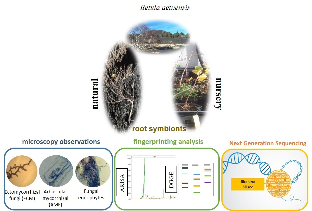
The Root Mycobiota of Betula aetnensis Raf., an Endemic Tree Species Colonizing the Lavas of Mt. Etna (Italy)

1
Department of Agricultural, Food and Forest Sciences—SAAF, University of Palermo, Viale delle Scienze, blg. 4, 90128 Palermo, Italy
2
Department of Biological, Chemical and Pharmaceutical Sciences and Technologies—STEBICEF, University of Palermo, Viale delle Scienze, blg. 16, 90128 Palermo, Italy
*
Author to whom correspondence should be addressed.
The first two authors, E.B. and V.C. contributed equally to the work.
Forests 2021, 12(12), 1624; https://doi.org/10.3390/f12121624
Submission received: 24 September 2021 / Revised: 17 November 2021 / Accepted: 19 November 2021 / Published: 24 November 2021
(This article belongs to the Section Forest Ecophysiology and Biology)

Abstract

:
Betula aetnensis is an endemic tree of high conservation value, which thrives on the nutrient-poor volcanic soils of Mount Etna. Since plant–microbe interactions could play a crucial role in plant growth, resource uptake, and resistance to abiotic stresses, we aimed to characterize the root and rhizosphere microbial communities. Individuals from natural habitat (NAT) and forest nursery (NURS) were surveyed through microscopy observations and molecular tools: bacterial and fungal automated ribosomal intergenic spacer analysis (ARISA), fungal denaturing gradient gel electrophoresis (DGGE). B. aetnensis was found to be simultaneously colonized by arbuscular (AM), ectomycorrhizal (ECM), ericoid (ERM) fungi, and dark septate endophytes (DSE). A high diversity of the bacterial community was observed whilst the root fungal assemblage of NAT plants was richer than that of NURS. Root and rhizosphere fungal communities from NAT plants were characterized by Illumina MiSeq sequencing. Most of the identified sequences were affiliated to Helotiales, Pezizales, and Malasseziales. Ascomycota and Basidiomycota dominated roots and rhizosphere but differed in community structure and composition. ECM in the roots mainly belonged to Tylospora and Leccinum, while Rhizopogon was abundant in the rhizosphere. The Helotiales, including ERM (mostly Oidiodendron) and DSE (mostly Phialocephala), appeared the dominant component of the fungal community. B. aetnensis harbors an extraordinarily wide array of root-associated soil microorganisms, which are likely to be involved in the adaptation and resistance mechanisms to the extreme environmental conditions in volcano Etna. We argue that nursery-produced seedlings could lack the necessary microbiota for growth and development in natural conditions.

Graphical Abstract

1. Introduction

Betula aetnensis Raf. (Family Betulaceae) is a tree species endemic to Sicily, where it mostly thrives on the north-eastern slopes of volcano Etna, from 1400 to 2100 m a.s.l. [1,2]. Betula aetnensis is thought to be originated from Betula pendula Roth., a European-wide distributed birch [3]. Particularly, during the last glacial period, Betula pendula may have reached Sicily, where the geographical isolation and the peculiar ecological conditions of Mt. Etna may have allowed the differentiation of Betula aetnensis [4]. Despite its taxonomic rank is still under debate, B. aetnensis is considered a separated taxon in the Italian flora [4]. This species has an excellent ability to colonize volcanic substrates, where it plays a prominent role as a typical pioneer species at the beginning of the primary succession, showing high ecological importance. The adaptation to such harsh habitats by B. aetnensis reflects its ability to tolerate soils with limited organic matter, as well as nutrient and water availability, where it may benefit from the absence of plant competitors [5].
In this regard, B. aetnensis behaves similarly to other European birches, which are light-demanding species mostly linked to early successional communities, with a strong pioneer attitude, and capable of establishing even in harsh and nutrient-limited conditions [3]. For instance, Betula pendula is particularly successful in colonizing disturbed sites, including little developed and contaminated soils, as well as coal mining spoils [6,7,8]. Interestingly, Betula pendula stands occurring in similar volcanic soils are found in Vesuvius volcanic complex (Campania region, Italy) [9] but information about soil microorganisms is lacking.
In the last decades, the research on B. aetnensis has been mostly focused on understanding the reasons underlying the increasing decaying of mature trees, that seems in part related to Armillaria mellea (Vahl) P. Kumm., a pathogenic fungus which typically affects aged and/or stressed and/or unhealthy individuals [10]. Conversely, less attention has been paid to soil microbial communities, including mycorrhizae [11] and fungal endophytes, which are expected to play a crucial role both in early and later life stages, significantly affecting plant growth and development, resource uptake, and resistance to abiotic stresses [12,13,14]. The decisive importance of mutualistic interactions with ectomycorrhizal (ECM) and arbuscular mycorrhizal (AM) fungi, in which higher plants may benefit from enhanced nutrient and water uptake, especially in harsh and critical environments, whereas symbiotic fungi receive the organic carbon produced by plants, is widely acknowledged for most terrestrial plants including forest species [15]. By contrast, the knowledge of other root-inhabiting species and the ecological role played by endophytic fungi is very limited [13]. Root-associated microbes such as mycorrhiza helper bacteria and nitrogen-fixing actinobacteria could benefit plant fitness in harsh environments and scarce soil nutrient conditions [16], all this without considering that symbiotic fungi community of the plant species in the edge of the distribution range [17], such as B. aetnensis, may be particularly affected by changes in environmental conditions (e.g., due to climate change) [18]. Furthermore, plant–soil microbe interactions may play a crucial role in shaping the structure and composition of plant communities, thus affecting biodiversity patterns and the functioning of natural ecosystems [19]. Plant–soil biota feedbacks have been increasingly recognized as important drivers of plant coexistence and competitive ability [20]. For instance, the progressive accumulation of soil-borne pathogens associated with higher densities of conspecifics has been considered a mechanism allowing the establishment of more diverse plant communities [21]. Conversely, positive feedbacks may arise from community shift, for instance due to the spread of invasive alien plants [22].
The knowledge of the root-inhabiting microbial community of B. aetnensis is limited as specific surveys are lacking up to now. However, this information is of key importance for elucidating the biological and ecological role of soil microorganisms, as well as for the potentially relevant consequences for the conservation of this endemic species. To fill this knowledge gap, we studied symbiotic fungi, endophytic fungi, and bacterial communities occurring in B. aetnensis roots. Furthermore, the comparison between individuals from the natural habitat (NAT) and nursery-grown seedlings (NURS) allowed us to assess whether the plant material and soil used for afforestation purposes bore the same soil microorganisms. Root-associated microorganisms were surveyed by means of microscope observations and molecular approaches (PCR-based DNA fingerprinting techniques), providing a rapid and sensitive detection of microbial diversity regardless of their culturability. To understand the potential contribution of plant–microbiota interactions for Betula survival in hostile environment as the extreme edaphic conditions in volcano Etna, we studied the root-associated and rhizosphere soil mycobiota of B. aetnensis individuals from the natural habitat through Illumina MiSeq. High throughput sequencing of the ribosomal internal transcribed spacer (ITS) region provides accurate semi-quantitative information about the diversity, structure, and composition of a fungal community, which is partially or not at all obtainable from traditional culture-based analyses, fingerprinting methods, or microscopy observation. At present, this technology has been widely applied to studies of microbial diversity in roots and rhizosphere soil of plants. While an increasing number of studies have evaluated root-associated fungi, including endophytes, most studies are focused on one or two groups of microrganisms, whilst the coexistence of different mycorrhizal symbionts, fungal endophytes, and bacteria in the same root system has rarely been investigated (e.g., [23]).

2. Materials and Methods

2.1. Collection Sites and Plant Traits

Betula aetnensis individuals were collected in the natural habitat (NAT) and in a forest nursery (NURS) (Table 1) with their rhizosphere soil (zone immediately surrounding the roots) (Figure 1). The natural habitat (NAT, “Mareneve” locality) is located at an altitude of 1574 m a.s.l., in the north-eastern slopes of Etna volcano (Figure 2), within the upper supramediterranean humid-hyperhumid bioclimatic belt [2]. In the nearest weather station (Piano Provenzana, Etna Nord, altitude of 1825 m a.s.l.), the mean annual temperature is 8.4 °C and mean annual precipitation is 930 mm. The study site is undergoing primary succession, with lavas in the process of plant colonization, characterized by pedogenic substrates, affected by strong abiotic stresses and limited nutrient and water availability [5]. The natural vegetation of the area is dominated by scattered individuals of Pinus nigra subsp. calabrica (Loud.) A. E. Murray, sometimes forming small thickets, and, more rarely, by mature plants of B. aetnensis and Populus tremula L. Since the natural regeneration of B. aetnensis in the study area is very limited, and young plants are crucial for the conservation of the species, only two individuals fully established were collected (Table 1). One-year old seedlings were collected from the regional forestry nursery “Flascio” (NURS), which is located at an altitude of 865 m a.s.l. Since soil and plant material for the nursery were collected in natural sites, they could be considered representative of field conditions. Seedlings in the nursery were subject to regular watering but not to fertilization treatments. Because of the rarity of this endemic species and the importance of nursery-grown individuals for reforestation activities, we could not use more than three seedlings.
Plant height and basal diameter (both in cm) of all individuals were measured soon after the collection. After taking the cross-sectional discs, the age of NAT individuals was determined by counting annual rings. Tree ring boundaries were marked under a binocular and the width of the increment zones was measured to the nearest 0.01 mm along at two disk radii using a moving table and software TSAP-Win Time Series Analysis (Frank Rinn, Heidelberg, Germany) [25].

2.2. Soil Analysis

Soil was sampled near the two plants in the natural habitat and also directly collected from the pot of each nursery-grown seedling. Soil analysis was carried out to evaluate soil organic matter (SOM), carbon content (C) soil nitrogen (N), C/N ratio, and pH, which were assessed using the methods described in Schuelke [27]. Available soil phosphorus (P2O5) was determined through extraction with sodium bicarbonate (NaHCO3) according to the Olsen method [28], while K content (as K2O) was determined using the barium chloride-triethanolamine method of Mehlich [29].

2.3. Ectomycorrhizal Colonization

The whole root system of each individual was deeply cleaned and laid on a horizontal plane. Then, along a vertical section, the whole root system was split into two identical parts, half of which was observed for searching ectomycorrhizal structures. Ectomycorrhizal colonization was assessed by means of a stereoscope at 4–40× magnification. The most typical features of ectomycorrhizal symbiosis, such as root tip shape, branching, and color were considered [30]. The total root length was measured, and the ectomycorrhizal colonization was quantified according to Brundrett et al. [31].

2.4. Arbuscular Mycorrhizal and Endophytic Colonization

The half of the root system not used for ectomycorrhizal assessment was analyzed for observation of arbuscular mycorrhizal and endophytic structures. Root fragments not more than 0.5–1 mm thick were thoroughly cleaned and split in small pieces 2–3 cm long. The colonization by arbuscular mycorrhizal fungi (AMF) and by endophytic fungi was assessed following the standard procedure by Phillips and Hayman (1970), which involves three main phases: root clearing with KOH (10%), acidification with HCl (2%), and staining with Trypan blue (0.05%) in glycerol. Stained roots were then immersed in lactic acid and stored in Petri dishes at 4 °C. Observations were made under a light microscope (Leica Microsystems, Heerbrugg, Switzerland, Leica DFC 420C©). The most peculiar fungal structures of AMF (i.e., non-septate hyphae, arbuscules, vesicles, and coils) and of fungal endophytes (i.e., septate hyphae and microsclerotia) were searched at 20 and 40× magnification. The presence of arbuscules, vesicles, coils, and septate hyphae was expressed as a percentage of the number of observed root fragments. Then, the quantitative assessment of AM colonization was made following the procedure by Trouvelot et al. [24] and using the spreadsheet provided by Mercy (2017). Such method allows to determine the following parameters: frequency of root infection (F%), colonization intensity of the root cortex (M%), colonization intensity of the mycorrhizal root cortex (m%), abundance of arbuscules in the root cortex (A%), and abundance of arbuscules in the mycorrhizal root cortex (a%). For the presence of AMF, we took into account the occurrence of intracellular hyphae and, above all, of arbuscules inside the cortical cells, as they are the most distinctive feature of this peculiar plant–fungus symbiosis [32].

2.5. DNA Extraction

Total DNA of B. aetnensis roots and rhizosphere soil (the zone immediately surrounding the roots) was extracted from randomly selected thin roots, considering each individual from natural habitat (NAT1, NAT2) and pooled samples from nursery (NURS1+2+3). Rhizosphere soil was collected after digging all around the selected plants, pulling up the root system and shaking it vigorously; then, the soil still attached to roots was collected as rhizosphere soil [33]. Before DNA extraction, a scalpel blade was used to cut the plant roots in sections of about 2 cm, the plant roots were surface sterilized by immersion in 75% ethanol for 5 min, and immersed in sodium hypochlorite solution (0.9%, w/v) for 10 min. The roots were then washed with sterile water for 5 min to remove surface sterilization agents and blotted dry with sterile filter paper. Analyses were carried out using the FastDNA Spin kit for soil (MP Biomedicals, Santa Ana, CA, USA), according to the manufacturer’s instructions. For DNA extraction, 500 mg of root samples or rhizosphere soil were used. Root samples were cut into 2 mm fragments and were frozen in liquid nitrogen in 1.5 mL tubes, crushed thoroughly using a micropestle. Root samples fragments or rhizosphere soil were placed into 2.0 mL tubes containing Lysing Matrix E. Homogenization in the FastPrep®® instrument was performed for 40 s at a speed setting of 6.0. The proteins were precipitated with PPS buffer, the DNA solution was transferred to a SPIN Filter Tube and washed with SEWS-M Solution. The purified DNA was eluted in 50 µL of DNase-RNase-free water. Yield was increased by incubation of SPIN™ Filter DNA binding for 5 min at 55 °C in a heat block. The DNA samples were analyzed by electrophoresis in 1% agarose gel with 1% ethidium bromide and stored at −20 °C until further analysis. DNA quality and concentration was determined using a NanoDrop ND-1000 Spectrophotometer (NanoDrop Technologies Inc., Wilmington, DE, USA).

2.6. Automated Ribosomal Intergenic Spacer Analysis (ARISA)

The structure of microbial communities was investigated by the bacterial and fungal automated ribosomal intergenic spacer analysis (B-ARISA and F-ARISA, respectively), using the bacterial primers ITSF/ITSReub [34], and the fungal primers 2234C/3126T [35] (Table S1). The forward primer (2234C) was modified including a degenerated position [36], improving the sensitivity of analyses and aiming to limit biases. The PCR fragments were labeled as described in Schuelke [37], using a sequence-specific forward primer with M13 tail at its 5′ end (TGT AAA ACG ACG GCC AGT), a sequence-specific reverse primer not modified, and the universal fluorescent-labeled M13 primer (FAM-TGT AAA ACG ACG GCC AGT-3′). PCR mixtures (30 μL) contained buffer Phire Hot Start 1X (Thermo Scientific, Waltham, MA, USA), dNTP 0.2 mM, sequence-specific forward primer with M13 tail 0.8 µM, sequence-specific reverse primer 0.3 µM, the universal fluorescent-labeled M13primer, BSA (New England Biolabs, Massachusetts, USA) 0.1%, Phire Hot Start II DNA Polymerase (Thermo Scientific, Waltham, MA, USA) 0.4 µL, and 20 ng of DNA. The amplification was as follows: 98 °C for 30 s, followed by 35 cycles consisting of 98 °C for 15 s, 50 °C for 15 s, 72 °C for 30 s; 72 °C for 1 min. PCR products (5 µL) were loaded on a 2% agarose gel and separated by electrophoresis. Gels were stained with 1% ethidium bromide. The samples were separated on a capillary electrophoresis Bioanalyzer ABIPrism 3100 Genetic Analyzer (Applied Biosystems, Foster City, CA, USA) and analyzed as described in Novara et al. [38].

2.7. PCR-DGGE of Fungal Endophytes

Nested PCR was used for the amplification of the ITS1 region of fungal rDNA to increase the resolution yield of denaturing gradient gel electrophoresis (DGGE) [39]. A fragment comprising both ITS1 and ITS2 was amplified in the first round of amplification using the universal primers ITS1F [40] and ITS4 [41] (Table S1). PCR amplification was performed in a total volume of 30 μL, containing 20 ng of template DNA, 0.5 mM of each primer, 0.3 µM of dNTPs, buffer Phire Hot Start 1× (Thermo Scientific, Waltham, MA, USA), Phire Hot Start II DNA Polymerase (Thermo Scientific, Waltham, MA, USA) 0.4 µL. The amplified reaction was performed using an initial denaturation at 84 °C for 30 s, followed by 35 cycles of 98 °C for 15 s, 50 °C for 15 s, and 72 °C for 10 s, with a final extension phase of 1 min at 72 °C. The amplification product from the first PCR round was diluted 10-fold; 1 μL of the dilution was used as the template for the second round of PCR. The ITS1 region was specifically amplified using the ITS1f [40] and ITS2 primers [41]. A GC clamp (5′-CGC CCG CCG CGC GCG GCG GGC GGG GCG GGG GCA CGG GGG G-3′) [42] was added to the 5′ end of the ITS1f primer for denaturing gradient gel electrophoresis (DGGE) analysis. PCR and cycling conditions were as described above, except that the annealing temperature that was set to 55 °C. All amplification products were electrophoresed in 1.5% (w/v) agarose gels, stained with ethidium bromide, and visualized under UV light. Molecular analysis of arbuscular mycorrhizal fungi (AMF) was performed using NS1 and NS41 [43] primers as described in Yergeau et al. [44] and AM1 [45] and the NS31-GC primers [46] according to Santos et al. [47]. Denaturing gradient gel electrophoresis (DGGE) analyses were performed on a 20–50% denaturant gradient (100% is defined as 40% (v/v) deionized formamide and 7 M urea) using a INGENY phor-U2 system instrument (Ingeny, Leiden, NL [48]. The gels were 1.5 mm thick (20 × 20 cm) and contained 9% (w/v) polyacrylamide (37.5:1 acrylamide/bis-acrylamide) plus 1× Tris-Acetate-EDTA buffer [38]. Approximately 500 ng of each nested PCR product was loaded onto the gels and electrophoresis was performed in 1× TAE at 60 °C for 17 h. Gels were stained with SYBR Gold nucleic acid gel stain (Molecular Probes, Invitrogen, Carlsbad, CA, USA) in accordance with the manufacturer’s instructions, and the gel image was captured with a Gel Doc 2000 (Bio-Rad Laboratories, Hercules, CA, USA). Some dominant DGGE bands were excised with a sterile pipette tip and resuspended over-night in 20 µL DNA/RNA free water (GIBCO, Milano, Italy) at 4 °C. Then, 2 μL of the eluted DNA was used as the template and amplified under the same conditions described above.

2.8. Illumina MiSeq Sequencing and Data Analysis

The root and rhizosphere mycobiota of B. aetnensis was identified by high throughput sequencing of the ribosomal ITS region. The PCR was conducted with the ITS3 and ITS4 primers [41] (Table S1) using a 50 μL total volume with the following components: 25 μL PCR Master Mix (Roche, Indianapolis, IN, USA), 1.5 μL 10mM primers, and 2 μL DNA template. PCR consisted of 95 °C (5 min), followed by 36 cycles of 95 °C (30 s), 55 °C (30 s), and 72 °C (60 s) for each cycle extension, and 72 °C (10 min) for final extension. Amplicons were pooled in equimolar and paired-end sequenced (2 × 300) on an Illumina MiSeq platform according to the standard protocols. Raw fastq files were demultiplexed, quality-filtered using QIIME (version 1.17). Operational units (OTUs) were clustered with 97% similarity cutoff, chimeric sequences were identified and removed using UPARSE-OTU algorithm. A taxonomic identity for each representative phylotype was performed using the fungal UNITE ITS database (https://unite.ut.ee/, accessed on 10 March 2021) [49]. To assign putative ecology to detected sequences, a literature review was conducted considering the highest match(es) obtained for each sequence.

3. Results

3.1. Plant Traits and Substrate Characteristics

The comparison of plant traits (Table 1) showed a large difference in the growth rate between NAT and NURS plants. Indeed, 8–12 years old NAT plants (mean height 44 cm, mean basal diameter 0.48 cm) were almost similarly developed in respect to one-year old nursery-grown plants (mean height 36 cm, basal diameter 0.58 cm), although being much older, confirming the strong influence of substrate and harsh environmental conditions for B. aetnenis growth. Soils from the natural habitat were not identical although sampled in the same area and they were much poorer than soils from the forest nursery (NURS) (Table S2). Soil organic carbon content was about 14 times higher in the NURS than in the NAT soil (2.45 vs. 0.17%), whilst phosphorus and nitrogen availability were more than 10 times higher and about 6 times higher in the NURS, respectively. In any case, soil data confirmed the low edaphic fertility of both substrates. Potassium in the NAT was lower than the detection limit of the method used. The C/N ratio was notably higher in the nursery (≈8) than in the natural habitat (≈3), while the pH was very similar, being 7.5–7.6 in all soil samples. Such large differences are due to the fact that the soils for the forest nursery came from lava flows older than those in the natural habitat, and were therefore more developed and fertile, as well as deeper. Conversely, the substrates in the natural habitat are only little evolved, being at the beginning of the primary succession.

3.2. Ectomycorrhizal Structures

At least 1000 root tips per individual were observed, ranging from 1021 to 5496 per individual, and approaching a total of almost 18,000 root tips (Table 1). Overall, about 330 cm of root length in NAT individuals and 1430 cm of root length in NURS plants were observed. Betula aetnensis roots exhibited a high level of colonization in all samples, with ectomycorrhizal tips (Figure 3) exceeding, on average, 74% of total observed tips in the natural soil, and 75% in the nursery soil. The ectomycorrhizal colonization was, on average, 74.7% in the natural soil and 77.8% in the nursery soil. Clear morphological and chromatic differences in the ectomycorrhizal root tips suggested the occurrence of different fungal species. Although there were notable qualitative differences in color, shape, and type of branching of the ectomycorrhizal roots, the most frequently observed root tips were brown-black in color, dichotomous branching, and straight (Figure 3). A small number of roots had an ivory-bronze color and regular dichotomous branching. The microscopic observations showed that Betula aetnensis mycorrhizal root tips resembled those formed by typical ectomycorrhizal genera, also described in Betula pendula [50].

3.3. Arbuscular Mycorrhizal and Endophytic Structures

Overall, 100 1-cm long root fragments per specimen were observed. The typical AM structures observed in Betula aetnensis roots (Figure 3) were arbuscules, that occurred in about half of the root samples (on average, in 50.5% of NAT roots and 46.8% of NURS roots, data not shown), and above all vesicles, which occurred, on average, in 80.0% of NAT roots and in 89.3% of NURS roots. Coils were also well represented, ranging from 77 to 85% in all root samples. According to Trouvelot et al. [24] estimations (Table 1), the frequency of root infection (F%) was high in all the individuals, ranging from 95% in natural habitat and 97% in the nursery but the abundance of arbuscules was similarly low both in NURS and NAT roots. Typical endophytic structures were also observed, including microsclerotia and septate hyphae (Figure 3). Septate hyphae occurred, on average, on 43% of NAT roots and on 52% of NURS roots (data not shown).

3.4. Bacterial and Fungal Diversity

The automated ribosomal intergenic spacer analysis (ARISA) was performed to analyze the structure of root microbial communities of the two Betula aetnensis individuals collected in the natural habitat (NAT1 and NAT2), and the three young individuals from the nursery and the NURS root sample (NURS1+2+3). We detected 25 distinct prokaryotic OTUs and 10 fungal OTUs with range sizes from 51 to 424 bp (data not shown). Bacterial richness was higher than fungal richness in all the root systems, and a higher microbial diversity was observed in the NAT samples with respect to the NURS sample. NAT2, in particular, showed the highest root microbiota diversity (Figure 4).

3.5. Diversity of Fungal Root Endophytes

Diversity and composition of the Betula aetnensis root mycobiota was analyzed by ITS PCR–DGGE. About 6–8 discernible bands were observed for each sample using the primers targeting fungal ribosomal intergenic spacer ITS1-ITS2 with variable intensities revealing similar and restricted putative OTUs in all the samples. Cluster analysis based on the DGGE profiles indicated that the fungal communities from the natural habitat were grouped together and were separated from those of the forest nursery (data not shown). Sequences of 12 random bands DGGE gel of B. aetnensis from NAT and NURS roots were affiliated to Ascomycota (92%) and Basidiomycota (8%) division (Table 2). The unique sequence affiliated to Basidiomycota division, detected only in the natural habitat, belonged to the genus Malassezia. Within the dominant division Ascomycota, Helotiales was the most abundant order (82%), followed by Pezizales (genus Tricharina) (18%); both were detected in NAT and NURS roots. The most abundant sequences affiliated to the Helotiales belonged to the genus Phialocephala, known as a DSE, while the remaining sequences were related to uncultured Helotiales. Surprisingly, no PCR product was obtained using primers targeting AMF sequences.

3.6. Identification of B. aetnensis Root Mycobiota

The root and rhizosphere mycobiota of B. aetnensis grown in the natural habitat were analyzed to a finer level by Mi-seq Illumina sequencing of the fungal ITS region. After chimera removal, a total of 80,372 and 102,820 reads were obtained from NAT1 and NAT2 roots (NAT1R and NAT2R), that were grouped into 43 and 121 OTUs, respectively. From rhizosphere soil samples NAT1RS and NAT2RS, 82,505 and 71,990 reads were obtained and were grouped in 89 and 101 OTUs, respectively. Diversity indices calculated at genus level (Table 3) were higher in NAT2 than in NAT1 both for roots and rhizosphere soil confirming the trends obtained from ARISA (Figure 4). B. aetnensis roots and rhizosphere were both dominated by Ascomycota and Basidiomycota. Ascomycota were more abundant than Basidiomycota both in the roots (67 vs. 29–32%) and in the rhizosphere soil (48–90 vs. 8–48%) (Figure 5). Other less abundant sequences (<5%) were assigned to the phyla Chytridiomycota, Rozellomycota, and Mucoromycota. Ascomycota were mainly represented by the order Helotiales in all samples, with abundances higher than 50% (Figure 3), followed by Eurotiales and Pleosporales, which were more abundant in the rhizosphere soil than in roots. Saccharomycetales, Capnodiales, and Dothideales were exclusively present in roots, while Venturiales, Sordariales, Hypocreales, and Chaetothyriales were exclusively detected in the rhizosphere soil. Basidiomycota were represented by Malasseziales, Agaricales, and Boletales in the roots and mainly by Boletales in the rhizosphere soil. At genus level, the root mycobiota was clearly distinct from the rhizosphere mycobiota (Figure 6), and differences between the two NAT plants were also found. In the roots, only Phialocephala (Dark Septate Endophytes, DSE) reached the same high abundance in both NAT1 and NAT2. Malassezia (DSE) and Oidiodendron (ERM, Ericaceous mycorrhizal fungi) genera were more abundant in NAT1 whilst Gyoerffyella (ERM) was more abundant in NAT2. The most remarkable difference was in the contribution of ECM fungi, with six genera detected only in NAT2, and including Tylospora, Leccinum and Cladosporium (Figure 6). The sequences of Oidiodendron were identified at the species level and were affiliated to O. maius, while Meliniomyces sequences were affiliated to M. bicolor. In contrast, the rhizosphere-associated fungal community of B. aetnensis was characterized by high abundance of Rhizopogon, Helicodendron, Cladophialophora, and Penicillium.

4. Discussion

Microscope observations, fingerprinting methods, and high-throughput Illumina sequencing analyses provided for the first time a detailed overview of the root microbiota of Betula aetnensis, an endemic tree species of high conservation value, native to Sicily and thriving on volcanic lavas of Mt. Etna [2]. We assessed the root-inhabiting bacterial and fungal community of plants grown in the natural habitat and the surrounding rhizosphere soil, as well as of nursery-grown seedlings. Due to the particularly threatened condition of the species, only two adult plants growing in the natural habitat were collected and studied. We recognize this is a limitation of the research, but at the same time, we deem it to provide interesting and novel insights into the belowground microbiota of B. aetnensis, which could serve to implement successful conservation strategies of this species. Root-associated microbial communities were highly diverse, showing the colonization by a rich community of endophytic bacteria and a diversified array of soil fungi, encompassing mycorrhizal fungi (AM, ECM, and ERM) endophytes with an interesting or poorly known ecological role, such as dark septate endophytes (DSE) and others producing secondary metabolites with potential beneficial effects for plants. Fingerprinting analysis ARISA showed a higher diversity and richness of the prokaryotic and eukaryotic community in the roots of plants from the NAT habitat compared to the NURS. These rich microbial communities detected in natural habitat may be crucial for plant survival in the nutrient-limited volcanic substrates of Mt. Etna (C = 0.17%, N = 0.05%, P = 0.0004%). Indeed, soil organic carbon in NAT soils was in line with that of volcanic soils at Mt. Etna at an early developmental stage [51]. Conversely, SOC of NURS soils was within the range found in more developed soils of Mt. Etna, as well as in volcanic islands in the Mediterranean basin [27,52,53]. Nitrogen content in our study sites (0.03–0.31%) fell within the range found in literature (0.01–0.74%) [27,52,53], confirming the low nutrient content of primary succession sites. P and K content were much lower than those found in deeper and more fertile volcanic soils used for agricultural crops [54].
To identify the fungal communities inhabiting B. aetnensis roots, having a key role in survival in the nutrient-limited natural habitat, we performed microscopic observations and molecular analyses. Members of Betulaceae are predominantly ectomycorrhizal [11,15,32] and our observations followed this general trend. The typical ECM symbiotic structures were observed (e.g., mycorrhizal root tips), with high abundance, both in the NAT and in the NURS habitat. Interestingly, we showed for the first time that B. aetnensis also harbors AM fungi, as arbuscules, non-septate hyphae, and vesicles were clearly identified by microscopy. The co-occurrence of ECM and AM fungi in the same root system has been rarely observed in tree species [55]. These fungal symbionts are suspected to act at different stages of plant life [56,57], with AM being more frequently observed in the early stages, while being progressively replaced by ECM, as found in Eucalyptus dumosa A. Cunn. ex J. Oxley [58]. However, we did not find evidence of a similar behavior in B. aetnensis. Indeed, ECM and AMF structures were detected both in 1 year-old NURS seedlings and in 8–12 years old NAT plants. Hence, we could hypothesize that under very stressful environmental conditions, such as the nutrient-limited lavas of our study site, AMF could persist over time, while maintaining a low occurrence of arbuscules, the functional structures for nutrient exchange. Moreover, the marked difference in growth rate between natural and nursery-grown plants may help explain this pattern. In the nursery, seedlings growing under better edaphic conditions showed a growth rate about ten-fold faster than in the natural habitat. Conversely, the very slow growth rate in the volcanic soils of Mt. Etna probably determines a higher need for complex groups of root microbes, including both ECM and AMF. We could hypothesize a synergic effect of the co-occurence of ECM and AM fungi in B. aetnensis, with AM fungi involved in making available soil phosphorus, and ECM fungi in improving the uptake of mineral nutrients and water, together increasing plant survival and growth in nutrient-poor environments [58,59].
Denaturing gradient gel electrophoresis of the ITS-PCR amplicons was performed to elucidate fungal communities diversity associated with B. aetnensis from the NAT and NURS habitat. Random DGGE bands were selected, excised, and sequenced to identified phylotypes. All sequences displaying reasonable similarity (94% or higher) to fungal sequences in GenBank were considered, while sequences with less than 94% similarity were discarded as possible band artifacts which could be due to multiple sequences associated with a single band position [60]. The sequence analysis mainly revealed the presence of Ascomycetes (Phialocephala and Tricharina) and Basidiomycetes (Malassezia) [61]. Among them, Phialocephala fortinii is known as a root endophyte capable of establishing ECM associations [62].
Surprisingly, we failed in the detection of typical ECM and AM fungi by PCR using ECM primers and two different couples of Glomalean specific primers that were successfully used in other plant species [45,63]. The absence of sequences affiliated to typical ECM and AM fungi could be due to biases that ITS primers may introduce during PCR amplification, as primer mismatches might favor some taxonomic groups in respect to others [64]. Moreover, the lack of detection of AMF could be due to the low levels of AMF hyphae in Betula roots, as hypothesized for Betula pendula [23].
Due to the incongruences between DGGE sequencing and microscopic observations, which showed the presence of ECM and AM structures, high throughput sequencing approach and taxonomic identification (UNITE database) were performed, which provided a much more accurate picture of root and rhizosphere soil mycobiota of B. aetnensis in its natural habitat. A large difference in the community structure and composition of root-inhabiting fungi between the two NAT plants was observed. The root mycobiota was much richer in NAT2 than in NAT1 (8 and 12 years old, respectively), with only four genera in common, but differing in abundance levels. Hence, we may hypothesize that a progressive shift in community assemblage occurred, with the colonization by ECM fungi mostly focused in later stages of plant growth, while AMF and DSE are soon needed in the early life stages [57]. However, due to the very limited samples number in our research, further surveys, possibly non-destructive, are needed to elucidate this aspect, once we shed light on the overall diversity of the belowground microbial communities of B. aetnensis.
The most abundant ECM basidiomycete retrieved in B. aetnensis roots was Tylospora, followed by Leccinum, Anphinema, Inocybe, and Rhizopogon. All these genera were detected only in the root system of NAT2, except Rhizopogon, which was also abundant in the rhizosphere soil. Much information is available about the mycorrhizal status, and the promoting effect of fungal symbionts, for the taxonomically closest birch to B. aetnensis, i.e., Betula pendula [23], due to its widespread occurrence throughout temperate and boreal Europe [3,65,66]. ECM were found to enhance seedling growth rates, due to the improved uptake of mineral nutrients and water, especially in low fertility substrates [58]. Therefore, we may suggest a similar favorable effect could exist for B. aetnensis.
Beyond ECM, Illumina sequencing revealed the abundant presence of endomycorrhizal fungi classified as ericoid mycorrhizal fungi (ERM), affiliated to Helotiales order. The dual role as endophytic and mycorrhizal fungi is one of the most intriguing aspects of ERM [67]. The most abundant species was Oidiodendron maius in the phylum Ascomycota, which is known both as a root symbiont of Ericaceae (e.g., on Vaccinium myrtillus L. and Calluna vulgaris (L.) Hull), and as an endophyte of non-ericaceous woody plants such as Betula, Picea and Abies in boreal forests [68]. Interestingly, Oidiodendron maius enhanced the plant growth of Betula pendula [69] and was found to inhibit in vitro Heterobasidium annosum [70], a well-known root pathogen of B. aetnensis [10]. Other Helotiales ERM fungi detected in B. aetnensis were Meliniomyces, Gyoerffyella, and Acephala. Meliniomyces was found to significantly improve the growth rate of Betula pendula plants, by transferring carbon and nitrogen [71]. The ericoid fungus Gyoerffyella spp. is also known as a DSE present in Picea abies (L.) H. Karst. roots, where it improves nutrient mobilization from soil organic matter to the plant [72].
Among non-ERM Helotiales, the genus Phialocephala was the most relevant in B. aetnensis roots, being first detected by DGGE band sequencing and then confirmed as highly abundant through Illumina sequencing. Phialocephala spp. are known as DSE and are suggested to behave similarly to mycorrhizae, utilizing plant photosynthates and providing mutual advantages under certain conditions, while being neutral or negative under others [62]; yet only Acephala applanata Grünig and T.N. Sieber established typical ectomycorrhizal structures [73]. Stressful conditions and ecosystems with high ecological constraints could trigger stronger and more positive interactions between DSE and host plants [74,75]. Since the natural habitat of B. aetnensis is characterized by strong abiotic stresses, being severely water- and nutrient-limited, and endophytic fungi were abundant in its roots, the ecological role of DSE for this species could be high, for instance, acting to potentially make available mineral nutrients for plants [76].
Some peculiar associations between DSE and ECM in soils seem to exist as specific pairs DSE-ECM were found in clustered distribution [77]. In this respect, the members of the Helotiales are considered of major interest, including both OTUs which are exclusive either of AMF or ECM symbionts, as well as OTUs with a generalist behavior, indifferently associated to both symbioses [78]. Such evidence proves that the co-occurrence of root-inhabiting fungi may give rise to different and unpredictable responses in host plants with respect to single colonization patterns. Hence, ECM, AMF, and DSE could act in synergy for improving the nutritional status of B. aetnensis in its stressful habitat.
Interesting insights were retrieved from the comparison between root-inhabiting mycobiota of B. aetnensis individuals growing in NAT habitat and the surrounding rhizosphere soil. The rhizosphere fungal community was distantly related to the root mycobiota, showing a lower diversity of ECM, ERM, and DSE, and a higher incidence of taxa with pathogenic activity. The rhizosphere was mainly dominated by extremophilic fungi (i.e., black fungi known for their ability to cope with stressful environmental conditions [79], P-solubilizing saprobes, biotrophic fungal pathogens, saprotrophs, and fungi producing secondary metabolites with beneficial effects for plants such as Trichoderma, Penicillum, Aspergillus, and Cladosporium [80].
B. aetnensis individuals from the nursery showed the same colonization levels by ECM and AM fungi as NAT plants but molecular fingerprinting analyses showed a much lower fungal diversity in the NURS roots, suggesting that the adaptation of NURS plants to natural conditions could be hampered by a poor mycobiota. Conversely, the prokaryotic diversity was similar between NAT and NURS plants. From a practical point of view, our results suggest that the use in forest nurseries of an apparently less evolved substrate may, in fact, have potentially higher beneficial effects for planted individuals, harboring richer and more complete soil microbial communities.

5. Conclusions

The knowledge of the co-occurrence of different root-inhabiting fungi (symbionts and endophytes) in host plants is limited up to now. In this study, we reported for the first time, through microscopic observations and molecular analyses, that root-associated microbial communities of Betula aetnensis encompass both typical symbiotic fungi and other fungi with an undefined or little known ecological role, including DSE and an oligospecific community of endophytic prokaryotes, occurring both in the natural habitat and the forest nursery regardless of the differences in soil organic carbon and nutrient content. This suggests that, in the harsh and nutrient-limited volcanic substrates of Mt. Etna, B. aetnensis plants are able to find enough propagules to establish such complex multipartite interactions, which are expected to be crucial for plant establishment, growth, and survival. It is extremely difficult to forecast the overall effects of different co-occurring fungal groups, with recognized (ECM, AMF, and ERM) and still under-investigated functions (DSE), while interacting and competing with each other in the same root system. Hence, future research is needed to understand the relative role of different root-inhabiting fungi for the conservation of this endemic tree species, including the assessment of its mycorrhizal dependence. In addition, the bacterial diversity of B. aetnensis deserves to be further explored. Soil microbial communities are involved in the strategies to acquire water and nutrients in limited conditions, as well as in overcoming abiotic and biotic stress, conditions which are frequently faced by B. aetnensis in its natural habitat. Such wealth of knowledge could be used to start targeted nursery programs aimed to produce plantlets bearing the necessary beneficial soil microorganisms and excluding species with suspected or demonstrated pathogenicity, thus improving the conservation status of this endemic and localized tree taxon. This work contributes to the knowledge of root-inhabiting microbiota of B. aetnensis, showing that complex belowground webs are involved encompassing not only mycorrhizal fungi but also diverse non-mycorrhizal fungal endophytes, as well as soil bacteria. This research also contributes to the study of the ecology of primary succession. Therefore, a full understanding of the functioning of symbiosis in trees would never be complete without taking into account the entire association networks involving the whole microbial community.

Supplementary Materials

The following are available online at https://www.mdpi.com/article/10.3390/f12121624/s1, Table S1: Primers used in this work for amplification of bacterial or fungal ribosomal DNA; Table S2: Soil characteristics of the study areas. NAT = Natural; NURS = Nursery.

Author Contributions

E.B., V.C., T.L.M. and P.Q. contributed to the study conception and design. Material preparation, data collection, and analysis were performed by E.B., V.C., S.S., M.T.S. and G.S. The first draft of the manuscript was written by E.B. and V.C.; all authors commented on previous versions of the manuscript. All authors have read and agreed to the published version of the manuscript.

Funding

The research was partially funded by the Italian Ministry of Education, University and Research (PRIN2010-11 “CARBOTREES”, “Climate change mitigation strategies in tree crops and forestry in Italy”), and by the University of Palermo with the “Fondo Finalizzato alla Ricerca di Ateneo” to Tommaso La Mantia 2018/2021 (FFR-D13).

Data Availability Statement

The authors confirm that the data supporting the findings of this study are available within the article and/or its Supplementary Materials.

Acknowledgments

We thank Giuliano Cerasa, Agata Novara, and Milko Sinacori for their valuable support during laboratory activities. We are indebted to Nino Santoro for soil analysis. We also thank the Department of Rural and Territorial Development of Sicily Region and the Etna Natural Park for the support given in all phases of the research. The research was partially funded by the University of Palermo with the “Fondo Finalizzato alla Ricerca di Ateneo” to Maria Teresa Sardina (FFR-D13).

Conflicts of Interest

E.B. declares that he has no conflict of interest/competing interests. V.C. declares that she has no conflict of interest/competing interests. S.S. declares that she has no conflict of interest/competing interests. M.T.S. declares that she has no conflict of interest/competing interests. G.S. declares that she has no conflict of interest/competing interests. T.L.M. declares that he has no conflict of interest/competing interests. P.Q. declares that she has no conflict of interest/competing interests.

References

  1. La Mantia, T.; Pasta, S. The Sicilian phanerophytes: Still a noteworthy patrimony, soon a lost resource? IUFRO Conference 15 November 2003, Firenze “Monitoring and indicators of forest biodiversity in Europe—From ideas to operationality”. EFI Proc. 2005, 51, 515–526. [Google Scholar]
  2. Brullo, C.; Brullo, S.; Del Galdo, G.G.; Guarino, R.; Siracusa, G.; Sciandrello, S. The class Querco-Fagetea sylvaticae in Sicily: An example of boreo-temperate vegetation in the central Mediterranean region. Ann. Bot. 2012, 2, 19–38. [Google Scholar] [CrossRef]
  3. Beck, P.; Tinner, W.; Caudullo, G.; De Rigo, D. Betula pendula, Betula pubescens and other birches in Europe: Distribution, habitat, usage and threats. Eur. Atlas For. Tree Species 2016, 70–73. [Google Scholar] [CrossRef]
  4. Pignatti, S.; Guarino, R.; La Rosa, M. Flora d’Italia; Edagricole: Bologna, Italy, 2017; Volume 1, ISBN 8850652429. [Google Scholar]
  5. Leonardi, S.; Rapp, M.; Failla, M.; Komaromy, E. Organic matter and nutrient cycling within an endemic birch stand in the Etna massif (Sicily): Betula aetnensis Rafin. Vegetatio 1994, 111, 45–57. [Google Scholar]
  6. Řehounková, K.; Lencová, K.; Prach, K. Spontaneous establishment of woodland during succession in a variety of central European disturbed sites. Ecol. Eng. 2018, 111, 94–99. [Google Scholar] [CrossRef]
  7. Kompała-Bąba, A.; Bierza, W.; Błońska, A.; Sierka, E.; Magurno, F.; Chmura, D.; Besenyei, L.; Radosz, Ł.; Woźniak, G. Vegetation diversity on coal mine spoil heaps–how important is the texture of the soil substrate? Biologia (Bratisl) 2019, 74, 419–436. [Google Scholar] [CrossRef] [Green Version]
  8. Bierza, W.; Bierza, K.; Trzebny, A.; Greń, I.; Dabert, M.; Ciepał, R.; Trocha, L.K. The communities of ectomycorrhizal fungal species associated with Betula pendula R oth and Pinus sylvestris L. growing in heavy-metal contaminated soils. Plant Soil 2020, 457, 321–338. [Google Scholar] [CrossRef]
  9. De Luca, D.; Paino, L.; Del Guacchio, E. The genetic structure of silver birch (Betula pendula Roth) in Campania (southern Italy). Delpinoa 2016–2017, 58–59, 41–53. Available online: http://www.biologiavegetale.unina.it/delpinoa_files/58-59_41-53.pdf. (accessed on 17 November 2021).
  10. Sidoti, A.; Lione, G.; Gugliemo, F.; Giordano, L.; Pasotti, L.G.P. Indagini preliminari su struttura e distribuzione delle popolazioni di Armillaria mellea e Heterobasidion annosum associate a piante deperienti di Betula aetnensis in Sicilia. Micol. Ital. 2013, 42, 68–72. [Google Scholar]
  11. Napoli, M. Ricerche micocenologiche in betuleti dell’Etna. Micol. Veg. Mediterr. 1993, 8, 113–124. [Google Scholar]
  12. Smith, S.; Read, D. Mycorrhizal Symbiosis; Academic Press: London, UK, 2008. [Google Scholar]
  13. Terhonen, E.; Blumenstein, K.; Kovalchuk, A.; Asiegbu, F.O. Forest Tree Microbiomes and Associated Fungal Endophytes: Functional Roles and Impact on Forest Health. Forests 2019, 10, 42. [Google Scholar] [CrossRef] [Green Version]
  14. Badalamenti, E.; La Mantia, T.; Quatrini, P. Arbuscular mycorrhizal fungi positively affect growth of Ailanthus altissima (Mill.) Swingle seedlings and show a strong association with this invasive species in Mediterranean woodlands. J. Torrey Bot. Soc. 2015, 142, 127–139. [Google Scholar] [CrossRef]
  15. Wang, Z.; Johnston, P.R.; Takamatsu, S.; Spatafora, J.W.; Hibbett, D.S. Toward a phylogenetic classificationof the Leotiomycetes based on rDNA data. Mycologia 2006, 98, 1066–1076. [Google Scholar] [CrossRef]
  16. Burke, D.J.; Dunham, S.M.; Kretzer, A.M. Molecular analysis of bacterial communities associated with the roots of Douglas fir (Pseudotsuga menziesii) colonized by different ectomycorrhizal fungi. FEMS Microbiol. Ecol. 2008, 65, 299–309. [Google Scholar] [CrossRef] [PubMed] [Green Version]
  17. Mucha, J.; Peay, K.G.; Smith, D.P.; Reich, P.; Stefański, A.; Hobbie, S.E. Effect of Simulated Climate Warming on the Ectomycorrhizal Fungal Community of Boreal and Temperate Host Species Growing Near Their Shared Ecotonal Range Limits. Microb. Ecol. 2017, 75, 348–363. [Google Scholar] [CrossRef] [PubMed] [Green Version]
  18. Pautasso, M.; Schlegel, M.; Holdenrieder, O. Forest Health in a Changing World. Microb. Ecol. 2014, 69, 826–842. [Google Scholar] [CrossRef] [PubMed]
  19. Klironomos, J.N. Feedback with soil biota contributes to plant rarity and invasiveness in communities. Nature 2002, 417, 67–70. [Google Scholar] [CrossRef] [PubMed]
  20. Bever, J.D.; Platt, T.G.; Morton, E.R. Microbial Population and Community Dynamics on Plant Roots and Their Feedbacks on Plant Communities. Annu. Rev. Microbiol. 2012, 66, 265–283. [Google Scholar] [CrossRef] [Green Version]
  21. Bever, J.D. Soil community feedback and the coexistence of competitors: Conceptual frameworks and empirical tests. New Phytol. 2003, 157, 465–473. [Google Scholar] [CrossRef] [Green Version]
  22. Callaway, R.M.; Thelen, G.C.; Rodriguez, A.; Holben, W.E. Soil biota and exotic plant invasion. Nature 2004, 427, 731–733. [Google Scholar] [CrossRef]
  23. Kolaříková, Z.; Kohout, P.; Krüger, C.; Janoušková, M.; Mrnka, L.; Rydlová, J. Root-associated fungal communities along a primary succession on a mine spoil: Distinct ecological guilds assemble differently. Soil Biol. Biochem. 2017, 113, 143–152. [Google Scholar] [CrossRef]
  24. Trouvelot, A.; Kouch, J.; Gianinazzi-Pearson, V. Mesure du taux de mycorhization VA d’un système radiculaire: Recherche de méthodes d’estimation ayant une signification fonctionnelle. In Les Mycorhizes: Physiologie et Génétique, 1er Séminaire Européen sur les Mycorhizes; Gianinazzi, S., Ed.; Dijon INRA: Dijon, France, 1986; pp. 217–221. [Google Scholar]
  25. Stokes, M.A.; Smiley, T.L. An Introduction to Tree-Ring Dating; University Arizona Press: Tucson, AZ, USA, 1996. [Google Scholar]
  26. Caudullo, G.; Welk, E.; San-Miguel-Ayanz, J. Chorological maps for the main European woody species. Data Brief 2017, 12, 662–666. [Google Scholar] [CrossRef] [PubMed]
  27. Badalamenti, E.; Gristina, L.; Laudicina, V.A.; Novara, A.; Pasta, S.; La Mantia, T. The impact of Carpobrotus cfr. acinaciformis (L.) L. Bolus on soil nutrients, microbial communities structure and native plant communities in Mediterranean ecosystems. Plant Soil 2016, 409, 19–34. [Google Scholar] [CrossRef]
  28. Olsen, S.R.; Cole, C.V.; Watanabe, F.S.; Dean, L. Estimation of Available Phosphorus in Soils by Extraction with Sodium Bicarbonate; United States Department of Agriculture: Washington, DC, USA, 1954.
  29. Mehlich, A. Use of Triethanolamine Acetate-Barium Hydroxide Buffer for the Determination of Some Base Exchange Properties and Lime Requirement of Soil. Soil Sci. Soc. Am. J. 1939, 3, 162–166. [Google Scholar] [CrossRef]
  30. Agerer, R. Fungal relationships and structural identity of their ectomycorrhizae. Mycol. Prog. 2006, 5, 67–107. [Google Scholar] [CrossRef]
  31. Brundrett, M.; Bougher, N.; Dell, B.; Grove, T.; Malajczuk, N. Working with Mycorrhizas in Forestry and Agriculture. Ann. Bot. 1996, 102, 374. [Google Scholar] [CrossRef]
  32. Brundrett, M.C. Mycorrhizal associations and other means of nutrition of vascular plants: Understnding global diversity of host plants by resolving conflicting information and developing reliable means of diagnosis. Plant Soil 2009, 320, 37–77. [Google Scholar] [CrossRef]
  33. Barillot, C.D.C.; Sarde, C.-O.; Bert, V.; Tarnaud, E.; Cochet, N. A standardized method for the sampling of rhizosphere and rhizoplan soil bacteria associated to a herbaceous root system. Ann. Microbiol. 2012, 63, 471–476. [Google Scholar] [CrossRef]
  34. Cardinale, M.; Brusetti, L.; Quatrini, P.; Borin, S.; Puglia, A.M.; Rizzi, A.; Sorlini, C.; Corselli, C.; Zanardini, E.; Daffonchio, D. Desenvolvimento de Matrizes Tridimensionais Poliméricas para Aplicação em Engenharia de Tecido Ósseo. Appl. Environ. Microbiol. 2004, 70, 6147–6156. [Google Scholar] [CrossRef] [Green Version]
  35. Ranjard, L.; Poly, F.; Lata, J.-C.; Mougel, C.; Thioulouse, J.; Nazaret, S. Characterization of Bacterial and Fungal Soil Communities by Automated Ribosomal Intergenic Spacer Analysis Fingerprints: Biological and Methodological Variability. Appl. Environ. Microbiol. 2001, 67, 4479–4487. [Google Scholar] [CrossRef] [Green Version]
  36. La Marca, E.C.; Catania, V.; Tagliavia, M.; Mannino, A.M.; Chemello, R.; Quatrini, P. Temporal dynamic of biofilms enhances the settlement of the central-Mediterranean reef-builder Dendropoma cristatum (Biondi, 1859). Mar. Environ. Res. 2021, 172, 105484. [Google Scholar] [CrossRef] [PubMed]
  37. Schuelke, M. An economic method for the fluorescent labeling of PCR fragments. Nat. Biotechnol. 2000, 18, 233–234. [Google Scholar] [CrossRef]
  38. Novara, A.; Catania, V.; Tolone, M.; Gristina, L.; Laudicina, V.A.; Quatrini, P. Cover Crop Impact on Soil Organic Carbon, Nitrogen Dynamics and Microbial Diversity in a Mediterranean Semiarid Vineyard. Sustainability 2020, 12, 3256. [Google Scholar] [CrossRef] [Green Version]
  39. Anderson, I.C.; Campbell, C.D.; Prosser, J.I. Diversity of fungi in organic soils under a moorland—Scots pine (Pinus sylvestris L.) gradient. Environ. Microbiol. 2003, 5, 1121–1132. [Google Scholar] [CrossRef] [PubMed]
  40. Gardes, M.; Bruns, T.D. ITS primers with enhanced specificity for basidiomycetes—Application to the identification of mycorrhizae and rusts. Mol. Ecol. 1993, 2, 113–118. [Google Scholar] [CrossRef] [PubMed]
  41. White, T.J.; Bruns, T.D.; Lee, S.; Taylor, J. Analysis of phylogenetic relationships by amplification and direct sequencing of ribosomal RNA genes. PCR Protoc. A Guid. Methods Appl. 1990, 18, 315–322. [Google Scholar]
  42. Muyzer, G.; de Waal, E.C.; Uitterlinden, A.G. Profiling of complex microbial populations by denaturing gradient gel electrophoresis analysis of polymerase chain reaction-amplified genes coding for 16S rRNA. Appl. Environ. Microbiol. 1993, 59, 695–700. [Google Scholar] [CrossRef] [Green Version]
  43. Simon, L.; Lalonde, M.; Bruns, T.D. Specific amplification of 18S fungal ribosomal genes from vesicular-arbuscular mycorrhizal fungal communities. Appl. Environ. Microbiol. 1992, 58, 291–295. [Google Scholar] [CrossRef] [Green Version]
  44. Yergeau, E.; Vujanovic, V.; St-Arnaud, M. Changes in Communities of Fusarium and Arbuscular Mycorrhizal Fungi as Related to Different Asparagus Cultural Factors. Microb. Ecol. 2006, 52, 104–113. [Google Scholar] [CrossRef]
  45. Helgason, T.; Daniell, T.J.; Husband, R.; Fitter, A.H.; Young, J.P.W. Ploughing up the wood-wide web? Nature 1998, 394, 431. [Google Scholar] [CrossRef]
  46. Kowalchuk, G.A.; De Souza, F.A.; Van Veen, J.A. Community analysis of arbuscular mycorrhizal fungi associated with Ammophila arenaria in Dutch coastal sand dunes. Mol. Ecol. 2002, 11, 571–581. [Google Scholar] [CrossRef] [PubMed]
  47. Santos, J.C.; Finlay, R.D.; Tehler, A. Molecular analysis of arbuscular mycorrhizal fungi colonising a semi-natural grassland along a fertilisation gradient. New Phytol. 2006, 172, 159–168. [Google Scholar] [CrossRef]
  48. Catania, V.; Cappello, S.; Di Giorgi, V.; Santisi, S.; Di Maria, R.; Mazzola, A.; Vizzini, S.; Quatrini, P. Microbial communities of polluted sub-surface marine sediments. Mar. Pollut. Bull. 2018, 131, 396–406. [Google Scholar] [CrossRef] [PubMed]
  49. Jonsson, L.; Dahlberg, A.; Nilsson, M.; Zackrisson, O.; Kårén, O. Ectomycorrhizal fungal communities in late-successional Swedish boreal forests, and their composition following wildfire. Mol. Ecol. 2003, 8, 205–215. [Google Scholar] [CrossRef]
  50. Ingleby, K.; Mason, P.A.; Last, F.T. Identification of Ectomycorrhizas; HMSO: London, UK, 1990.
  51. James, P.; Chester, D.; Duncan, A.M. Development and spatial distribution of soils on an active volcano: Mt Etna, Sicily. CATENA 2016, 137, 277–297. [Google Scholar] [CrossRef]
  52. Jones, T.E. Evolving approaches to volcanic tourism crisis management: An investigation of long-term recovery models at Toya-Usu Geopark. J. Hosp. Tour. Manag. 2016, 28, 31–40. [Google Scholar] [CrossRef]
  53. Hopkins, D.W.; Badalucco, L.; English, L.C.; Meli, S.M.; Chudek, J.A.; Ioppolo, A. Plant litter decomposition and microbial characteristics in volcanic soils (Mt Etna, Sicily) at different stages of development. Biol. Fertil. Soils 2006, 43, 461–469. [Google Scholar] [CrossRef]
  54. D’Antone, C.; Punturo, R.; Vaccaro, C. Rare earth elements distribution in grapevine varieties grown on volcanic soils: An example from Mount Etna (Sicily, Italy). Environ. Monit. Assess. 2017, 189, 1914. [Google Scholar] [CrossRef]
  55. Wang, B.; Qiu, Y.-L. Phylogenetic distribution and evolution of mycorrhizas in land plants. Mycorrhiza 2006, 16, 299–363. [Google Scholar] [CrossRef]
  56. Dhillion, S.S. Ectomycorrhizae, arbuscular mycorrhizae, and Rhizoctonia sp. of alpine and boreal Salix spp. in Norway. Arct. Alp. Res. 1994, 26, 304–307. [Google Scholar] [CrossRef]
  57. Van der Heijden, E.W. Differential benefits of arbuscular mycorrhizal and ectomycorrhizal infection of Salix repens. Mycorrhiza 2001, 10, 185–193. [Google Scholar] [CrossRef]
  58. Chilvers, G.A.; Lapeyrie, F.F.; Horan, D.P. Ectomycorrhizal vs Endomycorrhizal Fungi within the Same Root System. New Phytol. 1987, 107, 441–448. [Google Scholar] [CrossRef] [PubMed]
  59. Jonsson, L.M.; Nilsson, M.-C.; Wardle, D.A.; Zackrisson, O. Context dependent effects of ectomycorrhizal species richness on tree seedling productivity. Oikos 2001, 93, 353–364. [Google Scholar] [CrossRef]
  60. Neilson, J.W.; Jordan, F.L.; Maier, R.M. Analysis of artifacts suggests DGGE should not be used for quantitative diversity analysis. J. Microbiol. Methods 2013, 92, 256–263. [Google Scholar] [CrossRef] [PubMed] [Green Version]
  61. Amend, A. From Dandruff to Deep-Sea Vents: Malassezia-like Fungi Are Ecologically Hyper-diverse. PLoS Pathog. 2014, 10, e1004277. [Google Scholar] [CrossRef] [Green Version]
  62. Jumpponen, A. Dark septate endophytes—Are they mycorrhizal? Mycorrhiza 2001, 11, 207–211. [Google Scholar] [CrossRef]
  63. Hassan, S.E.D.; Boon, E.; St-Arnaud, M.; Hijri, M. Molecular biodiversity of arbuscular mycorrhizal fungi in trace metal-polluted soils. Mol. Ecol. 2011, 20, 3469–3483. [Google Scholar] [CrossRef]
  64. Bellemain, E.; Carlsen, T.; Brochmann, C.; Coissac, E.; Taberlet, P.; Kauserud, H. ITS as an environmental DNA barcode for fungi: An in silico approach reveals potential PCR biases. BMC Microbiol. 2010, 10, 189. [Google Scholar] [CrossRef] [Green Version]
  65. Dasila, K.; Pandey, A.; Samant, S.S.; Pande, V. Endophytes associated with Himalayan silver birch (Betula utilis D. Don) roots in relation to season and soil parameters. Appl. Soil Ecol. 2020, 149, 103513. [Google Scholar] [CrossRef]
  66. Kříbek, B.; Míková, J.; Knésl, I.; Mihaljevič, M.; Sýkorová, I. Uptake of trace elements and isotope fractionation of Cu and Zn by birch (Betula pendula) growing on mineralized coal waste pile. Appl. Geochem. 2020, 122, 104741. [Google Scholar] [CrossRef]
  67. Vrålstad, T. Are ericoid and ectomycorrhizal fungi part of a common guild? New Phytol. 2004, 164, 7–10. [Google Scholar] [CrossRef]
  68. Perotto, S.; Daghino, S.; Martino, E. Ericoid mycorrhizal fungi and their genomes: Another side to the mycorrhizal symbiosis? New Phytol. 2018, 220, 1141–1147. [Google Scholar] [CrossRef] [Green Version]
  69. Abuzinadah, R.A.; Read, D.J. The role of proteins in the nitrogen nutrition of ectomycorrhizal plants: V. Nitrogen transfer in birch (Betula pendula) grown in association with mycorrhizal and non-mycorrhizal fungi. New Phytol. 1989, 112, 61–68. [Google Scholar] [CrossRef]
  70. Qian, X.; El-Ashker, A.; Kottke, I.; Oberwinkler, F. Studies of pathogenic and antagonistic microfungal populations and their potential interactions in the mycorrhizoplane of Norway spruce (Picea abies (L.) Karst.) and beech (Fagus sylvatica L.) on acidified and limed plots. Plant Soil 1998, 199, 111–116. [Google Scholar] [CrossRef]
  71. Grelet, G.; Johnson, D.; Paterson, E.; Anderson, I.C.; Alexander, I.J. Reciprocal carbon and nitrogen transfer between an ericaceous dwarf shrub and fungi isolated from Piceirhiza bicolorata ectomycorrhizas. New Phytol. 2009, 182, 359–366. [Google Scholar] [CrossRef]
  72. Selosse, M.; Vohník, M.; Chauvet, E. Out of the rivers: Are some aquatic hyphomycetes plant endophytes? New Phytol. 2008, 178, 3–7. [Google Scholar] [CrossRef] [PubMed]
  73. Lukešová, T.; Kohout, P.; Větrovský, T.; Vohník, M. The Potential of Dark Septate Endophytes to Form Root Symbioses with Ectomycorrhizal and Ericoid Mycorrhizal Middle European Forest Plants. PLoS ONE 2015, 10, e0124752. [Google Scholar] [CrossRef] [Green Version]
  74. Galitskaya, P.; Biktasheva, L.; Blagodatsky, S.; Selivanovskaya, S. Response of bacterial and fungal communities to high petroleum pollution in different soils. Sci. Rep. 2021, 11, 164. [Google Scholar] [CrossRef] [PubMed]
  75. Rho, H.; Hsieh, M.; Kandel, S.L.; Cantillo, J.; Doty, S.L.; Kim, S.-H. Do Endophytes Promote Growth of Host Plants Under Stress? A Meta-Analysis on Plant Stress Mitigation by Endophytes. Microb. Ecol. 2017, 75, 407–418. [Google Scholar] [CrossRef]
  76. Caldwell, B.A.; Jumpponen, A.; Trappe, J.M. Utilization of Major Detrital Substrates by Dark-Septate, Root Endophytes. Mycologia 2000, 92, 230. [Google Scholar] [CrossRef] [Green Version]
  77. Yamamoto, S.; Sato, H.; Tanabe, A.; Hidaka, A.; Kadowaki, K.; Toju, H. Spatial Segregation and Aggregation of Ectomycorrhizal and Root-Endophytic Fungi in the Seedlings of Two Quercus Species. PLoS ONE 2014, 9, e96363. [Google Scholar] [CrossRef] [PubMed] [Green Version]
  78. Toju, H.; Sato, H. Root-Associated Fungi Shared Between Arbuscular Mycorrhizal and Ectomycorrhizal Conifers in a Temperate Forest. Front. Microbiol. 2018, 9, 433. [Google Scholar] [CrossRef] [PubMed] [Green Version]
  79. Sterflinger, K.; Tesei, D.; Zakharova, K. Fungi in hot and cold deserts with particular reference to microcolonial fungi. Fungal Ecol. 2012, 5, 453–462. [Google Scholar] [CrossRef]
  80. Poveda, J.; Abril-Urias, P.; Escobar, C. Biological Control of Plant-Parasitic Nematodes by Filamentous Fungi Inducers of Resistance: Trichoderma, Mycorrhizal and Endophytic Fungi. Front. Microbiol. 2020, 11, 992. [Google Scholar] [CrossRef]
Figure 1. Betula aetnensis growing in its typical habitat; (A) young individual thriving on a lava slope, (B) an older plant growing on bare volcanic soils. Note that the area is totally devoid of other plants and only mosses and lichens are present.
Forests 12 01624 g001
Figure 2. The narrow range of Betula aetnensis in Sicily in the context of the wide European distribution of Betula pendula, the most taxonomically close birch species (from Caudullo et al. [26], modified, licensed under a Creative Commons Attribution 4.0 International license).
Forests 12 01624 g002
Figure 3. Microscope views of Betula aetnensis roots. (a,b) ectomycorrhizal root tips; (c,d) arbuscular mycorrhizal structures, with arbuscules in evidence; (e) structures of endophytic fungi, particular of an intracellular microsclerotium, (f) septate hyphae of endophytes. Bars = 200 µm (a,b); 20 µm (cf).
Forests 12 01624 g003
Figure 4. Diversity (Shannon index) and richness (S) of the bacterial and fungal communities estimated from automated ribosomal intergenic spacer analysis (ARISA) in Betula aetnensis roots from the natural habitat (NAT1, NAT2) and nursery-grown seedlings (NURS1+2+3).
Forests 12 01624 g004
Figure 5. Relative abundance of fungal (a) phyla and (b) orders detected by Illumina sequencing in the roots and rhizosphere soil of Betula aetnensis individuals (NAT1 and NAT2) from the natural habitat. Only the taxa >0.5% are shown.
Forests 12 01624 g005
Figure 6. Heatmap showing the most abundant fungal genera detected in roots and rhizosphere soil of Betula aetnensis individuals (NAT1 and NAT2) from the natural habitat. The colors correspond to the relative abundance of each genus in the sample (indicated by the color legend). The genera also detected by DGGE analysis are in bold. The four assemblages were clustered according to Bray-Curtis. NAT1R, NAT2R: root samples; NAT1RS, NAT2RS: rhizosphere soil samples; PAT: pathogens; BYF: black yeast fungi; SAP: saprotrophs; ERM: ericoid mycorrhizal fungi; DSE: dark-septate endophytes; ECM: ectomycorrhizal fungi; BEN: beneficial fungi; Y: yeasts. Putative ecology was assigned by referring to the definition reported in the literature for the genus.
Forests 12 01624 g006
Table 1. Main characteristics of the surveyed individuals and quantitative assessment of ectomycorrhizal and arbuscular mycorrhizal colonization of Betula aetnensis roots. NAT: Natural habitat; NURS: Forest nursery; F%: frequency of root infection; M%: colonization intensity of the root cortex; m%: colonization intensity of the mycorrhizal root cortex; A%: abundance of arbuscules in the root cortex; a%: abundance of arbuscules in the mycorrhizal root cortex. The above reported parameters were calculated according to Trouvelot et al. [24].
Sample CodeHeight (cm)Basal Diameter (cm)Age (Years)Ectomycorrhizal ColonizationArbuscular Mycorrhizal Colonization
Observed Root Tips (N)Observed Root Length (cm)Colonization (%)F%M%m%A%a%
NAT1390.4082.730217.068.494.454.055.915.027.8
NAT2500.55121.077115.081.095.746.348.57.3515.8
NURS1370.4713.534275.393.197.251.853.711.221.6
NURS2370.6314.081404.395.395.855.657.412.221.9
NURS3360.6315.496650.094.398.757.058.811.420.0
Table 2. Phylogenetic affiliation of Betula aetnensis root endophytes detected by DGGE analysis and band sequencing. NAT: Natural habitat; NURS: Forest nursery. DSE: dark-septate endophytes; ERM: ericoid mycorrhizae; SAP: saprotrophs. Putative ecology was assigned by referring to the definition reported in the literature for the genus.
SampleBand n°Sequence Length (bp)DivisionClassOrderPutative EcologyClosest Sequence MatchSimilarity (%)Accession Number
NAT11142AscomycotaLeotiomycetesHelotialesDSE, ERM, SAPUncultured Phialocephala clone otu19_mt201598%AB354287.1
3259 Uncultured Phialocephala99%HF947843.1
4242 Phialocephala helvetica isolate RSF_Q10495%EU103612.1
6169 PezizomycetesPezizalesDSEUncultured Tricharina isolate DGGE gel band ZA494%KM200057.1
5240BasidiomycotaExobasidiomycetesMalassezialesDSEUncultured Malassezia clone TS1-9013 18S95%KC525787.1
NAT29177AscomycotaLeotiomycetesHelotialesDSE, ERM, SAPPhialocephala fortinii isolate FFP810 18S97%JQ711957.1
10239 Uncultured Phialocephala clone WD_S2_8_55a_1 195%JX630399.1
11243 Uncultured Helotiales clone AhedenL3698%FJ475791.1
13148 Phialocephala fortinii isolate m1498%MH931279.1
NURS1+2+315177AscomycotaLeotiomycetesHelotialesDSE, ERM, SAPPhialocephala fortinii isolate m1498%MH931279.1
16258 Phialocephala fortinii isolate m1497%MH931279.1
20257 PezizomycetesPezizalesDSEUncultured Tricharina isolate DGGE gel band ZA496%KM200057.1
Table 3. Diversity indices of fungal communities at genus level estimated by Illumina sequencing in the roots (NAT1R and NAT2R) and rhizosphere soil (NAT1RS and NAT2RS) of Betula aetnensis individuals from the natural habitat.
IndexSample
NAT1RNAT2RNAT1RSNAT2RS
Taxa_S715912
Simpson_1-D0.6860.87060.60420.8407
Shannon_H1.2952.3421.2692.043
Evenness_e^H/S0.52170.69330.39530.6428
Publisher’s Note: MDPI stays neutral with regard to jurisdictional claims in published maps and institutional affiliations.

