Article

In Vitro Evaluation of Candida albicans Adhesion on Heat-Cured Resin-Based Dental Composites

by
Francesco De Angelis
1,†,
Simonetta D’Ercole
1,*,†,
Mara Di Giulio
2,
Mirco Vadini
1,
Virginia Biferi
1,
Matteo Buonvivere
1,
Lorenzo Vanini
3,
Luigina Cellini
2,
Silvia Di Lodovico
2,† and
Camillo D’Arcangelo
1,†
1
Department of Medical, Oral and Biotechnological Sciences, “G. d’Annunzio” University of Chieti–Pescara, 66100 Chieti, Italy
2
Department of Pharmacy, “G. d’Annunzio” University of Chieti–Pescara, 66100 Chieti, Italy
3
Corso S. Gottardo 25, 6830 Chiasso, Switzerland
*
Author to whom correspondence should be addressed.
These authors contributed equally to this work.
Materials 2023, 16(17), 5818; https://doi.org/10.3390/ma16175818
Submission received: 28 July 2023 / Revised: 18 August 2023 / Accepted: 22 August 2023 / Published: 25 August 2023
(This article belongs to the Special Issue Advanced Materials for Oral Application (Volume II))

Abstract

:
Microbial adhesion on dental restorative materials may jeopardize the restorative treatment long-term outcome. The goal of this in vitro study was to assess Candida albicans capability to adhere and form a biofilm on the surface of heat-cured dental composites having different formulations but subjected to identical surface treatments and polymerization protocols. Three commercially available composites were evaluated: GrandioSO (GR), Venus Diamond (VD) and Enamel Plus HRi Biofunction (BF). Cylindrical specimens were prepared for quantitative determination of C. albicans S5 planktonic CFU count, sessile cells CFU count and biomass optical density (OD570 nm). Qualitative Concanavalin-A assays (for extracellular polymeric substances of a biofilm matrix) and Scanning Electron Microscope (SEM) analyses (for the morphology of sessile colonies) were also performed. Focusing on planktonic CFU count, a slight but not significant reduction was observed with VD as compared to GR. Regarding sessile cells CFU count and biomass OD570 nm, a significant increase was observed for VD compared to GR and BF. Concanavalin-A assays and SEM analyses confirmed the quantitative results. Different formulations of commercially available resin composites may differently interact with C. albicans. The present results showed a relatively more pronounced antiadhesive effect for BF and GR, with a reduction in sessile cells CFU count and biomass quantification.

1. Introduction

Due to their current improvements in mechanical properties and aesthetic performance, dental resin composites (DRCs) have become the most preferred filling material in direct dental restorations. [1,2,3,4,5]. DRCs are typically made out of an organic matrix, inorganic fillers and a silane coupling agent, which connects the two components [6]. The organic matrix consists of several monomers, such as BisGMA (2,2-bis [p-(2’-hidroxy-3’-methacryloxypropoxy) phenyl]- propane), UDMA (urethane dimethacrylate), TEGDMA (trietylenglycol di-methacrylate), DMAEMA (dimethylaminoethyl methacrylate) and various additives (photoinitiators-camphoroquinone, inhibitors, stabilizers). The organic matrix, in the form of a three-dimensional cross-linked network, is created during the polymerization reaction of the monomer mixture of resin composites [7]. The most used technique for starting the polymerization reaction of resin composites is photo-activation [7]. However, monomer conversion is never complete during the polymerization of light-cured resin composites [8] and, according to data from the literature, up to 45% of monomer and double bonds may go unreacted as methacrylate pendant groups [9]. The degree of conversion of double carbon bonds to single carbon bonds achieved during polymerization directly affects the final characteristics of photo-cured composites [10]. When resin composites are used for indirect restorations, before final restoration delivery, they can be further processed in the dental laboratory; here, they are typically subjected to additional heat curing protocols that, according to several studies, allow for enhanced material’s microhardness, flexural strength, fracture toughness, wear resistance, tensile strength and color stability [11,12,13,14]. The conversion of monomers into durable polymer chains is increased as a result of such a heat curing [15,16]. As stated in the literature, in fact, without a post-curing heat treatment process, the polymer matrix is not strong enough, and failure of composites under loading is typically caused by the crushing of the polymer matrix [17]. Many different heat-curing protocols are described in the literature, in which an additional material photopolymerization is performed for a prolonged time (ranging from 10 min to a maximum of 6 h) at an increased temperature (even up to 120 °C) [18,19,20].
Dental materials have been widely investigated in terms of their mechanical performances [21,22,23,24]. At the same time, their biological properties should also be considered of paramount clinical relevance. Oral microorganisms are found in highly structured and ordered microbial communities called biofilms, where microbial cells are embedded in a self-produced extracellular polymeric substance [25,26,27,28]. In this environment, several interactions between species occur, so that the presence of one microorganism is able to create a niche for others, which helps to promote colonization and retention [29,30]. The ability of Streptococcus mutans to create very large amounts of glucans, release acid and survive in an acidic environment, combined with its strong binding to teeth, ultimately leads to the disintegration of hydroxyapatite in tooth enamel and dentin [31,32,33,34,35]. Cariogenic bacteria, such as S. mutans, can cause degradation of resin composites through bacterial production of acidic derivatives [36,37,38,39], mainly acting at the adhesive tooth-restoration interface, which can limit the functional and esthetic longevity of a composite restoration [40,41,42] resulting in interfacial gaps [43].
Although S. mutans has traditionally been seen as the primary cariogenic species, current research appears to suggest that Candida albicans, through interactions with S. mutans, may also play a significant cariogenic role [44,45,46,47]. Alike S. mutans, C. albicans is a natural commensal colonizer of the oral cavity [30,48]. However, this opportunistic organism can quickly turn into a pathogen that causes oral candidiasis under conditions of immune suppression or changes in the host environment [49,50,51]. It is well known that C. albicans and S. mutans interact in the oral cavity in a synergistic way [52,53,54,55,56,57,58] and both physical connections and metabolic interactions have been shown. For example, S. mutans uses lactic acid to supply a carbon source for C. albicans growth, which in turn causes the oxygen tension to drop to levels favorable to facultative streptococci [59]. C. albicans has been described on mineralized tooth surfaces, has shown the ability to adapt in dental biofilms and to possess virulence attributes associated with caries pathogenesis, such as the ability to produce acids, to grow under low pH conditions, combined with a high proteolytic activity [60]. The results of a recent study have clearly shown how the different chemical composition of different light-cured composites subjected to identical surface treatments can determine significant differences in terms of adhesion for S. mutans [61]. Similarly, it would be of definite clinical interest to further investigate on the potential of C. albicans to adhere and proliferate on the surface of different DRCs, particularly on the surface of composite materials subjected to additional heat-curing protocols aimed at improving their mechanical properties.
Thus, the aim of the present in vitro study was to analyze the ability of C. albicans to adhere and form biofilms on the surface of three commercially available heat cured dental composite resins having different chemical formulations but subjected to identical surface treatment and polymerization protocols.

2. Materials and Methods

Table 1 shows the experimental groups and composite resins used in the current study design and provides details on the material composition.

2.1. Realization of Composite Discs

Disc-shaped specimens were manufactured by placing the gel material in polyvinylsiloxane molds, 2 mm high and having a 4 mm inner hole. To extrude the surplus material, filled molds were positioned between two glass slides held in place with a paper clip. A light-emitting diode curing unit (Celalux 3, VOCO, Cuxhaven, Germany) with an 8 mm tip diameter and an output power of 1300 mW/cm2 was used to light-cure the material for 20 s. Each final composite disc had a total surface area of 50.27 mm2. An ultrasonic bath was used to clean every disc, which were then subjected to an additional cycle of heat curing in a composite oven at 70 °C for 10 min (Bulb PlusT, Micerium, Avegno, Genova, Italy).

2.2. Saliva Collection

Human saliva samples were collected according to D’Ercole et al. (2022) and the Ethics Committee of “G. d’Annunzio” University, Chieti–Pescara, Italy (approval code SALI, N. 19 of the 10 September 2020) [61]. Healthy volunteers were enrolled who had not brushed or flossed their teeth for two hours, without active oral caries, periodontitis, dental care in progress or antibiotic therapy for at least three months before the study start. Saliva was collected, combined, centrifuged (16.000× g for 1 h at 4 °C) and filtered through filters with pores of 0.8, 0.45 and 0.2 mm to remove microorganisms. After being incubated for 24–48 h at 37 °C, saliva samples were deemed to be sterile if no growth could be seen in either an aerobic or anaerobic environment [61]. To perform the investigation, sterile saliva was collected into sterile tubes and stored in the freezer. Saliva was used to allow the saliva-acquired pellicle formation on the disks to reproduce a human-like environment.

2.3. Microbial Strain

Candida albicans S5 clinical strain, isolated from the oral cavity of patients who gave their informed consent for this study (reference number: BONEISTO N. 22-10.07.2021, G. d’Annunzio University, Chieti–Pescara, 10 July 2021), was used for the experiments. C. albicans S5, grown on Sabouraud dextrose agar (SAB, Oxoid, Milan, Italy) was cultured in RPMI 1640 (Sigma-Aldrich, Milan, Italy) plus 2% glucose and standardized to Optical Density (OD600) = 0.15 corresponding to ≈106 CFU/mL [62]. C. albicans S5 was characterized for its capability to form a biofilm on a polystyrene surface, and it was a good biofilm producer.

2.4. Experimental Design

All disks, previously sterilized with ultraviolet UV light for 40 min, were placed on 96-well polystyrene microtiter plates and inoculated for 2 h in saliva at 37 °C in a shaking incubator with slight agitation to form the protein pellicle layer on the surface and to provide microbial adhesion. Subsequently, 200 µL of standardized C. albicans S5 suspension was added in each well with disk and incubated at 37 °C for 48 h in aerobic atmosphere.
For negative control, each composite disk was inoculated with only medium without the microbial culture and incubated as described above. For each test, a negative control was evaluated. After incubation, the evaluations were performed following the experimental plan (Figure 1).
The planktonic phase was carefully removed from each well, diluted, spread on SAB and incubated at 37 °C for 48 h for the planktonic CFU count (CFU/mL), according to previous studies [55,57]. For the planktonic evaluation, 39 disks (10 tests and 3 negative controls for each different material), in triplicate, for a total of 117 disks were used.
The amount of C. albicans S5 adhered to each disk was evaluated for the following:
(i)
Sessile CFU count (CFU/mL);
(ii)
Biofilm biomass production;
(iii)
Extracellular polymeric substances (EPS) of the biofilm matrix;
(iv)
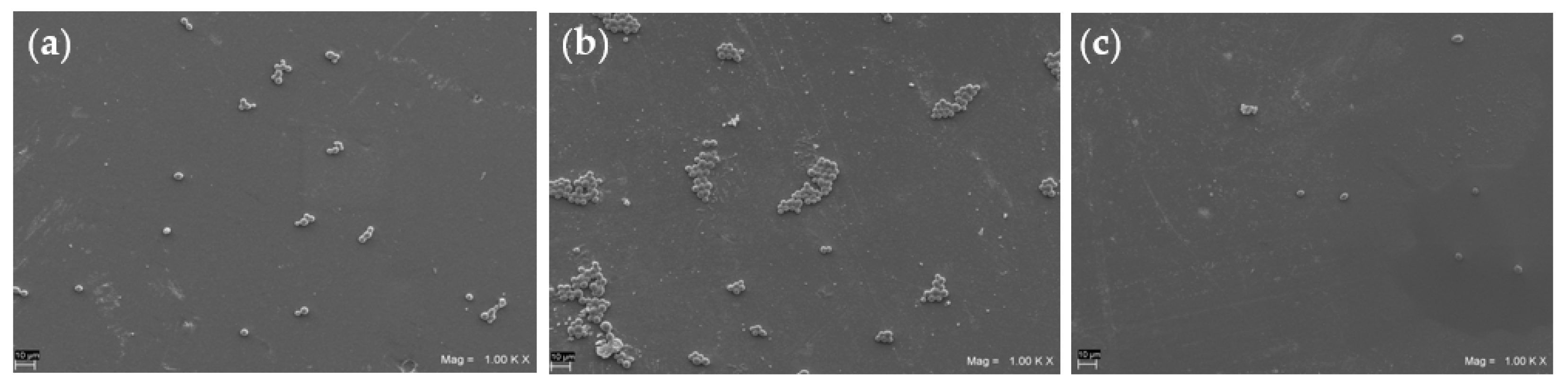
Morphology of the sessile colonies by Scanning Electron Microscopy (SEM).
The experimental planning was developed according to D’Ercole et al. [61].
Briefly, for sessile CFU/mL detection, after incubation for 48 h, the adherent viable cells were washed with PBS, detached by ultrasonication and vortexing, spread on SAB plates, incubated for 48 h at 37 °C and counted. The C. albicans S5 biomass was determined by Crystal violet (CV) staining at OD570 nm. A Concanavalin-A assay was carried out to evaluate the EPS of the biofilm’s matrix. SEM evaluation was performed to morphologically analyze the sessile colonies. After 48 h of in vitro biofilm formation, 5 specimens from each group were fixed for 1 h in 2.5% glutaraldehyde, dehydrated in six ethanol washes (10%, 25%, 50%, 75% and 90% for 20 min and 100% for 1 h) and then dried overnight in a bacteriological incubator at 37 °C. Then, they were coated with gold (Emitech K550, Emitech Ltd., Ashford, Kent, UK) and observed carefully under a SEM (EVO 50 XVP LaB6, Carl Zeiss SMT Ltd., Cambridge, UK) at 15 kV, under 1000× and 5000× magnifications.
For the sessile phase, a total of 294 specimens (10 tests and 3 negative controls for each different material; 117 disks for sessile CFU/mL count and 117 disks for biofilm biomass quantification; 5 disks from each group for Concanavalin A assay and SEM evaluation) were used.

2.5. Statistical Analysis

In each group, means and standard deviations were calculated from raw data related to quantitative tests (planktonic CFU count, sessile cells CFU count and biomass quantification by OD570 nm). Statistical analysis was carried out according to the analysis of variance (ANOVA) and the Tukey’s test for post hoc intergroup comparisons, using the SPSS for Windows version 21 (IBM SPSS Inc, Chicago, IL, USA). Levene’s and Kolmogorov–Smirnov tests were used to validate the homogeneity of the variances and the normality of data, respectively. The level of α was set at 0.05 for all tests.

3. Results

Table 2 summarizes the results achieved from the quantitative tests carried out.

3.1. Planktonic CFU/mL Count

Figure 2 shows the C. albicans S5 CFU/mL count in planktonic phase. Not statistically significant differences were detected among all groups (p > 0.05). A slight, but not significant, C. albicans S5 CFU/mL reduction was obtained with heat-cured VD (27.40 × 10⁵ ± 2.07 × 10⁵ CFU/mL) as compared to heat-cured GR (29.80 × 10⁵ ± 3.16 × 10⁵ CFU/mL) and BF (31.00 × 10⁵ ± 2.49 × 10⁵ CFU/mL).

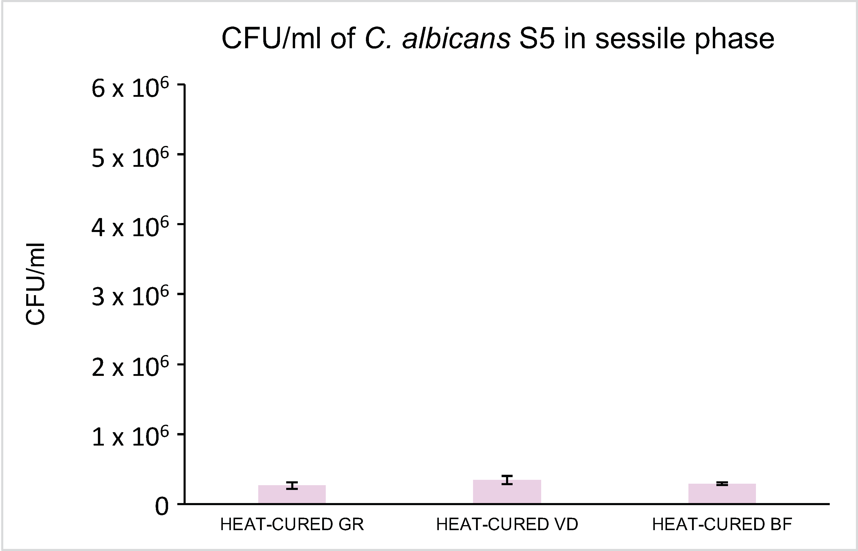
3.2. Sessile Cells CFU/mL Count

The CFU/mL of C. albicans S5 adherent to each tested disk are showed in Figure 3. In detail, the least fungal growth was observed on GR heat-cured (2.65 × 10⁵ ± 0.47 × 10⁵ CFU/mL), with no statistically significant difference (p < 0.05) compared to BF heat-cured (2.90 × 10⁵ ± 0.17 × 10⁵ CFU/mL). A significantly increased number of CFU/mL was observed for VD (3.44 × 10⁵ ± 0.56 × 10⁵) (p < 0.05).

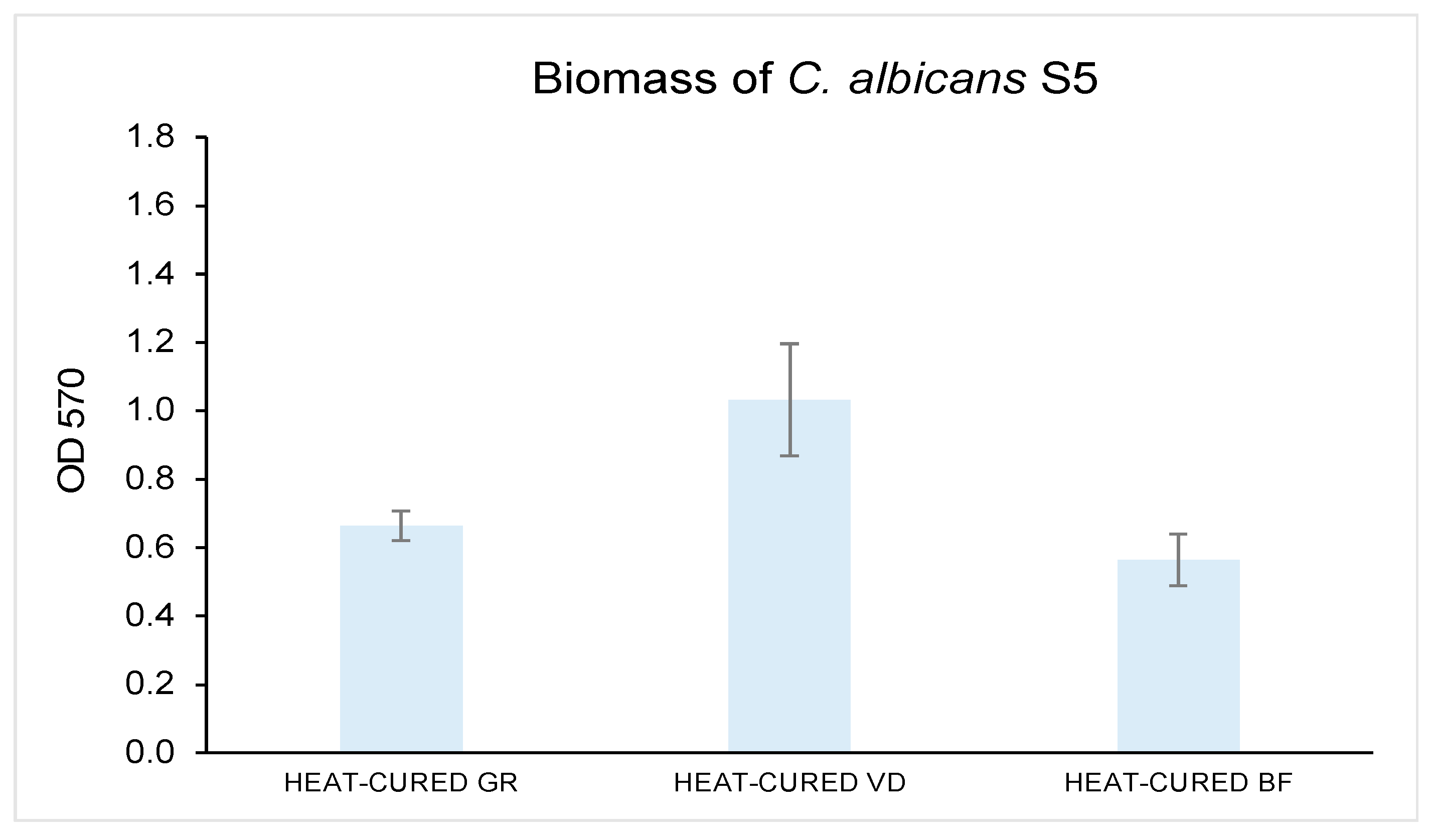
3.3. Biofilm Biomass Quantification by Optical Density (OD570 nm)

The biomass production of C. albicans S5 adherent to each tested disk (Figure 4) showed a trend similar to that observed for the sessile cells CFU/mL count. The best results were displayed by heat-cured BF, with an OD570 nm of the produced biofilm biomass equal to 0.564 ± 0.0752, significantly reduced as compared to both heat-cured GR (0.6637 ± 0.0427) (p < 0.05) and VD (1.0327 ± 0.1642) (p < 0.05). The biofilm biomass quantification on GR was significantly lower than VD (p < 0.05).

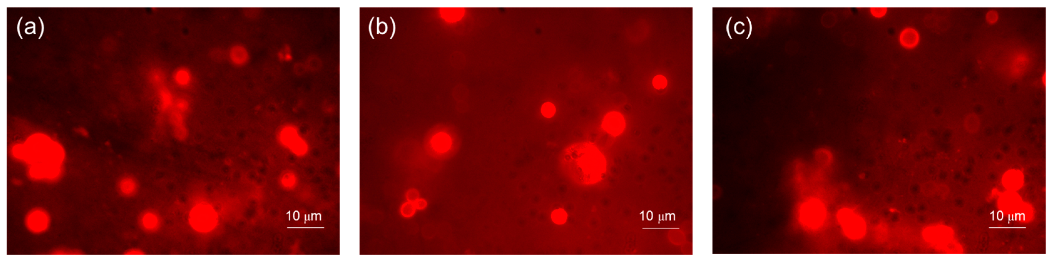
3.4. Concanavalin Assay

Figure 5 shows the polysaccharide matrix (EPS) production by C. albicans S5 biofilms on different composites disks. The images confirmed the biofilm biomass results with a major production of carbohydrates displayed on a heat-cured VD disk as compared to the other composites. A slight EPS reduction was also observed when comparing heat-cured BF to heat-cured GR.

3.5. SEM Analysis

Figure 6 shows representative SEM microphotographs of the C. albicans S5 biofilm formation on the composite disks. The VD group, which contained the greatest amount of fungal cells on its surface, showed relatively large adherent aggregates after 48 h of in vitro biofilm development (Figure 6b), while GR and BF showed smaller aggregates and an apparent reduced biofilm formation.

4. Discussion

In the present investigation, the behavior of C. albicans S5 in the presence of three commercially available, nanohybrid DCRs, coated with human saliva, was evaluated by assessing the planktonic and sessile growth modes, the biofilm biomass quantification and the qualitative analysis of the extracellular polymeric substances (EPS) of the biofilm matrix by the Concanavalin-A assay and SEM observation. All samples were polymerized using the same standardized heat-curing protocol and received the same surface treatment. This made it possible to exclude the impact of possible confounding factors and allowed us to properly assess the effect of the only one variable under investigation (i.e., the unique chemical formulation of each different resin) on the biofilm formation [63,64]. Focusing on planktonic CFU assay, a not statistically significant difference among the tested groups emerged. A slight, but not significant, C. albicans S5 planktonic CFU/mL reduction was observed with VD as compared to GR. Regarding the sessile cells CFU count, a significant increase was observed for VD compared to GR and BF. The current findings demonstrated a relevant antiadhesive effect for GR and BF, with a significant decrease in the number of sessile CFUs, in the biomass quantification and in the presence of EPS matrix. BF had the most promising behavior when compared to the other composite, both in terms of antiadhesive activity and due to a general decrease in the biofilm biomass quantification. The C. albicans capability to adhere to disk is affected by the nature of the surface, the molecules involved in quorum sensing and the production of adhesin [65]. In particular, Park et al. demonstrated that the C. albicans adhesion on resin was reduced by the electrostatic repulsion force created and hydrophobic interactions between the surface and yeast [66]. Probably, even in the present study, a decreased yeast cells growth might be related to an increased surface hydrophilicity. As already reported by Kim et al., cells grown under bis-GMA showed significantly increased surface hydrophobicity, which could potentially enhance the ability of C. albicans S5 to adhere to hydrophobic surfaces [67]. As a result, in the current investigation, both bis-GMA-based composites (GR and VD), when compared to a bis-GMA free material (BF), showed a slight rise in the sessile CFU count.
Of course, antiadhesive and antibacterial qualities need to be carefully considered together with any potential cytotoxicity brought on by the monomers present in (and released from) dental materials [68,69]. Numerous in vitro investigations have shown that the free methacrylate monomer residues left over after the polymerization phase, which may cause the production of prostaglandin E2 (PGE2), the expression of cyclooxygenase 2 and an activation of the proinflammatory response via an increase in interleukin-1 (IL-1), IL-6 and nitric oxide (NO), are primarily responsible for the potential cytotoxicity of the CR organic components [70,71]. It has also been observed that resin monomers can affect cellular physiology and adaptive cell responses by boosting ROS generation [72,73]. In a recent study, De Angelis et al. investigated the biological effect of different resin-based composites on human gingival fibroblast by means of flow cytometry analysis and immunofluorescence. Interestingly, based on their results, GrandioSo (Voco GmbH) and Enamel Plus HRi Biofunction (Micerium) seemed the least cytotoxic among the materials they tested, leading to reduced oxidative stress and fewer genotoxic effects [74]. Therefore, for the abovementioned resins (GR and BF), the antiadhesive action and the general reduction in biomass quantification herein observed seems even more promising and clinically relevant, if combined with the reduced cytotoxicity shown in previous studies [70,71,72,73]. Furthermore, as far as GrandioSo (Voco GmbH) is concerned, in a recent study on light-cured materials, D’Ercole et al. found a stronger capability to reduce S. mutans growth (in terms of sessile CFU count, biofilm biomass, metabolic activity and polysaccharide production) compared to that of Venus Diamond (Kulzer GmbH), somehow strengthening the clinical relevance of the present findings [61]. In fact, several studies provided striking evidence that S. mutans and C. albicans develop a symbiotic relationship that enhances the virulence of cospecies plaque biofilms formed on tooth surfaces, ultimately amplifying the severity of disease [53,56,57]. Such an association between C. albicans and S. mutans appears to be largely mediated by a physical interaction that relies on the production of glucans, which are produced by bacterial exoenzymes (Gtfs), on yeast and hyphal cell surfaces. These interactions are essential for the assembly of an EPS-rich matrix, the formation of enlarged microcolonies containing densely packed S. mutans cells and the development of cospecies biofilms [45]. For these reasons, it should be underlined how the ability suggested for GR to limit the proliferation of both C. albicans and S. mutans might definitely be promising.
A lower amount of biofilm matrix, with a reduced biofilm thickness and less carbohydrate amount accumulation, may be clinically advantageous, as a biofilm with a reduced biomass is supposed to be less resistant to the salivary buffering effect, antibacterial agents and to the action of fluoride ions [61]. The presence of a thick and stable plaque biofilm, on the contrary, makes those defense mechanisms less effective, compromising the long-term outcome of composite resin restorations [43] and potentially increasing the risk for secondary caries. Thus, from a clinical point of view, it would be advisable to select a material that impairs adhesion, proliferation or even just extracellular matrix production of pathogenic microorganisms, in particular (based on the results of the present study) when dealing with indirect composites subjected to post-polymerization thermal cycles.
Among the limitations of the present study, it should be underlined that only the heat-curing protocol was herein evaluated, without taking into account the simple light-curing procedure. Photoactivation is the most prevalent method used to activate and promote the polymerization of resin based dental composites. The irradiance emitted by different models/brands of light-curing devices is variable, usually ranging between 400 and 1200 mW/cm2 [75]. However, as already mentioned, monomer conversion is never complete during the polymerization of light-cured resin composites. This is the reasons why, when dealing with indirect restorations, resin composites are conveniently subjected to further heat curing. Such an additional heat treatment leads to an increase in the degree of conversion and improved mechanical properties such as microhardness, flexural strength, fracture toughness, wear resistance, tensile strength, color stability and a reduction of wear [11,12,13,14]. Thus, it seemed of greater clinical interest to investigate the antimicrobial activity of materials when they are used in their best performing modes.
As already mentioned, several studies have reported high prevalence for S. mutans in dental biofilms where C. albicans resides, suggesting that such a fungal–bacterial interaction can potentially contribute to dental caries [25,26,44,45]. Considering this association between C. albicans S5 and S. mutans, further studies seem necessary to better investigate the combined susceptibility of both microorganisms to resin-based material formulation in terms of adhesion and proliferation.
In brief, based on the present results, it can be concluded that different formulations of commercially available heat-cured resin composites may interact differently with C. albicans. In comparison to VD, GR and BF led to a decreased biofilm growth in terms of sessile CFU count, biofilm biomass and polysaccharide synthesis. Despite the fact that they also permitted C. albicans development to a certain level, these characteristics would suggest that GR and BF as more suitable restorative materials. Understanding that heat-cured composites may show a different affinity with Candida albicans, depending on their intrinsic chemical composition and despite the surface treatment, may be of great clinical relevance. In fact, an informed clinician could wisely select those materials that are able to limit the formation of biofilms by potentially cariogenic species, which might lead to secondary caries over time. Moreover, a better understanding about the affinity between restorative materials and cariogenic species would also encourage manufacturers of the dental industry towards the production and commercialization of resin composites that are less prone to allow an intense microbial proliferation.

Author Contributions

Conceptualization, F.D.A., S.D., M.V., L.V. and C.D.; methodology, F.D.A., S.D., M.V., L.V., S.D.L. and C.D.; software, F.D.A.; validation, F.D.A., S.D.L., M.D.G. and S.D.; formal analysis, F.D.A., S.D. and C.D.; investigation, F.D.A., V.B., M.V., M.B., S.D.L. and S.D.; resources, L.V. and C.D.; data curation, F.D.A., V.B., M.B. and S.D.; writing—original draft preparation, F.D.A., V.B., S.D.L. and S.D.; writing—review and editing, F.D.A., V.B., M.V., C.D. and L.C.; visualization, F.D.A., M.B. and M.D.G.; supervision, F.D.A., C.D., L.C. and S.D.; project administration, C.D. and L.C.; funding acquisition, C.D. All authors have read and agreed to the published version of the manuscript.

Funding

This research received no external funding.

Institutional Review Board Statement

This study was conducted according to the guidelines of the Declaration of Helsinki and approved by the Ethics Committee of “G. d’Annunzio” University, Chieti–Pescara, Italy (approval code SALI, N. 19 of 10 September 2020).

Informed Consent Statement

Informed consent was obtained from all subjects involved in this study.

Data Availability Statement

The data presented in this study are available on request from the corresponding author.

Conflicts of Interest

The authors declare no conflict of interest.

References

  1. Van Landuyt, K.L.; Yoshida, Y.; Hirata, I.; Snauwaert, J.; De Munck, J.; Okazaki, M.; Suzuki, K.; Lambrechts, P.; Van Meerbeek, B. Influence of the chemical structure of functional monomers on their adhesive performance. J. Dent. Res. 2008, 87, 757–761. [Google Scholar] [CrossRef] [PubMed]
  2. De Angelis, F.; D’Arcangelo, C.; Maliskova, N.; Vanini, L.; Vadini, M. Wear properties of different additive restorative materials used for onlay/overlay posterior restorations. Oper. Dent. 2020, 45, E156–E166. [Google Scholar] [CrossRef]
  3. D’Arcangelo, C.; Vanini, L.; Rondoni, G.D.; Vadini, M.; De Angelis, F. Wear evaluation of prosthetic materials opposing themselves. Oper. Dent. 2018, 43, 38–50. [Google Scholar] [CrossRef]
  4. D’Arcangelo, C.; Vanini, L.; Rondoni, G.D.; Pirani, M.; Vadini, M.; Gattone, M.; De Angelis, F. Wear properties of a novel resin composite compared to human enamel and other restorative materials. Oper. Dent. 2014, 39, 612–618. [Google Scholar] [CrossRef]
  5. D’Arcangelo, C.; Vadini, M.; Buonvivere, M.; De Angelis, F. Safe clinical technique for increasing the occlusal vertical dimension in case of erosive wear and missing teeth. Clin. Case Rep. 2021, 9, e04747. [Google Scholar] [CrossRef] [PubMed]
  6. Ferracane, J.L. Current trends in dental composites. Crit. Rev. Oral Biol. Med. 1995, 6, 302–318. [Google Scholar] [CrossRef] [PubMed]
  7. Obici, A.C.; Sinhoreti, M.A.C.; Frollini, E.; Correr Sobrinho, L.; Consani, S. Degree of conversion of z250 composite determined by fourier transform infrared spectroscopy: Comparison of techniques, storage periods and photo-activation methods. Mater. Res. 2004, 7, 605–610. [Google Scholar] [CrossRef]
  8. Peutzfeldt, A. Resin composites in dentistry: The monomer systems. Eur. J. Oral Sci. 1997, 105, 97–116. [Google Scholar] [CrossRef]
  9. Amirouche-Korichi, A.; Mouzali, M.; Watts, D.C. Effects of monomer ratios and highly radiopaque fillers on degree of conversion and shrinkage-strain of dental resin composites. Dent. Mater. 2009, 25, 1411–1418. [Google Scholar] [CrossRef]
  10. Daronch, M.; Rueggeberg, F.; De Goes, M. Monomer conversion of pre-heated composite. J. Dent. Res. 2005, 84, 663–667. [Google Scholar] [CrossRef]
  11. Grazioli, G.; Francia, A.; Cuevas-Suárez, C.E.; Zanchi, C.H.; Moraes, R.R.D. Simple and low-cost thermal treatments on direct resin composites for indirect use. Braz. Dent. J. 2019, 30, 279–284. [Google Scholar] [CrossRef]
  12. Lapesqueur, M.; Surriaga, P.; Masache, M.E.; Vásquez, B.; Peña, M.; Gómes, O.M.M.; Domínguez, J.A. Efectos sobre microdureza y grado de conversión de dos tipos de resinas sometidas a tratamientos de pospolimerización. Rev. Nac. Odontol. 2015, 11, 49–56. [Google Scholar] [CrossRef]
  13. Cruz, F.L.; Carvalho, R.F.; Batista, C.H.T.; Siqueira-Júnior, H.M.; Queiroz, J.R.C.; Leite, F.P. Efecto del tratamiento térmico y de fibras de polietileno en la resistencia a la flexión de resinas compuestas. Acta Odontol. Venez. 2014, 52, 3–4. [Google Scholar]
  14. Zamalloa-Quintana, M.; López-Gurreonero, C.; Santander-Rengifo, F.M.; Ladera-Castañeda, M.; Castro-Pérez Vargas, A.; Cornejo-Pinto, A.; Cervantes-Ganoza, L.; Cayo-Rojas, C. Effect of additional dry heat curing on microflexural strength in three types of resin composite: An in vitro study. Crystals 2022, 12, 1045. [Google Scholar] [CrossRef]
  15. Al-Zain, A.O.; Platt, J.A. Effect of light-curing distance and curing time on composite microflexural strength. Dent. Mater. J. 2021, 40, 202–208. [Google Scholar] [CrossRef] [PubMed]
  16. Al-Zain, A.O.; Marghalani, H.Y. Influence of light-curing distances on microflexural strength of two resin-based composites. Oper. Dent. 2020, 45, 297–305. [Google Scholar] [CrossRef]
  17. Kumar, D.; Shukla, M.; Mahato, K.; Rathore, D.; Prusty, R.; Ray, B. Effect of Post-Curing on Thermal and Mechanical Behavior of gfrp Composites. IOP Conf. Ser. Mater. Sci. Eng. 2015, 75, 012012. [Google Scholar] [CrossRef]
  18. Lucena-Martin, C.; Gonzalez-Lopez, S.; Navajas-Rodriguez de Mondelo, J.M. The effect of various surface treatments and bonding agents on the repaired strength of heat-treated composites. J. Prosthet. Dent. 2001, 86, 481–488. [Google Scholar] [CrossRef] [PubMed]
  19. Uzay, C.; Boztepe, M.H.; Bayramoğlu, M.; Geren, N. Effect of post-curing heat treatment on mechanical properties of fiber reinforced polymer (frp) composites. Mater. Test. 2017, 59, 366–372. [Google Scholar] [CrossRef]
  20. Takeshige, F.; Kinomoto, Y.; Torii, M. Additional heat-curing of light-cured composite resin for inlay restoration. J. Osaka Univ. Dent. Sch. 1995, 35, 59–66. [Google Scholar]
  21. De Angelis, F.; Minnoni, A.; Vitalone, L.M.; Carluccio, F.; Vadini, M.; Paolantonio, M.; D’Arcangelo, C. Bond strength evaluation of three self-adhesive luting systems used for cementing composite and porcelain. Oper. Dent. 2011, 36, 626–634. [Google Scholar] [CrossRef] [PubMed]
  22. De Angelis, F.; Vadini, M.; Buonvivere, M.; Valerio, A.; Di Cosola, M.; Piattelli, A.; Biferi, V.; D’Arcangelo, C. In vitro mechanical properties of a novel graphene-reinforced pmma-based dental restorative material. Polymers 2023, 15, 622. [Google Scholar] [CrossRef]
  23. Re, D.; De Angelis, F.; Augusti, G.; Augusti, D.; Caputi, S.; D’Amario, M.; D’Arcangelo, C. Mechanical properties of elastomeric impression materials: An in vitro comparison. Int. J. Dent. 2015, 2015, 428286. [Google Scholar] [CrossRef] [PubMed]
  24. D’Amario, M.; De Angelis, F.; Mancino, M.; Frascaria, M.; Capogreco, M.; D’Arcangelo, C. Canal shaping of different single-file systems in curved root canals. J. Dent. Sci. 2017, 12, 328–332. [Google Scholar] [CrossRef] [PubMed]
  25. Jenkinson, H.F.; Lala, H.C.; Shepherd, M.G. Coaggregation of Streptococcus sanguis and other streptococci with Candida albicans. Infect. Immun. 1990, 58, 1429–1436. [Google Scholar] [CrossRef]
  26. Kolenbrander, P.E.; Andersen, R.N.; Blehert, D.S.; Egland, P.G.; Foster, J.S.; Palmer, R.J., Jr. Communication among oral bacteria. Microbiol. Mol. Biol. Rev. 2002, 66, 486–505. [Google Scholar] [CrossRef]
  27. Rickard, A.H.; Gilbert, P.; High, N.J.; Kolenbrander, P.E.; Handley, P.S. Bacterial coaggregation: An integral process in the development of multi-species biofilms. Trends. Microbiol. 2003, 11, 94–100. [Google Scholar] [CrossRef] [PubMed]
  28. Vu, B.; Chen, M.; Crawford, R.J.; Ivanova, E.P. Bacterial extracellular polysaccharides involved in biofilm formation. Molecules 2009, 14, 2535–2554. [Google Scholar] [CrossRef]
  29. Xiao, J.; Klein, M.I.; Falsetta, M.L.; Lu, B.; Delahunty, C.M.; Yates, J.R., III; Heydorn, A.; Koo, H. The exopolysaccharide matrix modulates the interaction between 3d architecture and virulence of a mixed-species oral biofilm. PLoS Pathog. 2012, 8, e1002623. [Google Scholar] [CrossRef]
  30. Sultan, A.S.; Kong, E.F.; Rizk, A.M.; Jabra-Rizk, M.A. The oral microbiome: A lesson in coexistence. PLoS Pathog. 2018, 14, e1006719. [Google Scholar] [CrossRef]
  31. Islam, B.; Khan, S.N.; Khan, A.U. Dental caries: From infection to prevention. Med. Sci. Monit. 2007, 13, RA196-203. [Google Scholar]
  32. Koo, H.; Falsetta, M.L.; Klein, M.I. The exopolysaccharide matrix: A virulence determinant of cariogenic biofilm. J. Dent. Res. 2013, 92, 1065–1073. [Google Scholar] [CrossRef] [PubMed]
  33. Lemos, J.A.; Quivey, R.G.; Koo, H.; Abranches, J. Streptococcus mutans: A new Gram-positive paradigm? Microbiology 2013, 159, 436–445. [Google Scholar] [CrossRef]
  34. Klein, M.I.; Hwang, G.; Santos, P.H.; Campanella, O.H.; Koo, H. Streptococcus mutans-derived extracellular matrix in cariogenic oral biofilms. Front. Cell. Infect. Microbiol. 2015, 5, 10. [Google Scholar] [CrossRef] [PubMed]
  35. Valm, A.M. The structure of dental plaque microbial communities in the transition from health to dental caries and periodontal disease. J. Mol. Biol. 2019, 431, 2957–2969. [Google Scholar] [CrossRef] [PubMed]
  36. Bourbia, M.; Ma, D.; Cvitkovitch, D.G.; Santerre, J.P.; Finer, Y. Cariogenic bacteria degrade dental resin composites and adhesives. J. Dent. Res. 2013, 92, 989–994. [Google Scholar] [CrossRef]
  37. Huang, B.; Siqueira, W.L.; Cvitkovitch, D.G.; Finer, Y. Esterase from a cariogenic bacterium hydrolyzes dental resins. Acta Biomater. 2018, 71, 330–338. [Google Scholar] [CrossRef]
  38. Kruger, J.; Maletz, R.; Ottl, P.; Warkentin, M. In vitro aging behavior of dental composites considering the influence of filler content, storage media and incubation time. PLoS ONE 2018, 13, e0195160. [Google Scholar] [CrossRef]
  39. Marashdeh, M.Q.; Gitalis, R.; Levesque, C.; Finer, Y. Enterococcus faecalis hydrolyzes dental resin composites and adhesives. J. Endod. 2018, 44, 609–613. [Google Scholar] [CrossRef]
  40. Delaviz, Y.; Finer, Y.; Santerre, J.P. Biodegradation of resin composites and adhesives by oral bacteria and saliva: A rationale for new material designs that consider the clinical environment and treatment challenges. Dent. Mater. 2014, 30, 16–32. [Google Scholar] [CrossRef]
  41. Opdam, N.J.; van de Sande, F.H.; Bronkhorst, E.; Cenci, M.S.; Bottenberg, P.; Pallesen, U.; Gaengler, P.; Lindberg, A.; Huysmans, M.C.; van Dijken, J.W. Longevity of posterior composite restorations: A systematic review and meta-analysis. J. Dent. Res. 2014, 93, 943–949. [Google Scholar] [CrossRef] [PubMed]
  42. Demarco, F.F.; Collares, K.; Coelho-de-Souza, F.H.; Correa, M.B.; Cenci, M.S.; Moraes, R.R.; Opdam, N.J. Anterior composite restorations: A systematic review on long-term survival and reasons for failure. Dent. Mater. 2015, 31, 1214–1224. [Google Scholar] [CrossRef] [PubMed]
  43. Kusuma Yulianto, H.D.; Rinastiti, M.; Cune, M.S.; de Haan-Visser, W.; Atema-Smit, J.; Busscher, H.J.; van der Mei, H.C. Biofilm composition and composite degradation during intra-oral wear. Dent. Mater. 2019, 35, 740–750. [Google Scholar] [CrossRef]
  44. Metwalli, K.H.; Khan, S.A.; Krom, B.P.; Jabra-Rizk, M.A. Streptococcus mutans, Candida albicans, and the human mouth: A sticky situation. PLoS Pathog. 2013, 9, e1003616. [Google Scholar] [CrossRef] [PubMed]
  45. Falsetta, M.L.; Klein, M.I.; Colonne, P.M.; Scott-Anne, K.; Gregoire, S.; Pai, C.-H.; Gonzalez-Begne, M.; Watson, G.; Krysan, D.J.; Bowen, W.H. Symbiotic relationship between Streptococcus mutans and Candida albicans synergizes virulence of plaque biofilms in vivo. Infect. Immun. 2014, 82, 1968–1981. [Google Scholar] [CrossRef]
  46. Pereira, D.F.A.; Seneviratne, C.J.; Koga-Ito, C.Y.; Samaranayake, L.P. Is the oral fungal pathogen Candida albicans a cariogen? Oral Dis. 2018, 24, 518–526. [Google Scholar] [CrossRef] [PubMed]
  47. Xiao, J.; Huang, X.; Alkhers, N.; Alzamil, H.; Alzoubi, S.; Wu, T.T.; Castillo, D.A.; Campbell, F.; Davis, J.; Herzog, K.; et al. Candida albicans and early childhood caries: A systematic review and meta-analysis. Caries Res. 2018, 52, 102–112. [Google Scholar] [CrossRef]
  48. Krom, B.P.; Kidwai, S.; Ten Cate, J.M. Candida and other fungal species: Forgotten players of healthy oral microbiota. J. Dent. Res. 2014, 93, 445–451. [Google Scholar] [CrossRef]
  49. Fidel, P.L., Jr. Candida-host interactions in hiv disease: Implications for oropharyngeal candidiasis. Adv. Dent. Res. 2011, 23, 45–49. [Google Scholar] [CrossRef]
  50. Williams, D.; Lewis, M. Pathogenesis and treatment of oral candidosis. J. Oral Microbiol. 2011, 3, 5771. [Google Scholar] [CrossRef]
  51. Jabra-Rizk, M.A.; Kong, E.F.; Tsui, C.; Nguyen, M.H.; Clancy, C.J.; Fidel, P.L., Jr.; Noverr, M. Candida albicans pathogenesis: Fitting within the host-microbe damage response framework. Infect. Immun. 2016, 84, 2724–2739. [Google Scholar] [CrossRef] [PubMed]
  52. Jenkinson, H.; Barbour, M.; Jagger, D.; Miles, M.; Bamford, C.; Nobbs, A.; Dutton, L.; Silverman, R.; McNally, L.; Vickerman, M. Candida albicans-bacteria interactions in biofilms and disease. Univ. Bristol. Dent. Sch. 2008, 16. [Google Scholar] [CrossRef]
  53. Diaz, P.I.; Xie, Z.; Sobue, T.; Thompson, A.; Biyikoglu, B.; Ricker, A.; Ikonomou, L.; Dongari-Bagtzoglou, A. Synergistic interaction between Candida albicans and commensal oral streptococci in a novel in vitro mucosal model. Infect. Immun. 2012, 80, 620–632. [Google Scholar] [CrossRef] [PubMed]
  54. Xu, H.; Dongari-Bagtzoglou, A. Shaping the oral mycobiota: Interactions of opportunistic fungi with oral bacteria and the host. Curr. Opin. Microbiol. 2015, 26, 65–70. [Google Scholar] [CrossRef]
  55. Ellepola, K.; Liu, Y.; Cao, T.; Koo, H.; Seneviratne, C.J. Bacterial gtfb augments Candida albicans accumulation in cross-kingdom biofilms. J. Dent. Res. 2017, 96, 1129–1135. [Google Scholar] [CrossRef] [PubMed]
  56. Koo, H.; Andes, D.R.; Krysan, D.J. Candida-streptococcal interactions in biofilm-associated oral diseases. PLoS Pathog. 2018, 14, e1007342. [Google Scholar] [CrossRef]
  57. Montelongo-Jauregui, D.; Lopez-Ribot, J.L. Candida interactions with the oral bacterial microbiota. J. Fungi 2018, 4, 122. [Google Scholar] [CrossRef]
  58. Montelongo-Jauregui, D.; Saville, S.P.; Lopez-Ribot, J.L. Contributions of Candida albicans dimorphism, adhesive interactions, and extracellular matrix to the formation of dual-species biofilms with Streptococcus gordonii. mBio 2019, 10, e01179-19. [Google Scholar] [CrossRef]
  59. Jenkinson, H.F.; Lamont, R.J. Oral microbial communities in sickness and in health. Trends Microbiol. 2005, 13, 589–595. [Google Scholar] [CrossRef] [PubMed]
  60. Xiang, Z.; Wakade, R.S.; Ribeiro, A.A.; Hu, W.; Bittinger, K.; Simon-Soro, A.; Kim, D.; Li, J.; Krysan, D.J.; Liu, Y.; et al. Human tooth as a fungal niche: Candida albicans traits in dental plaque isolates. mBio 2023, 14, e0276922. [Google Scholar] [CrossRef]
  61. D’Ercole, S.; De Angelis, F.; Biferi, V.; Noviello, C.; Tripodi, D.; Di Lodovico, S.; Cellini, L.; D’Arcangelo, C. Antibacterial and antibiofilm properties of three resin-based dental composites against Streptococcus mutans. Materials 2022, 15, 6633. [Google Scholar] [CrossRef]
  62. Di Lodovico, S.; Dotta, T.C.; Cellini, L.; Iezzi, G.; D’Ercole, S.; Petrini, M. The antibacterial and antifungal capacity of eight commercially available types of mouthwash against oral microorganisms: An in vitro study. Antibiotics 2023, 12, 675. [Google Scholar] [CrossRef] [PubMed]
  63. Mena Silva, P.A.; Garcia, I.M.; Nunes, J.; Visioli, F.; Castelo Branco Leitune, V.; Melo, M.A.; Collares, F.M. Myristyltrimethylammonium bromide (mytab) as a cationic surface agent to inhibit Streptococcus mutans grown over dental resins: An in vitro study. J. Funct. Biomater. 2020, 11, 9. [Google Scholar] [CrossRef] [PubMed]
  64. Cazzaniga, G.; Ottobelli, M.; Ionescu, A.; Garcia-Godoy, F.; Brambilla, E. Surface properties of resin-based composite materials and biofilm formation: A review of the current literature. Am. J. Dent. 2015, 28, 311–320. [Google Scholar]
  65. Gad, M.M.; Fouda, S.M. Current perspectives and the future of Candida albicans-associated denture stomatitis treatment. Dent. Med. Probl. 2020, 57, 95–102. [Google Scholar] [CrossRef]
  66. Park, S.E.; Blissett, R.; Susarla, S.M.; Weber, H.P. Candida albicans adherence to surface-modified denture resin surfaces. J. Prosthodont. 2008, 17, 365–369. [Google Scholar] [CrossRef]
  67. Masuoka, J.; Hazen, K.C. Cell wall protein mannosylation determines Candida albicans cell surface hydrophobicity. Microbiology 1997, 143, 3015–3021. [Google Scholar] [CrossRef] [PubMed]
  68. Trubiani, O.; Caputi, S.; Di Iorio, D.D.; Amario, M.; Paludi, M.; Giancola, R.; Di Nardo Di Maio, F.; De Angelis, F.; D’Arcangelo, C. The cytotoxic effects of resin-based sealers on dental pulp stem cells. Int. Endod. J. 2010, 43, 646–653. [Google Scholar] [CrossRef]
  69. Trubiani, O.; Cataldi, A.; De Angelis, F.; D’Arcangelo, C.; Caputi, S. Overexpression of interleukin-6 and -8, cell growth inhibition and morphological changes in 2-hydroxyethyl methacrylate-treated human dental pulp mesenchymal stem cells. Int. Endod. J. 2012, 45, 19–25. [Google Scholar] [CrossRef] [PubMed]
  70. Kuan, Y.H.; Huang, F.M.; Lee, S.S.; Li, Y.C.; Chang, Y.C. Bisgma stimulates prostaglandin e2 production in macrophages via cyclooxygenase-2, cytosolic phospholipase a2, and mitogen-activated protein kinases family. PLoS ONE 2013, 8, e82942. [Google Scholar] [CrossRef] [PubMed]
  71. Huang, F.M.; Chang, Y.C.; Lee, S.S.; Yeh, C.H.; Lee, K.G.; Huang, Y.C.; Chen, C.J.; Chen, W.Y.; Pan, P.H.; Kuan, Y.H. Bisgma-induced cytotoxicity and genotoxicity in macrophages are attenuated by wogonin via reduction of intrinsic caspase pathway activation. Environ. Toxicol. 2016, 31, 176–184. [Google Scholar] [CrossRef] [PubMed]
  72. Lottner, S.; Shehata, M.; Hickel, R.; Reichl, F.X.; Durner, J. Effects of antioxidants on DNA-double strand breaks in human gingival fibroblasts exposed to methacrylate based monomers. Dent. Mater. 2013, 29, 991–998. [Google Scholar] [CrossRef]
  73. Gallorini, M.; Petzel, C.; Bolay, C.; Hiller, K.A.; Cataldi, A.; Buchalla, W.; Krifka, S.; Schweikl, H. Activation of the nrf2-regulated antioxidant cell response inhibits hema-induced oxidative stress and supports cell viability. Biomaterials 2015, 56, 114–128. [Google Scholar] [CrossRef] [PubMed]
  74. De Angelis, F.; Mandatori, D.; Schiavone, V.; Melito, F.P.; Valentinuzzi, S.; Vadini, M.; Di Tomo, P.; Vanini, L.; Pelusi, L.; Pipino, C.; et al. Cytotoxic and genotoxic effects of composite resins on cultured human gingival fibroblasts. Materials 2021, 14, 5225. [Google Scholar] [CrossRef] [PubMed]
  75. Rueggeberg, F.A.; Giannini, M.; Arrais, C.A.G.; Price, R.B.T. Light curing in dentistry and clinical implications: A literature review. Braz. Oral Res. 2017, 31, e61. [Google Scholar] [CrossRef]
Figure 1. Schematic summary of the study design.
Materials 16 05818 g001
Figure 2. Planktonic C. albicans S5 CFU/mL count (×105 CFU/mL).
Materials 16 05818 g002
Figure 3. Sessile C. albicans S5 CFU/mL count (×103 CFU/mL).
Materials 16 05818 g003
Figure 4. Biomass quantification by optical density (OD570 nm).
Materials 16 05818 g004
Figure 5. Representative images (original magnification 100×) of polysaccharide matrix (EPS) production by C. albicans S5 biofilms observed using Concanavalin-A on heat-cured GR (a), heat-cured VD (b) and heat-cured BF specimens (c).
Materials 16 05818 g005
Figure 6. Representative SEM images (original magnification 10,000×) of C. albicans S5 biofilm formed on disk-shaped specimens from heat-cured GR (a), heat-cured VD (b) and heat-cured BF (c) groups.
Materials 16 05818 g006
Table 1. Composite resins included in this study.
Experimental GroupMaterialManufacturerBatchComposition
GRGrandioSO
- Shade A2 -
(Nanohybrid)
Voco GmbH
(Cuxhaven, Germany)
202845989% (w/w) fillers (1 μm glass ceramic filler, 20 nm–40 nm silicon dioxide fillers), Bis-GMA, Bis-EMA, TEGDMA.
VDVenus Diamond
- Shade A2 -
(Nanohybrid)
Kulzer GmbH (Hanau, Germany)E829271% (w/w) fillers (0.2 μm Si-Zr fillers), Bis-GMA, TEGDMA.
BFEnamel Plus
HRi Biofunction
- Shade BF2 -
(Nanohybrid)
Micerium (Avegno, Genova, Italy)202100624774% in weight (60% in volume) fillers (0.005 μm–0.05 μm silicon dioxide fillers), (0.2–3.0 μm glass fillers), Urethane dimethacrylate, Tricyclodecane dimethanol dimethacrylate.
Table 2. C. albicans S5 detection on the three resin composites investigated.
Experimental Group
GRVDBF
Planktonic CFU count (×105 CFU/mL)
(SD)
29.80 a27.40 a31.00 a
(3.16)(2.07)(2.49)
Sessile Cells CFU count (×103 CFU/mL)
(SD)
2.65 b3.44 a2.90 b
(0.47)(0.56)(0.17)
Biomass quantification OD570 nm
(SD)
0.6637 b1.0327 a0.5641 c
(0.0427)(0.1642)(0.0752)
Different superscript letters indicate statistically significant differences.
Disclaimer/Publisher’s Note: The statements, opinions and data contained in all publications are solely those of the individual author(s) and contributor(s) and not of MDPI and/or the editor(s). MDPI and/or the editor(s) disclaim responsibility for any injury to people or property resulting from any ideas, methods, instructions or products referred to in the content.

